studies in carpal tunnel syndrome and cold intolerance
TRANSCRIPT

Studies in Carpal Tunnel Syndrome
and Cold Intolerance

2
Studies in Carpal Tunnel Syndrome
and Cold Intolerance
By Khalid M. Salem-Saqer, MB, ChB,
MRCS (Edinburgh), MRCS (Glasgow)
Thesis submitted to the University of Nottingham for the
degree of Doctor in Medicine
September 2007
The work was undertaken in the University of Nottingham, Division
of Vascular Medicine, Derby City General Hospital, Derby
Hospitals NHS Foundation Trust, Derby and Ilkeston Community
Hospital, Erewash Primary Care Trust, Ilkeston.
3
Table of Contents
Abbreviations -----------------------------------------------------------------------------8
List of Illustrations --------------------------------------------------------------------------- 11
List of Tables --------------------------------------------------------------------------- 13
Acknowledgements--------------------------------------------------------------------------- 16
Declaration --------------------------------------------------------------------------- 17
Research Presentations--------------------------------------------------------------------- 18
Abstract --------------------------------------------------------------------------- 19
CHAPTER 1: INTRODUCTION----------------------------------------------------------- 23
1.1 CARPAL TUNNEL SYNDROME (CTS) ---------------------------------------- 24
1.1.1 Definition and prevalence ------------------------------------------------ 24
1.1.2 Pathophysiology of CTS-------------------------------------------------- 25
1.1.3 Clinical picture -------------------------------------------------------------- 29
1.1.4 Diagnosis criteria----------------------------------------------------------- 32
1.1.5 Risk and prognostic factors---------------------------------------------- 34
1.1.6 Treatment of CTS ---------------------------------------------------------- 36
1.2 POST-TRAUMATIC COLD INTOLERANCE (PTCI)------------------------- 44
1.2.1 Definition --------------------------------------------------------------------- 44
1.2.2 Clinical presentation------------------------------------------------------- 45
1.2.3 Pathophysiology------------------------------------------------------------ 46
1.2.4 Incidence and natural history of PTCI--------------------------------- 48
1.2.5 Nature and severity of symptoms -------------------------------------- 50
1.2.6 Symptom scoring systems ----------------------------------------------- 51
1.2.7 Resolution-------------------------------------------------------------------- 53
1.2.8 Treatment -------------------------------------------------------------------- 54
1.3 RAYNAUD�S PHENOMENON (RP) --------------------------------------------- 57
1.3.1 Classification ---------------------------------------------------------------- 58
1.3.2 Prevalence------------------------------------------------------------------- 59
1.3.3 Pathophysiology of RP---------------------------------------------------- 61
4
1.4 HAND ARM VIBRATION SYNDROME (HAVS) ------------------------------ 63
1.4.1 Prevalence------------------------------------------------------------------- 64
1.4.2 Patient assessment and diagnosis------------------------------------- 64
1.4.3 Pathology--------------------------------------------------------------------- 67
1.4.4 Pathophysiology------------------------------------------------------------ 68
1.5 LASER DOPPLER PERFUSION IMAGING (LDI)---------------------------- 70
1.5.1 The technology ------------------------------------------------------------- 71
1.5.2 Factors affecting measurement by LDI ------------------------------- 73
1.5.3 Advantages of LDI --------------------------------------------------------- 73
1.5.4 Pitfalls in LDI ---------------------------------------------------------------- 74
1.5.5 Applications of LDI --------------------------------------------------------- 75
1.6 ENDOTHELIAL ROLE IN VASOMOTION REGULATION ----------------- 76
1.7 AIMS --------------------------------------------------------------------------- 79
CHAPTER 2: GENERAL METHODS --------------------------------------------------- 81
2.1 INTRODUCTION --------------------------------------------------------------------- 82
2.2 ETHICAL APPROVAL -------------------------------------------------------------- 82
2.3 CLINICAL SETTING----------------------------------------------------------------- 82
2.4 SUBJECT RECRUITMENT-------------------------------------------------------- 82
2.5 PATIENT CONSENT---------------------------------------------------------------- 84
2.6 DEMOGRAPHIC INFORMATION------------------------------------------------ 84
2.7 BLOOD PRESSURE (BP) MEASUREMENT---------------------------------- 84
2.8 DATA ANALYSIS--------------------------------------------------------------------- 85
2.9 IONTOPHORESIS ------------------------------------------------------------------- 87
2.9.1 Introduction ------------------------------------------------------------------ 87
2.9.2 Theory ------------------------------------------------------------------------ 87
2.9.3 Equipment-------------------------------------------------------------------- 88
2.9.4 Investigational drugs ------------------------------------------------------ 89
2.9.5 Precautions and considerations ---------------------------------------- 89
2.9.6 Iontophoresis protocol ---------------------------------------------------- 90
5
2.10 LASER DOPPLER IMAGING (LDI)---------------------------------------------- 91
2.10.1 Introduction ------------------------------------------------------------------ 91
2.10.2 Equipment-------------------------------------------------------------------- 91
2.10.3 Precautions and considerations ---------------------------------------- 92
2.10.4 Procedure/protocol--------------------------------------------------------- 93
2.11 COLD PROVOCATION TESTING (CPT) -------------------------------------- 94
2.11.1 Introduction ------------------------------------------------------------------ 94
2.11.2 Equipment-------------------------------------------------------------------- 94
2.11.3 Precautions and considerations ---------------------------------------- 95
2.11.4 Procedure/protocol--------------------------------------------------------- 95
2.12 CLINICAL AUDIT--------------------------------------------------------------------- 96
2.12.1 Introduction ------------------------------------------------------------------ 96
2.12.2 The audit process ---------------------------------------------------------- 97
2.12.3 Research or audit ---------------------------------------------------------- 98
2.12.4 Audit procedures ----------------------------------------------------------- 99
2.12.5 CTS assessment tools -------------------------------------------------- 100
CHAPTER 3: DEVELOPMENT OF METHODS ------------------------------------ 105
3.1 INTRODUCTION ------------------------------------------------------------------- 106
3.1.1 CPT modification --------------------------------------------------------- 106
3.1.2 Repeatability of methods ----------------------------------------------- 106
3.2 METHODS ------------------------------------------------------------------------- 107
3.2.1 CPT modification and repeatability ---------------------------------- 107
3.2.2 LDI repeatability ---------------------------------------------------------- 108
3.3 DATA ANALYSIS------------------------------------------------------------------- 109
3.3.1 CPT modification --------------------------------------------------------- 109
3.3.2 Test repeatability --------------------------------------------------------- 110
3.4 RESULTS ------------------------------------------------------------------------- 110
3.4.1 CPT modification --------------------------------------------------------- 110
3.4.2 Repeatability of the modified CPT ----------------------------------- 118
3.4.3 Repeatability of LDI in the hand -------------------------------------- 123
6
3.5 DISCUSSION ----------------------------------------------------------------------- 129
3.5.1 The modified CPT-------------------------------------------------------- 129
3.5.2 The repeatability of methods ------------------------------------------ 131
3.6 CONCLUSION---------------------------------------------------------------------- 134
CHAPTER 4: THE EFFECT OF CARPAL TUNNEL SYNDROME ON THE
MICROVASCULAR RESPONSE TO COLD IN THE HAND 135
4.1 INTRODUCTION ------------------------------------------------------------------- 136
4.2 AIMS ------------------------------------------------------------------------- 136
4.3 METHODS ------------------------------------------------------------------------- 136
4.4 SAMPLE SIZE AND DATA ANALYSIS --------------------------------------- 138
4.5 RESULTS ------------------------------------------------------------------------- 138
4.5.1 Control group-------------------------------------------------------------- 139
4.5.2 The effect of CTS on the microvascular and rewarming
response of the hand---------------------------------------------------- 142
4.5.3 The microvascular response following CTR----------------------- 143
4.6 DISCUSSION ----------------------------------------------------------------------- 147
4.7 CONCLUSION---------------------------------------------------------------------- 149
CHAPTER 5: THE EFFECT OF HAND INJURY ON THE MICROVASCULAR
RESPONSE IN THE HAND------------------------------------------ 150
5.1 INTRODUCTION ------------------------------------------------------------------- 151
5.2 AIMS ------------------------------------------------------------------------- 151
5.3 METHODS ------------------------------------------------------------------------- 151
5.4 RESULTS ------------------------------------------------------------------------- 152
5.4.1 Recruitment and demographics -------------------------------------- 152
5.4.2 The microvascular perfusion response to iontophoresis
challenge ------------------------------------------------------------------- 154
5.4.3 The cold provocation test in hand injury ---------------------------- 156
5.4.4 The hand injury severity score (HISS) and vibration white
finger (VWF) questionnaire -------------------------------------------- 156

7
5.5 DISCUSSION ----------------------------------------------------------------------- 157
5.6 CONCLUSION---------------------------------------------------------------------- 157
CHAPTER 6: THE MANAGEMENT OF CTS IN A COMMUNITY
HOSPITAL � AN AUDIT STUDY----------------------------------- 159
6.1 INTRODUCTION ------------------------------------------------------------------- 160
6.2 AIMS ------------------------------------------------------------------------- 161
6.3 METHODS ------------------------------------------------------------------------- 161
6.3.1 An audit of the nurse-led CTS clinic --------------------------------- 161
6.3.2 Registrar-led CTS clinic audit ----------------------------------------- 162
6.3.3 Data analysis-------------------------------------------------------------- 163
6.4 RESULTS ------------------------------------------------------------------------- 163
6.4.1 The nurse-led CTS clinic audit ---------------------------------------- 163
6.4.2 Registrar-led CTS clinic audit ----------------------------------------- 167
6.5 DISCUSSION ----------------------------------------------------------------------- 174
6.6 CONCLUSION---------------------------------------------------------------------- 177
CHAPTER 7: GENERAL DISCUSSION---------------------------------------------- 178
7.1 MICROCIRCULATORY MEASURES ----------------------------------------- 179
7.2 CTS, HI AND COLD INTOLERANCE ----------------------------------------- 180
7.3 CTS CLINICS ----------------------------------------------------------------------- 181
7.4 AREAS OF FUTURE RESEARCH--------------------------------------------- 181
CHAPTER 8: REFERENCES ----------------------------------------------------------- 183
APPENDICES ------------------------------------------------------------------------- 227
8
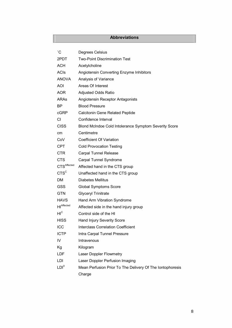
Abbreviations
ûC Degrees Celsius
2PDT Two-Point Discrimination Test
ACH Acetylcholine
ACIs Angiotensin Converting Enzyme Inhibitors
ANOVA Analysis of Variance
AOI Areas Of Interest
AOR Adjusted Odds Ratio
ARAs Angiotensin Receptor Antagonists
BP Blood Pressure
cGRP Calcitonin Gene Related Peptide
CI Confidence Interval
CISS Blond McIndoe Cold Intolerance Symptom Severity Score
cm Centimetre
CoV Coefficient Of Variation
CPT Cold Provocation Testing
CTR Carpal Tunnel Release
CTS Carpal Tunnel Syndrome
CTSAffected Affected hand in the CTS group
CTSC Unaffected hand in the CTS group
DM Diabetes Mellitus
GSS Global Symptoms Score
GTN Glyceryl Trinitrate
HAVS Hand Arm Vibration Syndrome
HIAffected Affected side in the hand injury group
HIC Control side of the HI
HISS Hand Injury Severity Score
ICC Interclass Correlation Coefficient
ICTP Intra Carpal Tunnel Pressure
IV Intravenous
Kg Kilogram
LDF Laser Doppler Flowmetry
LDI Laser Doppler Perfusion Imaging
LDI0 Mean Perfusion Prior To The Delivery Of The Iontophoresis
Charge

9
LDIMax Maximum post-iontophoresis perfusion readings
LDIMean Mean perfusion values of the first three images post-delivery of
iontophoresis
LOA Limits Of Agreement
m 2PDT Moving Two-Point Discrimination Test
MC CTS Medically Confirmed Carpal Tunnel Syndrome
mm Millimetres
mmHg Millimetres Mercury
MRI Magnetic Resonance Imaging
ms Millisecond
Mv Microvolt
NC Normal control group
NCLeft Normal control group, Left hand
NCRight Normal control group, Right hand
NCS Nerve Conduction Studies
NHS National Health Service
Nm Nanometer
NO Nitric Oxide
NOS Nitric Oxide Synthase
NSAID Non-Steroidal Anti-Inflammatory Drugs
OR Odds Ratio
PL Palmaris Longus
PPT Purdue Pegboard Test
PRP Primary Raynaud�s Phenomenon
PTCI Post Traumatic Cold Intolerance
r Pearson Correlation Coefficient
RBC Red Blood Cells
RP Raynaud�s Phenomenon
sec Second
s 2PDT Static Two Point Discrimination
SD Standard Deviation
SN Sensorineural
SNP Sodium Nitroprusside
SPSS Statistical Package For Social Sciences
SSc Scleroderma

10
SSS Symptom Severity Score
SWM Semmes-Weinstein Monofilament Pressure Aethesiometer
SWS Stockholm Workshop Scales
T30secs Temperature Rise In The First 30 Seconds
T5oC Time Taken For The Hands To Re-Warm By 5°C
TA Thermal Aesthesiometry
TICAS Trauma Induced Cold Associated Symptoms
V Vascular
VWF Vibration White Finger
WBC White Blood Cell

11
List of Illustrations
CHAPTER 1: INTRODUCTION
Figure 1.1 The pre- and post-op interstitial pressure in CTS 26 Figure 1.2 The annular elements around the wrist and their movement with
flexion and extension
28
Figure 1.3 The difference in the skin incision between the conventional and
limited incision carpal tunnel release
42
Figure 1.4 RP affecting both hands 58 Figure 1.5 The LDI apparatus 72 Figure 1.6 Mechanisms of impairment of endothelial-dependent control of
vasomotor tone
79
CHAPTER 2: GENERAL METHODS
Figure 2.1 Guide flow chart for normal continuous data analysis 86 Figure 2.2 Guide flow chart for skewed continuous data analysis 86 Figure 2.3 Simultaneous cathodal and anodal Iontophoresis 88 Figure 2.4 PeriIont-382 Power Supply 88 Figure 2.5 Two Iontophoretic Silver/Silver coated electrodes attached to the
back of the hand
91
Figure 2.6 Periscan PIM II Laser Doppler Perfusion Imager 91 Figure 2.7 Data Output after completing LDI 94 Figure 2.8 The cooling water bath with thermocouples attached to the terminal
pulp of the digits in both hands
96
Figure 2.9 The phases of the re-warming curve 96 Figure 2.10 The Audit Cycle 97 Figure 2.11 Audit stages 98 Figure 2.12 The DISK-CRIMINATOR® 101 Figure 2.13 Touch-Test® Sensory Evaluator 102 Figure 2.14 Grip and pinch strength dynamometers 104
12
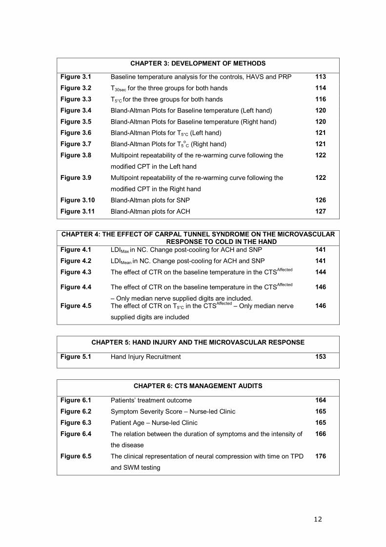
CHAPTER 3: DEVELOPMENT OF METHODS
Figure 3.1 Baseline temperature analysis for the controls, HAVS and PRP 113 Figure 3.2 T30sec for the three groups for both hands 114 Figure 3.3 T5°C for the three groups for both hands 116 Figure 3.4 Bland-Altman Plots for Baseline temperature (Left hand) 120 Figure 3.5 Bland-Altman Plots for Baseline temperature (Right hand) 120 Figure 3.6 Bland-Altman Plots for T5°C (Left hand) 121 Figure 3.7 Bland-Altman Plots for T5
oC (Right hand) 121
Figure 3.8 Multipoint repeatability of the re-warming curve following the
modified CPT in the Left hand
122
Figure 3.9 Multipoint repeatability of the re-warming curve following the
modified CPT in the Right hand
122
Figure 3.10 Bland-Altman plots for SNP 126 Figure 3.11 Bland-Altman plots for ACH 127
CHAPTER 4: THE EFFECT OF CARPAL TUNNEL SYNDROME ON THE MICROVASCULAR RESPONSE TO COLD IN THE HAND
Figure 4.1 LDIMax in NC. Change post-cooling for ACH and SNP 141 Figure 4.2 LDIMean in NC. Change post-cooling for ACH and SNP 141 Figure 4.3 The effect of CTR on the baseline temperature in the CTSAffected
144
Figure 4.4 The effect of CTR on the baseline temperature in the CTSAffected
� Only median nerve supplied digits are included.
146
Figure 4.5 The effect of CTR on T5°C in the CTSAffected � Only median nerve
supplied digits are included
146
CHAPTER 5: HAND INJURY AND THE MICROVASCULAR RESPONSE
Figure 5.1 Hand Injury Recruitment 153
CHAPTER 6: CTS MANAGEMENT AUDITS
Figure 6.1 Patients� treatment outcome 164 Figure 6.2 Symptom Severity Score � Nurse-led Clinic 165 Figure 6.3 Patient Age � Nurse-led Clinic 165 Figure 6.4 The relation between the duration of symptoms and the intensity of
the disease
166
Figure 6.5 The clinical representation of neural compression with time on TPD
and SWM testing
176
13
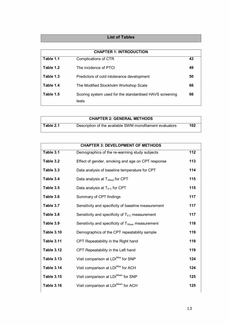
List of Tables
CHAPTER 1: INTRODUCTION
Table 1.1 Complications of CTR 43
Table 1.2 The incidence of PTCI 49
Table 1.3 Predictors of cold intolerance development 50
Table 1.4 The Modified Stockholm Workshop Scale 66
Table 1.5 Scoring system used for the standardised HAVS screening
tests
66
CHAPTER 2: GENERAL METHODS
Table 2.1 Description of the available SWM monofilament evaluators 102
CHAPTER 3: DEVELOPMENT OF METHODS
Table 3.1 Demographics of the re-warming study subjects 112
Table 3.2 Effect of gender, smoking and age on CPT response 113
Table 3.3 Data analysis of baseline temperature for CPT 114
Table 3.4 Data analysis at T30sec for CPT 115
Table 3.5 Data analysis at T5°C for CPT 115
Table 3.6 Summary of CPT findings 117
Table 3.7 Sensitivity and specificity of baseline measurement 117
Table 3.8 Sensitivity and specificity of T5ûC measurement 117
Table 3.9 Sensitivity and specificity of T30sec measurement 118
Table 3.10 Demographics of the CPT repeatability sample 119
Table 3.11 CPT Repeatability in the Right hand 119
Table 3.12 CPT Repeatability in the Left hand 119
Table 3.13 Visit comparison at LDIMax for SNP 124
Table 3.14 Visit comparison at LDIMax for ACH 124
Table 3.15 Visit comparison at LDIMean for SNP 125
Table 3.16 Visit comparison at LDIMean for ACH 125
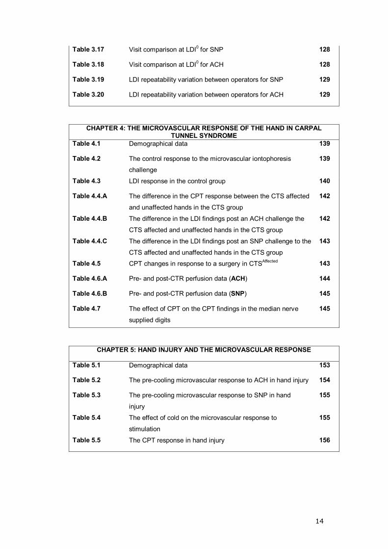
14
Table 3.17 Visit comparison at LDI0 for SNP 128
Table 3.18 Visit comparison at LDI0 for ACH 128
Table 3.19 LDI repeatability variation between operators for SNP 129
Table 3.20 LDI repeatability variation between operators for ACH 129
CHAPTER 4: THE MICROVASCULAR RESPONSE OF THE HAND IN CARPAL TUNNEL SYNDROME
Table 4.1 Demographical data 139
Table 4.2 The control response to the microvascular iontophoresis
challenge
139
Table 4.3 LDI response in the control group 140
Table 4.4.A The difference in the CPT response between the CTS affected
and unaffected hands in the CTS group
142
Table 4.4.B The difference in the LDI findings post an ACH challenge the
CTS affected and unaffected hands in the CTS group
142
Table 4.4.C The difference in the LDI findings post an SNP challenge to the
CTS affected and unaffected hands in the CTS group
143
Table 4.5 CPT changes in response to a surgery in CTSAffected 143
Table 4.6.A Pre- and post-CTR perfusion data (ACH)
144
Table 4.6.B Pre- and post-CTR perfusion data (SNP)
145
Table 4.7 The effect of CPT on the CPT findings in the median nerve
supplied digits
145
CHAPTER 5: HAND INJURY AND THE MICROVASCULAR RESPONSE
Table 5.1 Demographical data 153
Table 5.2 The pre-cooling microvascular response to ACH in hand injury 154
Table 5.3 The pre-cooling microvascular response to SNP in hand injury
155
Table 5.4 The effect of cold on the microvascular response to stimulation
155
Table 5.5 The CPT response in hand injury 156
15
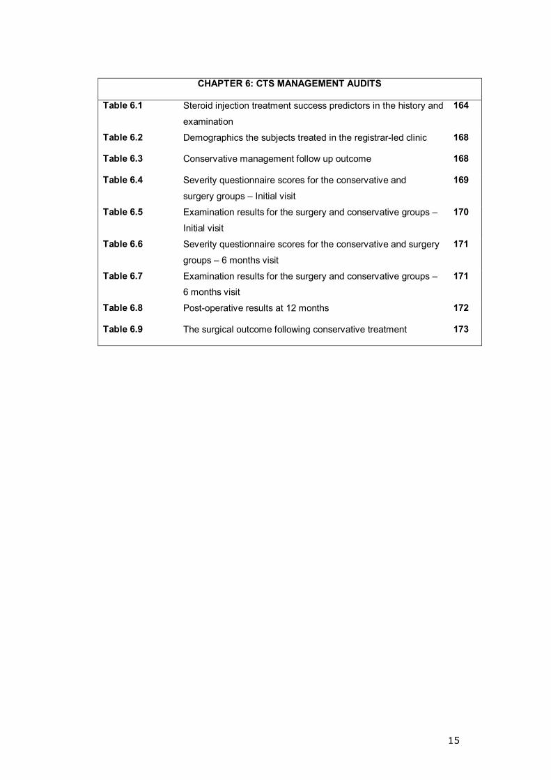
CHAPTER 6: CTS MANAGEMENT AUDITS
Table 6.1 Steroid injection treatment success predictors in the history and
examination
164
Table 6.2 Demographics the subjects treated in the registrar-led clinic
168
Table 6.3 Conservative management follow up outcome 168
Table 6.4 Severity questionnaire scores for the conservative and surgery groups � Initial visit
169
Table 6.5 Examination results for the surgery and conservative groups �
Initial visit
170
Table 6.6 Severity questionnaire scores for the conservative and surgery
groups � 6 months visit
171
Table 6.7 Examination results for the surgery and conservative groups �
6 months visit
171
Table 6.8 Post-operative results at 12 months 172
Table 6.9 The surgical outcome following conservative treatment 173

16
Acknowledgements
At this point I would like to thank the many people who have supported my work
and guided me along the way. I would like to start by thanking Mr L C
Bainbridge, whose determination and strength of conviction paved the path for
my research and led me into this journey. His ideas and comments lit the path
on numerous occasions and helped me to overcome some of the hurdles during
my research. Thank you.
I would like to thank Dr G Manning, whose organisation, patience and academic
approach to research were enlightening and her dedication to research remains
inspiring. Thank you.
I would like to thank all the members of the Division of Vascular Medicine:
Professor R Donnelly, Mrs Jean Clarke and Sue Lane. My deepest gratitude
has to go to Mrs Margaret Baker and Mrs Kelly Mitchell, both of whom were and
will always be my family away from home and stood by me when I needed help
the most. Thank you.
This piece of work was not only the product of the cooperation between the
members of our research group but also the R&D Department staff at Derby
Hospitals NHS Foundation Trust, especially Dr R Hilliam and Mr A Fakis. My
sincere thanks to both.

17
Declaration
The author declares that:
1- All the work contained in this thesis is my own, unless stated.
2- My role in the modification of the cold provocation testing was to
undertake a detailed analysis of the data from subjects that had been
studied by the team prior to the start of my research post.
3- I recruited and studied the majority of the studies within Chapters 4 and
5 with technical support from Mrs M Baker.
4- The clinical audit studies were entirely my own work.

18
Research Presentations
Various parts of this thesis have been published in peer reviewed scientific
journals and/or presented in abstract form in national and international
meetings.
A list of these publications is shown below: Published
1. Salem KM, Baker M, Manning G, Bainbridge LC, Donnelly R Analysis of Rewarming curves in patients with Raynaud�s phenomenon of various aetiologies Revesta Iberoamericana De Cirugia de la Mano (2005) 33(66): 76 (Abstract)
Accepted
2. Salem K, Baker M, Hilliam RM, Davies S, Deighton C, Bainbridge LC, Manning G Analysis of rewarming curves in patients with Raynaud�s phenomenon of various aetiologies (Original Article) The Journal of Hand Surgery [Br] (Manuscript number: JHSB-D-06-00228), Submission date: June 2006
Submitted
3. Salem K, Donnelly R, Manning G, Bainbridge LC Post traumatic cold intolerance, a review (Review Article) The Journal of Hand Surgery [Br] (Manuscript number: JHSB-D-06-00347), Submission Date: August 2006
Poster presentations
1. Does Cold Provocation Testing have a role in the assessment of people with suspected Hand Arm Vibration White Finger Syndrome (HAVS)? Society of Occupational Medicine Annual Scientific Meeting, Science in Practice, Manchester June 2005
International and National meeting oral Presentations
2. Analysis of re-warming curves in patients with Raynaud�s phenomenon of various aetiologies XVII National Congress SECMA Surgery of the Hand, in association with the British Society for Surgery of the Hand. Valladolid- Spain, April 2005
3. A new objective re-warming test Advanced Course for Hand Surgery,
Pulvertaft Hand Unit, Derbyshire Royal Infirmary, May 2005
19
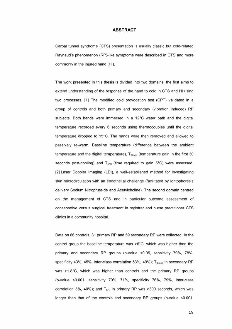
ABSTRACT
Carpal tunnel syndrome (CTS) presentation is usually classic but cold-related
Raynaud�s phenomenon (RP)-like symptoms were described in CTS and more
commonly in the injured hand (HI).
The work presented in this thesis is divided into two domains; the first aims to
extend understanding of the response of the hand to cold in CTS and HI using
two processes. [1] The modified cold provocation test (CPT) validated in a
group of controls and both primary and secondary (vibration induced) RP
subjects. Both hands were immersed in a 12°C water bath and the digital
temperature recorded every 6 seconds using thermocouples until the digital
temperature dropped to 15°C. The hands were then removed and allowed to
passively re-warm. Baseline temperature (difference between the ambient
temperature and the digital temperature), T30sec (temperature gain in the first 30
seconds post-cooling) and T5°C (time required to gain 5°C) were assessed.
[2] Laser Doppler Imaging (LDI), a well-established method for investigating
skin microcirculation with an endothelial challenge (facilitated by iontophoresis
delivery Sodium Nitroprusside and Acetylcholine). The second domain centred
on the management of CTS and in particular outcome assessment of
conservative versus surgical treatment in registrar and nurse practitioner CTS
clinics in a community hospital.
Data on 86 controls, 31 primary RP and 59 secondary RP were collected. In the
control group the baseline temperature was >6°C, which was higher than the
primary and secondary RP groups (p-value <0.05, sensitivity 79%, 78%,
specificity 43%, 45%, inter-class correlation 53%, 49%); T30sec in secondary RP
was >1.8°C, which was higher than controls and the primary RP groups
(p-value <0.001, sensitivity 70%, 71%, specificity 76%, 79%, inter-class
correlation 3%, 40%); and T5°C in primary RP was >300 seconds, which was
longer than that of the controls and secondary RP groups (p-value <0.001,

20
sensitivity 64%, 61%, specificity 70%, 64%, inter-class correlation 70%, 70%);
data given for left and right hands respectively.
CPT and LDI studies were undertaken on 60 controls and 60 CTS patients pre-
operatively and repeated on 40 subjects 5-7 months post-decompression. Post-
operatively, the baseline temperature increased by 1.5°C (p-value <0.05) in
both hands and 2.5°C (p-value <0.001) in the median nerve supplied digits, T5oC
was reduced in the hands (pre- versus post-operative from 474 to 348 seconds)
(p-value 0.06) and from 468 to 273 seconds in the median nerve supplied digits
(p-value 0.01). Endothelial dependent and independent control at mean and
maximum pre- and post-cooling perfusion was significantly depressed (p-value
0.05) post-cold exposure in the control group. LDI limited to the dorsum of the
hand identified no significant difference pre- and post-operatively (p-value
>0.05).
HI subject recruitment was challenging: the absence of a financial incentive and
the possible income loss during working days for a young working cohort might
have contributed to the poor recruitment. Of the 60 subject targets only 14
recruited and the injury severity varied widely between the recruits; the data
gathered through CPT and LDI in this group did not show a significant
difference from that collected in controls.
CTS management audits on 74 subjects in a nurse-led clinic and 173 subjects
in a registrar-led clinic identified a high failure rate of the conservative
management (60%) at 6 months follow up in both clinics with unclear success
predictors suggesting an extra burden on clinics providing decompression
surgery.
21
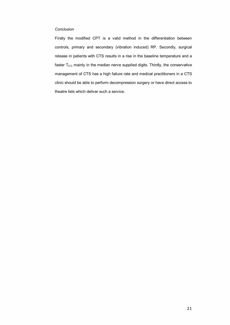
Conclusion
Firstly the modified CPT is a valid method in the differentiation between
controls, primary and secondary (vibration induced) RP. Secondly, surgical
release in patients with CTS results in a rise in the baseline temperature and a
faster T5°C mainly in the median nerve supplied digits. Thirdly, the conservative
management of CTS has a high failure rate and medical practitioners in a CTS
clinic should be able to perform decompression surgery or have direct access to
theatre lists which deliver such a service.

22
To my parents, without
whom nothing would ever
have been possible

23
CHAPTER 1: INTRODUCTION
24
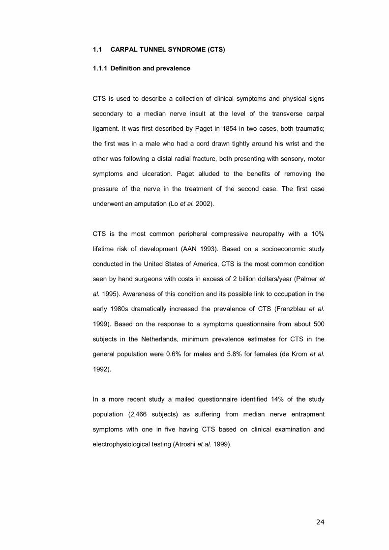
1.1 CARPAL TUNNEL SYNDROME (CTS)
1.1.1 Definition and prevalence
CTS is used to describe a collection of clinical symptoms and physical signs
secondary to a median nerve insult at the level of the transverse carpal
ligament. It was first described by Paget in 1854 in two cases, both traumatic;
the first was in a male who had a cord drawn tightly around his wrist and the
other was following a distal radial fracture, both presenting with sensory, motor
symptoms and ulceration. Paget alluded to the benefits of removing the
pressure of the nerve in the treatment of the second case. The first case
underwent an amputation (Lo et al. 2002).
CTS is the most common peripheral compressive neuropathy with a 10%
lifetime risk of development (AAN 1993). Based on a socioeconomic study
conducted in the United States of America, CTS is the most common condition
seen by hand surgeons with costs in excess of 2 billion dollars/year (Palmer et
al. 1995). Awareness of this condition and its possible link to occupation in the
early 1980s dramatically increased the prevalence of CTS (Franzblau et al.
1999). Based on the response to a symptoms questionnaire from about 500
subjects in the Netherlands, minimum prevalence estimates for CTS in the
general population were 0.6% for males and 5.8% for females (de Krom et al.
1992).
In a more recent study a mailed questionnaire identified 14% of the study
population (2,466 subjects) as suffering from median nerve entrapment
symptoms with one in five having CTS based on clinical examination and
electrophysiological testing (Atroshi et al. 1999).

25
1.1.2 Pathophysiology of CTS
The pathophysiology of CTS is multifaceted, characterised by an increased
intra-carpal tunnel pressure (ICTP) as a final common pathway (Szabo et al.
1987). Using the forearms of five cadavers, Cobb et al. perfused the flexor
compartment with saline in an attempt to determine whether elevated pressure
in this compartment is transmitted to the carpal tunnel. The author concluded
that the carpal tunnel functions as a relatively closed compartment with respect
to the transfer of pressure from the flexor compartment of the forearm (Cobb et
al. 1995).
1.1.2.1 The carpal tunnel pressure
Using a wick catheter, measurements of the ICTP obtained from 12 normal
subjects were lowest when the wrist was in neutral (mean 2.5 mmHg (0-7)), with
pressure readings rising to 30 mmHg during flexion and extension (Gelberman
et al. 1981b).
To investigate the pressure threshold for peripheral nerve dysfunction, a human
model of acute nerve compression was used by applying a controlled external
pressure on the carpal tunnel and measuring the ICTP using a wick catheter.
External pressure challenges of 40, 50, 60 and 70 mmHg were applied with the
wrist in neutral. Using nerve conduction studies (NCS) pre, 30 and 240 minutes
into the compression phase and during the recovery phase, the authors
concluded that at 40 mmHg some nerve function was lost but at 50 mmHg all
nerve function was blocked. They hypothesised the presence of a critical
pressure between 40 and 50 mmHg where nerve function is acutely
endangered (Gelberman et al. 1983b).
The above study was repeated on the non-dominant wrist of nine hypertensive
controls (diastolic blood pressure (BP) ≥90 mmHg). The critical pressure

26
threshold in the hypertensive group was greater than that previously reported
for normotensive subjects (40-50 mmHg). Further analysis revealed that the
critical ICTP threshold was consistently 30 mmHg below diastolic BP
(approximately 45 mmHg below mean arterial BP) for both the normotensive
and hypertensive population (Szabo et al. 1983). In support of this finding, CTS
was reported in hypertensive patients following the successful treatment of their
hypertension (Emara et al. 1988).
When the ICTP was measured in 15 patients with CTS the mean pressure was
32 mmHg in neutral increasing to 94 and 110 mmHg in flexion and extension
respectively. Surgical release produced an immediate and sustained reduction
in pressure (Gelberman et al. 1981b). This drop in the ICTP was statistically
significant using both open and endoscopic release (Figure 1.1) (Brown et al.
1993).
Figure 1.1 The pre- and post-op interstitial pressure in CTS (whiskers=SD)
(n=15) (Brown et al. 1993)
27
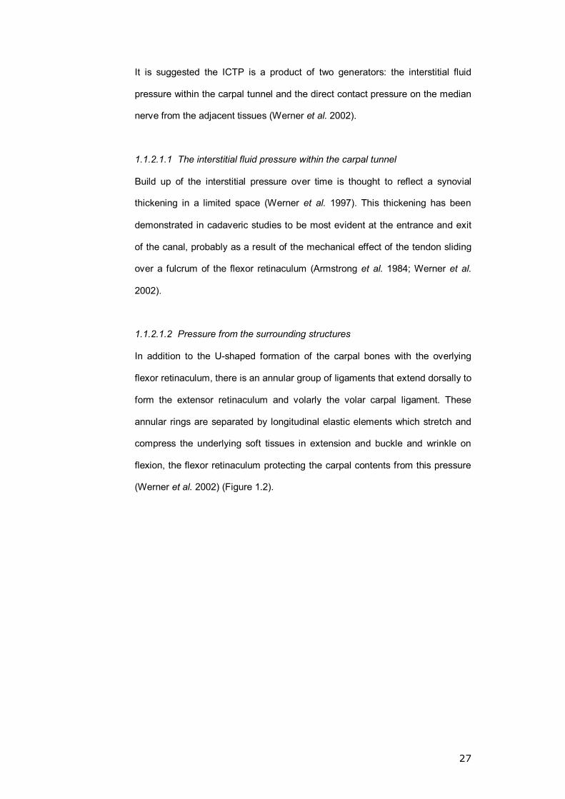
It is suggested the ICTP is a product of two generators: the interstitial fluid
pressure within the carpal tunnel and the direct contact pressure on the median
nerve from the adjacent tissues (Werner et al. 2002).
1.1.2.1.1 The interstitial fluid pressure within the carpal tunnel
Build up of the interstitial pressure over time is thought to reflect a synovial
thickening in a limited space (Werner et al. 1997). This thickening has been
demonstrated in cadaveric studies to be most evident at the entrance and exit
of the canal, probably as a result of the mechanical effect of the tendon sliding
over a fulcrum of the flexor retinaculum (Armstrong et al. 1984; Werner et al.
2002).
1.1.2.1.2 Pressure from the surrounding structures
In addition to the U-shaped formation of the carpal bones with the overlying
flexor retinaculum, there is an annular group of ligaments that extend dorsally to
form the extensor retinaculum and volarly the volar carpal ligament. These
annular rings are separated by longitudinal elastic elements which stretch and
compress the underlying soft tissues in extension and buckle and wrinkle on
flexion, the flexor retinaculum protecting the carpal contents from this pressure
(Werner et al. 2002) (Figure 1.2).

28
Figure 1.2 The annular elements around the wrist and their movement with flexion and extension (Werner et al. 2002)
The movement of the carpal bones during extension result in the proximal
carpal margin gliding volarly against the flexor retinaculum, consequently
squeezing the carpal contents between the bone and ligamentous components
of the tunnel.
Digital and wrist extension draws the more proximal, thicker part of the tendons
into the carpal tunnel leading to an increase in the content volume and an
increased pressure on the nerve. It is hypothesised that this also occurs in
volar, radial and ulnar deviation but to a lesser extent (Werner et al. 2002).
Histological studies of the tenosynovium in patients with CTS support ischemia-
induced changes rather than inflammation. These findings include cloudy white
hypertrophy of the tenosynovium with no inflammatory infiltrate. Fibrous
hyperplasia was also noted with an increased disorganised collagen deposition
(Fuchs et al. 1991; Kerr et al. 1992). In a retrospective review of synovial
specimens from 625 carpal tunnel releases, the authors reported 96% (601) to
have a synovial tissue histological diagnosis of benign fibrous tissue without
29
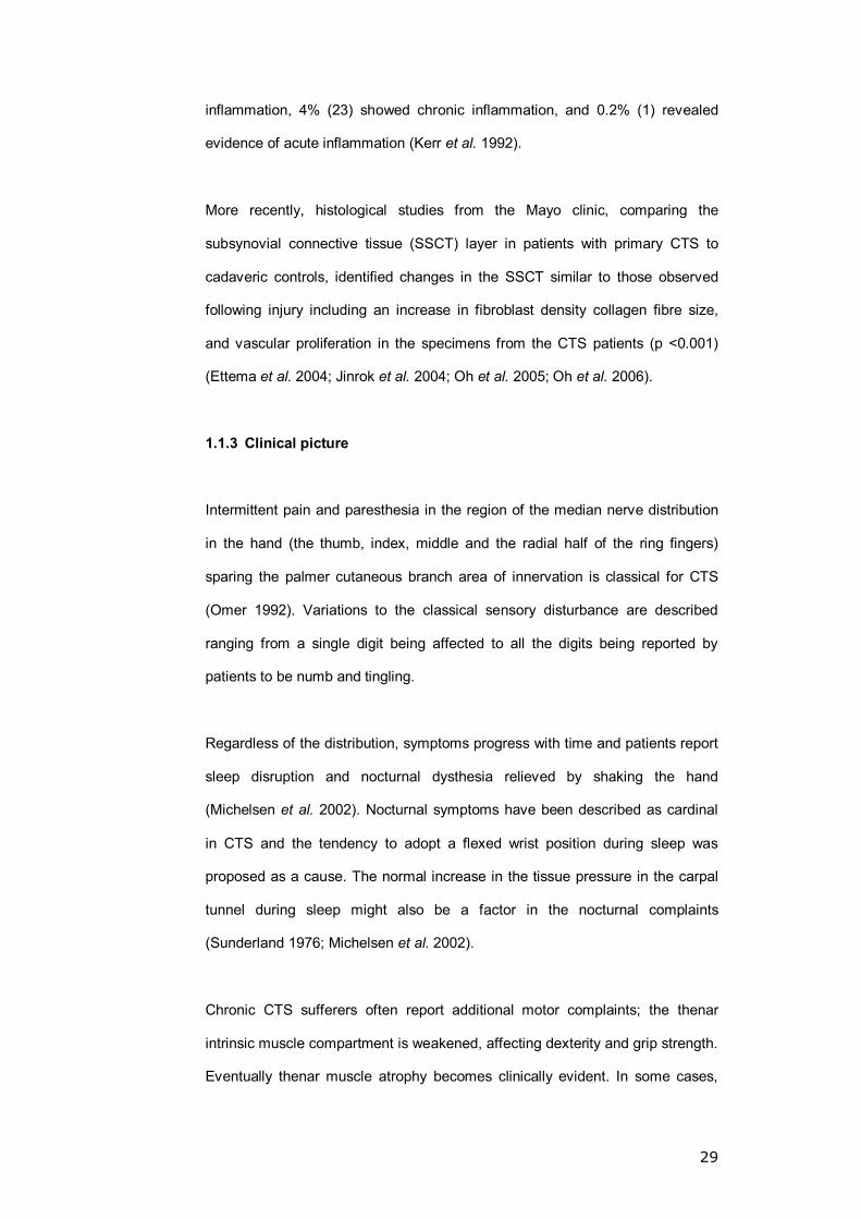
inflammation, 4% (23) showed chronic inflammation, and 0.2% (1) revealed
evidence of acute inflammation (Kerr et al. 1992).
More recently, histological studies from the Mayo clinic, comparing the
subsynovial connective tissue (SSCT) layer in patients with primary CTS to
cadaveric controls, identified changes in the SSCT similar to those observed
following injury including an increase in fibroblast density collagen fibre size,
and vascular proliferation in the specimens from the CTS patients (p <0.001)
(Ettema et al. 2004; Jinrok et al. 2004; Oh et al. 2005; Oh et al. 2006).
1.1.3 Clinical picture
Intermittent pain and paresthesia in the region of the median nerve distribution
in the hand (the thumb, index, middle and the radial half of the ring fingers)
sparing the palmer cutaneous branch area of innervation is classical for CTS
(Omer 1992). Variations to the classical sensory disturbance are described
ranging from a single digit being affected to all the digits being reported by
patients to be numb and tingling.
Regardless of the distribution, symptoms progress with time and patients report
sleep disruption and nocturnal dysthesia relieved by shaking the hand
(Michelsen et al. 2002). Nocturnal symptoms have been described as cardinal
in CTS and the tendency to adopt a flexed wrist position during sleep was
proposed as a cause. The normal increase in the tissue pressure in the carpal
tunnel during sleep might also be a factor in the nocturnal complaints
(Sunderland 1976; Michelsen et al. 2002).
Chronic CTS sufferers often report additional motor complaints; the thenar
intrinsic muscle compartment is weakened, affecting dexterity and grip strength.
Eventually thenar muscle atrophy becomes clinically evident. In some cases,

30
the sensory fascicles are spared and the compression damages the motor
component of the nerve leading to a delayed presentation. The patient is often
unaware of the problem until significant muscle atrophy develops (Michelsen et
al. 2002; Burke et al. 2003).
In a literature review, CTS was classified into early, intermediate, advanced and
acute stages based on an observed progression of symptoms, and clinical and
electro-diagnostic findings. The early stage was less than one year with sensory
but no motor component while the intermediate and late stages were associated
with weakness for which operative treatment was advocated (Gelberman et al.
1988).
Based on a hypothesis suggesting that the sympathetic supply to the hand is
mostly carried by the median nerve, Phalen suggested that the vasomotor
control of the hand will be affected by compressive neuropathy to the median
nerve (Phalen 1966). Despite the high prevalence of CTS and the large amount
of published literature related to it, the autonomic disturbance associated with
CTS is frequently missed clinically or unreported in the literature. First reported
in 1957, RP-like symptoms in CTS have subsequently been addressed in a very
limited number of studies (Garland et al. 1957; Linscheid et al. 1967; Pal et al.
1996).
More recently, a prospective review of 139 cases in 76 CTS patients identified
62% of the patients to have �dysautonomia�. Verghese et al. (2000) considered
eight items related to the autonomic function and reported the incidence of
those in the study cohort as follows: swelling of the digits (60%), dry skin (40%),
RP (33%), blanching (32%). No patients presented with digital ulceration or nail
dystrophy. Cold sensation in the fingertips was found to be non-specific in a
pilot study and was excluded. Although this study subjectively demonstrates the
high prevalence of the autonomic symptoms, several limitations can be

31
identified: unclear clinical definition swelling, dryness and RP, the exclusion of
sweating, and the large number of females in the cohort resulting in a bias in
prevalence calculations (higher in females).
The prevalence of the autonomic symptoms reported in other studies varied. A
retrospective review of the notes of 2,800 patients in the Mayo Clinic in the
1960s reported 28 complaints of autonomic symptoms but a systematic analysis
of the autonomic pattern was lacking and minor symptoms were not recorded
(Linscheid et al. 1967). In a more recent study, the incidence of RP was higher
(36%) in a CTS group (93 subjects) compared to a control group 12% (57
subjects) (Pal et al. 1996) . Reporting on 30 subjects, Chung et al. found 60%
complaining of RP and then reported an improvement in 78% of the affected
following surgical release in a subsequent publication (Chung et al. 1999;
Chung et al. 2000).
A variety of provocation tests designed to reproduce or provoke the sensory
component of the symptoms was suggested such as Phalen�s test (Phalen
1972), Reverse Phalen�s (Werner et al. 1994), Tinel�s (Bowles et al. 1983; Tinel
2005) and Durkan�s tests (Bowles et al. 1983). In a systematic review of
literature to determine the accuracy of history taking and physical examination
in the diagnosis of CTS, the authors concluded that elements in the history such
as age and nocturnal symptoms and in the physical examination such as thenar
atrophy, Tinel�s sign and Phalen�s sign, pressure provocation tests and sensory
tests have limited or no value in distinguishing patients with CTS from controls
(D'Arcy et al. 2000). In another study, the prevalence of Tinel�s sign in a control
group was reported to be as high as 45% (Seror 1987).

32
1.1.4 Diagnosis criteria
Although the presentation in the majority of CTS cases is classical, the
diagnosis in some cases remains difficult. This is mainly due to the variations in
the dysthesia distribution and the limited usefulness of provocation tests in
cases where no other clinical signs are present (weakness and muscle
wasting). In an effort to identify diagnostic and staging criteria for CTS, 14
experienced researchers could identify no gold standard for the diagnosis within
the literature (Rempel et al. 1998). This reflects the problem junior doctors and
nurse practitioners face in their clinical practice.
In his diagnosis, Phalen required CTS patients to have one or more of three
signs: dysthesia limited to the area of the median nerve distribution, a positive
Tinel�s sign and a positive Phalen�s sign (Phalen 1981). In an attempt to
establish a consensus on case definitions for several common work-related
upper limb pain syndromes, a core group of 29 United Kingdom experts
representing various disciplines managing these conditions (including
rheumatologists, surgeons, GPs and therapists) concluded that a diagnosis of
CTS requires sensory symptoms limited to the median nerve distribution in
addition to one or more of the following: nocturnal symptoms, median nerve
motor signs, a positive Tinel�s sign, a positive Phalen�s sign or an abnormality of
the nerve conduction studies (NCS) of the median nerve across the wrist
(Harrington et al. 1998).
In a survey of the members of the American Society for Surgery of the Hand
only 33% of the surgeons used NCS as an aid in the diagnosis of CTS (Duncan
et al. 1987). This reflects the tentative views on the usefulness of NCS amongst
surgeons. In correspondence, the �under utilisation� of the NCS in the
management of CTS in the USA was reported despite guidance advocating its
use (Mainous et al. 1996). This �under use� relates to the test�s shortcomings:
33
the lack of standardisation even within the same department, the absence of
population-based reference intervals, the limited sensitivity (70%) and specificity
(76%), the absence of evidence for the test�s ability to predict or improve the
treatment outcome and the lack of correlation with the patient�s symptoms
(Atroshi et al. 1999; Kilmer et al. 2002; Smith 2002).
In an attempt to evaluate the agreement between the clinical examination, the
sensory nerve conduction testing and symptom surveys including hand
diagrams, only 23 out of 449 subjects with one or more symptoms suggestive of
CTS met all three criteria (symptoms, examination and NCS) with a very poor
agreement between the assessment parameters (kappa coefficient between
0.00 and 0.18) (Homan et al. 1999). In the same study, the majority of the
subjects with NCS evidence of median neuropathy were asymptomatic and vice
versa (Homan et al. 1999). In an 11-year longitudinal study following up 558
subjects with an abnormal NCS, the authors concluded that an abnormal NCS
is not a predictor of future CTS (Nathan et al. 1998).
CTS severity assessment questionnaires have been proposed. Levine et al.
developed a self-administered CTS symptoms and function assessment
questionnaire. The authors concluded that the questionnaire is reproducible,
internally consistent, and responsive to clinical change when studied on a
sample of 76 patients diagnosed clinically and by NCS to have CTS. These
conclusions were based on repeatability assessment using the Pearson
correlation coefficient (r) = 0.91 and 0.93 for severity of symptoms and
functional status respectively, a statistically significant drop in severity scores
following surgical treatment (Levine et al. 1993). It is clear that this study had
limitations; the authors used an inappropriate test (r) to assess repeatability, the
study lacked a control group limiting its conclusions and, despite a significant
improvement in the scores post-operatively, there is no evidence to suggest that
it is responsive in the conservative management group. This may explain the

34
findings of a subsequent study which concluded that the Levine questionnaire
has only limited value in establishing the diagnosis of CTS (You et al. 1999).
More recently Bland described a questionnaire with an overall sensitivity of 79%
and specificity of 55% for the diagnosis of CTS and an ability to predict the
findings of nerve conduction studies based on a study of 8,223 suspected CTS
subjects and compared with the neurophysiological findings (Bland 2000).
The use of tools such as ultrasound, CT and MRI scanning has been reported
but their role in the diagnosis of CTS is not established (D'Arcy et al. 2000;
Beekman et al. 2002).
1.1.5 Risk and prognostic factors
The cause in the majority of CTS cases is unknown (idiopathic) (Kerwin et al.
1996) but some of the risk factors have been identified. A case-control analysis
of the data from 3,391 CTS cases from the UK General Practice Research
Database identified a higher risk in patients suffering rheumatoid arthritis
(OR=2.23), osteoarthritis of the wrist and carpus (OR=1.89), previous wrist
fracture (OR=2.29), obesity and thyroxin. The authors also acknowledged
Diabetes Mellitus (DM) (OR=2.06) and its treatment (Insulin, Sulphonylureas,
Metformin) as risk factors, yet the reported risk with DM treatment may be
biased by the higher risk of developing the syndrome in the diabetics to start
with (Geoghegan et al. 2004).
Most studies report a prevalence three to fourfold higher in females compared
to males (D'Arcy et al. 2000; Burke et al. 2003). The cause of this higher
prevalence was explained by a narrower carpal tunnel outlet, therefore a
pronounced decrease in the tunnel volume in females compared to males
(Dekel et al. 1980).

35
The strongest factor associated with the development of CTS is vibration
exposure (Mackinnon 2002). In a population assessed for compensation for
Hand Arm Vibration Syndrome (HAVS) through the miner�s compensation
scheme in the UK, a prevalence of 15% was reported. This figure is likely to be
higher than expected due to the potential financial gain for those diagnosed with
both conditions within the scheme (Burke et al. 2005). In a small prospective
review of eight patients (15 hands) diagnosed with both CTS and HAVS, a 6-
month follow up from surgical decompression for CTS revealed an improvement
in the CTS symptoms in all subjects and HAVS symptoms improvement in 50%
of the participants suggesting a link between the two conditions (Savage et al.
1990).
In recent years, claims for compensation due to occupation-related disease
have increased. This is true for work-related repetitive strain injuries of which
only about 2% are CTS (Mackinnon et al. 1997). In a study based on the 1988
National Health Interview Survey in the United States, the data on
approximately 30,000 workers were analysed to identify the prevalence of CTS
and its risk factors. The authors reported 1.5% of the study population to have
what they termed self-reported CTS (not medically confirmed) and 0.5%
medically confirmed CTS cases (MC CTS). Within the MC CTS group the
following risk factors were identified: exposure to bending and twisting at work,
(Adjusted Odds Ratio (AOR) 5.5); exposure to vibration (AOR 1.9); white race
(AOR 16.7); female gender (AOR 2.3); BMI >25 (AOR 2.0); history of cigarette
smoking (AOR 1.6); age >40 (AOR 1.2); and education >12 years (AOR 1.2)
(Tanaka et al. 1997). Findings supporting the association between repetitive
movement of the hand and CTS have also been reported by others (Bernard
1997; Lo et al. 2002). Nonetheless, a direct relation between specific forms of
work (including the use of a keyboard) and CTS is still lacking (Burke et al.
2003).
36
A strong family history due to a hereditary neuropathy was reported to result in
a higher predisposition to pressure palsies (Lane et al. 2001).
The prediction of the response to treatment remains difficult. In a prospective
study of 96 patients undergoing CTS surgery, the pre-operative health profile
measured using the Nottingham Health Profile score was significantly worse in
the 27 cases dissatisfied with their surgery (Rege et al. 2001). In their analysis,
the authors included diabetic patients and grouped the patients with post-
operative complications (14 cases) with those whose symptoms did not
improve, rendering conclusions based on this group greatly biased by the
iatrogenic side-effects of surgery and they did not identify the effect of diabetes
(a known cause of mono-neuropathy) on poor outcome.
Another study suggested that complete restoration of clinical and
electrophysiological nerve function was observed only in patients with mild CTS
based on history, clinical examination and NCS (Aulisa et al. 1998) but these
views are not accepted by all. Glowacki et al. presented 227 CTS cases treated
surgically and showed no correlation between the NCS findings and the post-
operative improvement (Glowacki et al. 1996).
Other factors reported to affect the prognosis are compensation claims which
point to a poor post-operative course (De Smet et al. 1995) and favourable
response to steroid injection constituting a good prognostic sign (Green 1984).
1.1.6 Treatment of CTS
1.1.6.1 Conservative treatment
The surgical risk, the increased number of patients seeking treatment for CTS,
the reported success rate for conservative treatment ranging from 13-92% in
some of the studies and the mild-to-moderate presentation picture in some
cases keeps the conservative treatment options as a more appealing choice as

37
a first line of treatment (Osterman et al. 2002). Various modalities of non-
operative treatment have been suggested. These include:
1- Lifestyle modification
Avoiding repetitive wrist and hand motion may reduce the intensity of symptoms
(Viera 2003). This advice is based on the hypothesis that repetitive use of the
hands provokes flexor tenosynovitis (von Schroeder et al. 1996). However, the
results of a synovial tissue histological study on 625 patients diagnosed with
idiopathic CTS uncovered only 4% (23) as having histological evidence of
chronic inflammation and 0.2% (1) to have acute tenosynovitis. The authors
concluded that tenosynovitis is not a part of the pathophysiological process in
chronic idiopathic carpal tunnel syndrome (Kerr et al. 1992).
The same applies to using a wrist support and/or improved wrist positioning
when using a computer (Viera 2003). Avoiding vibration exposure is believed to
reduce the frequency and intensity of the symptoms (Stevens et al. 1992).
Adjustment of work height or tools to optimise the position of the wrist in neutral
and avoid extreme ranges has also been suggested (Burke et al. 2003).
2- Exercise
Tendon and nerve gliding exercises have been shown to reduce the need for
surgical intervention in CTS; in a study of 240 CTS cases, patients were divided
into two groups. Both groups received similar conservative treatment but an
additional programme of nerve and tendon gliding exercises was added to one
of the groups. Of those who did not perform the nerve and tendon gliding
exercises, 71.2% underwent surgery compared with only 43% of patients who
did perform them. Out of 47 patients presenting at a mean of 23 months, 70.2%
reported good or excellent results (Rozmaryn et al. 1998). It is thought that
nerve tethering and adhesions are minimised through nerve mobilisation, and
38
oedema and congestion are reduced by facilitating the venous return (Burke et
al. 2003).
3- Wrist splints
The pressure in the carpal tunnel is lowest in the neutral position (0-7 mmHg):
Gelberman et al. measured the intra-carpal pressures at neutral, 90° flexion and
90° extension in 15 idiopathic CTS cases and 12 controls; pressures in neutral
were lowest in both groups but higher in the CTS group compared to the
controls (32 mmHg in CTS compared to 2.5 mmHg in controls, means)
(Gelberman et al. 1981b). The splint aims to hold the wrist in a neutral position
and is reported to be helpful in mild-to-moderate cases and less effective in
cases of continuous paresthesia or numbness (Osterman et al. 2002; Burke et
al. 2003).
It is common practice to advise patients to use the splint at night and only
occasionally during the day if an activity is expected to exacerbate the
symptoms. However, the symptoms and functional deficits, measured by
Levine's self-administered questionnaire and physiologic impairment measured
by NCS assessed in a small randomised trial with short follow up (six weeks),
were better in the group wearing the splint full-time compared to the group
wearing the splint at night (Walker et al. 2000).
4- Oral medications
In a survey of American hand surgeons, diuretics, Vitamin B6 supplement
(Pyridoxine), Non-steroidal anti-inflammatory drugs (NSAID) and oral steroid
treatment were used in the treatment of CTS (Duncan et al. 1987). A recent
prospective randomised double blind and placebo-controlled trial evaluated the
effectiveness of commonly used oral medications in the treatment of mild-to-
moderate CTS based on NCS findings. The authors reported no difference
between the placebo, NSAID and diuretics in outcome based on a global
39
symptom score (GSS) (Herskovitz et al. 1995) at two and four weeks. The
authors found a significant improvement in the symptom scores of patients on
oral steroid (using prednisolone 20 mg/day for two weeks followed by two
weeks on 10 mg/day, p = 0.00001) despite the small sample size (18 subjects)
(Chang et al. 1998).
A more recent meta-analysis of the randomised controlled trials evaluating the
efficacy of conservative treatment in CTS showed similar conclusions.
Additionally, pyridoxine and laser acupuncture were reported as ineffective in
providing short-term symptom relief (Gerritsen et al. 2002).
5- Local steroid injection
Local steroid infiltration has been shown to give superior results compared to an
oral systemic steroid. In a prospective randomised double blind trial comparing
the effectiveness of a local steroid (methylprednisolone) to a systemic oral
steroid (prednisolone 25 mg/day for 10 days) using GSS, the authors reported a
significantly better symptomatic relief in the infiltration group (p <0.05) at two,
eight and 12 weeks (Wong et al. 2001).
The patient�s forearm is placed on the examination table with the palm facing
up. Using a sterile technique, the needle is advanced from the point of entry at
the distal wrist crease adjacent to the ulnar aspect of the Palmaris Longus (PL)
tendon. The bevel of the needle should run parallel to the longitudinal nerve
fibres to minimise trauma. The needle is advanced distally at 45° towards the
carpal tunnel. If the patient reports paresthesia during the insertion, the needle
should be withdrawn and repositioned (Girlanda et al. 1993; Armstrong et al.
2004). The injectable steroid is water soluble and can be combined with an
anaesthetic to minimise the discomfort associated with the injection (Osterman
et al. 2002).
40
Despite the care taken during the infiltration process, steroid injections are not
hazard free and the most important complication is a direct needle injury to, or
an intra-neural injection into, the median nerve (Burke et al. 2003). To avoid this
potentially disastrous complication and, based on median nerve anatomical
observations in 93 wrists undergoing endoscopic surgical release for CTS, it
was found that the median nerve extends ulnar to the PL tendon in 82% of the
cases by a mean of 4 mm and may extend radial to the PL over 6 mm. The
authors suggested that the safest location to insert a needle in the carpal tunnel
for a steroid injection is through the Flexor carpi radialis tendon (Racasan et al.
2005).
A randomised trial with one year follow-up compared steroid injection to limited
incision CTS release in 123 cases (57 injected, 66 treated surgically). The
authors reported a better subjective outcome in the injection group at the early
stages following treatment. No significant difference in outcome was reported at
one year apart from a perceived better functional outcome in the surgical group
(Andreu et al. 2006). The study had several limitations; the researchers did not
include patients with thenar atrophy, which represents the more severe stage of
the disease. Nocturnal symptoms were used as a primary outcome measure for
improvement, which is not a universal feature in all the patients (Michelsen et al.
2002). Subjective measurement was used to determine patient improvement
and the follow-up period was short (one year) after which a significant body of
evidence suggests a high failure rate ranging between 50% and 86% of the
injected cases (Green 1984; Ozdogan et al. 1984; Weiss et al. 1994; Dammers
et al. 1999b; D'Arcy et al. 2000; Kanaan et al. 2001; Burke et al. 2003; Hui et al.
2004).
There was no significant difference in the outcome measured by the GSS
following the treatment with one versus two steroid injections in idiopathic CTS
41
at eight, 24 and 40 weeks according to a randomised controlled double blind
trial on a group of 40 subjects (Wong et al. 2005).
A prospective study on 50 CTS cases aiming to define the role of steroid
injection and splinting as a method of treatment of CTS concluded that patients
with mild symptoms, a short history of less than one year, normal sensibility,
normal thenar strength and mass, and one- to two-millisecond prolongations of
either distal median motor or sensory latencies had the most satisfactory
responses to injections and splinting (Gelberman et al. 1980).
1.1.6.2 Surgical treatment
Despite the good short-term response to local steroid infiltration, long-term
failure rates are high. This makes the surgical option amongst hand surgeons
treating CTS preferable (Duncan et al. 1987). The surgical management of CTS
has advanced in the last three decades. Surgery through a limited palmer
approach has been advocated with comparable results to the older wide
incision decompression and more recently the endoscopic CTS release is being
popularised.
The traditional open incision technique with a longitudinal incision crossing the
wrist flexion crease was described as an effective approach of treatment with a
low complication rate in major operative text books (Eversmann 1988;
Gelberman 1991). In 1998, Lee et al. described a limited incision approach
utilising a 1-1.5 cm palmer incision for the release of the transverse carpal
ligament with the advantage of avoiding a long post-operative sensitive scar
crossing the wrist crease and avoiding the incision of the facial convergence
between the thenar and hypothenar compartments, a problem blamed for the
slower post-operative recovery in the conventional release (Lee et al. 1998)
(Figure 1.3).

42
Figure 1.3 The difference in the skin incision between the conventional and limited incision carpal tunnel release (Jugovac et al. 2002)
A randomised trial investigating the outcome difference between the
conventional and limited carpal tunnel release in 72 cases (36 in each limb of
the study) with a three-month follow-up concluded that there is no difference in
the symptomatic relief and the post-operative NCS parameters between the two
approaches. However, the functional recovery of the hand measured by the
return to daily activities and the return to work was significantly quicker in the
limited approach group (return to daily activities: 5 days vs. 10 days, return to
work 15 days vs. 30 days, p = 0.001 results are means for limited and
conventional approaches respectively) (Jugovac et al. 2002).
Endoscopic procedures to treat CTS were introduced with the claimed
advantage of decreased post-operative pain and faster recovery. The findings
of a number of randomised trials designed to assess the differences between
endoscopic and traditional surgical decompression were consistently in favour
of the endoscopic release when post-operative scar pain was assessed, yet no
difference in the recovery of strength, return to daily activities or work was
noticed between the groups. The cost of the endoscopic technique was higher
and more complications were reported in this group, too (Brown et al. 1993;
Atroshi et al. 2006; van den Bekerom et al. 2006).
43
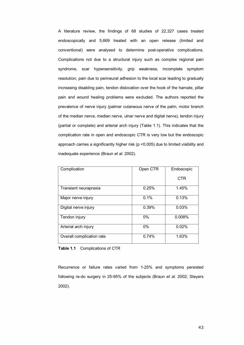
A literature review, the findings of 68 studies of 22,327 cases treated
endoscopically and 5,669 treated with an open release (limited and
conventional) were analysed to determine post-operative complications.
Complications not due to a structural injury such as complex regional pain
syndrome, scar hypersensitivity, grip weakness, incomplete symptom
resolution, pain due to perineural adhesion to the local scar leading to gradually
increasing disabling pain, tendon dislocation over the hook of the hamate, pillar
pain and wound healing problems were excluded. The authors reported the
prevalence of nerve injury (palmer cutaneous nerve of the palm, motor branch
of the median nerve, median nerve, ulnar nerve and digital nerve), tendon injury
(partial or complete) and arterial arch injury (Table 1.1). This indicates that the
complication rate in open and endoscopic CTR is very low but the endoscopic
approach carries a significantly higher risk (p <0.005) due to limited visibility and
inadequate experience (Braun et al. 2002).
Complication Open CTR Endoscopic
CTR
Transient neurapraxia 0.25% 1.45%
Major nerve injury 0.1% 0.13%
Digital nerve injury 0.39% 0.03%
Tendon injury 0% 0.008%
Arterial arch injury 0% 0.02%
Overall complication rate 0.74% 1.63%
Table 1.1 Complications of CTR
Recurrence or failure rates varied from 1-25% and symptoms persisted
following re-do surgery in 25-95% of the subjects (Braun et al. 2002; Steyers
2002).
44
Patients with CTS have an isolated insult to the median nerve within the carpal
tunnel. As discussed before, this is associated with a group of autonomic
symptoms that have also been described following hand injury. There is no
evidence to suggest the symptoms relate to a specific injury. The following
review presents the current views concerning post-traumatic cold intolerance
and its pathophysiology.
1.2 POST-TRAUMATIC COLD INTOLERANCE (PTCI)
Cold intolerance is a common problem in the injured hand with significant
consequences for a patient�s work and leisure activities. Many patients consider
it the most disabling outcome from their hand injury (Lithell et al. 1997).
Attempts have been made to unveil the underlying pathophysiology and tailor
treatment accordingly, but the cause of cold intolerance remains uncertain. In
this paper we review the available literature.
1.2.1 Definition
The selection of a satisfactory working definition has been challenging; this has
led to inconsistency in case reporting and follow-up. �An icy cold feeling that can
progress to pain sometimes lasting for several hours following cold exposure� is
an early attempt to define the condition (Engkvist et al. 1985). However, this
definition did not extend to cover the varied spectrum of symptoms including
stiffness and colour changes, which meant a proportion of sufferers were
missed (Lithell et al. 1997). Other definitions were proposed such as �An
exaggerated or abnormal reaction to cold exposure of the injured part causing
discomfort or the avoidance of cold� (Kay 1985), �Symptoms triggered by
exposure to cold and represent discomfort or problems that are perceived by
the patient as a sequel to their hand injury� (Lithell et al. 1997), and a �collection

45
of acquired symptoms resulting in an abnormal aversion to cold� (Campbell et
al. 1998).
Although the majority of the studies continue to refer to cold intolerance as such
(Freedlander 1986), various alternative terms have been used; cold sensitivity
(Nylander et al. 1987), cold hypersensitivity (Craigen et al. 1999), and finally
�trauma induced cold associated Symptoms (TICAS)� (Campbell et al. 1998). In
this review we opt to refer to this syndrome as Post-Traumatic Cold Intolerance
(PTCI); this term reflects the causation (trauma), the trigger (cold) and generally
describes the reaction to cold (intolerance).
1.2.2 Clinical presentation
In broad terms cold-related symptoms fall into four main groups: 1- Pain/
discomfort, 2- Altered sensation, 3- Stiffness, 4- Colour changes (Campbell et
al. 1998). In a review of 128 subjects complaining of cold-related symptoms
post-hand injury, pain was the most prevalent symptom, affecting 77%, followed
by stiffness in 60% and colour changes and sensory symptoms in around 50%
of the subjects (Campbell et al. 1998). In 398 patients, Irwin et al. reported
numbness and stiffness as being most prevalent followed by weakness, pain
and colour changes. Proximal radiation was noted as far as the wrist (but not
more proximally) in 33% of cases (Irwin et al. 1997).
The effects of the syndrome are significant and far reaching, forcing changes in
habits, hobbies and even work (Backman et al. 1993; Collins et al. 1996).
Although the incidence of medico legal complaints due to PTCI is not well
reported in the published literature, it is thought that those who complain run a
more severe course (Koman et al. 1998).
46
1.2.3 Pathophysiology
The pathogenesis of PTCI remains unclear. Educated hypotheses based on
vascular and neurological assessment of the effected suggest a multi-factorial
origin.
The hypothesis of reduced digital perfusion following hand injury was first
investigated by Porter et al. using Doppler digital flowmetery (Porter 1968).
Using digital pulse pressure, Gelberman et al. demonstrated that values less
than 75% in the injured digit, compared to the corresponding digit in the contra-
lateral hand, are associated with more severe symptoms while the symptoms
were milder with values >80% (Gelberman et al. 1978). A low digital baseline
temperature (<2.2±1.9°C compared to the unaffected side) was also reported in
patients suffering from PTCI (Engkvist et al. 1985).
Two-digital-artery re-anastomosis was reported to positively correlate with
reduced symptoms in a review of 100 digital replantation or revascularisation
cases but the development of cold intolerance was felt to be due to both arterial
and neural impairment (Morrison et al. 1978). Using a cold challenge and pulp
temperature measurement aided by thermocouples on 14 patients following
digital revascularisation or replantation, Kay found no correlation between the
digital re-warming time (an indirect indicator of perfusion) and symptom severity
(using a questionnaire). In his study, Kay identified patients with better sensory
recovery to be mildly affected and vice versa suggesting a defect in local blood
flow neuro-regulation but his conclusions were based on a small sample size
with varied injury severity (Kay 1985).
Based on a prolonged response (symptoms free >12 months) to an IV
guanethidine block (a sympathetic blocking drug that reduces the release of
catecholamines) in nine out of 24 patients with intolerance to cold after partial or

47
complete finger amputations, and based on a transient elevation in a
temperature of 2.7±2.1°C above the corresponding area on the uninjured hand
with a significant symptomatic improvement even after the return to baseline
temperature 2-4 weeks in all participants, Engkvist et al. speculated that
neurogenic rather than vascular dysfunction is mainly behind PTCI. The author
suggested that cold-related symptoms are due to either a direct neuronal
sensitivity to cold or indirectly through hypersensitivity to a normally increased
noradrenalin following cold exposure. Earlier findings suggest neuronal
sensitivity to mechanical stimuli and noradrenalin (Wall et al. 1974; Engkvist et
al. 1985). Other studies have shown that two-point discrimination does not
correlate with the severity of the symptoms in PTCI patients (Freedlander 1986;
Nylander et al. 1987; Lenoble et al. 1990; Backman et al. 1991). The cause of
this is unknown but a possible hypothesis would be an all or nothing effect of
trauma on the damaged neural ends with resultant cold intolerance independent
of the extent of the nerve damage sustained.
Multiple limitations in studies touching on the underlying mechanisms in PTCI
are obvious: small sample size, retrospective, non-specific assessment
technique or a combination of these factors. We still understand very little about
PTCI pathogenesis. In recent years, the utility and reproducibility of laser
Doppler techniques have improved considerably. Our own work has shown that
laser Doppler fluximetry, combined with trans-cutaneous iontophoretic
administration of endothelial-dependent and -independent vasodilators
(acetylcholine and sodium nitroprusside, respectively), provides a reproducible,
non-invasive method for exploring cutaneous microvascular dysfunction in
diverse clinical conditions ranging from pre-eclampsia to diabetes and
peripheral arterial disease (Davis et al. 2001; Klonizakis et al. 2003; Klonizakis
et al. 2006). These techniques provide new opportunities to study the regulation
of cutaneous microvessels in the hand following injury, and to assess the
vascular effects of local or systemic interventions.

48
1.2.4 Incidence and natural history of PTCI
The lack of a specific definition led to a wide range for the prevalence reported
(50-100%), although >70% is generally accepted (Table 1.2). There is
agreement on the early onset of this condition (Backman et al. 1991; Backman
et al. 1993; Lithell et al. 1997; Craigen et al. 1999). In a study of 65 patients,
more than 50% of the patients developed cold intolerance immediately after
injury, with a further 39% developing symptoms within six months of the injury
(Nancarrow et al. 1996). A review of 331 patients with hand injuries reported a
PTCI prevalence of 83% with 90% of those affected developing symptoms
within the first six months of injury and 35% developing the symptoms
immediately post-trauma (Irwin et al. 1997). Predictors of PTCI development are
listed in Table 1.3.

St
udy
Sa
mpl
e
size
Stud
y ty
pe
Inju
ry re
view
ed
Rep
orte
d PT
CI I
ncid
ence
M
ean
follo
w-u
p pe
riod
(Gel
berm
an e
t al.
1978
) 29
Pr
ospe
ctiv
e D
igita
l rep
lant
atio
n fo
r a c
ompl
ete
ampu
tatio
n 10
0%
Not
giv
en
(Mor
rison
et a
l. 19
78)
100
Ret
rosp
ectiv
e D
igita
l rep
lant
atio
n an
d re
vasc
ular
isat
ion
10
0%
NA
(Pop
pen
et a
l. 19
83)
10
Pros
pect
ive
Gre
at to
e to
thum
b tra
nsfe
r 10
0%
42 m
onth
s
(Ear
ley
et a
l. 19
84)
14
Pros
pect
ive
Thum
b re
plan
tatio
n 71
%
Not
giv
en
(Kay
198
5)
14
Pros
pect
ive
Dig
ital r
epla
ntat
ion
and
reva
scul
aris
atio
n 73
%
33 m
onth
s
(Tar
k et
al.
1989
) 15
3 R
etro
spec
tive
Dig
ital r
epla
ntat
ion
and
reva
scul
aris
atio
n
100%
N
A
(Len
oble
et a
l. 19
90)
82
Pros
pect
ive
Uln
ar a
nd m
edia
n ne
rve
inju
ry
75%
pos
t-uln
ar in
jury
50%
pos
t-med
ian
inju
ry
42 m
onth
s
(Pov
lsen
et a
l. 19
95)
8
Pros
pect
ive
Dig
ital r
epla
ntat
ion
5 pa
tient
s
12 y
ears
(Nan
carr
ow e
t al.
1996
) 65
Pr
ospe
ctiv
e H
and
inju
ry re
quiri
ng s
urgi
cal i
nter
vent
ion
65
%
5.8
year
s
(Lith
ell e
t al.
1997
) 40
R
etro
spec
tive
Dig
ital t
raum
a
100%
N
A
(Irw
in e
t al.
1997
) 39
8 R
etro
spec
tive
Upp
er li
mb
perip
hera
l ner
ve in
jury
83
%
NA
(Cam
pbel
l et a
l. 19
98)
167
Pros
pect
ive
Han
d in
jury
(non
spe
cific
) 73
%
24 m
onth
s
(Kom
an e
t al.
1998
) 26
1 R
etro
spec
tive
Han
d in
jury
(non
spe
cific
) 62
%
NA
(Cra
igen
et a
l. 19
99)
123
Pros
pect
ive
H
and
inju
ry (n
on s
peci
fic)
85%
11
mon
ths,
3 y
ears
for s
ever
e ca
ses
Tabl
e 1.
2 Th
e in
cide
nce
of P
TCI
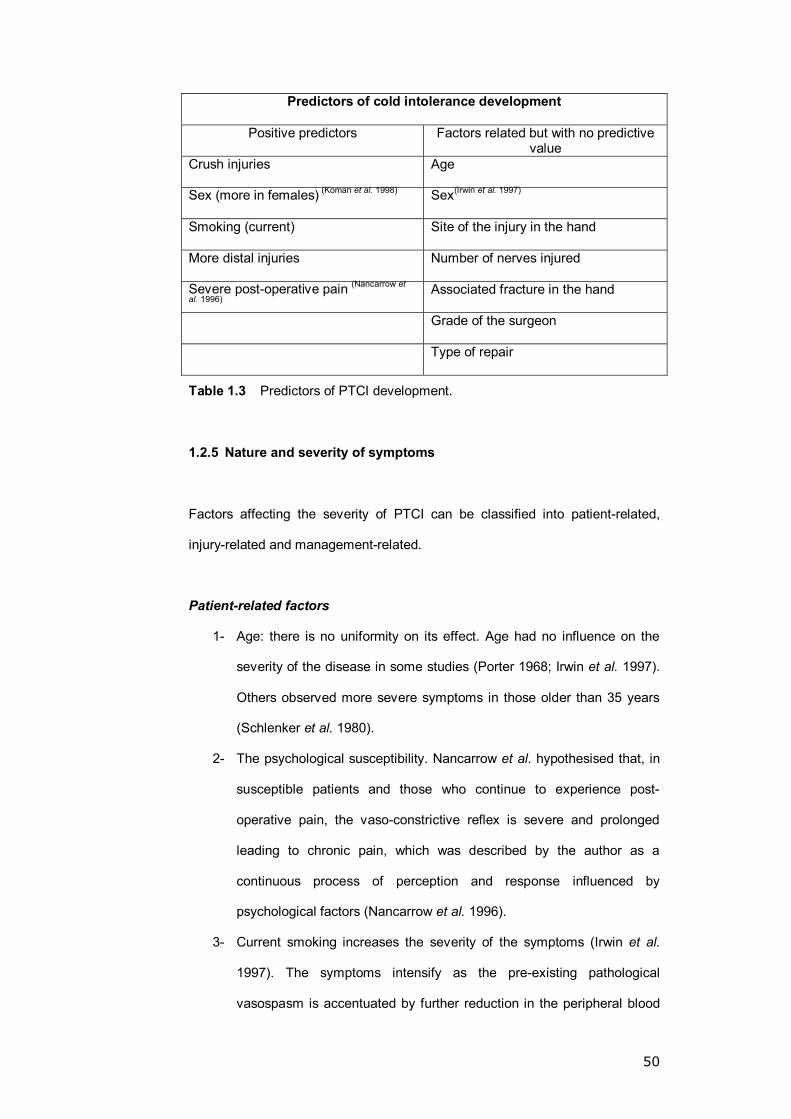
50
Predictors of cold intolerance development
Positive predictors Factors related but with no predictive value
Crush injuries Age
Sex (more in females) (Koman et al. 1998) Sex(Irwin et al. 1997)
Smoking (current) Site of the injury in the hand
More distal injuries Number of nerves injured
Severe post-operative pain (Nancarrow et
al. 1996) Associated fracture in the hand
Grade of the surgeon
Type of repair
Table 1.3 Predictors of PTCI development.
1.2.5 Nature and severity of symptoms
Factors affecting the severity of PTCI can be classified into patient-related,
injury-related and management-related.
Patient-related factors
1- Age: there is no uniformity on its effect. Age had no influence on the
severity of the disease in some studies (Porter 1968; Irwin et al. 1997).
Others observed more severe symptoms in those older than 35 years
(Schlenker et al. 1980).
2- The psychological susceptibility. Nancarrow et al. hypothesised that, in
susceptible patients and those who continue to experience post-
operative pain, the vaso-constrictive reflex is severe and prolonged
leading to chronic pain, which was described by the author as a
continuous process of perception and response influenced by
psychological factors (Nancarrow et al. 1996).
3- Current smoking increases the severity of the symptoms (Irwin et al.
1997). The symptoms intensify as the pre-existing pathological
vasospasm is accentuated by further reduction in the peripheral blood

51
flow after smoking (Mosely et al. 1977; van Adrichem et al. 1992;
Backman et al. 1993; Monfrecola et al. 1998).
4- An ongoing compensation claim (Koman et al. 1998).
Gender and hand dominance have no bearing on the severity of the symptoms.
Injury-related factors
Retrospective studies of upper limb peripheral nerve injuries have shown the
following to be associated with worse symptoms: complete nerve division,
presence of a vessel injury and an associated fracture (Irwin et al. 1997; Koman
et al. 1998). The mechanism of injury (sharp or crush) has no bearing on the
severity of the symptoms.
Management-related factors
Patients with both the digital arteries repaired in replantation cases are less
affected by cold-related symptoms post-operatively compared to those with a
single arterial repair (Morrison et al. 1978; Freedlander 1986; Tark et al. 1989).
The number of injuries to any single nerve does not affect outcome, nor does
the number of nerves injured, the type of surgical repair, the grade of the
surgeon or the time of repair after the injury (Nystrom et al. 1991; Irwin et al.
1997).
1.2.6 Symptom scoring systems
Cold intolerance is difficult to score or measure objectively (Koman et al. 1998).
However, there have been attempts to score this condition.
1 � Kay�s numerical cold intolerance score (Kay 1985)
Based on a questionnaire with points awarded for the following:
A- the presence of any of the main four symptoms (pain, numbness,
stiffness and loss of function)
52
B- the degree of avoidance of cold exposure
C- the degree to which the patient�s activities and work are affected by the
symptoms
D- the patient�s own assessment of the severity of their symptoms.
Neither the text of the questionnaire nor evidence of validation was provided by
the author. The maximum possible score was 22.
2 � Cold sensitivity scale (McCabe et al. 1991)
Designed to reflect the patient�s subjective grading of the disability caused by
cold exposure in given situations. To eliminate the variability of presentation
between the patients, direct symptom-specific questions were avoided.
Questions targeted cold-related scenarios, which were divided into two scales:
1- the cold sensitivity severity scale.
2- potential work exposure scale.
The items were selected from circumstances that patients commonly felt
provoked cold-induced symptoms in their hands. The response to each item
was obtained by marking a 100 mm line to reflect the severity of the symptom in
a manner similar to a visual analogue scale. The sum of the scores in each
subscale in millimetres gives the corresponding final score for that scale. The
test/re-test correlation coefficient for the Cold Sensitivity Severity Scale was
reported at 0.92 and for the potential work exposure scale at 0.94. The authors
presented a statistically significant difference in the score between the nerve-
injured patients and the control group with mean values of 171 and 80
respectively.
53
3 � Blond McIndoe Cold Intolerance Symptom Severity (CISS) Score (Irwin
et al. 1997)
This is a modification on the cold sensitivity scale, designed to provide more
information on a wide range of symptoms and stratify the frequency and
persistence of symptoms and how they disturb daily activities.
The maximum score is 100 grouped into four ranges (0-25, 26-50, 51-75, 76-
100) corresponding to mild, moderate, severe, and extreme severity
respectively. A score/re-score correlation coefficient of CISS for 26 subjects
completed six months apart was reported as 0.90 and a mean difference
between scores of 0.48 (SD=11.4). No information on validation was provided
but the severity score corresponded well to the patient�s subjective assessment
of the symptoms, particularly in the severe and extreme groups.
1.2.7 Resolution
The variability in the follow-up duration, subjectivity of severity assessment and
variability in symptoms considered related to PTCI yielded variation in resolution
observations. Short- to medium-term studies (up to five years follow-up)
revealed only partial improvement (less pain) in less than one-fifth of the
sufferers while the majority continued to have the same symptoms and 2% were
getting worse (Gelberman et al. 1978; Freedlander 1986; Backman et al. 1993;
Nancarrow et al. 1996). Long-term follow-up studies varied from no
improvement at 12 years follow-up (Povlsen et al. 1995), partial improvement
with time (Kleinert et al. 1980), and an up to 63% complete resolution at 10
years (Matsuda et al. 1999).
The most important predictors of improvement were reported as the Blond
McIndoe Cold Intolerance Symptom Severity (CISS) Score and the smoking

54
status of the patient (i.e. the higher the score, the worse the prognosis and that
smoking adversely affects the clinical course of the condition) (Irwin et al. 1997).
1.2.8 Treatment
Cold evasion appears to be the most practical way to manage PTCI, although
this is not always possible. Conservative modalities and surgical techniques
(sympathectomy) have been described with limited success. It is worth
considering different approaches to potential medical therapy, and in particular
how these treatments might be evaluated in placebo-controlled studies in PTCI.
1.2.8.1 Vasodilator therapies
Drugs which relax vascular smooth muscle in medium-large arteries and
arterioles, especially the resistance vessels that are important determinants of
systemic BP, have been tried with varying success in PTCI. For example, α-
Blockers have been used to enhance the digital blood flow with limited or no
effect (Nylander et al. 1987; Isogai et al. 2004). Similarly, the calcium channel
blocker nifedipine has been shown to effectively reduce the frequency and
severity of symptoms in Raynaud�s phenomenon (Smith et al. 1982), but no
studies have yet assessed calcium antagonists in PTCI. In a controlled double
blind trial, nifedipine was shown not to affect the skin temperature recovery time
(Rodeheffer et al. 1983). The role of other vasodilators is unknown: in particular
blockade of the renin-angiotensin system using angiotensin converting enzyme
inhibitors (ACIs) or angiotensin receptor antagonists (ARAs) is associated with
non-BP-dependent vascular effects, including improved microvascular function
(e.g. in the kidney & retina), which could be useful in patients with PTCI.
Randomised trials are needed.
In future clinical studies, apart from evaluating symptoms, it would be useful to
monitor the effects of vasodilators on large and small vessels in the hand using
55
newer non-invasive techniques such as laser Doppler fluximetry and pulse
wave analysis. These techniques are now available, more reproducible and less
operator-dependent, but few if any studies have applied the methodologies to
study the hand.
1.2.8.2 Antiplatelet therapies
Platelet activation and aggregation in response to vascular injury plays an
important role in microangiopathic complications, which prompts interesting
hypotheses about whether aspirin or clopidogrel might improve microvascular
cutaneous perfusion in PTCI.
1.2.8.3 Sympathetic nervous system blockade
In general, beta-adrenoceptor antagonists have an adverse effect on peripheral
blood flow, but these drugs, in theory, may have beneficial effects in attenuating
sympathetic nervous system pathways involved in arterio-venous shunting and
cutaneous vasoconstriction. Newer beta blockers with vasodilator properties,
e.g. carvedilol, offer a pharmacological profile which, theoretically, could
improve some of the vascular mechanisms which are known to control skin
blood flow and cutaneous responses to temperature change.
1.2.8.4 Prostacyclin analogues
Beraprost has been studied in 11 patients affected by cold intolerance at a dose
of 60 µg three times daily for two weeks with a significant reduction in symptom
severity in nine patients and no side effects. Beraprost elevated the skin
temperature in eight of the cases (Isogai et al. 2004). The authors suggested
that failure to respond to beraprost indicates that poor vascular flow is unlikely
to be the cause of the symptoms in that particular case and that the mechanism
might be related to a sympathetic dysfunction, psychological trauma due to the
injury or the patient�s personality. For resistant cases the authors suggested the
use of nerve blocks, antidepressants or anxiolytics. They also expressed the
56
opinion that long-term therapy with beraprost may be useful in some patients
with gradual dose reduction. No evidence to support the long-term usefulness of
beraprost in such cases has been reported in the literature.
1.2.8.5 Regional neuronal block
Guanethidine intravenous regional blocks have been used to treat this
condition; 20 mg of guanethidine are diluted in 40 ml of saline and injected over
a minute through a vein on the dorsum of the hand after occlusion of the
circulation with a tourniquet applied to the upper arm. Tourniquet pressure is
maintained 50 mmHg above the systolic blood pressure and the tourniquet
released after 20 minutes. Such blocks have been used with only limited short-
term success (one-third of 20 patients showed symptomatic relief two to 12
weeks post-block). However some patients can be kept relatively symptom free
during the cold season with repeated blocks (Engkvist et al. 1985).
1.2.8.6 Psychological interventions in symptom management
Success in using biofeedback, autogenic training and classical conditioning in
the management of patients with Raynaud�s phenomenon inspired the use of
classical conditioning in the treatment of post-traumatic cold intolerance with
encouraging results (Jobe et al. 1985; Jobe et al. 1986). In a small study of 10
patients, classical conditioning improved the symptoms in eight subjects and
improved baseline temperature difference between the affected digits and the
corresponding contralateral uninjured from 1.5°C to 0.1°C (Brown et al. 1986).
1.2.8.7 Surgical interventions
Surgical intervention to treat severe forms of Raynaud�s phenomenon (RP) and
scleroderma (SSc) has been described (Egloff et al. 1983; Ward et al. 1995).
Although cervical sympathectomy was often unsuccessful due to the incomplete
sympathetic denervation of the hand (Wilgis 1981), the selective digital
approach is effective in preventing digital amputation due to severe ischemia in

57
this group of patients (RP, SSC) (McCall et al. 1999). There are no studies
describing the use of digital sympathectomy in the treatment of PTCI. Hussel et
al. described single digit treatment in four patients using a 12 cm thoracodorsal
vascular bundle implanted microsurgically through an end-to-side proximal
anastomosis with the radial artery in the radial fossa, passing the graft
subcutaneously to reach the tip of the injured digit where its end is covered by a
split skin graft. Details of the technique and the extent of improvement in the
other digits after surgery was not described (Hussel et al. 1989). Repair of the
two digital arteries and adequate post-operative pain management are
associated with better PTCI symptom (Fagius et al. 1985; Tark et al. 1989;
Isogai et al. 2004).
In conclusion, cold intolerance remains a significant problem after hand injury
with its effects touching various aspects of the patient�s life. The precise cause
remains uncertain although a mixed neurovascular pathophysiology is thought
likely. More vigorous studies are needed to help understand the exact nature of
this problem. Adjustments to lifestyle can be very helpful in controlling
symptoms. Biofeedback, conditioning and some pharmacological agents have
been found to offer some additional benefits. Surgical intervention is unlikely to
be helpful.
1.3 RAYNAUD�S PHENOMENON (RP)
Raynaud�s Phenomenon (RP) describes a recurrent exacerbated vasospastic
response to cold or emotional strain, mainly affecting the hands and feet while
the nose, ears and even the nipples can be affected (Block et al. 2001; Wigley
2002). This response presents clinically as skin colour changes (white �
ischemia, blue � deoxygenation, then red or crimson � reperfusion) associated
with discomfort or pain in the affected part and numbness and tingling (Bowling
et al. 2003) (Figure 1.4).
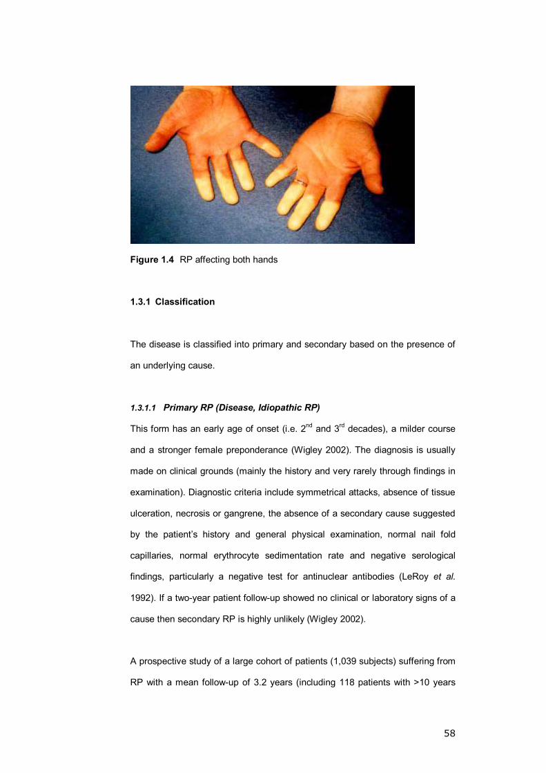
58
Figure 1.4 RP affecting both hands
1.3.1 Classification
The disease is classified into primary and secondary based on the presence of
an underlying cause.
1.3.1.1 Primary RP (Disease, Idiopathic RP)
This form has an early age of onset (i.e. 2nd and 3rd decades), a milder course
and a stronger female preponderance (Wigley 2002). The diagnosis is usually
made on clinical grounds (mainly the history and very rarely through findings in
examination). Diagnostic criteria include symmetrical attacks, absence of tissue
ulceration, necrosis or gangrene, the absence of a secondary cause suggested
by the patient�s history and general physical examination, normal nail fold
capillaries, normal erythrocyte sedimentation rate and negative serological
findings, particularly a negative test for antinuclear antibodies (LeRoy et al.
1992). If a two-year patient follow-up showed no clinical or laboratory signs of a
cause then secondary RP is highly unlikely (Wigley 2002).
A prospective study of a large cohort of patients (1,039 subjects) suffering from
RP with a mean follow-up of 3.2 years (including 118 patients with >10 years
59
follow-up) identified the incidence of progression to develop connective tissue
disease in RP based on Nielsen�s test (Nielsen et al. 1977), plethysmography,
digital arterial systolic blood pressure and serology (antinuclear antibodies and
rheumatoid factor). The incidence was found to vary between four subgroups of
patients: sero-negative spastic RP (2%, 6.3%), sero-positive spastic RP (16%,
33%), sero-negative obstructive RP (8.5%, 18%) and sero-positive obstructive
RP (30%, 82%). The above figures are: over all incidence; incidence after 10
years (Landry et al. 1996).
1.3.1.2 Secondary RP
This is commonly the consequence of a systemic disease (occurring in 90% of
SSc patients, 10-45% of Systemic Lupus Erythamotosis and 10-20% of
Rheumatoid Arthritis) but a long list of predisposing factors has been outlined,
e.g. mechanical injury, arterial disease, malignancy and infections (Block et al.
2001).
This form of RP runs a more aggressive course and those affected are usually
over 40 years old. Secondary RP should be suspected if the patient is over 30
years old, suffering from intensely painful asymmetrical symptoms or associated
with ischemic skin lesions. The presence of joint pain, stiffness, abnormal lung
function tests, abnormal blood tests (inflammatory markers and serology) and
evidence of microvascular disease on microscopy of the nail fold capillaries are
also suggestive of an underlying cause (Kallenberg et al. 1988; Kallenberg
1990; Wigley 2002).
1.3.2 Prevalence
The incidence of RP in the general population is unknown. Prevalence rates
range between 1-25% for men and 1.8-30% for women depending on the
60
population and the geographical regions studied (i.e. more prevalent in colder
areas) (Maricq et al. 1993; Maricq et al. 1997; Fraenkel 2002; Wigley 2002).
Gender seems to be the most consistent risk factor in RP with a female:male
ratio of approximately 4:1. A positive association between the family history and
RP in women suggests a strong genetical expression of RP in females. Males
did not show a similar association (Valter et al. 1998). Other observations such
as a higher prevalence amongst women between menarche and menopause
and a higher post-menopausal incidence in women using unopposed oestrogen
replacement therapy suggests that oestrogen might be a significant reason for
the skewed ratio towards females (Fraenkel et al. 1998; Fraenkel 2002).
A recent study of 720 children aged 12-15 reported an overall 15% prevalence;
it also showed RP to be more common in girls (18%) than boys (12%) (Jones et
al. 2003).
The effect of age, alcohol, smoking and marital status has also been
investigated. In a cross-sectional study of 800 women and 725 men
participating in the Framingham Offspring Study, the authors reported RP
prevalence of 9.6% in women and 5.8% in men. In women, marital status and
alcohol use were each associated with prevalent RP (for marital status adjusted
odds ratio [AOR] 2.3, 95% confidence interval [95% CI] 1.4-3.9; for alcohol use
AOR 2.2, 95% CI 1.0-5.2), whereas these factors were not associated with RP
in men (marital status AOR 1.4, 95% CI 0.6-3.5; alcohol use AOR 1.0, 95% CI
0.2-4.4). In men, older age (AOR 2.3, 95% CI 1.0-5.2) and smoking (AOR 2.6,
95% CI 1.1-6.3) were associated with prevalent RP; these factors were not
associated with RP in women (older age AOR 0.8, 95% CI 0.4-1.6; smoking
AOR 0.7, 95% CI 0.4-1.1). DM, hypertension, and hypercholesterolemia were
not associated with RP in either sex (Fraenkel et al. 1999).
61
Both current and ex-smokers were reported to have higher RP prevalence
compared to people who never smoked (Fraenkel et al. 1999; Palmer et al.
2000). The link between smoking, alcohol and RP was not consistently found in
the literature (Palesch et al. 1999; Voulgari et al. 2000). Other risk factors
include β-Blockers, Carpal Tunnel Syndrome and Rheumatoid Arthritis (Brand
et al. 1997).
1.3.3 Pathophysiology of RP
The pathophysiological pathway that leads to microvascular vasospasm in
primary RP is unclear. Despite the wealth of research conducted on this
process, only proposals for possible mechanisms have been suggested.
1.3.3.1 Central neurogenic factors
A dysfunction in the central sympathetic control of the microvasculature was
first proposed by Maurice Raynaud (1862). Studies on the effect of stress
(Freedman et al. 1983), sympathectomy and sympatholytic drugs (Olsen 1987;
Olsen et al. 1991) all suggested that a sympathetic over-activity plays a role in
the pathogenesis of RP. Whether this is a direct neurogenic effect or an
abnormal neurovascular interaction (nerve-vessel wall dysfunction) is unclear
(Fagius et al. 1985).
1.3.3.2 Local factors
�The Local Fault theory� was suggested as early as 1929 after an aberrant cold-
induced vasospasm was noted in patients with RP post-sympathectomy (Lewis
1929). Neurogenic, humeral and endothelial faults have been reported.
Nonetheless, the interaction between those factors makes it difficult to
differentiate the cause from the effect in most cases (Turton et al. 1998).
62
A. Vascular α- Adrenoceptors
Both α-1 and α-2 receptors mediate the sympathetic response in the skin
microcirculation despite a predominance in the effect of the α-2 subtype
(Willette et al. 1991). An enhanced response to catecholamines is believed to
play a role in the pathophysiology of RP through an exaggerated density and/or
sensitivity of the α-2 receptors (Freedman et al. 1989).
Further work by Freedman et al. (1995) demonstrated that it is the activation of
the α-2 receptors and not the α-1 adrenergic receptors that is necessary for the
production of vasospastic attacks in idiopathic RP (Freedman et al. 1995).
B. Calcitonin gene-related peptide (CGRP)
This is a potent vasodilating neuropeptide found in nerve endings of the
cutaneous unmylinated sensory neurons (Gibbins et al. 1987). Although the
systemic levels in the RP patients are not different from the control group
(Bunker et al. 1996), the favourable response to an IV infusion of CGRP on the
hands of patients with RP suggests a local deficiency in those patients
(Shawket et al. 1989).
C. Endothelin-1
This is a potent endothelial-derived vasoconstrictor (Turton et al. 1998). It has
been shown that baseline and post-cold provocation measurements of
endothelin-1 were elevated and continued to be elevated in patients with RP
suggesting a role in the aetiology (Zamora et al. 1990; Biondi et al. 1991).
Although the agreement on the role of endothelin-1 is not universally accepted
(Bottomley et al. 1994), it is better considered as a marker of endothelial
damage (Mangiafico et al. 1996).

63
D. Oestrogen
It is thought that oestrogen may sensitise the peripheral vascular bed to
endogenous circulating vasoconstriction catecholamines � this may explain the
4:1 predominance of RP in females (Altura 1975).
E. Nitric Oxide (NO)
The vasodilatory ability is reduced in response to acetylcholine and
nitroprusside in RP (Khan et al. 1997). The production of NO is also believed to
be suboptimal in RP patients (Khan et al. 1994).
1.3.3.3 Blood cell-related factors
A. Neutrophils
In both primary and secondary RP, evidence exists suggesting an abnormal
White Blood Cell (WBC) activation and a pathological release of free radicals
(Lau et al. 1992a; Lau et al. 1992b). Cold exposure is thought to activate the
neutrophils during the vasospastic attack in RP leading to increased
neutrophilic-endothelial adhesion (Ciuffetti et al. 1995).
B. Platelets
An exaggerated expression of α-2 receptors on the platelets, mirroring a similar
increase on the endothelium is thought to be the cause of the abnormal platelet
aggregation and mediator release leading to vasoconstriction and increased
permeability in patients with RP (Ikehata et al. 1980; Turton et al. 1998). This is
magnified by the blunted responsiveness of the platelets to inhibiting factors
such as prostacyclin and prostaglandin E1 (Hutton et al. 1984).
1.4 HAND ARM VIBRATION SYNDROME (HAVS)
The high incidence of RP in vibrating tool operators was first reported by Loriga
(Loriga 1911). Seven years later, a direct link between RP and vibration as a

64
cause was suggested (Hamilton 1918). As the understanding of the multi-
systemic nature of the condition grew, the term �vibration white finger� (VWF)
was replaced by a more representative name (Hand Arm Vibration Syndrome
(HAVS)) in 1985. More importantly, and in the same year, HAVS became a
prescribed disease for which sufferers can claim compensation (HMSO 1985;
Taylor 1985).
1.4.1 Prevalence
According to a cross-sectional study published in 2000, it is estimated that
vibration exposure is responsible for extensive blanching affecting at least eight
digits or 15 phalanxes in about 222,000 cases in the UK (McGeoch et al. 2000),
with approximately 100,000 of those claiming compensation (McGeoch et al.
2005).
The ratio between male and female is unclear but the causation (prolonged
exposure to vibration mainly through work) tips the balance in favour of a male
majority amongst the affected.
1.4.2 Patient assessment and diagnosis
Both subjective and objective methods of assessment and diagnosis have been
described but confirming the diagnosis is difficult and very subjective (Lawson
et al. 2003; McGeoch et al. 2005). A medical assessment process was created
to aid in the diagnosis of HAVS and was implemented in all the assessment
centres in the UK. The process included the following.
A- A questionnaire to ascertain the history of exposure to hand arm vibration
followed by a questionnaire regarding any history of symptoms of HAVS
(subjective tool).

65
B- A questionnaire to help with the differential diagnosis and the detection of
any dual pathology (subjective tool).
C- Upper limb and neck examination to exclude other mimicking clinical
condition.
D- Staging using the Stockholm Workshop Scales (SWS) (Brammer et al.
1987) (Table 1.4) (subjective tool).
E- Five standardised tests for both the sensorineural (SN) and vascular (V)
disease components (objective tool).
- Sensorineural component assessment tests
1- Vibrotactile perception threshold test (VPT)
2- Thermal aesthesiometry (TA)
3- Purdue pegboard test (PPT)
4- Grip strength using a Jamar Dynamometer
- Vascular component assessment test
5- Cold provocation testing (CPT)
The standardised tests outcome is scored (Table 1.4) and the scores are
incorporated within the modified SWS.

66
Modification Of The Neurological Component Of The Stockholm Workshop Scales Stage Criteria
0 SN Vibration exposure but no symptoms
1 SN Intermittent numbness and/or tingling with a sensorineural
score of ≥3<6
2 SN (Early) Intermittent or persistent numbness, and/or tingling,
reduced sensory perception with a score of ≥6<9
2 SN (Late) As 2 SN (Early) but with a score of ≥9<16
3 SN Intermittent or persistent numbness, and/or tingling,
reduced manual dexterity and an SN score of ≥19
Modification of the vascular component of the SWS
Stage Criteria
0V No attacks
1V Attacks only affecting the tips of the distal phalanges of one
or more fingers � usually a blanching score of 1-4
2V Occasional attacks of whiteness affecting the distal and
middle (rarely also the proximal) phalanges of one or more
fingers � usually a blanching score of 5-16
3V Frequent attacks of whiteness affecting all the phalanges of
most of the fingers � usually a blanching score of 18 or
more
4V As 3V and trophic changes
Table 1.4 The Modified Stockholm Workshop Scales (SWS) (McGeoch et al.
2005)
Scoring system used for the standardised tests 31.4 Hz <0.3 ms² = 0 ≥0.3 ms², <0.4 ms² = 1 ≥0.4 ms² = 2 VPT
(index and little finger) 125 Hz <0.7 ms² = 0 ≥0.7 ms², <1.0 ms² = 1 ≥1.0 ms² = 2 TA 1ºC/s (index and Little finger)
<21ºC = 0 ≥21ºC, <27ºC = 2 ≥27ºC = 4
CPT (15ºC for 5 min, 10 min recovery)
T(+4º) ≤300 s = 0 >300 s, ≤600 s = 1 >600 s = 2
Table 1.5 Scoring system used for the standardised tests

67
In a cross-sectional study of 165 workers exposed to vibration for a mean of 23
years (range 3-47) investigating the correlation between the subjective
assessment (i.e. SWS) and the objective tests, the authors reported a
significant (p <0.001) correlation between the SWS neurological stage and the
findings of the neurological tests but could not demonstrate a correlation
between the vascular stage and the CPT findings (McGeoch et al. 2000). In this
study the overall prevalence of neurological findings of 62% was greater than
the overall prevalence of vascular findings, which was 33%. Findings confirming
the limited value of the CPT in the diagnosis of HAVS was also reported on
review of approximately 20,000 CPT tests done through the Department of
Trade and Industry scheme for the evaluation of miners (Proud et al. 2003b).
Based on the above, the diagnosis of HAVS is largely subjective and unreliable
due to the complicating expected financial gain.
Of note is the incorporation of the sensorineural score in the modified SWS
within the neurological part while using a �blanching score� (Rigby et al. 1984)
within the vascular part instead of the CPT score. This obviously creates an
inbuilt bias within the score and may explain the significant correlation in the
sensorineural examination and the SWS findings.
1.4.3 Pathology
Three major findings were noticed in finger biopsies from patients suffering from
the condition (Takeuchi et al. 1986):
[I] intense thickening of the arterial muscular wall with individual cell
hypertrophy;
[II] peripheral demyelination neuropathy with increased numbers of
Schwann cells and fibroblasts;
[III] increased amounts of connective tissue causing perineural and
perivascular fibrosis.
68
1.4.4 Pathophysiology
The causation remains unclear; generally speaking it can be divided into neural,
vascular and cellular factors with some similarities to RP.
1.4.4.1 Neural factors
A- Autonomic dysfunction
It is thought that patients with HAVS have an increased sympathetic tone and/
or reduced parasympathetic function probably due to Pacinian corpuscle over
stimulation (Bovenzi 1986).
It is interesting to note that long-lasting vibration exposure was found to provoke
central sympathetic vasoconstrictor reflex mechanisms, which trigger primarily
episodic arterial closure typical of HAVS (Olsen 1990).
B- Receptor and nerve ending dysfunction
Immunohistochemistry testing of skin biopsies obtained from patients exposed
to vibration demonstrated that vibration exposure damages the local nerve
endings, particularly the digital cutaneous perivascular nerves which contain
neuropeptides with powerful vasodilator properties, i.e. CGRP (Goldsmith et al.
1994; Bunker et al. 1996).
Pyykko and Gemne (1987) described an aberrant vasoconstriction in response
to cold in patients with HAVS secondary to excessive affinity of the efferent
receptors to substances potentiated by cooling (Pyykko et al. 1987). Using
iontophoresis and laser Doppler imaging, Ekenvall and Lindblad (1986)
demonstrated an adrenoceptor imbalance in response to noradrenaline
(stimulating both α-1 and α-2 receptors), phenylephrine (an α-1 stimulator) and
B-HT 933 (an α-2 stimulator) with evidence suggesting α-1 adrenoceptor

69
dysfunction and predominance in the α-2 receptors in the digital arteries
(Ekenvall et al. 1986). Similar findings were reported in RP (Section 1.3.3.2).
1.4.4.2 Vascular factors
A- Endothelial damage
The vascular response to methacholine (an endothelium-dependent vasodilator)
was found to be reduced when measured by laser Doppler imaging (Taccola et
al. 1998; Stoyneva et al. 2003). To support this, thrombomodulin plasma levels
were found to be elevated in the symptomatic and asymptomatic vibration-
exposed individuals (Kanazuka et al. 1996).
B- Endothelial dysregulation
The serum concentration of Endothelin-1 (a potent endothelial-derived
vasoconstrictor) was found to be elevated in symptomatic HAVS patients
(Toibana et al. 1995) compared to vibration-exposed asymptomatic individuals
(Palmer et al. 1996). Nonetheless, an increase in the serum concentration of
Endothelin-1 has been noted after cold challenge testing in the asymptomatic
vibration-exposed individuals (Taccola et al. 1991; Stoyneva et al. 2003).
It seems likely that Endothelin-1 concentration is a reflection of symptom
severity, being highest amongst the most symptomatic (Kohout et al. 1995). On
the other hand it might merely be a marker of endothelial damage (Mangiafico
et al. 1996).
It is thought that the increase in the Endothelin-1 concentration creates an
imbalance with the CGRP resulting in the inhibition of the Nitric Oxide (NO)
myo-relaxing effect on the smooth muscles (Stoyneva et al. 2003). This results
in an accentuation of the Endothelin-1 vasospastic properties (Noel 2000).

70
1.4.4.3 Cellular activation
The endothelial damage consequent to the mechanical trauma and shear stress
will result in leukocyte and platelet activation with consequences similar to those
mentioned in the pathophysiology of primary RP (Bovenzi et al. 1983; Jen et al.
1984). Leukocytes are thought to play a role in the microvascular damage in
patients with HAVS as it was found that the production of Leukotriene B4 � a
pro-inflammatory product � and free radicals is increased (Lau et al. 1992b;
Stoyneva et al. 2003).
1.5 LASER DOPPLER PERFUSION IMAGING (LDI)
The skin�s microvascular blood supply is part of the thermoregulatory control
system in the body and might play a role in the local cold-induced peripheral
symptoms in patients with RP, HAVS and PTCI. The proposed vascular
dysfunction was further non-invasively studied using laser Doppler techniques.
The use of a laser emitting device to asses the skin microcirculation is by no
means a novel idea (Stern 1975). Blood flow was measured under a single spot
of skin defined by the incident and Doppler shifted reflected light � Laser
Doppler Flowmetry (LDF); this represented an in situ sample of about 1 mm3
(Holloway et al. 1977). Nevertheless, major site variation in blood flow recording
with single spot devices was noticed and three or more measurements were
often required to obtain an average estimate of the cutaneous blood flow
(Bircher et al. 1994).
To evaluate the cutaneous perfusion, it is important to realise that not only a
temporal variability exists but a spatial one as well; this is a reflection of the
heterogeneity of the cutaneous microvascular network with arteriovenous
anastomosis in some areas and capillaries in others (Nilsson 1997). The
temporal changes can be assessed by continuous measurement using LDF but
the spatial changes cannot be reliably assessed. This is due to the localised

71
nature of the test flow signal and the gross variability in the microvascular bed
which effectively makes inter- and intra-individual comparison meaningless
(Obeid et al. 1990).
LDI is a relatively new technology (Essex et al. 1991) described as �a non-
contact system with a laser beam position controlled in the X and Y axes by a
system of mirrors, in which a digital image composed of numerous single point
recordings form a two dimensional flow map of the blood flow variability over an
extended skin surface� (Fullerton et al. 2002). Its advantage is the ability to
acquire a number of measurements to be taken at different sites almost in real
time, a characteristic that takes the spatial changes in the microvascular flow
variability into account (Svedman et al. 1998).
1.5.1 The technology
The concept rests on the idea that a laser beam scans the tissue step-wise by
means of a computer-controlled mirror system around two perpendicular axes
allowing the scanning of a square area. The system is housed in a camera-like
scanner head positioned 10-30 cm above the tissue surface. The laser beam
generated is a low power (1 mV) helium-neon red laser with a wavelength of
632.8 nm forming a light spot with a diameter of about 1 mm2 (Fullerton et al.
2002).
The surface scanned can range from 1 mm2 to a maximum of approximately
144 cm2 scanned in about 5 minutes containing 4,069 measurement points in
the processes. The laser beam generated penetrates the skin variably to a
depth of 0.6mm (Anderson et al. 1981). The gradually decreasing sensitivity
with depth reduces the sampling volume to be probed in a non-uniform manner
with an average measurement depth of 0.3 mm (Jakobsson et al. 1993).
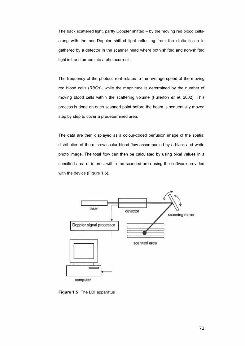
72
The back scattered light, partly Doppler shifted � by the moving red blood cells-
along with the non-Doppler shifted light reflecting from the static tissue is
gathered by a detector in the scanner head where both shifted and non-shifted
light is transformed into a photocurrent.
The frequency of the photocurrent relates to the average speed of the moving
red blood cells (RBCs), while the magnitude is determined by the number of
moving blood cells within the scattering volume (Fullerton et al. 2002). This
process is done on each scanned point before the beam is sequentially moved
step by step to cover a predetermined area.
The data are then displayed as a colour-coded perfusion image of the spatial
distribution of the microvascular blood flow accompanied by a black and white
photo image. The total flow can then be calculated by using pixel values in a
specified area of interest within the scanned area using the software provided
with the device (Figure 1.5).
Figure 1.5 The LDI apparatus

73
1.5.2 Factors affecting measurement by LDI
Increasing the height of the scanner was shown to increase the LDI output both
in vivo and in vitro. Although the manufacturer suggests a scanner head �
tissue distance of 15 cm (Fullerton et al. 2002), no reason was given for this
recommendation but at this level a good linear velocity response is maintained
up to a rate of 3 mm/sec (Kernick et al. 2000).
A non-linear reduction in the LDI signal was observed with the increase in the
skin thickness. This observation led to the conclusion that the technique is more
sensitive to changes in the superficial blood flow as the sensitivity to changes in
the flow will decrease exponentially as a function of the vascular depth.
It was found that increasing the RBC concentration increased the LDI output.
This observation, however, demonstrated evidence of linearity between the LDI
output and red cell velocity over a low heamatocrit of up to 1%. As the
heamatocrit increased, the linearity was lost at lower velocities due to the
multiple scattering that occurs at higher RBC concentrations (Kernick et al.
2000).
1.5.3 Advantages of LDI
The ability to measure the flow in up to 4,096 points in a single scan minimises
the importance of point-to-point variability and gives greater confidence in
comparing differences observed between one patient and the other (Fullerton et
al. 2002). Another advantage is that the characteristics of the blood flow map
are consistent from day to day and the relative difference between the spots
generally persists (Nilsson 1997). Furthermore, the reproducibility of
measurement at the same site on different days was significantly repeatable
when tested in the forearm (Morris et al. 1996; Kubli et al. 2000) and gaiter area

74
(Klonizakis et al. 2003). The authors reported a small mean coefficient of
variation values between the two visits as follows: (<10%, 10-20% (Kubli et al.
2000), 23%, 31% (Morris et al. 1996)) in the forearm and (10-20%, 10-20%
(Kolonizakis 2004)) for the gaiter area, for ACH and SNP respectively.
1.5.4 Pitfalls in LDI
Although images can be recorded under different light conditions, the influence
of the ambient light may alter the magnitude of the actual perfusion values to a
degree dependent on the light source and its intensity. It is therefore advisable
to record the image with the light on and off and then calculate the influence of
the light of the perfusion values to judge on the feasibility of tolerating the
ambient light in an actual test situation (Fullerton et al. 2002).
Factors affecting the cutaneous blood perfusion should be accounted for and
standardised as much as possible. These include: temperature, anatomical site,
physical and mental activity, food and drug intake, day-to-day and diurnal
variations, posture, age, sex, menstrual cycle and race (Bircher et al. 1994;
Fullerton et al. 2002).
In settings with possible altered skin transparency (i.e. skin ulceration with
concomitant necrotic debris) there may be an influence on the laser Doppler
results. Nevertheless, such effects appear to be less of a problem compared to
other study techniques like capillary microscopy (Gschwandtner et al. 1999).
It is ambiguous to express the cutaneous blood flow in absolute terms due to
the inter-sample and between the sample variability in the biological structure
and function of the microvasculature. Therefore the experimental conditions,
instruments� validation and setting, selection and pre-conditioning of study
subjects, the measurement site selected, the environment and laboratory room
75
control and the experimental design should carefully be described to allow
comparable results (Fullerton et al. 2002).
1.5.5 Applications of LDI 1.5.5.1 Inflammation
LDI was reported to be of benefit in the follow-up of patients with lateral
epicondylitis through demonstrating a normalised pattern of skin perfusion
compared to the contralateral side post-steroid injection treatment (Ferrell et al.
2000). Investigations into the role of histamine in the local dermal inflammatory
response were also done using LDI (Clough et al. 1998). LDI was also used to
further investigate the endothelial function in patients with vasospastic disorders
(i.e. RP and SSc) through the analysis of the vascular response to
iontophoresis of vasoactive substances which demonstrated impaired
endothelium-dependent and endothelium-independent responses in the SSc
group and a similar response to controls in the RP group (Anderson et al.
2004).
1.5.5.2 Healing processes
LDI combined with capillary microscopy has been utilised as an aid in
identifying the healing capacity in various areas within an ischemic ulcer by
assessing the laser Doppler area of flux (highest in healing granulation tissue)
and capillary density (highest in normal skin followed by granulation tissue
compared to very low density in areas without healing) (Gschwandtner et al.
1999) and investigating the blood flow patterns in patients at risk from pressure
sores to aid in the early diagnosis in the condition (Nixon et al. 1999).
1.5.5.3 Burn assessment
LDI aids in obtaining accurate objective assessment of a burn�s depth (clinical
accuracy is 65-75%, boosted to 100%). The idea behind the use of this test in
burns rests on the fact that burns with a high perfusion are superficial, needing

76
only dressing, while low perfusion burns require surgery (Kloppenberg et al.
2001; Pape et al. 2001).
1.5.5.4 Intra-operative measurement
Laser Doppler Flowmetry was used to demonstrate a significant reduction in the
colonic perfusion particularly distal to the site of the mesenteric division in 10
subjects scanned after colon mobilisation during large bowel resection (Boyle et
al. 2000). Similar perfusion drops were noted in the stomach and the jejunum
following mobilisation during oesophageal resection (Boyle et al. 1998).
1.5.5.5 Dermatology
The uses of LDI in this field are numerous, including skin irritation
measurements (Fullerton et al. 1995), patch tests (Bjarnason et al. 1999) and
the diagnosis of malignant skin tumours including melanoma (Stucker et al.
1999).
1.6 ENDOTHELIAL ROLE IN VASOMOTION REGULATION
The vascular endothelium is a single layer of cells separating the blood from the
vascular smooth muscles. The endothelium plays a pivotal role in the regulation
of the vascular tone and growth, thrombosis, thrombolysis and platelets and
leukocyte interaction in response to a stimulus (physical or chemical). Its
function is mediated by the synthesis and secretion of substances such as nitric
oxide (NO), prostacyclin, endothelins, interleukins and endothelial growth
factors (Sader et al. 2002).
Following the publication of work by Furchgott and Zawadzki, which established
that the aortic rings in the rabbit relaxed to acetylcholine (ACH) only in the
presence of an intact endothelium through the secretion of a relaxing factor
which they identified as NO (Furchgott et al. 1980), considerable interest
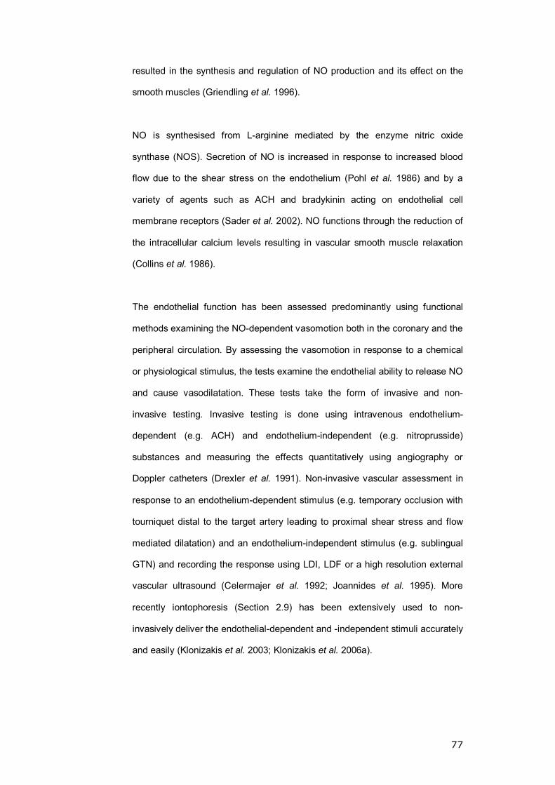
77
resulted in the synthesis and regulation of NO production and its effect on the
smooth muscles (Griendling et al. 1996).
NO is synthesised from L-arginine mediated by the enzyme nitric oxide
synthase (NOS). Secretion of NO is increased in response to increased blood
flow due to the shear stress on the endothelium (Pohl et al. 1986) and by a
variety of agents such as ACH and bradykinin acting on endothelial cell
membrane receptors (Sader et al. 2002). NO functions through the reduction of
the intracellular calcium levels resulting in vascular smooth muscle relaxation
(Collins et al. 1986).
The endothelial function has been assessed predominantly using functional
methods examining the NO-dependent vasomotion both in the coronary and the
peripheral circulation. By assessing the vasomotion in response to a chemical
or physiological stimulus, the tests examine the endothelial ability to release NO
and cause vasodilatation. These tests take the form of invasive and non-
invasive testing. Invasive testing is done using intravenous endothelium-
dependent (e.g. ACH) and endothelium-independent (e.g. nitroprusside)
substances and measuring the effects quantitatively using angiography or
Doppler catheters (Drexler et al. 1991). Non-invasive vascular assessment in
response to an endothelium-dependent stimulus (e.g. temporary occlusion with
tourniquet distal to the target artery leading to proximal shear stress and flow
mediated dilatation) and an endothelium-independent stimulus (e.g. sublingual
GTN) and recording the response using LDI, LDF or a high resolution external
vascular ultrasound (Celermajer et al. 1992; Joannides et al. 1995). More
recently iontophoresis (Section 2.9) has been extensively used to non-
invasively deliver the endothelial-dependent and -independent stimuli accurately
and easily (Klonizakis et al. 2003; Klonizakis et al. 2006a).

78
A literature review revealed a gender difference in the effect of the sex
hormones on the endothelial function and vascular reactivity. Oestrogen
potentiated endothelial vasodilatation in both the coronary and systemic
circulations in both genders, although this may require long-term administration
in males (Gilligan et al. 1994; Sader et al. 2002). Using brachial artery
ultrasound during the three phases of the menstrual cycle to assess the effect
of oestrogen on the endothelial function, variation in vasodilatation between the
different phases was noted with a maximal vasodilatation in the follicular and
leuteal phases (Hashimoto et al. 1995).
Abnormalities in the endothelial vasoregulatory function is the hallmark of
several vascular diseases including DM, hypertension and chronic
hypercholesterolemia (Griendling et al. 1996). Several mechanisms underlying
the endothelial dysfunction have been identified.
1- Substrate (Arginine) deficiency.
2- Poor NOS stimulation due to an alteration in the second messenger release
resulting in a reduced calcium signal.
3- Receptor dysfunction due to an impaired agonist-receptor interaction or a
reduced receptor expression.
4- Over-production of the constriction factors.
5- NO inactivation due to super-oxide anion (Figure 1.6).
79
Figure 1.6 Potential mechanisms of impaired endothelial-dependent control of vasomotor tone by NO (Griendling et al. 1996)
1.7 AIMS
The overall aim of this work presentation is to investigate aspects within CTS
through experimental microvascular research and reflect on applied practice-
based audits to achieve the following aims.
1- Analyse cold provocation tests conducted on a series of subjects with
HAVS, RP and normal controls to develop and standardise the data
analysis methods for cold provocation testing.
2- Determine the methodology of techniques used in the methodology,
namely CPT and LDI in the hand.
3- Determine the normal vascular response of the hand to a cold challenge
using CPT and LDI.

80
4- Determine the effect of CTS and hand injury on the microcirculation of the
hand.
5- Assess the effect of CTR and hand injury on the vascular response in the
hand.
6- Determine the outcome of conservative management of CTS and its
predictors, and compare the outcome of conservative to surgical treatment
in a carpal tunnel clinic.

81
CHAPTER 2: GENERAL METHODS
82
2.1 INTRODUCTION
This chapter reviews the general background information on the research
subjects and the methods I have used in each study. The review explains the
function, limitations, protocol and rationale for the use of each technique.
2.2 ETHICAL APPROVAL
Research studies were commenced following approval by the Local Research
Ethics Committee and the NHS Trust concerned (Southern Derbyshire Local
Research Ethics Committee and Derby Hospitals NHS Foundation Trust).
Audits performed at Ilkeston Community Hospital (Erewash Primary Care Trust)
were formally reported to the local Clinical Governance body in the Primary
Care Trust and written approval was obtained in accordance with the policy and
the procedures of the Trust. No identifiable data was recorded during the
implementation of the research protocol (psuedonomised: referred to with a
code) or the data collection for the audit, in accordance with the Data Protection
Act 1998 (Al-Shahi et al. 2000; Strobl et al. 2000).
2.3 CLINICAL SETTING
Patient studies involving Laser Doppler Imaging and Cold Provocation Testing
were performed in the clinical investigation facilities within the Clinical Sciences
Wing of the Graduate Entry Medical School of The University of Nottingham,
based at Derby.
2.4 SUBJECT RECRUITMENT
Subjects were invited to participate by direct contact, mail or through study
posters displayed in areas where potential study candidates might attend within
83
the premises of the Derby Hospitals NHS Foundation Trust and The Graduate
Entry Medical School.
Subjects were recruited from:
The Orthopaedics and Hand Outpatient Clinics
Current and past patients attending orthopaedic and hand clinics at Derbyshire
Royal Infirmary. CTS and RP patients were invited to participate through
information leaflets provided in clinics or by mail.
The Pulvertaft Centre Hand Surgery Database (Delta Database)
The Pulvertaft Hand Centre keeps an up-to-date database of all the hand
surgery patients reviewed in the unit since 1987 with details of their clinic
appointments and last operative intervention date. This system was designed
in-house as a clinical office management system and is updated on a daily
basis by the secretaries as they process the clinical tapes. The database
currently holds information on approximately 80,000 patients.
Normal (control) volunteers
Control subjects were recruited from a database in the Division of Vascular
Medicine, via posters within the Graduate Entry Medical School and through
invitation letters to suitable people registered in a medical practice in which one
of the lead investigators works.
Advertising in miners welfare clubs
Advertising leaflets and information sheets were sent to the surrounding miners
welfare clubs (nine clubs contacted).
The premises of the Derby Hospitals NHS Foundation Trust
Advertised within the premises in clinics (Vascular and Rheumatology).
84
The HAVS patients undergoing CPT for medical reports in the private practice
of the chief investigator had their fully anonymised data included in the study.
Patients attending a Registrar-Led Carpal Tunnel Clinic in Ilkeston Community
Hospital (Erewash Primary Care Trust).
2.5 PATIENT CONSENT
All study patients gave written informed consent at least 24 hours after reading
the information sheet for patients/volunteers prior to participating in a research
study. Copies of the consent form and patient information sheet were filed in the
hospital notes.
2.6 DEMOGRAPHIC INFORMATION
Basic information on each study subject was obtained and recorded using a
standardised data collection sheet (referenced for each study in relevant
chapters). Relevant history including age, sex, exposure to vibration, history of
RP, DM, drug history and hand dominance was obtained. Clinical data including
height (cm), weight (Kg), blood pressure (Section 2.7), clinical peripheral or
central cyanosis, timed Allan�s test (Gelberman et al. 1981a) and hand
examination for neurological signs were collected and documented as per
protocol.
2.7 BLOOD PRESSURE (BP) MEASUREMENT
A validated, maintained and calibrated OMRON 703 CP device was used
(Medical Devices Directive (MDD: 93/42/EEC)) to measure the sitting BP in
study subjects using a cuff of appropriate size. Two BP readings were taken; if
a marked difference (10-15 mmHg in systolic or diastolic BP) between the
85
readings was obtained, a third reading was taken (Williams et al. 2004). The
mean BP for each subject was used in the final analysis.
2.8 DATA ANALYSIS
All data collected were stored on an Excel® spreadsheet and analysed using
one of two statistical packages (SPSS V13.0: statistical package for social
sciences and MedCalc®). Ten per cent of the data entries in each database
were checked by Mrs M. Baker (senior research assistant) for errors prior to
analysis. A further detailed data review was planned when/if errors were
detected in the pilot review. The Derby Hospitals NHS Foundation Trust
statisticians (Dr R. Hilliam and Mr A. Fakis) were consulted on the most
appropriate statistical methods of analysis at each step of the process and
specific reference to the appropriate tests on each data set is provided in the
corresponding chapter.
The data was analysed based on its type and distribution. Categorical data
were analysed using the Х2 test, continuous data were analysed based on their
distribution. Data normality was assessed based on the sample size using the
Kolmogorov-Smirnov test for samples larger than 50 and the Shapiro-Wilk test
for samples of 50 or smaller; a non-significant p-value (>0.05) indicates
normality in either test. Data histograms were also inspected. The statistical test
choice was then made as demonstrated in Figures 2.1 and 2.2 (paired
data=data from the same subject). The data are presented as mean (SD)
unless stated otherwise.
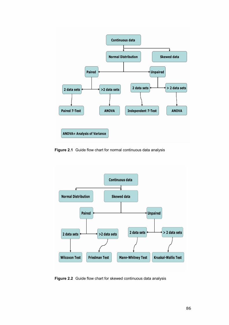
86
Continuous data
Skewed dataNormal Distribution
Paired Unpaired
>2 data sets
Paired T-Test
2 data sets > 2 data sets
Independent T-Test
2 data sets
ANOVA ANOVA
ANOVA= Analysis of Variance
Figure 2.1 Guide flow chart for normal continuous data analysis
Continuous data
Skewed dataNormal Distribution
Paired Unpaired
>2 data sets 2 data sets > 2 data sets
Mann-Whitney Test
2 data sets
Friedman Test Kruskal-Wallis Test Wilcoxon Test
Figure 2.2 Guide flow chart for skewed continuous data analysis
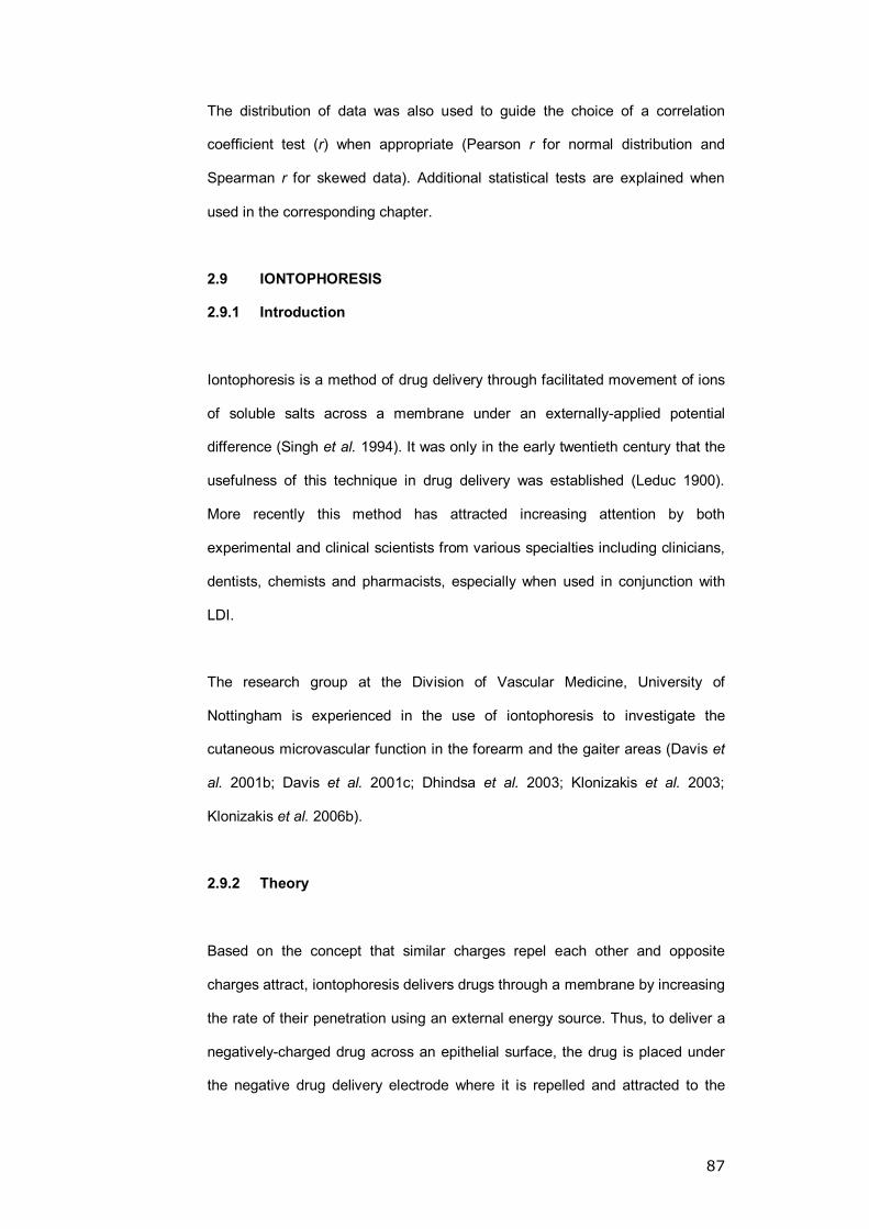
87
The distribution of data was also used to guide the choice of a correlation
coefficient test (r) when appropriate (Pearson r for normal distribution and
Spearman r for skewed data). Additional statistical tests are explained when
used in the corresponding chapter.
2.9 IONTOPHORESIS
2.9.1 Introduction
Iontophoresis is a method of drug delivery through facilitated movement of ions
of soluble salts across a membrane under an externally-applied potential
difference (Singh et al. 1994). It was only in the early twentieth century that the
usefulness of this technique in drug delivery was established (Leduc 1900).
More recently this method has attracted increasing attention by both
experimental and clinical scientists from various specialties including clinicians,
dentists, chemists and pharmacists, especially when used in conjunction with
LDI.
The research group at the Division of Vascular Medicine, University of
Nottingham is experienced in the use of iontophoresis to investigate the
cutaneous microvascular function in the forearm and the gaiter areas (Davis et
al. 2001b; Davis et al. 2001c; Dhindsa et al. 2003; Klonizakis et al. 2003;
Klonizakis et al. 2006b).
2.9.2 Theory
Based on the concept that similar charges repel each other and opposite
charges attract, iontophoresis delivers drugs through a membrane by increasing
the rate of their penetration using an external energy source. Thus, to deliver a
negatively-charged drug across an epithelial surface, the drug is placed under
the negative drug delivery electrode where it is repelled and attracted to the
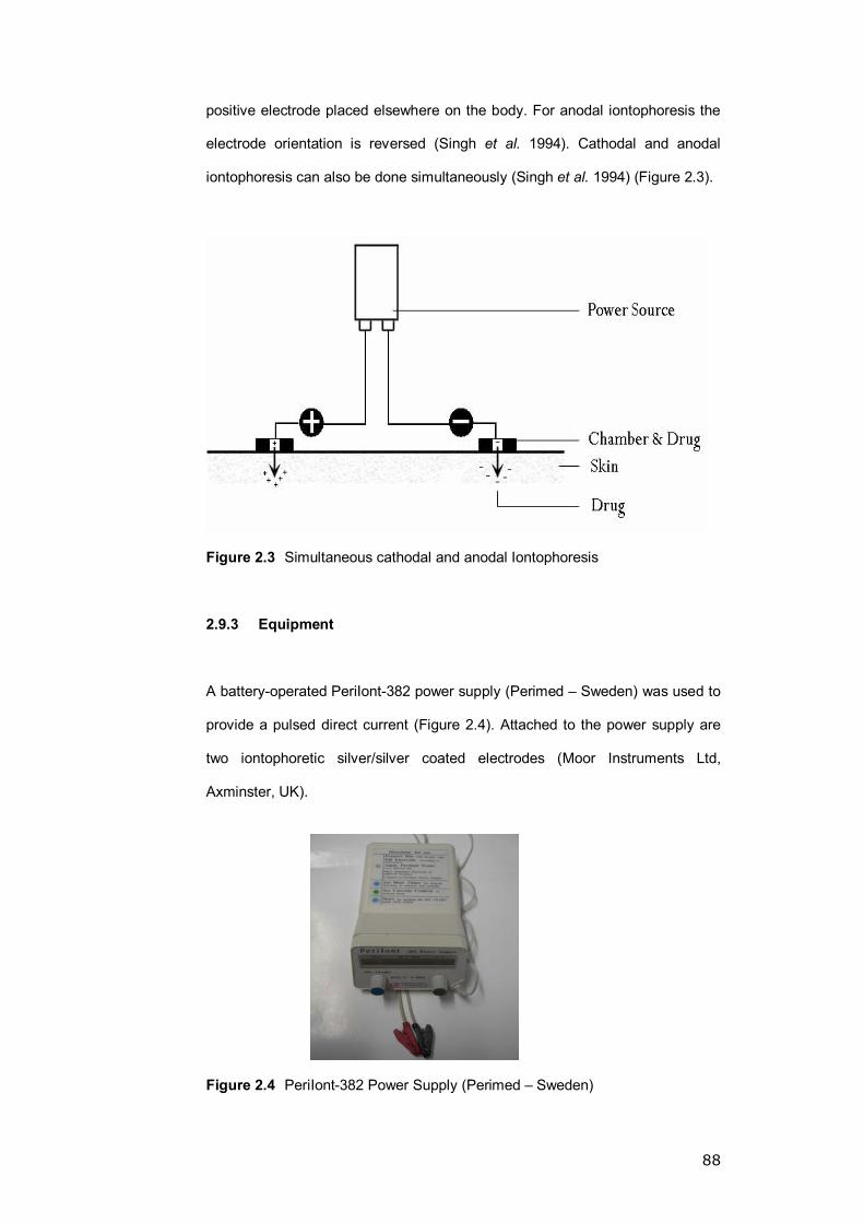
88
positive electrode placed elsewhere on the body. For anodal iontophoresis the
electrode orientation is reversed (Singh et al. 1994). Cathodal and anodal
iontophoresis can also be done simultaneously (Singh et al. 1994) (Figure 2.3).
Figure 2.3 Simultaneous cathodal and anodal Iontophoresis
2.9.3 Equipment
A battery-operated PeriIont-382 power supply (Perimed � Sweden) was used to
provide a pulsed direct current (Figure 2.4). Attached to the power supply are
two iontophoretic silver/silver coated electrodes (Moor Instruments Ltd,
Axminster, UK).
Figure 2.4 PeriIont-382 Power Supply (Perimed � Sweden)

89
2.9.4 Investigational drugs
Acetylcholine (ACH) (Sigma Chemicals, UK) and sodium nitorprusside (SNP)
(Nipride, Roche Pharmaceuticals Ltd) were prepared as 1% solutions using
deionised HPLC-Grade water. Higher drug concentrations had previously been
shown not to result in a faster response and were associated with a risk of non-
specific vasodilatation (Droog et al. 2003; Droog et al. 2004). The drugs were
prepared in a quantity sufficient to cover a period of four weeks. The vials were
covered with aluminium paper and the preparations were stored in a freezer to
prevent the denaturation of the drugs due to high temperature or light exposure.
To minimise errors, two persons were present during the preparation according
to a standard operating procedure.
The choice of the drugs was based on previous work undertaken in the
department (Davis et al. 2001c; Klonizakis et al. 2003; Klonizakis et al. 2006b).
ACH is a non-specific vasodilator with effects on the C-fibres, vascular
endothelium and smooth muscles; SNP acts on the vascular smooth muscles
only (Green et al. 1996; Fujimoto et al. 1997; Berghoff et al. 2002).
2.9.5 Precautions and considerations
Factors known to affect the delivery of drugs using iontophoresis were
controlled. The use of the same drugs with identical concentrations, delivered
through an identical protocol, eliminated variations due to drug physiochemical
properties (molecular size, charge and concentration), drug formulation (type of
vehicle, buffer, pH, viscosity and the presence of other ions) and equipment
factors (current used, the type of electrodes and the duration of the
iontophoresis) (Singh et al. 1994).

90
Biological variation such as age and sex were balanced as far as possible
during recruitment. All studies were conducted in a controlled environment with
the air temperature maintained between 20-25°C. Subjects were allowed to
acclimatise to the ambient temperature for 25-30 minutes before each study.
The electrodes were applied on the dorsum of the chosen hand/hands as per
study protocol.
2.9.6 Iontophoresis protocol
After a period of acclimatisation, the dorsal skin of the hand was carefully
cleaned with an alcohol swab and dried thoroughly to minimise the risk of local
spots of high current density (which might result in C-fibre activation, non-drug-
related vasodilatation and local micro burns) (Gazelius 2005). Two chambers
(electrodes) were used simultaneously on the dorsum of the hand and
positioned 1-3 cm apart avoiding major dorsal veins. One of the chambers was
connected to the anodal lead of the iontophoresis controller (PeriIont-382 Power
Supply (Perimed � Sweden)) and filled with 0.5 ml of the positively-charged
ACH, the other was connected to the cathodal lead and filled with 0.5 ml of the
negatively-charged SNP (Figure 2.5).
The iontophoresis protocol used was the following:
Time Current Charge
20 seconds 120µamp 2400µCb
The same protocol was used in all study subjects.

91
Figure 2.5 Two Iontophoretic Silver/Silver coated electrodes (Moor Instruments Ltd, Axminster, UK) attached to the back of the hand
2.10 LASER DOPPLER IMAGING (LDI)
2.10.1 Introduction
A comprehensive overview of the LDI was given earlier (Section 1.5).
2.10.2 Equipment
Periscan PIM II Laser Doppler Perfusion Imager (Perimed � Sweden) was
connected to a computer (Compaq, Intel PIII), equipped with PeriScan Software
� LDIwin 2.6 (Perimed � Sweden) (Figure 2.6).
Figure 2.6 Periscan PIM II Laser Doppler Perfusion Imager (Perimed �Sweden)

92
2.10.3 Precautions and considerations
A number of variables were controlled to achieve consistency in measurements.
Temperature, anatomical site, physical activity, food and drug intake were
accounted for (Bircher et al. 1994; Fullerton et al. 2002).
Changes in the ambient temperature variation are associated with changes in
cutaneous flux readings. A light intensity of 6-8 microvolts was maintained in the
test room during the imaging to minimise the effect of the ambient light (Nilsson
1997).
Temperatures >30°C markedly increase the skin microcirculation (Winsor et al.
1989); hence, all subjects were allowed to acclimatise in a temperature-
controlled room (20-25°C for 30 minutes) to allow the skin temperature to
stabilise.
The distance of the scanner head from the point of interest affects the sensor
unit in the LDI; distances <10 cm saturate the detector unit while >30cm create
an impaired signal-noise ratio (Fullerton et al. 2002). The scanner head was
positioned 20 cm from the area of interest.
Food intake, age, sex, race, blood and serum parameters did not affect mean
values of skin flux (Bircher et al. 1994). No blood samples were taken from
study subjects.
Smoking has a negative correlation with the flux (Waeber et al. 1984). Smokers
were instructed to refrain from smoking 6 hours prior to the test and were
prioritised to be tested early in the morning session. Alcohol increases the
cutaneous circulation (Wilkin 1986; Bircher et al. 1994) and subjects were
instructed to abstain from alcohol for 6 hours prior to the test.
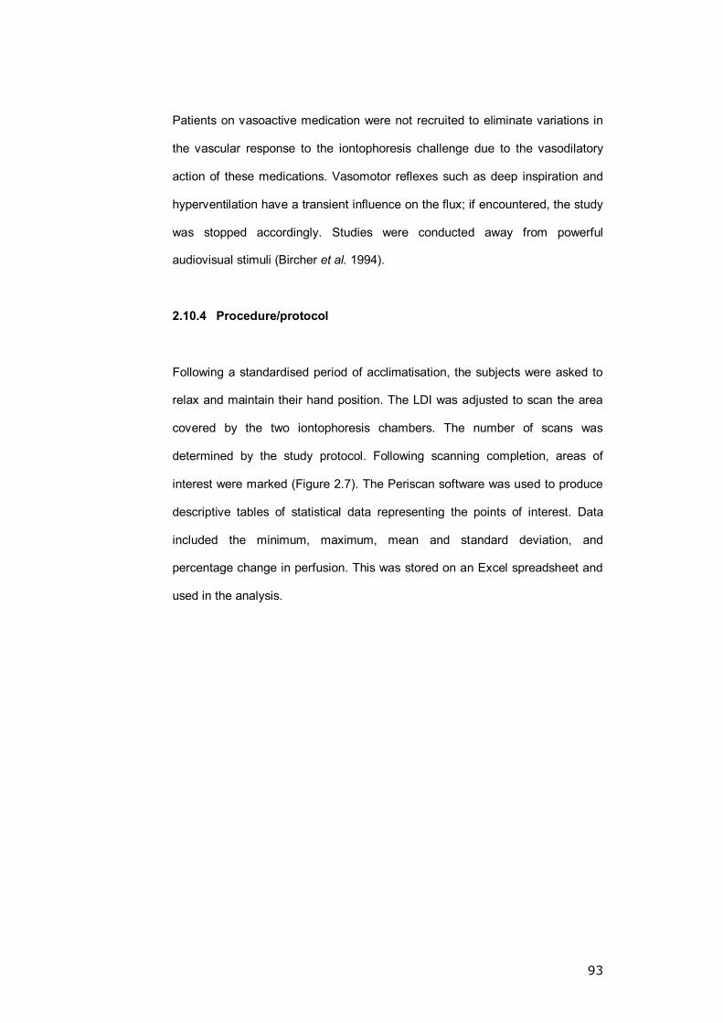
93
Patients on vasoactive medication were not recruited to eliminate variations in
the vascular response to the iontophoresis challenge due to the vasodilatory
action of these medications. Vasomotor reflexes such as deep inspiration and
hyperventilation have a transient influence on the flux; if encountered, the study
was stopped accordingly. Studies were conducted away from powerful
audiovisual stimuli (Bircher et al. 1994).
2.10.4 Procedure/protocol
Following a standardised period of acclimatisation, the subjects were asked to
relax and maintain their hand position. The LDI was adjusted to scan the area
covered by the two iontophoresis chambers. The number of scans was
determined by the study protocol. Following scanning completion, areas of
interest were marked (Figure 2.7). The Periscan software was used to produce
descriptive tables of statistical data representing the points of interest. Data
included the minimum, maximum, mean and standard deviation, and
percentage change in perfusion. This was stored on an Excel spreadsheet and
used in the analysis.

94
Figure 2.7 Data output after completing the imaging
2.11 COLD PROVOCATION TESTING (CPT)
2.11.1 Introduction
A comprehensive overview of the LDI is given in Sections 1.4.2 and 3.2.
2.11.2 Equipment
A laboratory water bath with a continuously-stirred temperature-controlled
cooling and heating element was used, the water temperature being controlled
to within 0.1°C (Clifton NE4-D, Digital Thermostatic Control, Nickel Electro. Ltd-
UK). Thermocouples used were standard type-K thermocouples (T. M.
Electronics (UK) Ltd) connected through Pico Technology TC-08 thermocouple
adapters (8 channel thermocouple data logger).
Test descriptive data were generated and stored using a software package
based on Microsoft Excel (1998).
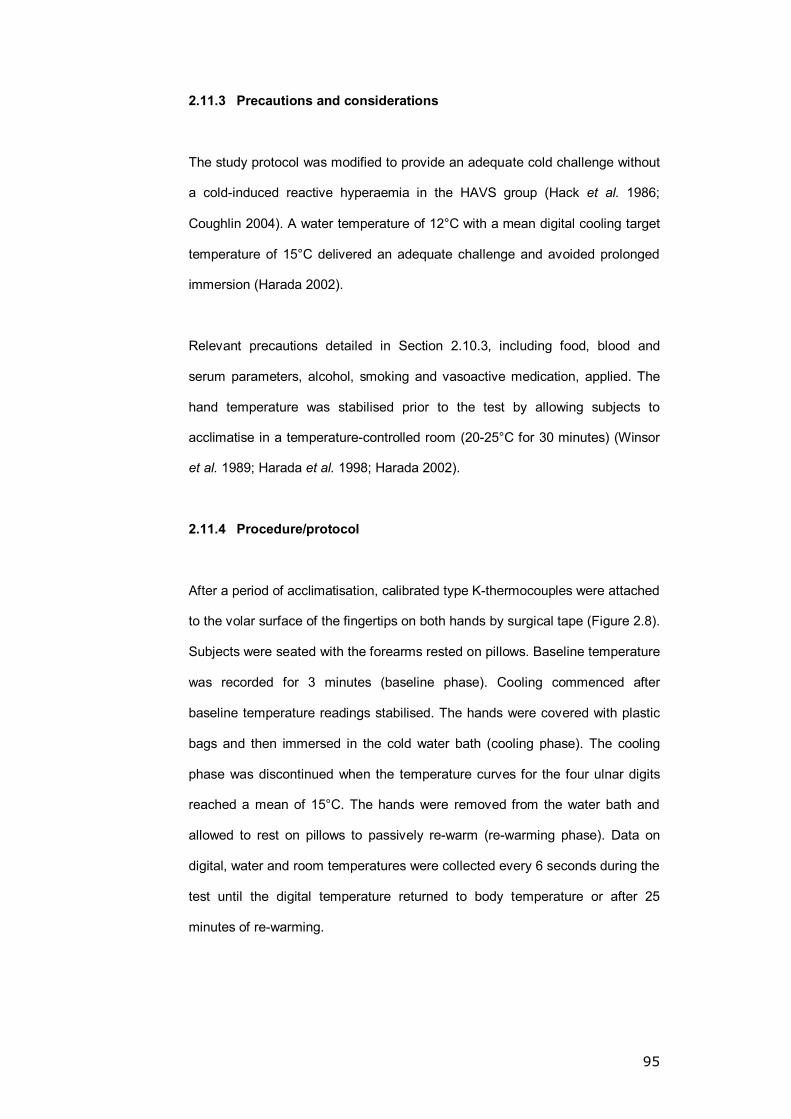
95
2.11.3 Precautions and considerations
The study protocol was modified to provide an adequate cold challenge without
a cold-induced reactive hyperaemia in the HAVS group (Hack et al. 1986;
Coughlin 2004). A water temperature of 12°C with a mean digital cooling target
temperature of 15°C delivered an adequate challenge and avoided prolonged
immersion (Harada 2002).
Relevant precautions detailed in Section 2.10.3, including food, blood and
serum parameters, alcohol, smoking and vasoactive medication, applied. The
hand temperature was stabilised prior to the test by allowing subjects to
acclimatise in a temperature-controlled room (20-25°C for 30 minutes) (Winsor
et al. 1989; Harada et al. 1998; Harada 2002).
2.11.4 Procedure/protocol
After a period of acclimatisation, calibrated type K-thermocouples were attached
to the volar surface of the fingertips on both hands by surgical tape (Figure 2.8).
Subjects were seated with the forearms rested on pillows. Baseline temperature
was recorded for 3 minutes (baseline phase). Cooling commenced after
baseline temperature readings stabilised. The hands were covered with plastic
bags and then immersed in the cold water bath (cooling phase). The cooling
phase was discontinued when the temperature curves for the four ulnar digits
reached a mean of 15°C. The hands were removed from the water bath and
allowed to rest on pillows to passively re-warm (re-warming phase). Data on
digital, water and room temperatures were collected every 6 seconds during the
test until the digital temperature returned to body temperature or after 25
minutes of re-warming.
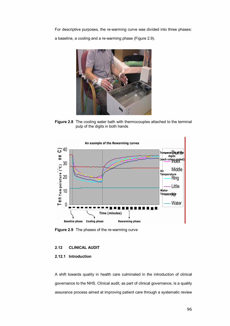
96
For descriptive purposes, the re-warming curve was divided into three phases:
a baseline, a cooling and a re-warming phase (Figure 2.9).
Figure 2.8 The cooling water bath with thermocouples attached to the terminal
pulp of the digits in both hands
0
10
20
30
40
Time (minutes)
Tem
pera
ture
(deg
ree
C) ThumbIndexMiddleRingLittleAirWater
Baseline phase Cooling phase Rewarming phase
Time (minutes)
Te
mp
era
ture
(°C
)
An example of the Rewarming curves
Air Temperature
Water Temperature
Temperature of the digits
(each colour coded)
Figure 2.9 The phases of the re-warming curve
2.12 CLINICAL AUDIT
2.12.1 Introduction
A shift towards quality in health care culminated in the introduction of clinical
governance to the NHS. Clinical audit, as part of clinical governance, is a quality
assurance process aimed at improving patient care through a systematic review
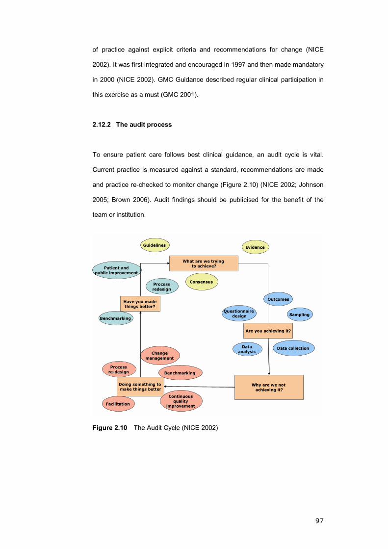
97
of practice against explicit criteria and recommendations for change (NICE
2002). It was first integrated and encouraged in 1997 and then made mandatory
in 2000 (NICE 2002). GMC Guidance described regular clinical participation in
this exercise as a must (GMC 2001).
2.12.2 The audit process
To ensure patient care follows best clinical guidance, an audit cycle is vital.
Current practice is measured against a standard, recommendations are made
and practice re-checked to monitor change (Figure 2.10) (NICE 2002; Johnson
2005; Brown 2006). Audit findings should be publicised for the benefit of the
team or institution.
What are we trying to achieve?
Why are we not achieving it?
Doing something to make things better
Are you achieving it?
Have you made things better?
Guidelines
Patient and public improvement
Evidence
Benchmarking
Process redesign
Consensus
Data analysis
Process re-design
Questionnaire design
Continuous quality
improvement
Benchmarking
Change management
Facilitation
Data collection
Sampling
Outcomes
Figure 2.10 The Audit Cycle (NICE 2002)
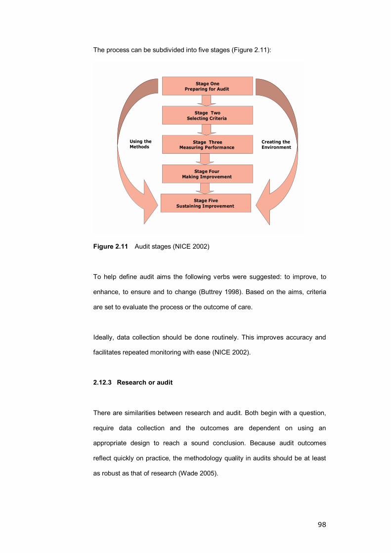
98
The process can be subdivided into five stages (Figure 2.11):
Stage OnePreparing for Audit
Stage TwoSelecting Criteria
Stage ThreeMeasuring Performance
Stage Four Making Improvement
Stage FiveSustaining Improvement
Creating the Environment
Using the Methods
Figure 2.11 Audit stages (NICE 2002)
To help define audit aims the following verbs were suggested: to improve, to
enhance, to ensure and to change (Buttrey 1998). Based on the aims, criteria
are set to evaluate the process or the outcome of care.
Ideally, data collection should be done routinely. This improves accuracy and
facilitates repeated monitoring with ease (NICE 2002).
2.12.3 Research or audit
There are similarities between research and audit. Both begin with a question,
require data collection and the outcomes are dependent on using an
appropriate design to reach a sound conclusion. Because audit outcomes
reflect quickly on practice, the methodology quality in audits should be at least
as robust as that of research (Wade 2005).

99
More recently, a national audit on diabetic retinopathy was presented to 28
ethics committees and 25 trusts for approval. Only six bodies considered the
project an audit and not research (Wilson et al. 1999). Evident is the lack of
uniformity in the decision as seen above, particularly when a substantial amount
of data is collected (Wade 2005). It was argued that minimal changes in clinical
practice such as collecting extra data amounting to a minor burden on the
patient (5-10 minute questionnaire, taking measurements such as blood
pressure and pulse rate) would rarely require ethical consideration (Kinn 1997;
Wade 2005). Others are of the view that an audit should never involve
disturbances of patients over and above normal clinical management
requirements with no extra data collection and no extra clinical assessment
(RDSU).
Although clinical governance guidance is available, it is primarily the
responsibility of the investigator to seek the appropriate approval.
While conducting the CTS audit, the patient management was not influenced by
the data collection. The data collection is part of a continuous process to
monitor the standard of practice in the clinic. Measurements of sensation and
power are common practice in the Pulvertaft Hand Centre and the
questionnaires given to the patients take <5 minutes to complete.
2.12.4 Audit procedures
Two audit projects were designed and approved by the local clinical governance
office: a retrospective audit of the outcome of patients managed by a nurse
practitioner in a CTS clinic and a prospective audit on the clinic following the
appointment of a hand fellow (myself).
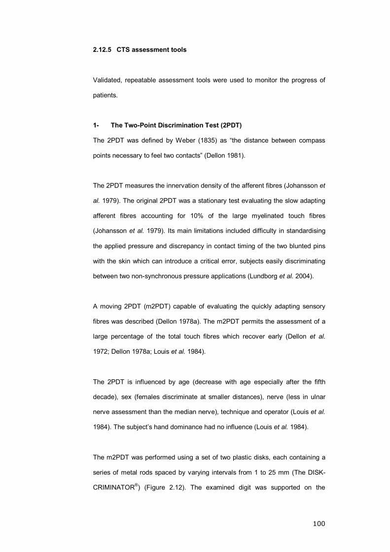
100
2.12.5 CTS assessment tools
Validated, repeatable assessment tools were used to monitor the progress of
patients.
1- The Two-Point Discrimination Test (2PDT)
The 2PDT was defined by Weber (1835) as �the distance between compass
points necessary to feel two contacts� (Dellon 1981).
The 2PDT measures the innervation density of the afferent fibres (Johansson et
al. 1979). The original 2PDT was a stationary test evaluating the slow adapting
afferent fibres accounting for 10% of the large myelinated touch fibres
(Johansson et al. 1979). Its main limitations included difficulty in standardising
the applied pressure and discrepancy in contact timing of the two blunted pins
with the skin which can introduce a critical error, subjects easily discriminating
between two non-synchronous pressure applications (Lundborg et al. 2004).
A moving 2PDT (m2PDT) capable of evaluating the quickly adapting sensory
fibres was described (Dellon 1978a). The m2PDT permits the assessment of a
large percentage of the total touch fibres which recover early (Dellon et al.
1972; Dellon 1978a; Louis et al. 1984).
The 2PDT is influenced by age (decrease with age especially after the fifth
decade), sex (females discriminate at smaller distances), nerve (less in ulnar
nerve assessment than the median nerve), technique and operator (Louis et al.
1984). The subject�s hand dominance had no influence (Louis et al. 1984).
The m2PDT was performed using a set of two plastic disks, each containing a
series of metal rods spaced by varying intervals from 1 to 25 mm (The DISK-
CRIMINATOR®) (Figure 2.12). The examined digit was supported on the
101
examination table. Using the 5-8 mm interval rods moving from proximal to
distal and proceeding in stages down to 2 mm spaced rods (Dellon 1978a). The
index finger was used as the sensory denominator for the median nerve and is
tested in both hands (Moore et al. 2005). Values of less than 6 mm were
considered normal (Gellis et al. 1977; Dellon 1981). A 2PDT >15 mm
represented a useless digit and measurements between 6 and 15 mm
suggested �some tactile gnosis� is preserved (Moberg 1964; Lundborg et al.
2004).
Figure 2.12 The DISK-CRIMINATOR®
2- The Semmes-Weinstein pressure aethesiometer (SWM)
A sensory threshold test, this measures the function of slow adapting afferent
fibres (Levin et al. 1978; Dellon 1980). The SWM correlates accurately with
symptoms of nerve compression and electro diagnostic tests (Gelberman et al.
1983a). It is used in the assessment of peripheral neuropathy in diabetes
mellitus (Lee et al. 2003), and compressive and traumatic neuropathy (Dellon
1978a; Gelberman et al. 1983a).

102
The SWM was conducted using a Touch-Test® Sensory Evaluator (North Coast
Medical, Inc. Morgan Hill, CA) (Figure 2.13), which consists of six individually-
calibrated monofilament pencils (Table 2.1).
Figure 2.13 Touch Test® Sensory Evaluator (North Coast Medical, Inc. Morgan Hill, CA)
Evaluator size Target force to bow
evaluator (grams) Sensory hand threshold
2.83 0.07 Normal
3.61 0.4 Diminished light touch
4.31 2 Diminished protective
sensation
4.56 4
5.07 10 Loss of protective sensation
6.65 300 Deep pressure sensation only
Table 2.1 Description of the SWM monofilament evaluators
After explaining the test, subjects were asked to rest the extremity on a stable
surface and look away from the test site. The evaluator was pressed at 90° to
the surface of the skin avoiding callosities until it bowed, pressure was
maintained for 1-2 seconds and then removed. The test started with a 2.83
evaluator and was repeated using a larger monofilament if a response was
lacking. The size of the evaluator dictated the number of applications (2.83 &
3.61 applied 1-3 times, 4.31-6.65 applied only once). The pulp of the index
103
finger was used as a denominator for median nerve sensory innervations
(Moore et al. 2005).
3- Self-administered questionnaires
A. Levine-Katz symptoms and functional questionnaire
The questionnaire consists of two main parts. [a] Symptom severity scale
assessing the six critical domains in CTS evaluation (pain, paresthesia,
numbness, weakness, night symptoms and the overall functional status of the
patient). Eleven questions with multiple choice responses scored from 1:
(mildest) to 5: (most severe) and an overall symptom severity score calculated
as the mean of the 11 scores. [b] Functional score assessing eight activities
commonly performed. The answers were rated from 1: (no difficulty in
performing the activity) to 5: (cannot perform the activity at all). The overall
functional status was calculated as the mean of all eight scores (Levine et al.
1993) (Appendix 1.1).
The questionnaire was reported as repeatable (Pearson correlation coefficient
=0.91 and 0.93 for severity and functional status scales respectively) and
sensitive (defined as the response to clinical change) (Spearman coefficient
was 0.5 for the symptom severity score and 0.54 for the functional score in a
prospective cohort (p<0.01) (Duncan et al. 1987; Levine et al. 1993) (Section
1.1.3).
B. Quick Dash (disability of the arm, shoulder and hand) outcome
measurement
A standardised questionnaire evaluating impairment and activity limitations
through 30 questions with multiple choice responses ranging from 1 to 5 (1: no
difficulty to 5: unable) (Hudak et al. 1996; Jester et al. 2005). A shorter version
was devised containing 11 questions while maintaining an acceptable internal
consistency, the Quick Dash (Appendix 1.2) (Beaton et al. 2005). The

104
questionnaire is repeatable (Interclass Correlation Coefficient =0.94) and
validated (r≥0.64 with single item indices of pain and function) (Beaton et al.
2005).
In the CTS clinic, the Quick Dash was used. The total score was calculated if at
least 10 of the 11 questions were answered. The assigned value for all
completed responses was summed and averaged producing a score out of five.
This value was transformed to a score out of 100 by subtracting one and
multiplying by 25. This is done to facilitate comparing the outcome with other
measures scaled 0-100. The higher the score the greater the disability (Beaton
et al. 2005).
4- Dynamometers
Calibrated grip and pinch strength dynamometers were used (TEC, Clifton, New
Jersey). The best out of three readings (kg) was recorded for both hands
(Figure 2.14).
Figure 2.14 Grip and pinch strength dynamometers
This chapter has provided an overview of the methods used. The following
chapters describe the projects undertaken to meet the stated aims of my work.
105
CHAPTER 3: DEVELOPMENT OF METHODS
1- CPT modification
2- Repeatability of Methods
i. Modified Cold Provocation Test
ii. Laser Doppler Imaging in the hand
106
3.1 INTRODUCTION
3.1.1 CPT modification
The large number of claimants on the miners� compensation scheme carried out
by the Department of Trade and Industry (DTI) identified the need for an
objective test for the diagnosis of HAVS. In 1998, a standardised CPT was
adopted to assess the vascular component of HAVS (Griffin et al. 1998). The
weak correlation between this test�s findings and the vascular stage based on
the Stockholm Workshop Scale generated debate on the validity of the test
(McGeoch et al. 2000). Some authors suggested the standardised CPT to have
�no� or �limited� value in evaluating the presence or severity of HAVS (Mason et
al. 2003; Proud et al. 2003).
A modification on the standardised test was developed by Mr L. C. Bainbridge
(The Pulvertaft Hand Centre). The research team at the Division of Vascular
Medicine, The University of Nottingham, started validating this test in patients
with RP and HAVS two years ago.
3.1.2 Repeatability of methods
Repeatability is defined as outcome consistency under the same conditions (i.e.
same operator, same apparatus, within a short time interval and for the same
sample). It is an important test validation requirement (Bircher et al. 1994).
The standardised CPT was �positive one day and negative the next day� for
subjects exposed to vibration, and authors reported wide limits of agreement in
vibration-exposed subjects (i.e. LOA for middle finger 5 minutes post-cooling
was 2.3, 10.9 compared to 5.1, 6.9 in the control group; similar findings were
noticed in the other digits) (Griffin et al. 1998). Similar findings were reported

107
elsewhere (Mason et al. 2003). Other formats of the test exist but repeatability
evidence remains lacking (Coughlin et al. 2001).
The use of LDI in the assessment of a provoked microvascular response
minimised the importance of point-to-point variability and preserved the
representation of the blood flow map in day-to-day measurements (Nilsson
1997; Fullerton et al. 2002). Evidence supporting LDI repeatability exists in the
forearm (coefficient of variation <10% for ACH iontophoresis and 10-20% for
SNP) (Kubli et al. 2000) and the gaiter area (Klonizakis et al. 2003). Evidence of
LDI repeatability in the hand is lacking.
The aims of this chapter are: firstly, to present the data analysis I have
undertaken on CPT data collected by members of the research team to identify
characteristics in the re-warming curve that differentiates between subjects with
PRP, HAVS and controls; secondly, to investigate the modified CPT
repeatability; thirdly, to investigate the repeatability of LDI in the hand.
3.2 METHODS
3.2.1 CPT modification and repeatability
Subject recruitment and demographical data collection is outlined in Sections
2.4 and 2.6 respectively. The modified CPT is described in detail in Section
2.11. Subjects participating in the study were asked to re-attend for a repeat
test after 12-14 days.
Three groups were recruited:
[1] A control (C) group with no history of RP, cold intolerance symptoms,
history of peripheral vascular disease or exposure to vibration.
108
[2] A HAVS group with a history of exposure to vibration for more than five
years and stage (V2) or more on the Stockholm Workshop Score (McGeoch
et al. 2005) (Section 1.2.4).
[3] Subjects with Primary RP (PRP), no history of connective tissue disease or
exposure to vibration.
Exclusion criteria:
[1] Smoked within the last 6 hours.
[2] Currently taking medication for cardiac disease or hypertension including ß-
Blockers, vasodilators, ACE inhibitors.
[3] Currently taking long-term medication for migraine or RP.
[4] A history of a significant finger or limb injury.
[5] Under 18 years of age.
[6] Showed clinical evidence of central or peripheral cyanosis (not related to
cold weather).
[7] Have a prolonged timed Allan�s test.
[8] Failed to maintain their hands in the water for the necessary period.
3.2.2 LDI repeatability
Non-smoking healthy male and female volunteers aged 18 years or over were
recruited to undergo two LDI scanning sessions 12-14 days apart. In each
session the back of the hand was scanned nine times and the vascular
challenge delivered using iontophoresis following the third scan. Details of the
recruitment, LDI and iontophoresis methodology are covered in Chapter 2.
Three operators (myself, a PhD student and a senior research technician) each
performed the study on 20 subjects.
109
Subjects were excluded if they were taking vasoactive medication or gave a
history of Raynaud's phenomenon, exposure to vibration, hand injury or carpal
tunnel syndrome.
3.3 DATA ANALYSIS
Data analysis followed the methods outlined in Section 2.8. Additional statistical
tests are explained where appropriate.
3.3.1 CPT modification
The following measurements were obtained: baseline temperature (defined as
the difference between the pre-cooling digit temperature and the ambient
temperature), temperature rise in the first 30 seconds (T30secs), time taken for
the hands to re-warm by 5°C (T5°C) and the time variability in re-warming
between digits within the hand (defined as the time difference between the
fastest and slowest digit to re-warm to room temperature after cooling within the
hand). The latter was felt to be of particular importance in patients with vibration
exposure as the effect is expected to vary between digits in the same individual.
To study this observation the patients within each group were stratified in one of
five subgroups.
Group 1 0�1 minutes
Group 2 >1�2 minutes
Group 3 >2�3 minutes
Group 4 >3�4 minutes
Group 5 >4 minutes
Thumbs were excluded from all calculations to allow comparison with published
literature (Proud et al. 2003). It was estimated that a sample size of 60 subjects
in each group would allow the detection of 10% difference between the groups
110
with a power of 80% and a beta error of <0.05. Univariate linear regression was
used to study the effect of age, gender and smoking. Odds ratio analysis for the
effect of gender and Receiver Operator Curves (ROC) analysis for the
sensitivity and specificity of the CPT was provided by the trust statistician (Dr
Rachel Hilliam).
3.3.2 Test repeatability
Repeatability of the data was investigated using a random one-way intra-class
correlation (Howell 2002) and Bland-Altman Plots (Bland et al. 1986). To
facilitate the comparison with previously published literature, the correlation
coefficient (r) and the coefficient of variation (CoV) was also calculated. The
following parameters were investigated: baseline, T30secs and T5°C.
3.3.2.1 Repeatability of LDI in the hand
The repeatability of three parameters was investigated: (LDI0) (the mean
perfusion prior to the delivery of the iontophoresis charge), (LDIMax) (maximum
post-iontophoresis perfusion readings) and (LDIMean) (mean perfusion values of
the first three images post-delivery of iontophoresis). Further analysis was
undertaken to investigate operator repeatability variations.
3.4 RESULTS
3.4.1 CPT modification
One hundred and seventy-six subjects were included in the study: 86 controls,
31 PRP and 59 HAVS (Table 3.1). None of the recruited subjects failed to
complete the study.
Age and smoking had no effect on the individual response to cooling (p>0.05,
Linear Regression). The effect of gender was only significant at T30secs; males

111
demonstrated a higher mean temperature rise compared to females (e.g. Right
hand males 2°C (0.9), females 1.4°C (0.8); p = 0.003. Left hand males 1.9°C
(0.9), females 1.5°C (0.6); p = 0.001) (Table 3.2). Additionally, no significant
effect of gender was demonstrated when the odds ratio was calculated for
baseline, T30sec and T5°C (Appendix 3.1).
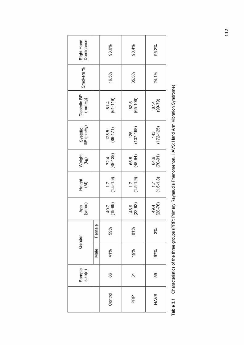
112
Gen
der
S
ampl
e si
ze(n
)
Mal
e Fe
mal
e
Age
(y
ears
) H
eigh
t (M
) W
eigh
t (k
g)
Sys
tolic
BP
(mm
Hg)
D
iast
olic
BP
(m
mH
g)
Smok
ers
%
Rig
ht H
and
Dom
inan
ce
Con
trol
86
41%
59
%
40.7
(1
9-69
) 1.
7 (1
.5-1
.9)
72.4
(4
8-12
8)
125.
5 (8
6-17
1)
81.4
(6
1-11
9)
16.5
%
93.0
%
PR
P
31
19
%
81
%
48.9
(2
3-82
) 1.
7 (1
.5-1
.9)
65.5
(4
8-94
) 12
5 (1
07-1
68)
82.5
(6
5-10
6)
35.5
%
90.4
%
HA
VS
59
97%
3%
49.4
(2
8-76
) 1.
7 (1
.6-1
.8)
84.6
(7
0-91
) 14
3 (1
72-1
25)
87.4
(9
9-79
) 24
.1%
95
.2%
Ta
ble
3.1
Cha
ract
eris
tics
of th
e th
ree
grou
ps (P
RP
: Prim
ary
Ray
naud
�s P
heno
men
on, H
AV
S: H
and
Arm
Vib
ratio
n Sy
ndro
me)
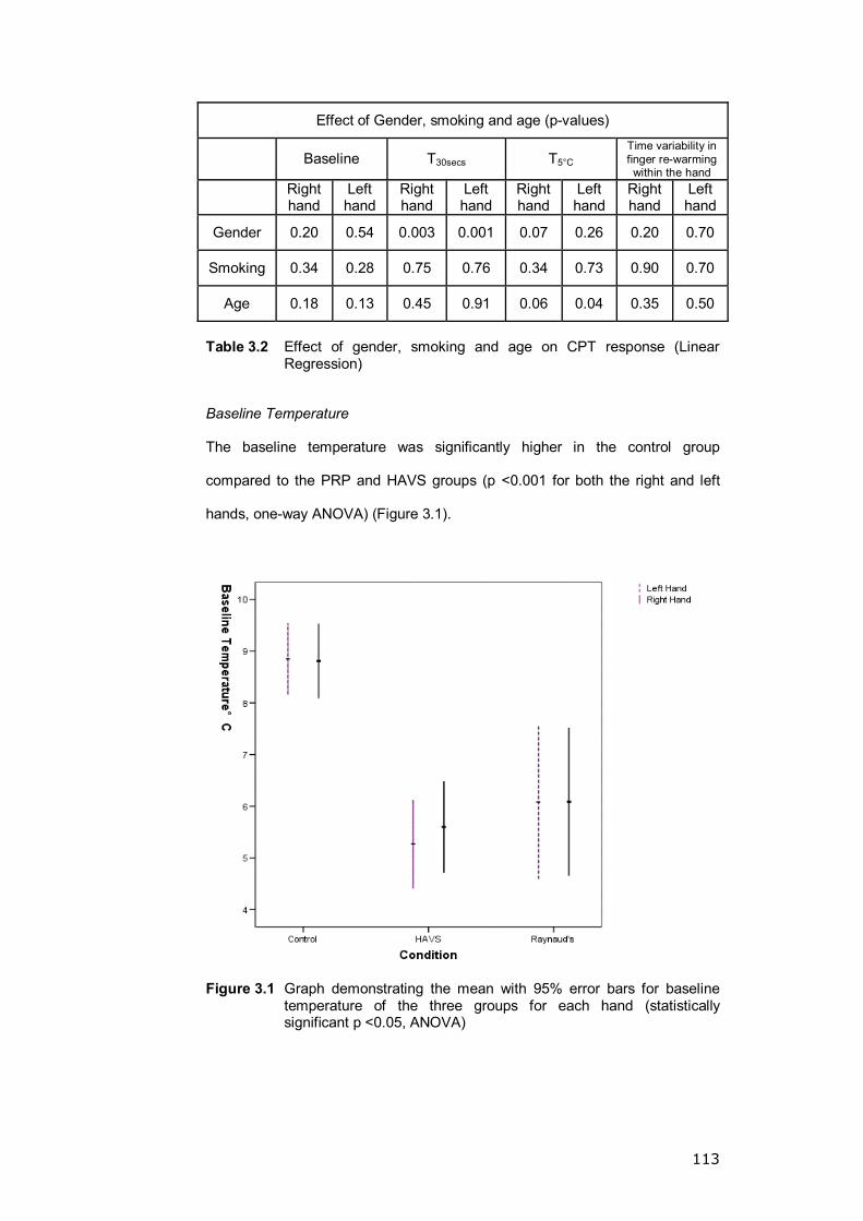
113
Effect of Gender, smoking and age (p-values)
Baseline T30secs T5°C Time variability in finger re-warming within the hand
Right hand
Left hand
Right hand
Left hand
Right hand
Left hand
Right hand
Left hand
Gender 0.20 0.54 0.003 0.001 0.07 0.26 0.20 0.70
Smoking 0.34 0.28 0.75 0.76 0.34 0.73 0.90 0.70
Age 0.18 0.13 0.45 0.91 0.06 0.04 0.35 0.50
Table 3.2 Effect of gender, smoking and age on CPT response (Linear
Regression)
Baseline Temperature
The baseline temperature was significantly higher in the control group
compared to the PRP and HAVS groups (p <0.001 for both the right and left
hands, one-way ANOVA) (Figure 3.1).
Figure 3.1 Graph demonstrating the mean with 95% error bars for baseline temperature of the three groups for each hand (statistically significant p <0.05, ANOVA)
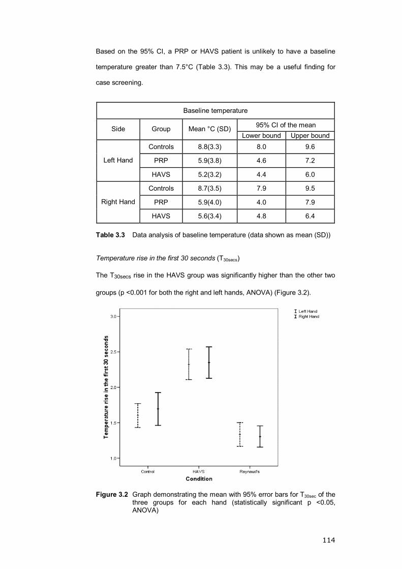
114
Based on the 95% CI, a PRP or HAVS patient is unlikely to have a baseline
temperature greater than 7.5°C (Table 3.3). This may be a useful finding for
case screening.
Baseline temperature
95% CI of the mean Side Group Mean °C (SD) Lower bound Upper bound
Controls 8.8(3.3) 8.0 9.6
PRP 5.9(3.8) 4.6 7.2 Left Hand
HAVS 5.2(3.2) 4.4 6.0
Controls 8.7(3.5) 7.9 9.5
PRP 5.9(4.0) 4.0 7.9 Right Hand
HAVS 5.6(3.4) 4.8 6.4 Table 3.3 Data analysis of baseline temperature (data shown as mean (SD))
Temperature rise in the first 30 seconds (T30secs)
The T30secs rise in the HAVS group was significantly higher than the other two
groups (p <0.001 for both the right and left hands, ANOVA) (Figure 3.2).
Figure 3.2 Graph demonstrating the mean with 95% error bars for T30sec of the three groups for each hand (statistically significant p <0.05, ANOVA)
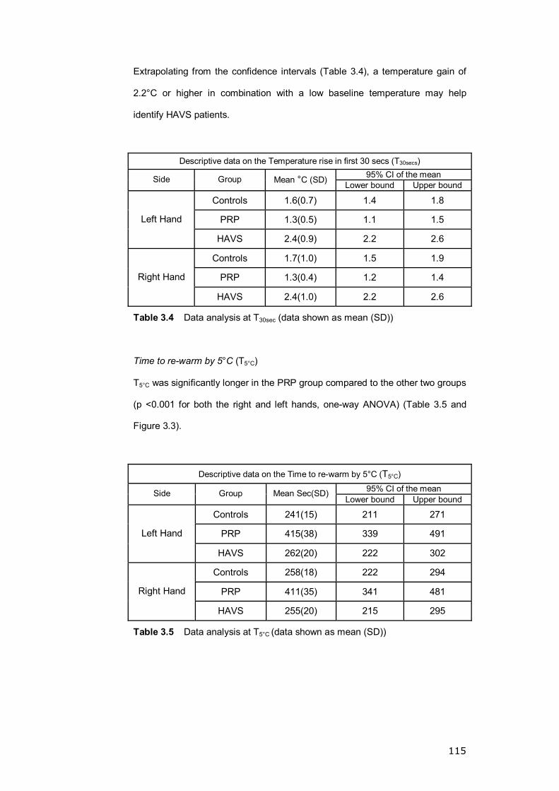
115
Extrapolating from the confidence intervals (Table 3.4), a temperature gain of
2.2°C or higher in combination with a low baseline temperature may help
identify HAVS patients.
Descriptive data on the Temperature rise in first 30 secs (T30secs) 95% CI of the mean Side Group Mean °C (SD) Lower bound Upper bound
Controls 1.6(0.7) 1.4 1.8
PRP 1.3(0.5) 1.1 1.5 Left Hand
HAVS 2.4(0.9) 2.2 2.6
Controls 1.7(1.0) 1.5 1.9
PRP 1.3(0.4) 1.2 1.4 Right Hand
HAVS 2.4(1.0) 2.2 2.6
Table 3.4 Data analysis at T30sec (data shown as mean (SD))
Time to re-warm by 5°C (T5°C)
T5°C was significantly longer in the PRP group compared to the other two groups
(p <0.001 for both the right and left hands, one-way ANOVA) (Table 3.5 and
Figure 3.3).
Descriptive data on the Time to re-warm by 5°C (T5°C)
95% CI of the mean Side Group Mean Sec(SD) Lower bound Upper bound
Controls 241(15) 211 271
PRP 415(38) 339 491 Left Hand
HAVS 262(20) 222 302
Controls 258(18) 222 294
PRP 411(35) 341 481 Right Hand
HAVS 255(20) 215 295
Table 3.5 Data analysis at T5°C (data shown as mean (SD))
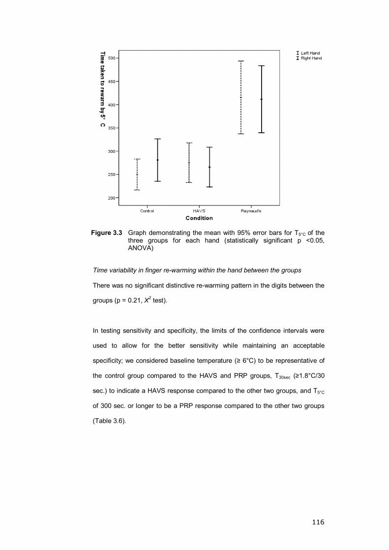
116
Figure 3.3 Graph demonstrating the mean with 95% error bars for T5°C of the three groups for each hand (statistically significant p <0.05, ANOVA)
Time variability in finger re-warming within the hand between the groups
There was no significant distinctive re-warming pattern in the digits between the
groups (p = 0.21, X2 test).
In testing sensitivity and specificity, the limits of the confidence intervals were
used to allow for the better sensitivity while maintaining an acceptable
specificity; we considered baseline temperature (≥ 6°C) to be representative of
the control group compared to the HAVS and PRP groups, T30sec (≥1.8°C/30
sec.) to indicate a HAVS response compared to the other two groups, and T5°C
of 300 sec. or longer to be a PRP response compared to the other two groups
(Table 3.6).
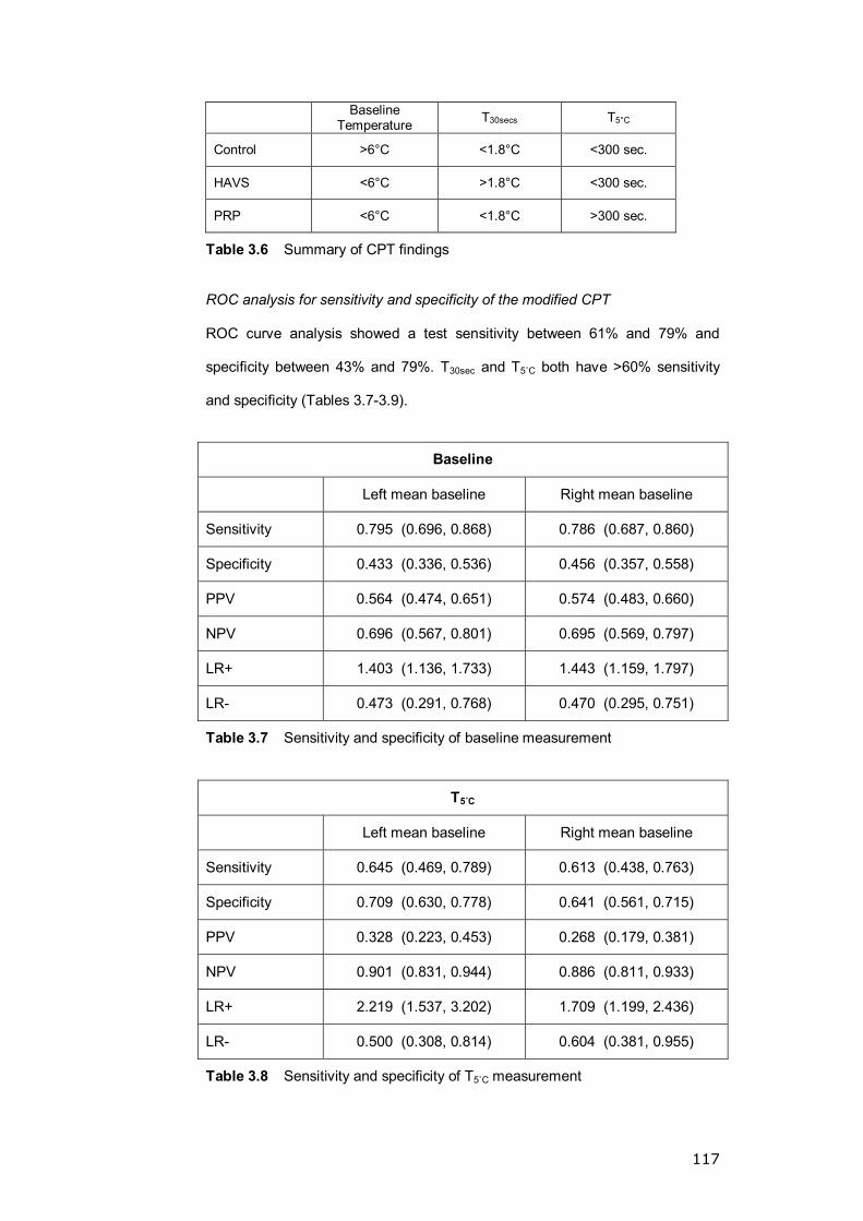
117
Baseline Temperature T30secs T5°C
Control >6°C <1.8°C <300 sec.
HAVS <6°C >1.8°C <300 sec.
PRP <6°C <1.8°C >300 sec.
Table 3.6 Summary of CPT findings
ROC analysis for sensitivity and specificity of the modified CPT
ROC curve analysis showed a test sensitivity between 61% and 79% and
specificity between 43% and 79%. T30sec and T5ûC both have >60% sensitivity
and specificity (Tables 3.7-3.9).
Baseline
Left mean baseline Right mean baseline
Sensitivity 0.795 (0.696, 0.868) 0.786 (0.687, 0.860)
Specificity 0.433 (0.336, 0.536) 0.456 (0.357, 0.558)
PPV 0.564 (0.474, 0.651) 0.574 (0.483, 0.660)
NPV 0.696 (0.567, 0.801) 0.695 (0.569, 0.797)
LR+ 1.403 (1.136, 1.733) 1.443 (1.159, 1.797)
LR- 0.473 (0.291, 0.768) 0.470 (0.295, 0.751)
Table 3.7 Sensitivity and specificity of baseline measurement
T5ûC
Left mean baseline Right mean baseline
Sensitivity 0.645 (0.469, 0.789) 0.613 (0.438, 0.763)
Specificity 0.709 (0.630, 0.778) 0.641 (0.561, 0.715)
PPV 0.328 (0.223, 0.453) 0.268 (0.179, 0.381)
NPV 0.901 (0.831, 0.944) 0.886 (0.811, 0.933)
LR+ 2.219 (1.537, 3.202) 1.709 (1.199, 2.436)
LR- 0.500 (0.308, 0.814) 0.604 (0.381, 0.955)
Table 3.8 Sensitivity and specificity of T5ûC measurement
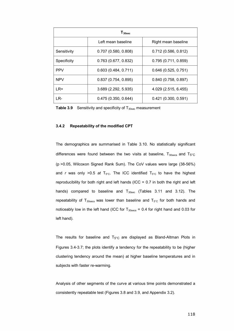
118
T30sec
Left mean baseline Right mean baseline
Sensitivity 0.707 (0.580, 0.808) 0.712 (0.586, 0.812)
Specificity 0.763 (0.677, 0.832) 0.795 (0.711, 0.859)
PPV 0.603 (0.484, 0.711) 0.646 (0.525, 0.751)
NPV 0.837 (0.754, 0.895) 0.840 (0.758, 0.897)
LR+ 3.689 (2.292, 5.935) 4.029 (2.515, 6.455)
LR- 0.475 (0.350, 0.644) 0.421 (0.300, 0.591)
Table 3.9 Sensitivity and specificity of T30sec measurement
3.4.2 Repeatability of the modified CPT
The demographics are summarised in Table 3.10. No statistically significant
differences were found between the two visits at baseline, T30secs and T5°C
(p >0.05, Wilcoxon Signed Rank Sum). The CoV values were large (38-56%)
and r was only >0.5 at T5°C. The ICC identified T5°C to have the highest
reproducibility for both right and left hands (ICC = 0.7 in both the right and left
hands) compared to baseline and T30sec (Tables 3.11 and 3.12). The
repeatability of T30secs was lower than baseline and T5°C for both hands and
noticeably low in the left hand (ICC for T30secs = 0.4 for right hand and 0.03 for
left hand).
The results for baseline and T5°C are displayed as Bland-Altman Plots in
Figures 3.4-3.7; the plots identify a tendency for the repeatability to be (higher
clustering tendency around the mean) at higher baseline temperatures and in
subjects with faster re-warming.
Analysis of other segments of the curve at various time points demonstrated a
consistently repeatable test (Figures 3.8 and 3.9, and Appendix 3.2).
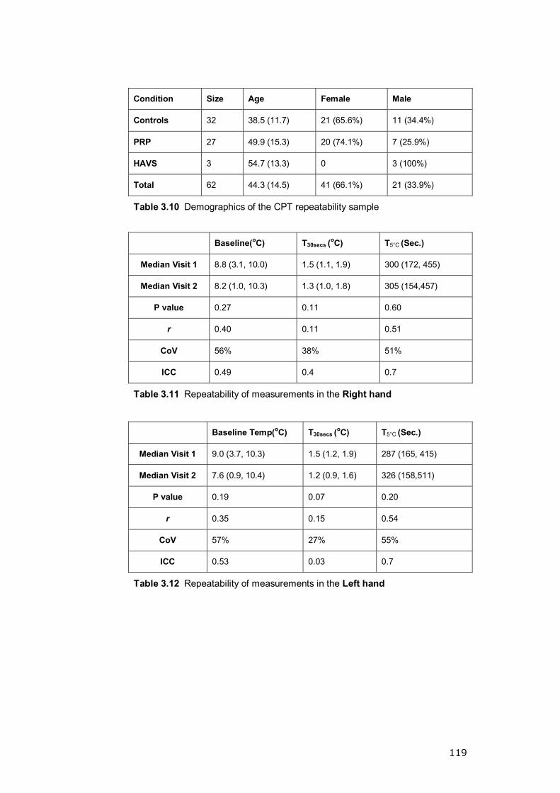
119
Condition Size Age Female Male
Controls 32 38.5 (11.7) 21 (65.6%) 11 (34.4%)
PRP 27 49.9 (15.3) 20 (74.1%) 7 (25.9%)
HAVS 3 54.7 (13.3) 0 3 (100%)
Total 62 44.3 (14.5) 41 (66.1%) 21 (33.9%)
Table 3.10 Demographics of the CPT repeatability sample
Baseline(oC) T30secs (oC) T5°C (Sec.)
Median Visit 1 8.8 (3.1, 10.0) 1.5 (1.1, 1.9) 300 (172, 455)
Median Visit 2 8.2 (1.0, 10.3) 1.3 (1.0, 1.8) 305 (154,457)
P value 0.27 0.11 0.60
r 0.40 0.11 0.51
CoV 56% 38% 51%
ICC 0.49 0.4 0.7
Table 3.11 Repeatability of measurements in the Right hand
Baseline Temp(oC) T30secs (oC) T5°C (Sec.)
Median Visit 1 9.0 (3.7, 10.3) 1.5 (1.2, 1.9) 287 (165, 415)
Median Visit 2 7.6 (0.9, 10.4) 1.2 (0.9, 1.6) 326 (158,511)
P value 0.19 0.07 0.20
r 0.35 0.15 0.54
CoV 57% 27% 55%
ICC 0.53 0.03 0.7
Table 3.12 Repeatability of measurements in the Left hand

120
Bland-Altman Graph for the Baseline temperature in the left hand
-5 0 5 10 15
AVERAGE of Baseline at 1 and Baseline at visit 2
15
10
5
0
-5
-10
-15
Bas
elin
e a
t vis
it 1
- B
ase
line a
t vi
sit
2
Mean
1.4
-1.96 SD
-8.4
+1.96 SD
11.3
Average of the mean baseline between the first and second visits � Left hand
Figure 3.4 Bland-Altman Plots for Baseline temperature (Left hand)
Bland-Altman Graph for Baseline Temperature in the Right hand
-5 0 5 10 15
AVERAGE of Baseline at visit 1 and Baseline at visit 2
20
15
10
5
0
-5
-10
-15
Bas
elin
eat
visi
t 1 -
Bas
elin
e a
t vi
sit
2
Mean
1.4
-1.96 SD
-8.9
+1.96 SD
11.7
Average of the mean baseline between the first and second visits � Right hand
Figure 3.5 Bland-Altman Plots for Baseline temperature (Right hand)
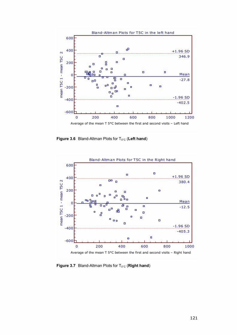
121
Bland-Altman Plots for T5C in the left hand
0 200 400 600 800 1000 1200
AVERAGE of mean T5C 1 and meanT5C 2
600
400
200
0
-200
-400
-600
mea
n T
5C 1
- m
ean T
5C
2
Mean
-27.8
-1.96 SD
-402.5
+1.96 SD
346.9
Average of the mean T 5°C between the first and second visits � Left hand
Figure 3.6 Bland-Altman Plots for T5°C (Left hand)
Bland-Altman Plots for T5C in the Right hand
0 200 400 600 800 1000
AVERAGE of mean T5C 1 and mean T5C 2
600
400
200
0
-200
-400
-600
mea
n T
5C 1
- m
ean T
5C 2
Mean
-12.5
-1.96 SD
-405.3
+1.96 SD
380.4
Average of the mean T 5°C between the first and second visits � Right hand
Figure 3.7 Bland-Altman Plots for T5°C (Right hand)

122
ICC at baseline = 0.5
ICC at T30sec = 0.03
Figure 3.8 The graph represents the mean values (95% CI error bars) at sequential time points during the CPT in the Left hand
ICC at baseline = 0.48
ICC at T30sec = 0.38
Figure 3.9 The graph represents the mean values (95% CI error bars) at
sequential time points during the CPT in the Right hand

123
3.4.3 Repeatability of LDI in the hand
The data of 60 subjects were analysed: the mean age of the sample was
48.51(17.9) years, Height (cm) 170.5(10), Weight (Kg) 78.01(14), mean systolic
BP (mmHg) 127(17) and mean diastolic BP (mmHg) 78(9). Fifty-one (85%)
subjects were right-handed.
The mean values in LDIMax had the highest ICC (ICC = 0.49 for SNP and 0.48
for ACH); those of the LDIMean had lower repeatability (ICC = 0.41 for SNP and
0.42 for ACH) (Tables 3.13-3.16).
When maximum, minimum and mean values for the LDIMax and LDIMean data
sets were compared, a statistically significant difference (p <0.05) was found for
all parameters. The correlation (r) between the two visits was weak (i.e. r< 0.15
for SNP and r = 0.19 -0.35 for ACH) and the CoV was wide more so for the
SNP (37-44%) than the ACH (48-69%). Bland-Altman plots revealed higher
repeatability at lower perfusion values (Figures 3.10 and 3.11).
Parameters at LDI0 were not reproducible for either SNP or ACH (Tables 3.17
and 3.18).
There was observed variation in test repeatability between the three operators
for each of the drugs and with the same operator when comparing the two
iontophoresis drugs (Tables 3.19 and 3.20, Appendices 3.3-3.5).

124
Stag
e Va
riabl
e M
ean(
SD)
95%
CI
Nor
mal
ity
(K-S
Tes
t) W
ilcox
on S
igne
d R
anks
Tes
t r (
P-Va
lue)
C
OV
ICC
Min
imum
1
0.44
(0.2
1)
0.40
, 0.4
8 0.
009
Min
imum
2
0.50
(0.2
4)
0.46
, 0.5
5 0.
19
0.09
0 0.
12 (0
.1)
60.0
%
0.15
Max
imum
1
1.42
(0.5
3)
1.33
, 1.5
2 0.
01
Max
imum
2
1.65
(1.0
0)
1.47
, 1.8
3 0.
06
0.03
0 0.
30 (0
.001
) 48
.3%
0.
23
Mea
n 1
0.82
(0.2
8)
0.77
, 0.8
7
0.20
LDIM
ax
Mea
n 2
0.90
(0.3
3)
0.84
, 0.9
6 0.
06
0.01
6 0.
30 (0
.001
) 40
.5%
0.
49
Tabl
e 3.
13 V
isit
com
paris
on a
t LD
IMax
for S
NP
Stag
e Va
riabl
e M
ean(
SD)
95%
CI
Nor
mal
ity
(K-S
Tes
t) W
ilcox
on S
igne
d R
anks
Tes
t r (
P-Va
lue)
C
OV
ICC
Min
imum
1
0.43
(0.2
2)
0.39
, 0.4
7 <0
.000
1
Min
imum
2
0.52
(0.2
5)
0.47
, 0.5
6 <0
.000
1
0.00
9 0.
13 (0
.15)
76
.2%
0.
19
Max
imum
1
1.49
(0.6
6)
1.37
, 1.6
1 0.
001
Max
imum
2
1.51
(0.7
1)
1.38
, 1.6
4 0.
200
0.66
3 0.
23 (0
.013
) 69
.5%
0.
29
Mea
n 1
0.84
(0.3
1)
0.78
, 0.8
9
<0.0
001
LDIM
ax
Mea
n 2
0.91
(0.3
5)
0.84
, 0.9
7 0.
028
0.03
2 0.
22 (0
.015
) 52
.3%
0.
48
Tabl
e 3.
14 V
isit
com
paris
on a
t LD
IMax
for A
CH
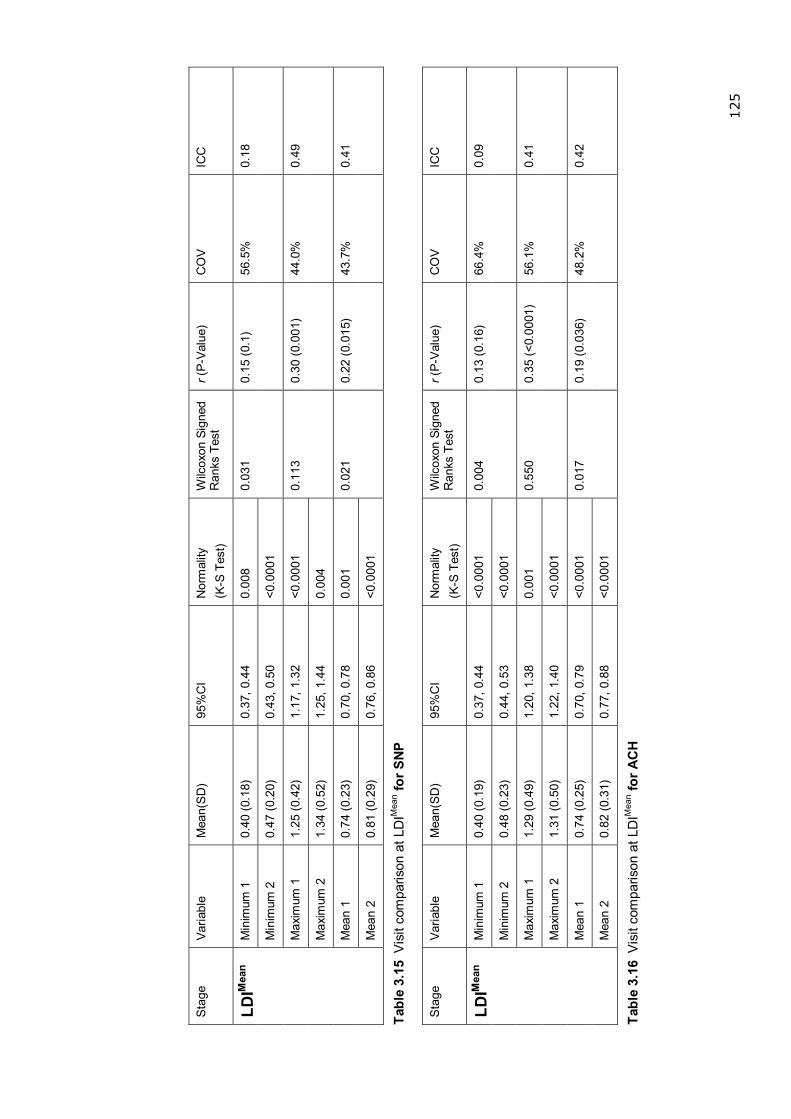
125
Stag
e Va
riabl
e M
ean(
SD)
95%
CI
Nor
mal
ity
(K-S
Tes
t) W
ilcox
on S
igne
d R
anks
Tes
t r (
P-Va
lue)
C
OV
ICC
Min
imum
1
0.40
(0.1
8)
0.37
, 0.4
4
0.00
8
Min
imum
2
0.47
(0.2
0)
0.43
, 0.5
0 <0
.000
1
0.03
1 0.
15 (0
.1)
56.5
%
0.18
Max
imum
1
1.25
(0.4
2)
1.17
, 1.3
2 <0
.000
1
Max
imum
2
1.34
(0.5
2)
1.25
, 1.4
4
0.00
4
0.11
3 0.
30 (0
.001
) 44
.0%
0.
49
Mea
n 1
0.74
(0.2
3)
0.70
, 0.7
8 0.
001
LDIM
ean
Mea
n 2
0.81
(0.2
9)
0.76
, 0.8
6 <0
.000
1
0.02
1 0.
22 (0
.015
) 43
.7%
0.
41
Tabl
e 3.
15 V
isit
com
paris
on a
t LD
IMea
n for S
NP
Stag
e Va
riabl
e M
ean(
SD)
95%
CI
Nor
mal
ity
(K-S
Tes
t) W
ilcox
on S
igne
d R
anks
Tes
t r (
P-Va
lue)
C
OV
ICC
Min
imum
1
0.40
(0.1
9)
0.37
, 0.4
4 <0
.000
1
Min
imum
2
0.48
(0.2
3)
0.44
, 0.5
3 <0
.000
1
0.00
4 0.
13 (0
.16)
66
.4%
0.
09
Max
imum
1
1.29
(0.4
9)
1.20
, 1.3
8 0.
001
Max
imum
2
1.31
(0.5
0)
1.22
, 1.4
0 <0
.000
1
0.55
0 0.
35 (<
0.00
01)
56.1
%
0.41
Mea
n 1
0.74
(0.2
5)
0.70
, 0.7
9 <0
.000
1
LDIM
ean
Mea
n 2
0.82
(0.3
1)
0.77
, 0.8
8 <0
.000
1
0.01
7 0.
19 (0
.036
) 48
.2%
0.
42
Tabl
e 3.
16 V
isit
com
paris
on a
t LD
IMea
n for A
CH

126
PERFUSION MEANS AT IMAGE OF MAXIMUM RESPONSE FOR SNP
0.0 0.5 1.0 1.5 2.0 2.5
AVERAGE of MEAN 1 and MEAN 2
1.5
1.0
0.5
0.0
-0.5
-1.0
-1.5
MEAN1 -
MEAN
2
Mean
-0.08
-1.96 SD
-0.75
+1.96 SD
0.60
Average LDImax of Visit 1 and visit 2 -SNP
MEAN OF MEAN VALUES FOR POST IONTO. SCANS FOR SNP
0.0 0.5 1.0 1.5 2.0
AVERAGE of MEAN 1 and MEAN 2
1.0
0.8
0.6
0.4
0.2
0.0
-0.2
-0.4
-0.6
-0.8
-1.0
MEA
N 1
- M
EAN
2
Mean
-0.07
-1.96 SD
-0.68
+1.96 SD
0.55
Average LDImean of Visit 1 and visit 2 -SNP
Figure 3.10 Bland-Altman plots for SNP

127
PERFUSION MEANS AT IMAGE OF MAXIMUM RESPONSE FOR ACH
0.0 0.5 1.0 1.5 2.0 2.5
AVERAGE of MEAN 1 and MEAN 2
1.5
1.0
0.5
0.0
-0.5
-1.0
-1.5
MEA
N 1
- M
EAN
2
Mean
-0.07
-1.96 SD
-0.81
+1.96 SD
0.68
Average LDImax of Visit 1 and visit 2 -ACH
MEAN OF MEAN VALUES OF POST IONTO. SCANS FOR ACH
0.0 0.5 1.0 1.5 2.0
AVERAGE of MEAN 1 and MEAN 2
1.0
0.5
0.0
-0.5
-1.0
-1.5
MEAN 1
- M
EAN
2
Mean
-0.08
-1.96 SD
-0.73
+1.96 SD
0.58
Average LDImean of Visit 1 and visit 2 -ACH
Figure 3.11 Bland-Altman plots for ACH
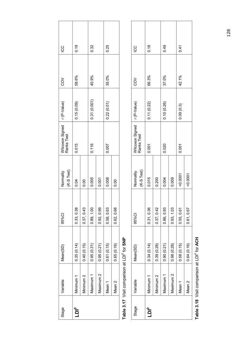
128
Stag
e Va
riabl
e M
ean(
SD)
95%
CI
Nor
mal
ity
(K-S
Tes
t) W
ilcox
on S
igne
d R
anks
Tes
t r (
P-Va
lue)
C
OV
ICC
Min
imum
1
0.35
(0.1
4)
0.33
, 0.3
8 0.
04
Min
imum
2
0.40
(0.1
5)
0.37
, 0.4
3 0.
00
0.01
5 0.
15 (0
.09)
58
.6%
0.
19
Max
imum
1
0.95
(0.3
1)
0.89
, 1.0
0 0.
005
Max
imum
2
0.95
(0.2
1)
0.92
, 0.9
9 0.
001
0.11
6 0.
31 (0
.001
) 40
.9%
0.
32
Mea
n 1
0.61
(0.1
5)
0.58
, 0.6
3 0.
008
LDI0
Mea
n 2
0.65
(0.1
6)
0.62
, 0.6
8 0.
00
0.00
7 0.
22 (0
.01)
35
.0%
0.
25
Tabl
e 3.
17 V
isit
com
paris
on a
t LD
I0 for S
NP
Sta
ge
Varia
ble
Mea
n(SD
) 95
%C
I N
orm
ality
(K
-S T
est)
Wilc
oxon
Sig
ned
Ran
ks T
est
r (P-
Valu
e)
CO
V IC
C
Min
imum
1
0.34
(0.1
4)
0.31
, 0.3
6 0.
015
Min
imum
2
0.39
(0.2
8)
0.37
, 0.4
2 0.
200
0.00
1 0.
11 (0
.22)
66
.3%
0.
18
Max
imum
1
0.90
(0.2
1)
0.86
, 0.9
3 0.
004
Max
imum
2
0.98
(0.2
8)
0.93
, 1.0
3 0.
009
0.02
0 0.
10 (0
.26)
37
.0%
0.
49
Mea
n 1
0.58
(0.1
5)
0.55
, 0.6
1 <0
.000
1
LDI0
Mea
n 2
0.64
(0.1
6)
0.61
, 0.6
7 <0
.000
1
0.00
1 0.
09 (0
.3)
42.1
%
0.41
Tabl
e 3.
18 V
isit
com
paris
on a
t LD
I0 for A
CH
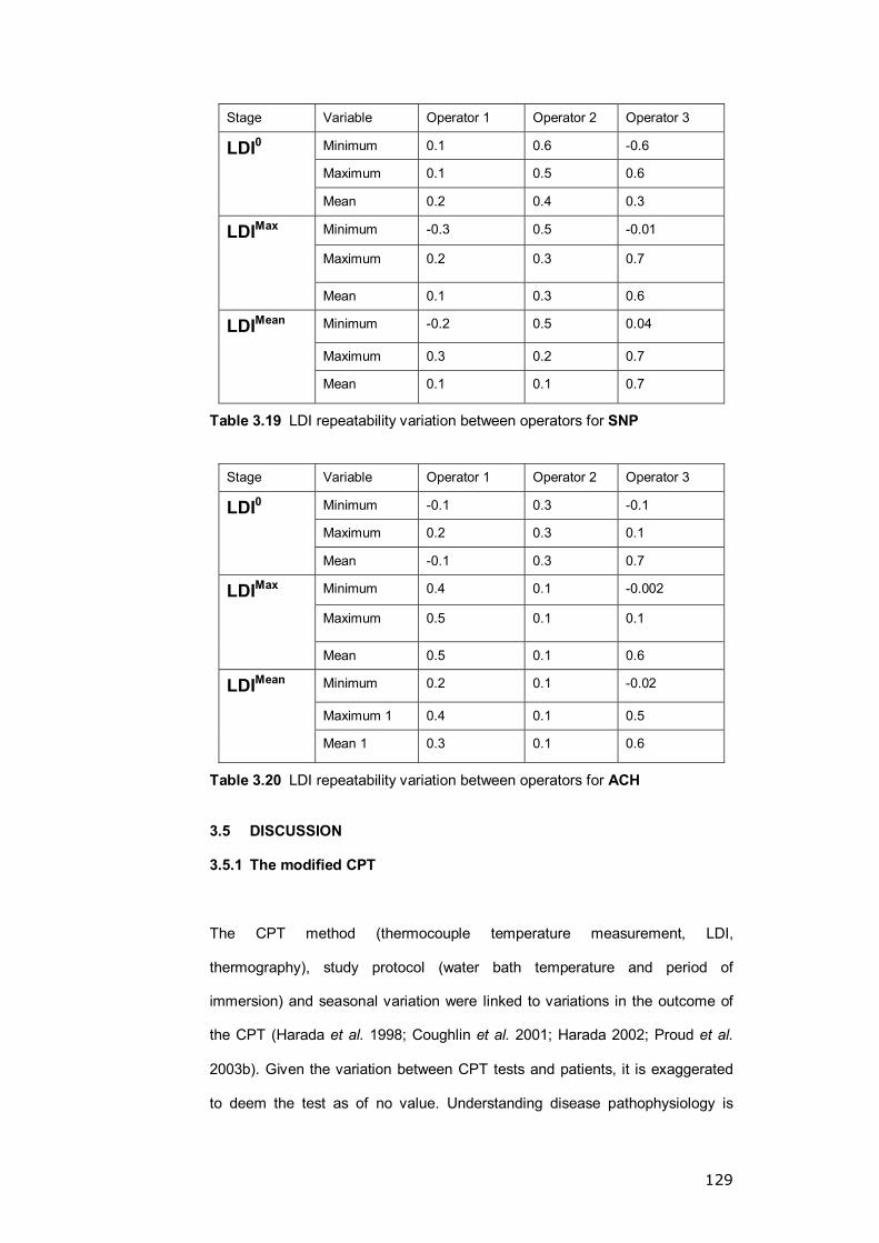
129
Stage Variable Operator 1 Operator 2 Operator 3
Minimum 0.1 0.6 -0.6
Maximum 0.1 0.5 0.6
LDI0
Mean 0.2 0.4 0.3
Minimum -0.3 0.5 -0.01
Maximum 0.2 0.3 0.7
LDIMax
Mean 0.1 0.3 0.6
Minimum -0.2 0.5 0.04
Maximum 0.3 0.2 0.7
LDIMean
Mean 0.1 0.1 0.7
Table 3.19 LDI repeatability variation between operators for SNP
Stage Variable Operator 1 Operator 2 Operator 3
Minimum -0.1 0.3 -0.1
Maximum 0.2 0.3 0.1
LDI0
Mean -0.1 0.3 0.7
Minimum 0.4 0.1 -0.002
Maximum 0.5 0.1 0.1
LDIMax
Mean 0.5 0.1 0.6
Minimum 0.2 0.1 -0.02
Maximum 1 0.4 0.1 0.5
LDIMean
Mean 1 0.3 0.1 0.6
Table 3.20 LDI repeatability variation between operators for ACH
3.5 DISCUSSION
3.5.1 The modified CPT
The CPT method (thermocouple temperature measurement, LDI,
thermography), study protocol (water bath temperature and period of
immersion) and seasonal variation were linked to variations in the outcome of
the CPT (Harada et al. 1998; Coughlin et al. 2001; Harada 2002; Proud et al.
2003b). Given the variation between CPT tests and patients, it is exaggerated
to deem the test as of no value. Understanding disease pathophysiology is
130
important when designing a diagnostic test for it (Stoyneva et al. 2003; Herrick
2005). Knowledge gaps still exist in our understanding of changes in RP and
HAVS, which makes it difficult to develop a validated test.
In 2000, Coughlin et al. reported a good sensitivity, specificity, and positive and
negative predictive values (100%, 88%, 95% and 100%, respectively at 8
minutes post-cold challenge). Their CPT used thermal imaging and a cooling
challenge of 5°C for 1 minute in 21 HAVS patients and 10 controls (Coughlin et
al. 2001). A wide range of sensitivities and specificities (sensitivity 22-73% and
specificity 43-100%) has been reported by other investigators (Hack et al. 1986;
Bovenzi 1987; Gautherie 1995). The modified CPT applied an adequate
challenge (water temperature = 12°C, hands must cool down to 15°C) whilst
avoiding the risk of reactive hyperaemia (Coughlin et al. 2001). Frequent (every
6 seconds) temperature measurements allowed accurate curve assessment at
various points.
The modified CPT identified a significantly lower baseline temperature amongst
the HAVS and PRP groups. This could be a useful screening tool for RP. The
test suggests normality when baseline is >7.5°C above the ambient
temperature; this excluded PRP and HAVS with a Stockholm Workshop
Vascular score of V2 or more. The lower baseline skin blood flow secondary to
a high sympathetic activity in the RP group is a possible cause for the low
baseline temperature and smaller response to vaso-constrictive stimuli
(Wollersheim et al. 1991). Cherkas et al. reported a similar observation
suggesting baseline temperature can help identify RP sufferers from the general
population with reservations on its low predictive power and the inability of the
test to stage the disease (Cherkas et al. 2003).
The modified CPT can help differentiate between PRP and HAVS. A T30sec
>2.2°C is indicative of HAVS and, combined with a low baseline temperature,

131
suggests HAVS as the diagnosis; this was not reported before. The HAVS�
response at T30sec is probably due to disturbed local vascular response to the
cold secondary to an underlying aberrant autonomic reflex. The pattern of re-
warming in HAVS after T30sec followed a normative course compared to PRP.
PRP had a slower re-warming time (T5°C >330 seconds), which in this study
helped to differentiate them from HAVS. This pattern of re-warming has not
been reported before.
3.5.2 The repeatability of methods
There are inherent limitations in the consistency of repeated measurements of
physiological variables. This applies to skin temperature and laser Doppler
microcirculation measurements (Bartelink et al. 1993; Bircher et al. 1994;
Beevers et al. 2001; Williams et al. 2004).
3.5.2.1 Repeatability of the modified CPT
Using ICC allowed the amalgamation of the three groups into one large sample
to reflect the repeatability of the test without making specific conclusions
relating to any particular group. ICC is designed to test the repeatability based
on the variation between the two visits within the same subject (Fleiss et al.
1973; Howell 2002). This method of analysis allowed the use of the test�s
repeatability outcome as a guide in choosing the parameters helpful in the
assessment of patients with hand injuries and CTS for which the repeatability of
the test has not been previously determined. The analysis of the three
parameters used in the differentiation between the groups (baseline, T30sec and
T5°C) revealed T5°C to be the most repeatable curve segment (ICC = 0.7 for both
hands). Baseline measurement had moderate repeatability (ICC = 0.49 on the
left and 0.53 on the right); henceforth, baseline and T5°C were used in
subsequent studies.
132
CPT repeatability has been reported as acceptable in controls. This was
demonstrated using a 15°C challenge for 5 minutes (Griffin et al. 1998), a 5-7°C
challenge for 15 minutes (Hack et al. 1986) and with a 16°C challenge for 5
minutes (Bartelink et al. 1993). Bartelink et al. also reported a good repeatability
in the RP group (Bartelink et al. 1993). In the standardisation report for CPT, a
recommendation for a repeat CPT test if the symptoms and CPT findings did
not match was made based on larger LOA in the HAVS group. This variation in
the repeatability between the two groups (control and HAVS) was thought to be
due to a larger inconsistency in the response of the central sympathetic nervous
system to the cold provocation in the HAVS group (Griffin et al. 1998).
Apart from some similarities in the clinical history and presentation of RP and
HAVS, the evidence to support a pathophysiological link between these
conditions is lacking (Pyykko et al. 1987; Noel 2000; Block et al. 2001).
Conclusions on the CPT repeatability in HAVS should not be made comparable
to these of RP. The evidence for CPT repeatability in HAVS is lacking. The
small sample size, lack of objective diagnostic gold standard and weak staging
tools make the available evidence questionable. Due to the small number of
HAVS in our repeatability study, no conclusions could be drawn regarding the
CPT repeatability in this group.
3.5.2.2 Repeatability of LDI in the hand
Early techniques utilising laser Doppler technology had poor repeatability
(Schabauer et al. 1994). The spatial variation within the microvascular network
is partly to blame (Kubli et al. 2000; Fullerton et al. 2002). Additionally, the
mechanical contact of the instruments modified the vascular response leading
to significant variations in repeat measurements (Obeid et al. 1990; Kernick et
al. 2000; Fullerton et al. 2002). LDI overcame these limitations (Section 1.5).
133
LDI repeatability in the forearm (Morris et al. 1996; Kubli et al. 2000) and gaiter
area (Klonizakis et al. 2003) has been explored. A good day-to-day repeatability
was reported for ACH and SNP in the forearm (CoV <10%, 10-20%, 23%, 31%
(Morris et al. 1996; Kubli et al. 2000)) and the gaiter area (10-20%, 10-20%
(Kolonizakis 2004)). Our study revealed a wider CoV for both SNP and ACH
(40-43% and 48-52% respectively) despite using identical LDI methodology to
that adopted in previous house studies (Klonizakis et al. 2003; Klonizakis et al.
2006). The ICC for the test was not high (0.41-0.49) confirming this finding.
Our findings might be explained by a possible inconsistency in the methodology
between the various operators (Appendices 3.3-3.5), difficulty in accurately
identifying the same areas of interest (AOI) on repeat testing, participant in
compliance with alcohol and cigarette abstinence and the lack of noise levels
control. Marking the AOI for two weeks was not practical; the use of anatomical
land marks was not always feasible while trying to avoid the variable superficial
venous anatomy and the large size of the iontophoresis electrode in relation to
the AOI was also a difficulty. Acceptable noise levels were not defined in the
literature; during the study, a conversation was held to answer queries from
some of the subjects and the effect of this on the outcome cannot be
determined. The position of the AOI in the hand might have played a bigger role
than expected; perfusion values for distal forearm measurements were reported
as higher than proximal forearm measurements using LDF in normal healthy
volunteers (de Boer et al. 1989; Bircher et al. 1994).
The list of factors influencing the results of the LDI test is long (Section 1.5.4).
This study adds to this list the influence of the operator. We were able to
demonstrate repeatability variations between the operators and within different
vasoactive challenges. The protocol was standardised but the approach to the
AOI selection and the skin handling was not. Perfusion values for distal forearm
measurements were reported to be higher than proximal forearm
134
measurements using LDF in normal healthy volunteers (de Boer et al. 1989;
Bircher et al. 1994). This prompts questions on whether the responsiveness of
the microcirculatory bed in the upper limb is higher distally than proximally
requiring a more specific LDI protocol taking into account the above-mentioned
limitations.
3.6 CONCLUSION
The use of CPT and LDI is influenced by environmental, technical and biological
variations. LDI and CPT in the hand have limited value in the assessment of
individual cases due to their moderate repeatability but are useful tools in
detecting group trends. The modified CPT can differentiate between PRP,
HAVS and healthy volunteers groups. The LDI repeatability in the hand needs
to be revisited with revised methodology and using smaller iontophoresis
electrodes.

135
CHAPTER 4: THE EFFECT OF CARPAL TUNNEL SYNDROME ON
THE MICROVASCULAR RESPONSE TO COLD IN THE HAND

136
4.1 INTRODUCTION
Autonomic dysfunction associated with CTS has been reported as early as
1957 (Garland et al. 1957). Phalen suggested that the median nerve carries
autonomic nerve fibres which are affected in CTS resulting in vasomotor control
disturbances (Phalen 1966).
The prevalence of autonomic symptoms in CTS has gradually increased with
heightened awareness and better documentation. In a retrospective review of
2,800 CTS patients, only 28 reported autonomic symptoms (Linscheid et al.
1967). More recently, the prevalence has been reported to be as high as 55-
60% (Chung et al. 1999; Verghese et al. 2000) and the symptoms seem to
respond favourably to CTR (Chung et al. 1999; Chung et al. 2000), but the high
prevalence has not yet helped unveil the underlying pathology.
4.2 AIMS
Firstly, to investigate the microvascular response in the hands of both normal
volunteers and patients with CTS to a cold challenge and, secondly, to
investigate the effect of CTR on the CPT in CTS patients.
4.3 METHODS
After obtaining the required ethical approval, subject recruitment and
demographic data collection were carried out as outlined in Chapter 2. A data
collection sheet was used (Appendix 5.1). Subjects were asked to complete a
vibration white finger (VWF) screening questionnaire (Appendix 4.5).

137
We recruited two groups:
1- CTS patients awaiting surgery
2- Normal controls (NC)
Inclusion criteria
• Male or female aged > 16 years
• No history of vibration exposure or history of RP prior to the development of
CTS
• CTS patients awaiting surgery
or
• Normal control (NC) subject with no history of hand injury or CTS
• Subjects who give written informed consent after reading the information
sheet for volunteers
Exclusion criteria
• Smoked within the last 6 hours
• Treatment with vasodilating medication including but not restricted to α-
blockers, β-blockers, calcium channel antagonists, ace inhibitors,
angiotensin II blockers, nitrates
• Intake of alcohol or caffeine-containing drinks within the last 12 hours
• Central or peripheral cyanosis on arrival at the examination
• Type I or Type II DM
• Subjects failing to maintain their hands in the water until the end of the test
All subjects underwent LDI of both hands at baseline followed by CPT (Sections
2.10 and 2.11). During the CPT re-warming phase, LDI was repeated on the
affected hand in CTS patients and on a randomly selected side in the NC based
on a randomisation table provided by the Trust statistician.

138
Subjects with CTS were asked to re-attend for a repeat test 5-7 months post-
CTR. For descriptive purposes the following definitions were used; left hand
control group (NCLeft), right hand control group (NCRight), CTS affected hand
(CTSAffected), CTS unaffected hand (CTSC).
4.4 SAMPLE SIZE AND DATA ANALYSIS
It was estimated that a sample size of 60 subjects in each group would allow
the detection of 10% difference between the groups with a power of 80% and a
beta error <0.05. During analysis, each hand was treated as a data unit
(Garland et al. 1957).
In the control group (NC), the data from NCLeft and NCRight was compared to
allow the amalgamation of the sample into one NC group. Unpaired data
analysis was used to compare the groups, except when the effect of CTR was
studied where paired data analysis methods were utilised. The data are given
as mean (SD) unless otherwise stated. Statistical advice was provided by the
Trust statistician. Data analysis was conducted as outlined in Section 2.8.
4.5 RESULTS
Recruitment was complete with 60 subjects in each group. To date, 40 CTS
subjects completed a 5-7 months post-surgery re-test. Recruitment history is
outlined in Appendix 4.1. Demographics are summarised in Table 4.1.
The VWF screening questionnaire identified four subjects with white finger
discoloration on exposure to cold in the CTS group and two in the control group.
The questionnaire lacked the ability to identify patients with cold intolerance
symptoms (Section 1.2.2) and was therefore not used in further analysis.
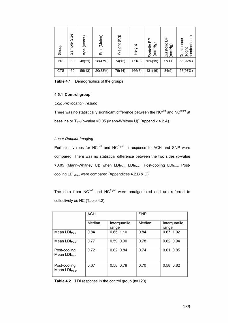
139
Gro
up
Sam
ple
Siz
e
Age
(yea
rs)
Sex
(Mal
es)
Wei
ght (
Kg)
Hei
ght
Sys
tolic
BP
(m
mH
g)
Dia
stol
ic B
P
(mm
Hg)
Dom
inan
ce
(Rig
ht
hand
edne
ss)
NC 60 48(21) 28(47%) 74(12) 171(8) 126(19) 77(11) 55(92%)
CTS 60 56(13) 20(33%) 79(14) 166(8) 131(16) 84(9) 58(97%)
Table 4.1 Demographics of the groups
4.5.1 Control group
Cold Provocation Testing
There was no statistically significant difference between the NCLeft and NCRight at
baseline or T5ûC (p-value >0.05 (Mann-Whitney U)) (Appendix 4.2.A).
Laser Doppler Imaging
Perfusion values for NCLeft and NCRight in response to ACH and SNP were
compared. There was no statistical difference between the two sides (p-value
>0.05 (Mann-Whitney U)) when LDIMax, LDIMean, Post-cooling LDIMax, Post-
cooling LDIMean were compared (Appendices 4.2.B & C).
The data from NCLeft and NCRight were amalgamated and are referred to
collectively as NC (Table 4.2).
ACH SNP
Median Interquartile range
Median Interquartile range
Mean LDIMax
0.84 0.65, 1.10 0.84 0.67, 1.02
Mean LDIMean
0.77 0.59, 0.90 0.78 0.62, 0.94
Post-cooling Mean LDIMax
0.72 0.62, 0.84 0.74 0.61, 0.85
Post-cooling Mean LDIMean
0.67 0.58, 0.78 0.70 0.58, 0.82
Table 4.2 LDI response in the control group (n=120)
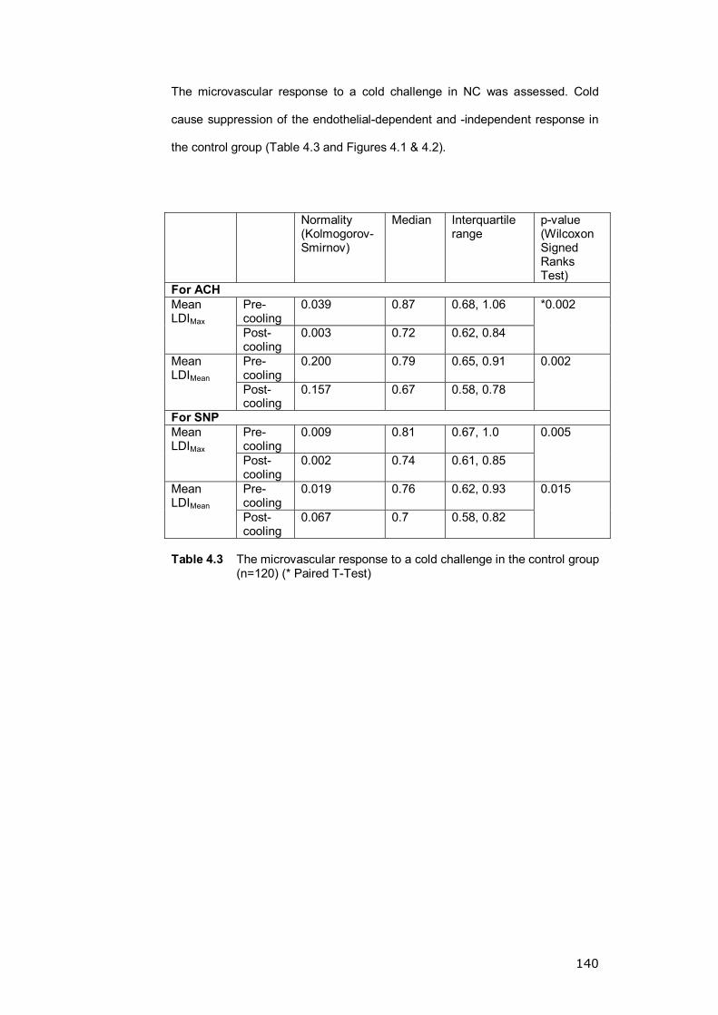
140
The microvascular response to a cold challenge in NC was assessed. Cold
cause suppression of the endothelial-dependent and -independent response in
the control group (Table 4.3 and Figures 4.1 & 4.2).
Normality
(Kolmogorov-Smirnov)
Median Interquartile range
p-value (Wilcoxon Signed Ranks Test)
For ACH Pre- cooling
0.039 0.87 0.68, 1.06
Mean LDIMax Post-
cooling 0.003 0.72 0.62, 0.84
*0.002
Pre- cooling
0.200 0.79 0.65, 0.91
Mean LDIMean Post-
cooling 0.157 0.67 0.58, 0.78
0.002
For SNP Pre- cooling
0.009 0.81 0.67, 1.0
Mean LDIMax Post-
cooling 0.002 0.74 0.61, 0.85
0.005
Pre- cooling
0.019 0.76 0.62, 0.93 Mean LDIMean Post-
cooling 0.067 0.7 0.58, 0.82
0.015
Table 4.3 The microvascular response to a cold challenge in the control group
(n=120) (* Paired T-Test)

141
Test Stage
Post Cooling TestPre Cooling Test
Mea
n at
LD
IMax
1.8
1.6
1.4
1.2
1.0
.8
.6
.4
.2
Drug
ACH
SNP
Figure 4.1 LDImax in NC. Change post-cooling for ACH and SNP (p-value = 0.002 and 0.005, respectively)
Test Stage
Post Cooling TestPre Cooling Test
Mea
n at
LD
IMea
n
1.8
1.6
1.4
1.2
1.0
.8
.6
.4
.2
Drug
ACH
SNP
Figure 4.2 LDIMean in NC. Change post-cooling for ACH and SNP (p-value = 0.002 and 0.15, respectively)
Mean at LDIMax
Mean at LDIMean

142
The CPT and LDI findings for NC and CTSC were compared and no significant
difference observed (p-value >0.05 (Mann-Whitney U)) (Appendix 4.3). CTSC
was used as a control against CTSAffected.
4.5.2 The effect of CTS on the microvascular and rewarming response
of the hand
The CPT and LDI data for CTSAffected and CTSC were compared and no
significant difference observed (Tables 4.4 A, B, C).
Normality
(Shapiro-Wilk)
Median Interquartile range
p-value (Mann-Whitney U)
CTSC 0.2 7.7 5.2, 9.3 Baseline
CTSAffected <0.0001 7.7 4.7, 9.7
0.97
CTSC 0.2 330 206, 512 T5oC
CTSAffected 0.2 372 191, 591
0.27*
Table 4.4.A CPT comparison between CTSAffected and CTSC (* T-Test)
Normality (Shapiro-Wilk)
Median Interquartile range
p-value (Mann-Whitney U)
CTSC 0.02 0.76 0.59, 0.98 Mean LDIMax CTSAffected 0.002 0.79 0.65, 0.99
0.4
CTSC 0.2 0.68 0.54, 0.92 Mean LDIMean CTSAffected <0.0001 0.73 0.62, 0.89
0.3
CTSC 0.002 0.68 0.58, 0.8
Post- cooling Mean LDIMax CTSAffected <0.0001 0.73 0.62, 0.84
0.3
CTSC 0.001 0.63 0.54, 0.76
Post- cooling Mean LDIMean CTSAffected 0.004 0.68 0.59, 0.78
0.3
Table 4.4.B LDI response to ACH between CTSAffected and CTSC
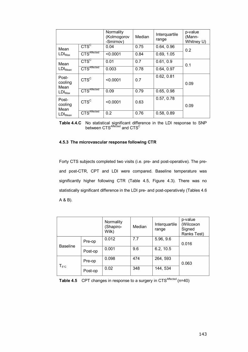
143
Normality (Kolmogorov-Smirnov)
Median Interquartile range
p-value (Mann-Whitney U)
CTSC 0.04 0.75 0.64, 0.96 Mean LDIMax CTSAffected <0.0001 0.84 0.69, 1.05
0.2
CTSC 0.01 0.7 0.61, 0.9 Mean LDIMean CTSAffected 0.003 0.78 0.64, 0.97
0.1
CTSC <0.0001 0.7 0.62, 0.81
Post- cooling Mean LDIMax CTSAffected 0.09 0.79 0.65, 0.98
0.09
CTSC <0.0001 0.63 0.57, 0.78
Post- cooling Mean LDIMean CTSAffected 0.2 0.76 0.58, 0.89
0.09
Table 4.4.C No statistical significant difference in the LDI response to SNP between CTSAffected and CTSC
4.5.3 The microvascular response following CTR
Forty CTS subjects completed two visits (i.e. pre- and post-operative). The pre-
and post-CTR, CPT and LDI were compared. Baseline temperature was
significantly higher following CTR (Table 4.5, Figure 4.3). There was no
statistically significant difference in the LDI pre- and post-operatively (Tables 4.6
A & B).
Normality (Shapiro-Wilk)
Median Interquartile range
p-value (Wilcoxon Signed Ranks Test)
Pre-op 0.012 7.7 5.96, 9.6
Baseline Post-op 0.001 9.6 6.2, 10.5
0.016
Pre-op 0.098 474 264, 593
T5°C Post-op 0.02 348 144, 534
0.063
Table 4.5 CPT changes in response to a surgery in CTSAffected (n=40)

144
Post Op. baselinePre op. baseline
Visit Number
10.00
5.00
0.00
Base
line
Tem
pera
ture
(C)
Figure 4.3 The change in the baseline temperature in the CTS-affected hand
post-surgery (n=40)
Normality (Shapiro-Wilk)
Median Interquartile range
p-value (Wilcoxon Signed Ranks Test)
Pre-op 0.001 0.79 0.64, 1.1 Mean LDIMax Post-op 0.005 0.82 0.66, 1.18
0.9
Pre-op 0.001 0.73 0.62, 0.94 Mean LDIMean Post-op 0.007 0.75 0.59, 1.09
0.7
Pre-op 0.001 0.73 0.6, 0.84 Post- cooling Mean LDIMax
Post-op 0.001 0.73 0.65, 0.86 0.7
Pre-op 0.001 0.70 0.56, 0.77 Post- cooling Mean LDIMean
Post-op <0.0001 0.64 0.58, 0.78 0.8
Table 4.6.A Pre- and post-CTR perfusion data (ACH) (n=40)
p-value=0.016
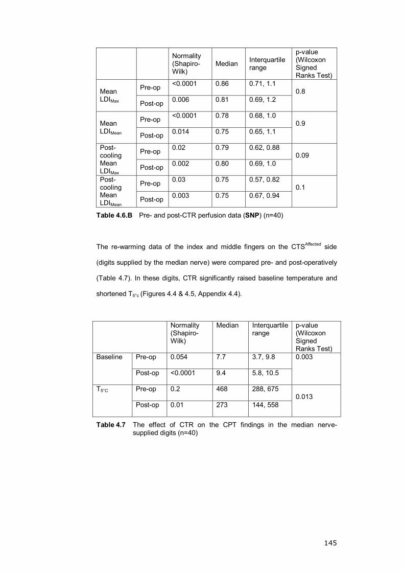
145
Normality (Shapiro-Wilk)
Median Interquartile range
p-value (Wilcoxon Signed Ranks Test)
Pre-op <0.0001 0.86 0.71, 1.1 Mean LDIMax Post-op 0.006 0.81 0.69, 1.2
0.8
Pre-op <0.0001 0.78 0.68, 1.0 Mean LDIMean Post-op 0.014 0.75 0.65, 1.1
0.9
Pre-op 0.02 0.79 0.62, 0.88 Post- cooling Mean LDIMax
Post-op 0.002 0.80 0.69, 1.0 0.09
Pre-op 0.03 0.75 0.57, 0.82 Post- cooling Mean LDIMean Post-op 0.003 0.75 0.67, 0.94
0.1
Table 4.6.B Pre- and post-CTR perfusion data (SNP) (n=40)
The re-warming data of the index and middle fingers on the CTSAffected side
(digits supplied by the median nerve) were compared pre- and post-operatively
(Table 4.7). In these digits, CTR significantly raised baseline temperature and
shortened T5°c (Figures 4.4 & 4.5, Appendix 4.4).
Normality (Shapiro-Wilk)
Median Interquartile range
p-value (Wilcoxon Signed Ranks Test)
Pre-op 0.054 7.7 3.7, 9.8 Baseline
Post-op <0.0001 9.4 5.8, 10.5
0.003
Pre-op 0.2 468 288, 675 T5°C
Post-op 0.01 273 144, 558 0.013
Table 4.7 The effect of CTR on the CPT findings in the median nerve-supplied digits (n=40)
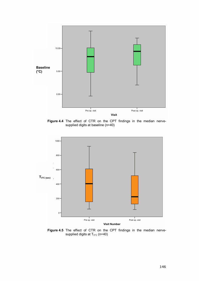
146
Post op. visitPre op. visit
Visit
10.00
5.00
0.00
Bas
elin
e Te
mpe
ratu
re (C
)
Figure 4.4 The effect of CTR on the CPT findings in the median nerve-
supplied digits at baseline (n=40)
Post op. visitPre op. visit
Visit Number
1000
800
600
400
200
0
time
to re
gain
5C
(sec
)
Figure 4.5 The effect of CTR on the CPT findings in the median nerve-
supplied digits at T5°C (n=40)
T5°C (sec)
Baseline (°C)

147
4.6 DISCUSSION
This study identified a significant rise in the digital baseline temperature
(>1.5°C) 5-7 months post-CTR, and a faster T5°C post-CTR particularly in the
median nerve-supplied digits. The study identified the effect of cold on
suppressing the response of both the endothelial-dependent and -independent
mechanisms to stimulation in the control group.
Following a series of publications documenting the autonomic symptoms
amongst CTS patients (Linscheid et al. 1967; Pal et al. 1996; Chung et al. 1999;
Chung et al. 2000; Verghese et al. 2000), the American Academy of Neurology
included dry skin, swelling and colour changes as part of the CTS picture (AAN
1993). Some authors considered this presentation a variant of RP (Linscheid et
al. 1967). Gradually the prevalence of such symptoms increased up to 60%
amongst CTS suffers.
A rise in the baseline temperature post-CTR is not a novel finding. Despite the
small sample size, unclear methodology and the lack of statistical significance,
Chung et al. evaluated the improvement of the autonomic (RP-like) symptoms
subjectively and objectively (using photoplethysmography following CPT) in a
group of CTS suffers pre- and post-operatively; they reported a 78%
improvement in 18 patients post-CTR (all bilateral, and three out of seven
unilateral CTRs improved) with a mean recovery time of 4 months (Chung et al.
1999; Chung et al. 2000). The faster T5°C post-CTR is a novel finding in our
study and once again recovery of the vasomotor sympathetic fibres to the
fingers carried by the median nerve might be the cause.
Endothelial function is mediated mainly by Nitrous Oxide (NO) with variable
contribution from substances such as prostaglandins and an endothelial-derived
hyperpolarisation factor (Furchgott et al. 1980; Vallance et al. 1989; Garland et

148
al. 1995; Kvandal et al. 2003). Whereas SNP acts directly on the smooth
muscle cells (Rapoport et al. 1983; Kvandal et al. 2003), ACH-induced
vasodilatation depends on an intact vascular endothelium to stimulating NO
synthesis in the endothelial cells. Therefore, endothelial function can be
evaluated by comparing the ACH- and SNP-induced vasodilatation (Griendling
et al. 1996). Our study clearly identified the effect of cold on suppressing the
endothelial function in normal controls. The effect of temperature on
microvascular perfusion has been reported previously (Bircher et al. 1994;
Fullerton et al. 2002), resulting in a lower perfusion response to an endothelial-
dependent stimulus (i.e. ACH).
Using infrared telethermography, a block-induced paralytic vasodilatation was
studied in 47 subjects (15 median blocks, 15 ulnar blocks and 17 radial blocks).
The authors concluded that the vasomotor supply of the skin of the hand
followed the sensory supply territory (Campero et al. 1993). These findings
might explain why CPT measured vasomotor changes post-surgery (measures
the temperature of the terminal phalanx), while LDI failed to detect similar
changes on the dorsum of the hand. This area receives its sensory supply from
the ulnar and radial nerves, not the median nerve.
There are some shortcomings in the LDI studies. To deliver an iontophoresis
challenge to the median nerve distribution region, small, easy to secure digital
chambers are required. Such chambers were not available and scanning the
back of the hand was a compromise. There is no evidence in the literature to
suggest that local neural injury causes a systemic response when cold-related
symptoms are in question (Section 1.2.3). Indirectly, these findings suggest that
changes in the microvascular response post-CTR is a local phenomenon.
The use of a cold intolerance assessment tool could have strengthened this
study. A vibration white finger screening questionnaire (Appendix 4.5) was used

149
but was not useful in screening cold intolerance. We introduced the CISS
questionnaire (McCabe et al. 1991; Ruijs et al. 2006) later on in the study; this
was sent by mail to the participants but the replies were incomplete and not
available for analysis. The use of the CISS questionnaire should allow
stratification of the subjects into subgroups which can be compared.
4.7 CONCLUSION
CTR significantly raises the baseline temperature and shortens the re-warming
time in the median nerve-supplied digits. Cold blunts the endothelial response
to direct and indirect stimulation. It would be interesting to investigate the effect
of CTR on other cold-related conditions like RP.

150
CHAPTER 5: THE EFFECT OF HAND INJURY ON THE
MICROVASCULAR RESPONSE IN THE HAND
151
5.1 INTRODUCTION
Cold-related symptoms in the injured hand are not uncommon with 50-100%
prevalence. The pathophysiology is unclear but a neural cause is favoured;
however, a direct correlation between the severity of symptoms and the extent
of the neural injury is lacking (Section 1.2.4) (Engkvist et al. 1985; Kay 1985;
Freedman et al. 1995).
Small-sized retrospective studies reported a lower digital baseline temperature
in the injured hand compared to controls (Engkvist et al. 1985) and a lower
pulse pressure (<75% of the unaffected contralateral digit), particularly in the
severely affected (Gelberman et al. 1978).
5.2 AIMS
To investigate the CPT and perfusion response in the injured hand compared to
normal controls.
5.3 METHODS
A data collection sheet was used to record demographical data (Appendix 5.1).
Subjects were required to complete a vibration white finger (VWF) screening
questionnaire (Appendix 4.5).
Two groups were recruited:
Group 1: Patients sustaining a hand injury (HI) in the previous three months
to five years
Group 2: Normal control (NC) volunteers
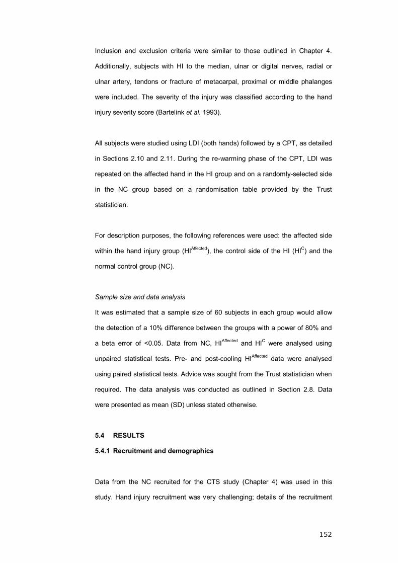
152
Inclusion and exclusion criteria were similar to those outlined in Chapter 4.
Additionally, subjects with HI to the median, ulnar or digital nerves, radial or
ulnar artery, tendons or fracture of metacarpal, proximal or middle phalanges
were included. The severity of the injury was classified according to the hand
injury severity score (Bartelink et al. 1993).
All subjects were studied using LDI (both hands) followed by a CPT, as detailed
in Sections 2.10 and 2.11. During the re-warming phase of the CPT, LDI was
repeated on the affected hand in the HI group and on a randomly-selected side
in the NC group based on a randomisation table provided by the Trust
statistician.
For description purposes, the following references were used: the affected side
within the hand injury group (HIAffected), the control side of the HI (HIC) and the
normal control group (NC).
Sample size and data analysis
It was estimated that a sample size of 60 subjects in each group would allow
the detection of a 10% difference between the groups with a power of 80% and
a beta error of <0.05. Data from NC, HIAffected and HIC were analysed using
unpaired statistical tests. Pre- and post-cooling HIAffected data were analysed
using paired statistical tests. Advice was sought from the Trust statistician when
required. The data analysis was conducted as outlined in Section 2.8. Data
were presented as mean (SD) unless stated otherwise.
5.4 RESULTS
5.4.1 Recruitment and demographics
Data from the NC recruited for the CTS study (Chapter 4) was used in this
study. Hand injury recruitment was very challenging; details of the recruitment
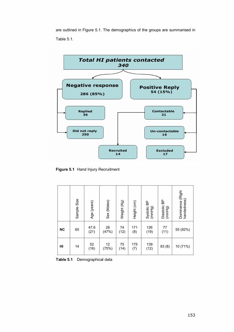
153
are outlined in Figure 5.1. The demographics of the groups are summarised in
Table 5.1.
Total HI patients contacted 340
Replied36
Did not reply 250
Negative response
286 (85%)
Contactable 21
Positive Reply 54 (15%)
Excluded 17
Un-contactable16
Recruited14
Figure 5.1 Hand Injury Recruitment
Sam
ple
Size
Age
(yea
rs)
Sex
(Mal
es)
Wei
ght (
Kg)
Hei
ght (
cm)
Syst
olic
BP
(mm
Hg)
Dia
stol
ic B
P (m
mH
g)
Dom
inan
ce (R
ight
ha
nded
ness
)
NC 60 47.6 (21)
28 (47%)
74 (12)
171 (8)
126 (19)
77 (11) 55 (92%)
HI 14 52 (16)
12 (75%)
75 (14)
175 (7)
139 (12) 83 (8) 10 (71%)
Table 5.1 Demographical data
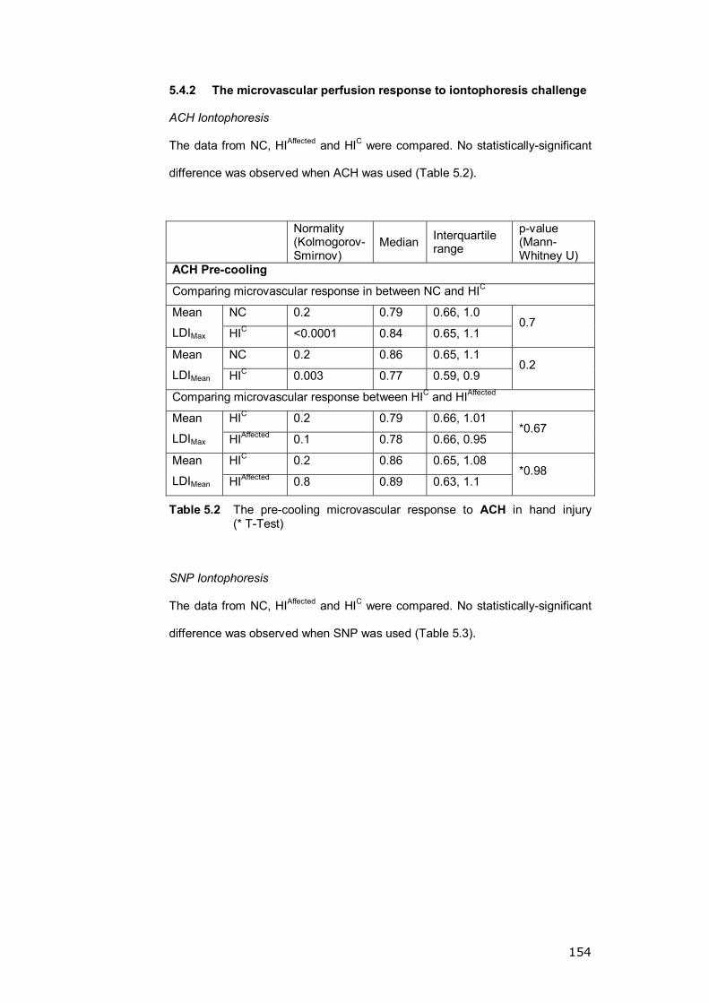
154
5.4.2 The microvascular perfusion response to iontophoresis challenge
ACH Iontophoresis
The data from NC, HIAffected and HIC were compared. No statistically-significant
difference was observed when ACH was used (Table 5.2).
Normality (Kolmogorov-Smirnov)
Median Interquartile range
p-value (Mann-Whitney U)
ACH Pre-cooling
Comparing microvascular response in between NC and HIC
NC 0.2 0.79 0.66, 1.0 Mean
LDIMax HIC <0.0001 0.84 0.65, 1.1 0.7
NC 0.2 0.86 0.65, 1.1 Mean
LDIMean HIC 0.003 0.77 0.59, 0.9 0.2
Comparing microvascular response between HIC and HIAffected
HIC 0.2 0.79 0.66, 1.01 Mean
LDIMax HIAffected 0.1 0.78 0.66, 0.95 *0.67
HIC 0.2 0.86 0.65, 1.08 Mean
LDIMean HIAffected 0.8 0.89 0.63, 1.1 *0.98
Table 5.2 The pre-cooling microvascular response to ACH in hand injury (* T-Test)
SNP Iontophoresis
The data from NC, HIAffected and HIC were compared. No statistically-significant
difference was observed when SNP was used (Table 5.3).

155
Normality (Kolmogorov-Smirnov)
Median Interquartile range
p-value (Mann-Whitney U)
SNP Pre-cooling
Comparing microvascular response in between NC and HIC
NC 0.001 0.74 0.58, 0.86 Mean
LDIMax HIC <0.0001 0.82 0.67, 1 0.2
NC 0.028 0.71 0.55, 1.02 Mean
LDIMean HIC <0.0001 0.78 0.62, 0.93 0.6
Comparing microvascular response between HIC and HIAffected
HIC 0.001 0.74 0.58, 0.86
Mean
LDIMax HIAffected 0.01 0.78 0.56, 0.91 0.98
HIC 0.03 0.71 0.55, 1.02
Mean
LDIMean HIAffected 0.3 0.77 0.6, 0.96 0.91
Table 5.3 The pre-cooling microvascular response to SNP in hand injury
LDI findings post-cooling in hand injury
As for NC (Section 4.5), the endothelium-dependent stimulation in the HIAffected
was blunted by cold. The independent-endothelial response did not mirror that
of the NC in response to cold in this small sample (Table 5.4).
Normality (Shapiro-Wilk)
Median Interquartile range
p-value (Wilcoxon Signed Ranks Test)
ACH Pre-cooling 0.1 0.78 0.66, 0.95
Mean LDIMax Post-
cooling <0.0001 0.68 0.59, 0.76 0.27
Pre-cooling 0.8 0.89 0.63, 1.1
Mean LDIMean Post-
cooling 0.001 0.65 0.55, 0.67 0.01
SNP Pre-cooling 0.01 0.78 0.56, 0.91
Mean LDIMax Post-
cooling 0.8 0.92 0.8, 1.08 0.08
Pre-cooling 0.3 0.77 0.6, 0.96
Mean LDIMean Post-
cooling 0.4 0.84 0.79, 0.98 0.2
Table 5.4 The effect of cold on the microvascular response to stimulation
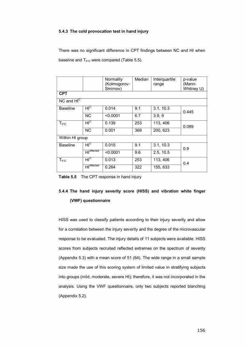
156
5.4.3 The cold provocation test in hand injury
There was no significant difference in CPT findings between NC and HI when
baseline and T5°C were compared (Table 5.5).
Normality (Kolmogorov-Smirnov)
Median Interquartile range
p-value (Mann-Whitney U)
CPT
NC and HIC
HIC 0.014 9.1 3.1, 10.3 Baseline
NC <0.0001 6.7 3.9, 9 0.445
HIC 0.139 253 113, 406 T5°C
NC 0.001 369 200, 623 0.089
Within HI group
HIC 0.015 9.1 3.1, 10.3 Baseline
HIAffected <0.0001 9.6 2.5, 10.5 0.9
HIC 0.013 253 113, 406 T5°C
HIAffected 0.264 322 155, 633 0.4
Table 5.5 The CPT response in hand injury
5.4.4 The hand injury severity score (HISS) and vibration white finger
(VWF) questionnaire
HISS was used to classify patients according to their injury severity and allow
for a correlation between the injury severity and the degree of the microvascular
response to be evaluated. The injury details of 11 subjects were available. HISS
scores from subjects recruited reflected extremes on the spectrum of severity
(Appendix 5.3) with a mean score of 51 (64). The wide range in a small sample
size made the use of this scoring system of limited value in stratifying subjects
into groups (mild, moderate, severe HI); therefore, it was not incorporated in the
analysis. Using the VWF questionnaire, only two subjects reported blanching
(Appendix 5.2).

157
5.5 DISCUSSION
In this study I was unable to identify statistically-significant changes in the CPT
or skin perfusion in HI patients in response to cold. This may reflect the small
sample size and wide variation in injury severity between the study subjects.
Bearing in mind that CPT is a technique better suited for the detecting trends in
a large sample size, the lack of significant findings is expected (Bartelink et al.
1993).
Recruitment was a major hurdle. Postal recruitment rates as low as 20% have
been reported (Hewison et al. 2006) but the recruitment rate in this study was
as low as 4%. Hand injury patients are usually young and may not be motivated
to participate in research, particularly in the absence of a financial incentive.
Additionally, the Pulvertaft Hand Unit's catchment area is very wide; therefore,
the commute distance in some cases can be too far to attend.
During the study period, a number of limitations were noted. A tool to identify
subjects with cold-related symptoms and objectively quantify the symptoms was
not incorporated within the protocol (Section 4.6). The VWF questionnaire was
not useful in the identification of CI symptoms as it only screened for white
discoloration of the digits and the hand. The iontophoresis was delivered to the
back of the hand rather than the affected digits; this was a compromise to
accommodate for the inability to secure the available iontophoresis chambers
on the affected digits.
5.6 CONCLUSION
It is vital to improve the recruitment technique (perhaps by providing a financial
incentive) to achieve an adequate sample size and get meaningful conclusions.
The spectrum of hand injuries is vast and a validated repeatable cold

158
intolerance assessment tool has to be incorporated in any future study of this
condition to facilitate patient grouping. This study is a start for future work on the
microvascular response of the injured hand to cold and the limitations faced
here will hopefully be recognised and avoided in the future.

159
CHAPTER 6: THE MANAGEMENT OF CTS IN A COMMUNITY
HOSPITAL � AN AUDIT STUDY
160
6.1 INTRODUCTION
CTS is the commonest entity seen by hand surgeons (Palmer et al. 1995).
Despite the classic symptoms and signs, the diagnosis can occasionally be
challenging due to atypical sensory symptoms, the absence of physical findings
and the lack of an objective gold standard test for the diagnosis (Rempel et al.
1998; Michelsen et al. 2002).
In short-term follow-up randomised trials, steroid injection treatment seems to
give better results compared to other non-operative treatments but on longer
follow-up surgery appears to give the best outcome (injection success rate is
8% at two years) (Dammers et al. 1999a; Viera 2003; Hui et al. 2005).
Both conservative and surgical modalities are available for the treatment of CTS
(Section 1.1.5). There is convincing evidence through randomised trials of the
short-term benefits of injectable steroid compared to other modalities of non-
operative treatment, but the long-term success rate remains very disappointing
and surgery remains the superior option.
In a local community hospital, a fortnightly nurse-led CTS management clinic (a
maximum of five new patients reviewed per clinic) with direct access to the lead
consultant�s consulting rooms was established in 2002. A nurse practitioner was
trained by a senior hand surgeon. In this clinic, patients were assessed based
on a set proforma and those diagnosed with CTS received initial conservative
treatment (night splint and steroid injection). Nerve conduction studies were
requested for the atypical cases and reviewed at a later date by the consultant.
In 2005, the nurse-led CTS clinic was replaced by a registrar-led hand clinic
twice weekly (a maximum of seven new patients per week) covering a spectrum

161
of hand referrals, mainly compressive neuropathy, base of thumb OA and hand
lumps.
6.2 AIMS
The aims of this work are: firstly, to assess the success rate of conservative
treatment for CTS in the nurse- and registrar-led clinics; secondly, to identify
predictors of non-operative treatment success; thirdly, to determine the impact
of time on the severity of symptoms; fourthly, to compare the conservative with
the surgical treatment of CTS; and, finally, to determine the impact of these
clinics on the consultant clinic waiting time.
6.3 METHODS
6.3.1 An audit of the nurse-led CTS clinic
Approval from the Primary Care Trust Clinical Governance office was obtained.
The notes of all patients treated for CTS through the nurse-led CTS clinic over a
period of 18 months were audited.
Data collection was based on the assessment proforma used in the clinic and
included age, gender and hand dominance. Additionally, relevant clinical
history, such as the symptomatic side, the time at which worse symptoms were
noted, digits affected, nocturnal symptoms and the duration of the complaint,
was also documented. Symptom severity score was determined (Levine Katz
Questionnaire (Levine et al. 1993)). Details of the clinical examination, including
evidence of thenar wasting, the response to provocation i.e. Phalen�s (Werner
et al. 1994), reverse Phalen�s (Bowles et al. 1983), Tinel�s (Bowles et al. 1983)
McMurtry�s and Durkan�s tests, NCS and management plan information, were
also available for analysis.
162
6.3.2 Registrar-led CTS clinic audit
Data on patients managed in this clinic over a period of 18 months were
collected from patient follow-up sheets (Appendix 6.1). Up to 6 months follow-up
data were available for subjects treated non-operatively (splint and steroid
injection) while up to 12 months follow-up data were available for the surgery
group.
In both clinics, steroid injections were prepared in clinic (40 mg of Kenalog®
(Triamcinolone Acetate) and 1 ml of Lignocaine 1%) and delivered at the level
of the proximal wrist crease half-way between the Palmaris Longus and Flexor
Carpi Ulnaris tendons using a 22-gauge needle angled 45° distally into the
carpal tunnel. A repeat injection was offered once at 2 months.
Exclusion criteria:
• Diabetes mellitus
• Concurrent compressive neuropathy (i.e. double-crush syndrome or cubital
tunnel syndrome) or a systemic neuropathy (i.e. multiple sclerosis)
After the diagnosis of CTS was established, the options of management
(conservative and surgical) were discussed. Patients conservatively treated
were reviewed at 2, 4 and 6 months; patients remaining asymptomatic were
discharged at 6 months. A repeat injection was offered once at 2 months and all
patients treated conservatively were given a night splint (FUTURA Splint). The
conservative management was considered to have failed if the patient
underwent CTR following conservative treatment. Patients treated surgically
were followed up for a year.

163
6.3.3 Data analysis
The data were stored on an Excel® spread sheet and analysed as outlined in
Section 2.8 using SPSS Version 13. Logistic regression was used to determine
predictors of injection success.
6.4 RESULTS
6.4.1 The nurse-led CTS clinic audit
Eighty-two new patients attended the clinic over the 18 months. The notes of 74
(90%) patients (31 males, 43 females) were available. The mean age of the
sample was 49 (14.4) years and 93% were right-handed. CTS was unilateral in
66 (92%) patients (right hand n=46, left hand n=20).
6.4.1.1 The success of the conservative management
Only 17 (23%) patients were successfully managed conservatively by the nurse.
The remaining 57 (77%) patients were referred to the consultant clinic for
further management (Figure 6.1). Of the patients who received conservative
treatment, 27 (61%) failed conservative treatment and required surgical
intervention.
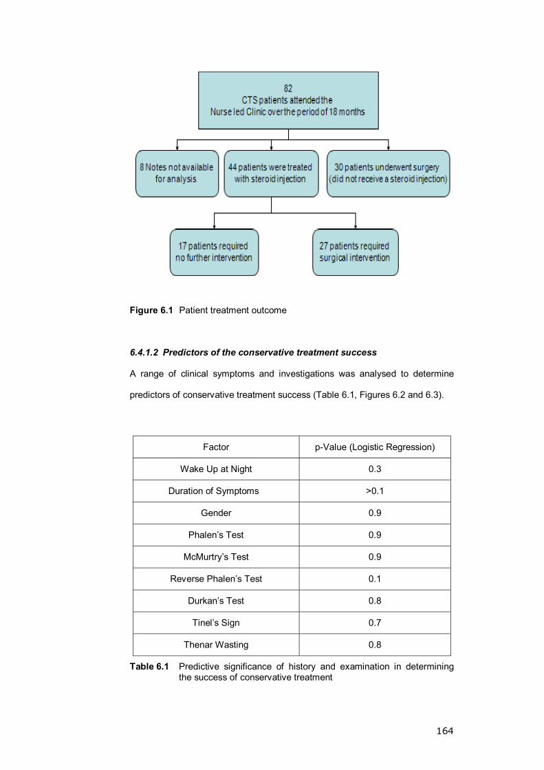
164
Figure 6.1 Patient treatment outcome
6.4.1.2 Predictors of the conservative treatment success
A range of clinical symptoms and investigations was analysed to determine
predictors of conservative treatment success (Table 6.1, Figures 6.2 and 6.3).
Factor p-Value (Logistic Regression)
Wake Up at Night 0.3
Duration of Symptoms >0.1
Gender 0.9
Phalen�s Test 0.9
McMurtry�s Test 0.9
Reverse Phalen�s Test 0.1
Durkan�s Test 0.8
Tinel�s Sign 0.7
Thenar Wasting 0.8
Table 6.1 Predictive significance of history and examination in determining the success of conservative treatment
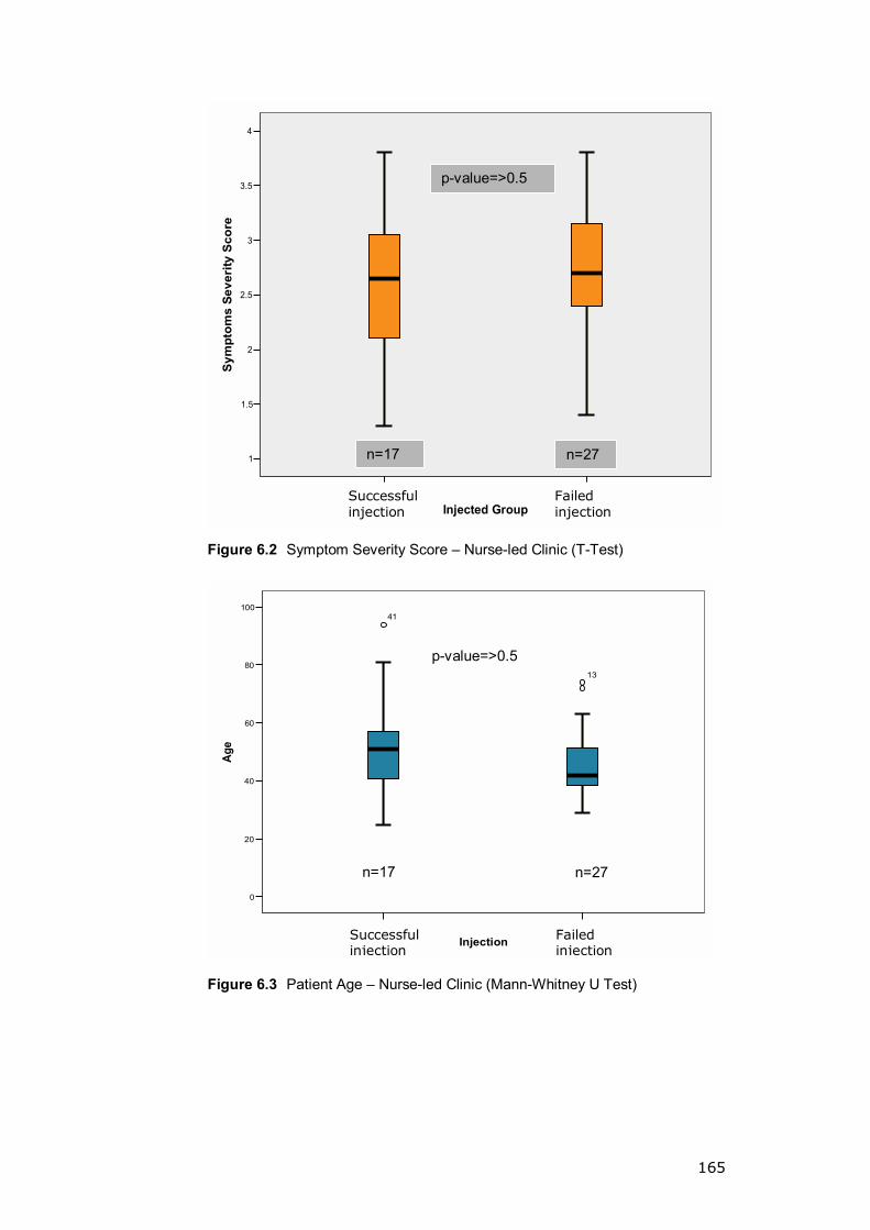
165
failed Injectionsucsessful Injection
Injected Group
4
3.5
3
2.5
2
1.5
1
Sym
ptom
s Se
veri
ty S
core
Figure 6.2 Symptom Severity Score � Nurse-led Clinic (T-Test)
faliuresucsess
Injection
100
80
60
40
20
0
Age
13
17
41
Figure 6.3 Patient Age � Nurse-led Clinic (Mann-Whitney U Test)
Successful injection
Failed injection
Failed injection
Successful injection
p-value=>0.5
n=27n=17
p-value=>0.5
n=17 n=27
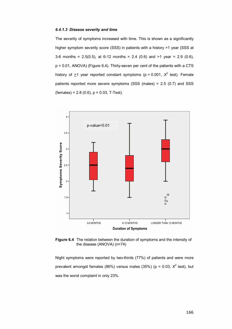
166
6.4.1.3 Disease severity and time
The severity of symptoms increased with time. This is shown as a significantly
higher symptom severity score (SSS) in patients with a history >1 year (SSS at
3-6 months = 2.5(0.5), at 6-12 months = 2.4 (0.6) and >1 year = 2.9 (0.6),
p = 0.01, ANOVA) (Figure 6.4). Thirty-seven per cent of the patients with a CTS
history of >1 year reported constant symptoms (p = 0.001, X2 test). Female
patients reported more severe symptoms (SSS (males) = 2.5 (0.7) and SSS
(females) = 2.8 (0.6), p = 0.03, T-Test).
LONGER THAN 12 MONTHS6-12 MONTHS3-6 MONTHS
Duration of Symptoms
4
3.5
3
2.5
2
1.5
1
Sym
pto
ms
Sev
erit
y S
core
39
4
Figure 6.4 The relation between the duration of symptoms and the intensity of the disease (ANOVA) (n=74)
Night symptoms were reported by two-thirds (77%) of patients and were more
prevalent amongst females (86%) versus males (35%) (p = 0.03, X2 test), but
was the worst complaint in only 23%.
p-value=0.01
167
6.4.1.4 The effect of a nurse-led CTS clinic on waiting times
The available databases at the hospital contained insufficient information to
assess this aspect of the study. This was due to the following: firstly, all the
patients were listed under the consultant�s name with no reference to the nurse
in the computer system in the hospital; secondly, patients seen by the nurse
came from a general pool of patients and not directly from the consultant�s clinic
waiting list.
Service observers in the hospital management confirmed that the waiting times
were not reduced by the nurse-led clinic activity (remained 27 weeks throughout
the duration of the clinic service).
6.4.2 Registrar-led CTS clinic audit
The notes of 173 consecutive cases fulfilling the inclusion criteria over a period
of 18 months were reviewed. Demographical data are summarised in Table 6.2.
Demographically, there was no significant difference between the injection and
surgery groups (Appendix 6.2). The data were skewed and are therefore
presented as median (interquartile range).

168
Injection Group Surgery Group
Sample size 112 61
Age (years) 55 (20) 56 (16)
Male 33 (29%) 15 (25%) Sex
Female 79 (71%) 46 (75%)
Right 92% 93% Dominance
Left 8% 7%
Yes 17% 16% Current smoking No 83% 84%
Right 14 (23%) 16 (14%)
Left 12 (20%) 12 (11%) Affected side
Bilateral 35 (57 %) 84 (75%)
Duration of symptoms (months) 12 (24) 12 (30)
Yes 3% 0% Off work
No 97% 100%
Table 6.2 Demographical data for injection and surgery groups
6.4.2.1 Conservative management outcome
At 6 months, 51% failed non-operative treatment. The failure rate was highest at
2 months follow-up (Table 6.3).
Total follow-up
VA score Outcome n (%)
Total failure rate
Surgery 20 (24%)
Re-inject with steroid 48 (56%) 2 months 85 4 (5.5)
Not given an Injection 17 (20%)
18%
Surgery 21 (39%) 4 Months 54 2 (3.5)
Follow-up 33 (61%) 37%
Surgery 16 (42%) 6 Months 38 3 (6.5)
Discharge 22 (58%) 51%
Table 6.3 Conservative management group follow-up outcome

169
6.4.2.2 Comparison between conservatively- and surgically-managed
patients
Patients opting for conservative management reported milder symptoms
compared to the surgery group. Both the DASH and VA Scores were
significantly lower in the conservative group (p <0.05, Mann-Whitney U Test).
The grip and pinch strength in the surgery group was significantly weaker
(p >0.05, Mann-Whitney U Test) (Tables 6.4 and 6.5).
Injection group Surgery Group
Score Completion
n (%) Score Completion n (%)
p-Value
LK Symptom
Score 3 (0.9)/5 91 (81%) 3.2 (0.8)/5 49 (80%) 0.07
LK Function
Score 2.3 (1.4)/5 89 (80%) 2.8 (1.5)/5 45 (74%) 0.08
DASH Score 36 (34)/100 71 (63%) 57
(18)/100 39 (64%) 0.004
VA Score 6 (3)/10 112 (100%) 7 (2)/10 61 (100%) 0.002
Table 6.4 Severity questionnaire scores for the conservative and surgery groups � Initial visit
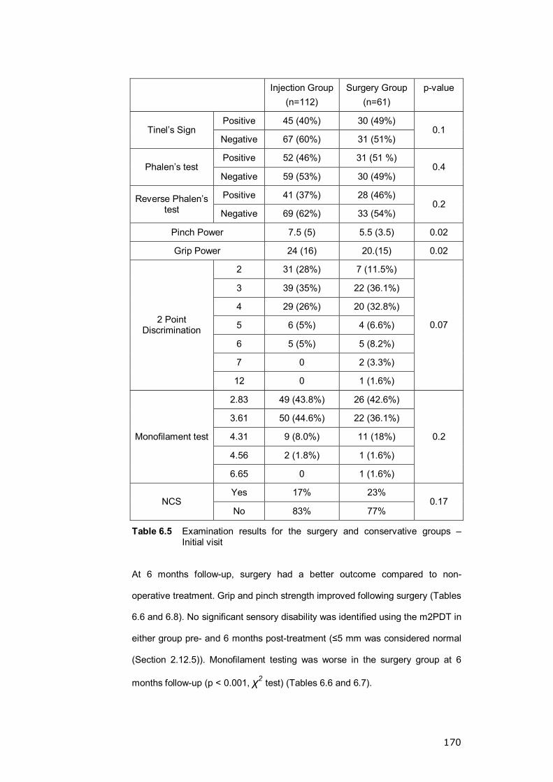
170
Injection Group
(n=112) Surgery Group
(n=61) p-value
Positive 45 (40%) 30 (49%) Tinel�s Sign
Negative 67 (60%) 31 (51%) 0.1
Positive 52 (46%) 31 (51 %) Phalen�s test
Negative 59 (53%) 30 (49%) 0.4
Positive 41 (37%) 28 (46%) Reverse Phalen�s test Negative 69 (62%) 33 (54%)
0.2
Pinch Power 7.5 (5) 5.5 (3.5) 0.02
Grip Power 24 (16) 20.(15) 0.02
2 31 (28%) 7 (11.5%)
3 39 (35%) 22 (36.1%)
4 29 (26%) 20 (32.8%)
5 6 (5%) 4 (6.6%)
6 5 (5%) 5 (8.2%)
7 0 2 (3.3%)
2 Point Discrimination
12 0 1 (1.6%)
0.07
2.83 49 (43.8%) 26 (42.6%)
3.61 50 (44.6%) 22 (36.1%)
4.31 9 (8.0%) 11 (18%)
4.56 2 (1.8%) 1 (1.6%)
Monofilament test
6.65 0 1 (1.6%)
0.2
Yes 17% 23% NCS
No 83% 77% 0.17
Table 6.5 Examination results for the surgery and conservative groups � Initial visit
At 6 months follow-up, surgery had a better outcome compared to non-
operative treatment. Grip and pinch strength improved following surgery (Tables
6.6 and 6.8). No significant sensory disability was identified using the m2PDT in
either group pre- and 6 months post-treatment (≤5 mm was considered normal
(Section 2.12.5)). Monofilament testing was worse in the surgery group at 6
months follow-up (p < 0.001, χ2 test) (Tables 6.6 and 6.7).

171
Injection group Surgery Group
Score Completion n (%) Score Completion
n (%) p-
Value
LK Symptom
Score 2.2 (2)/5 30 (83%) 1.5 (1.5)/5 38 (68%) 0.1
LK Function Score 2 (1.2)/5 26 (72%) 1.5 (1.3)/5 39 (70%) 0.6
DASH Score 32 (41)/100 26 (72%) 16 (27)/5 39 (70%) 0.05
VA Score 3 (6.5)/10 36 (100%) 1 (1)/10 56 (100%) 0.002
Table 6.6 Severity questionnaire scores for the conservative and surgery groups � 6 months visit
Injection Group(n=36)
Surgery Group (n=56)
p-value
Positive 6 (17%) 6 (7%) Tinel�s Sign
Negative 30 (83%) 50 (93%) 0.1
Positive 10 (27%) 6 (11%) Phalen�s test
Negative 26 (73%) 48 (89%) 0.04
Positive 5 (14%) 3 (5.7%) Reverse Phalen�s test Negative 31 (86%) 50 (94.3%)
0.1
Pinch Power 7.5 (3.3) 6 (4) 0.053
Grip Power 25 (12) 20 (17) 0.1
2 16 (44%) 19 (34%)
3 9 (25%) 22 (39%)
4 2 (6%) 12 (21%)
5 8 (22%) 2 (4%)
6 0 1 (2%)
2 Point Discrimination
15 1 (3%) 0
<0.001
2.83 18 (50%) 22 (42%)
3.61 14 (39%) 1 (2%)
4.31 1 (3%) 26 (49%)
4.56 2 (6%) 4 (8%)
Monofilament test
6.65 1 (3%) 0
<0.001
Table 6.7 Examination results for the surgery and conservative groups � 6 months visit

172
Patients continued to improve one year post-CTR (Table 6.8).
Surgery Group
Number 28
LK Symptom Score 1.4 (1)
LK Function Score 1.3 (0.8)
DASH Score 14 (23)
VA Score 0 (0.25)
Tinel�s Sign Positive (3) 11%
Phalen�s test Positive 11%
Reverse Phalen�s test Positive 11%
Pinch Power 5 (3.5)
Grip Power 18 (15)
2 52%
3 40%
4 4% 2 Point Discrimination
5 4%
2.83 37%
3.61 56% Monofilament test
4.31 7%
Table 6.8 Post-operative results at 12 months
6.4.2.3 Surgery following conservative treatment outcome
The outcome of CTR was not significantly different when surgery was done
primarily or after a period of non-operative treatment (Table 6.9).

173
Surgery following failed conservative treatment
(n=57)
Surgery without a trial of conservative
treatment (n=61)
p value
LK Symptom Score 0.08
LK Function Score 0.3
DASH Score 0.2
VA Score 0.7
Tinel�s Sign *0.7
Phalen�s test *0.5
Reverse Phalen�s test *0.2
Pinch Power 0.1
Grip Power 0.1
2 Point Discrimination *0.7
Monofilament test *0.5
Table 6.9 The surgical outcome is not affected by a failed conservative approach. Mann-Whitney U test except (* = χ2 test).
6.4.2.4 The effect of surgery on the sensory improvement using MFT and
TPD
There was no statistically-significant change in the sensory measurements
when MFT and TPD were used to assess the change at 2 weeks, 6 months and
one year following surgery (p-value >0.05, χ2 test).
6.4.2.5 The effect of the registrar-led clinic on the consultant clinic
waiting list
Service observers in the hospital management confirmed that the waiting time
to see a consultant dropped from 27 weeks to 5 weeks while the registrar-led
clinic was running.

174
6.5 DISCUSSION
The diagnosis and management of CTS can be challenging. The high failure
rate of the conservative treatment in both the nurse and registrar clinics leads to
increased clinic re-attendance suggesting surgery to be a better management
option. The lack of success predictors in conservative treatment makes patient
selection difficult and outcomes unpredictable. The LK symptom severity scores
(SSS) was proposed as a useful triage tool with higher values favouring surgery
(Boyd et al. 2005) but our findings did not mirror this observation as high SSS
was not predictive of non-operative treatment failure.
This audit identified failure rates >50% in both nurse- and registrar-led clinics.
The high failure rate of conservative treatment is in line with previously
published rates of between 50% and 86% (Green 1984; Ozdogan et al. 1984;
Weiss et al. 1994; Dammers et al. 1999; D'Arcy et al. 2000; Kanaan et al. 2001;
Burke et al. 2003; Hui et al. 2004). The follow-up period seems to determine the
reported failure rate. A randomised prospective study with one year follow-up of
163 patients reported 85% success rate (nocturnal symptoms improvement)
amongst the injection group (Ly-Pen et al. 2005). This is clearly a better result
than ours, but using the nocturnal symptoms as a primary indicator of success
is questionable as we have noticed that not all patients have it nor is it the most
concerning symptom from the patient point of view. On the other hand, Hui et al.
reported 11% success rate (patients did not have surgery) at 80 weeks follow-
up in 30 patients treated with a single Methylprednisolone acetate injection (Hui
et al. 2004). Various types of steroid including Methylprednisolone acetate,
Paramethasone acetonide and Triamcinolone acetonide have been used with
various doses, which makes it difficult to compare outcomes.
CTS severity increased with time. There is no evidence in the literature clearly
identifying the mechanism of steroid action in CTS, whether it is the result of the
175
nerve membrane stabilisation effect of steroid or a true drop in the intra-
compartmental pressure of the carpal tunnel secondary to soft tissue atrophy.
What is concerning is the high failure rate of non-operative treatment resulting
in a delay in the delivery of the definitive treatment (CTR) and increase in the
symptom severity with time.
Surgery in this study gave better symptomatic relief at 6 months. This was
statistically evident in the DASH and VA Scores but not with the LK
Questionnaire. This finding might be the result of significant weaknesses in the
validation of the LK questionnaire as discussed earlier (Section 1.6).
Tests to evaluate the sensory function in the skin are those for the slowly
adapting fibres (static two-point discrimination (sTPD) and Semmes-Weinstein
monofilament (SWM) test) and for the quickly adapting fibres (vibration and
moving two-point discrimination (mTPD)). SWM and vibration tests measure the
sensory threshold by measuring the function of a receptor or group of receptors
supplied by a single nerve fibre. mTPD and sTPD test innervations density,
which requires an overlap of different sensory units and complex cortical
integration (Szabo 1998). Threshold tests are more likely to detect a gradual,
progressive change in the nerve function while density tests are reliable in
assessing the functional nerve regeneration after nerve repair in which the brain
input is radically altered. In an experimental model of acute median nerve
compression, abnormalities in the SWM test were detected shortly after the
appearance of subjective symptoms (reduced sensation and paresthesia) while
the TPD exhibited an all-or-none phenomenon in which the abnormality was
only detected after a marked or total elimination of the electrical signal
(Gelberman et al. 1983) (Figure 6.5).
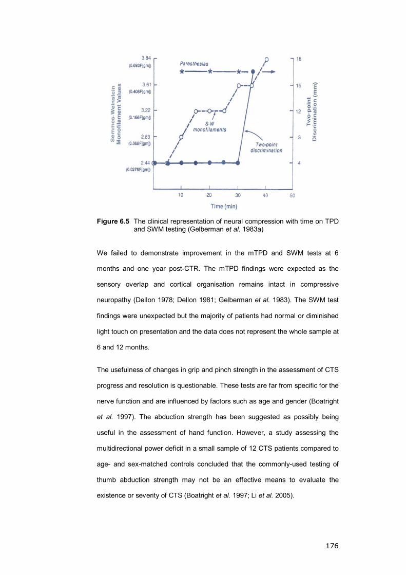
176
Figure 6.5 The clinical representation of neural compression with time on TPD and SWM testing (Gelberman et al. 1983a)
We failed to demonstrate improvement in the mTPD and SWM tests at 6
months and one year post-CTR. The mTPD findings were expected as the
sensory overlap and cortical organisation remains intact in compressive
neuropathy (Dellon 1978; Dellon 1981; Gelberman et al. 1983). The SWM test
findings were unexpected but the majority of patients had normal or diminished
light touch on presentation and the data does not represent the whole sample at
6 and 12 months.
The usefulness of changes in grip and pinch strength in the assessment of CTS
progress and resolution is questionable. These tests are far from specific for the
nerve function and are influenced by factors such as age and gender (Boatright
et al. 1997). The abduction strength has been suggested as possibly being
useful in the assessment of hand function. However, a study assessing the
multidirectional power deficit in a small sample of 12 CTS patients compared to
age- and sex-matched controls concluded that the commonly-used testing of
thumb abduction strength may not be an effective means to evaluate the
existence or severity of CTS (Boatright et al. 1997; Li et al. 2005).
177
The registrar-led clinics had a larger impact on the consultant�s clinic due to
several factors: clinics were larger and more frequent, attended to a wider
spectrum of hand conditions and patients requiring surgery were directly
managed in a registrar-led operating list. All this freed the consultant clinic for
more cases and reduced the waiting time. A study from Leicester reported a
reduction in CTR waiting time by 100 weeks within two years of the
commencement of the nurse-led specialist CTS clinic (Newey et al. 2006). An
adequately trained nurse practitioner performed approximately 400 procedures
on 300 patients with comparable outcomes to the accepted standards
presented in published literature. The nurse-led clinic in our study was not as
successful in reducing the waiting lists. If such clinics are to have a noticeable
effect, more responsibility has to be given to the nurse practitioner with
adequate training to operate and review more patients, a step not all lead
clinicians are happy to take (Murray 1998; Newey et al. 2006).
6.6 CONCLUSION
Non-operative CTS treatment has a high failure rate, surgery being the
definitive treatment with better outcomes. For nurse-led clinics to have an
impact, the role of the nurse has to be extended with sizable, more frequent
clinics and operative responsibility.

178
CHAPTER 7: GENERAL DISCUSSION
179
7.1 MICROCIRCULATORY MEASURES
The validation of biological variables measurement tools has to accommodate
for the dynamic nature of all living tissue. It is important for such tests to
represent the variable measured and reflect change but, at the same time, to
have reasonable repeatability. But, what is reasonable in such tests? I feel it is
the ability of the test to demonstrate a trend through repeated measurements,
not absolute values. Whether it is a simple blood pressure measurement of a
CPT or LDI, the same rule applies.
The CPT presented in this thesis is one of many possible formats of the test,
capable of detecting change. The modified CPT was able to differentiate
between controls, PRP and HAVS groups. Statistically the differences were
highly significant but the test repeatability was not high. This is expected. When
comparing the repeatability to blood pressure measurement repeatability, it is
proving to be very similar. So surely it is a very good test. It is the trend rather
than the absolute value that is important and the CPT is a lengthy and an
uncomfortable test which makes repeating it to determine a trend within a
subject cumbersome, but makes it a very good tool for assessing groups as we
have witnessed in this thesis. The baseline measurement might be useful; it is
easy to measure, can be repeated frequently and a trend higher than 7.5°C
suggests normality. This makes it a good screening measurement.
Pairing LDI and iontophoresis created an excellent opportunity to indirectly
study the endothelial response. By eliminating a lot of the LDF limitations it is an
ideal non-invasive technique but its repeatability in the hand appears to be
weaker compared to the forearm or gaiter. There is evidence suggesting its
repeatability to weaken, the more distal the target organ. This was noticed in the
hand and should be kept in mind when sample sizing for such studies.

180
7.2 CTS, HI AND COLD INTOLERANCE
Autonomic symptoms are increasingly reported with CTS and evidence
suggests they improve following CTR. Treatment for CTS is either operative or
non-operative; amongst non-operative options, steroid injections appear to give
the best results but they have a high failure rate. Surgery (open or endoscopic)
remains the preferred treatment option with better symptom and function
recovery.
It remains unclear why cold intolerance is highly prevalent amongst patients
with hand injury. Neural and vascular causes have been explored but the neural
hypothesis is favoured. The link between PRP, cold intolerance and the
autonomic symptoms amongst CTS patients is frequently eluded but not
supported by convincing evidence.
CPT findings post-CTR identified a significant rise in the digital baseline
temperature and a faster re-warming time in the median nerve-supplied digits.
This was an absolute rise in the digital temperature as pre-op testing identified
no difference between the affected and control hand in patients. The role of
CTR in RP would be an interesting aspect to explore.
It became evident during this work that cold intolerance severity measurement
tools are weak and improperly validated. A validated tool is important in
grouping subjects, particularly HI patients as the injury spectrum is very wide.
The recruitment of HI subjects is difficult. I feel that, in the young, active working
group, a financial incentive is of paramount importance for recruitment.
181
7.3 CTS CLINICS
For such clinics to be effective, they should be frequent, sizeable (>7 patients/
clinic) and run by a professional capable of managing CTS for diagnosis to CTR
if required. If such requirements are not available, the workload will only be
shifted but not managed. The debate on the role of nurse practitioners is such
that clinics remain controversial but a well-trained nurse should be able to cover
such responsibility successfully.
7.4 AREAS OF FUTURE RESEARCH
1- Identification of variation in the re-warming response in common
variants of RP such as SSc, Lupus and Dermatomyositis compared to
the idiopathic variant and controls. This may be aided by further
analysis of more segments in the available re-warming curves for the
controls and PRP to identify further segments with distinct disease
representation and acceptable repeatability.
2- Validation of CISS scoring as a tool in the assessment and
classification of cold-intolerant subjects.
3- A repeat of the assessment of the microvascular changes in the median
nerve distribution (using appropriate iontophoresis chambers),
especially comparing the pre- and post-operative response.
4- Study on the effect of carpal tunnel release in patients with PRP
complicated by carpal tunnel syndrome, assessing the symptomatic
and microvascular improvement post-operatively.

182
5- Study the effect of steroid injection therapy on the ICTP using a
pressure transducer pre- and 2-4 weeks post-injection to determine
whether steroid effect is due to its membrane stabilising capability or
due to a true effect on the ICTP possibly as a result of soft tissue
atrophy.

183
CHAPTER 8: REFERENCES

184
AAN (1993). "Practice parameter for carpal tunnel syndrome (summary
statement). Report of the Quality Standards Subcommittee of the American
Academy of Neurology." Neurology 43(11): 2406-9.
Al-Shahi, R. and C. Warlow (2000). "Using patient-identifiable data for
observational research and audit." BMJ 321(7268): 1031-2.
Altura, B. M. (1975). "Sex and estrogens and responsiveness of terminal
arterioles to neurohypophyseal hormones and catecholamines." Journal of
Pharmacology & Experimental Therapeutics 193(2): 403-12.
Anderson, M. E., T. L. Moore, et al. (2004). "Digital iontophoresis of vasoactive
substances as measured by laser Doppler imaging--a non-invasive technique
by which to measure microvascular dysfunction in Raynaud's phenomenon."
Rheumatology 43(8): 986-91.
Anderson, R. R. and J. A. Parrish (1981). "The optics of human skin." Journal of
Investigative Dermatology 77(1): 13-9.
Andreu, J. L. and D. Ly-Pen (2006). "A randomized controlled trial of surgery vs
steroid injection for carpal tunnel syndrome [comment]." Neurology 66(6):
955-6; author reply 955-6.
Armstrong, T., W. Devor, et al. (2004). "Intracarpal steroid injection is safe and
effective for short-term management of carpal tunnel syndrome." Muscle &
Nerve 29(1): 82-8.
Armstrong, T. J., W. A. Castelli, et al. (1984). "Some histological changes in
carpal tunnel contents and their biomechanical implications." Journal of
Occupational Medicine 26(3): 197-201.

185
Atroshi, I., C. Gummesson, et al. (1999). "Prevalence of carpal tunnel syndrome
in a general population." JAMA 282(2): 153-8.
Atroshi, I., G. U. Larsson, et al. (2006). "Outcomes of endoscopic surgery
compared with open surgery for carpal tunnel syndrome among employed
patients: randomised controlled trial [see comment]." BMJ 332(7556): 1473.
Aulisa, L., F. Tamburrelli, et al. (1998). "Carpal tunnel syndrome: indication for
surgical treatment based on electrophysiologic study." Journal of Hand
Surgery - American Volume 23(4): 687-91.
Backman, C., A. Nystrom, et al. (1991). "Cold-induced arterial spasm after
digital amputation." Journal of Hand Surgery - British Volume 16(4): 378-81.
Backman, C., A. Nystrom, et al. (1993). "Arterial spasticity and cold intolerance
in relation to time after digital replantation." Journal of Hand Surgery - British
Volume 18(5): 551-5.
Bartelink, M. L., H. Wollersheim, et al. (1993). "Reproducibility of the finger
cooling test." Microvascular Research 45(1): 65-73.
Beaton, D. E., J. G. Wright, et al. (2005). "Development of the QuickDASH:
comparison of three item-reduction approaches." Journal of Bone & Joint
Surgery - American Volume 87(5): 1038-46.
Beekman, R. and L. H. Visser (2002). "Sonographic detection of diffuse
peripheral nerve enlargement in hereditary neuropathy with liability to
pressure palsies." Journal of Clinical Ultrasound 30(7): 433-6.

186
Beekman, R. and L. H. Visser (2003). "Sonography in the diagnosis of carpal
tunnel syndrome: a critical review of the literature." Muscle & Nerve 27(1): 26-
33.
Beevers, G., G. Y. Lip, et al. (2001). "ABC of hypertension. Blood pressure
measurement. Part I-sphygmomanometry: factors common to all techniques
[see comment]." BMJ 322(7292): 981-5.
Berghoff, M., M. Kathpal, et al. (2002). "Vascular and neural mechanisms of
ACh-mediated vasodilation in the forearm cutaneous microcirculation."
Journal of Applied Physiology 92(2): 780-8.
Bernard, B. (1997). "Musculoskeletal Disorders and Workplace Factors: A
Critical Review of Epidemiologic Evidence for Work-Related Musculoskeletal
Disorders of the Neck, Upper Extremity, and Low Back." N. I. f. O. S. a.
health.
Biondi, M. L., B. Marasini, et al. (1991). "Increased plasma endothelin levels in
patients with Raynaud's phenomenon." New England Journal of Medicine
324(16): 1139-40.
Bircher, A., E. M. de Boer, et al. (1994). "Guidelines for measurement of
cutaneous blood flow by laser Doppler flowmetry. A report from the
Standardization Group of the European Society of Contact Dermatitis."
Contact Dermatitis 30(2): 65-72.
Bjarnason, B., E. Flosadottir, et al. (1999). "Objective non-invasive assessment
of patch tests with the laser Doppler perfusion scanning technique." Contact
Dermatitis 40(5): 251-60.

187
Bland, J. D. (2000). "The value of the history in the diagnosis of carpal tunnel
syndrome." Journal of Hand Surgery - British Volume 25(5): 445-50.
Bland, J. M. and D. G. Altman (1986). "Statistical methods for assessing
agreement between two methods of clinical measurement." Lancet 1(8476):
307-10.
Block, J. A. and W. Sequeira (2001). "Raynaud's phenomenon." Lancet
357(9273): 2042-8.
BMA (1995). Ethical issues in audit. British Medical Association.
Boatright, J. R., G. M. Kiebzak, et al. (1997). "Measurement of thumb abduction
strength: normative data and a comparison with grip and pinch strength [see
comment]." Journal of Hand Surgery - American Volume 22(5): 843-8.
Bottomley, W. and M. Goodfield (1994). "A pathogenic role for endothelin in
Raynaud's phenomenon?" Acta Dermato-Venereologica 74(6): 433-4.
Bovenzi, M. (1986). "Some pathophysiological aspects of vibration-induced
white finger." European Journal of Applied Physiology & Occupational
Physiology 55(4): 381-9.
Bovenzi, M. (1987). "Finger thermometry in the assessment of subjects with
vibration-induced white finger." Scandinavian Journal of Work, Environment &
Health 13(4): 348-51.
Bovenzi, M., A. Fiorito, et al. (1983). "Platelet function and clotting parameters
of vibration-exposed foundry workers." Scandinavian Journal of Work,
Environment & Health 9(4): 347-52.
188
Bowles, A. P., Jr., S. W. Asher, et al. (1983). "Use of Tinel's sign in carpal
tunnel syndrome." Annals of Neurology 13(6): 689-90.
Bowling, J. C. and P. M. Dowd (2003). "Raynaud's disease." Lancet 361(9374):
2078-80.
Boyd, K. U., B. S. Gan, et al. (2005). "Outcomes in carpal tunnel syndrome:
symptom severity, conservative management and progression to surgery."
Clinical & Investigative Medicine - Medecine Clinique et Experimentale 28(5):
254-60.
Boyle, N. H., D. Manifold, et al. (2000). "Intraoperative assessment of colonic
perfusion using scanning laser Doppler flowmetry during colonic resection."
Journal of the American College of Surgeons 191(5): 504-10.
Boyle, N. H., A. Pearce, et al. (1998). "Scanning laser Doppler flowmetry and
intraluminal recirculating gas tonometry in the assessment of gastric and
jejunal perfusion during oesophageal resection." British Journal of Surgery
85(10): 1407-11.
Brammer, A. J., W. Taylor, et al. (1987). "Sensorineural stages of the hand-arm
vibration syndrome." Scandinavian Journal of Work, Environment & Health
13(4): 279-83.
Brand, F. N., M. G. Larson, et al. (1997). "The occurrence of Raynaud's
phenomenon in a general population: the Framingham Study." Vascular
Medicine 2(4): 296-301.

189
Braun, R. M., M. Rechnic, et al. (2002). "Complications related to carpal tunnel
release." Hand Clinics 18(2): 347-57.
Brown, F. E., J. B. Jobe, et al. (1986). "Induced vasodilation in the treatment of
posttraumatic digital cold intolerance." Journal of Hand Surgery - American
Volume 11(3): 382-7.
Brown, P. M. (2006). "The audit cycle: a guide to success." British Journal of
Hospital Medicine 67(3): 116-7.
Brown, R. A., R. H. Gelberman, et al. (1993). "Carpal tunnel release. A
prospective, randomized assessment of open and endoscopic methods [see
comment]." Journal of Bone & Joint Surgery - American Volume 75(9): 1265-
75.
Bunker, C. B., P. C. Goldsmith, et al. (1996). "Calcitonin gene-related peptide,
endothelin-1, the cutaneous microvasculature and Raynaud's phenomenon."
British Journal of Dermatology 134(3): 399-406.
Burke, F. D., J. Ellis, et al. (2003). "Primary care management of carpal tunnel
syndrome." Postgraduate Medical Journal 79(934): 433-7.
Burke, F. D., I. J. Lawson, et al. (2005). "Carpal tunnel syndrome in association
with hand-arm vibration syndrome: a review of claimants seeking
compensation in the Mining Industry." Journal of Hand Surgery - British
Volume 30(2): 199-203.
Buttrey, Y. (1998). "Implementing Evidence Through Clinical Audit." Evidence-
based Healthcare Oxford, Butterworth-Hienemann: 182-207.
190
Campbell, D. A. and S. P. Kay (1998). "What is cold intolerance?" Journal of
Hand Surgery - British Volume 23(1): 3-5.
Campero, M., R. J. Verdugo, et al. (1993). "Vasomotor innervation of the skin of
the hand: a contribution to the study of human anatomy." Journal of Anatomy
182(Pt 3): 361-8.
Celermajer, D. S., K. E. Sorensen, et al. (1992). "Non-invasive detection of
endothelial dysfunction in children and adults at risk of atherosclerosis."
Lancet 340(8828): 1111-5.
Chang, M. H., H. T. Chiang, et al. (1998). "Oral drug of choice in carpal tunnel
syndrome." Neurology 51(2): 390-3.
Cherkas, L. F., L. Carter, et al. (2003). "Use of thermographic criteria to identify
Raynaud's phenomenon in a population setting." J Rheumatol 30(4): 720-2.
Chung, M. S., H. S. Gong, et al. (1999). "Prevalence of Raynaud's phenomenon
in patients with idiopathic carpal tunnel syndrome." Journal of Bone & Joint
Surgery - British Volume 81(6): 1017-9.
Chung, M. S., H. S. Gong, et al. (2000). "Raynaud's phenomenon in idiopathic
carpal tunnel syndrome: postoperative alteration in its prevalence." Journal of
Bone & Joint Surgery - British Volume 82(6): 818-9.
Ciuffetti, G., L. Pasqualini, et al. (1995). "Neutrophil-derived adhesion molecules
in human digital ischaemia and reperfusion." Vasa 24(2): 155-8.
Clough, G. F., A. R. Bennett, et al. (1998). "Effects of H1 antagonists on the
cutaneous vascular response to histamine and bradykinin: a study using
191
scanning laser Doppler imaging." British Journal of Dermatology 138(5): 806-
14.
Cobb, T. K., W. P. Cooney, et al. (1995). "Pressure dynamics of the carpal
tunnel and flexor compartment of the forearm." Journal of Hand Surgery -
American Volume 20(2): 193-8.
Collins, E. D., C. B. Novak, et al. (1996). "Long-term follow-up evaluation of cold
sensitivity following nerve injury." Journal of Hand Surgery - American Volume
21(6): 1078-85.
Collins, P., T. M. Griffith, et al. (1986). "Endothelium-derived relaxing factor
alters calcium fluxes in rabbit aorta: a cyclic guanosine monophosphate-
mediated effect." Journal of Physiology 381: 427-37.
Coughlin, P. (2004). "Letter 3: cold provocation testing and hand-arm vibration
syndrome - an audit of the results of the Department of Trade and Industry
scheme for the evaluation of miners (Br J Surg 2003; 90: 1076-1079)." British
Journal of Surgery 91(1): 124; author reply 124-5.
Coughlin, P. A., I. C. Chetter, et al. (2001). "The analysis of sensitivity,
specificity, positive predictive value and negative predictive value of cold
provocation thermography in the objective diagnosis of the hand-arm vibration
syndrome." Occup Med (Lond) 51(2): 75-80.
Craigen, M., J. M. Kleinert, et al. (1999). "Patient and injury characteristics in
the development of cold sensitivity of the hand: a prospective cohort study."
Journal of Hand Surgery - American Volume 24(1): 8-15.
192
D'Arcy, C. A. and S. McGee (2000). "The rational clinical examination. Does this
patient have carpal tunnel syndrome? [see comment] [erratum appears in
JAMA 2000 Sep 20;284(11):1384]." JAMA 283(23): 3110-7.
Dammers, J. W., M. M. Veering, et al. (1999a). "Injection with
methylprednisolone proximal to the carpal tunnel: randomised double blind
trial." BMJ 319(7214): 884-6.
Dammers, J. W., M. M. Veering, et al. (1999b). "Injection with
methylprednisolone proximal to the carpal tunnel: randomised double blind
trial [see comment]." BMJ 319(7214): 884-6.
Davis, K. R., H. Pearson, et al. (2001a). "Acute hyperhomocysteinaemia affects
pulse pressure but not microvascular vasodilator function." Br J Clin
Pharmacol 52(3): 327-32.
Davis, K. R., H. Pearson, et al. (2001b). "Acute hyperhomocysteinaemia affects
pulse pressure but not microvascular vasodilator function." British Journal of
Clinical Pharmacology 52(3): 327-32.
Davis, K. R., J. Ponnampalam, et al. (2001c). "Microvascular vasodilator
response to acetylcholine is increased in women with pre-eclampsia." BJOG:
An International Journal of Obstetrics & Gynaecology 108(6): 610-4.
de Boer, E. M., P. D. Bezemer, et al. (1989). "A standard method for repeated
recording of skin blood flow using laser Doppler flowmetry." Dermatosen in
Beruf und Umwelt Occupational & Environmental Dermatoses 37(2): 58-62.
193
de Krom, M. C., P. G. Knipschild, et al. (1992). "Carpal tunnel syndrome:
prevalence in the general population." Journal of Clinical Epidemiology 45(4):
373-6.
De Smet, L., A. Steenwerckx, et al. (1995). "Value of clinical provocative tests in
carpal tunnel syndrome." Acta Orthopaedica Belgica 61(3): 177-82.
Dekel, S., T. Papaioannou, et al. (1980). "Idiopathic carpal tunnel syndrome
caused by carpal stenosis." British Medical Journal 280(6227): 1297-9.
Dellon, A. L. (1978a). "The moving two-point discrimination test: Clinical
evaluation of the quickly-adapting fiber/receptor system." Journal of Hand
Surgery - American Volume 3: 474-481.
Dellon, A. L. (1978b). "The moving two-point discrimination test: clinical
evaluation of the quickly adapting fiber/receptor system." Journal of Hand
Surgery - American Volume 3(5): 474-81.
Dellon, A. L. (1980). "Clinical use of vibratory stimuli to evaluate peripheral
nerve injury and compression neuropathy." Plastic & Reconstructive Surgery
65(4): 466-76.
Dellon, A. L. (1981). Evaluation of sensibility and re-education of sensation in
the hand. Baltimore, Williams & Wilkins.
Dellon, A. L., R. M. Curtis, et al. (1972). "Evaluating recovery of sensation in the
hand following nerve injury." Johns Hopkins Medical Journal 130(4): 235-43.
Dhindsa, P., K. R. Davis, et al. (2003). "Comparison of the micro- and macro-
vascular effects of glimepiride and gliclazide in metformin-treated patients

194
with Type 2 diabetes: a double-blind, crossover study." British Journal of
Clinical Pharmacology 55(6): 616-9.
Drexler, H. and A. M. Zeiher (1991). "Endothelial function in human coronary
arteries in vivo. Focus on hypercholesterolemia." Hypertension 18(4 Suppl):
II90-9.
Droog, E. J., J. Henricson, et al. (2004). "A protocol for iontophoresis of
acetylcholine and sodium nitroprusside that minimises nonspecific
vasodilatory effects." Microvascular Research 67(2): 197-202.
Droog, E. J. and F. Sjoberg (2003). "Nonspecific vasodilatation during
transdermal iontophoresis-the effect of voltage over the skin." Microvascular
Research 65(3): 172-8.
Duncan, K. H., R. C. Lewis, Jr., et al. (1987). "Treatment of carpal tunnel
syndrome by members of the American Society for Surgery of the Hand:
results of a questionnaire." Journal of Hand Surgery - American Volume
12(3): 384-91.
Earley, M. J. and J. S. Watson (1984). "Twenty four thumb replantations."
Journal of Hand Surgery - British Volume 9(1): 98-102.
Egloff, D. V., R. P. Mifsud, et al. (1983). "Superselective digital sympathectomy
in Raynaud's phenomenon." Hand 15(1): 110-4.
Ekenvall, L. and L. E. Lindblad (1986). "Is vibration white finger a primary
sympathetic nerve injury?" British Journal of Industrial Medicine 43(10): 702-
6.
195
Emara, M. K. and A. M. Saadah (1988). "The carpal tunnel syndrome in
hypertensive patients treated with beta-blockers." Postgraduate Medical
Journal 64(749): 191-2.
Engkvist, O., L. K. Wahren, et al. (1985). "Effects of regional intravenous
guanethidine block in posttraumatic cold intolerance in hand amputees."
Journal of Hand Surgery - British Volume 10(2): 145-50.
Essex, T. J. and P. O. Byrne (1991). "A laser Doppler scanner for imaging blood
flow in skin." Journal of Biomedical Engineering 13(3): 189-94.
Ettema, A. M., P. C. Amadio, et al. (2004). "A histological and
immunohistochemical study of the subsynovial connective tissue in idiopathic
carpal tunnel syndrome." Journal of Bone & Joint Surgery - American Volume
86-A(7): 1458-66.
Eversmann, W. W. (1988). Entrapment and compression neuropathies. New
York, Churchill Livingstone.
Fagius, J. and H. Blumberg (1985). "Sympathetic outflow to the hand in patients
with Raynaud's phenomenon." Cardiovascular Research 19(5): 249-53.
Ferrell, W. R., P. V. Balint, et al. (2000). "Novel use of laser Doppler imaging for
investigating epicondylitis." Rheumatology 39(11): 1214-7.
Fleiss, J. L. and J. Cohen (1973). "The equivalence of weighted kappa and the
intraclass correlation coefficient as measures of reliability." Educ Psychol
Meas 33: 613-9.
196
Fraenkel, L. (2002). "Raynaud's phenomenon: epidemiology and risk factors."
Current Rheumatology Reports 4(2): 123-8.
Fraenkel, L., Y. Zhang, et al. (1998). "The association of estrogen replacement
therapy and the Raynaud phenomenon in postmenopausal women." Annals
of Internal Medicine 129(3): 208-11.
Fraenkel, L., Y. Zhang, et al. (1999). "Different factors influencing the
expression of Raynaud's phenomenon in men and women." Arthritis &
Rheumatism 42(2): 306-10.
Franzblau, A. and R. A. Werner (1999). "What is carpal tunnel syndrome?"
JAMA 282(2): 186-7.
Freedlander, E. (1986). "The relationship between cold intolerance and
cutaneous blood flow in digital replantation patients." Journal of Hand Surgery
- British Volume 11(1): 15-9.
Freedman, R. R., R. P. Baer, et al. (1995). "Blockade of vasospastic attacks by
alpha 2-adrenergic but not alpha 1-adrenergic antagonists in idiopathic
Raynaud's disease." Circulation 92(6): 1448-51.
Freedman, R. R. and P. Ianni (1983). "Role of cold and emotional stress in
Raynaud's disease and scleroderma." British Medical Journal Clinical
Research Ed 287(6404): 1499-502.
Freedman, R. R., S. C. Sabharal, et al. (1989). "Increased alpha-adrenergic
responsiveness in idiopathic Raynaud's disease." Arthritis & Rheumatism
32(1): 61-5.
197
Fuchs, P. C., P. A. Nathan, et al. (1991). "Synovial histology in carpal tunnel
syndrome." Journal of Hand Surgery - American Volume 16(4): 753-8.
Fujimoto, S. and T. Itoh (1997). "Role of nitric oxide and nitric oxide-
independent relaxing factor in contraction and relaxation of rabbit blood
vessels." European Journal of Pharmacology 330(2-3): 177-84.
Fullerton, A. and J. Serup (1995). "Laser Doppler image scanning for
assessment of skin irritation." Current Problems in Dermatology 23: 159-68.
Fullerton, A., M. Stucker, et al. (2002). "Guidelines for visualization of cutaneous
blood flow by laser Doppler perfusion imaging. A report from the
Standardization Group of the European Society of Contact Dermatitis based
upon the HIRELADO European community project." Contact Dermatitis 46(3):
129-40.
Furchgott, R. F. and J. V. Zawadzki (1980). "The obligatory role of endothelial
cells in the relaxation of arterial smooth muscle by acetylcholine." Nature
288(5789): 373-6.
Garland, C. J., F. Plane, et al. (1995). "Endothelium-dependent
hyperpolarization: a role in the control of vascular tone." Trends in
Pharmacological Sciences 16(1): 23-30.
Garland, H., J. P. Bradshaw, et al. (1957). "Compression of median nerve in
carpal tunnel and its relation to acroparaesthesiae." Br Med J. 30(1(5021)):
730-4.

198
Gautherie, M. (1995). "Clinical studies of the vibration syndrome using a cold
stress test measuring finger temperature." Central European Journal of Public
Health 3 Suppl: 5-10.
Gazelius, B. (2005). Iontophoresis - theory. Stockholm, Perimed AB.
Gelberman, H. R. (1991). Carpal tunnel release. Philadelphia, JB Lippincot.
Gelberman, R. H., D. Aronson, et al. (1980). "Carpal-tunnel syndrome. Results
of a prospective trial of steroid injection and splinting." Journal of Bone & Joint
Surgery - American Volume 62(7): 1181-4.
Gelberman, R. H. and J. P. Blasingame (1981a). "The timed Allen test." J
Trauma 21(6): 477-9.
Gelberman, R. H., P. T. Hergenroeder, et al. (1981b). "The carpal tunnel
syndrome. A study of carpal canal pressures." Journal of Bone & Joint
Surgery - American Volume 63(3): 380-3.
Gelberman, R. H., B. L. Rydevik, et al. (1988). "Carpal tunnel syndrome. A
scientific basis for clinical care." Orthopedic Clinics of North America 19(1):
115-24.
Gelberman, R. H., R. M. Szabo, et al. (1983a). "Sensibility testing in peripheral-
nerve compression syndromes. An experimental study in humans." Journal of
Bone & Joint Surgery - American Volume 65(5): 632-8.
Gelberman, R. H., R. M. Szabo, et al. (1983b). "Tissue pressure threshold for
peripheral nerve viability." Clinical Orthopaedics & Related Research (178):
285-91.
199
Gelberman, R. H., J. R. Urbaniak, et al. (1978). "Digital sensibility following
replantation." Journal of Hand Surgery - American Volume 3(4): 313-9.
Gellis, M. and R. Pool (1977). "Two-point discrimination distances in the normal
hand and forearm: application to various methods of fingertip reconstruction."
Plastic & Reconstructive Surgery 59(1): 57-63.
Geoghegan, J. M., D. I. Clark, et al. (2004). "Risk factors in carpal tunnel
syndrome." Journal of Hand Surgery - British Volume 29(4): 315-20.
Gerritsen, A. A., M. C. de Krom, et al. (2002). "Conservative treatment options
for carpal tunnel syndrome: a systematic review of randomised controlled
trials." Journal of Neurology 249(3): 272-80.
Gibbins, I. L., D. Wattchow, et al. (1987). "Two immunohistochemically identified
populations of calcitonin gene-related peptide (CGRP)-immunoreactive axons
in human skin." Brain Research 414(1): 143-8.
Gilligan, D. M., A. A. Quyyumi, et al. (1994). "Effects of physiological levels of
estrogen on coronary vasomotor function in postmenopausal women."
Circulation 89(6): 2545-51.
Girlanda, P., R. Dattola, et al. (1993). "Local steroid treatment in idiopathic
carpal tunnel syndrome: short- and long-term efficacy." Journal of Neurology
240(3): 187-90.
Glowacki, K. A., C. J. Breen, et al. (1996). "Electrodiagnostic testing and carpal
tunnel release outcome." Journal of Hand Surgery - American Volume 21(1):
117-21.
200
GMC (2001). Good Medical Practice. G. M. Council. London, General Medical
Council.
Goldsmith, P. C., F. A. Molina, et al. (1994). "Cutaneous nerve fibre depletion in
vibration white finger." Journal of the Royal Society of Medicine 87(7): 377-81.
Green, D. J., D. T. Fowler, et al. (1996). "Endothelium-derived nitric oxide
activity in forearm vessels of tennis players." Journal of Applied Physiology
81(2): 943-8.
Green, D. P. (1984). "Diagnostic and therapeutic value of carpal tunnel
injection." Journal of Hand Surgery - American Volume 9(6): 850-4.
Griendling, K. K. and R. W. Alexander (1996). "Endothelial control of the
cardiovascular system: recent advances." FASEB Journal 10(2): 283-92.
Griffin, M. J. and C. J. Lindsell (1998). "Cold provocation tests for the diagnosis
of vibration - induced white finger: Standardisation and repeatability."
173/1998.
Gschwandtner, M. E., E. Ambrozy, et al. (1999). "Laser Doppler imaging and
capillary microscopy in ischemic ulcers." Atherosclerosis 142(1): 225-32.
Hack, M., M. A. Boillat, et al. (1986). "Assessment of vibration induced white
finger: reliability and validity of two tests." British Journal of Industrial
Medicine 43(4): 284-7.
Hamilton, A. (1918). "A study of spastic anaemia in the hands of stone cutters."
Bulletin 19(236:US bureau of labour statistics): 53-66.

201
Harada, N. (2002). "Cold-stress tests involving finger skin temperature
measurement for evaluation of vascular disorders in hand-arm vibration
syndrome: review of the literature." International Archives of Occupational &
Environmental Health 75(1-2): 14-9.
Harada, N., M. Iwamoto, et al. (1998). "Effects of room temperature, seasonal
condition and food intake on finger skin temperature during cold exposure test
for diagnosing hand-arm vibration syndrome." Industrial Health 36(2): 166-70.
Harrington, J. M., J. T. Carter, et al. (1998). "Surveillance case definitions for
work related upper limb pain syndromes." Occupational & Environmental
Medicine 55(4): 264-71.
Hashimoto, M., M. Akishita, et al. (1995). "Modulation of endothelium-
dependent flow-mediated dilatation of the brachial artery by sex and
menstrual cycle [see comment]." Circulation 92(12): 3431-5.
Herrick, A. L. (2005). "Pathogenesis of Raynaud's phenomenon."
Rheumatology 44(5): 587-96.
Herskovitz, S., A. R. Berger, et al. (1995). "Low-dose, short-term oral
prednisone in the treatment of carpal tunnel syndrome." Neurology 45(10):
1923-5.
Hewison, J. and A. Haines (2006). "Overcoming barriers to recruitment in health
research." BMJ 333(7562): 300-2.
HMSO (1985). "Social Security (Industrial Injuries Prescribed Disease),
Amendment Regulations." (SI No1985/159).
202
Holloway, G. A. J. and D. W. Watkins (1977). "Laser Doppler measurement of
cutaneous blood flow." The Journal of Investigative Dermatology 69(3): 306-9.
Homan, M. M., A. Franzblau, et al. (1999). "Agreement between symptom
surveys, physical examination procedures and electrodiagnostic findings for
the carpal tunnel syndrome." Scandinavian Journal of Work, Environment &
Health 25(2): 115-24.
Howell, D. C. (2002). "Intraclass Correlation: For Unordered Pairs." From
http://www.uvm.edu/~dhowell/StatPages/More_Stuff/icc/icc.html.
Hudak, P. L., P. C. Amadio, et al. (1996). "Development of an upper extremity
outcome measure: the DASH (disabilities of the arm, shoulder and hand)
[corrected]. The Upper Extremity Collaborative Group (UECG) [erratum
appears in Am J Ind Med 1996 Sep; 30(3):372]." American Journal of
Industrial Medicine 29(6): 602-8.
Hui, A. C., S. Wong, et al. (2005). "A randomized controlled trial of surgery vs
steroid injection for carpal tunnel syndrome." Neurology 64(12): 2074-8.
Hui, A. C., S. M. Wong, et al. (2004). "Long-term outcome of carpal tunnel
syndrome after conservative treatment." International Journal of Clinical
Practice 58(4): 337-9.
Hussl, H., W. Jungwirth, et al. (1989). "Microsurgery for cold intolerance after
finger replantation." Lancet 2(8664): 690.

203
Hutton, R. A., D. P. Mikhailidis, et al. (1984). "Assessment of platelet function in
patients with Raynaud's syndrome." Journal of Clinical Pathology 37(2): 182-
7.
Ikehata, K., S. Kawauchi, et al. (1980). "Increased platelet function and von
Willebrand factor in vibration syndrome." Tokushima Journal of Experimental
Medicine 27(1-2): 23-8.
Irwin, M. S., S. E. Gilbert, et al. (1997). "Cold intolerance following peripheral
nerve injury. Natural history and factors predicting severity of symptoms."
Journal of Hand Surgery - British Volume 22(3): 308-16.
Isogai, N., Y. Miyasato, et al. (2004). "Prostacyclin analogue (beraprost) relief of
cold intolerance after digital replantation and revascularization." Journal of
Hand Surgery - British Volume 29(4): 406-8.
Jakobsson, A. and G. E. Nilsson (1993). "Prediction of sampling depth and
photon pathlength in laser Doppler flowmetry." Med Biol Engineering
Computing 31: 301-7.
Jen, C. J. and L. V. McIntire (1984). "Characteristics of shear-induced
aggregation in whole blood." Journal of Laboratory & Clinical Medicine 103(1):
115-24.
Jester, A., A. Harth, et al. (2005). "Disabilities of the arm, shoulder and hand
(DASH) questionnaire: Determining functional activity profiles in patients with
upper extremity disorders." Journal of Hand Surgery - British Volume 30(1):
23-8.
204
Jinrok, O., C. Zhao, et al. (2004). "Vascular pathologic changes in the flexor
tenosynovium (subsynovial connective tissue) in idiopathic carpal tunnel
syndrome." Journal of Orthopaedic Research 22(6): 1310-5.
Joannides, R., W. E. Haefeli, et al. (1995). "Nitric oxide is responsible for flow-
dependent dilatation of human peripheral conduit arteries in vivo." Circulation
91(5): 1314-9.
Jobe, J. B., W. P. J. Beetham, et al. (1985). "Induced vasodilation as a home
treatment for Raynaud's disease." Journal of Rheumatology 12(5): 953-6.
Jobe, J. B., J. B. Sampson, et al. (1986). "Comparison of behavioral treatments
for Raynaud's disease." Journal of Behavioral Medicine 9(1): 89-96.
Johansson, R. S. and A. B. Vallbo (1979). "Tactile sensibility in the human
hand: relative and absolute densities of four types of mechanoreceptive units
in glabrous skin." Journal of Physiology 286: 283-300.
Johnson, C. (2005). "Clinical audit." British Journal of Hospital Medicine 66(12):
M82-4.
Jones, G. T., A. L. Herrick, et al. (2003). "Occurrence of Raynaud's
phenomenon in children ages 12-15 years: prevalence and association with
other common symptoms." Arthritis & Rheumatism 48(12): 3518-21.
Jugovac, I., N. Burgic, et al. (2002). "Carpal tunnel release by limited palmar
incision vs traditional open technique: randomized controlled trial." Croatian
Medical Journal 43(1): 33-6.

205
Kallenberg, C. G. (1990). "Early detection of connective tissue disease in
patients with Raynaud's phenomenon." Rheumatic Diseases Clinics of North
America 16(1): 11-30.
Kallenberg, C. G., A. A. Wouda, et al. (1988). "Development of connective
tissue disease in patients presenting with Raynaud's phenomenon: a six year
follow up with emphasis on the predictive value of antinuclear antibodies as
detected by immunoblotting." Annals of the Rheumatic Diseases 47(8): 634-
41.
Kanaan, N. and R. A. Sawaya (2001). "Carpal tunnel syndrome: modern
diagnostic and management techniques." British Journal of General Practice
51(465): 311-4.
Kanazuka, M., T. Shigekiyo, et al. (1996). "Increase in plasma thrombomodulin
level in patients with vibration syndrome." Thrombosis Research 82(1): 51-6.
Kay, S. (1985). "Venous occlusion plethysmography in patients with cold related
symptoms after digital salvage procedures." Journal of Hand Surgery - British
Volume 10(2): 151-4.
Kernick, D. P. and A. C. Shore (2000). "Characteristics of laser Doppler
perfusion imaging in vitro and in vivo." Physiological Measurement 21(2): 333-
40.
Kerr, C. D., D. R. Sybert, et al. (1992). "An analysis of the flexor synovium in
idiopathic carpal tunnel syndrome: report of 625 cases." Journal of Hand
Surgery - American Volume 17(6): 1028-30.

206
Kerwin, G., C. S. Williams, et al. (1996). "The pathophysiology of carpal tunnel
syndrome." Hand Clinics 12(2): 243-51.
Khan, F. and J. D. Coffman (1994). "Enhanced cholinergic cutaneous
vasodilation in Raynaud's phenomenon." Circulation 89(3): 1183-8.
Khan, F., S. J. Litchfield, et al. (1997). "Oral L-arginine supplementation and
cutaneous vascular responses in patients with primary Raynaud's
phenomenon." Arthritis & Rheumatism 40(2): 352-7.
Kilmer, D. D. and B. A. Davis (2002). "Electrodiagnosis in carpal tunnel
syndrome." Hand Clinics 18(2): 243-55.
Kinn, S. (1997). "The relationship between clinical audit and ethics." Journal of
Medical Ethics 23(4): 250-3.
Kleinert, H. E., M. Jablon, et al. (1980). "An overview of replantation and results
of 347 replants in 245 patients." Journal of Trauma-Injury Infection & Critical
Care 20(5): 390-8.
Klonizakis, M., J. M. Yeung, et al. (2006a). "Contrasting effects of varicose vein
surgery on endothelial-dependent and -independent cutaneous vasodilation in
the perimalleolar region." European Journal of Vascular & Endovascular
Surgery 31(4): 434-8.
Klonizakis, M., J. M. Yeung, et al. (2003). "Effects of posture and venous
insufficiency on endothelial-dependent and -independent cutaneous
vasodilation in the perimalleolar region." European Journal of Vascular &
Endovascular Surgery 26(1): 100-4.
207
Klonizakis, M., J. M. C. Yeung, et al. (2006b). "Contrasting effects of varicose
vein surgery on endothelial-dependent and -independent cutaneous
vasodilation in the perimalleolar region." European Journal of Vascular &
Endovascular Surgery 31(4): 434-8.
Kloppenberg, F. W., G. I. Beerthuizen, et al. (2001). "Perfusion of burn wounds
assessed by laser doppler imaging is related to burn depth and healing time."
Burns 27(4): 359-63.
Kohout, J., O. Topolcan, et al. (1995). "The serum level of endothelin in patients
with hand-arm vibration syndrome." Central European Journal of Public
Health 3 Suppl: 43-4.
Kolonizakis, M. (2004). Microvascular studies in patients with lower limb venous
disease. Division of Vascular Medicine Nottingham, The University of
Nottingham. PhD: 194.
Koman, L. A. and J. A. Nunley (1986). "Thermoregulatory control after upper
extremity replantation." Journal of Hand Surgery - American Volume 11(4):
548-52.
Koman, L. A., S. A. Slone, et al. (1998). "Significance of cold intolerance in
upper extremity disorders." Journal of the Southern Orthopaedic Association
7(3): 192-7.
Kubli, S., B. Waeber, et al. (2000). "Reproducibility of laser Doppler imaging of
skin blood flow as a tool to assess endothelial function." Journal of
Cardiovascular Pharmacology 36(5): 640-8.
208
Kvandal, P., A. Stefanovska, et al. (2003). "Regulation of human cutaneous
circulation evaluated by laser Doppler flowmetry, iontophoresis, and spectral
analysis: importance of nitric oxide and prostaglandines [erratum appears in
Microvasc Res. 2006 May; 71(3):227 Note: Kvermmo, Hebe Desiree
[corrected to Kvernmo, Hebe Desiree]]." Microvascular Research 65(3): 160-
71.
Landry, G. J., J. M. Edwards, et al. (1996). "Long-term outcome of Raynaud's
syndrome in a prospectively analyzed patient cohort." Journal of Vascular
Surgery 23(1): 76-85; discussion 85-6.
Lane, J. E., G. D. Foulkes, et al. (2001). "Hereditary neuropathy with liability to
pressure palsies mimicking multifocal compression neuropathy [see
comment]." Journal of Hand Surgery - American Volume 26(4): 670-4.
Lau, C. S., A. B. Bridges, et al. (1992a). "Further evidence of increased
polymorphonuclear cell activity in patients with Raynaud's phenomenon [see
comment]." British Journal of Rheumatology 31(6): 375-80.
Lau, C. S., A. O'Dowd, et al. (1992b). "White blood cell activation in Raynaud's
phenomenon of systemic sclerosis and vibration induced white finger
syndrome." Annals of the Rheumatic Diseases 51(2): 249-52.
Lawson, I. J. and K. L. McGeoch (2003). "A medical assessment process for a
large volume of medico-legal compensation claims for hand-arm vibration
syndrome [see comment]." Occupational Medicine (Oxford) 53(5): 302-8.
Leduc, S. (1900). "Introduction of medicinal substances into the depth of tissues
by electric current." Ann. d' Electrobiol. 3: 545.
209
Lee, S., H. Kim, et al. (2003). "Clinical usefulness of the two-site Semmes-
Weinstein monofilament test for detecting diabetic peripheral neuropathy."
Journal of Korean Medical Science 18(1): 103-7.
Lee, W. P. and J. W. Strickland (1998). "Safe carpal tunnel release via a limited
palmar incision [see comment]." Plastic & Reconstructive Surgery 101(2):
418-24; discussion 425-6.
Lenoble, E., C. Dumontier, et al. (1990). "Cold sensitivity after median or ulnar
nerve injury based on a series of 82 cases." Annales de Chirurgie de la Main
et du Membre Superieur 9(1): 9-14.
LeRoy, E. C. and T. A. J. Medsger (1992). "Raynaud's phenomenon: a proposal
for classification." Clinical & Experimental Rheumatology 10(5): 485-8.
Levin, S., G. Pearsall, et al. (1978). "Von Frey's method of measuring pressure
sensibility in the hand: an engineering analysis of the Weinstein-Semmes
pressure aesthesiometer." Journal of Hand Surgery - American Volume 3(3):
211-6.
Levine, D. W., B. P. Simmons, et al. (1993). "A self-administered questionnaire
for the assessment of severity of symptoms and functional status in carpal
tunnel syndrome." Journal of Bone & Joint Surgery - American Volume
75(11): 1585-92.
Lewis, T. (1929). "Experiments relating to the peripheral mechanism involved in
spasmodic arrest of circulation in fingers, a variety of Raynaud's disease."
Heart 15: 7-101.
210
Li, Z.-M., D. A. Harkness, et al. (2005). "Thumb strength affected by carpal
tunnel syndrome." Clinical Orthopaedics & Related Research 441: 320-6.
Linscheid, R. L., L. F. Peterson, et al. (1967). "Carpal-tunnel syndrome
associated with vasospasm." Journal of Bone & Joint Surgery - American
Volume 49(6): 1141-6.
Lithell, M., C. Backman, et al. (1997). "Pattern recognition in post-traumatic cold
intolerance." Journal of Hand Surgery - British Volume 22(6): 783-7.
Lo, S. L., K. Raskin, et al. (2002). "Carpal tunnel syndrome: a historical
perspective." Hand Clinics 18(2): 211-7.
Loriga, G. (1911). "Il Labora Coi Martelli Pneumatici." Boll Ispett Lavoro(2): 35.
Louis, D. S., T. L. Greene, et al. (1984). "Evaluation of normal values for
stationary and moving two-point discrimination in the hand." Journal of Hand
Surgery - American Volume 9(4): 552-5.
Lundborg, G. and B. Rosen (2004). "The two-point discrimination test--time for a
re-appraisal? [see comment]." Journal of Hand Surgery - British Volume
29(5): 418-22.
Mackinnon, S. E. (2002). "Pathophysiology of nerve compression." Hand Clinics
18(2): 231-41.
Mackinnon, S. E. and C. B. Novak (1997). "Repetitive strain in the workplace."
Journal of Hand Surgery - American Volume 22(1): 2-18.
211
Mainous, A. G., 3rd and K. R. Nelson (1996). "How often are preoperative
electrodiagnostic studies obtained for carpal tunnel syndrome in a Medicaid
population?" Muscle & Nerve 19(2): 256-7.
Mangiafico, R. A., L. S. Malatino, et al. (1996). "Circulating endothelin-1 levels
in patients with "a frigore" vascular acrosyndromes." Panminerva Medica
38(4): 229-33.
Maricq, H. R., P. H. Carpentier, et al. (1993). "Geographic variation in the
prevalence of Raynaud's phenomenon: Charleston, SC, USA, vs Tarentaise,
Savoie, France." Journal of Rheumatology 20(1): 70-6.
Maricq, H. R., P. H. Carpentier, et al. (1997). "Geographic variation in the
prevalence of Raynaud's phenomenon: a 5 region comparison." Journal of
Rheumatology 24(5): 879-89.
Mason, H. J., K. Poole, et al. (2003). "A critique of a UK standardized test of
finger rewarming after cold provocation in the diagnosis and staging of hand-
arm vibration syndrome [see comment]." Occupational Medicine (Oxford)
53(5): 325-30.
Matsuda, M., K. Ikeda, et al. (1999). "Long-term results of atrophic digits after
replantation." Japanese Society for Surgery of the Hand 16(3): 437-440.
McCabe, S. J., C. Mizgala, et al. (1991). "The measurement of cold sensitivity of
the hand." Journal of Hand Surgery - American Volume 16(6): 1037-40.
McCall, T. E., D. P. Petersen, et al. (1999). "The use of digital artery
sympathectomy as a salvage procedure for severe ischemia of Raynaud's

212
disease and phenomenon." Journal of Hand Surgery - American Volume
24(1): 173-7.
McGeoch, K. L. and W. H. Gilmour (2000). "Cross sectional study of a
workforce exposed to hand-arm vibration: with objective tests and the
Stockholm workshop scales." Occupational & Environmental Medicine 57(1):
35-42.
McGeoch, K. L., I. J. Lawson, et al. (2005). "Diagnostic criteria and staging of
hand-arm vibration syndrome in the United Kingdom." Industrial Health 43(3):
527-34.
Michelsen, H. and M. A. Posner (2002). "Medical history of carpal tunnel
syndrome." Hand Clinics 18(2): 257-68.
Moberg, E. (1964). "Aspects of Sensation in Reconstructive Surgery of the
Upper Extremity." J Bone Joint Surg Am 46: 817-25.
Monfrecola, G., G. Riccio, et al. (1998). "The acute effect of smoking on
cutaneous microcirculation blood flow in habitual smokers and nonsmokers."
Dermatology 197(2): 115-8.
Moore, K. L. and A. F. Dalley II (2005). Clinically Oriented Anatomy.
Philadelphia, Lippincott Williams & Wilkins.
Morris, S. J. and A. C. Shore (1996). "Skin blood flow responses to the
iontophoresis of acetylcholine and sodium nitroprusside in man: possible
mechanisms." Journal of Physiology 496(Pt 2): 531-42.
213
Morrison, W. A., B. M. O'Brien, et al. (1978). "Digital replantation and
revascularisation. A long term review of one hundred cases." Hand 10(2):
125-34.
Mosely, L. H. and F. Finseth (1977). "Cigarette smoking: impairment of digital
blood flow and wound healing in the hand." Hand 9(2): 97-101.
Murray, W. J. (1998). "Nurses in surgery--opportunity or threat? A personal
view." Journal of the Royal College of Surgeons of Edinburgh 43(6): 372-3.
Nancarrow, J. D., S. A. Rai, et al. (1996). "The natural history of cold intolerance
of the hand." Injury 27(9): 607-11.
Nathan, P. A., R. C. Keniston, et al. (1998). "Natural history of median nerve
sensory conduction in industry: relationship to symptoms and carpal tunnel
syndrome in 558 hands over 11 years." Muscle & Nerve 21(6): 711-21.
Newey, M., M. Clarke, et al. (2006). "Nurse-led management of carpal tunnel
syndrome: an audit of outcomes and impact on waiting times." Annals of the
Royal College of Surgeons of England 88(4): 399-401.
NICE (2002). Principles of best practice in clinical audit. London Radcliffe
Medical Press Ltd.
Nielsen, S. L. and N. A. Lassen (1977). "Measurement of digital blood pressure
after local cooling." Journal of Applied Physiology: Respiratory, Environmental
& Exercise Physiology 43(5): 907-10.
Nilsson, G. E. (1997). "Laser Doppler perfusion imaging for assessment of
cutaneous microcirculation." Phlebologie 26: 87-91.
214
Nixon, J., S. Smye, et al. (1999). "The diagnosis of early pressure sores: report
of the pilot study." Journal of Tissue Viability 9(2): 62-6.
Noel, B. (2000). "Pathophysiology and classification of the vibration white
finger." International Archives of Occupational & Environmental Health 73(3):
150-5.
Nylander, G., E. Nylander, et al. (1987). "Cold sensitivity after replantation in
relation to arterial circulation and vasoregulation." Journal of Hand Surgery -
British Volume 12(1): 78-81.
Nystrom, A., C. Backman, et al. (1991). "Digital amputation, replantation, and
cold intolerance." Journal of Reconstructive Microsurgery 7(3): 175-8.
Obeid, A. N., N. J. Barnett, et al. (1990). "A critical review of laser Doppler
flowmetry." Journal of Medical Engineering & Technology 14(5): 178-81.
Oh, J., C. Zhao, et al. (2005). "Immunolocalization of collagen types in the
subsynovial connective tissue within the carpal tunnel in humans." Journal of
Orthopaedic Research 23(5): 1226-31.
Oh, J., C. Zhao, et al. (2006). "Morphological changes of collagen fibrils in the
subsynovial connective tissue in carpal tunnel syndrome." Journal of Bone &
Joint Surgery - American Volume 88(4): 824-31.
Olsen, N. (1987). "Centrally and locally mediated vasomotor activities in
Raynaud's phenomenon." Scandinavian Journal of Work, Environment &
Health 13(4): 309-12.

215
Olsen, N. (1990). "Hyperreactivity of the central sympathetic nervous system in
vibration-induced white finger." Kurume Medical Journal 37 Suppl: S109-16.
Olsen, N. and S. W. Hansen (1991). "Vasomotor functions of skin
microcirculation in vasospastic Raynaud's phenomena." Acta Physiologica
Scandinavica Supplementum 603: 101-7.
Omer, G. E., Jr. (1992). "Median nerve compression at the wrist." Hand Clinics
8(2): 317-24.
Osterman, A. L., M. Whitman, et al. (2002). "Nonoperative carpal tunnel
syndrome treatment." Hand Clinics 18(2): 279-89.
Ozdogan, H. and H. Yazici (1984). "The efficacy of local steroid injections in
idiopathic carpal tunnel syndrome: a double-blind study." British Journal of
Rheumatology 23(4): 272-5.
Pal, B., J. Keenan, et al. (1996). "Raynaud's phenomenon in idiopathic carpal
tunnel syndrome." Scandinavian Journal of Rheumatology 25(3): 143-5.
Palesch, Y. Y., I. Valter, et al. (1999). "Association between cigarette and
alcohol consumption and Raynaud's phenomenon." Journal of Clinical
Epidemiology 52(4): 321-8.
Palmer, D. H. and L. P. Hanrahan (1995). "Social and economic costs of carpal
tunnel surgery." Instructional Course Lectures 44: 167-72.
Palmer, K. T., M. J. Griffin, et al. (2000). "Prevalence of Raynaud's
phenomenon in Great Britain and its relation to hand transmitted vibration: a
216
national postal survey [see comment]." Occupational & Environmental
Medicine 57(7): 448-52.
Palmer, K. T. and H. J. Mason (1996). "Serum endothelin concentrations in
workers exposed to vibration." Occupational & Environmental Medicine 53(2):
118-24.
Pape, S. A., C. A. Skouras, et al. (2001). "An audit of the use of laser Doppler
imaging (LDI) in the assessment of burns of intermediate depth." Burns 27(3):
233-9.
Phalen, G. S. (1966). "The carpal-tunnel syndrome. Seventeen years'
experience in diagnosis and treatment of six hundred fifty-four hands."
Journal of Bone & Joint Surgery - American Volume 48(2): 211-28.
Phalen, G. S. (1972). "The carpal-tunnel syndrome. Clinical evaluation of 598
hands." Clinical Orthopaedics & Related Research 83: 29-40.
Phalen, G. S. (1981). "The birth of a syndrome, or carpal tunnel revisited."
Journal of Hand Surgery - American Volume 6(2): 109-10.
Pohl, U., J. Holtz, et al. (1986). "Crucial role of endothelium in the vasodilator
response to increased flow in vivo." Hypertension 8(1): 37-44.
Poppen, N. K., T. R. Norris, et al. (1983). "Evaluation of sensibility and function
with microsurgical free tissue transfer of the great toe to the hand for thumb
reconstruction." Journal of Hand Surgery - American Volume 8(5 Pt 1): 516-
31.
217
Porter, R. W. (1968). "Functional assessment of transplanted skin in volar
defects of the digits. A comparison between free grafts and flaps." Journal of
Bone & Joint Surgery - American Volume 50(5): 955-63.
Povlsen, B., G. Nylander, et al. (1995). "Cold-induced vasospasm after digital
replantation does not improve with time. A 12-year prospective study."
Journal of Hand Surgery - British Volume 20(2): 237-9.
Proud, G., F. Burke, et al. (2003a). "Cold provocation testing and hand-arm
vibration syndrome--an audit of the results of the Department of Trade and
Industry scheme for the evaluation of miners." Br J Surg 90(9): 1076-9.
Proud, G., F. Burke, et al. (2003b). "Cold provocation testing and hand-arm
vibration syndrome--an audit of the results of the Department of Trade and
Industry scheme for the evaluation of miners [see comment]." British Journal
of Surgery 90(9): 1076-9.
Pyykko, I. and G. Gemne (1987). "Pathophysiological aspects of peripheral
circulatory disorders in the vibration syndrome." Scandinavian Journal of
Work, Environment & Health 13(4): 313-6.
Racasan, O. and T. Dubert (2005). "The safest location for steroid injection in
the treatment of carpal tunnel syndrome." Journal of Hand Surgery - British
Volume 30(4): 412-4.
Rapoport, R. M., M. B. Draznin, et al. (1983). "Endothelium-dependent
relaxation in rat aorta may be mediated through cyclic GMP-dependent
protein phosphorylation." Nature 306(5939): 174-6.
218
Rawlins, R. (1997). "Local research ethics committees. Research discovers the
right thing to do; audit ensures that it is done right." BMJ 315(7120): 1464.
Raynaud, M. (1862). "Local Asphyxia and Symmetrical Gangrene of the
Extremities." translated by T. Barlow. New Sydenham Society, London,1888
RCP (1996). Guidelines on the practice of ethics committees in medical
research involving human subjects. 3rd. London, Royal College of Physicians
of London.
RDSU. (2006). "Research Ethics." Retrieved 15 Aug, 2006, from
www.hsru.ox.ac.uk/rdsc/ethics.htm.
Rege, A. J. and J. L. Sher (2001). "Can the outcome of carpal tunnel release be
predicted?" Journal of Hand Surgery - British Volume 26(2): 148-50.
Rempel, D., B. Evanoff, et al. (1998). "Consensus criteria for the classification of
carpal tunnel syndrome in epidemiologic studies." American Journal of Public
Health 88(10): 1447-51.
Rigby, T. A. and D. Cornish (1984). Vibration Syndrome Research Panel.
Derby, Guest Keen & Nettlefolds (GKN Forgings Ltd) and Rolls Royce.
Rodeheffer, R. J., J. A. Rommer, et al. (1983). "Controlled double-blind trial of
nifedipine in the treatment of Raynaud's phenomenon." New England Journal
of Medicine 308(15): 880-3.
Rozmaryn, L. M., S. Dovelle, et al. (1998). "Nerve and tendon gliding exercises
and the conservative management of carpal tunnel syndrome." Journal of
Hand Therapy 11(3): 171-9.

219
Ruijs, A. C. J., J. B. Jaquet, et al. (2006). "Cold intolerance of the hand
measured by the CISS questionnaire in a normative study population."
Journal of Hand Surgery - British Volume 31(5): 533-6.
Sader, M. A. and D. S. Celermajer (2002). "Endothelial function, vascular
reactivity and gender differences in the cardiovascular system."
Cardiovascular Research 53(3): 597-604.
Savage, R., F. D. Burke, et al. (1990). "Carpal tunnel syndrome in association
with vibration white finger." Journal of Hand Surgery - British Volume 15(1):
100-3.
Schabauer, A. M. and T. W. Rooke (1994). "Cutaneous laser Doppler
flowmetry: applications and findings." Mayo Clinic Proceedings 69(6): 564-74.
Schlenker, J. D., H. E. Kleinert, et al. (1980). "Methods and results of
replantation following traumatic amputation of the thumb in sixty-four
patients." Journal of Hand Surgery - American Volume 5(1): 63-70.
Scott, P. V. and C. A. Pinnock (1997). "Local research ethics committees.
BMA's advice about approval of clinical audit studies is confusing." BMJ
315(7099): 60.
Seddon, H. J. (1954). "Peripheral nerve injuries." Medical Research Council
Special Report Series, London, Her Majesty's Stationery Office 282.
Seror, P. (1987). "Tinel's sign in the diagnosis of carpal tunnel syndrome."
Journal of Hand Surgery - British Volume 12(3): 364-5.
220
Shawket, S., C. Dickerson, et al. (1989). "Selective suprasensitivity to
calcitonin-gene-related peptide in the hands in Raynaud's phenomenon [see
comment]." Lancet 2(8676): 1354-7.
Singh, P. and H. I. Maibach (1994). "Iontophoresis in drug delivery: basic
principles and applications." Crit Rev Ther Drug Carrier Syst 11(2-3): 161-
213.
Smith, C. D. and R. J. McKendry (1982). "Controlled trial of nifedipine in the
treatment of Raynaud's phenomenon." Lancet 2(8311): 1299-301.
Smith, N. J. (2002). "Nerve conduction studies for carpal tunnel syndrome:
essential prelude to surgery or unnecessary luxury?" Journal of Hand Surgery
- British Volume 27(1): 83-5.
Smith, R. (1992). "Audit and research." BMJ 305(6859): 905-6.
Stern, M. D. (1975). "In vivo evaluation of microcirculation by coherent light
scattering." Nature 6(254(5495)): 56-8.
Stevens, J. C., C. M. Beard, et al. (1992). "Conditions associated with carpal
tunnel syndrome." Mayo Clinic Proceedings 67(6): 541-8.
Steyers, C. M. (2002). "Recurrent carpal tunnel syndrome." Hand Clinics 18(2):
339-45.
Stoyneva, Z., M. Lyapina, et al. (2003). "Current pathophysiological views on
vibration-induced Raynaud's phenomenon." Cardiovascular Research 57(3):
615-24.
221
Strobl, J., E. Cave, et al. (2000). "Data protection legislation: interpretation and
barriers to research [see comment]." BMJ 321(7265): 890-2.
Stucker, M., I. Horstmann, et al. (1999). "Blood flow compared in benign
melanocytic naevi, malignant melanomas and basal cell carcinomas." Clinical
& Experimental Dermatology 24(2): 107-11.
Sunderland, S. (1976). "The nerve lesion in the carpal tunnel syndrome."
Journal of Neurology, Neurosurgery & Psychiatry 39(7): 615-26.
Svedman, C., G. W. Cherry, et al. (1998). "The veno-arteriolar reflex in venous
leg ulcer patients studied by laser Doppler imaging." Acta Dermato-
Venereologica 78(4): 258-61.
Szabo, R. M. (1998). "Acute carpal tunnel syndrome." Hand Clinics 14(3): 419-
29.
Szabo, R. M. and R. H. Gelberman (1987). "The pathophysiology of nerve
entrapment syndromes." Journal of Hand Surgery - American Volume 12(5 Pt
2): 880-4.
Szabo, R. M., R. H. Gelberman, et al. (1983). "Effects of increased systemic
blood pressure on the tissue fluid pressure threshold of peripheral nerve."
Journal of Orthopaedic Research 1(2): 172-8.
Taccola, A., C. Aprile, et al. (1991). "Sindromi professionali delle estremita. Il
ruolo del endothelin nel vasospasm della freddo-prova: pathogenetic un�
ipotesi." G Ital Med Lav 13(1-6): 71-5.
222
Taccola, A., D. Miotti, et al. (1998). "Esposizione professionale alla vibrazione:
Fenomeno di Raynaud e la risposta vascolare allo iontophoresis di
methacholine." G Ital Med Lav Ergonom 20(4): 243-8.
Takeuchi, T., M. Futatsuka, et al. (1986). "Pathological changes observed in the
finger biopsy of patients with vibration-induced white finger." Scandinavian
Journal of Work, Environment & Health 12(4 Spec No): 280-3.
Tanaka, S., D. K. Wild, et al. (1997). "Association of occupational and non-
occupational risk factors with the prevalence of self-reported carpal tunnel
syndrome in a national survey of the working population." American Journal of
Industrial Medicine 32(5): 550-6.
Tark, K. C., Y. W. Kim, et al. (1989). "Replantation and revascularization of
hands: clinical analysis and functional results of 261 cases." Journal of Hand
Surgery - American Volume 14(1): 17-27.
Taylor, W. (1985). "Vibration white finger: a newly prescribed disease." Br Med
J (Clin Res Ed) 291(6500): 921-2.
Tinel, J. (2005). " 'Tingling' signs with peripheral nerve injuries. 1915." Journal of
Hand Surgery - British Volume 30(1): 87-9.
Toibana, N., M. Kanazuka, et al. (1995). "High level of plasma thrombomodulin
(TM) concentration and correlation with endothelin (ET)-1 in vibration-
exposed patients." Central European Journal of Public Health 3 Suppl: 40-2.
Turton, E. P., P. J. Kent, et al. (1998). "The aetiology of Raynaud's
phenomenon." Cardiovascular Surgery 6(5): 431-40.
223
Vallance, P., J. Collier, et al. (1989). "Effects of endothelium-derived nitric oxide
on peripheral arteriolar tone in man [see comment]." Lancet 2(8670): 997-
1000.
Valter, I. and H. R. Maricq (1998). "Prevalence of Raynaud's phenomenon in 2
ethnic groups in the general population of Estonia." Journal of Rheumatology
25(4): 697-702.
Van Adrichem, L. N., S. E. Hovius, et al. (1992). "The acute effect of cigarette
smoking on the microcirculation of a replanted digit." Journal of Hand Surgery
- American Volume 17(2): 230-4.
van den Bekerom, M. P., E. Breemans, et al. (2006). "Outcome of open versus
endoscopic approach for the surgical treatment of carpal tunnel syndrome."
Acta Orthopaedica Belgica 72(3): 288-95.
Verghese, J., A. S. Galanopoulou, et al. (2000). "Autonomic dysfunction in
idiopathic carpal tunnel syndrome." Muscle & Nerve 23(8): 1209-13.
Viera, A. J. (2003). "Management of carpal tunnel syndrome." American Family
Physician 68(2): 265-72.
von Schroeder, H. P. and M. J. Botte (1996). "Carpal tunnel syndrome." Hand
Clinics 12(4): 643-55.
Voulgari, P. V., Y. Alamanos, et al. (2000). "Prevalence of Raynaud's
phenomenon in a healthy Greek population." Annals of the Rheumatic
Diseases 59(3): 206-10.
224
Wade, D. T. (2005). "Ethics, audit, and research: all shades of grey." BMJ
330(7489): 468-71.
Waeber, B., M. D. Schaller, et al. (1984). "Skin blood flow reduction induced by
cigarette smoking: role of vasopressin." American Journal of Physiology
247(6 Pt 2): H895-901.
Walker, W. C., M. Metzler, et al. (2000). "Neutral wrist splinting in carpal tunnel
syndrome: a comparison of night-only versus full-time wear instructions."
Archives of Physical Medicine & Rehabilitation 81(4): 424-9.
Wall, P. D. and M. Gutnick (1974). "Ongoing activity in peripheral nerves: the
physiology and pharmacology of impulses originating from a neuroma."
Experimental Neurology 43(3): 580-93.
Ward, W. A. and A. Van Moore (1995). "Management of finger ulcers in
scleroderma." Journal of Hand Surgery - American Volume 20(5): 868-72.
Weiss, A. P., K. Sachar, et al. (1994). "Conservative management of carpal
tunnel syndrome: a reexamination of steroid injection and splinting [see
comment]." Journal of Hand Surgery - American Volume 19(3): 410-5.
Werner, R. A. and M. Andary (2002). "Carpal tunnel syndrome: pathophysiology
and clinical neurophysiology." Clinical Neurophysiology 113(9): 1373-81.
Werner, R. A. and T. J. Armstrong (1997). "Carpal tunnel syndrome: Ergonomic
risk factors and intra carpal canal pressure, carpal tunnel syndrome." Phys
Med Rehabil Clin N Am 8(3): 555-69.
225
Werner, R. A., C. Bir, et al. (1994). "Reverse Phalen's maneuver as an aid in
diagnosing carpal tunnel syndrome." Archive of Physical Medicine and
Rehabilitation 75(7): 783-6.
Wigley, F. M. (2002). "Clinical practice. Raynaud's Phenomenon." New England
Journal of Medicine 347(13): 1001-8.
Wilgis, E. F. (1981). "Evaluation and treatment of chronic digital ischemia."
Annals of Surgery 193(6): 693-8.
Wilkin, J. K. (1986). "Quantitative assessment of alcohol-provoked flushing."
Archives of Dermatology 122(1): 63-5.
Willette, R. N., J. P. Hieble, et al. (1991). "The role of alpha adrenoceptor
subtypes in sympathetic control of the acral-cutaneous microcirculation."
Journal of Pharmacology & Experimental Therapeutics 256(2): 599-605.
Williams, B., N. R. Poulter, et al. (2004). "British Hypertension Society
guidelines for hypertension management 2004 (BHS-IV): summary [see
comment] [erratum appears in BMJ. 2004 Apr 17; 328(7445):926]." BMJ
328(7440): 634-40.
Wilson, A., G. Grimshaw, et al. (1999). "Differentiating between audit and
research: postal survey of health authorities' views." BMJ 319(7219): 1235.
Winsor, T., D. J. Haumschild, et al. (1989). "Influence of local and
environmental temperatures on cutaneous circulation with use of laser
Doppler flowmetry." Angiology 40(5): 421-8.

226
Wollersheim, H., H. Droste, et al. (1991). "Laser Doppler evaluation of skin
vasomotor reflexes during sympathetic stimulation in normals and in patients
with primary Raynaud's phenomenon." International Journal of
Microcirculation: Clinical & Experimental 10(1): 33-42.
Wong, S. M., A. C. Hui, et al. (2005). "Single vs. two steroid injections for carpal
tunnel syndrome: a randomised clinical trial." International Journal of Clinical
Practice 59(12): 1417-21.
Wong, S. M., A. C. Hui, et al. (2001). "Local vs systemic corticosteroids in the
treatment of carpal tunnel syndrome [see comment]." Neurology 56(11):
1565-7.
You, H., Z. Simmons, et al. (1999). "Relationships between clinical symptom
severity scales and nerve conduction measures in carpal tunnel syndrome."
Muscle & Nerve 22(4): 497-501.
Zamora, M. R., R. F. O'Brien, et al. (1990). "Serum endothelin-1 concentrations
and cold provocation in primary Raynaud's phenomenon [see comment]."
Lancet 336(8724): 1144-7.

227
APPENDICES

228
APPENDIX 1.1 THE LEVINE KATZ SELF-ADMINISTERED QUESTIONNAIRE
Study ID
Levine Katz self-administered questionnaire for the assessment of severity of symptoms and functional status in
carpal tunnel syndrome
Today�s date:

229
SYMPTOM SEVERITY SCALE The following questions refer to your symptoms for a typical 24-hour period during the past two weeks (circle on answer to each question) How severe is the hand or wrist pain that you have at night? 1 I don�t have hand or wrist pain at night 2 mild pain 3 moderate pain 4 severe pain 5 very severe pain How often did hand or wrist pain wake you up during a typical night in the past 2 weeks? 1 never 2 once 3 two or three times 4 four or five times 5 more than five times Do you typically have pain in your hand or wrist during the daytime? 1 I never have pain during the day 2 I have mild pain during the day 3 I have moderate pain during the day 4 I have severe pain during the day 5 I have very severe pain during the day How often do you have hand or wrist pain during the daytime? 1 never 2 once or twice a day 3 three to five times a day 4 more than five times a day 5 the pain is constant How long on average does an episode of pain last during the daytime? 1 I never get pain during the day 2 less than 10 minutes 3 10 to 60 minutes 4 greater than 60 minutes 5 the pain is constant throughout the day Do you have numbness (loss of sensation) in your hands? 1 no numbness 2 I have mild numbness 3 I have moderate numbness 4 I have severe numbness 5 I have very severe numbness Do you have weakness in your hand or wrist? 1 no weakness 2 mild weakness 3 moderate weakness 4 severe weakness 5 very severe weakness

230
Do you have tingling sensation in your hand? 1 no tingling 2 mild tingling 3 moderate tingling 4 severe tingling 5 very severe tingling How severe is the numbness (loss of sensation) or tingling at night? 1 I have no numbness or tingling at night 2 mild 3 moderate 4 severe 5 very severe How often did numbness or tingling wake you up during a typical night during the past 2 weeks? 1 never 2 once 3 two or three times 4 four or five times 5 more than five times Do you have difficulty with the grasping and use of small objects such as keys or pens? 1 no difficulty 2 mild difficulty 3 moderate difficulty 4 severe difficulty 5 very severe difficulty
FUNCTIONAL STATUS SCALE On a typical day during the past 2 weeks, have hand and wrist symptoms caused you to have any difficulty doing the activities listed below? Please circle one number that best describes your ability to do the activity.
ACTIVITY No difficulty
Mild difficulty
Moderate difficulty
Severe difficulty
Cannot do at all due to hand
or wrist symptoms
Writing 1 2 3 4 5
Buttoning of clothes 1 2 3 4 5
Holding a book while reading 1 2 3 4 5
Gripping of a telephone handle 1 2 3 4 5
Opening of jars 1 2 3 4 5
Household chores 1 2 3 4 5
Carrying of grocery bags 1 2 3 4 5
Bathing and dressing 1 2 3 4 5

231
APPENDIX 1.2 THE QUICK DASH QUESTIONNAIRE
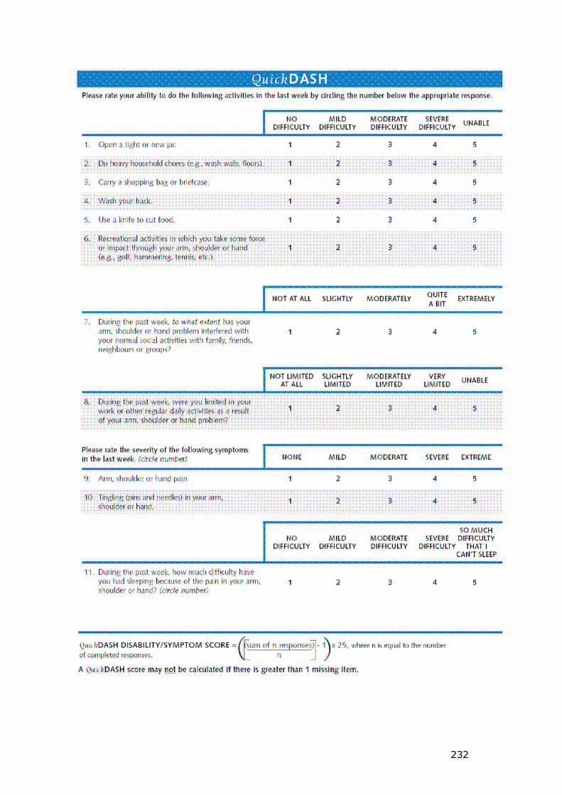
232
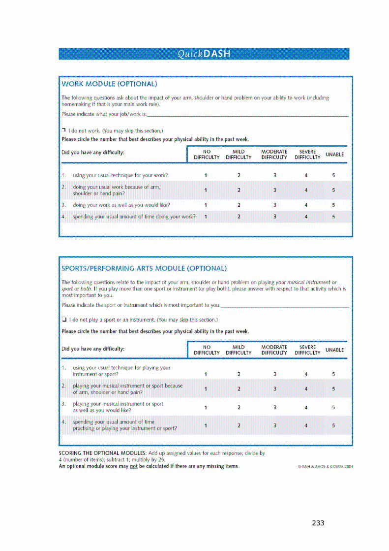
233
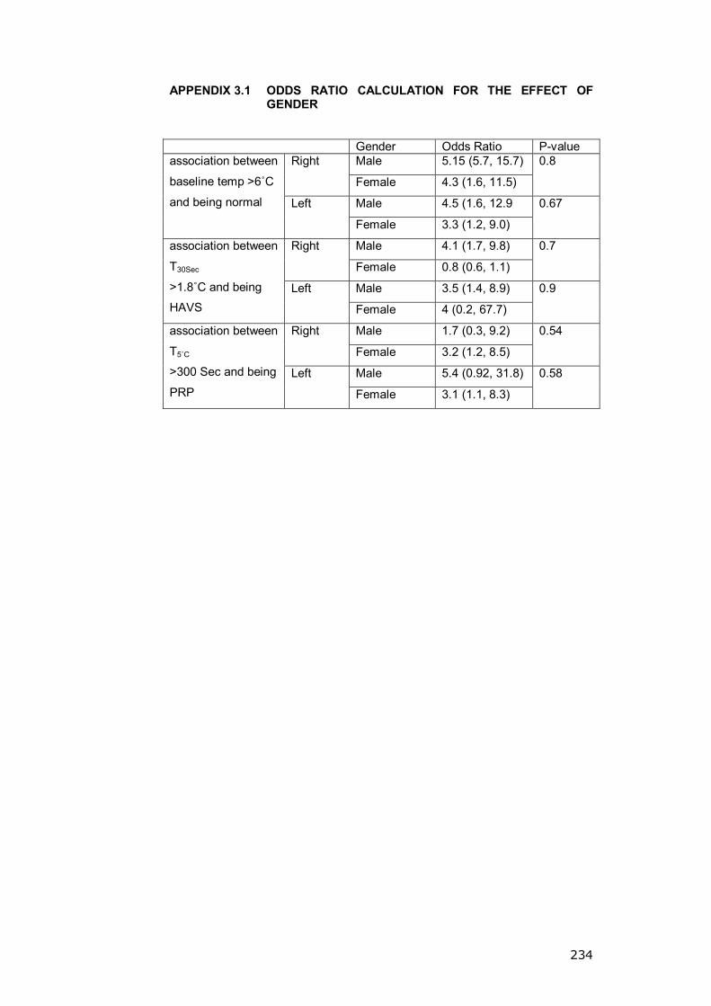
234
APPENDIX 3.1 ODDS RATIO CALCULATION FOR THE EFFECT OF GENDER
Gender Odds Ratio P-value
Male 5.15 (5.7, 15.7) Right
Female 4.3 (1.6, 11.5)
0.8
Male 4.5 (1.6, 12.9
association between
baseline temp >6ûC
and being normal Left
Female 3.3 (1.2, 9.0)
0.67
Male 4.1 (1.7, 9.8) Right
Female 0.8 (0.6, 1.1)
0.7
Male 3.5 (1.4, 8.9)
association between
T30Sec
>1.8ûC and being
HAVS Left
Female 4 (0.2, 67.7)
0.9
Male 1.7 (0.3, 9.2) Right
Female 3.2 (1.2, 8.5)
0.54
Male 5.4 (0.92, 31.8)
association between
T5ûC
>300 Sec and being
PRP Left
Female 3.1 (1.1, 8.3)
0.58
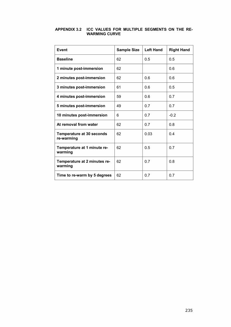
235
APPENDIX 3.2 ICC VALUES FOR MULTIPLE SEGMENTS ON THE RE-WARMING CURVE
Event Sample Size Left Hand Right Hand
Baseline 62 0.5 0.5
1 minute post-immersion 62 0.6
2 minutes post-immersion 62 0.6 0.6
3 minutes post-immersion 61 0.6 0.5
4 minutes post-immersion 59 0.6 0.7
5 minutes post-immersion 49 0.7 0.7
10 minutes post-immersion 6 0.7 -0.2
At removal from water 62 0.7 0.8
Temperature at 30 seconds re-warming
62 0.03 0.4
Temperature at 1 minute re-warming
62 0.5 0.7
Temperature at 2 minutes re-warming
62 0.7 0.8
Time to re-warm by 5 degrees 62 0.7 0.7

236
APPENDIX 3.3-A LDI REPEATABILITY ANALYSIS FOR OPERATOR [A] USING ACH
Stage Variable Normality
(K-S Test) Wilcoxon Signed Ranks Test
r (P-Value) COV ICC
Minimum 1 0.005
Minimum 2 0.20
0.004 0.02 (0.9) 73.5% -0.13
Maximum 1 0.09
Maximum 2 0.08
0.02 * 0.11 (0.46) 31.3% 0.19
Mean 1 0.20
Baseline
Mean 2 0.20
0.003 * 0.04 (0.8) 35.6% -0.08
Minimum 1 0.08
Minimum 2 0.19
0.02 *
0.28 (0.6) 57.2% 0.44
Maximum 1 0.19
Maximum 2 0.20
0.11 * 0.36 (0.01) 42.6% 0.51
Mean 1 0.001
Max post Ionto.
Mean 2 0.20
0.02 0.34 (0.02) 43.9% 0.52
Minimum 1 <0.0001
Minimum 2 0.001
0.02 0.06 (0.7) 67.3% 0.17
Maximum 1 0.02
Maximum 2 0.001
0.25 0.34 (0.02) 37.8% 0.43
Mean 1 0.01
Mean post Ionto.
Mean 2 0.004
0.03 0.12 (0.4) 45.9% 0.27
r: Spearman Correlation Coefficient; COV: Coefficient of Variation; ICC: Interclass Correlation Coefficient; (*) paired T-Test
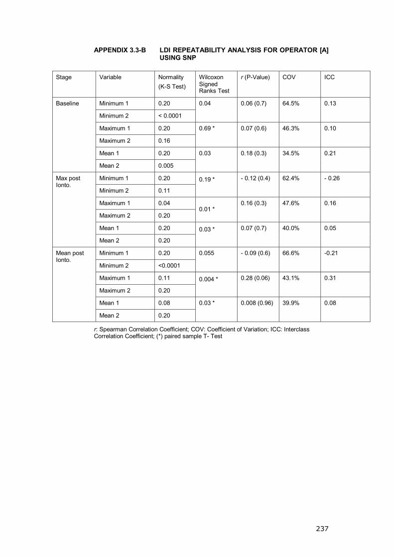
237
APPENDIX 3.3-B LDI REPEATABILITY ANALYSIS FOR OPERATOR [A] USING SNP
Stage Variable Normality
(K-S Test) Wilcoxon Signed Ranks Test
r (P-Value) COV ICC
Minimum 1 0.20
Minimum 2 < 0.0001
0.04 0.06 (0.7) 64.5% 0.13
Maximum 1 0.20
Maximum 2 0.16
0.69 * 0.07 (0.6) 46.3% 0.10
Mean 1 0.20
Baseline
Mean 2 0.005
0.03 0.18 (0.3) 34.5% 0.21
Minimum 1 0.20
Minimum 2 0.11
0.19 *
- 0.12 (0.4) 62.4% - 0.26
Maximum 1 0.04
Maximum 2 0.20 0.01 *
0.16 (0.3) 47.6% 0.16
Mean 1 0.20
Max post Ionto.
Mean 2 0.20
0.03 *
0.07 (0.7) 40.0% 0.05
Minimum 1 0.20
Minimum 2 <0.0001
0.055 - 0.09 (0.6) 66.6% -0.21
Maximum 1 0.11
Maximum 2 0.20
0.004 *
0.28 (0.06) 43.1% 0.31
Mean 1 0.08
Mean post Ionto.
Mean 2 0.20
0.03 * 0.008 (0.96) 39.9% 0.08
r: Spearman Correlation Coefficient; COV: Coefficient of Variation; ICC: Interclass Correlation Coefficient; (*) paired sample T- Test
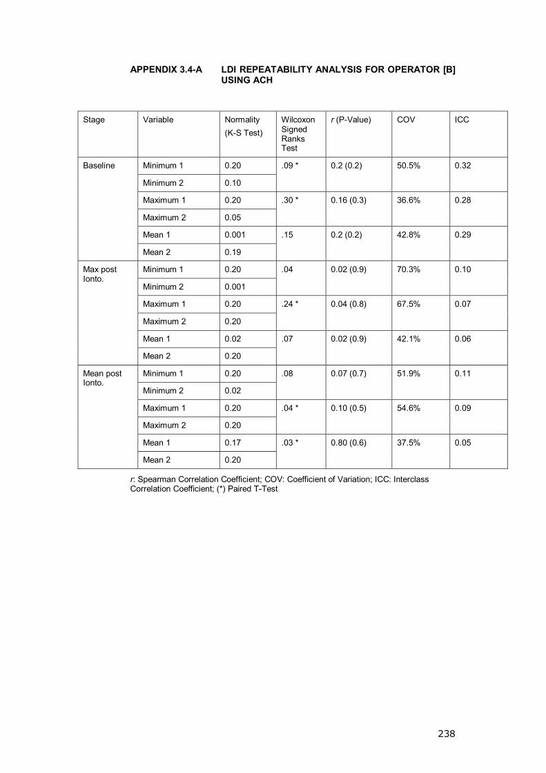
238
APPENDIX 3.4-A LDI REPEATABILITY ANALYSIS FOR OPERATOR [B] USING ACH
Stage Variable Normality (K-S Test)
Wilcoxon Signed Ranks Test
r (P-Value) COV ICC
Minimum 1 0.20
Minimum 2 0.10
.09 * 0.2 (0.2) 50.5% 0.32
Maximum 1 0.20
Maximum 2 0.05
.30 * 0.16 (0.3) 36.6% 0.28
Mean 1 0.001
Baseline
Mean 2 0.19
.15 0.2 (0.2) 42.8% 0.29
Minimum 1 0.20
Minimum 2 0.001
.04 0.02 (0.9) 70.3% 0.10
Maximum 1 0.20
Maximum 2 0.20
.24 * 0.04 (0.8) 67.5% 0.07
Mean 1 0.02
Max post Ionto.
Mean 2 0.20
.07 0.02 (0.9) 42.1% 0.06
Minimum 1 0.20
Minimum 2 0.02
.08 0.07 (0.7) 51.9% 0.11
Maximum 1 0.20
Maximum 2 0.20
.04 * 0.10 (0.5) 54.6% 0.09
Mean 1 0.17
Mean post Ionto.
Mean 2 0.20
.03 * 0.80 (0.6) 37.5% 0.05
r: Spearman Correlation Coefficient; COV: Coefficient of Variation; ICC: Interclass Correlation Coefficient; (*) Paired T-Test
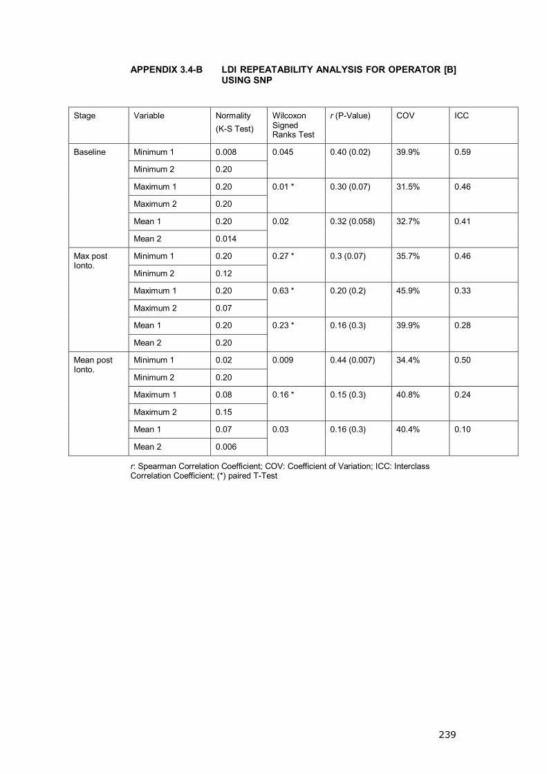
239
APPENDIX 3.4-B LDI REPEATABILITY ANALYSIS FOR OPERATOR [B] USING SNP
Stage Variable Normality (K-S Test)
Wilcoxon Signed Ranks Test
r (P-Value) COV ICC
Minimum 1 0.008
Minimum 2 0.20
0.045 0.40 (0.02) 39.9% 0.59
Maximum 1 0.20
Maximum 2 0.20
0.01 * 0.30 (0.07) 31.5% 0.46
Mean 1 0.20
Baseline
Mean 2 0.014
0.02 0.32 (0.058) 32.7% 0.41
Minimum 1 0.20
Minimum 2 0.12
0.27 * 0.3 (0.07) 35.7% 0.46
Maximum 1 0.20
Maximum 2 0.07
0.63 * 0.20 (0.2) 45.9% 0.33
Mean 1 0.20
Max post Ionto.
Mean 2 0.20
0.23 * 0.16 (0.3) 39.9% 0.28
Minimum 1 0.02
Minimum 2 0.20
0.009 0.44 (0.007) 34.4% 0.50
Maximum 1 0.08
Maximum 2 0.15
0.16 * 0.15 (0.3) 40.8% 0.24
Mean 1 0.07
Mean post Ionto.
Mean 2 0.006
0.03 0.16 (0.3) 40.4% 0.10
r: Spearman Correlation Coefficient; COV: Coefficient of Variation; ICC: Interclass Correlation Coefficient; (*) paired T-Test

240
APPENDIX 3.5-A LDI REPEATABILITY ANALYSIS FOR OPERATOR [C] USING ACH
Stage Variable Normality (K-S Test)
Wilcoxon Signed Ranks Test
r (P-Value) COV ICC
Minimum 1 0.20
Minimum 2 0.20
0.00 * 0.12 (0.44) 60% -0.127
Maximum 1 0.20
Maximum 2 0.002
0.004 0.15 (0.34) 29% 0.051
Mean 1 0.007
Baseline
Mean 2 0.00
0.00 0.37 (0.02) 26% 0.71
Minimum 1 0.20
Minimum 2 0.00
0.00 0.25 (0.11) 57% -0.002
Maximum 1 0.00
Maximum 2 0.20
0.59 0.04 (0.79) 84% 0.106
Mean 1 0.20
Max post Ionto.
Mean 2 0.20
0.02 * 0.45 (0.004) 50% 0.558
Minimum 1 0.02
Minimum 2 0.20
0.00 0.26 (0.11) 56% -0.024
Maximum 1 0.00
Maximum 2 0.02
0.09 0.39 (0.01) 47% 0.539
Mean 1 0.20
Mean post Ionto.
Mean 2 0.07
0.00 * 0.67 (<0.0001) 29% 0.632
r: Spearman Correlation Coefficient; COV: Coefficient of Variation; ICC: Interclass Correlation Coefficient; (*) paired T-Test was used in the analysis.
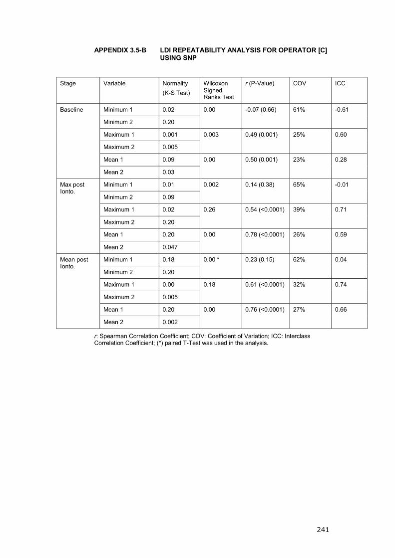
241
APPENDIX 3.5-B LDI REPEATABILITY ANALYSIS FOR OPERATOR [C] USING SNP
Stage Variable Normality (K-S Test)
Wilcoxon Signed Ranks Test
r (P-Value) COV ICC
Minimum 1 0.02
Minimum 2 0.20
0.00 -0.07 (0.66) 61% -0.61
Maximum 1 0.001
Maximum 2 0.005
0.003 0.49 (0.001) 25% 0.60
Mean 1 0.09
Baseline
Mean 2 0.03
0.00 0.50 (0.001) 23% 0.28
Minimum 1 0.01
Minimum 2 0.09
0.002 0.14 (0.38) 65% -0.01
Maximum 1 0.02
Maximum 2 0.20
0.26 0.54 (<0.0001) 39% 0.71
Mean 1 0.20
Max post Ionto.
Mean 2 0.047
0.00 0.78 (<0.0001) 26% 0.59
Minimum 1 0.18
Minimum 2 0.20
0.00 * 0.23 (0.15) 62% 0.04
Maximum 1 0.00
Maximum 2 0.005
0.18 0.61 (<0.0001) 32% 0.74
Mean 1 0.20
Mean post Ionto.
Mean 2 0.002
0.00 0.76 (<0.0001) 27% 0.66
r: Spearman Correlation Coefficient; COV: Coefficient of Variation; ICC: Interclass Correlation Coefficient; (*) paired T-Test was used in the analysis.

242
APPENDIX 4.1 RECRUITMENT FOR THE STUDY
Total CTS patients contacted 516
Replied67
Did not reply 228
Negative response
395 (77%)
Contactable 62
Positive Reply 121 (23%)
Excluded 45
Un-contactable16

243
APPENDIX 4.2 (A, B, C)
Normality (Kolmogorov-Smirnov)
Median Interquartile range
P-value (Mann-Whitney U)
Right Side <0.0001 7.3 3.9, 9.1 Baseline
Left Side 0.2 6.5 3.6, 8.9 0.4
Right Side 0.001 376 188, 639 T5C
Left Side 0.2 367 212, 615 0.98
A) No statistical difference in the CPT response between the two hands in the control group.
Normality (Kolmogorov-Smirnov)
Median Interquartile range
P-value (Mann-Whitney U)
Right Side .044 0.86 0.68, 1.15 Mean LDIMax Left Side .007 0.79 0.61, 0.94
0.09
Right Side .073 0.8 0.64, 0.99 Mean LDIMean Left Side .200 0.69 0.55, 0.85
*0.07
Right Side ** <0.0001 0.73 0.63, 0.84 Post-cooling Mean LDIMax Left Side ** 0.07 0.72 0.62, 0.85
0.8
Right Side ** 0.002 0.64 0.57, 0.78 Post- cooling Mean LDIMean Left Side ** 0.11 .69 0.59, 0.78
0.7
B) No statistical significant difference in the LDI response to ACH between the two hands in the control group (* independent sample T-Test used; ** Shapiro-Wilk test of normality used).
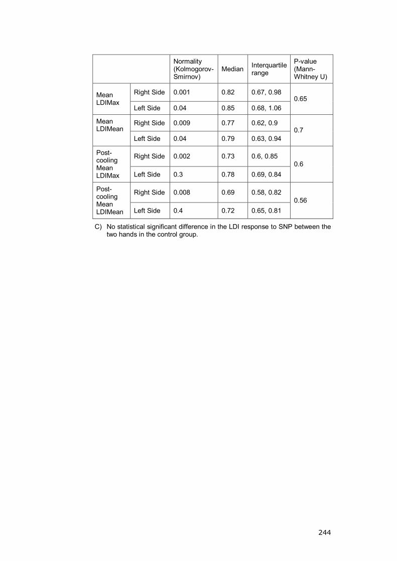
244
Normality (Kolmogorov-Smirnov)
Median Interquartile range
P-value (Mann-Whitney U)
Right Side 0.001 0.82 0.67, 0.98 Mean LDIMax
Left Side 0.04 0.85 0.68, 1.06 0.65
Right Side 0.009 0.77 0.62, 0.9 Mean LDIMean Left Side 0.04 0.79 0.63, 0.94
0.7
Right Side 0.002 0.73 0.6, 0.85 Post-cooling Mean LDIMax Left Side 0.3 0.78 0.69, 0.84
0.6
Right Side 0.008 0.69 0.58, 0.82 Post- cooling Mean LDIMean Left Side 0.4 0.72 0.65, 0.81
0.56
C) No statistical significant difference in the LDI response to SNP between the two hands in the control group.
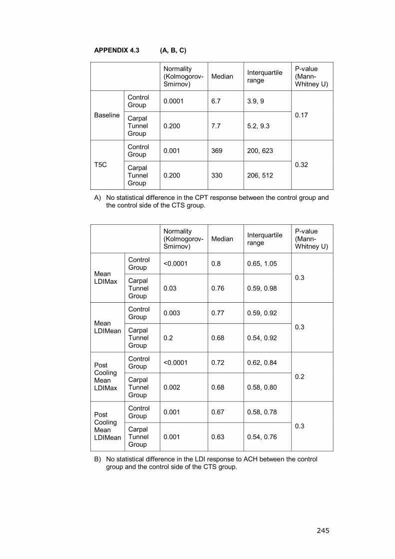
245
APPENDIX 4.3 (A, B, C)
Normality (Kolmogorov-Smirnov)
Median Interquartile range
P-value (Mann-Whitney U)
Control Group 0.0001 6.7 3.9, 9
Baseline Carpal Tunnel Group
0.200 7.7 5.2, 9.3 0.17
Control Group 0.001 369 200, 623
T5C Carpal Tunnel Group
0.200 330 206, 512 0.32
A) No statistical difference in the CPT response between the control group and the control side of the CTS group.
Normality (Kolmogorov-Smirnov)
Median Interquartile range
P-value (Mann-Whitney U)
Control Group <0.0001 0.8 0.65, 1.05
Mean LDIMax Carpal
Tunnel Group
0.03 0.76 0.59, 0.98 0.3
Control Group 0.003 0.77 0.59, 0.92
Mean LDIMean Carpal
Tunnel Group
0.2 0.68 0.54, 0.92 0.3
Control Group <0.0001 0.72 0.62, 0.84 Post
Cooling Mean LDIMax
Carpal Tunnel Group
0.002 0.68 0.58, 0.80 0.2
Control Group 0.001 0.67 0.58, 0.78 Post
Cooling Mean LDIMean
Carpal Tunnel Group
0.001 0.63 0.54, 0.76 0.3
B) No statistical difference in the LDI response to ACH between the control group and the control side of the CTS group.
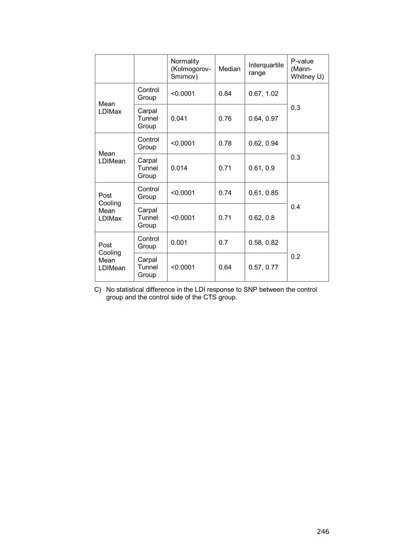
246
Normality (Kolmogorov-Smirnov)
Median Interquartile range
P-value (Mann-Whitney U)
Control Group <0.0001 0.84 0.67, 1.02
Mean LDIMax Carpal
Tunnel Group
0.041 0.76 0.64, 0.97 0.3
Control Group <0.0001 0.78 0.62, 0.94
Mean LDIMean Carpal
Tunnel Group
0.014 0.71 0.61, 0.9 0.3
Control Group <0.0001 0.74 0.61, 0.85 Post
Cooling Mean LDIMax
Carpal Tunnel Group
<0.0001 0.71 0.62, 0.8 0.4
Control Group 0.001 0.7 0.58, 0.82 Post
Cooling Mean LDIMean
Carpal Tunnel Group
<0.0001 0.64 0.57, 0.77 0.2
C) No statistical difference in the LDI response to SNP between the control group and the control side of the CTS group.
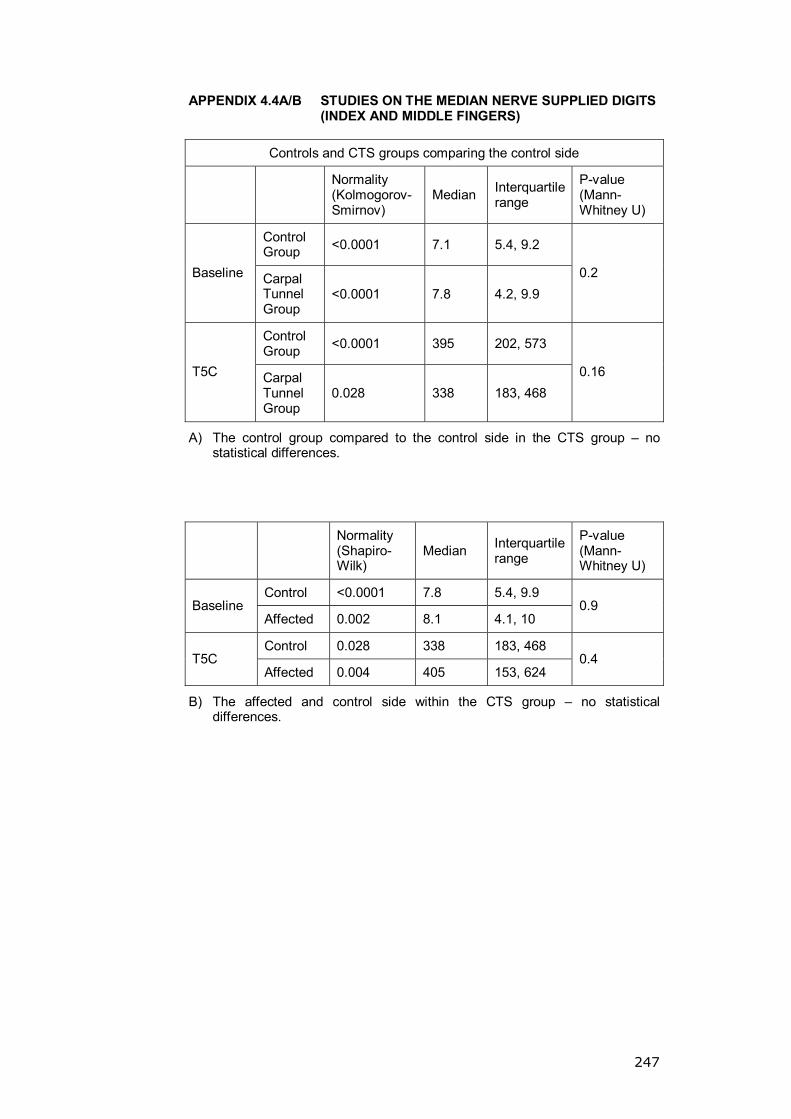
247
APPENDIX 4.4A/B STUDIES ON THE MEDIAN NERVE SUPPLIED DIGITS (INDEX AND MIDDLE FINGERS)
Controls and CTS groups comparing the control side
Normality (Kolmogorov-Smirnov)
Median Interquartile range
P-value (Mann-Whitney U)
Control Group <0.0001 7.1 5.4, 9.2
Baseline Carpal Tunnel Group
<0.0001 7.8 4.2, 9.9 0.2
Control Group <0.0001 395 202, 573
T5C Carpal Tunnel Group
0.028 338 183, 468 0.16
A) The control group compared to the control side in the CTS group � no statistical differences.
Normality (Shapiro-Wilk)
Median Interquartile range
P-value (Mann-Whitney U)
Control <0.0001 7.8 5.4, 9.9 Baseline
Affected 0.002 8.1 4.1, 10 0.9
Control 0.028 338 183, 468 T5C
Affected 0.004 405 153, 624 0.4
B) The affected and control side within the CTS group � no statistical differences.
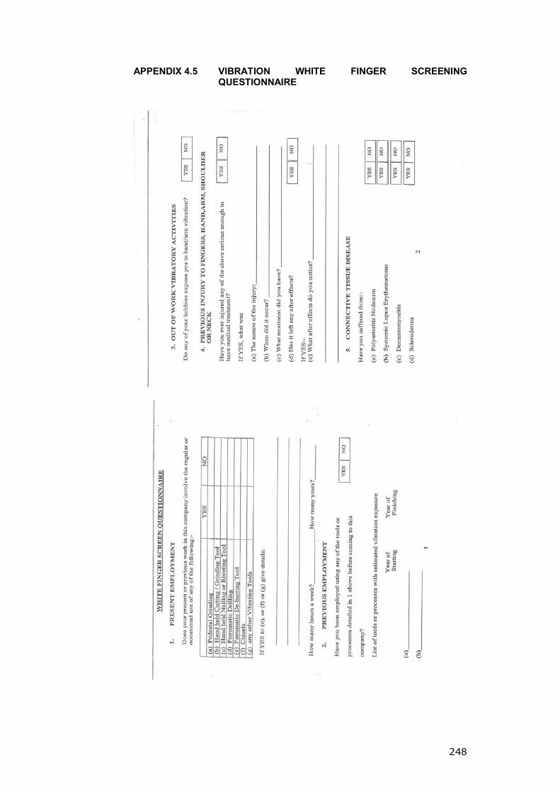
248
APPENDIX 4.5 VIBRATION WHITE FINGER SCREENING QUESTIONNAIRE
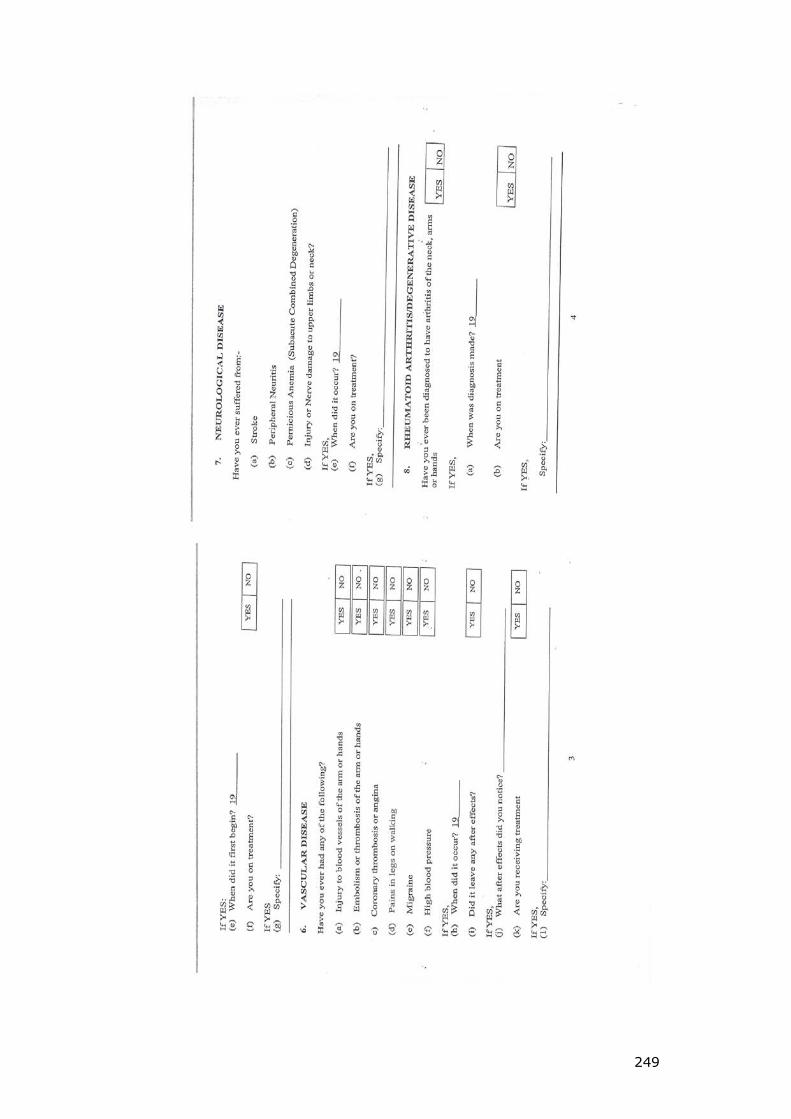
249

250
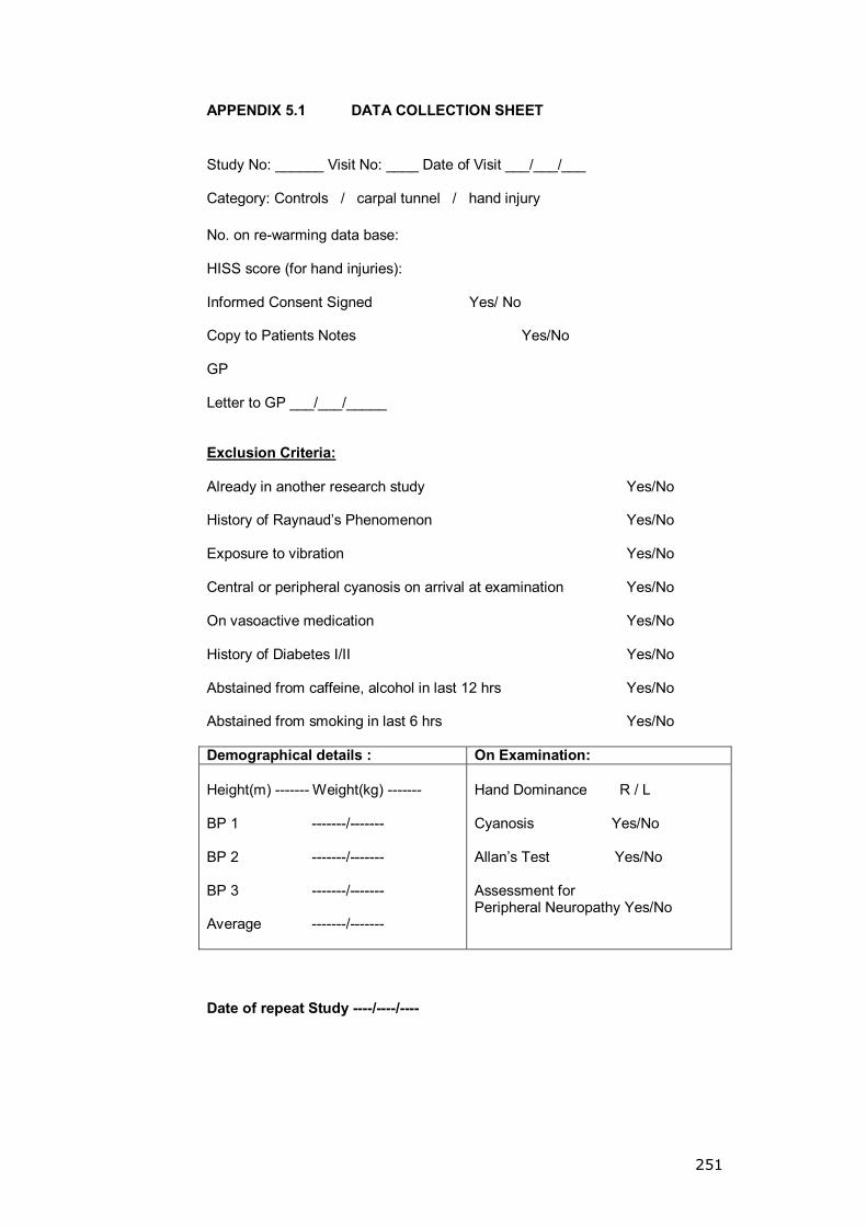
251
APPENDIX 5.1 DATA COLLECTION SHEET
Study No: ______ Visit No: ____ Date of Visit ___/___/___ Category: Controls / carpal tunnel / hand injury No. on re-warming data base: HISS score (for hand injuries): Informed Consent Signed Yes/ No Copy to Patients Notes Yes/No GP Letter to GP ___/___/_____ Exclusion Criteria: Already in another research study Yes/No History of Raynaud�s Phenomenon Yes/No Exposure to vibration Yes/No Central or peripheral cyanosis on arrival at examination Yes/No On vasoactive medication Yes/No
History of Diabetes I/II Yes/No Abstained from caffeine, alcohol in last 12 hrs Yes/No Abstained from smoking in last 6 hrs Yes/No Demographical details : On Examination: Height(m) ------- Weight(kg) ------- BP 1 -------/------- BP 2 -------/------- BP 3 -------/------- Average -------/-------
Hand Dominance R / L Cyanosis Yes/No Allan�s Test Yes/No Assessment for Peripheral Neuropathy Yes/No
Date of repeat Study ----/----/----
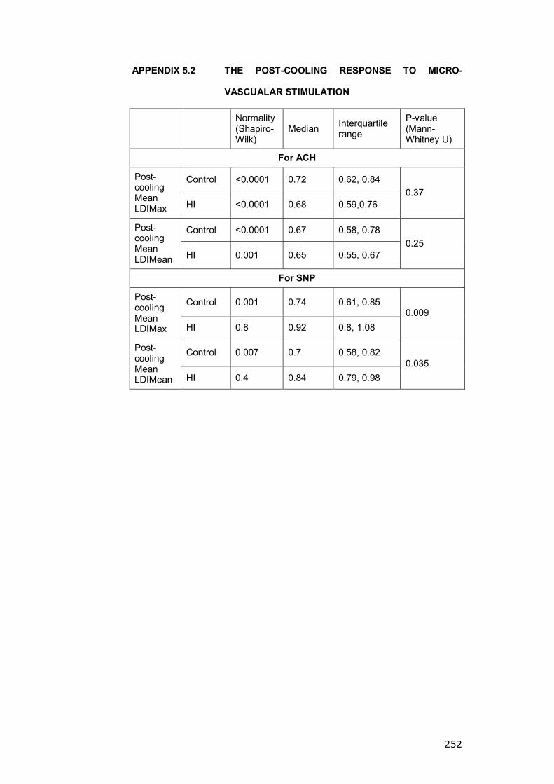
252
APPENDIX 5.2 THE POST-COOLING RESPONSE TO MICRO-
VASCUALAR STIMULATION
Normality (Shapiro-Wilk)
Median Interquartile range
P-value (Mann-Whitney U)
For ACH
Control <0.0001 0.72 0.62, 0.84 Post-cooling Mean LDIMax HI <0.0001 0.68 0.59,0.76
0.37
Control <0.0001 0.67 0.58, 0.78 Post-cooling Mean LDIMean HI 0.001 0.65 0.55, 0.67
0.25
For SNP
Control 0.001 0.74 0.61, 0.85 Post-cooling Mean LDIMax HI 0.8 0.92 0.8, 1.08
0.009
Control 0.007 0.7 0.58, 0.82 Post-cooling Mean LDIMean HI 0.4 0.84 0.79, 0.98
0.035
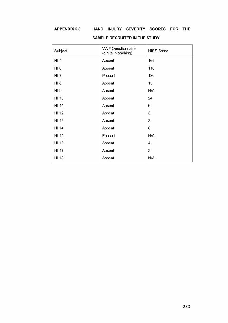
253
APPENDIX 5.3 HAND INJURY SEVERITY SCORES FOR THE
SAMPLE RECRUITED IN THE STUDY
Subject VWF Questionnaire (digital blanching) HISS Score
HI 4 Absent 165
HI 6 Absent 110
HI 7 Present 130
HI 8 Absent 15
HI 9 Absent N/A
HI 10 Absent 24
HI 11 Absent 6
HI 12 Absent 3
HI 13 Absent 2
HI 14 Absent 8
HI 15 Present N/A
HI 16 Absent 4
HI 17 Absent 3
HI 18 Absent N/A

254
APPENDIX 6.1 PATIENT FOLLOW-UP CHART IN THE REGISTRAR
LED CLINIC
Name DOB Sex Hospital No.
(Affix addressograph) Seen By:
Initial Visit Date: Right /Left Handed Occupation
Symptoms in Right / Left / Both Duration of symptoms
Off work because of symptoms Yes/No Smoker Yes/ No
Diabetic Yes/No
Test Right Left
Symptom Severity Score Levine Katz
score Functional score
Tinel�s Sign
Phalon Test
Reverse Phalon Test
Pinch Power
Grip Power
2 Point Discrimination
Semmes-Weinstien Monofilament Test
VA Score
Nerve conduction study
Injection Plan
Surgery
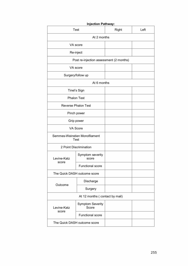
255
Injection Pathway:
Test Right Left
At 2 months
VA score
Re-inject
Post re-injection assessment (2 months)
VA score
Surgery/follow up
At 6 months
Tinel�s Sign
Phalon Test
Reverse Phalon Test
Pinch power
Grip power
VA Score
Semmes-Weinstien Monofilament Test
2 Point Discrimination
Symptom severity score Levine-Katz
score Functional score
The Quick DASH outcome score
Discharge Outcome
Surgery
At 12 months ( contact by mail)
Symptom Severity Score Levine-Katz
score Functional score
The Quick DASH outcome score

256
Surgery Pathway:
At 2 weeks
Wound healing
Dressing type
Tinel�s Sign
Phalon Test
Reverse Phalon Test
Pinch power
Grip power
VA Score
2 Point Discrimination
Semmes-Weinstien Monofilament Test
Symptom severity score
Levine-Katz score
Functional score
The Quick DASH outcome score
At 6 months
Tinel�s Sign
Phalon Test
Reverse Phalon Test
Pinch power
Grip power
VA Score
2 Point Discrimination
Semmes-Weinstien Monofilament Test
Symptom severity score
Levine-Katz score
Functional score
The Quick DASH outcome score
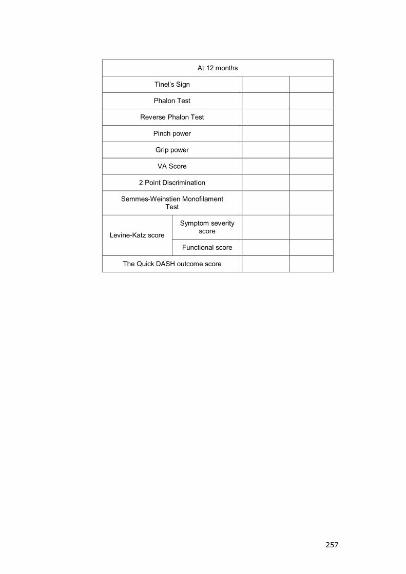
257
At 12 months
Tinel�s Sign
Phalon Test
Reverse Phalon Test
Pinch power
Grip power
VA Score
2 Point Discrimination
Semmes-Weinstien Monofilament Test
Symptom severity score
Levine-Katz score
Functional score
The Quick DASH outcome score

258
APPENDIX 6.2 DEMOGRAPHICAL VARIATION BETWEEN THE
INJECTION AND THE SURGERY GROUPS
Category Data type p-value
Age Normally distributed 0.46*
Sex Categorical data 0.35**
Dominance Categorical data 0.27
Smoking Categorical data 0.07
Affected side Categorical data 0.65
Duration of symptoms Skewed sample 0.29***
Off work Categorical data 0.41
* T-Test, **X2-test, *** Mann-Whitney Test