studies on the epidemiology of toxoplasmosis in …
TRANSCRIPT

STUDIES ON THE EPIDEMIOLOGY OF TOXOPLASMOSIS
IN SOUTH AFRICA
Kesenthri Kistiah
Dissertation submitted to the Faculty of Health Sciences, University of the
Witwatersrand, Johannesburg, in fulfilment of the requirements for the degree
of
Master of Science in Medicine.
Johannesburg, 2009
ii
I, Kesenthri Kistiah, declare that this dissertation is my own work. It is being
submitted for the degree of Master of Science in Medicine in the University of the
Witwatersrand, Johannesburg. It has not been submitted before for any degree or
examination at this or any other University.
This work was done under ethical clearance from the Human Research Ethics
Committee (Medical) of the University of the Witwatersrand (Clearance Certificates
M070637 and M070347) and the Animal Ethics Screening Committee (Clearance
Certificate 2008/50/01)
Kesenthri Kistiah
January 2009

iii
To my loving parents,
Alec and Kanthi Kistiah
iv
ABSTRACT
Toxoplasmosis is an infection of vertebrates caused by the obligate intracellular
protozoan parasite, Toxoplasma gondii. It is one of the most common parasitic
diseases of humans, infecting approximately one third of the world’s population. It is
a significant cause of congenital disease and an important opportunistic pathogen
which has become an increasing problem worldwide due to the AIDS epidemic. There
is limited historical information about the disease in South Africa. More knowledge is
needed at a regional level to properly consider solutions aimed at reducing the risk for
this disease. The seroprevalence of T. gondii in samples of selected populations at
risk, namely HIV-positive individuals and a more general population sample biased
towards pregnant women, was therefore investigated and found to be 9.8% (37/376)
and 6.4% (32/497) in the respective samples. The Pastorex Toxo latex agglutination
test was evaluated and found to be a cheap, reliable method to screen for T. gondii
exposure. PCR-based diagnostics were developed for direct diagnosis on tissue
samples. Rodent T. gondii infection prevalence was investigated, but did not yield any
positive results. This study helped to answer questions relating to the seroprevalence
and diagnosis of T. gondii in South Africa. Many questions still remain to be
answered, however to fully understand the impact of this parasite in our country.

v
ACKNOWLEDGEMENTS
Firstly, I would like to thank my supervisors, Dr John Frean, Dr Antonio Barragan
and Dr Jadwiga Winiecka-Krusnell for their valuable supervision, guidance, support
and encouragement throughout this project.
I would like to thank Dr Alan Karstaedt and the staff at the HIV clinic at Chris Hani
Baragwanath Hospital for their assistance with the collection of samples.
I would like to thank the staff of the Parasitology Reference Unit at the National
Institute for Communicable Diseases and the Department of Parasitology, Mycology
and Environmental Microbiology at the Swedish Institute for Infectious Diseases for
their help and kind use of their facilities.
I would also like to thank the following people for their assistance and suggestions:
Ms Leigh Dini, Ms Lorraine Arntzen, Dr Bernice Harris and Dr Cheryl Cohen.
Most of all, I would like to thank my parents, Alec and Kanthi, my sister Arshnie and
my brother Yuven for their incredible support and encouragement. Thank you for
always believing in me.
Finally, I would like to thank the Swedish Research Links Programme, Swedish
Research Council, Swedish International Development Cooperation Agency (SIDA),
the National Health Laboratory Service Research Trust and the Medical Research
Council for funding this project.

vi
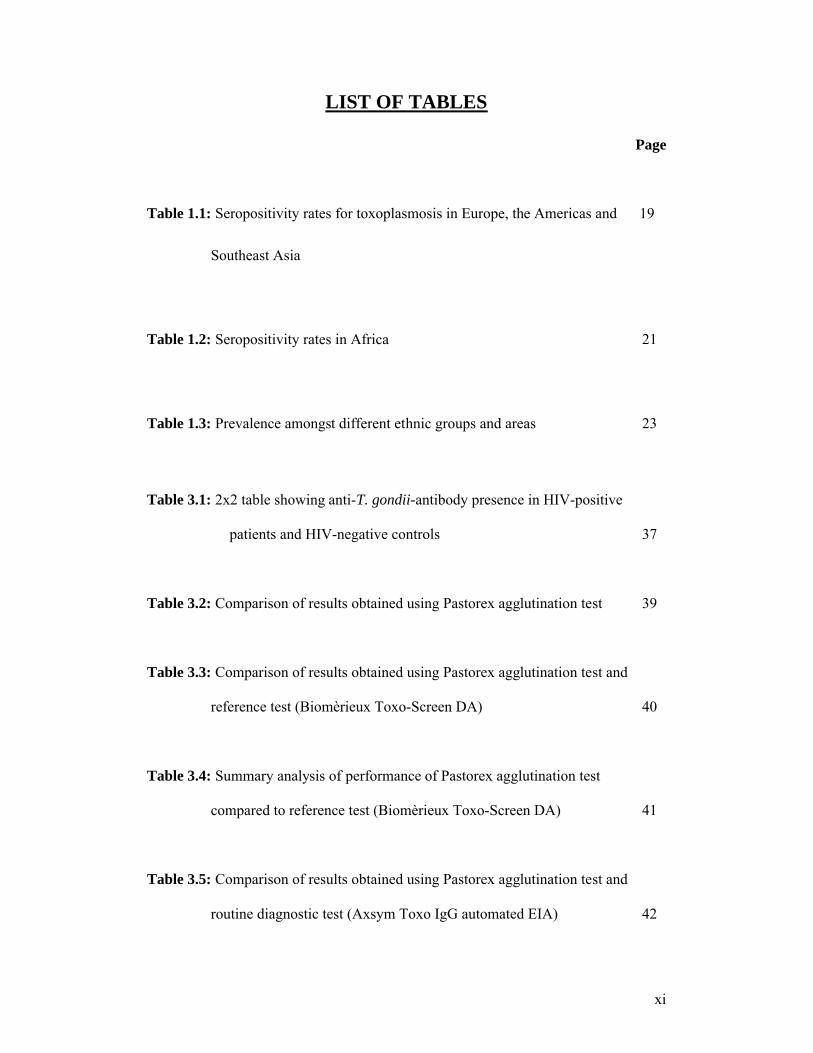
TABLE OF CONTENTS
Page
DECLARATION ii
DEDICATION iii
ABSTRACT iv
ACKNOWLEDGEMENTS v
TABLE OF CONTENTS vi
LIST OF FIGURES ix
LIST OF TABLES xi
ABBREVIATIONS xiii
1. INTRODUCTION 1
1.1 History 1
1.2 Morphology 3
1.3 Transmission 6
1.4 Life cycle 9
1.5 Strains 10
1.6 Toxoplasmosis 11
1.6.1 Clinical manifestations 11
1.6.2 Ocular toxoplasmosis 12
1.6.3 Congenital toxoplasmosis 13
1.6.4 Toxoplasmosis in immunocompromised hosts 14
1.6.5 Diagnosis 15
1.6.6 Prevention and treatment 18

vii
Page
1.7 Epidemiology 19
1.8 Climate and topography of South Africa 24
1.9 Aims and objectives 27
2. MATERIALS AND METHODS 29
2.1 HIV-positive prospective serosurvey 29
2.2 HIV-negative controls 32
2.3 Retrospective serosurvey 32
2.4 Prevalence in rodents 33
2.5 Toxoplasma cultivation 36
2.6 Data Analysis 36
3. RESULTS 37
3.1 Prevalence of anti-T. gondii antibodies in HIV-positive and HIV-negative
patients 37
3.2 Retrospective serosurvey 38
3.3 Evaluation of the Pastorex agglutination test 38
3.4 Prevalence in rodents 43
4. DISCUSSION 47
4.1 Introduction 47
4.2 Evaluation of the Pastorex agglutination test 47
4.3 Toxoplasma gondii in South Africa 53
4.4 Toxoplasma gondii in rodents 56
viii
Page
5. CONCLUSIONS 58
APPENDIX A: Subject enrolment information and consent forms, and ethical
clearance certificates 60
APPENDIX B: Protocols for procedures 65
REFERENCES 79
ix
LIST OF FIGURES
Page
Figure 1.1: Schematic diagram showing organelles of a tachyzoite of T. gondii 4
Figure 1.2: Schematic diagram showing organelles of a bradyzoite of T. gondii 5
Figure 1.3: An overview of the modes of transmission of T. gondii 7
Figure 1.4: Variation in T. gondii seroprevalence in Africa 22
Figure 1.5: Average temperature, humidity and rainfall of Cape Town 26
Figure 1.6: Average temperature, humidity and rainfall of Durban 26
Figure 1.7: Average temperature, humidity and rainfall of Johannesburg 27
Figure 3.1: Anti-T. gondii-antibody presence in HIV-positive patients and HIV-
negative controls 38
Figure 3.2: Electrophoresis gel of validation PCR 44

x
Page
Figure 3.3: Electrophoresis gel, showing atypical bands 45
Figure 3.4: Electrophoresis gel showing Toxoplasma gondii strains 46
xi
LIST OF TABLES
Page
Table 1.1: Seropositivity rates for toxoplasmosis in Europe, the Americas and 19
Southeast Asia
Table 1.2: Seropositivity rates in Africa 21
Table 1.3: Prevalence amongst different ethnic groups and areas 23
Table 3.1: 2x2 table showing anti-T. gondii-antibody presence in HIV-positive
patients and HIV-negative controls 37
Table 3.2: Comparison of results obtained using Pastorex agglutination test 39
Table 3.3: Comparison of results obtained using Pastorex agglutination test and
reference test (Biomèrieux Toxo-Screen DA) 40
Table 3.4: Summary analysis of performance of Pastorex agglutination test
compared to reference test (Biomèrieux Toxo-Screen DA) 41
Table 3.5: Comparison of results obtained using Pastorex agglutination test and
routine diagnostic test (Axsym Toxo IgG automated EIA) 42
xii
Page
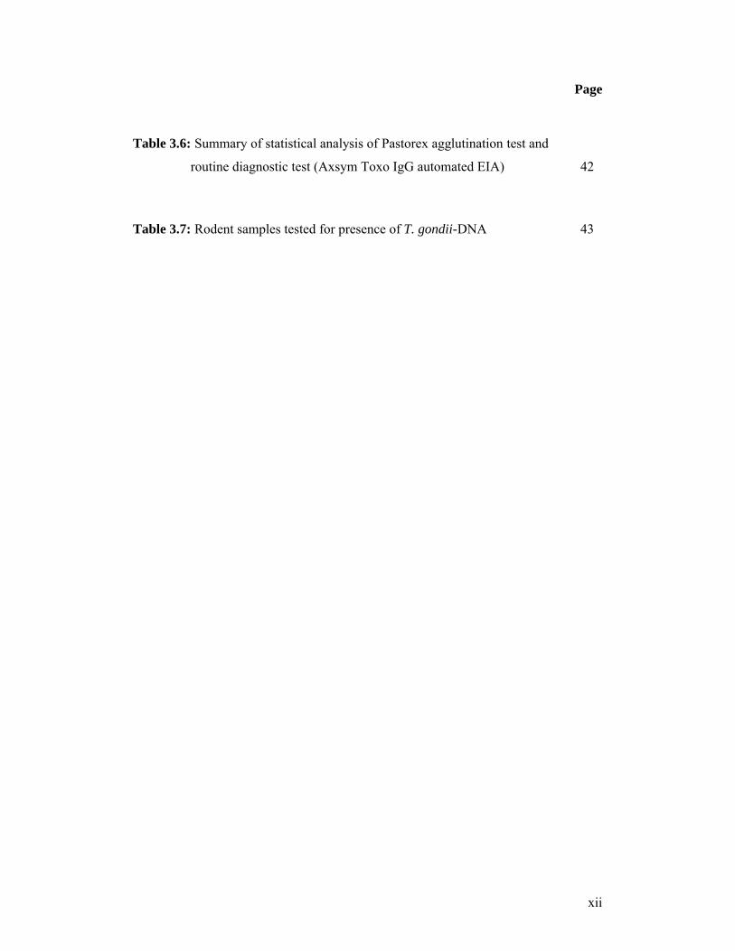
Table 3.6: Summary of statistical analysis of Pastorex agglutination test and
routine diagnostic test (Axsym Toxo IgG automated EIA) 42
Table 3.7: Rodent samples tested for presence of T. gondii-DNA 43

xiii
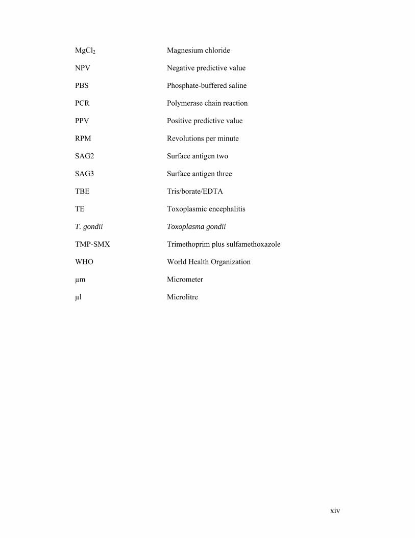
ABBREVIATIONS
AIDS Acquired immunodeficiency syndrome
ARV Antiretroviral
CD4 Cluster of differentiation 4
CFT Complement fixation test
CSF Cerebrospinal fluid
DMEM Dulbecco’s modified Eagle medium
DNA Deoxyribonucleic acid
dNTP Deoxyribonucleotide triphosphate
EDTA Ethylenediaminetetraacetic acid
EIA Enzyme immunoassay
ELISA Enzyme-linked immunosorbent assay
FCS Foetal calf serum
FITC Fluorescein isothiocyanate
HAART Highly active antiretroviral therapy
HIV Human immunodeficiency virus
HFF Human foreskin fibroblast cells
IFA Indirect fluorescent antibody assay
IFL Indirect immunofluorescence assay
IgA Immunoglobulin A
IgE Immunoglobulin E
IgG Immunoglobulin G
IgM Immunoglobulin M
LDM Long distance migration
xiv
MgCl2 Magnesium chloride
NPV Negative predictive value
PBS Phosphate-buffered saline
PCR Polymerase chain reaction
PPV Positive predictive value
RPM Revolutions per minute
SAG2 Surface antigen two
SAG3 Surface antigen three
TBE Tris/borate/EDTA
TE Toxoplasmic encephalitis
T. gondii Toxoplasma gondii
TMP-SMX Trimethoprim plus sulfamethoxazole
WHO World Health Organization
µm Micrometer
µl Microlitre
1
CHAPTER 1
INTRODUCTION
1.1. History
Toxoplasmosis is an infection of vertebrates caused by the obligate intracellular
protozoan parasite, Toxoplasma gondii (Tenter et al., 2000). T. gondii is one of the
most common parasites of humans worldwide, infecting approximately one third of
the world’s population (Tenter et al., 2000). It is a facultative heteroxenous parasite
whose definitive hosts are members of the family Felidae, but is capable of infecting
mammals, birds and reptiles as intermediate hosts. Its broad host range, high infection
rate, worldwide distribution and the ability to maintain a benign coexistence with its
host, are features of T. gondii which allow it to be widely regarded as one of the most
successful parasites on Earth (Carruthers, 2002).
T. gondii belongs to the Phylum Apicomplexa, class Sporozoasida and subclass
Coccidiasina. The genus name ‘Toxoplasma’ is derived from the Greek word ‘toxon’
meaning bow, which describes the crescentric shape of the tachyzoite stage, and the
species name ‘gondii’ from the rodent from which it was first isolated (Markus,
2003).
T. gondii was first discovered 100 years ago by Nicolle and Manceaux at the Pasteur
Institute in Tunisia, in 1908. Whilst conducting leishmaniasis research on the North
African rodent, Ctenodactylus gondi, the investigators isolated T. gondii merozoites
from its blood, liver and spleen (Frenkel, 1973; Cox, 2002; Sukthana, 2006). Around

2
the same time in São Paulo, Brazil, Splendore independently described T. gondii in a
laboratory rabbit, thus suggesting its worldwide distribution (reviewed by Frenkel,
1973).
The first human case of toxoplasmosis was described by Janků in 1923 (Cox, 2002;
Sukthana, 2006). He observed tissue cysts in the retina of an 11-month-old infant in
Prague (Sukthana, 2006). In 1939, Wolf et al. successfully isolated the parasite from
tissue from a neonate with encephalitis, by animal inoculation (Cox, 2002). This
served to be the first example of an organism causing disease in utero. Adult infection
with the parasite was first described by Pinkerton and Henderson in 1941 and
childhood infection by Sabin in 1942 (reviewed by Frenkel, 1973).
Toxoplasmic retinochoroiditis was first identified by Wilder in 1952 in the retinas of
50 human cases, and the parasite was later isolated from the eye in 1954 by Jacobs et
al. Reactivation of a latent infection in man was described by Frenkel in 1956
(reviewed by Frenkel, 1973).
In 1954, Weinman and Chandler first investigated the possible transmission of the
parasite through the consumption of undercooked meat. Evidence for this was
provided by Jacobs, Remington and Melton in 1960. They found that T. gondii cysts
from pigs, sheep and other experimental animals were resistant to proteolytic enzymes
and were readily infectious when swallowed (reviewed by Frenkel, 1973).
3
In 1970, more than 60 years after the first description of its asexual stage in
intermediate hosts, the life cycle of T. gondii was completely elucidated by the
discovery of a sexual stage in the small intestine of cats (Tenter et al., 2000).
1.2. Morphology
T. gondii has three infective stages: the sporozoite stage in oocysts in faeces, the
rapidly dividing tachyzoites found during an acute infection and the slowly dividing
bradyzoite stage in cysts during latent infection (Peterson and Dubey, 2001; Jones et
al., 2001b).
Tachyzoites are crescentic in shape, and approximately 6µm long and 2 µm wide
(Bhopale, 2003). They are coated by a three-unit membrane, namely a plasmalemma
and an inner membrane consisting of two closely situated membranes, all of which
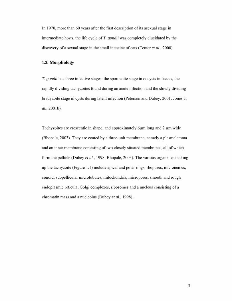
form the pellicle (Dubey et al., 1998; Bhopale, 2003). The various organelles making
up the tachyzoite (Figure 1.1) include apical and polar rings, rhoptries, micronemes,
conoid, subpellicular microtubules, mitochondria, micropores, smooth and rough
endoplasmic reticula, Golgi complexes, ribosomes and a nucleus consisting of a
chromatin mass and a nucleolus (Dubey et al., 1998).

4
Figure 1.1: Schematic diagram showing organelles of a tachyzoite of T. gondii.
(Adapted from Dubey et al., 1998.).
Tachyzoites are spread via the blood system in lymphocytes, macrophages and free in
the plasma and are able to infect almost any type of tissue, especially those in the eye,
central nervous system, heart, placenta and skeletal muscle (Montoya and Liesenfeld,
2004). They are capable of crossing tissue boundaries, such as the blood-brain barrier
and the placenta (Carruthers, 2002). They are able to multiply rapidly by
endodyogeny and such replication leads to cell necrosis when invaded cells can no
longer hold these parasites and erupt. Replication of tachyzoites occurs during the
first 8-12 days and accounts for the acute phase of infection (Carruthers, 2002). This
stage is responsible for the clinical manifestations of the disease as it produces a
strong inflammatory response (Montoya and Liesenfeld, 2004).
5
T. gondii induces a strong type 1 T-cell-mediated immune response, which promotes
a self-limiting infection, ensuring the survival of its host and thus itself (Carruthers,
2002). As the immune response progresses, interferon-γ, secreted by antigen-specific
T-cells, restricts tachyzoite replication.
Tachyzoites are sensitive to proteolytic enzymes and can therefore be destroyed
during gastric digestion. The pressure of the host’s immune system on the parasite
stimulates the formation of cysts and causes tachyzoites to be transformed into
bradyzoites, marking the beginning of the chronic phase of the infection (Carruthers,
2002; Montoya and Liesenfeld, 2004).
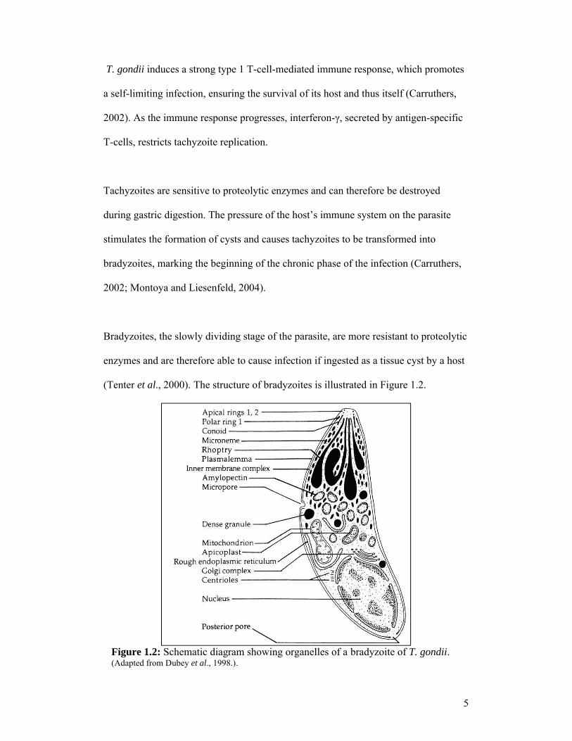
Bradyzoites, the slowly dividing stage of the parasite, are more resistant to proteolytic
enzymes and are therefore able to cause infection if ingested as a tissue cyst by a host
(Tenter et al., 2000). The structure of bradyzoites is illustrated in Figure 1.2.
Figure 1.2: Schematic diagram showing organelles of a bradyzoite of T. gondii. (Adapted from Dubey et al., 1998.).

6
They are found in quiescent tissue cysts, usually in the brain where cysts are
spheroidal in shape, and in muscle tissue where they are elongated (Dubey et al.,
1998; Roberts and McLeod, 1999). Tissue cysts are able to persist for the duration of
the host’s life and bradyzoites can be released from these cysts to form tachyzoites
again, causing a reactivated infection in immunocompromised hosts (Montoya and
Liesenfeld, 2004).
Oocysts are 10 µm to 12 µm in size and are produced within the intestine of the
definitive host. Sporozoites are found within these oocysts, which are shed in the
faeces of the definitive host (Peterson and Dubey, 2001).
1.3. Transmission
Transmission of the parasite can occur in several ways, as illustrated in Figure 1.3.
Definitive hosts can be infected by ingestion of oocysts from the environment or cysts
in prey. Transmission to humans can occur via accidental ingestion of infectious
stages through faecal-oral contact, the consumption of contaminated water, fruit or
vegetables, contaminated undercooked meat, via the placenta to the foetus, and rarely
via organ transplantation or blood transfusion (Montoya and Liesenfeld, 2004).

7
Figure 1.3: An overview of the modes of transmission of T. gondii.
In three large European case-control studies, undercooked meat was shown to be the
largest risk factor for T. gondii infection. The largest of these studies showed that 30-
60% of infections in pregnant women were due to the consumption of undercooked
contaminated meat (Kijlstra and Jongert, 2008).
Experimental infection of cattle, sheep, pigs and goats, all of which are used for
human consumption, showed that they are susceptible to T. gondii infection via
oocysts or tissue cyst intake (Kijlstra and Jongert, 2008). As a result of infection, most
animals harbour parasites in their tissues. Serological studies on cattle show
prevalences that can be as high as 92%, but tissue cysts are rarely found in beef as

8
cattle are known to show a high resistance to T. gondii (Tenter et al., 2000). Sheep are
fully susceptible to T. gondii infection and the consumption of mutton or lamb has
therefore been identified as a significant risk for T. gondii infection (Kijlstra and
Jongert, 2008).
Pork was generally considered a major source of infection in Europe and the USA,
but recent studies on fattening pigs in the Netherlands, Austria and Germany have
shown that T. gondii infections in pigs have dropped to less than 1% over the last ten
years (Tenter et al., 2000). This is largely due to modern, more hygienic farming
systems, as well as the increasing use of frozen meat (Kijlstra and Jongert, 2008).
Free-ranging chickens are increasingly being identified as an important source of
T. gondii infection, especially in developing countries. Seroprevalences of up to 65%
have been reported and parasite presence shown in 81% of these seropositive animals
(Kijlstra and Jongert, 2008).
T. gondii-infected rats may serve as reservoirs of infection for animals such as pigs,
dogs and cats, and are therefore epidemiologically potentially important in the
transmission of this parasite (Dubey and Frenkel, 1998). Serological data from
different countries showed that T. gondii prevalence in rats can be as high as 91% in
the water rat, Hydromys chrysogaster, in Queensland, Australia. Other areas such as
China, Costa Rica, Egypt, Finland, France, Germany, India, Japan, Mexico, Poland,
Italy and the USA and UK have varying prevalences from 1% to 70% in different
rodent species. A study in Africa by de Roever-Bonnet in 1972 showed an 8%
seroprevalence in Rattus rattus (Dubey and Frenkel, 1998).

9
A rodent control campaign on three organic pig farms over a four-month period
showed the disappearance of T. gondii from pigs in two of the three farms at the end
of the campaign and subsequent reappearance in one of these farms after the rodent
control campaign was stopped, thus emphasizing the role of rodents in the
transmission of T. gondii (Kijlstra et al., 2008).
The transmission of the parasite through contaminated water has generally been
considered uncommon; however, the widespread infection of marine mammals
indicates that that contaminated water may be a potential source of infection (Dubey,
2004). Recent outbreaks of toxoplasmosis linked to contaminated water supplies
provide further evidence for this (Dubey, 2004).
1.4. Life Cycle
T. gondii has a two-stage life cycle, consisting of a sexual phase in the definitive host,
and an asexual phase occurring in the intermediate host. Definitive hosts can be
infected by ingestion of parasites within tissue cysts or oocysts. There are five distinct
asexual phases that occur in enterocytes before gametogony begins. Microgametes
fertilize macrogametes in the enterocyte forming fertilized zygotes. A wall is then laid
around each zygote forming unsporulated oocysts, which are released into the
intestinal lumen when enterocytes rupture. These oocysts are then released into the
environment with the faeces (Peterson and Dubey, 2001). The prepatent period,
which is the period between ingestion and shedding, is approximately three to ten
days (Tenter et al., 2000; Peterson and Dubey, 2001). Cats not previously infected
with T. gondii shed oocysts after ingestion of infectious stages, whereas cats that have
10
been previously infected are generally immune against renewed oocyst shedding. This
immunity does not, however, continue throughout the duration of the cat’s life
(Petersen, 2003). Depending on the temperature, aeration and humidity of the
environment, oocysts begin to sporulate within five days, dividing into two
sporocysts, each containing four sporozoites. These sporulated oocysts can remain
infectious for months in the environment (Peterson and Dubey, 2001).
Once ingested by an intermediate host, the outer walls of cysts or oocysts are
disrupted by enzymatic degradation, releasing either bradyzoites or sporozoites into
the intestinal lumen. These then actively invade surrounding cells and transform into
tachyzoites. Upon entering the host cell, the parasite pulls the host cell membrane
around itself and is then surrounded by a parasitophorous vacuole (Petersen, 2003;
Bhopale, 2003) .Tachyzoites rapidly divide within the host cell, leading to its rupture,
releasing the parasites into the blood and lymphatic system, where they are carried to
other cells which they then invade, repeating this process (Bhopale, 2003; Montoya
and Rosso, 2005).
1.5. Strains
T. gondii has a highly unusual population structure comprised of three clonal lineages
(I, II and III). These differ in virulence and epidemiological pattern of occurrence
(Montoya and Liesenfeld, 2004). Studies using murine models show that the type I
strain is highly virulent and has a lethal dose of a single parasite regardless of the
genetic background of the mouse. Type II and III strains have a 50% lethal dose of
more than 103 parasites and the outcome is dependent on the genotype of the host
(Mordue et al., 2001). Type I and II strains have been reported in human cases, with

11
type I often associated with severe congenital and ocular disease, suggesting that it
may be more pathogenic in humans (Mordue et al., 2001). Type III has been shown to
be more common in animals (Montoya and Liesenfeld, 2004).
Until recently, T. gondii was considered to have little genetic variability. Recent
studies on T. gondii isolates from Brazil, however, show that they are both genetically
and biologically different from those in the USA and Europe (Velmurugan et al.,
2008). A recent study on T. gondii isolates from chickens in six different African
countries (Nigeria, Congo, Egypt, Burkina Faso, Kenya and Mali) revealed four
genotypes. Most isolates belonged to the clonal type II and III strains with one
Nigerian isolate having an atypical genotype (Velmurugan et al., 2008). There is no
information about the strains prevalent in South Africa.
1.6. Toxoplasmosis
Toxoplasmosis is a widespread disease and has been classified as the third most
common cause of food-borne deaths in the USA. Over the last twenty years, it has
also emerged as one of the most common opportunistic infections associated with
HIV and AIDS, and is a major cause of mortality in AIDS patients in developing
countries (Carruthers, 2002).
1.6.1. Clinical manifestations
The clinical manifestations of toxoplasmosis vary, depending on parasite
characteristics such as virulence of the strain and inoculum size, as well as host
factors such as genetic background and immune status (Montoya and Liesenfeld,
12
2004). Toxoplasmosis in immunocompetent individuals is typically mild or
asymptomatic and usually results in life-long immunity (Tenter et al., 2000; Markus,
2003). Symptoms most commonly include lymphadenopathy, which may be
accompanied by headache, fever, fatigue, muscular or abdominal pain as well as
myocarditis, hepatitis and pulmonary necrosis (Tenter et al., 2000; Bhopale, 2003;
Markus, 2003). However, there are cases of the infection where overtly serious
clinical symptoms may occur; these include ocular toxoplasmosis, congenital
toxoplasmosis, and the reactivation of a latent infection in immunocompromised
individuals.
1.6.2. Ocular toxoplasmosis
T. gondii is the most common cause of retinochoroiditis in humans worldwide,
accounting for 28% to 55% of all cases of posterior uveitis (Pavesio and Lightman,
1996; Lappalainen and Hedman, 2004; Vallochi et al., 2005; Bonfioli and Orefice,
2005).
Retinochoroiditis commonly occurs as a result of congenital infection but can also be
due to an acquired or reactivated infection (Bonfioli and Orefice, 2005; Dubey and
Jones, 2008). It is usually a self-limiting disease with lesions healing within six
weeks (Rothova, 1993). Active lesions usually present with a white focus of
necrotizing retinochoroiditis close to old pigmented scars. These lesions are usually
circular or oval in shape and they vary in size. In the congenital forms, the lesions are
usually bilateral and central and in the acquired forms, the lesions are usually
unilateral and solitary (Pavesio and Lightman, 1996).
13
Parasites reach the eye as free tachyzoites or as cysts which rupture, releasing
tachyzoites. Once infected cells lyse, tachyzoites invade the retina and multiply in the
surrounding cells causing an inflammatory response (Rothova, 1993; Roberts and
McLeod, 1999). Clinical manifestations of acute retinochoroiditis include tearing,
pain, photophobia and progressive loss of vision over time, especially when there is
macular or optic nerve involvement (Pavesio and Lightman, 1996; Dubey and Jones,
2008).
1.6.3. Congenital toxoplasmosis
Toxoplasmosis is a significant cause of congenital disease. Congenital toxoplasmosis
occurs in between 1 and 10 per 10 000 live births in Europe (Cook et al., 2000; Tenter
et al., 2000; Montoya and Liesenfeld, 2004). Transplacental transmission occurs
when an immunocompetent woman acquires a primary infection during pregnancy
(Hegab and Al-Mutawa, 2003), or may also be due to a reactivated infection in
immunocompromised women (Dubey and Jones, 2008). Primary infections acquired
four to six months before conception usually result in no transplacental transmission
to the foetus (Tenter et al., 2000; Jones et al., 2001b).
Primary infection during pregnancy may result in severe damage or death of the
foetus and long-term sequelae in the child. The risk of congenital infection increases
from the first trimester (10-25%) to the third trimester (60-90%) with the development
of a good blood flow (Jones et al., 2001b; Hegab and Al-Mutawa, 2003; Dubey and
Jones, 2008). The severity of the disease, however, is the highest in the first trimester
and lowest in the third trimester (Jones et al., 2001b; Hegab and Al-Mutawa, 2003).
Infection within the first two trimesters may result in death of the foetus in utero or
14
spontaneous abortion. Infection in the last trimester usually results in newborns that
are asymptomatic at birth, but may develop symptoms later in life (Montoya and
Liesenfeld, 2004).
Most children born with congenital toxoplasmosis are asymptomatic at birth;
however, approximately 80% of them will develop neurological or ocular sequlae
later in life (Rothova, 1993; Roberts and McLeod, 1999; Boyer, 2000; Jones et al.,
2003; Dubey and Jones, 2008). Approximately 10% of prenatal infections result in
abortion or neonatal death, and 10–23% of infected newborns show clinical signs of
toxoplasmosis at birth (Luft and Remington, 1992). There may be mild disease such
as reduced vision, or it may cause severe abnormalities such as blindness, mental
retardation and epilepsy (Tenter et al., 2000). The classic triad of signs is
hydrocephalus, retinochoroiditis and intracranial calcifications, and occurs in
approximately 10% of all infected newborns (Tenter et al., 2000).
1.6.4. Toxoplasmosis in immunocompromised hosts
Reactivated latent infections of toxoplasmosis in immunocompromised individuals,
such as those with HIV/AIDS, Hodgkin’s disease or those undergoing
immunosuppressive therapy, can be life threatening (Tenter et al., 2000). It has
become an increasing problem worldwide due to the AIDS epidemic. Studies prior to
widespread use of antiretroviral treatment show that approximately 10% of AIDS
patients in the USA and 30% in Europe die from toxoplasmosis (Luft and Remington,
1992). The brain is the most common site of infection in immunocompromised
individuals. Toxoplasmic encephalitis (TE) occurs in approximately 40% of AIDS
15
patients worldwide (Tenter et al., 2000; Aoun et al., 2006). Highly active
antiretroviral therapy (HAART), which helps to decrease the viral load and improve
CD4+ T-cell counts, and prophylactic treatment against reactivation of latent T. gondii
infections, have helped to decrease the incidence of TE (Tenter et al., 2000; Sukthana,
2006).
Early symptoms of TE include a persistent bilateral headache. Disease progression
leads to severe manifestations such as confusion, lethargy, mental state changes,
seizures, ataxia and coma, and the outcome may be fatal (Hill and Dubey, 2002;
Montoya and Liesenfeld, 2004). Approximately two-thirds of all people living with
HIV are in sub-Saharan Africa. According to the UNAIDS 2008 report on the global
AIDS epidemic, approximately 5.7 million South Africans were living with HIV at
the end of 2007. In areas such as this, where it is estimated that almost 1000 AIDS-
related deaths occur every day, toxoplasmosis could theoretically pose more of a
threat than almost anywhere else in the world.
1.6.5. Diagnosis
Toxoplasmosis is frequently asymptomatic and clinical manifestations, when present,
are usually non-specific and mimic other infections, making definitive clinical
diagnosis very difficult (Hill and Dubey, 2002; Kompalic-Cristo et al., 2004).
Diagnosis is usually made by immunological testing, histological identification,
isolation in tissue culture, recovery of the parasite DNA by the polymerase chain
reaction (PCR) or by a combination of these techniques. Cerebral toxoplasmosis can
also be diagnosed using computerized tomography and magnetic resonance imaging
(Hill and Dubey, 2002; Markus, 2003; Sukthana, 2006).
16
Serological tests are most widely used, yet they have the greatest limitations as they
often provide ambiguous results (Markus, 2003; Kompalic-Cristo et al., 2004).
Examples include the Sabin-Feldman dye test, which is the traditional gold standard,
indirect fluorescent antibody assay (IFA), complement fixation test (CFT) and the
enzyme-linked immunosorbent assay (ELISA) (Hill and Dubey, 2002).
Serological tests are used to detect increased antibody levels such as IgG, IgM, IgA
and IgE (Jones et al., 2003). In a primary T. gondii infection, IgM appears a few
weeks after infection, followed by IgA and IgE. These acute phase immunoglobulins
peak after about two months and are usually undetectable by serological tests by six to
nine months but can persist for longer periods of time (Montoya and Rosso, 2005;
Sukthana, 2006). IgG, which appears after IgM, peaks after four months and persists
at low levels throughout the duration of the host’s life (Sukthana, 2006).
A problem with serological tests is that the detection of antibodies in immuno-
compromised individuals may be difficult due to the deterioration of the immune
system (Schneider et al., 1992). A further problem is that IgM may persist for longer
than expected periods and discrimination between recent and older infections may
therefore be a problem (Ho-Yen et al., 1992; Remington et al., 2004). This is an
important factor when diagnosing toxoplasmosis in immunocompromised individuals
as the presence of IgG indicates a risk for the reactivation of a latent infection, and
IgM indicates the possibility of an acute infection. In pregnant women, positive IgM
results indicate the likely acquisition of infection during gestation and a positive IgG
and negative IgM result indicates a previous infection (Montoya and Rosso, 2005).
17
Avidity tests have helped to overcome this problem as they help differentiate between
recently and distantly acquired infections (Lappalainen and Hedman, 2004; Montoya
and Rosso, 2005). Avidity tests are based on the fact that during acute infections, IgG
antibodies bind antigen relatively weakly and therefore have a low avidity. Chronic
infections, however, have more strongly-binding antibodies and therefore have a high
avidity (Lappalainen and Hedman, 2004; Montoya and Rosso, 2005).
Some of these problems can be overcome with the use of PCR. This method has both
advantages and disadvantages. Advantages are that the detection of nucleic acid is
not affected by the condition of the immune system, it is generally more sensitive and
rapid than serological tests and diagnosis can be made from biopsies, blood,
cerebrospinal fluid (CSF) and amniotic fluid. Disadvantages are that false positive
results due to contamination may occur, it may be too sensitive in detecting non-
viable T. gondii remnants that do not cause disease, and may yield false negative
results due to inhibition (Johnson et al., 1993).
These problems with PCR can, however, be overcome and more rapid and sensitive
methods are regularly being developed. These advances in PCR techniques are
helping to make it an invaluable diagnostic tool.
18
1.6.6. Prevention and treatment
Toxoplasmosis is a curable but potentially fatal disease. To prevent infections in
humans, a number of measures can be taken. Individuals should practice good
hygiene. Hands should be washed thoroughly after handling meat or soil before
beginning any other tasks. All fruit and vegetables must also be washed before they
are to be consumed. All meat should be well cooked to a minimum temperature of
67 ºC before consuming so as to kill tissue cysts. Tissue cysts can also be killed by
cooling to -13 ºC. Pregnant women should be especially careful, and should limit
contact with cats, cat litter, soil and raw meat (Tenter et al., 2000; Hill and Dubey,
2002).
Treatment for immunocompetent individuals is usually not necessary. Sulfadiazine
plus pyrimethamine is the most commonly recommended therapy for congenital and
ocular toxoplasmosis, as well as infection in immunocompromised individuals
(Boyer, 2000; Hill and Dubey, 2002; Montoya and Liesenfeld, 2004). Alternative
treatments for patients intolerant to sulphonamides are also used. These include
clindamycin plus pyrimethamine, clarithromycin plus pyrimethamine, and atovaquone
(Arens et al., 2007). Co-trimoxazole (trimethoprim plus sulfamethoxazole, TMP-
SMX) is used in place of sulfadiazine and pyrimethamine in South Africa as
sulfadiazine has been removed from the South African market. A study by Arens et
al. in 2007 showed that there were no differences in clinical outcome between these
two regimens, but co-trimoxazole was better tolerated (Arens et al., 2007).
Maintenance therapy of half the dose of the therapeutic drugs is usually administered
19
as the drugs are not effective against tissue cysts, which could reactivate (Sukthana,
2006).
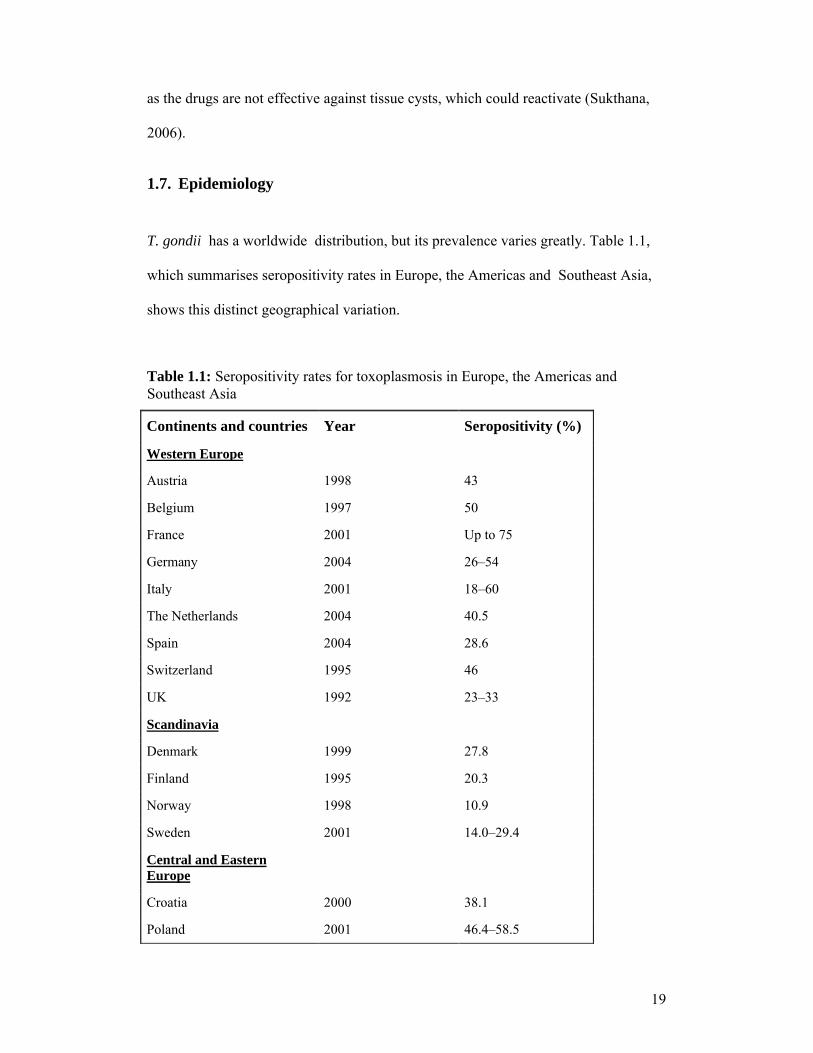
1.7. Epidemiology
T. gondii has a worldwide distribution, but its prevalence varies greatly. Table 1.1,
which summarises seropositivity rates in Europe, the Americas and Southeast Asia,
shows this distinct geographical variation.
Table 1.1: Seropositivity rates for toxoplasmosis in Europe, the Americas and Southeast Asia
Continents and countries Year Seropositivity (%)
Western Europe
Austria 1998 43
Belgium 1997 50
France 2001 Up to 75
Germany 2004 26–54
Italy 2001 18–60
The Netherlands 2004 40.5
Spain 2004 28.6
Switzerland 1995 46
UK 1992 23–33
Scandinavia
Denmark 1999 27.8
Finland 1995 20.3
Norway 1998 10.9
Sweden 2001 14.0–29.4
Central and Eastern Europe
Croatia 2000 38.1
Poland 2001 46.4–58.5
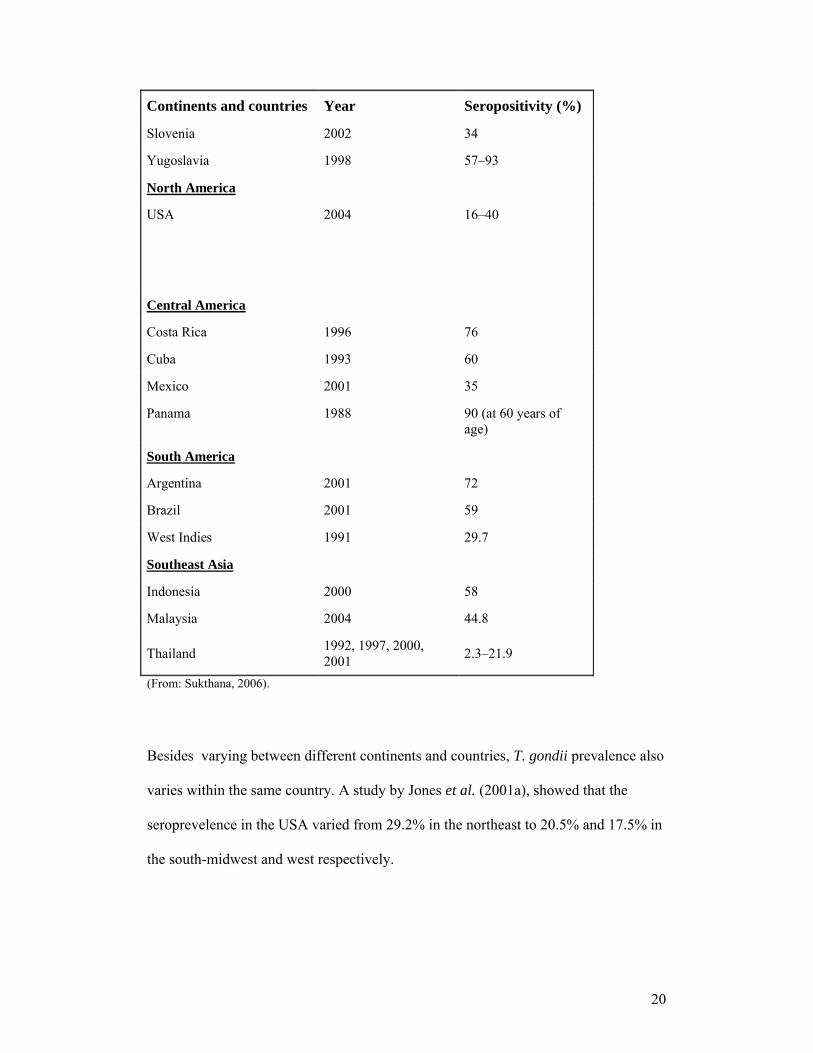
20
Continents and countries Year Seropositivity (%)
Slovenia 2002 34
Yugoslavia 1998 57–93
North America
USA 2004 16–40
Central America
Costa Rica 1996 76
Cuba 1993 60
Mexico 2001 35
Panama 1988 90 (at 60 years of age)
South America
Argentina 2001 72
Brazil 2001 59
West Indies 1991 29.7
Southeast Asia
Indonesia 2000 58
Malaysia 2004 44.8
Thailand1992, 1997, 2000, 2001
2.3–21.9
(From: Sukthana, 2006).
Besides varying between different continents and countries, T. gondii prevalence also
varies within the same country. A study by Jones et al. (2001a), showed that the
seroprevelence in the USA varied from 29.2% in the northeast to 20.5% and 17.5% in
the south-midwest and west respectively.

21
There is little known about T. gondii prevalence in Africa. Table 1.2 shows the results
of some studies carried out on this continent.
Table 1.2: Seropositivity rates in Africa
Country Year Study population
Seroprevalence (%)
Reference
South Africa and
Botswana
1978
See details in
text (below)
20
11
Jacobs and
Mason, 1978
Kenya 1991 HIV-positive
and -negative
adults
54
Brindle et al.,
1991
Sudan 1991 Apparently
healthy
individuals
41.7
Abdel-Hameed,
1991
Zambia 1991 HIV-positive
and -negative
adults
7
Zumla et al.,
1991
Cameroon 1992 Pregnant
women 77.1
Ndumbe et al.,
1992
Senegal 1993 Women
40.3
Diallo et al.,
1996
Republic of
Benin
1995 Pregnant
HIV-negative
women
53.6
Rodier et al.,
1995
Tanzania 1995 Pregnant
women 35
Doehring et al.,
1995
Ethiopia 1998 HIV-positive
and -negative
adults
80
Woldemichael et
al., 1998
Burkina Faso 2004-2005 Pregnant
women 25.3
Simpore et al.,
2006
Uganda 2006 HIV-positive
adults 54
Lindström et al.,
2006
22
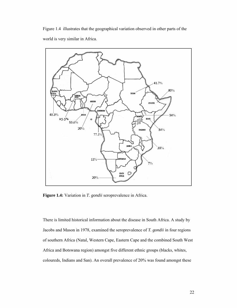
Figure 1.4 illustrates that the geographical variation observed in other parts of the
world is very similar in Africa.
Figure 1.4: Variation in T. gondii seroprevalence in Africa.
There is limited historical information about the disease in South Africa. A study by
Jacobs and Mason in 1978, examined the seroprevalence of T. gondii in four regions
of southern Africa (Natal, Western Cape, Eastern Cape and the combined South West
Africa and Botswana region) amongst five different ethnic groups (blacks, whites,
coloureds, Indians and San). An overall prevalence of 20% was found amongst these
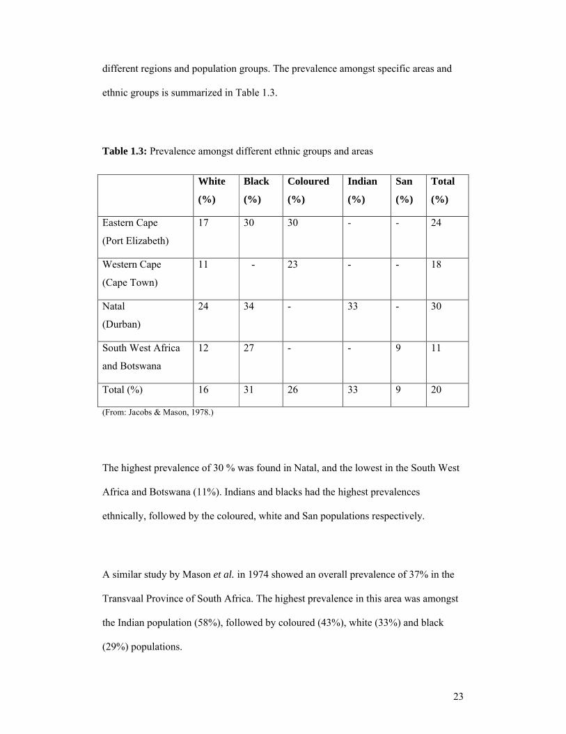
23
different regions and population groups. The prevalence amongst specific areas and
ethnic groups is summarized in Table 1.3.
Table 1.3: Prevalence amongst different ethnic groups and areas
White
(%)
Black
(%)
Coloured
(%)
Indian
(%)
San
(%)
Total
(%)
Eastern Cape
(Port Elizabeth)
17 30 30 - - 24
Western Cape
(Cape Town)
11 - 23 - - 18
Natal
(Durban)
24 34 - 33 - 30
South West Africa
and Botswana
12 27 - - 9 11
Total (%) 16 31 26 33 9 20
(From: Jacobs & Mason, 1978.)
The highest prevalence of 30 % was found in Natal, and the lowest in the South West
Africa and Botswana (11%). Indians and blacks had the highest prevalences
ethnically, followed by the coloured, white and San populations respectively.
A similar study by Mason et al. in 1974 showed an overall prevalence of 37% in the
Transvaal Province of South Africa. The highest prevalence in this area was amongst
the Indian population (58%), followed by coloured (43%), white (33%) and black
(29%) populations.
24
In 1992, Schneider et al. investigated the T. gondii prevalence in women of different
ethnic groups in Durban, KwaZulu-Natal Province. An overall prevalence of 31.3%
was found and the highest prevalence here was amongst the black population (46.2%),
followed by the Indian (36.9%), coloured (28.3%) and white (12.5%) populations.
A study on toxoplasmosis and HIV infection in South Africa by Sonnenberg et al.
(1998), was carried out at the Gold Fields West Mine Hospital. Patients originated
from several different home regions, such the Eastern Cape and KwaZulu-Natal
Provinces, Lesotho and Mozambique, and an overall prevalence of 24.6% was found.
The most recent study from the Gauteng Province by Hari et al. in 2007 was
conducted at the Helen Joseph Hospital in Johannesburg, and investigated T. gondii
seroprevalence in 307 consecutive HIV-infected medical inpatients. All patients were
black, antiretroviral naïve and did not receive toxoplasmosis prophylaxis. An overall
prevalence of 8% was found, with only two patients showing clinical manifestations.
Detailed recent demographic data of groups at risk is missing and more knowledge is
needed at a regional level, e.g. seroprevalence of infection, to implement solutions
aimed at reducing risk for this disease in South Africa.
1.8. Climate and topography of South Africa
South Africa is located on the southernmost tip of Africa. The country’s coastline
stretches more than 2 500km from the border of Mozambique along the Indian Ocean,

25
around the tip of Africa and up towards Namibia along the Atlantic Ocean. It consists
of a low-lying narrow coastal zone, mountainous escarpments and a high inland
plateau (Brand South Africa, 2008).
The country has nine provinces and five climatic regions. A temperate sub-tropical
central region is characterised by hot wet summers and cold, dry winters, with frost
and snow occurring in the mountains. The subtropical region is located on the eastern
border and coastline of the country, where summers are warm and humid and winters
are mild. Rain falls throughout the year with summer being the wettest season. The
Mediterranean climatic region experiences hot, dry summers with occasional
rainstorms and cool, wet winters. This region extends only over a narrow coastal area
near Cape Town. The semi-desert region experiences high temperatures and low
rainfall, which usually falls during the summer months, and the desert region
experiences very little rain and very high temperatures inland (Brand South Africa,
2008).
Much of South Africa is classified as semi-arid. The cold Benguela current from the
west coast and the warm Agulhas current from the east coast have a major effect on
the country’s climate. The high evaporation rate of the Agulhas current provides
significant rainfall on the east, while the Benguela current retains its moisture causing
desert conditions in the west. Mean temperatures vary from 15 ºC to 30 ºC during the
summer to 10 ºC to 25 ºC during the winter across different regions inland (Brand
South Africa, 2008).
26
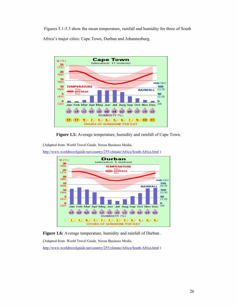
Figures 5.1-5.3 show the mean temperature, rainfall and humidity for three of South
Africa’s major cities: Cape Town, Durban and Johannesburg.
Figure 1.5: Average temperature, humidity and rainfall of Cape Town.
(Adapted from: World Travel Guide. Nexus Business Media.
http://www.worldtravelguide.net/country/255/climate/Africa/South-Africa.html )
Figure 1.6: Average temperature, humidity and rainfall of Durban .
(Adapted from: World Travel Guide. Nexus Business Media.
http://www.worldtravelguide.net/country/255/climate/Africa/South-Africa.html )
27
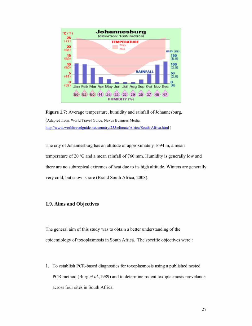
Figure 1.7: Average temperature, humidity and rainfall of Johannesburg.
(Adapted from: World Travel Guide. Nexus Business Media.
http://www.worldtravelguide.net/country/255/climate/Africa/South-Africa.html )
The city of Johannesburg has an altitude of approximately 1694 m, a mean
temperature of 20 ºC and a mean rainfall of 760 mm. Humidity is generally low and
there are no subtropical extremes of heat due to its high altitude. Winters are generally
very cold, but snow is rare (Brand South Africa, 2008).
1.9. Aims and Objectives
The general aim of this study was to obtain a better understanding of the
epidemiology of toxoplasmosis in South Africa. The specific objectives were :
1. To establish PCR-based diagnostics for toxoplasmosis using a published nested
PCR method (Burg et al.,1989) and to determine rodent toxoplasmosis prevelance
across four sites in South Africa.
28
2. To characterize prevalent Toxoplasma strains using rodent organ material. Types
I, II and III were identified by a nested PCR reaction (Grigg et al., 2001),
amplifying a SAG3 genetic marker and subsequent digestion with restriction
enzymes NciI or BcnI.
3. To conduct a retrospective human serosurvey, using a bank of residual serum,
targeting mainly, but not exclusively, pregnant women and children, for evidence
of toxoplasmosis, to establish prevalence rates across all population groups.
4. To conduct a prospective human serosurvey in HIV-positive patients compared to
HIV-negative controls.
5. To compare a standard enzyme immunoassay (EIA) serological test with a simple,
cheap latex agglutination test, which, if validated, would make screening for
Toxoplasma exposure more accessible for the general population.

29
CHAPTER 2
MATERIALS AND METHODS
2.1. HIV-Positive prospective serosurvey
A prospective human serosurvey for toxoplasmosis was conducted at the HIV clinic at
the Chris Hani Baragwanath Hospital in Soweto, Johannesburg. Patients enrolled in
the antiretroviral (ARV) treatment programme were invited to participate. Patients
were recruited at the time of collection of blood for routine investigations and
sampling was done on a convenience basis. Once informed consent was given, two
tubes of blood (1 EDTA-anticoagulated and 1 clotted blood) were drawn by
phlebotomists at the clinic, for this study (Appendix A, pp.60-61). Ethical clearance
was granted by the Human Research Ethics Committee, University of the
Witwatersrand. (Protocol Number: M070637), (Appendix A, p. 62).
The required minimum sample size, based on a mean toxoplasmosis prevalence of
30% (from published South African data), taking the maximum acceptable difference
between the population and the sample rate to be 4%, with a 95% confidence level, is
504, but the final number tested was 376 due to the limited amount of time available
to collect samples.
Samples were then taken to the Parasitology Reference Unit at the National Institute
for Communicable Diseases (NICD), which is one of the host laboratories for this
study. Samples were centrifuged at 3000 revolutions per minute (RPM) for 4.5
minutes and serum was collected and aliquoted. Aliquots were labelled with sample
30
numbers corresponding to the information sheets and consent forms filled in by
patients, so that patient identity was kept confidential.
Aliquots were taken to Department of Parasitology, Mycology and Environmental
Microbiology at the Swedish Institute for Infectious Disease Control (SMI),
Stockholm, Sweden, which was also a host laboratory, where all tests were carried
out by the principal investigator.
Using the Pastorex Toxo latex particle agglutination test, serum samples were tested
in duplicate according to the package protocol (Pastorex TOXO, package insert; Bio-
Rad, France; 2006), with the operator blinded to corresponding results. This technique
utilises soluble membrane and cytoplasmic antigens bound to latex particles, which
agglutinate when specific antibodies (IgM and/or IgG) are present in the samples. A
positive reaction is characterised by the formation of red aggregates on a green
background, whereas in a negative reaction, the suspension remains brown and
homogenous. Indeterminate results were categorized as those which showed a slight
green background or red aggregates as well as a brown suspension. Positive and
negative serum controls were included on every card.
All discrepant, positive and indeterminate results were then retested and titrated using
the BioMèrieux Toxo-Screen DA test, which was the reference test for this part of the
study as it corresponds to the Sabin-Feldman test, the traditional gold standard. The
Biomèrieux Toxo-Screen DA detects Toxoplasma IgG antibodies in human serum by
direct agglutination. The principle of this test is that formalin-treated Toxoplasma
zoites agglutinate in the presence of diluted serum which contains specific IgG

31
antibodies (Toxo-Screen DA, package insert; BioMèrieux SA, France; 2006). Once
again samples were tested in duplicate according to the package insert, with the
operator blinded to corresponding results. Positive, negative and PBS controls were
included in every experiment.
The discrepant, positive and indeterminate Pastorex results were also retested using
indirect immunofluorescence (IFL) according to a specific protocol (Appendix B,
p.65). Diluted patient serum samples were added to wells containing Toxoplasma
tachyzoites and allowed to incubate. During this stage, any anti-Toxoplasma-specific
antigens present in the serum bind specifically to the parasites. A second incubation
with rabbit anti-human antibodies conjugated with fluorescein isothiocyanate (FITC)
diluted in Evans blue detect any human antibodies present from the first incubation.
Evans blue helps to create a dark background for viewing slides. Positive and negative
controls, as well as a conjugate control of PBS were included with every experiment.
All BioMèrieux test-negative results were further tested for IgM using an IFL because
the BioMèrieux test only detects IgG whereas Pastorex test detects both IgG and IgM.
32
2.2. HIV-negative controls
HIV-negative controls were obtained from the HIV diagnostic virology laboratory at
the NICD. These residual human sera, previously collected under an antenatal survey
programme, served as appropriate controls. They matched the test group as they were
both collected in the Gauteng area from populations with similar living conditions and
expected rates of exposure. Samples were identifiable only by number and
confidentiality was thus ensured. Ethical approval was granted by the Human
Research Ethics Committee of the University of the Witwatersrand. (Protocol
Number: M 070637) (Appendix A, p.62).
Three hundred and seventy-six controls were tested in an identical manner to the
HIV-positive specimens. All samples were tested in duplicate in a blinded manner
using the Pastorex-Toxo agglutination test. Thereafter the BioMèrieux Toxo-Screen
DA test was used to confirm all positive and indeterminate results. Testing with IFL
was not carried out at this stage, as it was unlikely that IgM would be present in many
of these specimens.
2.3. Retrospective serosurvey
The retrospective serosurvey involved the use of serum samples, mainly but not
exclusively targeting pregnant women and children, randomly selected from a
residual serum bank of 7000 specimens that had been collected for a rubella
prevalence study. The age spectrum of selected subjects was wide, but the majority
were young adults. (Protocol number: M070347), (Appendix A, p.63). The required
minimum sample size, once again based on a mean toxoplasmosis prevalence of 30%,
33
taking the maximum acceptable difference between the population and the sample
rate to be 4%, with a 95% confidence level, was 504. Due to availability of samples,
however, only 497 were used.
Samples were sent to the Department of Immunology at the National Health
Laboratory Service (NHLS), where the Axsym Toxo IgG (Abbott) automated enzyme
immunoassay (EIA) was used to assay IgG antibodies and was the reference method
for this part of the study. This technique utilises Toxoplasma antigens bound to
microparticles, which are mixed with the serum sample. Specific antigen-antibody
complexes are immobilised on a glass fibre matrix, and enzyme-linked antihuman
globulin is used in a detection system, as in a normal plate EIA. Standardised
calibrators (referenced to WHO standards) are used to produce a calibration curve
against which the samples are read, to give a quantitative anti-Toxoplasma antibody
concentration (Axsym test information; Abbott Laboratories, USA; 2005).
The Pastorex Toxo latex particle agglutination test (Bio-Rad, France), described
earlier, was used on the same specimens, with the operator blinded to corresponding
EIA results.
2.4. Prevalence in Rodents
In order to determine T. gondii prevalence in rodents as well as to establish
PCR- based diagnostics for toxoplasmosis, 100 rodent spleen specimens were
retrospectively sampled (as residual specimens from a different study) from each of
three sites, namely, Port Elizabeth, Limpopo Province, and Durban. Twenty-five

34
rodent heart and brain samples were prospectively collected from the Johannesburg
area. Ethical approval was granted by the Animal Ethics Screening Committee,
University of the Witwatersrand (Clearance number: 2008/50/01), (Appendix A,
p.64).
The required minimum sample size, based on the fact that the background
Toxoplasma prevalence in rodents is unknown (and therefore assumed to be 50%),
and with 95% confidence of being within 5% of the true prevalence, was calculated to
be 384; however, due to problems with the availability of new material, only 325
samples could be collected.
Samples from Durban were mainly of the Rattus norvegicus species. Rhabdomys
pumilio was most common in the sample group from Port Elizabeth and Rattus rattus
was most common in Mapate. Samples from Johannesburg were not speciated.
DNA was extracted from the tissue according to the QIAgen tissue protocol, using a
QIAamp DNA mini kit (Qiagen Inc. Valencia, C.A, USA), (Appendix B, p.66-67).
Extracted DNA was then tested using a polymerase chain reaction (PCR) technique.
A nested PCR based on the identification of the 35-fold repetitive B1 gene, as
described by Burg et al. (1989), was validated using known Toxoplasma-positive and
negative specimens, checking for a predicted PCR band size of 97 base pairs. Once it
was validated, all extracted DNA was tested according to a specific protocol
(Appendix B, pp.68-70) using oligonucleotide primers, Toxoplasma B1 (5’- GGA
ACT GCA TCC GTT CAT GAG -3’), Toxoplasma B2 (5’- TCT TTA AAG CGT
35
TCG TGG TC -3’), Toxoplasma B3 (5’- TGC ATA GGT TGC AGT CAC TG -3’),
and Toxoplasma B4 (5’- GGC GAC CAA TCT GCG AAT ACA CC-3’).
The PCR reaction mix and cycling parameters are described in Appendix B, pp.68-70.
Gel electrophoresis using a 2% agarose gel, was performed as described in Appendix
B, pp.71-72, to identify the 97 base pair product.
DNA and PCR reagent controls were included in each experiment to monitor for
possible contamination. Furthermore, DNA extraction, PCR reaction mix preparation,
DNA addition, PCR amplification and gel electrophoresis were done in separate one-
way traffic-controlled rooms to minimize the risk of contamination.
All results with irregular bands from the previous PCR method were retested using a
nested PCR reaction as described by Grigg et al. (2001), amplifying the SAG3 gene.
Once again, this method was first validated using known positive and negative
specimens before testing was carried out according to a set protocol (Appendix B,
pp.73-75) using oligonucleotide primers Primer 1 (5’- CAA CTC TCA CCA TTC
CAC CC -3’), Primer 2 (5’- GCG CGT TGT TAG ACA AGA CA -3’), Primer 3 (5’-
TCT TGT CGG GTG TTC ACT CA-3’) and Primer 4 (5’- CAC AAG GAG ACC
GAG AAG GA-3’). The PCR reaction mix and cycling parameters are described in
Appendix B, pp.73-75.

36
DNA and PCR reagent controls were included in each experiment to monitor for
possible contamination. Gel electrophoresis using a 2% agarose gel was used to
identify a 226 base pair product.
The method of enzyme digestion of the SAG3 gene with restriction enzymes NciI and
BcnI to identify different strain types was validated using known type I, II and III
Toxoplasma strains. Enzymes cut the amplified sequence of type I parasites twice,
type III once and did not cut type II parasites (Appendix B, p.76).
2.5. Toxoplasma cultivation
T. gondii parasites were cultivated in cell culture and used as positive DNA controls
in PCR. Tachyzoites were fixed onto slides for use in IFL.
Flasks containing confluent human foreskin fibroblast cells (HFF) were infected with
freshly lysed Toxoplasma tachyzoites, RH-LDM strain (Barragan and Sibley, 2002).
Flasks were then incubated until cells lysed and parasites were collected in sediment
as described in Appendix B, pp.77-78.
DNA was extracted once again using the QIAGEN DNA mini kit and used as positive
PCR controls. Washed Toxoplasma tachyzoites were distributed on 8-well Teflon-
coated microscope slides, left to air dry and then fixed in cold acetone for 20 minutes
and stored frozen until used for IFL.
2.6. Data analysis
Data analysis was carried out using a statistical software package, STATISTICA
(StatSoft, USA).
37
CHAPTER 3
RESULTS
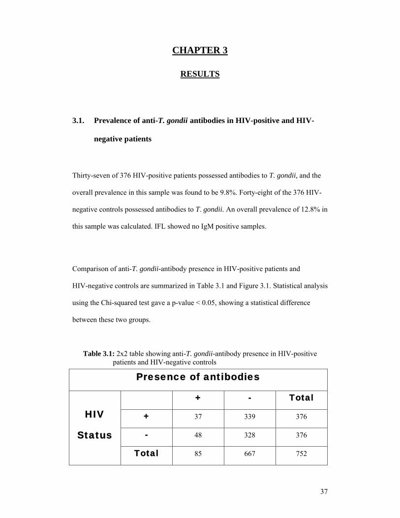
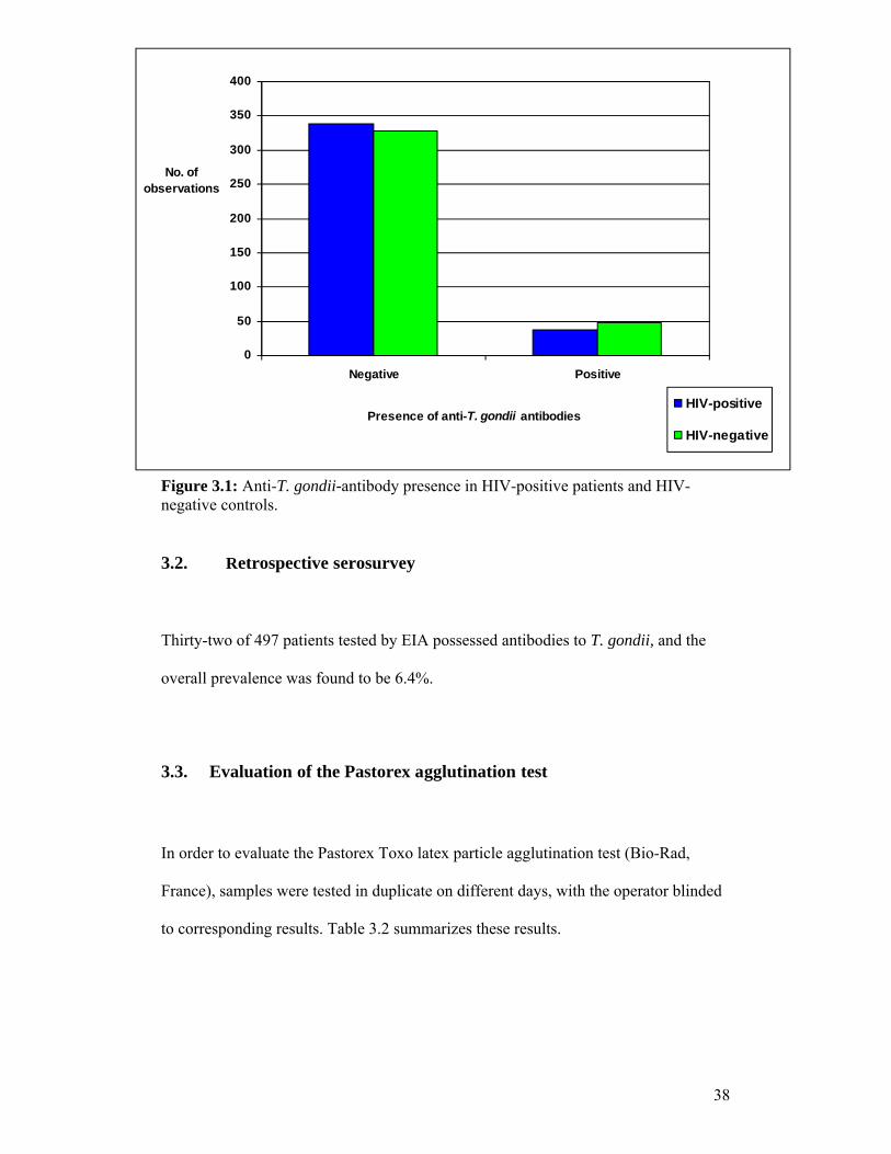
3.1. Prevalence of anti-T. gondii antibodies in HIV-positive and HIV-
negative patients
Thirty-seven of 376 HIV-positive patients possessed antibodies to T. gondii, and the
overall prevalence in this sample was found to be 9.8%. Forty-eight of the 376 HIV-
negative controls possessed antibodies to T. gondii. An overall prevalence of 12.8% in
this sample was calculated. IFL showed no IgM positive samples.
Comparison of anti-T. gondii-antibody presence in HIV-positive patients and
HIV-negative controls are summarized in Table 3.1 and Figure 3.1. Statistical analysis
using the Chi-squared test gave a p-value < 0.05, showing a statistical difference
between these two groups.
Table 3.1: 2x2 table showing anti-T. gondii-antibody presence in HIV-positive patients and HIV-negative controls
Presence of antibodies
HIV
Status
+ - Total
+ 37 339 376
- 48 328 376
Total 85 667 752
38
Figure 3.1: Anti-T. gondii-antibody presence in HIV-positive patients and HIV- negative controls.
3.2. Retrospective serosurvey
Thirty-two of 497 patients tested by EIA possessed antibodies to T. gondii, and the
overall prevalence was found to be 6.4%.
3.3. Evaluation of the Pastorex agglutination test
In order to evaluate the Pastorex Toxo latex particle agglutination test (Bio-Rad,
France), samples were tested in duplicate on different days, with the operator blinded
to corresponding results. Table 3.2 summarizes these results.
0
50
100
150
200
250
300
350
400
Negative Positive
Presence of anti-T. gondii antibodies
No. of observations
HIV-positive
HIV-negative
39
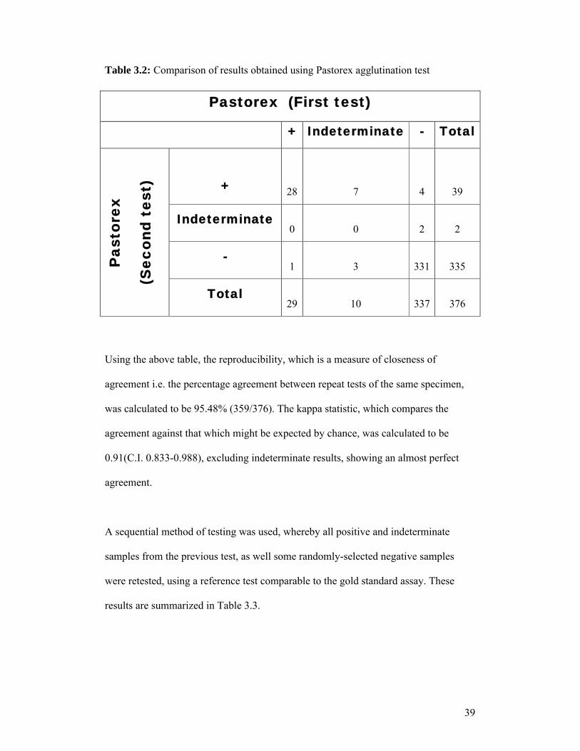
Table 3.2: Comparison of results obtained using Pastorex agglutination test
Pastorex (First test)
+ Indeterminate - TotalP
asto
rex
(Sec
ond
test
) + 28 7 4 39
Indeterminate0 0 2 2
-1 3 331 335
Total29 10 337 376
Using the above table, the reproducibility, which is a measure of closeness of
agreement i.e. the percentage agreement between repeat tests of the same specimen,
was calculated to be 95.48% (359/376). The kappa statistic, which compares the
agreement against that which might be expected by chance, was calculated to be
0.91(C.I. 0.833-0.988), excluding indeterminate results, showing an almost perfect
agreement.
A sequential method of testing was used, whereby all positive and indeterminate
samples from the previous test, as well some randomly-selected negative samples
were retested, using a reference test comparable to the gold standard assay. These
results are summarized in Table 3.3.

40
Table 3.3: Comparison of results obtained using Pastorex agglutination test and
reference test (Biomèrieux Toxo-Screen DA)
Twenty-seven true positive and 23 true negative results were found. One false
positive and no false negative results were found. Statistical analysis results,
excluding indeterminate results are summarized in Table 3.4.
Pastorex
+ Indeterminate - Total
Bio
mèr
ieux
Tox
o-
Scr
een
DA
+27
True +
10 0
False -
37
-1
False +
7 23
True -
31
Total 28 17 23 68
41
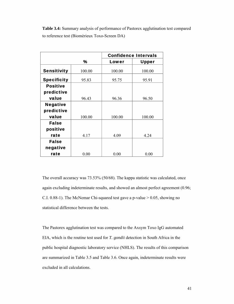
Table 3.4: Summary analysis of performance of Pastorex agglutination test compared
to reference test (Biomèrieux Toxo-Screen DA)
The overall accuracy was 73.53% (50/68). The kappa statistic was calculated, once
again excluding indeterminate results, and showed an almost perfect agreement (0.96;
C.I. 0.88-1). The McNemar Chi-squared test gave a p-value > 0.05, showing no
statistical difference between the tests.
The Pastorex agglutination test was compared to the Axsym Toxo IgG automated
EIA, which is the routine test used for T. gondii detection in South Africa in the
public hospital diagnostic laboratory service (NHLS). The results of this comparison
are summarized in Table 3.5 and Table 3.6. Once again, indeterminate results were
excluded in all calculations.
%Confidence IntervalsLower Upper
Sensitivity 100.00 100.00 100.00
Specificity 95.83 95.75 95.91
Positive predictive
value 96.43 96.36 96.50
Negative predictive
value 100.00 100.00 100.00
False positive
rate 4.17 4.09 4.24
False negative
rate 0.00 0.00 0.00

42
Table 3.5: Comparison of results obtained using Pastorex agglutination test and
routine diagnostic test (Axsym Toxo IgG automated EIA)
Table 3.6: Summary of statistical analysis of Pastorex agglutination test and
routine diagnostic test (Axsym Toxo IgG automated EIA)
%Confidence IntervalsLower Upper
Sensitivity 100.00 100.00 100.00
Specificity 93.99 93.96 94.00
Positive predictive
value 49.06 48.92 49.19
Negative predictive
value 100.00 100.00 100.00
False positive
rate 6.01 5.99 6.03
False negative
rate 0.00 0.00 0.00
Pastorex+ Indeterminate - Total
Axs
ym T
oxo
IgG
EIA +
26 True
+6
0 False
-32
Indeterminate0 1 4 5
-
27 False
+11
422 True
-460
Total 53 18 426 497
43
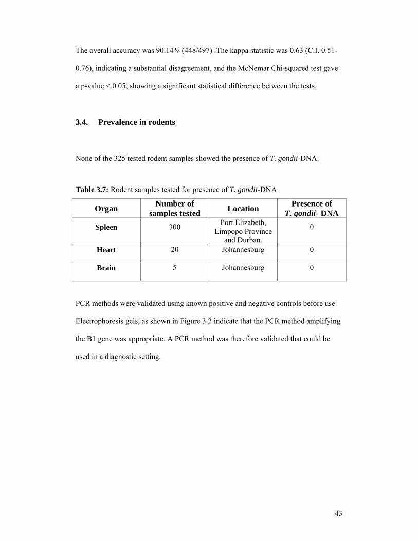
The overall accuracy was 90.14% (448/497) .The kappa statistic was 0.63 (C.I. 0.51-
0.76), indicating a substantial disagreement, and the McNemar Chi-squared test gave
a p-value < 0.05, showing a significant statistical difference between the tests.
3.4. Prevalence in rodents
None of the 325 tested rodent samples showed the presence of T. gondii-DNA.
Table 3.7: Rodent samples tested for presence of T. gondii-DNA
OrganNumber of
samples testedLocation
Presence of T. gondii- DNA
Spleen 300Port Elizabeth,
Limpopo Province and Durban.
0
Heart 20 Johannesburg 0
Brain 5 Johannesburg 0
PCR methods were validated using known positive and negative controls before use.
Electrophoresis gels, as shown in Figure 3.2 indicate that the PCR method amplifying
the B1 gene was appropriate. A PCR method was therefore validated that could be
used in a diagnostic setting.
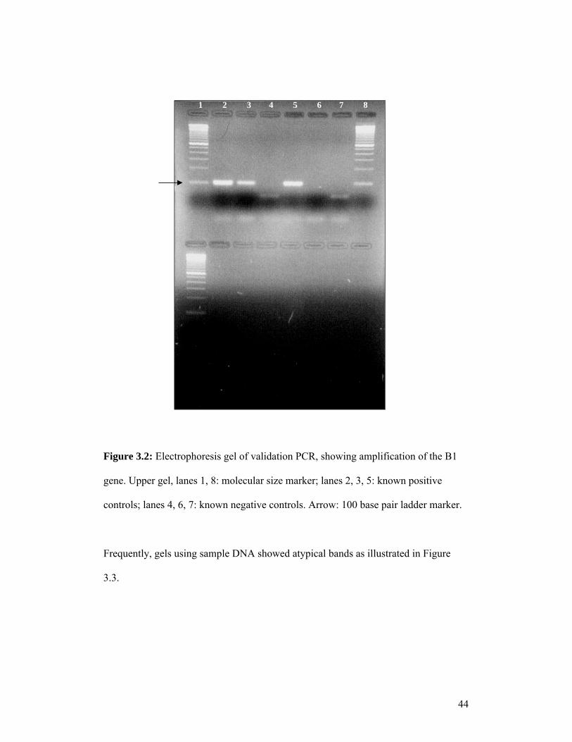
44
1 2 3 4 5 6 7 8
Marker Known Positive
Controls
Figure 3.2: Electrophoresis gel of validation PCR, showing amplification of the B1
gene. Upper gel, lanes 1, 8: molecular size marker; lanes 2, 3, 5: known positive
controls; lanes 4, 6, 7: known negative controls. Arrow: 100 base pair ladder marker.
` ControlsFrequently, gels using sample DNA showed atypical bands as illustrated in Figure
3.3.
45
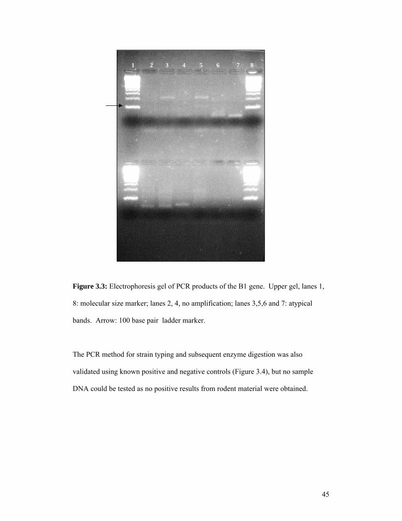
1 2 3 4 5 6 7 8
Figure 3.3: Electrophoresis gel of PCR products of the B1 gene. Upper gel, lanes 1,
8: molecular size marker; lanes 2, 4, no amplification; lanes 3,5,6 and 7: atypical
bands. Arrow: 100 base pair ladder marker.
The PCR method for strain typing and subsequent enzyme digestion was also
validated using known positive and negative controls (Figure 3.4), but no sample
DNA could be tested as no positive results from rodent material were obtained.

46
1 2 3 4 5 6 7 8
1 2 3 4 5 6 7 8
Figure 3.4: Electrophoresis gel showing restriction digests of PCR products of SAG3
gene, demonstrating the 3 Toxoplasma strains. Lanes 1, 8: molecular size markers;
lanes 2, 5: type I; lanes 3, 7: type II; lanes 4, 6: type III. Arrow: 100 base pair ladder
marker.
47
CHAPTER 4
DISCUSSION
4.1. Introduction
Since its discovery 100 years ago, T. gondii has become established as one of the
most versatile and successful of all parasites. Its worldwide distribution, broad host
range and ability to maintain a benign co-existence with its hosts have enabled its
success. The ability of T. gondii to be cultured, the fact that it is amenable to genetic
manipulation and has excellent animal models, has made studying this organism fairly
uncomplicated (Carruthers, 2002).
Although the parasite rarely causes acute disease in healthy individuals, its status as
the third most common cause of food-borne deaths in United States and its
exceptionally high infection rates show that it is a serious threat to human health
(Carruthers, 2002).
4.2. Toxoplasma gondii in South Africa
This study has shown the prevalence of anti-T. gondii antibodies in an HIV-positive
population in Soweto, Johannesburg, to be 9.8%. A comparable HIV-negative control
population showed a prevalence of 12.8 %. These prevalences are lower than those
reported in a previous study by Jacobs and Mason (1978), which reported a 20%
overall prevalence across four sites in South Africa. A more recent study by
48
Sonnenberg et al. (1998) showed a 24.6% prevalence in a cohort of HIV-positive
patients originating from rural areas around South Africa. In Johannesburg, a study by
Hari et al. (2007) showed a similar prevalence to the present report, namely, 8% in
HIV-positive individuals at the Helen Joseph Hospital.
The retrospective serosurvey reported here, targeting mainly pregnant women and
children, showed a 6.4% prevalence of anti-T. gondii antibodies. This prevalence is
lower than that reported in a similar study by Schneider et al. (1992), which examined
the prevalence of T. gondii infection in women in KwaZulu-Natal Province and found
a 31.3 % prevalence.
These prevalences are also lower than those reported for other parts of Africa as well
as other continents. Details of these studies are discussed in Chapter 1.
There are several possible explanations for the low prevalences found in this study, as
well as the reported historical differences in prevalences between regions.
In some HIV/AIDS patients, the body may be unable to make sufficient antibodies
due to its failing immune system. As a result, antibodies may be undetectable and this
may account for the lower prevalence found in the HIV-positive sample. However,
evidence against this being an important factor is that a sufficient proportion of HIV-
positive patients have detectable levels of antibodies to make such testing of clinical
value. All patients participating in this study were enrolled in an antiretroviral (ARV)
treatment programme and this may be one of the reasons why those possessing anti-
T. gondii antibodies did not present with clinical disease. Cotrimoxazole, routinely
49
used for prevention of Pneumocystis pneumonia, also suppresses reactivation of
toxoplasmosis. HIV-positive patients admitted to the hospital for treatment were not
recruited to this study and there is the possibility that the prevalence in this
subpopulation may be higher, and some such patients may be presenting with overt
toxoplasmosis.
A less pathogenic clonal lineage of the parasite may be present in South Africa
compared to the rest of Africa and other continents. Very little is known genetically
about T. gondii in Africa. However, a study by Lindström et al. (2006), found that the
genotype distribution in Uganda appears to be similar to that found in Europe
amongst immunocompromised individuals. The type II allele of the SAG2-locus was
the most common disease-causing strain found. Nothing is known about T. gondii
strains affecting humans in South Africa; further studies will be required in this
regard.
Previous studies on T. gondii in South Africa date back to 1974, which was during the
apartheid era and prior to the emergence of the HIV epidemic. Prevalences reported in
these earlier studies were higher than those found in this study and other more recent
ones. This may be due to differences in socio-economic and cultural factors present
nowadays.
During the apartheid era, a large proportion of the black population, in particular, did
not have adequate access to good sanitation, clean water, and many grew their own
fruit and vegetables as their main source of food. They had more contact with stray
animals and rodents, as housing facilities in rural areas were generally very poor.

50
Many worked on farms where they were at a higher risk of acquiring a T. gondii
infection due to their close contact with soil and domestic stock reservoirs of
toxoplasmosis. Studies on T. gondii prevalence during this time therefore found the
highest prevalence in the black population (Jacobs and Mason, 1978).
Since the birth of South Africa’s democracy in 1994, socio-economic conditions have
been changing, with rapid urbanisation. The majority of the population in urban areas
has improved access to clean water and sanitation facilities, although breakdowns in
these services occur from time to time. Contact with stray cats is limited and due to
rodent control campaigns and better living conditions, there might be generally less
contact with rodents, although this would be highly variable, as there is still
widespread poverty and a wide range of quality of life. Most people buy food from
shops and supermarkets and rarely rely on growing their own. There is more
education and awareness around good hygiene and washing of fruit and vegetables as
well as the importance of properly cooked food.
The development of better farming practices and the widespread use of freezers and
frozen meat, as well as pasteurized milk, means that many of the risk factors for
acquiring T. gondii infection have been reduced.
In the city of Johannesburg, where this study was carried out, there are a large number
of apartments and townhouses rather than traditional houses, many of which do not
have gardens and do not allow occupants to own pets. As a result, many children
growing up in such environments have less contact with soil and cats and therefore

51
have less chance of acquiring a T. gondii infection. Cats are not popular companion
animals in most black households, for reasons of superstition.
Climatic conditions may account in some part for the global variation in T. gondii
prevalence. The climate across much of Africa is generally uniform with considerably
less climatic variation between seasons, compared to Europe and North America.
T. gondii prevalence on this continent is lower than that of Europe and North
America; climatic and topographical factors may account for these lower prevalences.
In South Africa, the climate may also contribute to the differences in prevalence
between different provinces. In KwaZulu-Natal Province, a study by Bhigjee et al.
(1999) showed that toxoplasmosis was the most frequent cause of intracranial mass
lesions (comprising 52%) in HIV-positive patients. Modi et al. (2004), however,
found that tuberculosis, not toxoplasmosis, was the most frequent cause of focal brain
lesions in a Gauteng population. As discussed in Chapter 1, KwaZulu-Natal and
Gauteng Provinces fall in different climatic regions. KwaZulu-Natal is generally hot
with high humidity and rainfall while Gauteng generally has low humidity and
moderate rainfall. These differences in climate may to contribute to the differences in
prevalences. Prevalences might be expected to be higher in hotter, wetter areas as
these conditions are more favourable for the sporulation of oocysts. This trend is also
evident in the seroprevalence of T. gondii in sheep in South Africa, which is higher in
the humid costal regions (KwaZulu-Natal - 6.3%, Eastern Cape - 7.75%) and lowest
in the arid regions (Gauteng - 6%, Free State - 2.7%) (Samra et al., 2007).

52
Global variation in prevalence may also be attributed to differences in cultural habits.
In France, the high T. gondii prevalence (up to 75%) has been related to a preference
for eating raw or undercooked meat. In Thailand, where Buddhism is the main
religion, it is considered a sin to kill any living being. There are therefore large
numbers of stray cats which increase the risk of T. gondii transmission to humans
(Sukthana, 2006). Care of pets is also a factor influencing T. gondii prevalence in a
population. In Brazil and Mexico, cats are fed leftovers and raw viscera, which have
been identified as risk factors for human T. gondii infection, and in Thailand, cats are
rarely trained to defecate in litter boxes, making the contaminated environment a
major risk factor in the human population.
In South Africa, the number of feral cats is relatively low and there may be greater
awareness about good hygiene, especially with regard to the care of pets. Raw meat
is rarely consumed, and in some religions certain animals are not eaten at all. Hindu
and Muslim people, respectively, do not eat beef or pork, and therefore consume
relatively more mutton or lamb. As discussed in Chapter 1, sheep are more
susceptible to T. gondii infection and the handling and consumption of mutton and
lamb therefore poses more risk for acquiring infection. This may also account for the
higher occurrence of T. gondii in KwaZulu-Natal Province where there are a greater
number of Hindu and Muslim people.
Differences between reported prevalences may also be due to different sampling
procedures and assay methods. Various tests have different sensitivities, specificities,
false positive rates and false negative rates. These factors need to be considered when
comparing studies. Generally, more sensitive and specific assays are available
53
nowadays and this could account for the variability in prevalences reported in more
recent studies.
4.3. Evaluation of the Pastorex agglutination test
Diagnosis of toxoplasmosis can be made indirectly by serological techniques, or
directly by PCR or hybridization, histology or isolation. Direct methods are optimal
for a definitive diagnosis, especially in HIV-positive patients, where there is the risk
of a reactivated infection. This, however, is not always practical, especially in
developing countries such as South Africa, as these direct methods are often
expensive, technically demanding, and can be invasive and time-consuming.
Serological methods therefore are still widely used and can be very useful in the right
clinical setting. A negative serological result for T. gondii is helpful as it almost rules
out TE as a diagnosis in HIV/AIDS patients.
The Pastorex agglutination test was therefore evaluated as it is a simple, cheap test,
which, if validated, would make screening for toxoplasmosis exposure more accesible
for the general population.
The test showed a very high accuracy when first and second round results were
compared to one another, and when compared to both the reference (Biomèrieux
Toxo-Screen DA) and routine methods (Axsym Toxo IgG (Abbott) automated EIA).
This shows that results with the Pastorex test are highly reproducible. No statistical

54
difference was found between the Pastorex test and the reference method, but there
was a statistical difference between the agglutination test and the routine EIA method.
Different sets of sera were used for these two comparisons and this may account for
the different findings.
The sensitivity of a test is its ability to correctly yield a positive result. The sensitivity
of the Pastorex test when compared to both the routine and reference methods was
100%.
The ability of a test to correctly yield negative results is termed the specificity. The
specificity of the Pastorex test compared to both the routine and reference methods
was 95.83% and 93.99% respectively.
Values for both these parameters were very high for the Pastorex test, indicating that
it is both a very sensitive and specific test. The fact that the sensitivity is higher than
the specificity means that a lower rate of false negatives than false positives can be
anticipated. In the common clinical settings of toxoplasmosis testing
(immunosuppression and pregnancy), it is arguably better to pick up some false
positives and apply confirmatory testing, rather than to miss any and regard them as
negative.
A false positive result occurs when a test falsely identifies a person as having a
disease or infection when they in fact do not. False positive rates of 4.17% and 6.01%
were found when comparing the test to the reference and routine methods. A false
negative result occurs when a test falsely identifies a person as not having a disease or

55
infection when they in fact do have it. The false negative rate of this test was 0%,
indicating that the Pastorex agglutination test has a higher rate of finding patients
positive for anti-T. gondii antibodies when they do not in fact posses any, but it does
not identify patients who do have anti-T. gondii antibodies as negative. This indicates
that negative results with the Pastorex test are reliable.
Perhaps more important measures of a test are the positive predictive and negative
predictive values. The positive predictive value (PPV) is the probability that a person
who is identified as positive by the test, does in fact have the disease. The negative
predictive value (NPV) is the probability that a person identified as negative by the
test truly does not have the disease. For the purposes of a screening test the NPV is of
interest. Compared to both routine and reference methods, the NPV was 100%,
indicating again that negative results with the Pastorex test are reliable.
Overall the Pastorex agglutination test compared well to the other two methods. The
methodology was fairly simple and did not require a high level of expertise. The
interpretation of results, however, is subjective and this maybe problematic as
indeterminate results may often be found. A further problem is the occurrence of
false positive results, as these could have an impact on the treatment of patients.
A very good characteristic of this test is the fact that it does not yield false negative
results, indicating that negative results are reliable. This is an important factor in the
management of T. gondii infections, especially in HIV/AIDS patients.
56
4.4. Toxoplasma gondii in rodents
The role of rodents in the transmission of T. gondii has always been considered an
important one. In this study the T. gondii prevalence in rodents across four sites in
South Africa was investigated, but no positive results were obtained. It is unlikely that
T. gondii is absent in rodents and there are several possible reasons as to why no
positive results were obtained.
The testing method used may not have been appropriate or did not function
appropriately. In this study, however, PCR diagnostics were used. PCR is considered
to be a very sensitive and accurate technique and is widely used for T. gondii
diagnosis. The method was successfully validated using known positive and negative
specimens and each DNA extraction and PCR reaction mix included internal controls
ensuring good quality control. One-way traffic-controlled rooms were used in each
step of the testing procedure to further ensure no contamination. It is unlikely then
that the testing method accounted for the lack of positive results.
The quality of the material used in the testing may not have been optimal. Rodents
were trapped by either break-back or Sherman traps (Taylor et al., 2008). The time
taken for the rodents to be collected and organs removed and preserved from the time
that they were initially trapped is very important. The longer the time taken, the lower
the quality of the sample material becomes. There is evidence for this in this study as
many electrophoresis gels of PCR products showed atypical bands indicative of poor
material quality (Figure 3.4, Chapter 4). There was no way to control for this, as
residual material from a previous study was used.

57
The type of material available was also not optimal. Ideally brain or heart tissue
would be best used as there are usually greater numbers of T. gondii cysts found here.
Only spleen tissue was available for use in this study. The small number of brain and
heart tissue samples prospectively collected also did not yield any positive results, but
once again the quality of the material received could not be controlled.
There is also the possibility that rodents are no longer as important in the transmission
of T. gondii to humans as in the past. In principle, they have less contact with humans
due to urbanization, rodent control programmes, improving sanitary conditions and
increased health awareness. However, suboptimal human living conditions usually
favour increased rodent breeding and subsequent increased contact with humans
(Taylor et al., 2008). There is intermittent informal evidence (e.g. newspaper reports)
that commensal rodents emerge as public health problems from time to time in certain
areas in Gauteng Province and elsewhere.
Birds (including chickens), have recently been recognised as an important source of
infection. Chickens feed from the ground and therefore presumably have ready
contact with T. gondii oocysts, and have a greater chance of infecting humans through
food preparation and consumption. In rural and peri-urban areas of South Africa
where free-ranging chickens are found abundantly, they may have a greater role to
play than rodents. Further studies will be done to investigate this possibility.

58
CHAPTER 5
CONCLUSIONS
The results of this study have shown that T. gondii infection is present in South
Africa, but its prevalence is much lower than in other parts of the world. This low
prevalence means that previously unexposed people are at risk of acquiring an acute
infection, which may be passed on congenitally in pregnant women, or which could
be life-threatening in HIV/AIDS patients.
Rapid and accurate diagnosis in such patients is therefore essential to ensure effective
treatment and prevention of severe disease. This is not an easy task, especially in
South African populations where other infections, especially tuberculosis, are very
common. Distinguishing a tuberculosis central nervous system infection from
toxoplasmosis is a common diagnostic challenge and in populations such as this
where tuberculosis is highly endemic, patients are probably often not appropriately
treated for toxoplasmosis.
It is recommended that the Pastorex Toxo latex particle agglutination test be used as a
screening test as it is cheap, quick and reliable to rule out toxoplasmosis as a
diagnosis if negative. All positive and indeterminate results should be retested using
another more specific test or by direct methods such as PCR. Care should also be
taken when interpreting the results of tests in severely immunocompromised
individuals, as antibodies may be undetectable in some who do in fact have an
infection. Direct methods are preferable in such situations.

59
From the public health perspective, in a general population with such a low
prevalence and few risk factors for transmission of T. gondii, strict control measures
and screening programmes would be expensive and unnecessary. However,
interventions could benefit certain subpopulations at particular risk, as discussed
below.
Newly-diagnosed HIV-positive, or pregnant, patients should be screened for previous
T. gondii exposure so that adequate precautions can be taken by both the clinician and
the patient to prevent an acute or reactivated infection. Continued awareness around
good hygiene and infection control should be emphasised by clinicians and public
health professionals, especially with respect to pregnant women and HIV-positive
individuals.
This study helped to answer questions relating to the seroprevalence and diagnosis of
T. gondii in South Africa. Many questions still remain to be answered, however, to
fully understand the impact of this parasite in our country and to help to implement
solutions aimed at reducing risk for this curable but potentially fatal disease. The
growing AIDS epidemic and the recent detection of drug-resistant HIV strains is a
disturbing reminder that opportunistic infections such as toxoplasmosis, even in low
prevalences, remain a major potential threat to human health.
60
APPENDIX A
Patient information and consent forms
Title of Project:
Studies of Bartonella and Toxoplasma gondii species in human populations in South Africa
Patient information sheet
Hello. My name is ___________________________ and I would like to invite you to participate in a study. Your participation in this study is completely voluntary. If you decide not to participate, it will not affect your treatment in any way and you may also change your mind at any time. As we discuss the information, please feel free to ask me any questions.
I am studying infections called ‘toxoplasmosis’ and ‘bartonellosis’, caused by germs most commonly transmitted from humans animals and some foods. These infections can sometimes cause illness in humans. I would like to test your blood for signs of these infections, and ask your permission to take, at the same time that blood is taken from a vein in your arm for other tests routinely done in this clinic, two extra 5ml (2 teaspoons) tube of blood. This blood will be used to test for the presence of these infections and will not be used for any other purpose. Your blood will be disposed of safely once we have carried out these tests. Only the people involved in this study will have access to your blood and to these results. Your doctor will be given the results of my test, and will tell you about them when you return to the clinic for your normal appointment.
Thank you for your time. Once you have asked me any questions, you can decide in your own time whether you want to give the blood sample or not.
If you would like any more information on the study, you can contact any of the researchers that are here, or Miss K. Kistiah (011) 555 0332 or Miss A.N. Trataris (011) 555 0331
NICD
61
Title of Project:
Studies of Bartonella and Toxoplasma gondii species in human populations in South Africa
Informed consent form:
I have understood the information (verbal and/or printed) about this study, and agree to donate a sample of blood to be tested only for toxoplasmosis and bartonellosis , and I understand that any remaining blood will be destroyed and not used for any other purpose.
I understand that I have been invited to participate in this study, and I understand that my agreeing to participate is fully voluntary and I can withdraw at any time.
Subjects name:
Signature
Date:
Consent record for blood tests for toxoplasmosis and bartonellosis.
Age: Gender: Date:
Information provided and written consent witnessed by (print):
Sign:
Blood sample taken by (print): Sign:
Study sample number:
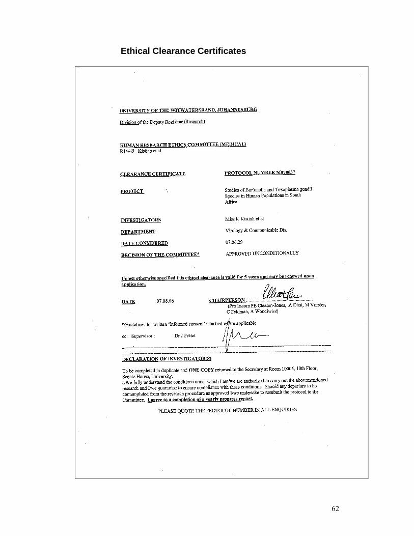
62
Ethical Clearance Certificates

63
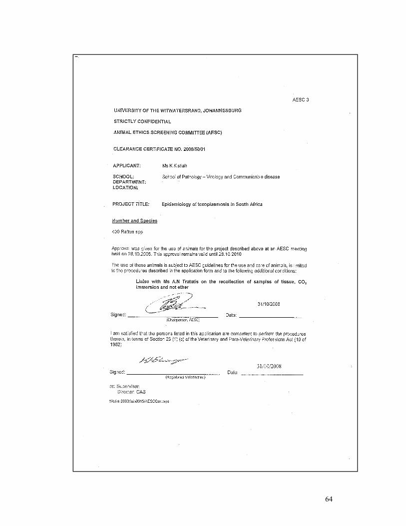
64
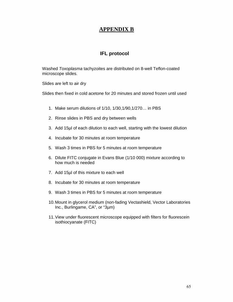
65
APPENDIX B
IFL protocol
Washed Toxoplasma tachyzoites are distributed on 8-well Teflon-coated microscope slides.
Slides are left to air dry
Slides then fixed in cold acetone for 20 minutes and stored frozen until used
1. Make serum dilutions of 1/10, 1/30,1/90,1/270… in PBS
2. Rinse slides in PBS and dry between wells
3. Add 15µl of each dilution to each well, starting with the lowest dilution
4. Incubate for 30 minutes at room temperature
5. Wash 3 times in PBS for 5 minutes at room temperature
6. Dilute FITC conjugate in Evans Blue (1/10 000) mixture according to how much is needed
7. Add 15µl of this mixture to each well
8. Incubate for 30 minutes at room temperature
9. Wash 3 times in PBS for 5 minutes at room temperature
10.Mount in glycerol medium (non-fading Vectashield, Vector Laboratories Inc., Burlingame, CA”, or “3µm)
11.View under fluorescent microscope equipped with filters for fluorescein isothiocyanate (FITC)

66
QIAgen tissue protocol,
(QIAamp DNA mini kit)
1. Cut up to 25 mg of tissue (up to 10 mg spleen) into small pieces, place in a
1.5 ml microcentrifuge tube, and add 180 µI of Buffer ATL
2. Add 20 µI Proteinase K, mix by vortexing, and incubate at 56°C until the
tissue is completely lysed. Vortex occasionally during incubation to disperse
the sample
3. Briefly centrifuge the 1.5 ml microcentrifuge tube to remove drops from the
inside of the lid
4. Set up a 200 µI control
4. Add 200 µI Buffer AL to the sample, mix by pulse-vortexing for 15 s, and
incubate at 70°C for 10 min. Briefly centrifuge the 1.5 ml microcentrifuge tube
to remove drops from inside the lid
5. Add 200 µI ethanol (96-100%) to the sample, and mix by pulse-vortexing
for 15 s. After mixing, briefly centrifuge the 1.5 ml microcentrifuge tube to
remove drops from inside the lid
6. Carefully apply the mixture from step 4 (including the precipitate) to
QIAamp spin column (in a 2 ml collection tube) without wetting the rim. Close

67
the cap, and centrifuge at 6000 x g (8000 rpm) for 1 min. Place the QIAamp
spin column in a clean 2 ml collection tube (provided), and discard the tube
containing the filtrate
7. Carefully open the QIAamp spin column and add 500 µI Buffer AW1 without
wetting the rim. Close the cap, and centrifuge at 6000 x g (8000 rpm) for 1
min. Place the QIAamp spin column in a clean 2 ml collection tube (provided),
and discard the collection tube containing the filtrate
8. Carefully open the QIAamp spin column and add 500 µI Buffer AW2 without
wetting the rim. Close the cap and centrifuge at full speed (20,000 x g; 14,000
rpm) for 3 min
9. Place the QIAamp spin column in a new 2 ml collection tube (not provided,
use 1.5 ml tube with lid removed) and discard the collection tube containing
the filtrate. Centrifuge at full speed for 1 min
10. Place the QIAamp spin column in a clean 1.5 ml microcentrifuge tube (not
provided), and discard the collection tube containing the filtrate. Carefully
open the QIAamp spin column and add 200 µI Buffer AE or distilled wafer.
Incubate at room temperature for 5 minutes and then centrifuge at 6000 x g
(8000 rpm) for 1 min
11. Repeat step 10
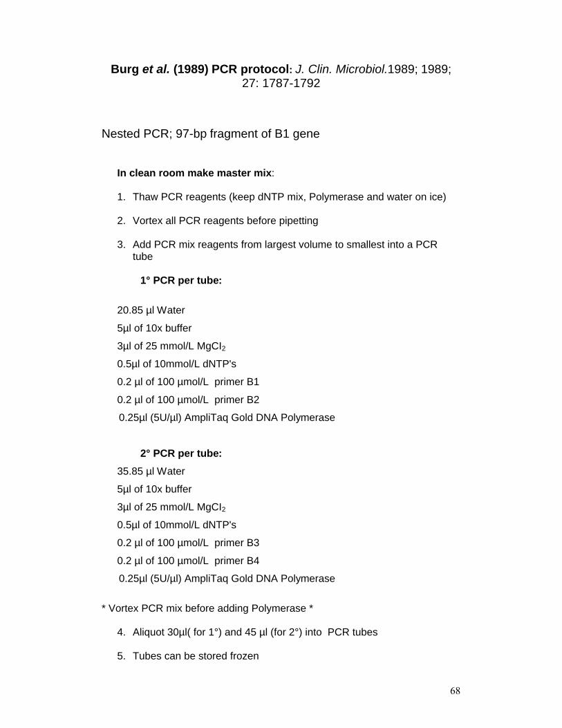
68
Burg et al. (1989) PCR protocol: J. Clin. Microbiol.1989; 1989;27: 1787-1792
Nested PCR; 97-bp fragment of B1 gene
In clean room make master mix:
1. Thaw PCR reagents (keep dNTP mix, Polymerase and water on ice)
2. Vortex all PCR reagents before pipetting
3. Add PCR mix reagents from largest volume to smallest into a PCR tube
1° PCR per tube:
20.85 µl Water
5µl of 10x buffer
3µl of 25 mmol/L MgCI2
0.5µl of 10mmol/L dNTP's
0.2 µl of 100 µmol/L primer B1
0.2 µl of 100 µmol/L primer B2
0.25µl (5U/µl) AmpliTaq Gold DNA Polymerase
2° PCR per tube:
35.85 µl Water
5µl of 10x buffer
3µl of 25 mmol/L MgCI2
0.5µl of 10mmol/L dNTP's
0.2 µl of 100 µmol/L primer B3
0.2 µl of 100 µmol/L primer B4
0.25µl (5U/µl) AmpliTaq Gold DNA Polymerase
* Vortex PCR mix before adding Polymerase *
4. Aliquot 30µl( for 1°) and 45 µl (for 2°) into PCR tubes
5. Tubes can be stored frozen
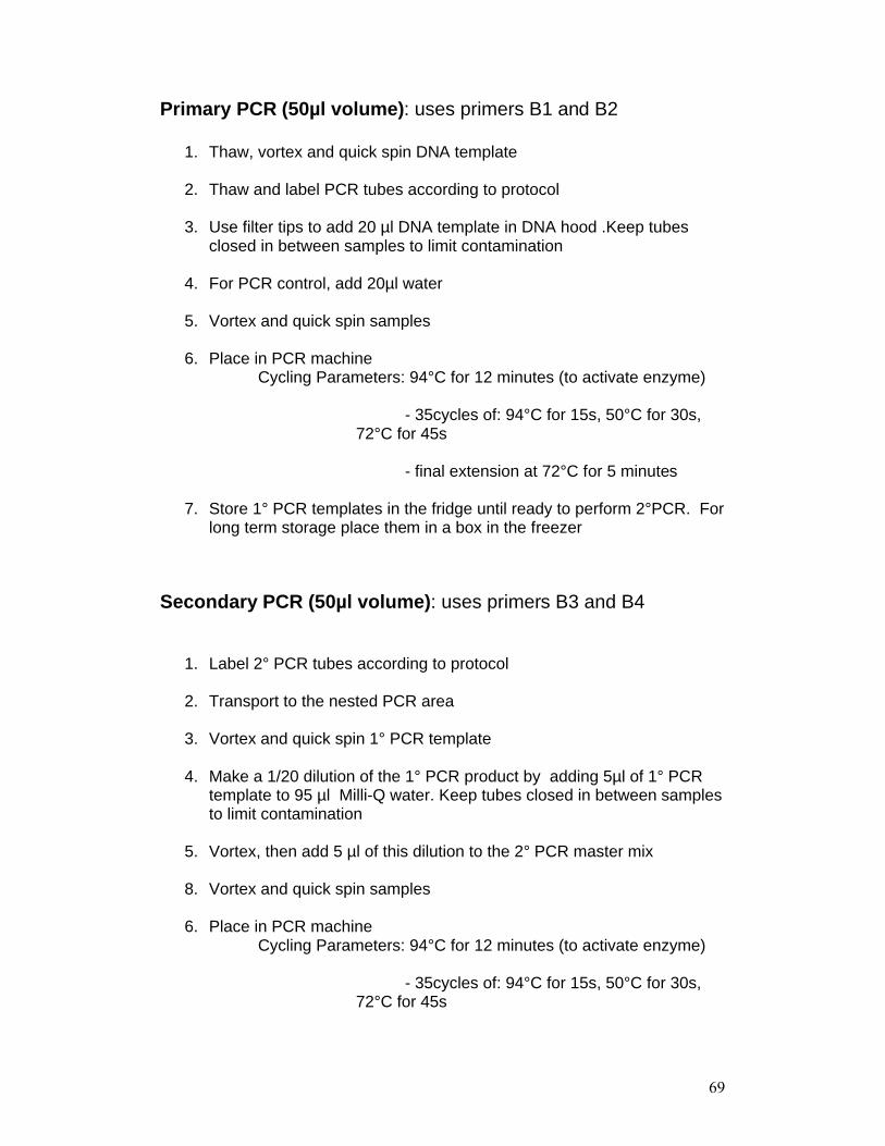
69
Primary PCR (50µl volume): uses primers B1 and B2
1. Thaw, vortex and quick spin DNA template
2. Thaw and label PCR tubes according to protocol
3. Use filter tips to add 20 µl DNA template in DNA hood .Keep tubes closed in between samples to limit contamination
4. For PCR control, add 20µl water
5. Vortex and quick spin samples
6. Place in PCR machineCycling Parameters: 94°C for 12 minutes (to activate enzyme)
- 35cycles of: 94°C for 15s, 50°C for 30s, 72°C for 45s
- final extension at 72°C for 5 minutes
7. Store 1° PCR templates in the fridge until ready to perform 2°PCR. For long term storage place them in a box in the freezer
Secondary PCR (50µl volume): uses primers B3 and B4
1. Label 2° PCR tubes according to protocol
2. Transport to the nested PCR area
3. Vortex and quick spin 1° PCR template
4. Make a 1/20 dilution of the 1° PCR product by adding 5µl of 1° PCR template to 95 µl Milli-Q water. Keep tubes closed in between samples to limit contamination
5. Vortex, then add 5 µl of this dilution to the 2° PCR master mix
8. Vortex and quick spin samples
6. Place in PCR machine Cycling Parameters: 94°C for 12 minutes (to activate enzyme)
- 35cycles of: 94°C for 15s, 50°C for 30s, 72°C for 45s

70
- final extension at 72°C for 5 minutes
7. 2° PCR templates can be left in the PCR machine overnight at 4°C, refrigerated or frozen until ready for electrophoresis
dNTP mix: 10mmol working dilution :
100 µl of each nucleotide
600 µl Milli-Q water
Total volume is 1ml
Primers :
Primers are reconstituted according to supplier’s specifications (Thermo Fisher Scientific,Germany) by adding Milli-Q water in the following volumes:
Toxoplasma B1 : 446,2 µl(5’- GGA ACT GCA TCC GTT CAT GAG -3’)
Toxoplasma B2 : 271,0 µl(5’- TCT TTA AAG CGT TCG TGG TC -3’)
Toxoplasma B3 : 255,1 µl(5’- TGC ATA GGT TGC AGT CAC TG -3’)
Toxoplasma B4 : 470,7 µl(5’- GGC GAC CAA TCT GCG AAT ACA CC-3’)

71
Gel electrophoresis
Method:
1. Wipe gel chamber with water and dry
2. Prepare the small gel-setting chamber by adding combs
3. Measure 0, 6 g of agar in a 100ml flask, add 30 ml TBE buffer and
mark level
4. Dissolve over a Bunsen burner or in a microwave; boil twice and leave
to cool for a few minutes (60°C).Add de-ionised water if any has
evaporated
5. Once cool, add one drop of ethidium bromide and swirl to mix
6. Pour the gel into the chamber and leave to set
7. Remove combs, place in tank and pour 1X TBE buffer over
8. Pipette 2µl of loading buffer (Blue Juice) onto parafilm for each PCR
sample
9. Add 10µl of PCR product to each drop, pipette to mix and load into
wells
10. Load 3µl of molecular size marker into the first and last well
11.Attach leads; run from red (in front) to black at 95 volts for 45 mins
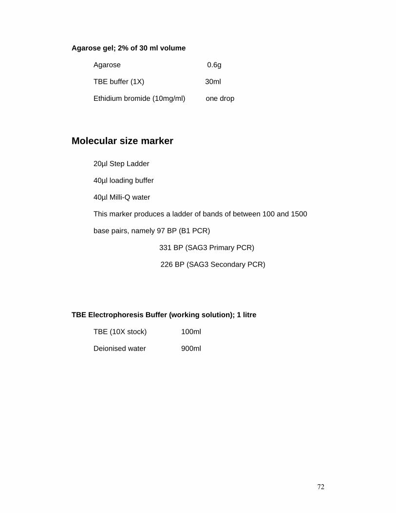
72
Agarose gel; 2% of 30 ml volume
Agarose 0.6g
TBE buffer (1X) 30ml
Ethidium bromide (10mg/ml) one drop
Molecular size marker
20µl Step Ladder
40µl loading buffer
40µl Milli-Q water
This marker produces a ladder of bands of between 100 and 1500
base pairs, namely 97 BP (B1 PCR)
331 BP (SAG3 Primary PCR)
226 BP (SAG3 Secondary PCR)
TBE Electrophoresis Buffer (working solution); 1 litre
TBE (10X stock) 100ml
Deionised water 900ml
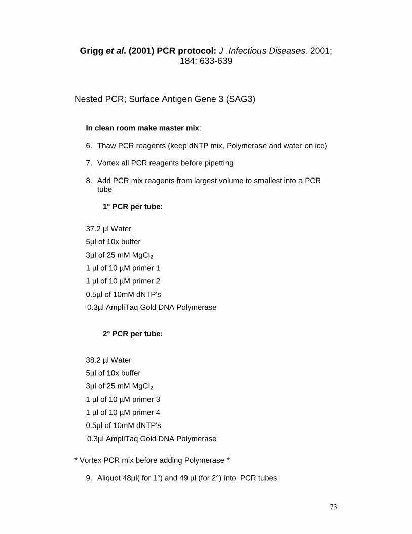
73
Grigg et al. (2001) PCR protocol: J .Infectious Diseases. 2001; 184: 633-639
Nested PCR; Surface Antigen Gene 3 (SAG3)
In clean room make master mix:
6. Thaw PCR reagents (keep dNTP mix, Polymerase and water on ice)
7. Vortex all PCR reagents before pipetting
8. Add PCR mix reagents from largest volume to smallest into a PCR tube
1° PCR per tube:
37.2 µl Water
5µl of 10x buffer
3µl of 25 mM MgCI2
1 µl of 10 µM primer 1
1 µl of 10 µM primer 2
0.5µl of 10mM dNTP's
0.3µl AmpliTaq Gold DNA Polymerase
2° PCR per tube:
38.2 µl Water
5µl of 10x buffer
3µl of 25 mM MgCI2
1 µl of 10 µM primer 3
1 µl of 10 µM primer 4
0.5µl of 10mM dNTP's
0.3µl AmpliTaq Gold DNA Polymerase
* Vortex PCR mix before adding Polymerase *
9. Aliquot 48µl( for 1°) and 49 µl (for 2°) into PCR tubes

74
10.Tubes can be stored frozen
Primary PCR (50µl volume): uses primers 1 and 2, 311bp fragment.
9. Thaw, vortex and quick spin DNA template
10.Thaw and label PCR tubes according to protocol
11.Use filter tips to add 2 µl DNA template in DNA hood. Keep tubes closed in between samples to limit contamination
12.For PCR control, add 2µl water
13.Vortex and quick spin samples
14.Place in PCR machineCycling Parameters: - 35cycles of: 94°C for 30s, 60°C for 30s,
72°C for 30s
15.Store 1° PCR templates in the fridge until ready to perform 2°PCR. For long term storage place them in a box in the freezer
Secondary PCR (50µl volume): uses primers 3 and 4, 226 bp fragment.
8. Label 2° PCR tubes according to protocol
9. Transport to the nested PCR area
10.Vortex and quick spin 1° PCR template.
11.Add 1 µl of 1° PCR template to 2° PCR master mix
12.Vortex and quick spin samples
13.Place in PCR machine
14.Cycling Parameters: - 35cycles of: 94°C for 30s, 60°C for 30s, 72°C for 30s
15.2° PCR templates can be left in the PCR machine overnight at 4°C, refrigerated or frozen until ready for electrophoresis
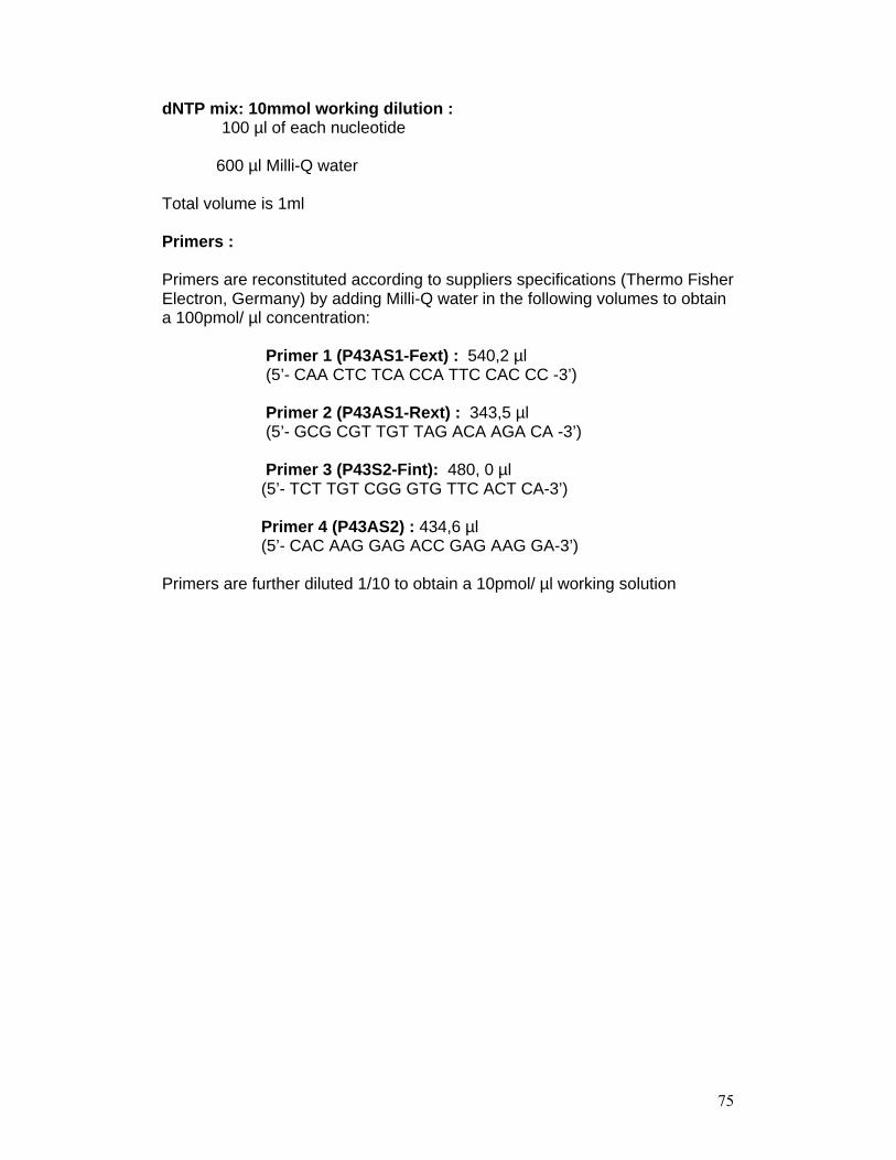
75
dNTP mix: 10mmol working dilution : 100 µl of each nucleotide
600 µl Milli-Q water
Total volume is 1ml
Primers :
Primers are reconstituted according to suppliers specifications (Thermo Fisher Electron, Germany) by adding Milli-Q water in the following volumes to obtain a 100pmol/ µl concentration:
Primer 1 (P43AS1-Fext) : 540,2 µl(5’- CAA CTC TCA CCA TTC CAC CC -3’)
Primer 2 (P43AS1-Rext) : 343,5 µl(5’- GCG CGT TGT TAG ACA AGA CA -3’)
Primer 3 (P43S2-Fint): 480, 0 µl(5’- TCT TGT CGG GTG TTC ACT CA-3’)
Primer 4 (P43AS2) : 434,6 µl(5’- CAC AAG GAG ACC GAG AAG GA-3’)
Primers are further diluted 1/10 to obtain a 10pmol/ µl working solution

76
Enzyme Digestion
Grigg et al. (2001)
Restriction enzyme: NciI
1. Mix :
7.5 µl MilliQ water
2.0 µl Buffer (TANGO X10)
0.5 µl NciI enzyme
10 µl PCR product
2. Incubate at 37°C for 3 hours
3. View using gel electrophoresis
Restriction enzyme: BcnI
1. Mix :
7.0 µl MilliQ water
2.0 µl Buffer (TANGO X10)
1.0 µl BcnI enzyme
10 µl PCR product
2. Incubate at 37°C for 3 hours
3. View using gel electrophoresis
4. Restriction enzymes cut type I strain – twice (154 BP)
type II strain- not cut (298 BP)
type III strain -once (220 BP)

77
Toxoplasma gondii cultivation
Human Foreskin Fibroblasts (HFF) Cells:
1. Remove geneticin medium from flask of confluent HFF cells
2. Wash with 5ml sterile PBS
3. Incubate with 1ml of 0,125 % trypsin diluted in PBS for 10 minutes at
37°C
4. Add 1ml of Gentamicin medium and spilt cells into 4 new flasks (0,5 ml
per flask)
5. Add 5ml Gentamicin medium to each of 3 flasks, and 5ml Geneticin
medium to the fourth flask, to maintain stock culture
6. Incubate at 37°C, 5% CO2 incubator until a confluent monolayer of
cells grow (+/- 1 week)
Toxoplasma gondii:
1. Select a confluent HFF cells flask, change medium
2. Select a freshly lysed HFF/Toxoplasma flask. Remove cells from the
walls with a pipette, transfer desired amount of Toxoplasma to the new
HFF flask
2 day passage: 6-10 drops
1 week passage: 50 µl
Overnight passage: 1-1, 5 ml
3. Recap flasks loosely and place in incubator

78
Collection of parasites:
1. Remove cells from walls with a pipette
2. Using a needle and syringe, remove parasites and medium
3. Remove needle and attach syringe to 3µM Isopore filters, Millipore,
Billerica, MA, USA
4. Press down gently to allow all liquid to pass through
5. Centrifuge at 2 500 rpm for 5 minutes
6. Discard supernatant
7. Disturb sediment slightly, wash with 5ml PBS (2 500 rpm for 5 mins)
8. Remove PBS and collect sediment
9. Can be frozen as a pellet at -20°C
Medium:
Prepare in sterile Falcon tube:
DMEM 44ml
Hepes (1M) 0.5 ml
FCS 5ml
L-Glutamine (200 Mm) 0.5ml
Gentamicin (10mg/ml) 0.1ml
For stock culture of HFF add Geneticin (0.4ml /50ml of stock 50 mg/ml)
instead of Gentamicin.
79
REFERENCES
1. Abdel-Hameed, A.A. 1991. Sero-epidemiology of toxoplasmosis in Gezira,
Sudan. Journal of Tropical Medicine and Hygiene. 94: 329-332.
2. Aoun, M., Georgala, A., Mboumi, K., De Bruyne, J.M., Duchateau, V.,
Lemort, M., Martiat, P. 2006. Changing the outcome of toxoplasmosis in bone
marrow transplant recipients. International Journal of Antimicrobial Agents.
27: 570-572.
3. Arens, J., Barnes, K., Crowley, N., Maartens, G. 2007. Treating AIDS-
associated cerebral toxoplasmosis – pyrimethamine plus sulfadiazine
compared with cotrimoxazole, and outcome with adjunctive glucocorticoids.
South African Medical Journal. 97: 956-958.
4. Barragan, A., Sibley, L.D. 2002. Transepithelial migration of Toxoplasma
gondii is linked to parasite motility and virulence. Journal of Experimental
Medicine. 195: 1625-1633.
5. Bhigjee, A.I., Naidoo, K., Patel, V.B., Govender, D. 1999. Intracranial mass
lesions in HIV-positive patients – the KwaZulu/Natal experience. South
African Medical Journal. 89: 284-288.
6. Bhopale, G.M. 2003. Pathogenesis of toxoplasmosis. Comparative
Immunology, Microbiology & Infectious Diseases. 26: 213-222.
80
7. Bonfioli, A.A., Orefice, F. 2005. Toxoplasmosis. Seminars in Ophthalmology.
20: 129-141.
8. Boyer, K.M. 2000. Congenital toxoplasmosis: Current status of diagnosis,
treatment and prevention. Seminars in Pediatric Infectious Diseases. 11: 165-
171.
9. Brand South Africa. South Africa’s geography. 2008.
<http://www.safrica.info/about/geography/geography.htm>
[Accessed 8 December 2008]
10. Brindle, R., Holliman, R., Gilks, C., Waiyaki, P. 1991. Toxoplasma antibodies
in HIV-positive patients from Nairobi. Transactions of the Royal Society of
Tropical Medicine and Hygiene. 85: 750-751.
11. Burg, J.L., Grover, C.M., Pouletty, P., Boothroyd, J.C. 1989. Direct and
sensitive detection of a pathogenic protozoan, Toxoplasma gondii, by
polymerase chain reaction. Journal of Clinical Microbiology. 27: 1787-1792.
12. Carruthers, V.B. 2002. Host cell invasion by the opportunistic pathogen
Toxoplasma gondii. Acta Tropica. 81: 111-122.
13. Cook, A.J.C., Gilbert, R.E., Buffolano, W., Zufferey, J., Peterson, E., Jenum,
P.A., Foulon, W., Semprini, A.E., Dunn, D.T., Holliman, R. 2000. Sources of
81
toxoplasma infection in pregnant women: European multicentre case-control
study. British Medical Journal. 321: 142-147.
14. Cox, F.E.G. 2002. History of Human Parasitology. Clinical Microbiology
Reviews. 15: 595-612.
15. Diallo, S., Ndir, O., Dieng, Y., Leye, A., Dieng, T., Bah, I.B., Diop, B.M.,
Gaye, O., Faye, O. 1996. A study of the seroprevalence of toxoplasmosis in
women during their reproductive years in Dakar, Senegal. Cahiers Santé. 6:
102-106.
16. Doehring, E., Reiter-Owona, I., Bauer, O., Kaisi, M., Hlobil, H., Quade, G.,
Hamudu, N.A.S., Seitz, H.M. 1995. Toxoplasma gondii antibodies in pregnant
women and their newborns in Dar Es Salaam, Tanzania. American Journal of
Tropical Medicine and Hygiene. 52: 546-548.
17. Dubey, J.P., Lindsay, D.S., Speer, C.A. 1998. Structures of Toxoplasma
gondii tachyzoites, bradyzoites, and sporozoites and biology and development
of tissue cysts. Clinical Microbiology Reviews. 11: 267-299.
18. Dubey, J.P., Frenkel, J.K. 1998. Toxoplasmosis of rats: A review, with
considerations of their value as an animal model and their possible role in
epidemiology. Veterinary Parasitology. 77: 1-32.
82
19. Dubey, J.P. 2004. Toxoplasmosis – a waterborne zoonosis. Veterinary
Parasitology. 126: 57-72.
20. Dubey, J.P., Jones, J.L. 2008. Toxoplasma gondii infection in humans and
animals in the United States. International Journal for Parasitology. 38: 1257-
1278.
21. Frenkel, J.K. 1973. Toxoplasma in and around us. Bioscience. 23: 343-352.
22. Grigg, M.E., Ganatra, J., Boothroyd, J.C., Margolis, T.P. 2001. Unusual
abundance of atypical strains associated with human ocular toxoplasmosis.
Journal of Infectious Diseases. 184: 633-639.
23. Hari, K.R., Modi, M.R., Mochan, A.H.D., Modi, G. 2007. Reduced risk of
toxoplasma encephalitis in HIV-infected patients – a prospective study from
Gauteng, South Africa. International Journal of STD & AIDS. 18: 555-558.
24. Hegab, S.M., Al-Mutawa, S.A. 2003. Immunopathogenesis of toxoplasmosis.
Clinical and Experimental Medicine. 3: 84-105.
25. Hill, D., Dubey, J.P. 2002. Toxoplasma gondii: transmission, diagnosis and
prevention. Clinical Microbiology and Infection. 8: 634-640.
26. Ho-Yen, D.O., Joss, A.W.L., Balfour, A.H., Smyth, E.T.M., Baird, D.,
Chatterton, J.M.W. 1992. The use of the polymerase chain reaction to detect
83
Toxoplasma gondii in human blood samples. Journal of Clinical Pathology.
45: 910-913.
27. Jacobs, M.R., Mason, P.R. 1978. Prevalence of Toxoplasma antibodies in
southern Africa. South African Medical Journal. 53: 619-621.
28. Johnson, J.D., Butcher, P.D., Savva, D., Holliman, R.E. 1993. Application of
the polymerase chain reaction to the diagnosis of human toxoplasmosis.
Journal of Infection. 26: 147-158.
29. Jones, J.L., Kruszon-Moran, D., Wilson, M., McQuillan, G., Navin, T.,
McAuley, J.B. 2001a. Toxoplasma gondii infection in the United States:
seroprevalence and risk factors. American Journal of Epidemiology. 154:
357–365.
30. Jones, J.L., Lopez, A., Wilson, M., Schulkin, J., Gibbs, R. 2001b. Congenital
toxoplasmosis: a review. Obstetrical and Gynecological Survey. 56:296-305.
31. Jones, J., Lopez, A., Wilson, M. 2003. Congenital toxoplasmosis. American
Family Physician. 67: 2131-2138.
32. Kijlstra, A., Jongert, E. 2008. Control of the risk of human toxoplasmosis
transmitted by meat. International Journal for Parasitology. 38: 1359-1370.
84
33. Kijlstra, A., Meerburg, B., Cornelissen, J., De Craeye, S., Vereijken, P.,
Jongert, E. 2008. The role of rodents and shrews in the transmission of
Toxoplasma gondii to pigs. Veterinary Parasitology. 156:183-190.
34. Kompalic-Cristo, A., Nogueira, S.A., Guedes, A.L., Frota, C., González, L.F.,
Brandão, A., Amendoeira, M.R., Britto, C., Fernandes, O. 2004. Lack of
technical specificity in the molecular diagnosis of toxoplasmosis. Transactions
of the Royal Society of Tropical Medicine and Hygiene. 98: 92-95.
35. Lappalainen, M., Hedman, K. 2004. Serodiagnosis of toxoplasmosis. The
impact of measurement of IgG avidity. Annali dell’Istituto Superiore di
Sanitá. 40: 81-88.
36. Lindström, I., Kaddu-Mulindwa, D., Kironde, F., Lindh, J. 2006. Prevalence of
latent and reactivated Toxoplasma gondii parasites in HIV-patients from
Uganda. Acta Tropica. 100: 218-222.
37. Luft, B.J., Remington, J.S. 1992. Toxoplasmic encephalitis in AIDS. Clinical
Infectious Diseases. 15: 211-222.
38. Markus, M.B. 2003. Toxoplasma gondii. In: International Handbook of
Foodborne Pathogens. Miliotis, M.D., Bier, J.W. (Eds). CRC Press. New
York, pp 511-523.
85
39. Mason, P.R., Jacobs, M.R., Fripp, P.J. 1974. Serological survey of
toxoplasmosis in the Transvaal. South African Medical Journal. 48: 1707-
1709.
40. Modi, M., Mochan, A., Modi, G. 2004. Management of HIV-associated focal
brain lesions in developing countries. Quarterly Journal of Medicine. 97: 413-
421.
41. Montoya, J.G., Liesenfeld, O. 2004.Toxoplasmosis. Lancet. 363: 1965-1976.
42. Montoya, J.G, Rosso, F. 2005. Diagnosis and management of toxoplasmosis.
Clinics in Perinatology. 32: 705-726.
43. Mordue, D.G., Monroy, F., La Regina, M., Dinarello, C.A., Sibley, L.D. 2001.
Acute toxoplasmosis leads to lethal overproduction of Th1 cytokines. Journal
of Immunology. 167: 4574-4584.
44. Ndumbe, P.M., Andela, A., Nkemnkeng-Asong, J., Watonsi, E., Nyambi, P.
1992. Prevalence of infections affecting the child among pregnant women in
Yaounde, Cameroon. Medical Microbiology and Immunology. 181:127-130.
45. Pavesio, C.E., Lightman, S. 1996. Toxoplasma gondii and ocular
toxoplasmosis: pathogenesis. British Journal of Ophthalmology. 80: 1099-
1107.
86
46. Peterson, E., Dubey, J.P. Biology of toxoplasmosis. 2001. In: Clinical
Toxoplasmosis: Prevention and Management. Joynson, D.H.M, Wreghitt, T.G.
(Eds). Cambridge University Press, Cambridge. pp 1-42.
47. Petersen, E. 2003. Toxoplasma gondii – Biology and pathogenesis. In:
Parasites of the Colder Climates. Akuffo, H., Ewert, L., Ljungström, I.,
Wahlgren, M. (Eds). Taylor and Francis, London. pp.132-148.
48. Remington, J.S., Thulliez, P., Montoya, J.G. 2004. Recent developments for
diagnosis of toxoplasmosis. Journal of Clinical Microbiology. 42: 941-945.
49. Roberts, F., McLeod, R. 1999. Pathogenesis of toxoplasmic retinochoroiditis.
Parasitology Today. 15: 51-57.
50. Rodier, M.H., Berthonneau, J., Bourgoin, A., Giraudeau, G., Agius, G.,
Burucoa, C., Hekpazo, A., Jacquemin, J.L. 1995. Seroprevalences of
toxoplasma, malaria, rubella, cytomegalovirus, HIV and treponemal infections
among pregnant women in Cotonou, Republic of Benin. Acta Tropica. 59:
271-277.
51. Rothova, A. 1993. Ocular involvement in toxoplasmosis. British Journal of
Ophthalmology. 77: 371-377.
87
52. Samra, N.A., McCrindle, C.M.E., Penzhorn, B.L., Cenci-Goga, B. 2007.
Seroprevalence of toxoplasmosis in sheep in South Africa. Journal of the
South African Veterinary Association. 78: 116-120.
53. Schneider, E., Schutte, C.H.J., Bommer, W. 1992. The prevalence of
Toxoplasma gondii infection in women of different ethnic groups in Natal,
South Africa. Southern African Journal of Epidemiology and Infection. 7: 41-
45.
54. Simpore, J., Savadogo, A., Ilboudo, D., Nadambega, M.C., Esposito, M.,
Yara, J., Pignatelli, S., Pietra, V., Musumeci, S. 2006. Toxoplasma gondii,
HCV, and HBV seroprevalence and co-infection among HIV-positive and -
negative pregnant women in Burkina Faso. Journal of Medical Virology. 78:
730–733.
55. Sonnenberg, P., Silber, E., Jentsch, U. 1998. Toxoplasmosis and HIV infection
in southern Africa. Southern African Journal of Epidemiology and Infection,
13: 104–106.
56. Sukthana, Y. 2006. Toxoplasmosis: beyond animals to humans. Trends in
Parasitology. 22:137-142.
57. Taylor, P.J., Arntzen, L., Hayter, M., Iles, M., Frean, J., Belmain, S. 2008.
Understanding and managing sanitary risks due to rodent zoonoses in an
African city: beyond the Boston Model. Integrative Zoology. 3: 38-50.

88
58. Tenter, A.M., Heckeroth, A.R., Weiss, L.M. 2000. Toxoplasma gondii: from
animals to humans. International Journal for Parasitology. 30: 1217-1258.
59. Vallochi, A.L., Muccioli, C., Martins, M.C., Silveira, C., Belfort, R., Rizzo,
L.V. 2005. The genotype of Toxoplasma gondii strains causing ocular
toxoplasmosis in humans in Brazil. American Journal of Ophthalmology. 139:
350-351.
60. Velmurugan, G.V., Dubey, J.P., Su, C. 2008. Genotyping studies of
Toxoplasma gondii isolates from Africa revealed that the archetypal clonal
lineages predominate as in North America and Europe. Veterinary
Parasitology. 155: 314-318.
61. Woldemichael, T., Fontanet, A.L., Sahlu, T., Gilis, H., Messele, T., Rinke de
Wit, T.F., Yeneneh, H., Coutinho, R.A., Van Gool, T. 1998. Evaluation of the
Eiken latex agglutination test for anti-Toxoplasma antibodies and
seroprevalence of Toxoplasma infection among factory workers in Addis
Ababa, Ethiopia. Transactions of the Royal Society of Tropical Medicine and
Hygiene. 92: 401-403.
62. World Travel Guide. Climate-South Africa. 2008.
<http://www.worldtravelguide.net/country/255/climate/Africa/South-
Africa.html > [Accessed 21 December 2008]
89
63. Zumla, A., Savva, D., Wheeler, R.B., Hira, S.K., Luo, N.P., Kaleebu, P.,
Sempala, S.K., Johnson, J.D., Holliman, R. 1991. Toxoplasma serology in
Zambian and Ugandan patients infected with the human immunodeficiency
virus. Transactions of the Royal Society of Tropical Medicine and Hygiene.
85: 227-229.