studies on the regulation of genes related to …
TRANSCRIPT

STUDIES ON THE REGULATION OF GENES RELATED TO NITROGEN FIXATION AND N-ASSIMILATION
IN Azoarcus sp. strain BH72: THE ROLE OF NtrBC
Abhijit Sarkar
2003
A THESIS SUBMITTED IN PARTIAL FULFILLMENT OF THE REQUIREMENTS FOR THE DEGREE OF
DOCTOR OF PHILOSOPHY IN (NATURAL) SCIENCE
(Dr. rer. nat.)
THE FACULTY OF BIOLOGY AND CHEMISTRY
UNIVERSITY OF BREMEN
GERMANY

Untersuchungen zur Regulation von Genen, die in der N2-fixierung und N-Assimilation von Azoarcus sp. Stamm BH72
involviert sind: Die Rolle von NtrBC
DISSERTATION
Zur Erlangung des Doktorgrades
der Naturwissenschaften
(Dr. rer. nat.)
Dem Fachbereich Biologie/Chemie der
Universität Bremen
vorgelegt von
Abhijit Sarkar
aus Indien
Bremen 2003

The studies of the presented work have been carried out from February 2000 till
March 2003 at the department of Biology/Chemistry of Bremen University, Germany,
under the guidance of Prof. Dr. Barbara Reinhold-Hurek.
Die Untersuchungen zur folgenden Arbeit wurden von Februar 2000 bis März 2003
am Fachbereich Biologie/Chemie der Universität Bremen unter der Leitung von Prof.
Dr. Barbara Reinhold-Hurek durchgeführt.
Vom Fachbereich Biologie/Chemie der Universität Bremen als Dissertation
angenommen am:
Datum der Disputation:
1. Erstgutachterin: Prof. Dr. Barbara Reinhold-Hurek
2. Zweitgutachterin: Prof. Dr. Friederike Koenig

Dedicated to My Parents …….Their dreams are my strength

Parts of the results presented in the following work have been published in:
Egener, T., Martin, D.E., Sarkar, A., and Reinhold-Hurek, B. (2001). Role of a
Ferredoxin Gene Cotranscribed with the nifHDK Operon in N2 Fixation and
Nitrogenase “Switch-Off” of Azoarcus sp. Strain BH72. J. Bacteriol. 183, 3752 – 3760
Egener, T., Sarkar, A., Martin, D.E., and Reinhold-Hurek, B. (2002). Identification
of a NifL-like protein in a diazotroph of the ß-subgroup of the Proteobacteria,
Azoarcus sp. strain BH72. Microbiol 148, 3202 – 3212.
Sarkar, A. and Reinhold-Hurek, B. (2002). Characterization of ntrBC of Azoarcus
sp. strain BH72. In Book of Abstracts, 5th European Nitrogen Fixation Conference,
Norwich, Great Britain.

Contents
Abbreviations 11 Summary 22 Introduction 43 Material and methods 14
3.1 Material 143.1.1 Chemicals 14
3.1.2 Gases 14
3.1.3 Strains and plasmids 15
3.2 Culture media and growth conditions 173.2.1 Media for E. coli 17
3.2.2 Media for Azoarcus sp. BH72 18
3.2.3 Antibiotic and other supplements 19
3.2,4 Cultures for E. coli 20
3.2.5 Cultures for Azoarcus sp. 20
3.2.6 Set up of N2 fixing cultures of Azoarcus sp. BH72 203.2.6.1 Cultures in semisolid medium 20
3.2.6.2 Batch cultures for N2 fixation in liquid medium 20
3.2.6.3 Cultures in Laboratory fermenter 21
3.3 Gas chromatography 213.3.1 Estimation of oxygen concentration 21
3.3.2 Estimation of ethylene concentration 21
3.4 Standard methods for working with nucleic acids 223.4.1 Sterilisation 22
3.4.2 Nucleic acid precipitation 22
3.4.3 Estimation of nucleic acids (DNA and RNA) 23
3.4.4 Restriction digestion 23
3.4.5 Agarose gel electrophoresis 23
3.5 Isolation of nucleic acids 243.5.1 Isolation of chromosomal DNA from Azoarcus sp. BH72 24
3.5.2 Plasmid DNA isolation 24
3.5.3 Isolation of DNA from agarose gel and solutions 25
3.5.4 Isolation of RNA 253.5.4.1 Hot phenol method for RNA isolation 25
3.5.4.2 Isolation of RNA using kit (peqGOLD Trifast) 25

3.5.4.3 DNase treatment of RNA 26
3.6 Cloning 263.6.1 The cloning vectors 26
3.6.2 Construction of recombinant plasmid 273.6.2.1 Preparation of vector and insert (along with its modification if
necessary) 27
3.6.2.2 Set up of ligation 27
3.7 Transfer of foreign DNA into bacterial cells 283.7.1 Transfer of DNA in E.coli cells 28
3.7.1.1 Transformation by CaCl2 and heat shock 28
3.7.1.2 Transformation by electroporation 29
3.7.2 Transfer of DNA in Azoarcus 29 3.7.1.1 Electroporation of Azoarcus 29
3.7.1.2 Conjugation of Azoarcus by triparental mating 30
3.8 DNA hybridisation techniques 30 3.8.1 DNA transfer to membrane 30
3.8.2 Labelling DNA probes for hybridisation 31
3.8.3 Hybridisation 31
3.8.4 Detection of the probe 32
3.9 Amplification of DNA by PCR 32 3.9.1 Standard method of amplification of plasmid or genomic DNA 32
3.9.2 PCR amplification using Proofstart polymerase 33
3.9.3 PCR amplification using RT-PCR beads 333.9.3.1 Semi-quantitative RT-PCR 33
3.10 Primer extension 35 3.11 Sequencing of DNA 36 3.12 Protein chemistry methods 37 3.12.1 SDS-PAGE 37
3.12.2 Gel staining 38
3.12.3 Western blot and immunodetection 38
3.12.4 2D-gel electrophoresis 39 3.12.4.1 Protein extraction 39
3.12.4.2 Isoelectric focussing (I dimension) 39
3.12.4.3 SDS-PAGE (II dimension) 40
3.13 Estimation of –gucuronidase activity (GUS assay) 40
3.14 Microscopy 41 3.15 Computer programmes used for data evaluation 41
4 Results 424.1 Transcript analyses of genes (nifH and nifLA) related to
N2 fixation in strain BH72 42
4.1.1 nifHDK is cotranscribed with fdxN from its upstream 54
promoter 43
4.1.2 nifA is cotranscribed with nifL utilizing the 54 promoter 44
4.1.3 nifA in strain BH72 is expressed differentially according to
N-availability. 45
4.2 Identification and genetic organization of the ntrBC genes 46 4.2.1 Cloning and sequencing of the ntrBC region. 46
4.2.2 Alignment of the NtrB and NtrC amino acid sequences of
Azoarcus sp. BH72 with known sequences from the datadases 48
4.2.3 Predicted functional motifs of the NtrB and NtrC from
Azoarcus sp. BH72 51
4.2.4 ntrB and ntrC in strain BH72 are transcriptionally linked. 52
4.3 Generation of a marker exchange ntrBC mutant of Azoarcus sp. strain BH72 53 4.3.1 Construction of a marker exchange deletion mutant of the ntrBC 53
4.3.2 Construction of a nonpolar ntrB mutant of strain BH72 54
4.3.3 Validation of constructs by Southern hybridisation and
genomic PCR amplification 55
4.4 Phenotypes of the ntrBC mutants 56 4.4.1 Growth characteristics of wild type and the ntrBC
mutants 56
4.4.2 Comparison of colony/cell morphologies of the wild type with the
ntrBC mutant growing on nitrate as sole N-source: phenotypes
of impairment exhibited by BntrBsp 57
4.4.3 Twitching motility is upregulated in BntrBsp 59
4.5 Transcriptional regulation of the ntrBC operon 60 4.5.1 Mapping the 5’ end of the ntrBC transcript (primer extension) 60
4.5.2 Undetectable expression of ntrB::gusA in strain BH72
grown on different nitrogen sources. 62
4.5.3 Effect of nitrogen sources on ntrB transcription: an RT PCR
approach. 63
4.5.4 ntrBC of strain BH72 is not likely to be autoregulated 64

4.6 Putative targets of NtrBC of strain BH72 654.6.1 Transcriptional regulation of N2 fixation genes by NtrBC 65
4.6.1.1 The nifA expression in strain BH72 is NtrBC regulated in
a nitrogen dependent manner. 65
4.6.1.2 Effect of nitrate on the derepression of nitrogenase genes
in BntrBsp 66
4.6.2 Transcription regulation of the gln genes of
Azoarcus sp. BH72: role of NtrBC 68 4.6.2.1 glnK regulation and effect of N2 on its expression: role of
NtrBC 68 4.6.2.1.1 Cotranscription of glnK and ugk 69
4.6.2.1.2 Primer extension studies to map the 5’ end of the
ugk and glnK transcript 70
4.6.2.1.3 Western blots to study the effect of nitrogen on GlnK
expression in strain BH72 72
4.6.2.1.4 Nitrogen-dependent differential glnK expression in
strain BH72 and its down-regulation in the ntrBC
mutant: RT-PCR approach 72
4.6.2.1.5 Confirmation of the nitrogen dependent glnK
expression by GUS reporter gene and its down
regulation in the ntrBC mutant 74
4.6.2.1.6 Retention of ammonium “switch off” response and
nitrogenase modification in BntrBsp under low glnK
expression levels 76
4.6.2.2 Analyses of the glnB expression and role of NtrBC 77 4.6.2.2.1 The glnB::gusA expression in strain BH72 is affected
by nitrogen 77
4.6.2.2.2 RT-PCR to analyse glnB expression in strain BH72
and its downregulation in BntrBsp 78
4.6.2.2.3 Comparable protein levels of GlnB in strain BH72
independent of nitrogen 79
4.6.2.3 Detection and analyses of the glnY in the ntrBC mutant 80 4.6.2.3.1 Detection of the glnY transcript in BntrBsp by RT-PCR 80
4.6.2.3.2 2D-gel and Western blot analyses confirming GlnY
expression along with the PII proteins 81
4.6.2.3.3 Low but detectable glnY::gusA expression in BntrBsp 82
4.6.3 Study of genes for N-assimilation in Azoarcus: role of NtrBC 83 4.6.3.1 Identification of a putative glutamine synthetase III in
Azoarcus sp. BH72 84
4.6.3.2 GSIII transcription in strain BH72 is nitrogen dependent
but free from NtrBC regulation 85

4.6.3.3 Identification of the genetic region in Azoarcus BH72
putatively encoding glutamine-2-oxoglutarate amino
transferase (GOGAT) 86
4.6.3.4 Effect of nitrogen on glt expression and its control by
NtrBC in strain BH72 87
4.6.3.5 Analysis of the nitrate assimilatory enzyme: nitrate
reductase 88 4.6.3.5.1 Identification of the genetic region in strain BH72
corresponding to the assimilatory nitrate reductase 88
4.6.3.5.2 The expression of the assimilatory nitrate reductase
gene in strain BH72 is NtrBC dependent 91
5 Discussion 936 References 111 Attachment Curriculum Vitae Acknowledgements Declaration

Abbreviations 1
Abbreviations
54-promoter RpoN dependent promoters having the –12/-24 consensus and
responsive to nitrogen
2D two dimensional
amtB, amtY genes encoding for putative proteins AmtB and AmtY respectively,
having similarity to membrane bound ammonium transport proteins.
APS Ammonium persulphate
EDTA Ethylene diamine tetra acetic acid
fdxN gene encoding for ferredoxin
glnB gene encoding for the signal transmitter protein PII or GlnB
glnIII gene encoding for glutamine synthetase three (GSIII).
glnK gene encoding for the PII paralogue, GlnK signal transmitter protein
glnY gene encoding for the third PII paralogue, GlnY signal transmitter
protein
glt gene encoding for glutamate synthetase (GOGAT)
GOGAT glutamine-2-oxoglutarate aminotransferase
GS glutamine synthetase
gusA gene encoding for -glucuronidase
nif nitrogen fixation, gene encoding for nitrogen fixation
NifA/NifL transcription activator of nif gene / inhibitor of NifA
NifH Dinitrogenase reductase
nifHDK structural genes for the nitrogenase enzyme complex
nifLA gene encoding for the NifL and NifA
nir gene encoding for assimilatory nitrate reductase
ntrBC genes encoding for NtrBC two component system
NtrBC two component regulatory system of N-metabolism
OD578 Optical density measured at a wavelength of 578 nm
PAGE polyacrylamide gel electrophoresis
PII-proteins signal transmitter protein of N-metabolism
SDS sodium dodecylsulphate
SSC standard saline citrate
TBS Tris-buffered saline
TEMED N,N,N’, N’- tetramethylethylene-diamine
Tris N-tris-(hydroxymethyl)-amino methane
ugk ORF upstream of glnK

Summary 2
Summary
The aim of this work is to study the regulation of genes related to nitrogen fixation
and ammonium assimilation in Azoarcus sp. strain BH72 and elucidate the role of
NtrBC in this regulatory process. The regulation has mostly been studied at the level
of transcription.
Characterization of the transcripts for nifHDK, encoding for the structural
genes of nitrogenase and nifLA, encoding for its transcription activator, revealed
unusual as well as common features in Azoarcus sp. BH72. Although both nifHDK
and nifLA utilized their respective upstream sigma 54 promoters for transcription
(transcriptional start sites verified by primer extension), nifHDK was unusually found
to be cotranscribed with its downstream fdxN (ferredoxin) gene. For these
experiments a protocol has been successfully developed for the first time by which all
the primer extensions were carried out non-radioactively, using an automated
sequencing machine. Also nifA was found to be transcriptionally linked to its
upstream nifL, a feature similar to the -subgroup of Proteobacteria, and its
expression in strain BH72 was found to be responsive to nitrogen.
Success has also been made to clone and characterize ntrBC from strain
BH72, which act as an important two-component system in sensing and initialising
the N-regulatory cascade. ntrB and ntrC from strain BH72 were also found to be
transcriptionally linked. Surprisingly, no standard promoter consensus could be
detected upstream of the ntrB transcriptional start site. Along with this, putative
Shine- Dalgarno sequences, representing translational initiation sites, were also
lacking upstream of their respective start codons. However, ntrB itself was found to
be expressed in an N-responsive manner and unusually found to be independent
from autoregulation.
A marker exchange deletion mutant of ntrBC (strain BntrBsp) verified that
these genes were not cryptic since the mutant had a phenotype and that several
genes were under its direct or indirect control. The mutation affected nitrogen
metabolism in several ways. Although nif+ in phenotype, the ntrBC mutant was
affected in N2 fixation, with reduced nifH::gusA expression. The expression of its
essential transcriptional activator nifLA on N2, was also found to be NtrBC regulated.
Nitrate had a severe effect on the ntrBC mutant, manifested by prolonged generation
time of growth, opaque rounded colonies, and upregulation of PHB biosynthesis in
BntrBsp. Even nitrogenase genes were not completely repressed in BntrBsp on
nitrate under microaerobiosis. This may be explained in terms of poor nitrate

Summary 3
assimilation, further substantiated by poor expression of the assimilatory nitrate
reductase in BntrBsp. However the ntrB non-polar mutant was not affected under
these conditions, suggesting the existence of an NtrB independent NtrC
phosphorylation (activation) mechanism. Unusually the “twitching motility” was found
to be upregulated in BntrBsp compared to that of the wild type, a phenotype which
was up to now not known to be linked to these genes.
Looking at the level of PII proteins in strain BH72, glnK was unexpectedly
found to be transcriptionally linked to its upstream ugk gene, encoding for a protein
putatively exported into the periplasm, and harbouring a signal peptide. Although a
putative sigma 54 promoter consensus is present upstream of ugk, a common
transcriptional start of glnK and ugk could not be mapped by primer extension,
possibly due to active processing of the single major transcript. Interestingly glnK
expression was also found to be down regulated in the ntrBC mutant in a similar
manner as that of glnB. The nitrogenase activity can be regulated at the
posttranslational level by ammonium, referred as “ammonium switch off”, which may
involve two different mechanisms. In strain BH72, GlnK and AmtB have been shown
to be essential for these responses. Strangely enough, ammonium “switch off”
response and posttranslational modification of nitrogenase still occurred in ntrBC
mutant, under low GlnK expression. Interestingly, for the first time, GlnY was found to
be expressed (albeit at low level) in the ntrBC mutant along with other PII proteins.
Thus studies on NtrBC may help in understanding the regulatory cascades of the
three paralogues, which is still not very clear in Azoarcus sp. BH72.
Analyses of the expression of nitrogen assimilatory genes encoding for
glutamine synthetase, GS and glutamate synthetase, GOGAT (using unreleased
sequence information from the Azoarcus genome project, Bielefeld) in strain BH72
revealed that the transcription of these assimilatory genes was affected by nitrogen.
Surprisingly, glnIII expression, encoding for GS, was found to be independent from
NtrBC control whereas glt encoding for putative GOGAT was found to be moderately
regulated by NtrBC. Thus it appears that NtrBC of Azoarcus has a significant
contribution on the N-metabolism, regulating nitrogenase expression via nifLA,
differentially regulating the three PII paralogues at least at the level of transcription
and affecting the nitrate assimilatory cascade. However, presence of several NtrBC
independent N-responses like physiological “switch off”, posttranslational nitrogenase
modification, and glutamine synthetase transcription provide hints for the existence of
another novel N-regulatory circuit apart from NtrBC in Azoarcus sp. BH72, which
makes it a more interesting model for the -subgroup of Proteobacteria.

Introduction 4
Introduction:
Today it is widely recognized that there is probably no ecological niche on earth
where bacteria have not evolved to exploit whatever nutrients are available to
support life. Despite this enormous versatility of bacterial metabolism, certain
fundamental mechanisms have to exist to regulate and integrate enzyme synthesis
and enzyme activity. At the centre of this metabolic control is the need to coordinate
the catabolism and assimilation of carbon and nitrogen sources so as to maximize
potential growth rates under any particular nutritional regime. Nitrogen is one of the
most important elements required for life as it is necessary for the production of
amino acids, nucleotides, amino sugars (required for the synthesis of
lipopolysaccharides and peptidoglycans), NAD, and p-aminobenzoate (a precursor in
folate biosynthesis). Consequently bacteria have developed a number of
mechanisms by which nitrogen can be assimilated from a variety of sources, ranging
from ammonium to atmospheric dinitrogen (N2). Ammonium is almost always the
preferred nitrogen source, as it can be assimilated directly into glutamine and
glutamate, the key donors for biosynthetic reactions and is therefore the least
energetically expensive substrate to process. By contrast organic sources such as
amino acids must first be degraded to ammonium and inorganic sources such as N2,
NO3- or NO2
- must be reduced before assimilation (Reitzer, 1996).
The process of biological nitrogen fixation is strictly restricted to prokaryotes.
The most spectacular highlight in this field before the 20th century was the discovery
by Hellriegel and Wilfarth that symbiotic N2 fixation by legumes was dependent upon
“ferments” in the soil, which are responsible for nodulation of roots. The nodulating
bacteria from Pisum sativum were isolated by Beijerinck. The process of nitrogen
fixation is highly energy demanding as nitrogenase (the key enzyme for N2 fixation)
requires 16 moles of ATP for the reduction of 1 mole of N2 to ammonium. So it is
quite likely that the synthesis of nitrogenase and its subsequent activity is tightly
regulated in organisms. Owing to high oxygen sensitivity of nitrogenase, the
environmental oxygen tension is a major regulatory factor in diazotrophs. Likewise
the availability of fixed nitrogen is a significant regulatory effector in free-living
diazotrophs but is of less importance for symbiotic organisms that are adapted to
export fixed nitrogen to their host. The structural organization of nitrogenase, reveals
the following salient features. The enzyme protein of nitrogenase is organized in a
complex of two proteins: dinitrogenase (or MoFe protein) and dinitrogenase
reductase (or Fe protein) (Burris, 1991). This nitrogenase is commonly referred to as
molybdenum nitrogenase (nif-encoded). A second nitrogenase with Vanadium as a

Introduction 5
cofactor and encoded by vnf had been reported from Azotobacter vinelandii
(Benemann et al., 1972); a third alternative heterometal-free nitrogenase (anf-
encoded) has also been reported for the first time in Azotobacter vinelandii and later
in Rhodobacter capsulatus (Masepohl et al., 2002). Nitrogenase MoFe protein is an
2 2 tetramer containing the cofactor FeMoco, believed to be the site for nitrogen
reduction while nitrogenase Fe-protein is a homodimer, containing an (Fe4S4) cluster
and acting as an obligate electron donor to MoFe protein (Howard et al., 1994).
The main enzymes for ammonium assimilation are glutamine synthetase (GS)
encoded by glnA and glutamine-2-oxoglutarate amino transferase (GOGAT).
Ammonium is assimilated to glutamine from glutamate in an ATP dependent reaction
catalysed by glutamine synthetase. Glutamine is reduced by NADPH and reacts with
cellular 2-ketoglutarate to form glutamate as product, the reaction being catalysed by
glutamate synthetase (GOGAT). Bacterial GS molecules are reported to be
dodecamers formed from two face-to-face hexameric rings of subunits, with 12 active
sites formed between the monomers in which the ATP and glutamate bind (Almassy
et al., 1986). The counterpart of glutamine synthetase is the GOGAT enzyme or
glutamate synthetase. In Corynebacterium glutamicum, gltB and gltD are the putative
genes coding for the large and small subunit respectively, and the transcription of this
operon depends on N-starvation (Beckers et al., 2001). NAD(P)H assimilary nitrate
reductase catalyzes the rate limiting and regulated step, the two electron reduction of
NO3- to NO2
-, in the pathway of inorganic nitrate assimilation, reported for Arabidopsis
sp. (Solomonson et al., 1990; Campbell et al., 1999). In most of the cases it has been
shown to be a multimeric protein composed of identical subunits each of which
contain FAD, a b-type cytochrome (cytochrome b557) and a molybdenum cofactor
(Solomonson et al., 1984). In this context it is important to mention that respiratory,
dissimilatory or “anaerobic” nitrate reductase is membrane bound and closely
associated with formate dehydrogenase. Absence of oxygen and presence of nitrate
/ nitrite induce its activity to reduce and dissimilate nitrate to nitrite and so the
dissimilatory nitrate reductase is strictly different from assimilatory nitrate reductase
mentioned earlier (Fedtke et al., 2002).
The process of biological N2 fixation as well as the pathways of nitrogen assimilation
are well coordinated and regulated in organisms. In most prokaryotes studied, the
control is exerted at the level of transcription of the N2 fixation genes (nif) or N-
assimilation genes (glnA). In certain organisms, including Rhodospirillum, regulation
can occur posttranslationally by covalent modification and consequent inactivation of
the nitrogenase Fe protein (Fitzmaurice et al., 1989). In other case, the nitrogenase

Introduction 6
can be subjected to a reversible conformational protection from oxygen damage;
nitrogenase in crude extracts of Azotobacter vinelandii, sediments as an air-tolerant
complex that contains a third redox protein called the Shethna, FeS II, or protective
protein (Shethna, 1970). The expression of the nif genes can also be a
developmentally regulated process as exemplified in heterocystous cyanobacteria,
where nitrogenase is synthesized and is most active within certain specialized cells
(heterocysts), where the oxygen evolving photosystem II is inactive and is thereby
spatially separated from the O2-evolving process of photosynthesis (Bottomeley et
al., 1979).
Typically the promoters of the nif genes share a unique consensus sequence
atypical in terms of general bacterial promoter sequences (-10/-35 consensus). The
K. pneumoniae nif promoters have a highly conserved sequence between positions –
11 and –26 with respect to the point of transcription initiation (Fig 2.1). This
sequence, which has a consensus of TGG-N8-TTGCA is quite unlike the consensus
TTGACA-N17-TATACA found in –35, -10 regions of most prokaryotic promoters.
Within this consensus, three residues remain invariant namely, the GG pairs at –25, -
24 and the G at –13. The –12 position is more or less conserved as C with exception
of a few rhizobial promoters where it is replaced by A. This difference suggested that
these promoters could be recognized by a modified form of RNA polymerase. It was
found indeed, that an alternative novel sigma factor of RNA polymerase ( 54),
encoded by the rpoN (ntrA) gene is needed to initiate transcription from these
promoters (Hirschmann et al., 1985). Furthermore, this sigma factor was found to be
totally dependent on an activator protein such as NifA or NtrC, for transcription
initiation. Interestingly, many non-nif genes such as E. coli glnAp2 (for N-assimilation)
and fdhF, Pseudomonas putida xylCAB are known to be RpoN dependent (Kustu et
al., 1986).
Fig 2.1 Bar diagram summarizing the sequence of 64 54-dependent promoters from 22 species (Morett and Buck, 1989). The frequency of each base is plotted at each position between -27 and –11 with respect to the transcription start site. The derived consensus is given below the histogram

Introduction 7
In enteric and many other bacteria, there is a general nitrogen regulation system (ntr)
that controls the expression of many genes concerned with nitrogen metabolism. It
comprises of four gene products: a uridylyltransferase (UTase) encoded by glnD,
trimeric effector proteins (PII) encoded by glnB and a pair of regulatory proteins
encoded by a single operon ntrBC which regulates 54 dependent transcription of
many nitrogen-regulated genes (Ninfa et al.,1995).
The ntrBC forms a part of bacterial two component systems of
phosphotransfer by histidine-aspartate signalling (Hoch and Silhavy, 1995). In its
simplest, the transmitter module of a sensor protein autophosphorylates at a
conserved histidine residue, then transfers the phosphoryl group to a conserved
aspartate in the receiver domain of a response regulator, which is often a
transcription activator. The primary structures of the receiver domains are relatively
well conserved and they fold as single units whose tertiary structure has been
determined in a number of cases (Stock et al., 1989; Volkman et al.,1995). Less well
conserved, the transmitter module has characteristic sequence motifs called the H,
N, G1, F and G2 boxes. Tertiary structures have been determined for histidine
kinases EnvZ (Tanaka et al., 1998) and CheA (Bilwes et al., 1999) revealing the
presence of separated phosphotransfer and kinase domains. NtrB, the histidine
kinase for nitrogen regulation, has a poorly characterized N- terminal sensor domain
that has homology to PAS domains (Taylor et al., 1999) and is joined to the
transmitter module by a Q-linker (Drummond et al., 1986). NtrB generally exists as
dimer (Ninfa et al., 1993). The response regulator NtrC is composed of three
domains: an N-terminal receiver domain which contains the site for phosphorylation
at aspartate position 54, a central output domain which is directly responsible for
activation of transcription by the 54 holoenzyme form of RNA polymerase and a C-
terminal DNA binding domain which carry the major dimerization determinants for the
protein and mediates binding to transcriptional enhancers. The receiver domain of
NtrC is connected to the central output domain by a flexible protease sensitive linker
(Keener et al., 1988). Phosphorylation–dependent interdomain interactions between
the receiver domain of one subunit and the output domain of its partner subunit in an
NtrC dimer precede and give rise to oligomerization needed for transcriptional
activation (Lee et al., 2000). Moreover yeast two hybrid systems have been used to
show protein contacts between two component regulators (Martinez-Argudo et al.,
2001).

Introduction 8
A cascade of regulatory interactions is generated to initiate the process of N2
fixation or N-assimilation (Fig 2.2). The UTase (uridylyltransferaes/uridylyl-removing)
enzyme, product of glnD, is considered to be the primary sensor to the cellular N-
status and responds to the ratio of the -ketglutarate to glutamine pools. In
Azospirillum sp. the N-regulated activity of the main N-assimilating enzyme (GS) is
not altered in glnD insertion mutant (Van Dommelen et al., 2002). In enteric bacteria,
the activity of GS is regulated by adenylation/deadenylation, depending on the
nitrogen availability. This modification is catalyzed by the bifunctional enzyme
adenyltransferase/ATase, the acitivity of which is in turn controlled by PII. So PII
occupies a pivotal position in the nitrogen regulation network. The nitrogen status of
the cell as sensed by uridylyltransferase, is signalled to PII by adjusting the degree of
uridylylation of the latter. Native PII indicates a nitrogen rich status, whereas PII-UMP
flags a nitrogen-poor status of the cell. Usually the GlnD senses glutamine as a
nitrogen signal. Glutamine inhibits uridylylation of PII by affecting the rate of UMP
transfer and in presence of Mg+2, stimulates deuridylylation of PII-UMP. Low levels of
oxoglutarate (sign of N-excess) stimulate the interaction of unmodified PII and NtrB,
leading to an inhibition of kinase activity and activation of phosphatase activity of
NtrB so that its response regulator NtrC is inactivated by dephosphorylation.
Under N-limitation, interaction of PII with NtrB is inhibited by uridylylation. At
the same time, binding of oxoglutarate to PII under N-deficiency inhibits PII-NtrB
interaction further. Under such a situation, histidine autokinase activity of NtrB is
stimulated and phosphorylates its response regulator NtrC. Phosphorylated NtrC
(active form) acts as a transcriptional activator of different ntr regulated operons. It
usually binds to enhancer sequences, upstream of the promoter and interacts with
the promoter-bound 54 by means of a DNA loop to activate the formation of the
transcription open complex at these promoters (Popham et al., 1989). On one hand it
activates transcription of its own operon along with glnA (where glnA is encoded
along with ntrBC, as in enterics). Side by side with an upregulated transcription of
glnA, the covalent modification of GS-AMP is removed by the PII-UMP stimulated
ATase (glnE) activity forming more of unmodified GS (active), subsequently
enhancing N-assimilation. The two other potential targets of phosphorylated NtrC are
the nifLA and glnKamtB where it acts as transcriptional activators for these N-
regulated genes. The nifA is positively regulated by phosphorylated NtrC as reported
in Herbaspirillum seropedicae (Wassem et al., 2002) or in Klebsiella pneumoniae
(Soupene et al., 1997). In a similar way, E. coli glnA, glnK, and nac promoters have
been found to be positively regulated by NtrC (Atkinson et al., 2002).

Introduction 9
AmtB
NtrC-P
nifHDK
nifLA NNiiffAA NifL
ntrBC
Nitrogenase
DraT/G
ggllnnAA
ggllnnKK aammttBBGlnK
PII UMP
NtrB
UTase (glnD) PII
GS (active)
ATase (glnE)
GS-AMP
NtrB-P
Low N/C
HighN/C
(inactive)
Fig 2.2 Schematic model illustrating different regulatory interactions generated during N2 fixation and ammonium assimilation in Proteobacteria. The grey block arrows represent transcriptional activation of the mentioned genes. Transcriptional activation for nifH gene expression (A) or nifLAexpression (F), posttranslational nitrogenase activity control (B), ammonium uptake (C), posttranslational activity control of GS (D) or its transcriptional activation (E). Transcriptional activation of glnK-amtBoperon (G) or expression of ntrBC (H). The scheme shows compilation of known signal transduction processes and may vary among different species.
AB
C
D
E
FG
H
The NifA protein acts as an essential proteobacterial transcription activator of
nifHDK, the structural genes for nitrogenase. In diazotrophs such as Proteobacteria
of the -subgroup or Herbaspirillum seropedicae belonging to subgroup (Souza et
al., 1991), the NifA proteins show a conserved cysteine motif in the central domain,
(not present in -Proteobacteria) which is probably the site of a redox sensitive Fe-S
cluster (Dixon, 1998; Fischer et al., 1988). Diazotrophs belonging to -Proteobacteria
such as Azotobacter vinelandii and Klebsiella pneumoniae are characterized by a

Introduction 10
NifL/A two-component regulatory system, with NifL being the sensor inhibiting the
NifA activity in response to O2 (Dixon, 1998). Stoichiometric amounts of both proteins
are needed to ensure proper transcription regulation (Dixon, 1998; Govantes et al.,
1996). For O2 sensing, the flavoprotein NifL inhibits NifA activity in the oxidized form
(Dixon, 1998; Hill et al., 1996). The mechanism by which the cellular N-status is
sensed and the signal transmitted is more complex and may vary considerably in
different diazotrophs. One level of control is the transcriptional regulation of the nifA
itself, which may be nitrogen regulated via the two component regulatory system
NtrBC as in Klebsiella pneumoniae (Drummond et al., 1983) or Herbaspirillum
seropedicae (Souza et al., 2000). At another level, the activity of NifA is modulated,
PII like proteins being the central signal transmitter proteins. The activity of
nitrogenase itself is posttranslationally regulated in some organisms like
Rhodospirillum rubrum, or Azospirillum brasilense by reversible mono-ADP
riobosylation of nitrogenase (Zhang et al., 1997; Ludden et al., 1989). In Azospirillum
brasilense it has been reported that ntrBC mutations have no effect on nif expression
but do alter the regulation of draT/G (genes responsible for ADP ribosylation) and
alter nitrogenase activity in response to ammonium (Zhang et al., 1994).
The small trimeric signal transmitter protein PII, product of the glnB gene,
plays an important role in regulation of N2 fixation and assimilation in several species
of Proteobacteria, cyanobacteria, Gram positives and Archea (reviewed by Merrick
and Edwards, 1995). Recently glnB like genes have also been identified in plants,
namely Porphyra sp., a red algae, in which they are encoded in the chloroplast DNA
(Reith et al., 1995), and in Arabidopsis thaliana, in which they are encoded in the
nuclear DNA (Hsieh et al., 1998). As mentioned earlier, GlnD (UTase/UR enzyme,
product of glnD) uridylylates GlnB under N-limiting conditions and deuridylylates it
under conditions of nitrogen excess, thereby stimulating NtrB-dependent
dephosphorylation of NtrC, resulting in prevention of transcription of ntr-dependent
operons. Moreover identification of a second PII like protein (named GlnK or GlnZ) in
several proteobacteria raised the question as to whether these proteins are also
involved in the control of nitrogen metabolism. Interestingly in many Bacteria and
Archea, the PII paralogue glnK occurs in an operon with an amtB gene coding for an
integral membrane protein, a homologue was identified as high affinity ammonium
transporter in Saccharamyces cerevisiae (Marini et al., 1994) and Arabidopsis
thaliana (Ninnemann et al., 1994). An involvement in ammonium transport is also
assumed for bacteria (De Zamaroczy et al., 1998, Michel-Reydellet et al., 1997,
Soupene et al., 1998). It has been proposed that the physical linkage of glnK and
amtB reflects a functional relationship and physical interaction of these proteins

Introduction 11
(Thomas et al., 2000). Not only is the glnKamtB operon a putative target of NtrC; in
E. coli, GlnK participates in the regulation of NtrC phosphorylation and GS
adenylation through adenyltransferase (ATase, glnE product) (Atkinson et al., 1998).
GlnK is also necessary to relieve NifL-dependent inactivation of NifA under
conditions of nitrogen limitation (He et al., 1998; Jack et al., 1999). Furthermore
(methyl)ammonium uptake appears to be regulated by GlnZ, a GlnK homologue in
Azospirillum brasilense (De Zamaroczy, 1998). A finer tuning to these regulatory
interactions is added by heterotrimer formation by GlnK and PII in vivo as has been
reported in E. coli (van Heeswijk et al., 2000), making the situation more interesting
to study.
The aim of this work is to find out how the genes related to nitrogen fixation
and N-assimilation are regulated in Azoarcus sp. strain BH72 with special emphasis
on NtrBC. So at this point, it is important to get introduced to Azoarcus. To reclaim
the salt-affected soils of the arid regions of Punjab of Pakistan, Kallar grass, a C4
plant (Leptochloa fusca (L.) Kunth), was introduced as a pioneer plant in the Punjab
region (Sandhu et al., 1975). Kallar grass has a high tolerance of waterlogged
conditions, soil salinity and alkalinity (Khan, 1966). Luxuriant growth of these plants
without nitrogen fertilizers along with acetylene reduction activity in its rhizosphere
(Malik et al., 1980) indicated a possible occurrence of nitrogen fixation. Analysis of
the microbial population in different zones of the rhizosphere showed that the
population of diazotrophic bacteria on the rhizoplane was found to be 35 fold higher
than in nonrhizospheric soil (Reinhold et al., 1986). Interestingly the rhizoplane was
dominated by Azospirillum and the endorhizospheric population was predominated
by unidentified diazotrophic rods of which one of them was later assigned to the
genus Azoarcus spp. (Reinhold-Hurek et al., 1993b). DNA-rRNA hybridization
studies placed them in separate rRNA branch in the -subdivision of Proteobacteria.
These strains showed a surprising diversity: they formed five groups distinct at
species level according to DNA-DNA hybridisation studies although they were very
similar to each other in morphology and nutritional requirements. Therefore instead of
five, only two named species, A. indigens and A. communis were proposed while
strain BH72 was included into the genus Azoarcus. The strains S5b2 (formerly
Azoarcus sp. Group C) and 6a3 (formerly Azoarcus sp. Group D) have recently been
placed under new genera, Azovibrio restrictus and Azospira oryzae respectively
(Reinhold-Hurek et al., 2002). Recently, two new species of Azoarcus have been
described: A. tolulyticus (Zhou et al., 1995) and A. evansii (Anders et al., 1995). They
are non-plant associated and originate from polluted sediments of US and South

Introduction 12
America. In contrast to plant-associated species, they are capable of degrading the
aromatic hydrocarbon anaerobically with nitrate as electron acceptor.
Azoarcus sp. strain BH72, is also capable of infecting rice seedlings in the
laboratory (Hurek et al., 1994). It can infect the roots of rice and spread systemically
into the shoot without causing symptoms of plant disease. Reporter gene studies
have shown that nitrogenase (nif) genes of Azoarcus spp. can be expressed
endophytically in the arenchyma of these seedlings, suggesting that the interior of the
rice roots provides a microenvironment suitable for N2 fixation (Egener et al., 1999).
Strain BH72 is unusual in that it can shift into a state of “hyperinduction” under
certain growth conditions that include extremely low oxygen concentrations (30 nM).
This state of strain BH72 is characterized by increased activity and efficiency of N2
fixation (Hurek et al., 1994), appearance of intracellular membrane stacks
(diazosomes), and association of the iron-protein of nitrogenase with diazosome
membranes (Hurek et al., 1995). Diazosome formation can be induced reproducibly
in the laboratory by cocultivating strain BH72 with the ascomycete Acremonium
alternatum which was isolated from the root interior of Kallar grass as well (Hurek et
al., 1998). The cells attach to the fungal mycelium, and the fungal respiration may
provide sufficient microaerobic niches for diazosome formation. The association of
nitrogenase with these membranes suggests that they are involved in efficient N2
fixation, possibly by providing a more efficient electron flux to nitrogenase.
Interestingly strain BH72 harbours unusual short type IV pili, encoded by the pilAB
locus which are involved in the colonization of both plant and fungal surfaces,
indicating that there may be common traits in the initial steps of interactions with
eukaryotic organisms of their habitat (Dörr et al., 1998).
At this point it is important to mention some of the characteristic features of
strain BH72 in context to genes related to nitrogen fixation, which have already been
reported for this organism. In contrast to most other bacteria, strain BH72 was found
to cotranscribe a ferredoxin gene with the structural nifHDK genes of nitrogenase.
Mutational analyses revealed that ferredoxin is not essential for N2 fixation but for the
rapid “switch off” of nitrogenase activity in response to ammonium addition (Egener
et al., 2001). The NifA in strain BH72 acts as an essential transcription activator for
nifHDK. Surprisingly a NifL like protein functionally similar to -Proteobacteria exists
in this -proteobacterial member and is encoded in the nifLA operon (Egener et al.,
2002). PII like proteins are the central signal transmitter proteins in sensing the N-
status of cells. Strain BH72 posses’ two paralogous gene copies, the glnB and the
glnK as other Proteobacteria; a third copy, the glnY, has recently been identified in

Introduction 13
this subgroup Proteobacterium. GlnY can only be detected in a glnB-glnK- double
knockout mutant and occurs only in the uridylylated state, irrespective of the N-status
of the cell (Martin et al., 2000). Moreover it has already been reported that PII like
proteins in strain BH72 have distinct roles for the physiological “switch off” and post
translational covalent modification of dinitrogenase reductase upon ammonium
addition or anaerobiosis. Moreover the AmtB protein (encoded from a glnK-amtB
operon in strain BH72) was found to be essential for ammonium induced switch-off,
serving probably as an ammonium sensor transmitting the signal to membrane
associated GlnK (Martin et al., 2002).
In this study, an attempt has been made on one hand, to map transcription
start points of some characterized nif operons (nifHDK and nifLA) from strain BH72
and verify their cotranscription status (nifHDK with fdxN and nifL with nifA). Side by
side, the effect of nitrogen on nifA transcription has been investigated by RT-PCR.
On the other hand, an effort has been made to clone and characterize the ntrBC-like
genes from strain BH72, map its transcription start by primer extension, check the
effect of nitrogen on its expression and find out whether ntrBC in strain BH72 is auto
regulated. A marker exchange ntrBC deletion mutant of strain BH72 was generated
to answer several questions related to its regulatory aspect. Examining such diverse
functions as “twitching motility” and trying to find out any role of NtrBC in this aspect,
studies were addressed to check the effect of nitrate on the ntrBC mutant, not only
on its growth and colony morphology but also on the expression of assimilatory
nitrate reductase. The effect of nitrate together with nitrogen has also been studied to
explore whether NtrBC mediated regulation of nifHDK and nifLA expression exists in
strain BH72. In context of N2 fixation, the PII proteins play an important role. So
attempt has been made on one hand, to study the effect of different nitrogen sources
on the expression of glnB and glnK by reporter gene fusion and RT-PCR and on the
other hand to check whether NtrBC from strain BH72 has any effect in modulating
their expression. Along with this, it is very interesting to check whether NtrBC has
any effect on the expression of the third PII paralogue GlnY, which is otherwise
expressed only in a glnB-K- background. Some of the other approaches made in the
following work include amplification of N-assimilatory genes encoding for GS and
GOGAT by RT-PCR using the information from the unreleased data of Azoarcus
genome project, Bielefeld, check the effect of nitrogen on their expression by RT-
PCR and find out whether NtrBC from strain BH72 exert a regulatory function on their
expression.

Material and methods 14
3 Material and methods
3.1 Material
3.1.1 Chemicals
As long as not mentioned specifically, chemicals and reagents were mostly
purchased from the following companies: Fluka (Buchs, Switzerland), Riedel de
Haen (Seelze), Serva (Heidelberg) and Sigma (Deisenhofen). Complex Media and
Agar were from Difco (Detroit, Michigan, USA). Restriction enzymes and DNA
dependent DNA polymerases were usually from New England Biolabs (Schwalbach)
and Amersham Biosciences. AMV Reverse transcriptase was purchased from
Roche. Agarose used for gel electrophoresis was from Biozym (Hess, Oldendorf) and
Serva (Heidelberg).
3.1.2 Gases
N2 and H2 (purity of each 99.993%), C2H2-acetylene (99.6%) and C2H4-ethylene
(51ppmv 2% in N2) were purchased from Messer Griesheim (Siegen).

Material and methods 15
3.1.3 Strains and plasmids
The bacterial strains used in this work are listed in Table 1. and plasmids used are
listed in Table 2.
Table 1. Bacterial strains used in this work with description.
Strain Description Source or reference
E. coli
DH5
MC1061
Azoarcus sp.
BH72
BntrBsp
BntrBKan
BGLK-GUS
BGLY-GUS
BGLB-GUS
BNtrBspGLK-GUS
BNtrBspGLY-GUS
BH72(pNHGUS)
BH72(pLGUS)
BntrBsp(pNHGUS)
BntrBsp(pLGUS)
BHNTR-GUS
F’ recA1 endA1 hsdR17(rk-, mk+) supE44 ( -
thi-1relA1 80dlacZ M15 (lacZYA-argF)U169
hsdR araD139 (araABC-leu)7679 (lac)
74galU galK rpsL thi
wild type
Sm/SpR, BH72 ntrBC::Sm/SpR
KmR, BH72 ntrB::KmR
ApR, BH72 glnK::gusA (chromosomal fusion)
ApR, BH72 glnY::gusA (chromosomal fusion)
ApR, BH72 glnB::gusA (chromosomal fusion)
ApR, BntrBsp glnK::gusA -chromosomal fusion
ApR, BntrBsp glnY::gusA -chromosomal fusion
TetR, Transconjugant BH72 with pNHGUS
TetR, Transconjugant BH72 with pNLGUS
TetR, Transconjugant BntrBsp with pNHGUS
TetR, Transconjugant BntrBsp with pNLGUS
ApR, BH72 ntrB::gusA (chromosomal fusion)
Hanahan (1983)
Meisnner et al.,
(1987)
Reinhold et al., (1986)
This study
This study
pSGLK-GUS
pSGLY-GUS
pSGLB-GUS
pSGLK-GUS
pSGLY-GUS
This study
This study
This study
This study
pSNTR-GUS
TABLE 2. Plasmids used in this work with description.
Plasmid Description Source or Reference
Cloning vectors
pBK-CMV
pBSKII
pBKSII
pUC4K
KmR, NeoR, ColE1-Replicon, f1(-)origin,
SV40ori.
ApR, ColE1-Replicon
ApR, ColE1-Replicon
ApR, KmR, Km-resistance cassette
Stratagene
Stratagene
Stratagene
Pharmacia

Material and methods 16
pHP45
pLAFR3
Constructs used
pDZD3
pDZD6
pDZD17
pDZD41
pNTRC2
pSNTR2.1
pSNTR2.7
pSNTR2.8
pSNTR2.9
pSNTR2.6
pSNTR2.5
pSNTR2.10
pSNTR2.2
pSNTR2.3
pSNTR2.1
pBKSNTR2.1
pBKSNTR2.1kan
pNHGUS
Sp/SmR, Sp/Sm-resistance cassette
TetR, Cosmid vector
KmR, 3.7 kb chromosomal Sau3AI fragment
having glnB locus in pBK-CMV
KmR, glnY-amtY locus on a 5.5 kb
chromosomal Sau3A fragment in pBK-CMV
KmR, glnK-amtB locus on a 5.8 kb
chromosomal Sau3A fragment in pBK-CMV
KmR, XhoI subclone of pDZD4 having
upstream of ugk in pBK-CMV
KmR, 6.5 kb phage bank clone bearing the
ntrC region in pBK-CMV
KmR, 4.5 kb subclone from pNTRC2; HindIII
digested and religated in pBK-CMV.
ApR, 1.2 kb SmaI fragment from pSNTR2.1;
subcloned in pBKSII
ApR, 1.87 kb SmaI fragment from pSNTR2.1;
subcloned in pBKSII
ApR, 0.54 kb PstI subclone from pSNTR2.1 in
pBKSII
ApR, 0.76 kb PstI subclone of pSNTR2.1 in
pBSKSII
ApR, 0.51 kb PstI subclone of pSNTR2.1 in
pBKSII
ApR, 0.3 kb PstI subclone of pSNTR2.1 in
pBKSII
KmR, PstI digested pNTRC2, and religated
KmR, XhoI digested pNTRC2, and religated
KmR, Sp/SmR, 1.55 kb region between the two
extreme NruI sites (after complete digest)
deleted from pSNTR2.1 and replaced by
cassette.
ApR, Asp718-SstI fragment from pSNTR2.1
(ntrBC region) cloned in pBKSII
ApR, KmR, Kanamycin cassette from pUC4K
inserted in the ntrB region at NruI site.
TetR, nifH::gusA fusion in pLAFR3
(Prentki and Krisch,
1984)
(Staskawicz et al.,
1987)
Martin et al., 2000
Martin et al., 2000
Martin et al., 2000
Martin,1996
Egener, 1998
This study
This study
This study.
This study
This study
This study
This study
This study.
This study
This study
This study
This study
Egener, 1998

Material and methods 17
pLGUS
pSGLK-GUS
pSGLY-GUS
pSGLB-GUS
TetR, HindIII-BsrBI-fragment of a part nifL
gene along with its upstream, cloned in
pLAFR3-GUS
ApR, 2.4 kb KpnI-SalI fragment from pDZD17
(glnK-region) cloned in pBluescript II SK; gusA
being inserted at EcoRI site in the vector at
right orientation.
ApR, 1.5 kb SalI fragment from pDZD18 (glnY-
region) cloned in pBluescript II SK; gusA
being inserted at EcoRI site in the vector at
right orientation.
ApR, 0.9 kb SstI-BglII fragment from pDZD3
(glnB-region) ligated to SstI-BamHI digested
pBKSII; gusA being inserted at EcoRV site in
the vector at right orientation.
Egener, 1998.
This study
This study
This study
3.2 Culture media and growth conditions.
All culture media were normally autoclaved at 121°C for 30 minutes. Non-
autoclavable stock solutions were usually sterile filtered using filters of pore size 0.2
m (Nr.FP 030/3 from Schleicher und Schuell, Dassel). For making solid media, I.5%
Agar was usually added to the medium.
3.2.1 Media for E. coli
LB medium (Luria-Bertani medium, Sambrook et al., 1989)
10g/l Bacto-Tryptone
5g/l Yeast Extract
10g/l NaCl
pH : 7 (adjusted with NaOH).
LB+ medium (Kushner, 1978)
10g/l Bacto-Tryptone
5g/l Yeast extract

Material and methods 18
10g/l NaCl
10mM MgSO4
10mM MgCl22.5mM KCl
pH 7.0 (adjusted with NaOH)
SOC medium
Bacto-Tryptone 20g/l
Yeast extract 5g/l
After autoclaving, the following solutions were added sterile to 1l of the medium
20mM Glucose
2.5mM KCl
10mM MgSO4
10mM NaCl
10mM MgCl2
3.2.2 Media for Azoarcus sp. BH72
VM-Ethanol medium (Reinhold-Hurek et al., 1993)
0.4g/l KH2PO4
0.6g/l K2HPO4
1.1g/l NaCl
0.5g/l NH4Cl
0.2g/l MgSO4.7H2O
26.4mg CaCl210mg/l MnSO4.7H2O
2mg/l Na2MoO4.2H2O
66mg/l Fe(III)-EDTA
1mg/l Yeast extract
3g/l Bacto-Tryptone
6ml/l Ethanol (sterile filtered and added after autoclaving)
pH 6.8
VM-Malate medium (Reinhold-Hurek et al., 1993)
Equivalent to VM-Ethanol medium; only 6ml/l Ethanol replaced by 5g/l Malic acid and
4.5g/l KOH.

Material and methods 19
SM medium (Reinhold et al., 1986)
0.4g/l KH2PO4
0.6g/l K2HPO4
0.1g/l NaCI
0.2g/l MgSO4.7H2O
26.4mg/l CaCl2.2H2O
10mg/l MnSO4.7H2O
2mg/l Na2MoO4.2H2O
66mg/l Fe(III)-EDTA
5g/l Malic acid
4.5g/l KOH
pH 6.8
SM medium with Nitrogen-Source (Reinhold et al., 1985)
Equivalent to SM medium: However each time, a Nitrogen source in form of 10mM
NH4Cl or KNO3 was added and called SM+N or SM+NO3 respectively.
KON medium
Similar to SM medium but supplemented with 5 g / l Yeast Extract and 1 g / l NaCl
Selection ( SEL) medium.
SM medium with 6 ml / l ethanol instead of potassium malate and (1 g / l) KNO3
3.2.3 Antibiotic and other supplements
While growing a strain resistant against a particular antibiotic, the medium was
supplemented with sterile filtered antibiotic which was added after the medium was
autoclaved and cooled down. Following antibiotic concentrations ( g/l) were used for
growing E. coli or Azoarcus sp. BH72 respectively: Ampicillin (150/50), Kanamycin
(50/50), Streptomycinsulphate (20/20), Spectinomycin (40/20), and Tetracyclin
(12/12).

Material and methods 20
3.2.4 Cultures for E. coli
Unless not stated, E. coli cultures were grown aerobically in LB medium at 37°C
under constant shaking at 200 rpm. Purity of the strain was controlled either by
microscopic examination or by testing the genetic markers (plasmids and antibiotic-
resistance). An effective long term preservation of E. coli strains were made by
mixing the culture in glycerine buffer (65% Glycerine, 0.1M MgSO4, 25mM Tris-HCl,
pH 8.0) 1:1 and then storing it at –80°C in glass vials.
3.2.5 Cultures for Azoarcus sp.
Unless stated otherwise, Azoarcus cultures were grown aerobically at 37°C in VM-
Ethanol medium with constant shaking. Media were usually supplemented with
appropriate antibiotic while growing mutant strains. Purity of strains was checked
either by microscopic examination or genetic markers. The strains could be
successfully stored at 4.C in VM –Ethanol-Agar plates for short term. For long term,
the cultures were stored in 10% Dimethylsulfoxide solution (DMSO) in liquid nitrogen.
3.2.6 Set up of N2 fixing cultures of Azoarcus sp. BH72
3.2.6.1 Cultures in semisolid medium
In order to observe the conditions for N2 fixation, the cells were usually grown in SM
medium with 0.2% Agar in glass vials. Under these conditions, the cells can make a
thin layer under the agar surface, following the O2 gradient in the semisolid medium.
3.2.6.2 Batch cultures for N2 fixation in liquid medium.
Unless stated otherwise, N2 fixing cells were grown on N-free SM medium under
microaerobic conditions in 1l Erlenmeyer flasks. Cells were precultured on SM
medium with combined nitrogen (0.05% NH4Cl, 0.01% yeast extract), washed two
times in N-free medium and then inoculated in the same at a final O.D578 0.05. To
simulate a N2 and microaerobic environment, the flasks were sealed with rubber
stoppers (suba seals) and the air inside was replaced by N2 gas. After that, 1.6%

Material and methods 21
headspace O2 concentration was set up by injecting sterile air into the flasks and the
cultures were usually incubated at 37°C by constant shaking at 100 rpm. In order to
measure for subsequent acetylene reduction by gas chromatography, acetylene gas
was injected in each flask to final 1% headspace concentration.
3.2.6.3 Cultures in Laboratory fermenter
For setting up N2 fixing cultures under definitive condition (constant dissolved O2
concentration of 0.1% at pH 6.8), cultures were grown in a 2 liter lab fermenter
(Biostat B, Braun Biotech, Melsungen). By giving air pressure and N2 injection a
stable O2 concentration could be maintained throughout the culture growth period.
3.3 Gas chromatography
3.3.1 Estimation of oxygen concentration
The oxygen concentrations of N2-fixing batch cultures were measured by gas
chromatograph, model HRGC-4000A (Konik, Barcelona, Spain). The separation of
gases was done in a molecular filter column (0.5 m, 80/100 mesh, 2 meter long and
internal diameter 0.2 cm) with helium as a carrier gas (1.5 bar column pressure). The
injection chamber had a temperature of 112°C and the column temperature was
80°C. The gases were measured by a heat-conducting detector. Room air was
injected to calibrate in the beginning.
3.3.2 Estimation of ethylene concentration
Acetylene Reduction Assay (ARA) was used to measure nitrogenase activity. The
detection method utilises the reduction of acetylene (substrate analogue of N2) to
ethylene by side products of nitrogenase reaction (H+). For the separation of
acetylene and ethylene, the following gas chromatograph equipped with Porapak-N-
column (80/100mesh, 2 m long, 0.2 cm internal diameter) was used. Nitrogen acted
as a carrier gas with a column pressure of 1.2 bar. During measurement the chamber
temperature was maintained at 112°C and column temperature remained 50°C. The
detection used a flame ionisation detector at 220°C fed with H2 and air pressure.
Ethylene (51ppm 2%, in N2) was used to calibrate the detector initially.

Material and methods 22
3.4 Standard methods for working with nucleic acids
Unless stated otherwise, most techniques used for analysis of nucleic acids (DNA
and RNA) was carried out according to standard procedures from Ausbel et al., 1987.
3.4.1 Sterilisation
All solutions, reagents and materials needed for working with nucleic acids were
autoclaved (30 min at 121°C and 1,3 bar). Glass pipettes and other glassware’s were
heat sterilised in oven at 180°C, overnight). Special care was taken while working
with RNA. All glassware’s used for RNA were baked overnight at 180°C. Distilled
water used for RNA work was first treated with 0.1% DEPC (diethylpyrocarbonate)
and stirred overnight at 37°C. All buffers and solutions used for RNA work were
made in DEPC treated water and autoclaved for 1 hour at 121°C. The working place
was made RNase free by wiping with 96% ethanol and subsequent flaming the area.
Hand gloves were always used while working with RNA for prevention against
degradation by stable RNase.
3.4.2 Nucleic acid precipitation
DNA was isolated from the solutions by precipitation. Unless otherwise stated, the
precipitation was usually done by adding 1/9 solution volume of 3 M sodium acetate
solution pH 5.2. This was followed either by adding 2.5 times the volume ice cold
ethanol and 15 min incubation at –80°C or by adding equal volume room
temperature isopropanol and incubating for 5 min at room temperature. Finally, the
nucleic acid pellet was obtained by centrifugation for 10 min at 13,000 rpm at 4°C.
The nucleic acid pellets were usually washed twice with 70% ethanol at room
temperature. The pellets were air-dried and finally dissolved in 1X TE buffer or
distilled autoclaved water. For RNA, pellets were not strongly dried and finally
dissolved in DEPC treated water.

Material and methods 23
3.4.3 Estimation of nucleic acids (DNA and RNA)
The Nucleic acid concentration of a solution was estimated by measuring its
absorption at 260nm with the help of a spectrophotometer (Ultrospec 2000,
Pharmacia, Freiburg). The DNA concentration was then calculated using the
empirical formula for double stranded DNA: g DNA/ml = E260 50 g/ml dilution
factor (Sambrook et al., 1989). The RNA concentration was measured by using the
empirical formula: g RNA /ml = E260 40 g/ml dilution factor.
3.4.4 Restriction digestion
Restriction digestion of DNA was done routine wise with 1 g plasmid DNA and 5
units of restriction endonuclease for 2 hours or with 3 g chromosomal DNA and 20
units of restriction endonuclease for 8 – 12 hours. The incubation temperature and
buffer conditions of each enzyme were chosen according to manufacturers
instruction.
3.4.5 Agarose gel electrophoreses
Unless otherwise stated, the separation of DNA was routinely done in 0.8% - 1.8%
agarose gels in TAE buffer (40 mM Tris-acetate, 2 mM EDTA, pH 8.0) with 3 – 5
V/cm. Electrophoreses of chromosomal DNA were usually done in gels with TBE
buffer (45 mM Tris borate, 1 mM EDTA, pH 8.0). DNA samples were usually treated
with 1/10 volume loading buffer {20% Ficoll 400, 0.1% bromophenol blue, 0.1%
xylenecyanol 0.1% orange G and 1 mg/ml ribonuclease A (optional)}. Samples with
low copy plasmids were incubated with loading dye, containing RNase at 37°C for 20
mins prior to gel loading. Bacteriophage lamda DNA (Amersham Biosciences)
digested with PstI restriction endonuclease was used as a standard molecular weight
marker (size marker) for DNA. For detection of DNA fragments after electrophoreses,
the agarose gel was incubated for 20 min in ethidium bromide solution (0.5 g/ml)
and viewed under UV light (302 nm) with a video camera (Image Master VDS,

Material and methods 24
amersham pharmacia biotech). Photographs were taken on special thermopapers
(K65HM, Mitsuibishi, Ratingen).
3.5 Isolation of nucleic acids
3.5.1 Isolation of chromosomal DNA from Azoarcus sp. BH72
For isolation of chromosomal DNA, the method from Dhaese et al., (1979), and
modified by Hurek et al., (1993), was routinely used. By this method, the cells were
lysed by detergent, proteins removed by enzymatic digest followed by phenol
chloroform extraction and DNA precipitation by ethanol. 1.5 ml overnight culture was
pelleted by 1 min centrifugation at 13000 rpm. The cells were washed in TES buffer
and suspended in 100 l TE buffer. Then this was treated with 300 l 5% N-
laurylsarcosin solution (Na salt in TE) so that Pronase E at the end concentration of
2.5 mg/ml could be added. This set up was mixed by vortexing and incubated for 1
hour at 37°C. This was followed by two times extraction with TE buffered phenol and
with TE buffered chloroform. Subsequently the genomic DNA was precipitated as
described before and finally dissolved in TE buffer.
3.5.2 Plasmid DNA isolation
Methods for plasmid DNA isolation from E. coli or Azoarcus sp. (usually low copy
plasmids) varied according to the purpose for its further utilization, although the basic
principle remained the same. However, for standard analytical purpose like cloning
and hybridisation, the standard plasmid preparation protocol from Birnboim and Doly
(1979), and later modified by Ausbel et al., (1987) was routinely used. 1.5 ml of
overnight culture (3 ml starting cultures when working with low copy plasmids) was
centrifuged down (13000 rpm, 1min) and the pellet suspended in 100 l TE. The cells
were then lysed by adding 200 l (200 mM NaOH / 0.1%SDS) and incubated for 5
min at room temperature. This was followed by adding 150 l ice-cold 3M sodium
acetate pH 5.2 for neutralisation. The precipitated protein and lysed cells were
centrifuged down and the supernatant containing the DNA was concentrated by
further precipitation and washed by 70% ethanol according to the standard methods
as had been discussed previously.

Material and methods 25
When the plasmid DNA was to be used for sequencing, the isolation was
made by using QIAGEN tip 100 columns (catalogue no. 10014, Qiagen, Hilden). This
method depends on reversible binding of DNA in the columns. The isolation process
using these columns was strictly according to manufacturer’s instruction.
3.5.3 Isolation of DNA from agarose gels and solutions
The isolation of DNA fragments from agarose gels and solutions were made using
QIAquick Gel Extraction Kit or QIAquick PCR Purification Kit respectively (Nr. 28704 /
28106, Qiagen, Hilden). The general principle of these kits depends on ion
dependent reversible binding of DNA fragments in anion exchange columns. The
process of using these kits was strictly according to manufacture’s instruction.
3.5.4 Isolation of RNA
While handling with bacterial RNA, special care was taken to prevent degradation by
RNases. All reagent and solutions were DEPC treated, autoclaved for 1 hour:
glasswares were baked and hand gloves were always used while working.
3.5.4.1 Hot phenol method for RNA isolation
This method follows from the protocol from Aiba (Aiba et al., 1981). 100 ml culture
(O.D. 0.5) was centrifuged down (4000 rpm, 10 min) and suspended in 3 ml “hot
phenol buffer” (20 mM sodium acetate, 0.5% SDS, 1mM EDTA, pH 5.5). This was
followed by three times extraction by hot (60°C) phenol (equilibrated with 20 mM
sodium acetate, pH 5.5. The RNA was finally precipitated from the upper phase by
adding 3 volumes of ethanol (-20°C) and finally dissolved in DEPC treated water. The
RNA concentration was estimated by using the standard formula, g RNA / ml = E260
40 g / ml dilution factor.
3.5.4.2 Isolation of RNA using kit ( peqGOLD Trifast)
For a fast and clean isolation of bacterial RNA, peqGOLD Trifast kit from peQLab
(catalogue no. 30-2010) was routinely used. Usually, 1 ml of peqGOLD solution was

Material and methods 26
added to frozen cells, collected from 50 ml of culture. The cells were suspended in
this solution and allowed to stand for 5 min. To this, 200 l of chloroform was added,
mixed and allowed to stand in room temperature for 10 min, during which phase
separation could be seen. After 5 min centrifugation, the upper phase (containing
RNA) was collected in new cup and precipitated by adding equal volume isopropanol.
The RNA pellet obtained after centrifugation was washed 2 times with 75% ethanol
and finally dissolved in DEPC treated water.
3.5.4.3 DNaseI treatment of RNA
The RNA isolated by routine procedures could not be used directly for RT PCR
(synthesis of cDNA and its subsequent amplification) as it often contained
contaminations from genomic DNA. To circumvent this, the isolated RNA was treated
with DNaseI. DNaseI is an endonuclease that degrades double stranded DNA (nicks
or breaks) to produce 3’-hydroxyl oligonucleotides. Divalent cations like Mg++ or Mn++
are absolutely crucial for its activity. About 2 -3 g RNA was mixed with DNase
reaction buffer (1X) and about 30 units of RNase free DNaseI (Roche) in a total 50 l
set up and incubated for 20 min at 37°C. This was followed by an immediate
inactivation of DNaseI by phenol chloroform isoamylalcohol (pH 4.0) treatment. RNA
was precipitated from upper phase using standard precipitation technique by chilled
ethanol washed 2 times with 75% ethanol, dissolved in DEPC treated water, and
estimated spectrophotometrically.
3.6 Cloning
3.6.1 The cloning vector
Unless stated otherwise, pBluescript SK / KS (Stratagene) and pLAFR3 (Staskawicz
et al., 1987) were usually used as cloning vectors. Mostly these vectors were used so
that there is a possibility to screen recombinant clones (containing the insert
fragment) by blue white selection. The plasmids pBSKII and pBKSII differed from
each other in context of orientation of their multiple cloning sites. Both used ColE1
Replications origin and could maintain high copy number (of about 200) per E. coli

Material and methods 27
cell The bluescript vectors contained a copy of the bla gene so that when
transformed by these plasmids the cells were resistant to ampicillin.
For transformation in Azoarcus, broad host range plasmid like pLAFR3A was
used. This vector provided tetracycline resistance to its host and could be maintained
in a low copy number inside the cells.
3.6.2 Construction of recombinant plasmid
3.6.2.1 Preparation of vector and insert (along with its modification if necessary)
For normal cloning, the vector and the cloned DNA fragment were so digested with
restriction endonucleases so that they might form compatible ends to be ligated.
When this option did not work, the cohesive ends produced by restriction
endonucleases were treated with modifying enzymes (T4 DNA polymerase or DNA
polymerase I (klenow fragment)) to produce blunt ends To prevent religation of vector
by its compatible ends, the digested vector was treated with alkaline phosphatase
(Roche) which removed the 5’ phosphate from the digested vector prior to ligation
with the insert fragment.
The cloning fragments (vector and insert) were restriction digested and run in
agarose gel for separation. The respective fragments were isolated and cleaned from
the gel slices as had been discussed earlier.
3.6.2.2 Set up of ligation
Normally for a good ligation, 3 –4 fold more of insert fragment was used compared to
that of the digested vector. A reduced reaction volume (10 l) of the ligation set up,
usually gave much better result.
For cohesive end ligation, 0.1 unit of T4 DNA Ligase (GIBCO BRL) was used.
For best result, the ligation was carried out for 1 hour at 12°C helping the sticky ends
to anneal to each other followed by 2-3 hour incubation at room temperature (22°C)
at which the ligase is highly active.
For setting up blunt end DNA ligation, a higher (insert: vector) ratio and more
units of T4 DNA ligase (1-2 unit) was used per set up. The ligation was carried out by
incubating the ligation mix at room temperature for 2 – 3 hours. Following ligation, E.

Material and methods 28
coli DH5 were transformed by the ligation mix as will be discussed in the following
section
3.7 Transfer of foreign DNA into bacterial cells.
3.7.1 Transfer of DNA in E. coli cells.
The transfer of DNA in E. coli cells was done usually by employing the method of
transformation. For this purpose the cells had to be made competent and were stored
in –80°C prior to transformation. Normally the cells could be made CaCl 2 competent
which then could be transformed by “heat shock method” (Kushner, 1978) or they
were made electro competent and subsequently transformed by electroporation.
3.7.1.1 Transformation by CaCl2 and heat shock
According to this method as described by (Kushner, 1978), the cells were washed in
a solution containing MnCl2, CaCl2 and RbCl2 so that the cells are competent to take
DNA. The exact mechanism of DNA uptake is still not known. From a fresh overnight
incubated LB plate, 5 ml preculture of LB+ medium was inoculated and allowed to
grow under constant shaking at 37°C until OD578 of 0.5. From this preculture, 80 ml of
main culture in LB+ medium was inoculated and allowed to grow under the same
conditions until OD578 of 0.4 was attained. The cells were then cooled down in ice,
centrifuged down (5 min, 5000rpm) and suspended in 20 ml TF1 (10 mM MES, 50
mM MnCl2, 10 mM CaCl2, 100 mM RbCl2). After another round of centrifugation, the
cells were resuspended in 2 ml ice cold TF2 (10 mM PIPES, 75 mM CaCl2, 75%
glycerol) and distributed in eppendorf cups in 100 l aliquots and immediately frozen
in dry ice and stored at –80°C, until transformation.
To 100 l thawed competent cells, 10 l of DNA to be transformed was
added. It was incubated in ice for 10 min, heat shocked at 42°C for 2 min and
immediately mixed with 1ml LB .The cells were incubated at 37°C for 1hour (shaking)
and then plated in LB containing antibiotic.

Material and methods 29
3.7.1.2 Transformation by electroporation
By this method the cells were subjected to a strong electric field so that the
membrane structure is disturbed and the foreign DNA is transferred inside the cells
(Neumann et al., 1982; Zimmermann et al., 1981). The method followed from Dower
(1988) and developed by Ausbel et al., (1987). From a preculture in exponential
phase, 500 ml of main culture was inoculated in Erlenmeyer flasks and cultured for 2
– 3 hours till OD578 of 0.5 was attained. The cells were then washed many times in
10% ice cold glycerol, suspended in 400 l of the same, distributed in 100 l aliquots
and immediately frozen. For electroporation, 1 – 10 g of DNA to be transformed was
dialysed against water in special filter plates (VSWPo 2500, Millipore, Eschborn). The
electroporation of E. coli was done in 0.2 cm cuvettes (Eurogenetic, Seraing,
Belgium) with a Gene Pulser (biorad, Munich). Following parameters were installed
before starting: 25 F, 200 , 2.5 kV and voltage 12.5 kV / cm. The time constant
after electroporation should be 0.48 – 0.5. After 1 hour of outgrowth in LB medium ,
the cells were plated on antibiotic plates.
3.7.1 Transfer of DNA in Azoarcus
In this work, foreign DNA in form of recombinant plasmids were transferred in
Azoarcus either exploiting transformation by electroporation or by triparental
conjugation. The former method was exploited when the gene of interest was
intended to integrate in the genome by stable recombination events. In this case the
insert was borne in low copy plasmids (eg pBluescript), which is usually not stable in
Azoarcus. On the other hand, when the gene of interest was intended to be
expressed in moderate level in trans, triparental mating by conjugation was the usual
choice. In this case, the insert was borne in low copy broad range host vectors like
pLAFR.
3.7.1.1 Electroporation of Azoarcus
The process of electroporation of Azoarcus is similar to E. coli in several aspects as
had been described. However there are certain differences. Once the cells were
made competent, they were electroporated immediately (cannot be stored in –80°C).
The cells were grown in VM malate for preculture and also for the main culture. After

Material and methods 30
electroporation, they were cultured in VM ethanol medium for 90 min and plated in
VM ethanol plates with antibiotics. Cuvette length of 0.1 cm was used and time
constant after electroporation usually exhibited lower value (0.45), in contrast to E.
coli electroporation events. Also the efficiency of transformation in Azoarcus is
drastically low as compared to that in E. coli.
3.7.1.2 Conjugation of Azoarcus by Triparental mating
Mobilized plasmids from a donor strain, taking the help of transfer gene tra from a
helper strain can be transferred to a recipient strain by the method of conjugation.
The donor (E. coli DH5 bearing the transferable plasmid -pLAFR), the helper (E.
coli DH5 bearing the natural plasmid pRK2013) and the recipient (Azoarcus sp.
BH72) were grown in separate cultures until an OD578 of 1.0 was attained. Each
culture was centrifuged down, resuspended to a final OD578 1.0, mixed in the ratio
(1:1:100) and plated on KON agar plates. After 5–7 hours of incubation at 37°C, the
cells were scraped from the surface of KON plates, washed in selective medium
(SEL), and finally plated on SEL plates with antibiotics with different serial dilution.
This process could select the transconjugants.
3.8 DNA hybridisation techniques
DNA fragments of interest specifically hybridised to a complementary and labelled
DNA fragment (probe) could be detected for analysis. In the following work,
Digoxygenin labelled DNA probes had been used. The hybridised probe could be
detected secondarily by a specific antibody, which could recognize a steroid haptene
and itself is linked to conjugated enzyme at its conserved Fc portion. This could then
catalyse a reaction transforming the substrate into a chemeluminogenic or
chromogenic product, which could be detected as signal. For hybridisation, the DNA
of interest must already bind single stranded to a membrane.
3.8.1 DNA transfer to the membrane (Southern blot)
By this method of Southern transfers (Southern, 1975), the DNA fragments
electrophoretically separated in agarose gel, were transferred into a membrane by

Material and methods 31
the help of capillary action and finally covalently attached to the membrane by UV
crosslinking. For the transfer of chromosomal DNA fragments, the agarose gel was
incubated for 10 mins in 0.25 M HCl for depurination. This was followed by alkaline
denaturation of the gel (2 20 min in 1.5 M NaCl, 0.5 M NaOH) for strand separation
so that the transfer is facilitated. Alkaline denaturation was followed by neutralization
(2 20 min in 0.5 M Tris-HCl, pH 7.2, and 1 mM EDTA) before the treated gel was
placed below a nylon membrane (Hybond N, Amersham, Braunschweig) for transfer,
along with high salt buffer (20X SSC). Following overnight transfer, the DNA was
permanently fixed with the membrane by UV crosslinking using 0.4 J/cm2 energy
(Transilluminator Fluonik, Biometra, Göttingen).
3.8.2 Labelling DNA probes for hybridisation
DNA fragments used as probes were labelled with Digoxygenin, using “the DIG –
DNA Labelling & Detection Kit “ from Roche. DNA fragment to be used, as probe was
first obtained by resriction digest or PCR amplification. The fragment was denatured
by boiling for 10 min in a 16 l reaction volume followed by quickly chilling in ice.
Then 4 l of DIG–High Prime mix (containing random primer, polymerase, dNTP and
Digoxygenin labelled UTP) was added to the denatured DNA and incubated for 1
hour or overnight at 37°C. The reaction was stopped by heating at 65°C for 10 min.
Finally the labelled probe was purified from free dNTPs using sieve chromatography
by passing it through Bio gel 250 and eluting it in 1X TE buffer pH: 8. A portion of this
purified probe was used in the hybridisation solution.
3.8.3 Hybridisation
The membrane with crosslinked DNA fragment was incubated for 1 hour at 65°C in
hybridisation solution (6X SSC, 5X Denhardt’s solution, 0.5% SDS) for
prehybridisation. The labelled probe mixed with the hybridisation solution was boiled
for 10 min before it was added to the membrane. Hybridisation was carried out at
65°C with constant shaking for minimum 8 hours; overnight incubation was however
the best option. Following hybridisation the membrane was washed at 65°C with (2X
SSC, for 15 min; 2X SSC / 0.1% SDS for 30 min; and finally 0.1X SSC / 0.1% SDS
for 10 min).

Material and methods 32
3.8.4 Detection of the probe
The hybridised probe was detected by specific antibody against Digoxygenin
epitopes. The Fc portion of the antibody was conjugated to enzyme alkaline
phosphatase. Depending on the nature of the substrate added at the end, a coloured
end product (colorimetric detection) or a precipitate of luminescent product
(chemeluminescent detection) occurred. The colorimetric detection was done using
the follow up of the “DNA-Labelling and Detection Kit” (Roche Applied Sciences)
using manufacturer’s instruction. Chemeluminescent detection was performed using
TROPIX- system (Serva, Heidelberg), following strictly the manufacturer’s instruction.
The light emission signal was permanently documented by exposing the membrane
to X ray films (X-OMAT, Kodak, Rochester, New York, USA) and subsequent
developing and fixing of the film.
3.9 Amplification of DNA by PCR
The polymerase chain reaction (PCR) was routinely used for amplification of DNA
fragments. The set up of PCR reactions varied according to purpose needed.
3.9.1 Standard method of amplification of plasmid or genomic DNA
For general purpose, DNA fragments were amplified following the standard protocol
from Innis and Gelfand (1990), using 1.25 Units Taq polymerase (Beckman, Munich),
standard buffer with 1.5 mM MgCl2, 20 M of each dNTP and 50 pmole of each
primer (forward and reverse) in 50 ul of reaction volume. Usually a standard PCR
programme consisted of a denaturation step at 95°C for 5 min linked to a loop of 35
to 45 cycles each having a denaturation step at 95°C, a primer annealing step
ranging between 50°C to 65°C (2 – 4°C below the primer melting temperature) and
an elongation step by the polymerase at 72°C. The programme usually ended with a
final extension step at 72°C for 10 min. The annealing temperature of the primers
with the template was determined according to the melting temperatures (Tm) for
each primers using the standard formula: Tm (°C) = 81.5 + 0.41 (% GC content)–
600/N +16.6 log10 M where N refers to the number of bases in the primer and logM

Material and methods 33
refers to the logarithm of the molarity of Na+ and K+ concentration present in the PCR
reaction.
3.9.2 PCR amplification using ProofStart polymerase
The proof reading activity of normal Taq polymerase is quite low, resulting in
incorporation of wrong bases in the PCR product. To circumvent this problem,
ProofStart DNA polymerase (Pfu) from Qiagen was used which provided robust PCR
performance in high-fidelity PCR. Some unique features adjoined to this method
were (an initial 5 min activation step at 95°C, a final concentration of 300 M of each
dNTP, longer extension times at 72°C and about 45 rounds of cycling). When a PCR
system did not work well under standard conditions, a special buffer (Q solution
provided with the kit) was added. This enabled amplification of a previously failed
reaction simply by changing the melting behaviour of DNA.
3.9.3 PCR amplification using RT-PCR beads
Ready-to-go-beads (Amersham Pharmacia Biotech) are basically RT-PCR beads
designed as pre-mixed, pre-dispensed reactions for performing RT-PCR. They are
provided as dried beads that are stable at room temperature and contain all of the
necessary reagents (including M-MuLV reverse transcriptase and Taq DNA
polymerase), except primer and template, for performing a one-tube one-step RT-
PCR reaction in a volume of 50 µl. The reactions were usually incubated for 30 min
at 42°C (formation of reverse transcribed cDNA), a denaturation at 95°C for 5 min
and eventual linking to the loop of denaturation, annealing and extension.
3.9.3.1 Semi quantitative RT-PCR
In order to have an idea about the amount of a particular transcript under a given
culture condition and to compare this with another condition, a semi quantitative
approach was employed. After a reverse transcription of the RNA, when the
amplification of cDNA was ongoing, equal amount of samples were pooled out from
different tubes (to be compared) at different time points (18-28 cycle number). When
electrophored in an agarose gel, the RT-PCR products exhibited differential ethidium
bromide staining intensities, which was a linear function (or clear reflection) of their

Material and methods 34
relative abundance. The primers used in this work (PCR / RT-PCR) are listed below
in form of Table 3.
TABLE 3. Description of various primers used for PCR / RT-PCR
Application name sequence
Cotranscription of
nifHDK & fdxN
Cotranscription of
nifL & nifA
RTPCR (nifA)
RTPCR(16SrRNA)
Cotranscription of
ntrB and ntrC
Cotranscription of ugk
and glnK
ntrB amplification
ntrC amplification
glnB amplification
glnK amplification
glnY amplification
GS3 amplification
nifKfw
Fdxrev
RTnifLAfor
RTnifArev
nifArev3RT
nifAfor1RT
nifArev2RT
1401rev
104f
1346rev
RTntrBbridgeF
RTntrCbridgeR
RTugk-F
RTglnK-R
ntrBfor
ntrBrev
ntrCfor
ntrCrev
glnBfor
glnBrev
glnKfor
glnKrev
glnYfor
glnYrev
GS3forRT
5’ CGCCTTCGCGGCACCCGT 3’
5’ CAGTCACCACAGGACGTGCAT 3’
5’ GAGAACGGCCAGGTCGACGTGGA 3’
5’ GTTGAAGCCGCACTCCTCGTCGAGCA 3’
5’ TCGTCCAGGTGCTCGCGGCTG 3’
5’ ATGAGCGCGGCCGGTCCGATG 3’
5’ CACGGTTTCGTGCCCGGCGCG 3’
5’ CGGTGTGTACAAGACCC 3’
5’ GGCGAACGGGTGMGTAAYGCACTGG 3’
5’ TAGCGATTCCGACTTCA 3’
5’ GGATGAGATTCGCGACAAGAT 3’
5’ CGGTCATGATGATGACCGGAA 3’
5’ GCGGCATATCCGGTTACCGAAGA 3’
5’ GATCTTCACCTTGGGCAGGAAGTCGA 3’
5’ AAGCTTGCTCGATCCTCCTCCTCCCAG 3’
5’ ATTAATTCAGGTCCGATCGGTAATCGGC 3’
5’AAGCTTAATACCGTCTGGATCGTGGATGACG 3’
5’ ATTAATTCAGTCCTCGTGCTCGCCTTCG 3’
5’ GCATGAAGAAGATCGAAGCGATCATC 3’
5’ CGGGATCCGATTGCGGCTTCGTTGGTTTC 3’
5’ GCATGAAGTTCATCACAGCGATCATC 3’
5’ CGGGATCCCAGGGCGTCGGCCCCGGTTTC 3’
5’ GCATGAAACTGATCACCGCAATCATC 3’
5’ CGGGATCCGAGGGCGTCGGCACCGGACTC 3’
5’GCTACGTCCTGGCCCAGTTCGTCGATA 3’

Material and methods 35
GOGAT amplification
Assimilatory nitrate
reductase
amplification
Sequencing primers
for pBluescript & pBK-
CMV
GS3revRT
GOGAT RTfor
GOGAT RTrev
nir forRT
NO3revRT
T3 forward
T7 reverse
5’GCAGTAGCGAGAACTCCGGCTCCA 3’
5’GAGTGTTCATCGAGCACGTCGGCATC3’
5’GTTGTGCAGATGGGACTTGCCGCAT3’
5’GCTGTCCTACGGCGTGCTCGAAGC3’
5’GCTGGCTGCTTGGACACCGG3’
5’ AATTAACCCTCACTAAAGGG 3’
5’ GTAATACGACTCACTATAGGG 3’
3.10 Primer extension
To map the transcription start point of genes, “Primer Extension Method” (Boorstein
and Craig, 1989) was performed. The method involved isolation of RNA followed by
annealing of the Cy5 labelled primer with the transcript of query and its subsequent
extension by AMV reverse transcriptase till the 5’ end of the transcript was reached.
Finally, by running a sequencing gel with the labelled single stranded cDNA product
along with a sequencing reaction of that particular gene, the precise base of the
gene, with which the 5’ base of the RNA matched (transcription start point of the
gene) could be located.
About 30 g of RNA was incubated with 2.5 pmole of Cy5 labelled primer and
1X AMV reverse transcriptase buffer (Roche Applied Sciences) at 70°C for 2 min in a
20 l reaction volume followed by “switching off” the thermo block and allowing it to
gradually cool to 42°C. The reverse transcription of RNA was performed
subsequently at 42°C for 30 min by addition of 1.25 mM each of dCTP, dGTP, dATP,
dTTP and 10 U AMV reverse transcriptase to the reaction. After a phenol chloroform
isoamylalcohol extraction and ethanol precipitation, the product was dissolved in 1X
TE. This was loaded in ALF sequencing gel along with sequencing reaction of the
gene, with a prior heating at 65°C for 2 min. The primers used for primer extension
are listed in Table 4.

Material and methods 36
TABLE 4. Description of Cy5 labelled primers used for primer extension.
Application name primer sequence
Primer extension nifH
Primer extension nifLA
Primer extension ntrB
Primer extension ntrB
Primer extension ugk
Primer extension glnK
TH25rCy
Cy5PEnifLA rev
Cy5PEntrB rev
Cy5PEntrB revUS
Cy5PEugk rev
Cy5PEglnK rev
5’TGCCGATACCGCCCTTGCC3’
5’GATCGCCGACTGCTCCACGG3’
5’CTCTCTGCCGCGATCGGC3’
5’GCGCTCGTGCAGGTCGAC3’
5’GTGGGCGGTACCGATCAG3’
5’GAACGGCTTGATGATCGCTG3’
3.11 Sequencing of DNA
DNA sequencing was routinely performed in the laboratory following dideoxymethod
(Sanger et al., 1977). The sequencing reaction was set up using “Thermo Sequenase
fluorescent labelled primer cycle sequencing kit with 7- deaza- dGTP“ (Amersham,
Braunschweig) following the manufacturer’s instruction with Cy5 labelled primers
(Thermo-Hybaid) and template DNA (clean plasmid preparations, minimum 5 g for
sequencing each clone). For each clone, 4 reactions were set up each having
ddATP, ddCTP, ddGTP or ddTTP as terminators of these particular bases
respectively. The reactions were carried out in a thermo cycler (Cyclogene, Thermo-
Dux, Wertheim), under the following set up parameters: 1 min 97°C, followed by 25
cycles of 30 s at 95°C (denaturation) and 30 s at 60°C (primer annealing and DNA
synthesis). The sequencing samples were later loaded in a 21% polyacrylamide gel “
ReproGelTM Long Read Solutions” (Amarsham Pharmacia Biotech, Uppsala,
Sweden), containing 6.2 M urea and separated with 0.6 X ALF TBE buffer in the
“ALF Express DNA Sequencer”. The fluorescent labelled fragments could be
detected by laser and photosensors. The evaluation of the sequencing reactions was
done with “ALFwin Software”.

Material and methods 37
3.12 Protein chemistry methods
3.12.1 SDS–PAGE
Preparation of total cell extracts
Cells grown in liquid culture were centrifuged down and suspended in 1 ml extraction
buffer (62 mM Tris-HCl pH 6.8, 5% -mercaptoethanol, 10% glycerol) per 150 mg
fresh weight along with 2% SDS and incubated at 95°C for 15 min. The preparation
was then centrifuged for 2 min at 13000 rpm and the clear supernatant was collected
in a new eppendorf cup and stored at –70°C.
Estimation of protein concentration
The protein concentration when required was estimated following the principle from
Bradford (1976) using the BioRad Protein Assay kit (BioRad, Munich), following
manufacturer’s instruction.
SDS polyacrylamide gel electrophoresis (SDS-PAGE)
SDS PAGE was performed following the principle from Laemmli (1970) using
discontinuous or disc electrophoresis (Ornstein, 1964) and (Davis, 1964) where a
non-restrictive large pore gel, referred to as stacking gel is layered on top of a
separating gel. Each gel layer is made with a different buffer and the running buffer is
different from the gel buffers. Set up: separation gel: 10-18% polyacrylamide, (30:1
acrylamide: basacrylamide), 0.375 M Tris-HCl, pH 8.8, 0.1% SDS; stacking gel: 5%
polyacrylamide, 0.125 M Tris-HCl, pH 6.8, and 0.1% SDS. The polymerisation took
place only after addition of 10% ammoniumpersulphate (3 l / ml) and TEMED (2 l /
ml). For the separation of the two bands of nitrogenase, “a low cross linker gel” with a
different polyacrylamide mix (acrylamide: bisacrylamide :: 172 : 1) was used.
Unless stated otherwise, the protein samples to be loaded were treated with
4X loading buffer (62.5 mM Tris-HCl pH 6.8, 15% glycerol, 2.3% SDS, 1.25% -
mercaptoethanol and 0.03% bromophenol blue) and denatured by heating at 95°C
for 3 min, before loading in the gel. As molecular weight marker, SDS-PAGE
standards, Broad Range (BioRad, catalogue: 161-031) was used when the gels were
prepared only for Coomassie staining. Gels, which were transferred to membranes
for Western blot, were usually loaded with “Colour Markers For SDS-PAGE and
Protein Transfer, Wide Range (MW 6.5KD – 205KD)” (Sigma, C-3437). 1X TGS

Material and methods 38
(0.025 M Tris-HCl pH 8.4, 0.192 M glycine, and 0.1% SDS) was used as standard
running buffer for SDS-PAGE. The gel electrophoresis was done in a mini gel
chamber (Biometra, Göttingen) or Mini PROTEAN gel chamber (BioRad). The gels
were usually run initially at 10 – 15 mA constant current; the current being increased
to 20 – 25 mA when the bromophenol blue entered the separating gel.
3.12.2 Gel staining
The staining of gels was done by Coomassie method. The gels were stained in
staining solution (0.25% Coomassie Brilliant Blue R-250 in 45% v/v methanol and
10% v/v acetic acid) for one hour followed by subsequent destaining in (45%
methanol and 10% acetic acid) until the protein bands became clear.
3.12.3 Western blot and immunodetection
For the purpose of identification of proteins with specific antibodies, the proteins after
being separated by SDS-PAGE were electrotransfered (30 min, 8V) with the help of
a Semi-Dry-Electroblotters (Trans-Blot Sd, BioRad, Munich) on a nitrocellulose
membrane (BB 85, Schleicher and Schüll, Dassel) placed below the gel. The semidry
condition was maintained over the period of transfer by soaking pieces of Whatmann
filter papers with transfer buffer (25 mM Tris, 0.2 M glycine, 0.0075% SDS in 20%
methanol) and placing the gel & membrane in between them. After the transfer, the
membrane was incubated with 5% fat free milk powder in 1X TBS (150 mM NaCl, 10
mM Tris-HCl pH7.5) for 1 hour to prevent non-specific binding of antibodies to the
membrane (blocking). The primary antibody (polyclonal) was added in 1:5000 dilution
in TBS along with 0.5% BSA and incubated with the membrane overnight at 4°C with
constant shaking. Following several washes in TBS (5 –6 times, 5 mins each), the
membrane was treated with secondary antibody (pork anti rabbit) for 2 hours in
(1:10000) dilution in TBS with 0.5% BSA. After repeated wash in TBS there after, the
membrane was ready for chemiluminiscent detection by “ECL Western blotting
detection reagents” (Amersham, Braunschweig) following manufacturer’s instruction.
The signals were detected by exposing the membrane on X Ray films (Kodak) and
subsequent development in developer and fixation. The heterologous antibodies for
dinitrogenase reductase (NifH) from R. rubrum (presented by P.Ludden) was used to
detect the NifH from Azoarcus sp.

Material and methods 39
3.12.4 2D – gel electrophoresis
This process involves separation of proteins first according to its charge (isoelectric
point, pI) and then according to its mass. Therefore the cells were treated with a non-
ionising detergent (Nonidet P 40) and finally via phenol extraction and precipitation,
the protein pellet was isolated.
3.12.4.1 Protein extraction
Cells grown in liquid cultures were centrifuged down and pellet washed in ice cold
PBS (150 mM NaCl, 10 mM phosphate pH 7.0). Cells were suspended in 1 ml
extraction buffer (0.7 M Saccharine, 0.5 M Tris, 30 mM HCl, 100 mM KCl, 2% -
mercaptoethanol) per 50 mg fresh weight and lysed by ultrasonication (Branson
Sonifer, Modell 250). After 30 min incubation in ice, the cell lysate was centrifuged
down for 5 min at 1000g. The supernatant was collected, mixed with 700 l phenol
(buffered with 100 mM Tris-HCl, pH 8) and incubated in ice for 30 min. After
centrifugation (1000g, 4°C, 10 min), the upper phase was collected and precipitated
overnight at –20°C with 5-volume precipitation buffer (0.1 M ammonium acetate in
methanol). After centrifugation (5000g, 4°C, 10 min), the protein precipitate was
dissolved in loading buffer (9,5 M urea, 2% nonidet P40, 2% ampholine pH 3.5 – 10
(Amersham Pharmacia), 100 mM dithiothreitol) and stored at –70°C for future use.
3.12.4.2 Isoelectric focussing (I dimension)
For isoelectric focussing, IPG – strips (Pharmacia) were used; they contain different
immobilised ampholyte (immobiline) which build up a linear pH gradient. Once placed
within a strong voltage, in this gradient, the proteins position themselves according to
their characteristic isoelectric points, and therefore separate themselves from each
other. It is possible to use IPG-strips having pH gradient ranging from 3.5 to 10 or 4
to 7. Once the strips are rehydrated, it is necessary that the strips are soaked in
protein solution (300 – 800 ug protein, 8 M urea, 0.5% nonidet P40, 100 mM DTT
and 0.5% ampholyte (pH 3.5 – 10)) minimum for 6 hours. The first dimension
electrophoresis was carried out in Multiphor system (Pharmacia) according to
manufacture’s instruction. The IPG strips after isoelctric focussing could be stored at
–70°C for future use.
Material and methods 40
3.12.4.3 SDS PAGE (II dimension)
After the isoelectric focussing, the IPG stripes were treated with equilibration
solutions (A & B), each for 15 min, before proceeding towards the II dimension (SDS
PAGE); (equilibration solutions: 50 mM Tris HCl pH 6.8, 8 M urea, 30% glycerol, 4%
SDS and 3.5 mg/ml DTT (solution A) and 45 mg/ml idoacetamide (solution B)). By
this treatment the disulphide bridges are reduced by DTT and iodoacetamide helped
in covalent modification. At the end, the IPG strips were placed on 12–18%
discontinuous gels (with stacking and resolving zones) and embedded on top of the
same with a embedding solution (SDS PAGE buffer with 1.5% agarose) so that the
strips are held firmly and the loss of proteins from the strips to the SDS PAGE are
minimised. The SDS PAGE was usually run at 55 volts, overnight and then the gel
was usually processed for a Coomassie staining or western blot.
3.13 Estimation of -glucuronidase activity (GUS assay)
The -Glucuronidase encoded by the gusA gene from E. coli was used as reporter
gene to study the expression of different promoters in Azoarcus sp. (transcriptional
fusions). The plasmid constructs, having the gene of interest fused with the reporter
gene, were either integrated with the Azoarcus genome or expressed from a low
copy stable plasmid (pLAFR3A), extra chromosomally. Examples of few genes used
as transcription fusion with gusA, reported in this work are the nifH, nifLA, glnB, glnK,
glnY etc.
Initially, the successful incorporation of gusA in the transformed strains was
detected by the enzymatic conversion of Xglu to a blue chromogenic product;
however the detection method is qualitative in nature. For quantitative estimation of
the enzyme activity, para-nitrophenyl- -D-glucuronide (pNPGlu) was used as a
chromogenic substrate for the enzyme whereby the yellow end product could be
spectrophotometrically estimated. For this, cells were grown in liquid medium till the
optical density of 0.4 was attained. 2 ml of these cells were centrifuged down,
washed and suspended in 2 ml of GusA buffer (60 mM Na2HPO4, 40 mM NaH2PO4
pH 7.0, 1 mM EDTA and 14 mM -mercaptoethanol). From this, 1ml of cell
suspension was used for OD estimation at 600nm and remaining 2 0.5 ml for
setting up of the enzyme test. For partial perforation, 0.5 ml of each cell suspension
was treated with 25 l TE buffer and 25 l 1% SDS, mixed completely by vortexing

Material and methods 41
and incubated at 37°C for 10 min. Thereafter followed the addition of the substrate
(100 l from 4mg/ml stock solution) and the reaction was stopped by adding 200 l of
stop buffer (2.5 M 2-amino-2-methylpropandiol), when the assay developed yellow
colour. The exact time between the addition of substrate and addition of stop buffer
was noted down which was used for calculating the enzyme activity. Following
centrifugation, the extinction of the yellow supernatant from the enzymatic reaction
was measured at 420 nm against a blank (without the cells) under the same
conditions. The -glucuronidase activity was calculated using the standard formula:
Miller Units (U) = E420 1000 / (t min OD600).
3.14 Microscopy
For general examination of colonies, the compound microscope (Axioplan 2, Zeiss)
was routinely used. On the other hand, to investigate detailed feature of colonies and
examine them under high resolution, the binocular microscope from (Olympus,
model: SZX – ILLB200, Olympus optical co. LTD, Japan) was used. Phase contrast
images both at high and low resolutions were obtained using the same instrument,
both under dark and bright fields.
3.15 Computer programmes used for data evaluation
For the generation of nucleic acid sequences and determination of restriction maps,
as well as for the translation of amino acid sequences along with the determination of
open reading frames, DNASTAR-programme was used routinely (DNASTAR,
London, Great Britain). Translation of protein sequences was also performed using
“Translate tool” (www. Expasy.ch/tools/dna.html). Determination of functional
domains within protein sequences was carried out using “Smart-Suchmaschine”
(http://smart.embl-heidelberg.de) or NPS@ (Network Protein Sequence @nalysis,
Lyon, France). Search for homologous proteins was carried out using the data banks
of BLAST-programme (Altschul et al., 1989). Alignment of homologous proteins with
protein of interest was performed using clustal X programme.

Results 42
4 RESULTS
The aim of the presented work is to study the regulation of genes related to N2
fixation and N-assimilation in strain BH72 and the involvement of NtrBC in such
regulation. Under section 4.1, promoter characterization of the nifH and the nifLA of
strain BH72 by primer extension is carried out along with the study of cotranscription
of these genes with their linked partners. Emphasis has also been given to study the
transcription regulation of nifLA by RT-PCR. Both these candidates are profusely
expressed during N2 fixation and are active members of the N-regulatory cascade.
Section 4.2 deals with the identification and genetic organization of the ntrBC genes
in strain BH72. In section 4.3 the generation of a marker exchange deletion mutant of
the ntrBC of strain BH72 is in focus. Section 4.4 deals with the study of typical
phenotypes manifested by the ntrBC mutant. Several aspects of regulation of the
ntrBC operon in strain BH72 have been studied in section 4.5. Section 4.6 is a broad
section dealing with the putative targets under the NtrBC control. This aspect has
been dealt under three broad subheadings: transcriptional regulation of genes for N2
fixation by NtrBC (4.6.1), transcriptional regulation of gln genes of strain BH72 and
role of NtrBC (4.6.2) and study of genes for N-assimilation and the role of NtrBC in
this respect (4.6.3).
4.1 Transcript analyses of genes (nifH and nifLA) related to N2
fixation in strain BH72
The aim of this section is to analyse the transcripts of important genes required in
strain BH72 for N2 fixation. nifH encoding the dinitrogenase reductase and nifA
encoding its essential transcription activator are major players of the N-regulatory
cascade and highly expressed during N2 fixation. That is why for the initial
standardization of transcript analyses experiments in strain BH72; they are the ideal
candidates of choice. The transcription analysis carried out in this section basically
involved mapping the 5’ end of the nifHDK and the nifLA transcripts by primer
extension and proving the cotranscription status of genes linked to nifHDK or nifA,
respectively. Differential expression analysis of the nifA transcript according to N-
availability is also carried out.
4.1.1 nifHDK is cotranscribed with fdxN from its upstream 54
promoter

Results 43
The upstream-untranslated region of nifHDK in strain BH72 has a high sequence
identity with the sigma(54) promoter consensus (Fig 4.1 A) (Egener et al., 2001). To
verify that this promoter is utilized for the nifHDK transcription, the transcription start
site of this gene was mapped by primer extension. The result shown in Fig 4.1B
indicates the transcription start at minute 158.6 corresponding to nucleotides 541/542
(159 and 158.2 min). The –12/-24 region of the promoter is precisely located 12 base
upstream of this start point base. So it is quite likely that this promoter is utilized in
transcribing the nifHDK. It was thus possible that primer extension was done non-
radioactively, with automated sequencing.
To proof that the nifHDK operon in BH72 is transcriptionally linked with its
downstream gene fdxN, a PCR involving an reverse transcription step was carried
out with RNA isolated from strain BH72 fixing N2. The first primer for the RT reaction
and the PCR was designed to anneal to the 5’ end of the ferredoxin gene, while the
second primer was targeted to nifK. An RT PCR product of expected size (Fig 4.1C)
was detected only in presence of RNA and active reverse transcription and not after
heat inactivation RT (control for DNA contamination) or without addition of RNA
(Egener et al., 2001).
Fig 4.1 Promoter region of nifHDK and its transcript analysis to determine the transcriptional start point along with the detection of its cotranscription with fdxN. In (A) the promoter region of nifHDK with the putative sigma(54) region along with upstream elements is shown. An arrow shows the transcriptional start point. In (B), the transcription start of nifH determined by primer extension is shown. The transcription start point is localized at 158.6 min corresponding to nucleotides 541/542 (159 and 158.2 min) using the primer TH25rCy and plasmid pEN322 bearing the nifH region and it’s upstream as template. Top, sequencing reaction using pEN322 as template and TH25rCy as reverse primer; bottom, primer extension with RNA from N2 fixing cultures of BH72 as template and the same primer as the sequencing reaction. In (C), analysis of the nifHDK and fdxN cotranscription by RT-PCR using 1 g RNA from N2 fixing cells of strain BH72 and primers annealing to nifK (nifKfw) and fdxN(Fdxrev) spanning 384 bp. Size marker (lane 1), negative control (no RNA added) (lane 2), RT inactivated prior to RNA addition (lane 3), RNA added without heat inactivation (lane 4) and PCR amplification from genomic DNA (lane 5).

Results 44
Time [min]130 135 140 145 150 155
AT G AT C T C G T G C A T T G C G G G C AC C C G T C C T G T A G C A A GC G T TTime [min]130 135 140 145 150 155
*
C
Fig 4.2A Primer extension analysis of the nifLAtranscript localizing the transcription start point at 145.16 min corresponding to the nucleotide 253 ((marked with an asterisk). The plasmid pSO8, bearing the nifLAupstream region was used as a template for the sequencing reaction using Cy5-labelled reverse primer (Cy5PEnifLArev). The same primer was used for the primer extension reaction with RNA from strain BH72 fixing N2 as starting template. Top, sequencing reaction; bottom primer extension.
Fig 4.2B Analysis of nifLA cotranscription by RT PCR. Primers RTnifLAfor and RTnifArev were used to amplify a 422 bp intergenic region between nifL and nifA after reverse transcribing the nifLA transcript from 1 gRNA isolated from N2 fixing cultures of strain BH72. Lane 1 & 6, size marker (PstI digested DNA); Lane 2, no RNA added; Lane 3, heat inactivated reverse transcriptase; Lane 4, RT-PCR reaction with RNA from N2 fixing cells; and Lane 5, PCR amplification from genomic DNA of strain BH72.
1 2 3 4 5 6
A
BC
1 2 3 4 5
4.1.2 nifA is cotranscribed with nifL utilizing the 54 promoter
The next candidate for transcript analysis was nifLA, NifA being the essential
transcription activator of the nifHDK (Egener et al., 2002). The ORFs of the nifL and
the nifA of strain BH72 are closely adjacent (intergenic region 78 bp). Only upstream
of nifL, are motifs characteristic of 54-dependent promoters corresponding well to the
consensus (Merrick, 1992). Primer extension with an RNA preparation from N2 fixing
culture of strain BH72 and Cy5-labelled reverse primer (Cy5PenifLArev) corroborated
the transcription start site. Using plasmid pSO8 as template for parallel sequencing
reaction and the same primer as above, the transcription start was localized at
minute 145.16, corresponding to the nucleotide marked by an asterisk in (Fig 4.2A)
(minute 145.11) which was at position +1 with respect to the –12 / -24 motif of 54
promoter.
Cotranscription of the nifL and nifA was confirmed by RT PCR using a
forward primer annealing to the 3’ end of the nifL and a reverse primer annealing to
the 5’ end of the nifA: RT PCR using DNaseI treated RNA of strain BH72 revealed an
amplification product of the expected size (422 bp) which was also obtained by PCR
using chromosomal DNA as template but not in an RNA free control or after heat
inactivation of reverse transcriptase(Fig 4.2B).

Results 45
Fig 4.3 Analysis of nifA expression by RT-PCR. 1 g RNA isolated from strain BH72 cells grown under ammonium excess (+N) or on N2, and from the nifL:: mutant BHLAO grown on ammonium, was used for RT-PCR with primers specific for nifA (nifArev3RT, nifA for1RT and nifArev2RT) (A). Samples were taken after 18 (lane 1), 23 (lane 2), and 28 (lane 3) cycles; lane 4, reverse transcriptase heat inactivated; lane 5, DNA size marker. Product of expected size 421 bp for nifAobtained. (B). As a quality control of the RNA, the same RNA preparations were used to get a 1242 bp product obtained from 16S rRNA-directed RT-PCR using primers 1401rev, 104f and 1346rev with 10 ng RNA as template; lanes are numbered as in A
1 2 3 4 5 1 2 3 4 5 1 2 3 4
BH72 (+N) BH72 (N2) BHLAO (+N)
A
B
4.1.3 nifA in strain BH72 is expressed differentially according to N-availability.
It has already been reported by Northern blot experiments that the nifLA transcript
was detectable in aerobically grown cells on combined N2 but was more abundant
during N2 fixation (Egener et al., 2002). The differential expression of nifA was
confirmed by RT PCR: One microgram RNA isolated from strain BH72 cells growing
under ammonium excess (+N) or on N2, and from the nifLA:: mutant (BHLAO)
growing on ammonium was used in semi quantitative RT-PCR for amplification of
the nifA gene (Fig 4.3A). Samples were removed after 18 (lane1), 23 (lane2), or 28
(lane3) PCR cycles, respectively (Egener et al., 2002). RT PCR amplification
products of the nifA (0.42 kb band) were more abundant in RNA extracts from N2
fixing cells than in extracts from ammonium grown cells and was completely absent
in extracts from ammonium grown cells of BHLAO (nifLA insertion mutant) (Fig 4.3A).
The use of equal amounts of RNA in all the three extracts was confirmed in a similar
way by directing an RT PCR using primers for 16S rRNA (1.24 kb band) (Fig 4.3B).

Results 46
4.2 Identification and genetic organization of the ntrBC genes
Identification of putative ntrC clones of Azoarcus was carried out by triparental
mating and subsequent complementation of Azotobacter vinelandii transconjugants
(Tanja Egener, 1999). Partial chromosomal DNA digests of Azoarcus sp. BH72 had
been cloned in low copy vector pLAFR3A in order to generate a cosmid gene bank.
After subsequent transfection of the gene bank in E. coli, it was used as a donor
strain (bearing the mobilizable plasmid pLAFR3A) for triparental conjugation, the
recipient being an ntrC mutant of Azotobacter vinelandii (unable to use nitrate as sole
nitrogen source), the helper being E. coli strain bearing plasmid pRK2013. Finally the
transconjugants which could complement growth in selection medium (BS-medium
with 10 mM nitrate as only nitrogen source) were chosen. The ntrBC coding regions
were present on a big plasmid (pTECE) having six PstI sites. A 6.5 kb Sau3A-BamHI
fragment from pTECE was further subcloned into pBK-CMV vector and named as
pNTRC2 (Egener, 1999). This clone was used to generate further subclones for
sequencing of the ntrBC region. Further attempts have been made in this section to
align the translated NtrB and NtrC with known protein sequences from the data base,
characterize important motifs within these putative proteins, analyse the ntrB and
ntrC transcripts and elucidate their cotranscription.
4.2.1 Cloning and sequencing of the ntrBC region
For the purpose of sequencing the ntrBC region and to get an idea about the
possible restriction sites within it, a phage bank clone bearing the ntrBC along with its
upstream and downstream regions was chosen. This clone (pNTRC2) had a 6.5 kb
Sau3A - BamHI fragment in the pBK-CMV vector. Initially pNTRC2 was digested
individually by HindIll, PstI and Xhol and in each case the largest fragment i.e., the

Results 47
portion of the clone still attached to the vector (9.1 kb, 8.3 kb and 5,7 kb respectively)
were separated from other fragments after running in a gel and then religated. In this
way, using general Cy5 labelled T3 and T7 primers annealing to the vector,
sequencing could be more easily accomplished. By this approach ntrBC and its
flanking regions could be sequenced. The HindIII, PstI and XhoI religated subclones
were named as pSNTR2.1, pSNTR2.2 and pSNTR2.3 respectively. In order to
facilitate sequencing of the ntrBC region, pSNTR2.1 was further digested by SmaI
and PstI. The 1.2 kb and 1.8 kb fragments coming from SmaI digest were further
subcloned into pBluescript-KS and named as pSNTR2.7 and pSNTR2.8,
respectively. Similarly, PstI digested fragments (0.54, 0.76, 0.50, and 0.30 kb) from
pSNTR2.1 were subcloned to pBluescript-KS and named as pSNTR2.9, pSNTR2.6,
pSNTR2.5 and pSNTR2.10, respectively. The subclones along with their putative
restriction sites are shown in Fig 4.4.
Overlapping sequences from these fragments were arranged in the form of contigs
using seqman (DNASTAR programme). Finally several contigs were arranged to give
consensus sequences. The final consensus was blasted against the NCBI data
bases. The putative NtrB protein from Azoarcus sp. BH72 showed 48% identity and
PP
XXHH
PP
PP
ppSSNNTTRR22..77
ppSSNNTTRR22..88
ppSSNNTTRR22..99ppSSNNTTRR22..66
ppSSNNTTRR22..55
ppSSNNTTRR22..1100
ppNNTTRRCC22
nnttrrBB nnttrrCC bbiirrAAhhrrbbpp
HH
ppSSNNTTRR22..11 ((44..55 kkbb))
P N P P S N S P N P P S
Fig 4.4, Chromosomal organization of the ntrB and the ntrC genes along with its flanking genes, restriction map and subclones generated for sequencing. The description of each of the subclones is provided in Table 2 (Material and Method). The restriction map exhibits the restriction sites of the following enzymes: H, HindIII, P, PstI, X, XhoI, S, SmaI, and N, NruI. ntrBC is flanked by genes homologous to hrbp(human RNA binding protein ) and birA (biotin CoA bifunctional protein).
1 kb

Results 48
60% similarity to NtrB from Ralstonia solanacearum or 45% identity and 57%
similarity to Herbaspirillum seropedicae histidine kinase or 39% identity to NtrB
protein from Pseudomonas aeruginosa. In all cases these function as sensor
kinases. The putative NtrC protein from Azoarcus shared its highest homology with
NtrC proteins from Ralstonia solanacearum (63% identity and 73% similarity) and
Herbaspirillum seropedicae (62% identity and 74% similarity).These proteins function
as N-response regulators and transcriptional activators. Thus both these putative
proteins from Azoarcus showed high homology to proteobacterial relatives.
Sequence analysis of the genomic region upstream of the ntrBC showed
homology to the RNA binding proteins from Homo sapiens (38% identity). These are
predicted to be proline rich proteins. Similar proline rich proteins have also been
reported from plants such as putative proline rich proteins from Arabidopsis thaliana
(Lin et al., 1999). Sequence analysis of the downstream region of the ntrBC showed
32% identity to the birA gene from E. coli and 31% identity to the birA gene from
Salmonella typhimurium. This gene is known to code for a bifunctional protein acting
as biotin operon repressor and biotin synthetase holoenzyme. The sequence,
reading frames, and putative restriction sites of the ntrB and the ntrC are presented
as attachment 4.5
4.2.2 Alignment of the NtrB and NtrC amino acid sequences of Azoarcus sp. BH72 with known sequences from databases
For this purpose, the translated protein sequence from the ntrBC open reading
frames was aligned with known protein sequence from the databank using the
Clustal X method. The protein sequence generated from the first ORF aligned well
with other known NtrB proteins (Fig 4.6). The protein sequence from Azoarcus was
compared not only with members of other -but also with -and - Proteobacteria. In
general, Azoarcus NtrB was found to be more or less similar in length compared to
other members. However amino acid similarities were higher with those of
Herbaspirillum, E. coli or Klebsiella compared to that with Rhodobacter sphaeroides
of the subgroup. From the alignment, it appears that the NtrB proteins are much
less conserved in their N terminal ends whereas the central and C terminal domains
show short and discrete patches of highly conserved regions. This is quite in
correlation with the function of NtrB like sensor kinases where a relatively non-
conserved N-terminal domain (input domain) helps to integrate discrete and different
signals, which can then be processed by a more general histidine kinase activity of
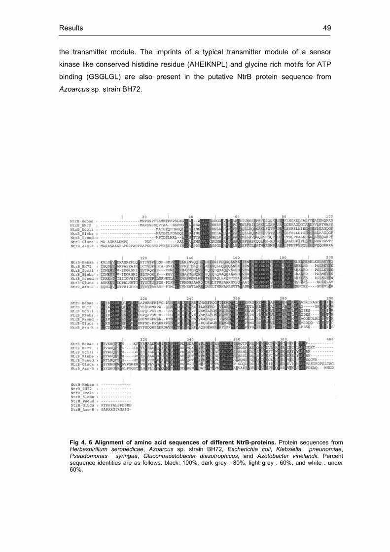
Results 49
Fig 4. 6 Alignment of amino acid sequences of different NtrB-proteins. Protein sequences from Herbaspirillum seropedicae, Azoarcus sp. strain BH72, Escherichia coli, Klebsiella pneunomiae,Pseudomonas syringae, Gluconoacetobacter diazotrophicus, and Azotobacter vinelandii. Percent sequence identities are as follows: black: 100%, dark grey : 80%, light grey : 60%, and white : under 60%.
the transmitter module. The imprints of a typical transmitter module of a sensor
kinase like conserved histidine residue (AHEIKNPL) and glycine rich motifs for ATP
binding (GSGLGL) are also present in the putative NtrB protein sequence from
Azoarcus sp. strain BH72.

Results 50
Fig 4. 7 Alignment of amino acid sequences of different NtrC proteins. Protein sequences from G.diazotrophicus, A. brasilense, Azoarcus sp. strain BH72, H. seropedicae, K. pneumoniae, and E. coli.Percent sequence identities are as follows: black : 100%, dark grey : 80%, light grey : 60% and white : under 60%.
The NtrC proteins act as global transcription activators for many ntr regulons.
Alignment of Azoarcus NtrC with its close relatives revealed the following features
(Fig 4.7). In contrast to NtrB, it shared more conserved regions. The N terminal
receiver domain of Azoarcus NtrC possesses conserved aspartate residues (DDD): a
typical site for phosphotransfer. This is linked to the central domain by a Q linker. A
common feature of the central domain of most response regulators is a stretch of
conserved residues GESGTGKE. This is also present in Azoarcus NtrC (between
residues 170 to 180) and is probably responsible for ATP binding and subsequent
transcription activation.

Results 51
4.2.3 Predicted functional motifs of the NtrB and NtrC from Azoarcus sp. BH72
The nucleotide sequence of ntrB and ntrC was translated by ExPASy
(www.expasy.ch/tools/dna.html) and generated predicted proteins for both the genes
under query. For NtrB, this predicted protein had 357 amino acids (molecular weight
39.69 kD, pI 6.56) and for NtrC 476 amino acids (Molecular weight 53.15 kD, pI
5.87).
In order to further investigate the functional motifs present in these predicted
proteins the translated sequences were analysed using the programme SMART
(Simple Modular Architecture Research Tool). Website address:
(www.smart.embl-heidelberg.de)
For NtrB from strain BH72, SMART predicted the following motifs: 12-79
residues PAS domain, 137-202 residues histidine kinase domain and 240-355
residues histidine kinase like ATPases. PAS motifs appear in Archaea, Bacteria and
Eucarya and are involved as signal sensor domains. Several PAS domain proteins
are known to detect their signal by way of an associated cofactor. The histidine
kinase A N-terminal domain is a dimerisation and phosphoacceptor domain of
histidine kinases and is found in many bacterial sensor proteins. The histidine kinase-
like ATPases include several ATP binding proteins like histidine kinase, DNA gyrase
B, topoisomerases, phytochrome-like ATPases and DNA mismatch repair proteins.
With NtrC as query, SMART prediction was as follows: 2-115 residues REC,
158-301 AAA and 303-314 low complexity. The REC (cheY-homologous receiver
domain) is present in CheY and involved in clockwise rotation of E. coli flagellar
motors. This domain contains a phosphoacceptor site that is phosphorylated by
histidine kinase homologues and forms a part of bacterial two component-systems. It
is usually found N-terminal to a DNA binding effecter domain. AAA (ATPases
associated with a variety of cellular activities) profiles detected a fraction of this vast
family. The key feature of this motif is that it shares a conserved region of 220 amino
acids that contain one ATP binding site. The AAA domains in these proteins act as
ATP dependent protein clamps and function in nucleotide binding. Surprisingly, no
DNA binding helix-turn-helix (HTH) motif, a characteristic feature for DNA binding
response regulators was predicted by this programme. However it is known that in
SMART diagrams domains with low scores are not exhibited. Therefore the putative
NtrC protein sequence from strain BH72 was submitted to another programme:
NPS@ (Network Protein Sequence @nalysis) to find out whether such a motif really
exists in the protein. The result produced a score of 6.15 for sequence

Results 52
GRRIEAAQLLGIGRNTITRKIQ at position 436. This score suggests a very high
probability that the protein of interest contains a helix-turn-helix motif.
4.2.4 ntrB and ntrC in strain BH72 are transcriptionally linked
From sequencing and reading frame analyses it is quite clear that both the sensor
histidine kinase NtrB and its response regulator NtrC are oriented in the Azoarcus
genome in same direction one next to the other. However a known promoter
consensus could not be detected before the start codon of the ntrB. It has been
shown previously that members of two component systems are physically and
functionally linked in the form of an operon, transcribed together and are often
controlled for cooperative translation (Govantes et al, 1998). Therefore in order to
check the size of the ntrBC transcript and to know whether both components are
transcriptionally linked (as would be the case if they share a common operon
architecture), total RNA was isolated from N2 fixing microaerobic batch cultures of
strain BH72. After a DNaseI treatment, the transcripts of ntrB and ntrC were reverse
transcribed separately using Amersham pharmacia beads with ntrBrev and ntrCrev
primers for 30 min at 42°C followed by subsequent amplification of the cDNA with
ntrBfor and ntrCfor primers respectively; the cycling conditions were 95°C 1min, 60°C
1min and 72°C 1,5 min for 35 cycles. The RT PCR amplification products from this
experiment showed bands of expected size (1.07 kb ntrB fragment and 1.43 kb ntrC
fragment) along with a negative control with heat inactivated beads (Fig 4.8 A). To
check the quality of RNA, RT-PCR of 16S rRNA was performed from the same RNA
preparation using 10 ng and using 1401 rev, 104f, and 1346rev primers.
To further verify whether ntrB and ntrC of strain BH72 are transcriptionally
linked (cotranscribed), the RNA from N2 fixing cultures of strain BH72 was reverse
transcribed by RTntrCbridgeR and PCR amplified with primers annealing to the 5’
end of the ntrC (RtntrCbridge R) and 3’ end of the ntrB (RtntrBbridge F). The reverse
transcription was carried out at 42°C. Following 35 PCR cycles of 95°C for 1 min,
55.5°C for 1 min, and 72°C for 1 min an amplification product of expected size (460
base pairs) which was also obtained from chromosomal DNA as template but not
obtained in an RNA-free control or after heat inactivation of reverse transcriptase (Fig
4.8B). Therefore, ntrB and ntrC are clearly cotranscribed.

Results 53
1 2 3 4 5 6 7 1 2 3 4 5
Fig 4.8 RT-PCR analyses of the ntrBC operon of Azoarcus sp. strain BH72. (A) Analysis of ntrB and ntrCtranscripts by RT-PCR. Lane 1, ntrB primers without RNA; Lane 2, ntrC primers without RNA; Lane 3, heat inactivated reverse transcriptase prior to RNA addition; Lane 4, ntrB- and Lane 5, ntrC-amplification product from 1 g RNA of N2 fixing cells of strain BH72, respectively; Lane 6, size marker; Lane 7, 16S rRNA directed RT PCR as control. (B) Analysis of the ntrBC cotranscription by RT-PCR. Lane 1, no RNA added; Lane 2, RT heat inactivated prior to RNA addition; Lane 3, RT PCR product from RNA without heat inactivation; Lane 4, amplification from genomic DNA; and Lane 5, size marker.
A B
4.3 Generation of a marker exchange ntrBC mutant of Azoarcus sp. strain BH72
These experiments were carried out to investigate, how ntrBC in strain BH72
regulates different cellular processes and whether it is absolutely crucial for its
survival.
4.3.1 Construction of a marker exchange deletion mutant of the ntrBC
The strategy was to delete a major portion of the genetic region of ntrBC and
replacing it by a resistance cassette. The cassette used in this case was a
spectinomycin / streptomycin resistance cartridge (2kb), derived from plasmid
pHP45 (4.3Kb). The Sp/Sm cartridge is flanked both sides by short inverted repeats
carrying a transcriptional and translational stop signals in all the three reading frames
along with a short polylinker stretch flanking symmetrically on both sides. The
resistance cartridge generates a polar effect on the transcription of the operons in
which it is inserted. For this purpose, the Smal digested fragment from pHP45

Results 54
was cloned in the NruI digested pSNTR2.1. Three NruI sites are present within the
ntrBC region: two within the ntrB region and the other in the middle of ntrC region.
Clone (pSNTR2.l ) was chosen by checking its restriction pattern with Hindlll digest
to ensure that the cartridge is inserted into the two outermost NruI sites, replacing the
portion in between. Finally pSNTR2.1 was transformed in Azoarcus sp. BH72 by
electroporation. Double recombinants were then chosen (rare clones where two point
homologous recombination took place) by plating the transformation mix on VM
ethanol plates with spectinomycin and streptomycin and choosing the resistant.
4.3.2 Construction of a nonpolar ntrB mutant of strain BH72
These experiments were carried out to check whether the response regulator is still
functional when the sensor is knocked out. For this purpose the kanamycin
resistance cartridge (1.2 kb) in the pUC4K vector was digested by EcoRI and the
fragment was treated with Klenow fragment to generate blunt ends as described in
material and methods. To generate the vector, Asp718 and SstI fragments from
pSNTR2.1 (having the entire the ntrBC region) were cloned in pBluescript-KS using
the same enzymes. This strategy imposed ampicillin resistance to the vector (pBS-
KS NTR2.1) to distinguish it from the cartridge resistance (kanamycin). A partial
digest of this vector was carried out using NruI and the bands were separated in an
agarose gel. After cloning of the kanamycin cassette in this partially digested vector,
the positive clone (pBS-KS NTR2.1 kan) was selected where the cartridge is cloned
into the first two NruI sites by replacing part of ntrB while the third NruI site in the ntrC
was still intact. Following an electroporation of this plasmid in Azoarcus sp. strain
BH72, transformed strains were selected on plates with kanamycin where both the
single and double recombinants grew. Finally the double recombinants were
negatively selected by transferring the transformants in ampicillin plates where only
the single recombinants grew. Thus only kanamycin resistant double recombinants
are the putative nonpolar mutants where the kanamycin promoter can still drive the
transcription of its downstream gene.

Results 55
4.3.3 Validation of constructs by Southern hybridisation and genomic PCR amplification.
To confirm the ntrBC mutant genetically, a Southern blot was performed using
genomic DNA from both the wild type and the mutant strain. The genomic DNA from
both the strains (wild type and the ntrBC mutant) (3 g) were digested with Hindlll
and PstI respectively. A 540 base pair PstI digested fragment within the ntrB region
was used as a probe for southern hybridisation at 65°C and washings were done
under conditions of high stringency. The result of the Southern hybridisation is
presented in Fig. 4.9. The probe hybridised with a large 6 kb HindIII fragment in the
wild type strain. The ntrBC mutant strain under similar condition hybridised to a 750
bp fragment: the internal Hindlll site being generated from the insertion of the
cartridge. Similarly the PstI digested wild type genomic DNA hybridised with a short
540 bp fragment corresponding to the size of the probe. Since the internal PstI sites
of the mutant are replaced by the cartridge, the probe hybridised to a larger 2.5 kb
fragment instead.
The genomic arrangement of the ntrB non polar mutant was verified by PCR
amplification of ntrB and ntrC from its genomic DNA using the standard forward and
reverse primers for the respective genes; the cycling condition were 30 cycles of
95°C for 1 min, 63°C for 1 min and 72°C for 3 min. As expected, a 1.43 kb fragment
was generated from the ntrC amplification, suggesting intactness of this gene. Using
ntrB
N N N
cassette
ntrC
A
Fig 4.9 Schematic diagram (A) and Southern blot (B), of the ntrBC marker exchange deletion mutant BntrBsp. In (A), N represents NruI sites used for inserting the cassette. In (B), Hand P denote restriction digestion of genomic DNA by HindIII and PstI of strain BH72 (1 and 3) and BntrBsp (2 and 4).
H H P P
6 kb
2.5 kb
0.750 kb
1 2 3 4
B
0.5 kb

Results 56
the ntrB primers a 1.58 kb fragment was amplified instead of 1.07 kb; the correct size
of the intact ntrB (Fig 4.10). This can only be explained if 0.771 kb internal NruI
fragment is replaced by 1.28 kb Kanamycin cartridge (1.07 – 0.771 + 1.282 = 1.584).
The presence of the intact NruI site within the amplified ntrC product was further
confirmed when the 1.43 kb ntrC amplification product fragmented into 0.858 kb and
0.573 kb when digested by NruI.
4.4 Phenotypes of the ntrBC mutants
The ntrBC mutant (BntrBsp) and the nonpolar ntrB mutant BntrBkan generated by
marker exchange mutagenesis were viable and able to fix N2 under microaerobic
conditions. The growth characteristics, morphology and physiology of these mutants
were compared with wild type strain BH72 in order to provide deeper insights about
NtrBC function and hints about its putative targets.
4.4.1 Growth characteristics of wild type and the ntrBC mutants
One of the ways to study growth characteristics of an organism is to follow up its
growth kinetics. The growth is usually carried out either on complete or synthetic
media with different N sources or N-free medium (N2 fixing). Samples were collected
at defined time intervals (both from the wild type and the mutant strains) to measure
OD at 578 nm. The plot of the OD against time intervals produced growth curves of a
Fig 4.10 Schematic diagram (A) and PCR amplification (B) of the ntrB marker exchange nonpolar mutant BntrBKan. In (A), N represents the NruI sites within the ntrBC region for the insertion of the kanamycin cassette. The ntrB nonpolar mutant is generated by replacing the ntrB region flanked by two NruI sites with the Kan cassette, the entire ntrC being intact. (B) PCR confirmation of the ntrB nonpolar mutant. Primers used to amplify ntrB are ntrBfor and ntrBrev respectively while those used to amplify ntrC are ntrCfor and ntrCrev. Lane 1 (size marker), lane2 (PCR amplification of entire ntrB from BH72), lane 4 and lane 5 (amplification product of the ntrB and the ntrC respectively from BntrBkan) and lane 3 (restriction digestion of the ntrC amplification product from lane 5 by NruI)
N N N
ntrB
AKan
ntrC 0.5 kb
1 2 3 4 5
B

Results 57
particular strain from which growth rate and generation times could be calculated
using the standard formula: growth rate ( ) = lnOD2 – lnOD1 / (t2 – t1) where OD2 and
OD1 are the respective OD’s at time t2 and t1, respectively. The generation time
calculated as (ln2 / ) from this formula is a measure of its growth characteristics.
The results from this experiment are presented in TABLE 1. The generation times of
all the three strains were comparable on complex medium, as well as on mineral
medium with ammonium as N-source (1.3 hrs in average for all the three strains,
compared). So ammonium as a N-source did not have a pronounced effect on the
growth rate of the mutants. One of the interesting observations is the prolonged
generation time of BntrBsp (9.56 hr) while growing aerobically with nitrate as sole N
source compared to strain BH72 (2.1 hr) or BntrBkan (3.3 hr) under similar condition.
A similar effect of nitrate has been observed in glnB-K- double mutant of strain BH72
(Martin et al., 2000). The N2-dependent growth was made in a fermenter with 0.8%
oxygen where the growth of BntrBsp was found to be affected (generation time: 3.62
hr) in comparison to the wild type strain BH72 (generation time: 1.8 hr).
TABLE 1. Growth of Azoarcus sp. strain BH72 (wild type), the ntrBC and the ntrB knockout mutants on different nitrogen sources. The sign (-) means not carried out. All data are mean values of at least three independent experiments.
Generation time in (h) during growth on
strain Relevant genotype N2 10mM NH4Cl 10mM NO3- VM ethanol
BH72 wild type 1.8 1.32 2.1 1.3
BntrBsp ntrBC- 3.62 1.3 9.55 1.39
BntrBkan ntrB- - 1.38 3.3 1.53
4.4.2 Comparison of colony/cell morphologies of the wild type with the ntrBC mutant growing on nitrate as sole N-source: phenotypes of impairment exhibited by BntrBsp
The colonies of the strain BH72 and strain BntBsp looked identical while growing on
complex medium agar plates. However when streaked on plates with nitrate as sole
N source and incubated at 37°C, the mutant colonies appeared drastically different.
BntrBsp colonies were more rounded and centrally opaque in contrast to whitish
BH72 colonies. Microscopic observation of the same under bright field illumination
revealed the colony profile in much more detail (Fig 4.11). The cells of BntrBsp
colonies appeared rounded and smooth surfaced with deep central invagination. In

Results 58
contrast, strain BH72 cells appeared rough surfaced with no such concavities. Under
dark field illumination the ntrBC mutant colonies appeared more refractive. This was
even clearer under phase contrast with bright field illumination where the ntrBC
mutant cells appeared elongated with a central shining region much in contrast with
wild type cells. It seems as if the ntrBC mutant was severely N-limited during growth
on nitrate and accumulated polyhydroxy butrate under these conditions. The
unreleased data from Azoarcus genome project have revealed the existence of
putative genes encoding enzymes for PHB biosynthesis. So it is quite reasonable to
speculate that PHB biosynthesis is upregulated in the ntrBC mutant due to N-
limitation in presence of nitrate.

Results 59
4.4.3 Twitching motility is upregulated in BntrBsp
Twitching motility is a form of bacterial translocation over firm surfaces that require
retractile type IV pili (Liu et al., 2001). It is already known that Azoarcus sp. strain
BH72 is capable of exhibiting twitching on solid complex medium when incubated at
37°C for 3 days and then left at room temperature. It has already been reported that
heterologous expression of type IV pilin gene from Pseudomonas syringae pv.
tomato DC3000 in strain Azoarcus sp. BH1599, an isogeneic mutant having
transposon insertion in pilA, could functionally complement the defect of this mutant
in “twitching motility” (Plessel, 2001). So it is evident that type IV pili are involved in
twitching motility in strain BH72. Interestingly it was observed that BntrBsp twitched
more under similar conditions as compared to the wild type. For quantification of
twitching motility overnight precultures of strain BH72 and BntrBsp in complex
medium were washed and suspended in physical saline. A dilution was made in
order to have 50 cells per 100 l per VM ethanol plates. They were incubated at 37°C
for 3 days when the colonies started to show twitching. Plates were scanned and the
colony areas from scanned images were calculated using the software “image quant”
of the fluoro phosphoimager (Typhoon). About 47 BH72 colonies from total 5 plates
and 121 BntrBsp colonies from 3 plates were used for calculating the area (square
units) occupied by individual colonies by outlining individual colonies manually. The
results are shown in Fig 4.12. It appeared that BntrBsp exhibited 2.2 times more
larger colony areas as compared to the wild type under similar conditions. Since it
has been already shown that both the wild type and the ntrBC mutant have similar
growth rates in complex medium (generation time: 1.3 hr) it can concluded that the
larger colony areas of BntrBsp was not due to faster growth but as a result of better
twitching compared to strain BH72.
Fig 4.12 Comparison of twitching motility of strain BH72 and strain BntrBsp. The area of 47 independent colonies of strain BH72 from 5 independent plates were compared with 121 colony areas of BntrBsp from 3 independent plates. The bar in each case represents standard deviations of calculated areas. 0
200
400
600
800
1000
BH72 BntrBsp
strains
Ave
rage
are
a (s
q un
its)

Results 60
4.5 Transcriptional regulation of the ntrBC operon
In the following section the main question addressed is the transcription regulation of
ntrBC in strain BH72. Generally the enteric ntrBC ‘s are linked to an upstream glnA
gene and transcribed from –12 / -24 type N-regulated promoters (MacFarlane et al.,
1985). On the other hand, ntrBC of the Proteobacteria including rhizobia share a –
10 /- 35 promoter consensus and are transcriptionally linked to an upstream ORF1 of
unknown function. To get a clear scenario about the organization of the ntrBC
promoter of Azoarcus sp. strain BH72 an attempt was made to map the 5’ end of the
ntrBC transcript. The other questions addressed in this section deal with the
expression profile of ntrB and ask whether any autoregulation mechanism exists to
control its expression.
4.5.1 Mapping the 5’ end of the ntrBC transcript (primer extension)
The promoters of the principal nif genes of strain BH72 (nifH and nifLA), studied in
section 4.1 share a -12 / -24 consensus, typical for N-regulated promoters. The
transcription start point of these genes mapped precisely with respect to the location
of the upstream consensus. However no putative –12 /-24 consensus was detected
upstream of ntrB; neither a bacterial –10 / -35 sigma (70) promoter consensus could
be found. Interestingly no proper Shine-Dalgarno sequences which are usually
located 3 – 8 base pair upstream of the start codon and are necessary for ribosomal
binding and translation initiation, could be detected upstream of the ntrB and the ntrC
start codons of Azoarcus sp. BH72.
In order to investigate the transcription start point of ntrB, two Cy5 labelled
reverse primers (Cy5PntrBrev and Cy5PEntrbrevUS) were designed, one annealing
at about 30 – 35 bp down stream from the start codon of ntrB and the other
annealing at about 30 – 35 bp upstream (US) of ntrB start codon. The rationale for
designing primers annealing at such sites is to cover a greater probability of
transcripts originating at undefined origins, upstream of ntrB. Primer extension was
carried out according to the method described earlier with RNA isolated from N2
fixing cultures of strain BH72. A peak was detected using the upstream primer very
close to the primer peak. The sequencing reaction at such an early time point is
often erratic, and so the starting base could not be detected precisely with such an
approach (data not shown). However the downstream primer under similar conditions

Results 61
produced a single extension peak at 213 min, corresponding to the base C (Fig
4.13). Thus the start point was mapped about 150 bp upstream from the ntrB start
codon. However attempts failed to detect a known bacterial promoter consensus
upstream of this putative transcription start site.
CGAAGCCCGCAAGAGCCTGGGCGAGCAGGAAGCGGTCCGGCTCGGCGACGAGCGCAACTACCA
GCGCGTGCTCGACCGCCTGCAGCCGTTCAAGGACAAGGTCGACCTGCACGAGCGCAATATCGAA
GCACTGAAGCGCGAGATCGCCAACCTGCGCTGACGCGAAGCCGGGGAGCACGCGCCCCGGTCC
GACTCACCTCCGTTCCGGTCGCCCGTTCCCCATGGCTCGATCCTCCTCCTCCCAGCCGATCGCGG
CAGAGA
A
start codon
G A C C T T G T C C T T G A A C G G C T G C A G G C G G T C G A G C A C G C G C T G G T A G T TTime [min]190 195 200 205 210 215 220 225
Time [min]190 195 200 205 210 215 220 225
B
Fig 4.13 Analysis of transcription start point of ntrBC of strain BH72 by primer extension. (A), sequence of the upstream region of ntrB beyond its start codon. No standard bacterial promoter consensus could be detected in this upstream region. (B), Primer extension analysis localizing the transcription start at 213.07 min, corresponding to the nucleotide “C” marked with an arrow. Top, sequencing reaction and bottom, primer extension.

Results 62
4.5.2 Undetectable expression of the ntrB::gusA in strain BH72 grown on different nitrogen sources.
One of the ways to quantify gene expression is to construct a transcriptional fusion of
the upstream region of the gene of interest with a promoter less gusA reporter gene.
While screening the putative pSNTR2.1 , some clones were obtained where the
cartridge was inserted into an NruI site deleting a portion of the ntrC region while the
ntrB with its upstream region was still intact (refer to section 4.). By this cloning, an
extra HindIII site is generated downstream of ntrB coming from the catridge in
addition to the original HindIII site present upstream of ntrB; This clone was digested
by HindIII and ligated to HindIII digested pBluescript-GUS-SK. By this strategy, the
transcription termination loops present within the catridge could be avoided from
being incorporated within the reporter construct. The correct orientation of the
reporter construct was checked by BamHI digest; the resulting plasmid (pSNTRGUS)
was electroporated in Azoarcus and ampicillin resistant single recombinants were
isolated representing the desired reporter strain. GUS assays were performed with
this strain (BHNTRGUS) under N excess (ammonium or nitrate as N-source) as well
as under N limitation (N2 fixing). The results are shown in Fig 4.14. Very low and
close to undetectable levels of GUS units were measured in all cases even after
incubation for 8 hrs. A positive control assay under similar conditions with
glnK::gusA, could be done in 2 mins, as expected. So no conclusions could be drawn
on the effect of different N-sources on ntrB expression in strain BH72.
Fig 4.14 Expression of the ntrB measured by GUS activity.(A)Cloning strategy of the ntrB::gusA forchromosomal integration. H – HindIII, B – BamHI, represents resistance catridge and represents the location of transcription termination loop. (B)Very low or near to undetectable levels of GUS activity was estimated in each case by culturing BHNTRGUS in ammonium (NH4), nitrate (NO3) or N2 fixation (N2).
0
5
10
15
20
25
NH4 NO3 N2
N sour c e
HH B ntrB
HindIII digest
ntrBgusA
pBluescript SK
HB B HB

Results 63
1 2 3 4 5 1 2 3 4 5
1 2 3 4 5 2 3 4 5 6 7 1NH4
+
NH4+ N2
N2
A
B
Fig 4.15 Analysis of the ntrB expression by RT-PCR. (A) 1 g RNA isolated from strain BH72 under ammonium excess (NH4
+) or N-limiting (N2) was used for RT-PCR with primers specific for ntrB. Samples were taken after 18, 20, 23, and 26 PCR cycles respectively, corresponding to lanes 2, 3, 4, and 5 respectively; lane 6, PCR amplification from genomic DNA; lane 7, RNA with heat inactivated beads; and lane 1, size marker. (B). As a RNA control, the same RNA preparations were used for 16S rRNA directed RT PCR. Samples were removed after 18, 22, and 26 PCR cycles (lanes 2, 3, 4 respectively); RNA with heat-inactivated beads in lane 1 and size marker in lane 5, respectively.
4.5.3 Effect of nitrogen sources on the ntrB transcription: RT PCR approach.
In order to analyse about the transcriptional regulation of the ntrBC, an RT-PCR
approach was used, since reporter gene studies failed. RNA was isolated both from
ammonium grown and N2 fixing cells of strain BH72. DNaseI treated RNA from both
were reverse transcribed by ntrBrev primers using the RT PCR beads at 42°C for 30
min followed by subsequent cDNA amplification of the ntrB transcript as described in
section 4.1. At defined time points of amplification cycles, samples were removed
and separated in an agarose gel (Fig 4.15 A). To ensure that an equal amount of
RNA was taken as starting template in both the cases, 16S rRNA was reverse
transcribed at 42°C for 30 min by 1401 rev fro 10 ng of same RNA preparations and
PCR amplified with 104f and 1346rev primers at 95°C 1 min, 65°C 2 min, and 72°C 2
min (Fig 4.15 B). From the figure it is clear that the abundance of the ntrB transcript
is much higher during N2 fixation as compared to ammonium grown cultures. This
points to the differential expression of ntrB depending on the N availability. 16S rDNA
bands of equal intensities in both the cases confirmed that equal amounts of RNA
were used under both the conditions for amplification.

Results 64
1 2 3 4 5 6 7 8 1 2 3 4 5 6 7
1 2 3 4 1 2 3 4
N2
N2
NH4+
NH4+
A
B
Fig 4.16 Analysis of autoregulation of ntrBC by RT- PCR. (A) 0.5 g RNA from BntrBsp growing under N2 fixation (N2) or combined nitrogen (NH4
+) was used to reverse transcribe and amplify 150 bp fragment from the N-terminal of ntrBC still intact in BntrBsp. Samples were collected after 16, 18, 20, 23, 25, and 28 cycles (lanes 1, 2, 3, 4, 5, 6, respectively) ; RNA with heat inactivated beads (lane 7) and size marker (lane 8). (B). Control for quality and equal amount of RNA as starting template was done by 16S rRNA directed RT-PCR by taking 0.5 g RNA each time and removing the samples after 18, 23, and 26 cycles (lanes 1, 2, & 3 respectively); the size marker being in lane 4.
4.5.4 ntrBC of strain BH72 is not likely to be autoregulated
NtrC as a transcription activator is known to regulate its own expression apart from
regulating its targets. Typical NtrC binding motifs (GCTGGCGCAGGTGC) have been
detected in the upstream region of many ntrC regulated promoters. In Rhizobium
leguminosarum it has been reported that NtrC negatively regulates its own operon
(Patriarca et al., 1993). An attempt to detect NtrC binding motifs upstream of ntrBC
from strain BH72 failed. On the other hand to get a clear picture about the existence
of autoregulation, a 150 bp fragment near the ntrB start which is still intact in the
ntrBC insertion mutant within the ntrB (before the NruI site where the cartridge is
inserted) was reverse transcribed by the primer RTntrBrev at 42°C from DNaseI
treated RNA (500 ng) isolated from the ntrBC mutant growing in ammonium or N2
fixing respectively. The cDNA was amplified using RTntrBfor primer in cycles of 95°C
1min, 59°C 1min and 72°C 1 min. Samples were removed after 18, 20, 23, 25 and 28
cycles and run in a gel along with a negative control for DNA contamination (heat
inactivated beads) as had been mentioned earlier (Fig 4.16). The result shows the
expression of the 150 bp N-terminal fragment in the ntrBC mutant under both
conditions tested. Interestingly, there is still a differential expression (N2 / NH4+) of the
N-terminal ntrB fragment in the ntrBC mutant, which is also an argument for no
autoregulation. A similar experiment with strain BH72 produced a comparable
expression profile as that of BntrBsp (data not shown). A control for equal amounts
of RNA for both the cases was carried out by RT PCR of 16S rRNA as before. The
results of the experiment indicate that the ntrB from Azoarcus sp. BH72 is not
involved in regulating its own expression (autoregulation).

Results 65
4.6 Putative targets of NtrBC of strain BH72
In this section, the goal is to analyse how the NtrBC of Azoarcus sp. strain BH72
plays a role in regulating the expression of putative downstream target genes. The
ntr regulon is generally known to control various genes involved in N2 fixation and N-
assimilation. These two aspects are studied under two different broad subheadings.
4.6.1 Transcriptional regulation of N2 fixation genes by NtrBC
In this section, the regulation exerted by NtrBC of strain BH72 on the nif genes
coding for structural genes for nitrogenase (nifHDK) and its transcription activator
(nifLA) has been studied. At the same time, the regulatory function of NtrBC on gln
genes acting as sensors for the N status of the cell and in many ways related to N2
fixation have been investigated.
4.6.1.1 The nifA expression in strain BH72 is NtrBC regulated in a nitrogen dependent manner
In Azoarcus sp. BH72, N2 fixation and nifHDK transcription occur only under
microaerobic and N-limiting conditions (Egener et al., 1999; Hurek et al., 1987). It is
also known that nifA is an essential transcription activator for nifHDK, the structural
genes for nitrogenase. In contrast to other members of Proteobacteria, strain BH72
harbours a nifL like gene similar to the members of Proteobacteria and helps in
inactivating NifA in response to O2 and combined nitrogen (Egener et al., 2002). In
order to study the effects of the ntrBC mutation on nifHDK and nifLA expression,
upstream regions of nifH and nifLA were transcriptionally fused with a gusA reporter
gene and cloned in a low copy vector pLAFR3A (pNHGUS and pLGUS) respectively
( Egener, 1999). These plasmids were conjugated into strains BH72 and BntrBsp.
The transconjugant strains arising from the wild type - BH72(pNHGUS) and
BH72(pLGUS) and from the ntrBC mutant: BntrBsp(pNHGUS) and BntrB(pLGUS)
were assayed for GUS activity by growing all the transconjugant strains on
ammonium or N2, respectively. The results are presented in Fig 4.17.

Results 66
Fig 4.17 Expression of nifH and nifLA in strain BH72 and BntrBsp. Cultures were grown either inpresence of combined nitrogen (10 mM ammonium) aerobically or microaerobically (0.8% O2 in head space)with N2 as substrate. The GUS activity was measured after 10-12 hours of growth (exponential growth phase).The data represent results from three parallel measurements of two independent experiments.
0
500
1000
1500
2000
2500
3000
3500
4000
nifH ::gusA NH4
nifLA::gusA NH4
GUS activity (in Miller units) BH72
BntrBsp
nifH ::gusA N2
nifLA::gusA N2
From the GUS measurements it is clear that the nifH promoter activity in strain BH72
is strongly induced under N2 fixation. In strain BntrBsp the nifH promoter is also
induced under N2 fixation and suppressed under N excess. However the nifH GUS
activity in the ntrBC mutant fixing N2 is relatively lower as compared to the wild type
under similar condition. On the other hand, the nifLA GUS activity in strain BH72 is
not completely repressed under N excess and induced about three fold during N2
fixation. This is quite consistent with previous nifLA expression patterns studied in
northern blot experiments (Egener et al., 2002). Interestingly in the ntrBC mutant,
the nifLA expression was not enhanced on N2 compared to its expression on
ammonium, along with reduced nifH expression on N2 suggesting that NtrBC might
play a role in regulating the nifLA transcription in response to N-deficiency. This in
turn may have a direct or indirect influence on nifH::gusA expression
4.6.1.2 Effect of nitrate on the derepression of nitrogenase genes in BntrBsp
One of the interesting features of the ntrBC mutant is its inability to completely
repress N2 fixation when growing microaerobically on nitrate as sole N-source. The
nitrogenase activity measured as (nmol of C2H4 / mg of protein) by acetylene
reduction assay (ARA) showed that BntrBsp exhibited acetylene reduction activity of
6,230 440, on nitrate which is much higher as compared to its fixation on N2 (3,980
230). However the nitrogenase activity detected for the wild type strain BH72 on N2
was estimated to be (9,238 440). The expression of nifH and nifLA in wild type and

Results 67
ntrBC mutant while growing microaerobically on nitrate has been compared in Fig
4.18. The high GUS activity as a consequence of nifH expression in BntrBsp
suggests derepression of the nitrogenase. Consistent with the nitrogenase activity,
the nifH::gusA expression in BntrBsp was found to be higher on nitrate as compared
to that on N2. It is interesting to point out that the nitrogenase is derepressed in a
similar way in gln B-K- double mutant while growing on nitrate with nifH::gusA
expression of 2,860 miller units and nitrogenase activity of 4,010 360 units (Martin
et al., 2002). So it is clear from these data that the extent of derepression of
nitrogenase on nitrate in ntrBC mutant is comparable to gln B-K- double mutant of
strain BH72. Another interesting observation here is the relative high (compared to
the nifLA expression on N2) and more or less comparable nifLA expression in both
the wild type and the ntrBC mutant. Thus in the same ntrBC mutant background, the
expression of nifLA is differential according to different nitrogen source (nitrate or N2).
So it is clear from these results that source of nitrogen (ammonium or nitrate) also
have an effect on nifLA expression in strain BH72.
Fig 4.18 . Effect of nitrate on the nifH::gusA and nifLA::gusA expression in strain BH72 and BntrBsp,respectively. Cultures were grown microaerobically on 10 mM nitrate (0.8% O2 in head space ) and GUSactivity was measured after 10 hours. The data represent values from three parallel measurements from twoindependent experiments.
01000
200030004000
500060007000
80009000
nifH::gusA
strains with reporter genes
GUS activity (in Miller units)
BH72
BntrBsp
nifLA::gusA

Results 68
4.6.2 Transcriptional regulation of the gln genes of Azoarcus sp. BH72: role of NtrBC
PII like signal transmitter proteins, found in Bacteria, Archea and plants mediate
control of nitrogen / carbon assimilation. They indirectly regulate the activity of key
metabolic enzymes and transcription factors by protein – protein interactions. Many
proteobacteria harbor two paralogous PII like proteins, GlnB and GlnK, whereas a
novel third PII paralogue (GlnY) has recently been identified in strain BH72 (Martin et
al., 2000). It is already known that PII like proteins in strain BH72 play distinct roles in
mediating nitrogen and oxygen control of the nif gene transcription and nitrogenase
activity (Martin et al., 2002). However the regulation of these genes in strain BH72, is
still not clear, especially at the level of transcription. At the same time, sequences
upstream of the gln genes lack conserved bacterial promoter sequences, which
make it even difficult to get an idea about the nature of regulation. So attempt has
been made in this section to study the transcriptional regulation of these gln genes by
RT-PCR and GUS assays and find the role of NtrBC in this context.
4.6.2.1 glnK regulation and effect of N2 on its expression: role of NtrBC
In Azoarcus sp BH72, the glnK gene is downstream genetically linked to amtB (a
putative ammonium transporter). This physical linkage of glnK with amtB has been
reported for many bacteria. Northern blot analysis to study glnK transcription in strain
BH72 revealed detectable expression of a large 1.8 kb transcript only during N2
fixation, which was repressed in presence of ammonium (Martin et al., 2000). This
speaks in favour of an N-regulated expression of the glnK operon. However a typical
sigma 54 promoter (-24 / -12) or general sigma 70 (-10 / -35) type promoter
consensus was not apparent in the glnK upstream region; instead a 750 bp ORF is
present. This ORF termed ugk (upstream glnK) was found to have 43% identity to a
putative protein harbouring signal peptide from Ralstonia solanacearum and 35%
identity to a similar protein from Caulobacter crescentus. Interestingly, a typical
sigma 54 promoter consensus sequence characteristic for many N-regulated
promoters is present upstream of ugk. Therefore, it is interesting to explore whether a
functional linkage exists between the glnK and the ugk of strain BH72 and whether
the 54- promoter consensus upstream of the ugk can still drive transcription of glnK
gene located downstream. Thus it can be speculated that Ugk harbouring a signal
peptide may be secreted into the periplasm, which may aid the sensor AmtB to sense
ammonium and propagate a signal transduction within the cell with the help of GlnK.
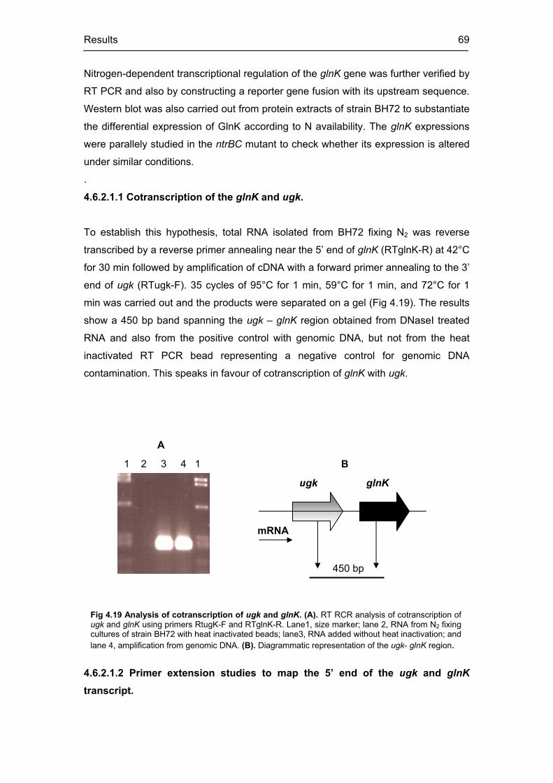
Results 69
ugk glnK
1 2 3 4 1
450 bp
mRNA
Fig 4.19 Analysis of cotranscription of ugk and glnK. (A). RT RCR analysis of cotranscription of ugk and glnK using primers RtugK-F and RTglnK-R. Lane1, size marker; lane 2, RNA from N2 fixing cultures of strain BH72 with heat inactivated beads; lane3, RNA added without heat inactivation; and lane 4, amplification from genomic DNA. (B). Diagrammatic representation of the ugk- glnK region.
A
B
Nitrogen-dependent transcriptional regulation of the glnK gene was further verified by
RT PCR and also by constructing a reporter gene fusion with its upstream sequence.
Western blot was also carried out from protein extracts of strain BH72 to substantiate
the differential expression of GlnK according to N availability. The glnK expressions
were parallely studied in the ntrBC mutant to check whether its expression is altered
under similar conditions.
.
4.6.2.1.1 Cotranscription of the glnK and ugk.
To establish this hypothesis, total RNA isolated from BH72 fixing N2 was reverse
transcribed by a reverse primer annealing near the 5’ end of glnK (RTglnK-R) at 42°C
for 30 min followed by amplification of cDNA with a forward primer annealing to the 3’
end of ugk (RTugk-F). 35 cycles of 95°C for 1 min, 59°C for 1 min, and 72°C for 1
min was carried out and the products were separated on a gel (Fig 4.19). The results
show a 450 bp band spanning the ugk – glnK region obtained from DNaseI treated
RNA and also from the positive control with genomic DNA, but not from the heat
inactivated RT PCR bead representing a negative control for genomic DNA
contamination. This speaks in favour of cotranscription of glnK with ugk.
4.6.2.1.2 Primer extension studies to map the 5’ end of the ugk and glnK
transcript.

Results 70
Fig 4.20 Analysis of transcription start point of the ugk and the glnK by primer extension analysis in strain BH72. (A) Sequence of the upstream region of ugk showing its start codon, and the putative RBS. Asterisks mark the bases that are conserved in the sigma (54) promoter region present at its upstream. The Arrow marks the putative transcription start point of ugk. (B) Sequence of the genomic region upstream of glnK, showing the glnK start codon and spanning the intergenic region between ugkand glnK. The thin arrow indicates the start point of glnK transcription as mapped by primer extension analysis; the thick arrow representing the stop codon of ugk. (C) Primer extension analyses of ugkshowing the sequencing reaction (top) and the primer extension reaction (second). The primer extension peak at 120.87 min match with base” A” of sequencing reaction indicated by an arrow. Primer extension of glnK transcript with sequencing reaction (third) and extension reaction (fourth) of Fig 4.20 C. The primer extension peak at 163.8 min match with base “A” indicated by an arrow in the sequencing reaction.
In order to locate the transcriptional start point of the glnK transcript, a primer
extension analysis was carried out from RNA isolated from strain BH72 fixing N2.The
Cy5 labelled reverse primer (Cy5PeglnK rev) annealed about 20 bp downstream
from the glnK start codon. The result of the primer extension is shown in Fig 4.20 C.
The primer extension peak at 163.8 min matched with the base A (163.56 min) of
the sequencing reaction (Fig 4.20 B). An attempt to find a known promoter
consensus upstream of this start point failed. It is also quite likely that the predicted
position for a transcriptional start may represent a site for active transcript
processing. However the primer extension result substantiated not only the
transcriptional linkage glnK with ugk (Fig 4.20 B) but also for the processing of a
large transcript. On the other hand, Northern blot experiments, detected a single 1.8
kb transcript in glnB- with a glnK probe under nitrogen fixation, indicating
cotranscription of glnK with amtB (Martin et al., 2000). Thus it is reasonable to think
for a large transcript bearing ugk, glnK and amtB and its subsequent processing
under N2 fixation. A similar approach was made to map the transcriptional start point
of ugk with a Cy5 labelled reverse primer (Cy5PEugk rev) annealing about 50 bp
downstream from the putative start codon of the ugk. The primer extension peak at
120.87 min mapped with base T (121.03 min) of the sequencing reaction (Fig 4.20
C). In fact it maps within the putative Shine-Dalgarno sequence, present immediately
upstream of the ugK start codon and so base spacing required between the
transcription start site and the upstream sigma (54) consensus is not strictly
maintained (Fig 4. 20 A). This can also be interpreted in terms processing of the
major transcript. However a northern blot should be performed to get a better
overview about the nature of transcript processing.
* * * * * * * * * * * * *
A

Results 71
CGTTTGGTGCTTTGCGACAAAAGGGGCGCACCAATCCGGTGCAGACGCGCCCG
CACCGCAGGGGGCGCTGCCCGAAAAGGCCTGCGACATGCCCCTCGCTTTCGT
CAAAACCCCAGTCGTTGCGGGGTTTGCGACAAGCTGGCACGGACCCTGCATTA
GCTGCCCTGCGATAAACACATCAACGCTGGAGAAAGACATGCGCAAGAACATTC
ATATGAAGGATAAGAGCGATTATGACGCTGACGCACGCTATATCCTGTCCGTCA
CCAAATCGTTTTAATTGTCATAGGCTTCCGGCCGCGGGTGGCGACACCCTGGC
CGGGTAACAGGAGAACCCGATGAAGTTCATCACAGCGATCATCAAGCCGTTCA

Results 72
NH4+N2
Fig 4.21 Western blot analysis of GlnK from strain BH72 growing with N2 or ammonium. The upper band represents GlnK.
4.6.2.1.3 Western blots to study the effect of nitrogen on GlnK expression in strain BH72.
Equal amounts of total protein extracts of strain BH72 isolated from ammonium
grown and N2 fixing cultures, respectively, were separated by SDS PAGE,
electrotransfered to a membrane and used for Western blot analysis with a GlnK
antiserum. The result (Fig 4.21) shows weak expression of GlnK in presence of
combined nitrogen and strong induction during N2 fixation, speaking in favour of its N-
regulated expression in the wild type strain. The lower band in both cases represents
an unspecific cross-reaction of the GlnK antiserum to a protein of slightly lower
molecular weight that does not belong to the PII protein family (Martin et al., 2000).
4.6.2.1.4 Nitrogen-dependent differential glnK expression in strain BH72 and its down- regulation in the ntrBC mutant: RT-PCR approach
RNA isolated from strain BH72 as well as BntrBsp growing in combined nitrogen or
N2 fixing conditions, respectively, were used for reverse transcription and subsequent
amplification of the glnK transcript using glnKfor and glnKrev primers. RT PCR was
carried out with glnKrev primer at 42°C for 30 min followed by PCR amplification with
glnKfor and glnKrev primers at 95°C 1 min, 57°C 1 min, and 72°C 2 min. Samples
were removed at defined time intervals of the PCR cycles. A similar approach was
made to amplify the 16S rRNA in each case to check the quality of RNA, taking 10 ng
of the same RNA preparation in each case, reverse transcribing at 42°C 30 min by
1401 rev primer and PCR amplifying with 104f and 1346 rev at 95°C 1 min, 65°C 2
min and 72°C 2 min. The results are shown in Fig 4.22. Consistent with the results
obtained from Western blot analysis, a differential expression of the glnK transcript
according to the N-availability was detected by RT-PCR analysis in strain BH72. The
abundance of the glnK transcript was much higher when cells were grown on N2 as
compared to that on combined nitrogen. A more quantitative evaluation of the

Results 73
differential expression of the glnK transcript in strain BH72 emerged when the same
gel was evaluated after scanning with the fluoro phosphoimager, Typhoon (Fig 4.22
C). The transcript abundance of the glnK in strain BH72 quantified by the fluoro
phosphoimager was found to be three times higher, when cells were grown on N2 as
compared to its abundance on combined nitrogen. This is in good agreement with the
GUS measurements of the glnK expression under similar conditions, which is
discussed in the following section. However attempts to amplify glnK transcripts from
total RNA of the ntrBC mutant failed under both conditions tested (Fig 4.22 A).
However 16S rRNA directed RT PCR produced an amplification product of correct
size (1.2 kb) in all cases tested, confirming that an equal amount of RNA was used
as starting template (Fig 4.22 B). Therefore it can be concluded that glnK exhibits an
N-regulated differential expression profile in strain BH72 and is strongly
downregulated in BntrBsp, suggesting a possible function of NtrBC in regulating glnK
expression.

Results 74
4.6.2.1.5 Confirmation of nitrogen dependent glnK expression by GUS reporter gene and its down regulation in the ntrBC mutant
The aim of this section is to construct transcription fusion of glnK::gusA and analyse
the effect of nitrogen on glnK expression in wild type and ntrBC mutant. A 2.4 kb
Asp718 – SalI fragment from pDZD17 was ligated into pBluescriptII SK. The gusA
gene had been already cloned in this vector into the EcoRI site against the lac
promoter. Both wild type BH72 and BntrBsp strains were transformed with this
plasmid (pSGLK-GUS) by electroporation, as described in material and method.
Fig 4.22 Analysis of the glnK expression by RT PCR in strains BH72 and BnrBsp. (A). Differential expression of glnK (strongly induced under N2 fixation). The glnK transcript was reverse transcribed and samples were taken after 16, 18, 20, 23, 25, and 28 PCR cycles (lane 1, 2, 3, 4, 5, and 6) from wild type and BntrBsp (both grown on combined nitrogen or nitrogen, respectively) middle and the lower panel; top panel, image of the middle panel scanned with fluoro phosphoimager. Lane 7, heat inactivated reverse transcriptase prior to RNA addition, and lane 8, size marker. (B) RT-PCR directed amplification of 16S rRNA gene from all of the four combinations, BH72 and BntrBsp each, growing in presence of NH4
+ or N2, respectively; samples being taken at 16, 23, and 28 PCR cycles (lanes 1, 2, and 3). Lane 4, size marker. (C) Quantification of the bands from the scanned gel of (A) showing that the glnK transcript is relatively more abundant during N2fixation than in the presence of combined nitrogen in strain BH72 (C.1) C.2, standard PCR kinetics of any PCR reaction, showing the exponential increase of products with time.

Results 75
Ampicillin resistant single recombinants from both the strains were chosen; the
genetic recombination status was confirmed by Southern blot hybridisation (data not
shown). The recombinant strains BGLK-GUS and BNtrBGLK-GUS generated from
the wild type or ntrBC mutant, respectively were checked for GUS activity by growing
them on N excess (ammonium) and N2-dependently. The results are shown in Fig 4.
23. It shows that the glnK::gusA expression in BGLK-GUS is twice as high under N2
fixation as compared to ammonium. At the same time there is a drastic reduction of
GUS activity in the ntrBC mutant at both conditions tested. Thus the GUS
measurement profile corroborated the interpretation made from RT-PCR
experiments, suggesting a possible role of the NtrBC in regulating the glnK
transcription in strain BH72.
Fig 4. 23 Expression of the glnK::gusA fusion in BH72 grown on different N-sources (A), and comparison of the glnK expression in strain BH72 and BntrBsp (B) along with restriction map (C). Cultures were grown in VM Ethanol (complex medium), SM+N (with 10 mM nitrate), SM+N/NH4 (10 mM ammonium) or on N2 in strain BH72 (A) or strains BH72 and BntrBsp (B). Results are from 3 parallel measurements from two independent experiments. pBluescript with glnK::gusA used for electroporation (C). A, Asp718; S, SalI; and E,EcoRI.
A B
A S E E
glnK with its upstream
gusApBluescript-SK
C
0
50 0
10 0 0
150 0
2 0 0 0
250 0
3 0 0 0
350 0
VM Eth SM+NO3 SM+NH4 N2 fixing
g ro w t h in d if f e re nt me d ia
s train BH72
0
500
1000
1500
2000
2500
3000
3500
S M+N N2 fixing
Me d i a ( N - e x c e s s o r N - f r e e )
BH72 Bt rBsp
4.6.2.1.6 Retention of ammonium “switch off” response and nitrogenase modification in BntrBsp under low glnK expression levels
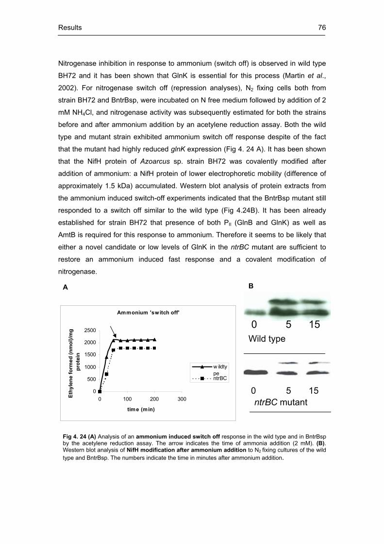
Results 76
Nitrogenase inhibition in response to ammonium (switch off) is observed in wild type
BH72 and it has been shown that GlnK is essential for this process (Martin et al.,
2002). For nitrogenase switch off (repression analyses), N2 fixing cells both from
strain BH72 and BntrBsp, were incubated on N free medium followed by addition of 2
mM NH4Cl, and nitrogenase activity was subsequently estimated for both the strains
before and after ammonium addition by an acetylene reduction assay. Both the wild
type and mutant strain exhibited ammonium switch off response despite of the fact
that the mutant had highly reduced glnK expression (Fig 4. 24 A). It has been shown
that the NifH protein of Azoarcus sp. strain BH72 was covalently modified after
addition of ammonium: a NifH protein of lower electrophoretic mobility (difference of
approximately 1.5 kDa) accumulated. Western blot analysis of protein extracts from
the ammonium induced switch-off experiments indicated that the BntrBsp mutant still
responded to a switch off similar to the wild type (Fig 4.24B). It has been already
established for strain BH72 that presence of both PII (GlnB and GlnK) as well as
AmtB is required for this response to ammonium. Therefore it seems to be likely that
either a novel candidate or low levels of GlnK in the ntrBC mutant are sufficient to
restore an ammonium induced fast response and a covalent modification of
nitrogenase.
Fig 4. 24 (A) Analysis of an ammonium induced switch off response in the wild type and in BntrBsp by the acetylene reduction assay. The arrow indicates the time of ammonia addition (2 mM). (B).Western blot analysis of NifH modification after ammonium addition to N2 fixing cultures of the wild type and BntrBsp. The numbers indicate the time in minutes after ammonium addition.
Ammonium 'switch off'
0
500
1000
1500
2000
2500
0 100 200 300
time (min)
Ethy
lene
form
ed (n
mol
)/mg
prot
ein
w ildtypentrBC
0 5 15 ntrBC mutant
Wild type 0 5 15
A B

Results 77
4.6.2.2 Analyses of the glnB expression and role of NtrBC
The glnB gene has been reported to encode for small trimeric PII proteins and plays
an important role in regulating nitrogen fixation and N-assimilation in many
prokaryotes (reviewed by Merrick and Edwards, 1995). One of the functions of GlnB
is to stimulate NtrB dependent dephosphorylation of NtrC under N-excess, thereby
preventing transcription from the ntr-dependent operons. Therefore, glnB belongs to
the ntr regulatory cascade. It is the goal of this section is to study glnB expression in
Azoarcus sp. BH72 (by GUS reporter gene analysis and RT PCR) and to find out the
role of the NtrBC in this context.
4.6.2.2.1 The glnB::gusA expression in strain BH72 is affected by nitrogen
To construct a transcriptional fusion of glnB::gusA, plasmid pDZD3 was digested by
SstI and BglII so that part of the glnB along with its upstream region falls out. This
DNA fragment was then ligated to SstI and BamHI digested pBluescript-KS. To this
construct, the HindIII and SmaI digested GUS fragment from pBluescript–SK was
modified by Klenow fragment and ligated into EcoRV digested glnB-pBluescript-KS.
The orientation of the gusA gene with respect to the glnB region was confirmed by
restriction digestion. This plasmid (pSGLB-GUS) was used to transform strains BH72
and BntrBsp by electroporation. Attempts to transform BntrBsp failed, but single
recombinants of strain BH72, which were ampicillin resistant and had the gusA
reporter gene integrated in the genome upstream of glnB could be rescued. This
recombinant strain BGLB-GUS was tested for its GUS activity by growing the strain
in various N-sources and on N2. The results of the GUS activity are presented in Fig
4. 25. Surprisingly, the strain exhibited a differential expression pattern; being highly
induced on N2 and suppressed in complex medium or minimal medium with N
(ammonium or nitrate). This is quite contradictory to previous reports where the glnB
expression (studied by Northern analyses) appeared to be constitutive, independent
of the N-status of the external medium (Martin et al., 2000).

Results 78
4.6.2.2.2 RT-PCR to analyse glnB expression in strain BH72 and its down regulation in BntrBsp
To further investigate whether glnB expression in strain BH72 is indeed differential in
response to the N-status and to have an idea about its expression profile in BntrBsp
(since no GUS reporter strain could be obtained for BntrBsp), RT PCR was carried
out to amplify the glnB gene. Equal amounts of RNA isolated from strain BH72 and
BntrBsp each growing in ammonium or under N2 fixation, respectively, were reverse
transcribed at 42°C for 30 min by reverse primer glnB rev and subsequently PCR
amplified by glnBfor and glnBrev primers at 95°C 1 min, 57°C 1 min, and 72°C 2 min
using RT-PCR beads. Equal aliquots of samples were removed at defined time
points of the cycles from both the strains and the PCR products were separated on a
gel. To ensure that equal amounts of template were used in all cases, an RT PCR of
the 16S rRNA gene was performed with 10 ng RNA from the same preparations at
42°C 30 min with the reverse primer 1401 rev followed by PCR cycles of 1 min 95°C,
2 min 65°C and 2 min 72°C with 104f and 1346rev primers The result of this
experiment is shown in Fig 4.26. It appeared that in strain BH72, the glnB abundance
Fig 4. 25 Expression of glnB::gusA in strain BH72 along with the restriction map of the construct. (A) Restriction map of the glnB::gusA construct used for electroporation. S, SstI; B, BamHI / BglII; E, EcoRV. (B) Expression of glnB::gusA in strain BH72. GUS activity was measured from cultures grown in complex medium (VM ethanol), minimal medium with 10 mM ammonium (SM+N / SM+NH4), minimal medium with 10 mM nitrate (SM+NO3), or N2 fixing (N2). Each data represent 3 parallel measurements from 2 independent experiments.
0
5000
10000
15000
20000
25000
VM ethno SM+N SM+NH4 SM+NO3 N2
Growth medium
GU
S (in
Mill
er u
nits
)
S B E E
glnB + upstream gusApBluescript-KS
A
B

Results 79
1 2 3 4 5 6 7 8 1 2 3 4 5 6 7 8
1 2 3 4 1 2 3 4 1 2 3 4 1 2 3 4
1 2 3 4 5 6 7 8 1 2 3 4 5 6 7
NH4+
N2NH4+
N2
N2N2
NH4+
NH4+
BH72
BntrBspA B
BH72 BntrBsp
Fig 4.26 Analysis of the glnB transcription by RT-PCR. (A) Cultures of strain BH72 and BntrBsp growing either in presence of combined nitrogen or N2 fixing were used for RNA isolation and subsequent glnB-directed RT-PCR. Samples were taken after 16, 18, 20, 23, 26, and 28 PCR cycles (lanes 1, 2, 3, 4, 5, and 6 for strain BH72 and lanes 2, 3, 4, 5, 6, and 7 for strain BntrBsp); RNA with heat inactivated beads (lane 7 for BH72 and lane 1 for BntrBsp) and size marker in lane 8. (B). 16S rRNA directed RT-PCR from equal amounts of all RNA preparations as control. Samples taken at 18, 23 and 28 PCR cycles (lanes 1, 2, and 3 respectively); lane 4, size marker.
levels were more or less comparable under N2 fixation and ammonium. At this point it
is important to note that predictions from the GUS assays do not correspond to these
findings. Interestingly in strain BntrBsp, the transcript (expression) appeared to be
down-regulated under N2 fixation as compared to that in ammonium.
4.6.2.2.3 Comparable protein levels of GlnB in strain BH72 independent of nitrogen
Since results coming from reporter gene fusion and RT-PCR analyses of glnB
transcriptional regulation differed from each other, attempt was made to study the
expression pattern at the level of protein. Equal amounts of protein isolated from
ammonium grown or nitrogen fixing cultures of strain BH72 were separated by SDS-
PAGE electrophoresis and transferred to membranes for Western blot analysis with
antiserum against GlnB. The result of the Western blot analysis is presented in Fig
4.27. It appeared that the GlnB was expressed in strain BH72 and that the
expression level under N-excess and N2 fixation was comparable. Thus at least for
strain BH72, the Western blot results correspond to the findings of RT PCR analysis
of the glnB transcript.

Results 80
Fig 4.27 Western Blot analysis of GlnB from strain BH72.Equal amounts of protein isolated from cultures in ammonium or N2 fixing were used for SDS PAGE and Western blot with GlnB antiserum
NH4+ N2
4.6.2.3 Detection and analyses of the glnY in the ntrBC mutant
As mentioned earlier, Azoarcus sp. BH72 is unique to have a third GlnY like PII
protein along with GlnB and GlnK. The GlnY was only detected in the glnK glnB
double mutant and occurs only in its uridylyted state (Martin et al., 2002). An ORF
coding for a putative ammonium transporter (amtY) is located immediately
downstream of glnY (Martin et al., 2000). Interestingly GlnY could be detected in
strain BntrBsp of Azoarcus sp. along with the other PII proteins (verified by RT PCR
and 2D gel electrophoresis analysis). Also an attempt has been made to analyse the
nature of the glnY expression with GUS reporter gene.
4.6.2.3.1 Detection of the glnY transcript in BntrBsp by RT PCR
For this purpose, RNA was isolated from strain BntrBsp growing microaerobically in a
minimal medium with nitrate as sole N-source. As has been mentioned earlier,
nitrogenase is not completely repressed under such conditions and the ntrBC mutant
still fixes N2. About 1 g of RNA isolated from this culture was treated with DNase I
and used for reverse transcription by the reverse primer (glnYrev) at 42°C for 30 min
with RT PCR beads (amersham pharmacia) as mentioned in the material and
methods section. The cDNA was subsequently amplified by PCR (95°C for 1 min,
57°C for 1 min and 72°C for 2 min) with primers specific for glnY (glnYfor and
glnYrev). The glnB and glnK genes were similarly reverse transcribed and PCR
amplified with their specific primers (glnB for and glnB rev and glnK for and glnK rev),
respectively. The results of this experiment are shown in Fig 4.28. The glnY transcript
was specifically amplified along with glnB and glnK while no amplification product
was obtained with heat-inactivated beads (thus test for DNA contamination in the
template) or with water (without any template).

Results 81
Fig 4.28 Analyses of glnY expression by RT PCR along with glnB and glnKfrom BntrBsp growing microaerobically on nitrate. Lane 1, size marker; lane 2, negative control without RNA; lane 3, heat inactivated reverse transcriptase prior to RNA addition; lane 4, 5, and 6, (RT PCR product of glnB, glnK and glnY from DNaseI treated RNA (A) or PCR products from genomic DNA of BntrBsp (B)
1 2 3 4 5 6 1 4 5 6A B
4.6.2.3.2 2D-gel and Western blot analyses confirming GlnY expression along with the PII proteins
For confirmation at the protein level, total protein was isolated from BntrBsp growing
microaerobically on nitrate. Two-dimensional gel electrophoresis (2D-PAGE), and
Western blot analyses were performed to separate and identify the PII proteins. The
antiserum raised against GlnK-MalE, which can detect both GlnK and GlnY, was
used to detect the PII proteins. The results of the 2D gel and Western blot are shown
in Fig 4.29. A single GlnY like spot appeared at a less acidic pI (pI 6.4) and could
clearly be differentiated from the other two more acidic PII – protein spots (pI 5.2).
The two distinct spots of PII represent its modified (uridylylated) and unmodified forms
with the spot towards the less acidic pI (pI 5.5) and less electrophoretic mobility
representing the uridylylated form. It has already been reported that in strain BH72
cells growing on various N2 sources except ammonium, two PII like protein spots
were always detectable in the wild type; both proteins were present as a mixture in a
single spot (Martin et al., 2000). So in this situation although the GlnK antiserum
binds to both, GlnK and GlnY, it is quite likely that the two GlnK spots may be a
mixture of two PII proteins (both GlnB and GlnK) in their modified and unmodified
form. Although the nitrogenase is not completely repressed in BntrBsp under this
condition, the unmodified form of PII is relatively more abundant.

Results 82
4.6.2.3.3 Low but detectable glnY::gus expression in BntrBsp
In order to construct a transcription fusion of glnY::gusA, a 1.5 kb SalI – SalI
fragment from pDZD18 was inserted into the SalI site of pBluescript-SK-GUS
(pSGLY-GUS). This plasmid was electroporated into strains BH72 and BntrBsp so
that the reporter gene fusion construct with glnY got integrated within the genome by
a single recombination event. The single recombinants were grown on different
nitrogen sources and the GUS activity was measured (Fig 4. 30). The GUS activity of
BGLY-GUS was nearly undetectable under all conditions tested. On the other hand,
the NTRGLY-GUS had relatively low but detectable GUS expression levels.
Interestingly the GUS activity was much higher in N-free medium (N2 fixation)
compared to that in SM+N. Also the glnY::gusA expression in BntrBsp was found to
be relatively higher while growing microaerobically on mineral medium with nitrate as
sole N-source as compared aerobically under similar condition.
Fig 4.29 Analysis of PII expression pattern along in BntrBsp by two-dimensional gel electrophoresis (A) and Western blot analysis (B) with antisera against GlnK. The ntrBCmutant strain was grown microaerobically with 10 mM nitrate as sole N-source. The arrow indicates the position of the modified PII (UMP). The star indicates the GlnY spot at a relative more basic pI (pI 6.8) compared to that of the PII proteins (GlnB and GlnK), both in their modified and unmodified forms.
A B
pI 5.2 5.5 6.4

Results 83
Fig 4. 30. Restriction map of glnY::gusA construct and its expression in strain BH72 and BntrBsp . (A) Restriction map of glnY::gusA construct in pBluescript-SK. S, SalI; E, EcoRI; B, BamHI; (B) Expression ofglnY in strains BH72 and BntrBsp. The GUS activity was below the detection level in the wild type strain BH72 under all conditions tested. SM+N (Minimal medium with 10 mM ammonium and 0.01% yeast extract), NH4 (in presence of only 10mM ammonium) or NO3 (with 10mM nitrate) growing aerobically –NH4 (a) and NO3 (a) or microaerobically with 0.8% 02 – NH4(ma) & NO3(ma). “N2 “represents growth under N2 fixation. Results were obtained from three parallel GUS measurements of two independent experiments. Bars represent standard deviations
020406080
100120140160
SM+N
NH4(a)
NH4(ma)
NO3(a)
NO3(ma) N2
Growth medium
GU
S (in
Mill
er u
nits
)
BH72BntrBsp
S S E B E
glnY + upstream gusA
pBluescript-SK
A
B
4.6.3 Study of genes for N–assimilation in Azoarcus : role of NtrBC
Genes involved in nitrogen assimilation are part of the ntrBC regulatory cascade. The
enzyme glutamine synthetase (GS) is the main assimilatory enzyme converting
ammonium to glutamine and strongly induced under N-limitations. Similarly,
glutamate synthetase (GOGAT) also acts as another key enzyme for ammonium
assimilation. On the other hand assimilatory nitrate reductase converts nitrate to
nitrite and acts as the rate-limiting enzyme when the source of nitrogen is nitrate. The
nitrite salts are toxic for the cells. Therefore cells detoxify nitrite rapidly by
assimilatory nitrite reductase, which reduces nitrite into ammonium, which can be

Results 84
easily utilized by the cell. It has been reported that transcription of many of these
assimilatory genes are NtrBC controlled (Persuhn et al., 2000)
In order to study the transcriptional regulation of a few of these assimilatory
genes, several contigs arising from the genome data of Azoarcus sp. strain BH72
(confidential and unreleased data from the Azoarcus genome project in cooperation
with University of Bielefeld, Germany) were blasted against Genbank using BlastX
(translated BLAST search tool). Primers were designed to specifically amplify certain
regions of these ORFs. After standardizing the conditions for PCR amplification from
genomic DNA for each of these putative assimilatory genes, transcripts both from the
wild type and the ntrBC mutants were reverse transcribed and amplified by PCR.
Differential transcription according to the N-source was observed, speaking in favour
of an N-regulated expression pattern of these genes.
4.6.3.1 Identification of a putative glutamine synthetase III in Azoarcus
sp. BH72
In order to identify the gene encoding for glutamine synthetase III, in strain BH72, a
contig of 21.251 kb (contig 281) was blasted in parts against Genbank. A 1500 bp
region located 13.500 kb downstream from the start of the contig was found to have
58% identity and 72% similarity to glutamine synthetase III from Mesorhizobium loti.
The corresponding genetic region from Azoarcus was translated into protein and a
putative protein with high similarity to glnIII gene from Mesorhizobium loti counter
part was chosen. This predicted protein was then aligned with known protein
sequences retrieved from related organisms by Clustal X. The result of this alignment
is presented in Fig 4.31. This alignment identified a putative glutamine synthetase III
in Azoarcus sp. BH72 by sequence comparison with GS proteins from other bacteria.

Results 85
4.6.3.2 GSIII transcription in strain BH72 is nitrogen dependent but free from NtrBC regulation
Equal amounts of RNA isolated from both strains - BH72 and BntrBsp grown in
ammonium and on N2 was reverse transcribed (using RT beads) using reverse
primer GS3 rev followed by PCR amplification (95°C for 1 min, 61°C for 1 min, and
72°C for 2 min.) of a central 350 bp fragment of the putative glnIII counterpart from
strain BH72 using GS3 rev and GS3 for primers respectively. The results are shown
in Fig 4. 32. No expression of glnIII was observed under N excess in both the
strains, BH72 and BntrBsp. However there was strong expression of the putative GS
protein under N- limitation (N2 fixation) for both strains. 16S rRNA directed RT PCR
Fig 4. 31 Alignment of amino acid sequences from different glutamine synthetase proteins (GS). Putative GS protein sequence from Azoarcus sp. BH72 aligned with sequences from M. loti,M. thermotrophicus, R. leguminosarum and S. meliloti. The degree of similarity in each case is represented as follows: black :100%, dark grey : 80% and white : below 80%.

Results 86
1 2 3 4 5 1 2 3 4 61 2 3 4 5 6 1
A B
BH72 BntrBsp
Fig 4.32 Analysis of glutamine synthetase III (glnIII gene) expression in strain BH72 and BntrBsp by RT-PCR. (A)RT-PCR of glnIII gene, using 1 g RNA from N2 fixing cultures of strain BH72 or BntrBsp. Reverse transcription carried out at 42°C for 30 min with the reverse primer GS3 rev followed by amplification for 35 cycles (95°C 1min, 61°C 1min and 72°C 2 min) using GS3 for and GS3 rev primers. RT-PCR product of glnIII from cells grown either in presence of ammonium with heat inactivation of reverse transcriptase prior to RNA addition (lane 1) or without heat inactivation (lane 2) or during N2 fixation with heat inactivation of reverse transcriptase (lane 3) or without heat inactivation (lane 4); size marker (lane 5); amplification product from genomic DNA (lane 6). (B), 16S rRNA directed RT-PCR from 10 ng RNA of the same preparation (as in A). Reverse transcription carried out with 1401rev primer (42°C 30 min) followed by amplification with 104f and 1346rev primers for 35 cycles of 1 min 95°C, 2 min 65°C, 2 min 72°C. 16S rRNA RT-PCR product from strains BH72 or BntrBsp growing in presence of ammonium (lane 3 and lane 5 respectively) or N2 fixing (lane 4 and lane 6 respectively); heat inactivated reverse transcriptase prior to RNA addition (lane 2); size marker (lane 1).
confirmed equal abundance of templates in all cases. These results indicated that
the putative glnIII gene is nitrogen regulated but not under the control of NtrBC.
4.6.3.3 Identification of the genetic region in Azoarcus BH72 putatively encoding glutamine-2-oxoglutarate amino transferase (GOGAT)
A similar approach was made to identify the glt gene encoding for putative GOGAT
enzyme as it was done to identify glnIII of Azoarcus sp. BH72. Within the same
contig (281), a 1.44 kb region (10620 bases downstream from the start) showed
homology to the Glutamate synthetase large subunit like protein of Sinorhizobium
meliloti (73% identity and 78% similarity). The translated amino acid sequences
generated using ExPASy was aligned with known GOGAT amino acid sequences of
other organisms using Clustal X. The alignment is shown in Fig 4.33. It shows that
the putative protein sequence from strain BH72 aligned well with others and showed
high level of identity with known sequences.

Results 87
4.6.3.4 Effect of nitrogen on glt expression and its control by NtrBC in strain BH72
Equal amounts of RNA from strain BH72 and BntrBsp were used to reverse
transcribe and amplify a 440 bp fragment from the central part of a putative gogat
counterpart using GOGATfor and GOGATrev primers. The reverse transcription was
carried out at 42°C and the annealing temperature for the primers was 61°C. The
result is shown in Fig 4.34. It clearly indicates that glt gene is differentially expressed
in both the strains, the transcript being much more abundant under N2 fixation, an
observation similar to the glnIII expression in strain BH72. However the expression of
glt in BntrBsp under N2 fixation is relatively lower as compared to that in strain BH72
under similar conditions. Therefore, the expression of glt gene in strain BH72
appears to be partially regulated by NtrBC.
Fig 4.33 Alignment of amino acid sequences from different glutamate synthetase (GOGAT) proteins. Putative GOGAT protein sequence from Azoarcus sp. BH72 aligned with protein sequences from S. meliloti and A.tumefaciens. Only identity (100% homology) represented by black shade is shown.

Results 88
4.6.3.5 Analysis of the nitrate assimilatory enzyme: nitrate reductase
As mentioned earlier, the ntrBC mutant (BntrBsp) showed signs of severe starvation
while growing on plates of minimal medium with nitrate as sole N-source. The growth
of the mutant is also prolonged when growing aerobically on such a medium.
Therefore it is quite tempting to speculate that NtrBC of strain BH72 has a regulatory
effect on the assimilatory nitrate reductase expression. To establish this fact, the
putative assimilatory nitrate reductase of Azoarcus was detected by homology with
other related genes and an attempt has been made to study its transcriptional
regulation.
4.6.3.5.1 Identification of the genetic region in strain BH72 corresponding to the assimilatory nitrate reductase
A similar approach was made to detect the assimilatory nitrate reductase gene from
strain BH72 as it was done previously to detect glnIII and glt using raw data from the
Azoarcus genome project, Bielefeld. A 2.58 kb region from the beginning of contig
342 was blasted against Genebank. This region was found to have maximum
homology to Pseudomonas aeruginosa PA01 assimilatory nitrate reductase (58%
similarity and 50% identity). This genetic region from contig 342 was further
translated into a protein sequence by ExPASy and the amino acid sequence
generated by this way was aligned with other assimilatory nitrate reductase protein
Fig 4.34 Analysis of glt gene (GOGAT) expression in strain BH72 and strain BntrBsp by RT-PCR. (A). One g RNA, in each case was reverse transcribed by GOGAT RTrev primer at 42°C for 30 min, followed by PCR amplification by GOGAT RTfor and GOGAT RTrev primers for 35 cycles of 95°C 1 min, 61°C 1 min, and 72°C 2 min. RT-PCR product of glt gene from cells grown either in presence of 10 mM ammonium (lane 3) or N2 fixation (lane 4); negative control with heat inactivated reverse transcriptase prior to the addition of RNA (lane 2); PCR amplification of glt from 100 ng genomic DNA using the same primers (lane 5); and size marker (lane 1). (B) 16S rRNA directed RT-PCR from equal amount of same RNA preparations (as in A) was carried out at 42°C for 30 min with 1401rev primer followed by amplification cycles of 95°C 1min, 65°C 2 min, 72°C 2 min by 104f and 1346rev primers. RT-PCR product from strains BH72 or BntrBsp growing in presence of ammonium (lane 3 and lane 5 respectively) or N2 fixation (lane 4 and lane 6 respectively); heat inactivated reverse transcriptase prior to RNA addition (lane 2); size marker (lane 1).
1 2 3 4 2 3 4 5 1 1 2 3 4 5 6
BH72 BntrBsp
A B

Results 89
sequences downloaded from the database using ClustalX. The result of this
alignment is presented in Fig 4.35. It appears that the putative protein sequence from
strain BH72 is very similar to other assimilatory nitrate reductases.
Nitrate reductases can be also of the dissimilatory type (respiratory nitrate
reductase). Usually these are membrane associated and involved in reduction of
nitrate to molecular N2. To ensure that the above mentioned gene is of the
assimilatory type and not a dissimilatory one, the protein sequence of this putative
nitrate reductase from strain BH72 was aligned with other dissimilatory nitrate
Fig 4.35 Alignment of amino acid sequences from different assimilatory nitrate reductase proteins.The putative protein sequence from Azoarcus sp. strain BH72 is aligned with the enzyme sequences derived from P. syringae, X. campestris, and A. tumefaciens. The degree of sequence similarity is represented as follows: black : 100%, dark grey : 80%, and white : less than 80%.

Results 90
reductase sequences from the data base. The result of such an alignment is shown
in Fig 4.36.
From the alignment in Fig 4. 36, it is quite clear that the putative assimilatory
nitrate reductase sequence from strain BH72 shows low similarity as well as identity
Fig 4.36 Alignment of the putative assimilatory nitrate reductase sequence from strain BH72 with different dissimilatory nitrate reductase sequences. The dissimilatory sequences from Shewanella violacea and Desulphovibrio are aligned with the putative assimilatory nitrate reductase sequence from strain BH72. Only regions of identical amino acids (100% sequence similarity and shaded in black) have been shown. The putative Azoarcuscounter part is much less similar to diifferent dissimilatory nitrate reductase sequences than to other assimilatory nitrate reductases from reference organisms (Fig 4.35).

Results 91
to potential dissimilatory nitrate reductase sequences of S. violacea and
Desulphovibrio. Nitrate reductases in general, share similar functional domains within
their structure. Usually a Molybdopterin oxidoreductase Fe4S4 domain is present near
the N-terminus of nitrate reductase. The central part of the enzyme bear typical
Molybdopterin dinucleotide cofactor binding domain which may constitute an entire
subunit and a Molybdopterin oxidoreductase domain. A 2Fe – 2S binding domain is
present near the C-terminus which function as a redox regulatory component and is
often absent in dissimilatory nitrate reductases. Dissimilatory nitrate reductases in
addition, bear signal peptide sequences and transmembrane helices at their N-
terminals, typical for membrane proteins. It was evident from protein sequence
analysis by programme SMART, that dissimilatory nitrate reductases from
Shewanella violacea or Desulphovibrio possessed signal peptide sequences and
transmembrane helices, which were absent in the putative assimilatory nitrate
reductase from strain BH72, in consistent to other cytoplasmic assimilatory nitrate
reductases from different organisms. This line of evidence is also clear from the
alignment, where the N-terminal region of the protein sequence from strain BH72,
appears shorter in length compared to the dissimilatory nitrate reductase sequences.
All these speak in favour of cytosolic and assimilatory nature of the putative nitrate
reductase sequence from strain BH72.
4.6.3.5.2 The expression of the assimilatory nitrate reductase gene in strain BH72 is NtrBC dependent.
An attempt was made to amplify the transcript for assimilatory nitrate reductase by
RT-PCR, both from strains BH72 and BntrBsp, to analyse its expression. Equal
amounts of RNA (1 g) from strain BH72 and BntrBsp growing aerobically on 10 mM
nitrate as sole N-source were DNaseI treated and amplified after reverse
transcription using primers specifically designed for assimilatory nitrate reductase.
Unfortunately the standard method of reverse transcription with RT beads was not
working in this case. Therefore a new method was adopted. Initially the reverse
primer (NO3revRT) was allowed to anneal to RNA by allowing the template primer
mix to gradually cool down from 70°C to 42°C, followed by reverse transcription with
AMV reverse transcriptase for 1 hour at 42°C. About 7.5 l of this reaction mix was
then used as a template for PCR amplification using the Proof start enzyme from
Qiagen: the cycling conditions being 94°C 30 sec, 63°C 30 sec and 72°C 25 sec for
45 cycles with nirforRT and NO3revRT primers, respectively. The results are shown
in Fig 4.37. Interestingly a PCR product (506 bp band) was obtained which was much

Results 92
less intense in BntrBsp compared to that of strain BH72, although equal amounts of
RNA were used as template for both the cases. A 16S rRNA directed RT-PCR was
carried out using equal amount (10 ng) of same RNA preparation from strains BH72
and BntrBsp respectively. Reverse transcription carried out at 42°C for 30 min with
1401rev primer followed by 35 PCR cycles of 95°C 1 min, 65°C 2 min, and 72°C 2
min with 104f and 1346rev primers produced bands of comparable intensities in both
the cases, verifying once more, that equal amount of starting template have been
taken for both the strains. These results show that the assimilatory nitrate reductase
gene in the ntrBC mutant (BntrBsp) is down regulated and that NtrBC is directly or
indirectly involved in the regulation of the assimilatory nitrate reductase in strain
BH72.
1 2 3 4 5 6 7 1 2 3 4
A B
Fig 4.37 Analysis of the expression of assimilatory nitrate reductase by RT-PCR in the wild type and the ntrBC mutant. (A) RT-PCR analysis of the assimilatory nitrate reductase transcript with RNA as templates which was isolated from BH72 and BntrBsp growing aerobically on 10 mM nitrate as sole N-source. Reverse transcriptase heat inactivated prior to addition of RNA from BntrBsp (lane 1) and BH72 (lane 2); size marker (lane 3), PCR amplification product from genomic DNA (lane 4), RT-PCR amplification of RNA after 45 cycles from BntrBsp growing on nitrate (lane 5), the same from strain BH72 (lane 6), size marker (lane 7). (B) 16S rRNA directed RT-PCR taking equal amounts of RNA from strain BH72 or BntrBsp, respectively. Size marker (lane 1), RNA with heat inactivated reverse transcriptase (lane 2), 16S rRNA amplification product from strain BH72 (lane 3) and from strain BntrBsp (lane 4).

Discussion 93
5 Discussion Although biological nitrogen fixation is carried out only by certain Eubacteria and
Archea, the capacity for N2 fixation is dispersed widely among a phylogenetically
diverse group of these organisms. In a broad sense, nitrogenases from
phylogenetically diverse diazotrophic organisms show similarity in structure and
enzymatic mechanism. However the nitrogenase protein sequence may vary at the
level of identity and similarity within different sub-groups of Proteobacteria. For
example, Herbaspirillum seropedicae belonging to the -subgroup of Proteobacteria,
has -type nitrogenase (Machado et al., 1996). So analysis of nitrogenase from
Azoarcus sp. strain BH72, is crucial, as it provides a platform for the first “true -type
nitrogenase“ to be analysed.
Characterization of nifHDK and nifLA transcripts reveals unusual as well as common features in Azoarcus sp. strain BH72.
The genetic organization of nitrogenase of Azoarcus sp. BH72 shows certain unique
features. Downstream of the nif genes, a sequence encoding for 2[4Fe-4S] cluster
carrying protein is present, having 59% amino acid identity to ferredoxin FdxN from
Rhodobacter capsulatus. This organization is quite unlike to the nifH organization of
K. pneumoniae and A. vinelandii where a nifT gene of unknown and nonessential
function is present downstream of nitrogenase genes (Simon et al., 1996). In
diazotrophs such as proteobacteria of the -subgroup or H. seropedicae belonging to
the -subgroup, NifA proteins act as essential transcription activators of nitrogenase
and are directly responsive to nitrogen and O2. Strangely enough, Azoarcus sp.
BH72 possesses NifL / NifA two-component regulatory system, with NifL inhibiting
NifA activity, in response to O2, very similar to that of diazotrophs of the subgroup of
the Proteobacteria. The promoters for both nifHDK as well as nifLA, have been well
characterized in Azoarcus sp. strain BH72 (Egener et al., 2001, Egener et al., 2002).
About 80% of the bases of the putative promoter region of nifHDK upstream of the
start codon match with the standard consensus defined for sigma-54 type promoters;
for the nifLA promoter region, the match is 73.3%. This speaks in favour of an RpoN-
dependent transcription from these promoters. Furthermore, typical NtrC-binding
palindrome motifs are present upstream from the consensus in both cases,

Discussion 94
suggesting an interaction of NtrC with the 54-type RNA polymerase, to initiate
transcription activation.
Interestingly, genes of N-regulatory pathways may possess two different types of
promoters. For example, the E. coli glnA is transcribed from two different promoters:
glnAp1 – a sigma(70) dependent promoter for expression during N excess and
glnAp2 – a sigma(54) dependent N-regulated promoter, used during N-starvation
(Tian et al., 2001). Similarly, the Azorhizobium nifA gene carries overlapping house
keeping type and sigma (54) type promoters that interactively respond to different
signals (Loroch et al., 1995). On the other hand in Azospirillum brasilense, two
overlapping upstream activator sequences have differential effects on the nifH
promoter activity (Passaglia et al., 1995); promoter mapping with S1 nuclease
revealed two start sites of the nifH transcript located 10bp and 40 bp downstream of
the NtrA-dependent promoter (de Zamaroczy et al., 1989). The situation can be more
complicated as is the case e.g. in the nifH promoter region from Rhizobium
leguminosarum, where a pseudo-promoter and a pseudo-upstream element are
present along with a sigma(54) N-regulated promoter consensus (Roelvink et al.,
1990). Therefore it is important to map the transcription start point in order to define
the promoter of a gene.
Usually the nifHDK and nifA / nifLA in diazotrophic proteobacteria are characterized
by 54 – type promoters, but exceptions are also present. In A. brasilense, consensus
sigma-54 and NifA binding sites are present only upstream of nifENX gene, and not
in the upstream of nifHDK (Potrich et al., 2001) whereas sigma (70) recognition sites
occur upstream of its nifA gene (Fadel-Picheth et al., 1999). In R. capsulatus, no
typical RpoN-activated promoters are present, upstream of nifA1 and nifA2, the
transcription activators of nifHDK gene (Foster-Harnett et al., 1992) On the other
hand, R. japonicum share two sigma54 promoters, upstream of nifH and nifDK
respectively (Adams et al., 1984). Whereas H. seropedicea, shares a typical sigma
(54) promoter consensus, similar to K. pneumoniae, and possesses typical NifA and
integration host factor (IHF) binding sites (Machado et al., 1996). In contrast,
methanogenic archaeons, like M. maripaludis are characterized by AT-rich Box-A
promoter sequences, 24 bp upstream of nifH gene ((Kessler et al., 1998). Primer
extension analysis carried out for nifHDK and nifLA of Azoarcus sp. BH72 revealed
that, indeed one major transcriptional start point is used for each of the above
mentioned genes when the bacteria are fixing nitrogen. Moreover the detected

Discussion 95
transcriptional start point in each case matched precisely with respect to the location
of the upstream –12 / -24 consensus, suggesting that this might represent an active
promoter for nifHDK and nifLA, respectively. Usually primer extensions are carried
out by radioactively labelled reverse primers, which might turn out to be much time
consuming and hazardous. However in this study, all primer extension experiments
have been carried out non-radioactively, using Cy5 labelled reverse primers and an
ALFexpress automated sequencer (Pharmacia biotech). It is clear from the
experimental results that the new methodology developed, is quite robust and well
reproducible.
The structural genes of the nitrogenase enzyme complex in strain BH72 are
genetically linked to a ferredoxin gene (fdxN) (Egener et al., 2001). Ferredoxins are
believed to be electron donors for nitrogenase in bacteria. In A. vinelandii, R.
capsulatus or S. meliloti, nif-specific ferredoxins have been identified that are not
genetically linked to the structural genes of nitrogenase (Klipp et al., 1989; Schatt et
al., 1989). In A. vinelandii, a ferredoxin like gene is localized downstream of vnf
coding for the iron protein of vanadium nitrogenase (Robson et al., 1986).
Cotranscription of nifHDK with fdxN was proved by RT PCR in strain BH72, by
amplifying a region of the transcript spanning between nifK and fdxN. This unusual
transcriptional linkage speaks in favour of a functional linkage of these two proteins in
strain BH72. In a similar way, it has been shown that indeed nifL and nifA in strain
BH72 are also cotranscribed. This situation is more similar to -Proteobacteria, where
nifL is transcribed along with nifA, NifL being the sensor inhibiting NifA activity in
response to O2 (Dixon, 1998). One of the major significance of coordinated synthesis
of antagonistic regulatory proteins, like NifL and NifA, is to maintain the correct
stoichiometry for efficient regulation of N2 fixation. In K. pneumoniae, a low
translation rate, as a result of inhibitory secondary structures arising at the Shine-
Dalgarno sequence of NifA has been reported, in the absence of NifL (Govantes et
al., 1996).
The nifLA transcript was detectable in aerobically grown cells on combined nitrogen
but was more abundant during N2 fixation in Northern blot analysis. This differential
expression pattern was confirmed by RT PCR amplification of the nifA transcript.
Also, no nifA transcript was detected in the polar mutant of nifA (BHLAO), while
growing in combined nitrogen, confirming cotranscription. In A. vinelandii,
transcription of nifLA is not repressed by ammonium (Blanco et al., 1993). However
in Klebsiella and Enterobacter, NifA expression is strongly enhanced (about 10-fold)

Discussion 96
under anaerobic conditions mediated by DNA-gyrase dependent negative
supercoiling. In this aspect strain BH72 shows similarities to K. pneumoniae nifLA
regulation, since the expression is enhanced both by ammonium deficiency and by
microaerobiosis, without the involvement of the FNR box (Hu et al., 2000).
The NtrBC proteins of strain BH72: the “third element” in the N-regulatory cascade.
The ntrBC two-component “sensor-response regulator” system has been identified in
many different bacteria. It plays an important role in sensing and evoking N-
regulatory cascades involved in N2 fixation and N-assimilation. NtrB acts as a
bifunctional histidine kinase (phosphatase) that in turn phosphorylates NtrC. NtrC-P
acts as transcriptional activator of many N-regulated genes. In the context of N2
fixation, NtrC-binding activates nifA transcription (second element), which in turn acts
as an essential transcriptional activator of nifHDK, the structural genes for
nitrogenase (first element). The concentration of NtrC is increased as cells become
starved for ammonia, concurrent with the activation of the ntr genes that have less
efficient NtrC binding sites like the nac and glnK promoters than does glnA (Atkinson
et al., 2002). In this way, NtrC regulated operons could be temporally separated
(with time) for expression, in the regulatory cascade.
In this study, ntrB and ntrC coding regions have been identified in strain BH72. The
ntrBC genes of strain BH72 share strong identity with Ralstonia solanacearum, and
Herbaspirillum seropedicea counterparts, both belonging to the -subgroup of
Proteobacteria. Analyses of the predicted NtrB and NtrC proteins by SMART
(www.smart.embl-heidelberg.de) revealed the presence of typical PAS, histidine
kinase and ATPases domains in NtrB, and phosphoacceptor, ATP-binding and “helix-
turn-helix” motifs in NtrC. All these domains are correlated to the function of sensor
and response regulator respectively. PAS domains constitute signal-receiving
domains of sensor kinases in association with bound cofactors. Autokinase activity is
exhibited by histidine kinase domain of NtrB like sensors whereas ATPase-like
domain is responsible for the phosphatase activity of the sensor, which is essential
for the down regulation of the cascade under nonfavourable conditions. The N-
terminal phosphoacceptor domain of NtrC-like response regulators have the
conserved aspartate residues, representing the active site for phosphotransfer. The
C-terminal helix-turn-helix domains are typical, not only for NtrC, but in general, for

Discussion 97
DNA-binding proteins. The central ATP binding domain is characteristic for NtrC-like
transcription activators, where binding of ATP in this domain helps in oligomerization
of NtrC and subsequent transcriptional activation of the sigma(54)-dependent RNA
polymerase, by formation of functional transcriptional open-complex. RT PCR
analyses revealed that both ntrB and ntrC transcripts of the expected size are
expressed during N2 fixation. Also an RT PCR, similar to that of nifLA in strain BH72
has also verified their cotranscription status.
Surprisingly, no proper ribosome-binding Shine-Dalgarno motif could be detected
before the putative start codons of ntrB and ntrC. Shine-Dalgarno (SD) dependent
translation initiation predominates in E. coli. However, this bacterium also has the
capacity to initiate translation in a SD independent manner. In contrast, plant
chloroplasts and their cyanobacterial ancestors appear to have adopted SD-
independent mechanisms for most of their translation initiations (Fargo et al., 1998).
In E. coli, the human calcitonin gene was more strongly expressed from a
nonconventional SD translation initiator depending on the binding energy (Mironona
et al., 1999). The +2 codon immediately following the start codon may sometimes act
as critical translation determinant (Stenstrom et al., 2001). On the other hand,
extended base pairing surrounding the start codon may compensate for the lack of a
classical SD (Esposito et al., 2001). A/T rich sequences upstream of the start codon
of E. coli K12 have been reported to correlate with translation initiation (Yamagishi et
al., 2002). Interestingly, an A+T element represented by a “TATTA” stretch could be
located 2 bases upstream of the NtrC start codon in strain BH72.
An attempt to find a standard bacterial promoter consensus (sigma-54 or sigma-70),
upstream of the ntrBC coding region failed. In -Proteobacteria, ntrBC is generally
encoded as ORF1-ntrBC operon, ORF1 being an upstream open reading frame of
unknown function. In Azospirillum brasilense, ORF1 has similarity to nifR3 of
Rhodobacter capsulatus and is transcribed from a vegetative sigma(70)-type
promoter (Machado et al., 1995). However in enterics, ntrBC transcription is linked to
a glnA gene located upstream of ntrBC in form of a glnA-ntrBC operon and
transcribed from a N-regulated sigma(54)-type promoter (MacFarlane et al., 1985).
Primer extension analysis to map the 5’ end of the transcript revealed in Azoarcus
sp., the presence of a single transcript which mapped at a position, 150 bp upstream
from the putative start codon of the ntrB, however also at the vicinity of this position a
promoter sequence consensus could not be detected.
Surprisingly, transcriptional fusions of the GUS reporter gene with the ntrBC
upstream region were not successful, as the expression levels were too low.
However, expression could be detected in RT PCR studies in strain BH72. It was

Discussion 98
shown that the ntrB is expressed both in presence of combined nitrogen and on N2 in
a differential manner. Under these circumstances transcript abundance was much
higher when cells fix N2, but not subjected to autoregulation. In Klebsiella
pneumoniae the ntrBC operon has a structure like P1-glnA-P2-ntrBC, P1 and P2
representing two promoters. P1 promotes transcription under N-deficiency and P2
under N-excess. P1 is regulated both positively and negatively, while P2 is only
negatively regulated by NtrC (Morales-Alvarez et al., 1984). In contrast, in
Rhizobium leguminosarum, the promoter is subjected to negative autoregulation by
NtrC (Patriarca et al., 1993). In this context, it’s quite unusual that NtrBC in strain
BH72 is not subjected to autoregulation. Another interesting finding in this study, is
that ntrB in strain BH72 is expressed in a N regulated manner, although a typical
sigma(54) promoter consensus is lacking upstream of ntrB. It is quite tempting to
speculate that ntrBC transcription in strain BH72 is probably regulated from some
distantly located upstream element and that the major transcript is subjected to
subsequent processing. On the other hand the possibility of transcription being driven
by some novel promoter elements cannot be ruled out. Another speculation in this
respect might be related to DNA-bending within the genome, which might mobilize
distantly located sigma(54)-type promoter elements, facilitate the binding of the
transcription machinery, and trigger transcription. Interestingly, a putative ORF
having homology to RNA binding protein from Homo sapiens is present in the
upstream of ntrBC of strain BH72 in the same orientation. So the possibility of
unusual transcriptional linkage of ntrBC with this ORF cannot be ruled out.
Nitrate assimilation in strain BH72 is positively controlled by NtrBC
The BntrBsp (ntrBC-mutant) of Azoarcus was impaired during growth on nitrate as
sole N-source. While growing on agar plates with nitrate as sole N-source, the colony
morphology appeared drastically different from the wild type; the mutant colonies
appeared more opaque and rounded. When viewed under phase contrast
microscope, the cells of BntrBsp appeared smooth surfaced with accumulation of
shining crystalline particles, centrally. When growth is limited by availability of an
essential nutrient other than carbon and energy source, many bacteria accumulate
reserve materials such as polyphosphate, PHB (poly- -hydroxybutrate) or glycogen

Discussion 99
(Dawes et al., 1973). A pronounced increase of PHB in culture of nitrogen-fixing A.
lipoferum Rp5 and A. halopraeferens Au4, and to a lower extent in Azoarcus sp.
BH72 have been reported at high O2 concentrations when the electrons could not be
channelled to nitrogenase and were used for PHB synthesis instead (Hurek et al.,
1987). Also the unreleased data from the sequenced genome of Azoarcus sp. BH72
provide evidence for the existence of several enzymes involved in the metabolism of
PHB. In Azospirillum brasilense Sp7, the ntrBC genes are involved in the regulation
of PHB synthesis by ammonia (Sun et al., 2000). So it is quite likely to assume that
these refractive particles represent PHB granules and NtrBC in strain BH72 either
directly or indirectly regulates PHB synthesis. However, an increase of PHB
biosynthesis in the ntrBC mutant could be a direct consequence from the down
regulation of PII proteins. The same has also been reported for A. brasilense Sp7
(Sun et al., 2002). The prolongation of the generation time (9.5 h) of the ntrBC
mutant growing aerobically on nitrate provides further evidence that BntrBsp was
impaired in growth on nitrate, similar to that has been reported for A. brasilense
(Liang et al., 1993). In this respect, it is relevant to mention that NtrBC of Azoarcus
sp. BH72 was found by complementation of A. vinelandii mutants unable to grow on
nitrate as sole N-source. Thus all these observations point towards an essential role
of NtrBC in nitrate assimilation in strain BH72. It can be recollected, that the glnB-K-
(double mutant) of strain BH72 fixes N2 and exhibits prolonged generation time while
growing on nitrate as sole N-source, a phenotype similar to that of ntrBC mutant. On
the other hand, no impairment of ntrB nonpolar mutant to grow on nitrate is an
unusual feature. Indeed, the nonpolar ntrB mutant shared phenotypes similar to that
of the wild type strain. This can be interpreted in terms of another phosphorylation
circuit, which might be still active in ntrB nonpolar mutant and is able phosphorylate
NtrC in absence of NtrB.
From the study of these phenotypes, it becomes apparently clear that NtrBC in strain
BH72 has a regulatory effect on the main enzyme of nitrate assimilation, nitrate
reductase. Enterobacteria can use nitrate and nitrite both as electron acceptors and
as source of nitrogen for biosynthesis. In Klebsiella, assimilatory nitrate and nitrite
reductase convert nitrate through nitrite to ammonium. The enzymes and structural
genes for nitrate/nitrite respiration and assimilation are distinct, and are subject to
different patterns of regulation. Respiratory enzyme synthesis is indifferent to the
availability of ammonium, and transcription is induced by anaerobiosis via the FNR
protein. In Enterobacteria, the respiratory enzyme is further induced by nitrate or
nitrite through a two-component system, where NarX and NarQ act as cognate
sensors and NARL and NARP being the response regulators, respectively (Stewart,

Discussion 100
V. 1994). In contrary, the assimilatory enzyme synthesis is indifferent to oxygen
availability, induced by ammonium and nitrate/nitrite limitation via NTRC or via the
transcriptional antiterminator protein NasR (Stewart, V. 1994). Assimilatory nitrate
reductase catalyzes the rate-limiting step in nitrate reduction with molybdenum as
cofactor (Campbell, W.H. 1999). The gene for assimilatory nitrate reductase was
amplified using sequence information from unreleased data of the Azoarcus genome
project. The corresponding Azoarcus counterpart shared high homology with other
assimilatory nitrate reductase protein sequences, but low sequence similarity with
dissimilatory nitrate reductase sequences. In Klebsiella, nasA and nasB (coding for
nitrate and nitrite reductase respectively) are linked in form of an operon.
Surprisingly, the nasBA operon is tightly linked to respiratory nitrate reductase
although it serves a different physiological function (Lin, et al., 1993). As expected
from the ntrBC mutant phenotype, the assimilatory nitrate reductase transcript was
abundant in strain BH72 while growing on nitrate but was strongly down regulated in
BntrBsp. This very weak expression of assimilatory nitrate reductase in the ntrBC
mutant may account for its prolonged generation time, while growing aerobically on
nitrate. Therefore it is likely to assume that assimilatory nitrate reductase in strain
BH72 is NtrBC regulated but still exhibits very low constitutive expression, unlike in
others. Interestingly, in Azospirillum brasilense, not NtrBC, but a novel NtrY/X two
component system is involved in nitrate assimilation instead. A cross talk between
these two components may add a fine-tuning to nitrate assimilation (Ishida, et al.,
2002).
Twitching motility in strain BH72 is down regulated by NtrBC.
“Twitching motility”- a term coined by Lautrop, describes flagella–independent
surface motility exhibited by a wide range of bacteria. It is equivalent to social gliding
motility in Myxococcus xanthus and is important in host colonization by a wide range
of plant and animal pathogens as well as for formation of biofilms. Type IV pili are
known to be important for twitching motility (Mattick, J.S. 2002) and this has also
been reported for Azoarcus sp. BH72 (Plessel, 2001). The ntrBC-mutant exhibited
two times more twitching compared to the wild type in complex medium. This is quite
surprising, as many type IV pilin genes (pilA) are known to be regulated by PilR,
which has a high sequence similarity at the amino acid level to the NtrC family of
response regulators (Wu et al., 1997). On the other hand, it has been shown that
signal transduction in Ntr and Che systems involves a common phosphotransfer

Discussion 101
mechanism; high intracellular concentration of NRII can suppress a smooth-
swimming phenotype of a cheA mutant involved in chemotaxis (Ninfa, et al., 1988).
Such cross-talks between N-assimilation and chemotaxis signals in bacterial signal
transduction pathways help in fine tuning. Therefore, it is possible that NtrBC from
strain BH72 directly or indirectly controls the twitching motility negatively. On the
other hand, it can be argued that intriguing cross talks may be involved in regulating
twitching. However such predictions are quite speculative and more evidence is
required to support this hypothesis.
The nifLA expression is positively regulated by NtrBC in strain BH72.
It has been well established that in strain BH72, the NifA acts as an essential
transcription activator for the nifHDK, coding for the structural genes of nitrogenase
(Egener et al., 2002). While in Azotobacter vinelandii, transcription of nifLA is not
repressed by ammonium and not dependent on NtrC or RpoN (Blanco et al., 1993), it
is NtrC-dependent in K. pneumoniae (Minchin et al., 1988) and Enterobacter
agglomerans (Siddavattam et al., 1995). In Klebsiella and Enterobacter cloacae,
nifLA expression is also enhanced (approx. tenfold) under anaerobic conditions
mediated by DNA gyrase dependent negative supercoiling (Hu et al., 2000). In ß-
Proteobacteria, like Herbaspirillum seropedicae, concerted action of NtrC, NifA and
IHF (Integration Host factor) is essential to fine-tune NifA expression in response to
ammonium and oxygen levels (Wasem et al., 2002). Unlike in Klebsiella, transcription
of nifA does not require NtrB/NtrC proteins in A. brasilense, and the expression of
nifHDK is controlled by posttranslational regulation of NifA activity (Zhang et al.,
1997). Indeed, nifLA gene expression in strain BH72 was found to be NtrBC
regulated, similar to that of -Proteobacteria. This fact was further exemplified by a
relative low nifH expression in strain BntrBsp (but nif+) compared to strain BH72
under derepression conditions and relative prolongation of generation time in strain
BntrBsp during N2 fixation. The question whether NtrC acts directly on the nifH
promoter or indirectly via NifA is debatable. Since nifA is invariably necessary for
nifHDK transcription, it is not likely that NtrC is an additional transcriptional activator
of nifHDK. However NtrC-binding tandem activation sequences are present upstream
of nifHDK promoter. Although binding of NtrC, upstream of nifHDK promoter has not
been reported for Klebsiella or A. vinelandii, the promoter of the alternative

Discussion 102
nitrogenase of R. capsulatus has been shown to be activated by NtrC binding
(Kutsche et al., 1996). While in A. brasilense, NtrC is not absolutely required for nifH
promoter activation (Broek et al., 1992), the glnG (ntrC) gene product might directly
regulate the symbiotic nitrogen fixation genes in Rhizobium (Sundaresan et al.,
1983). In Klebsiella pneumoniae, GlnK is required for relieving the inhibition of NifA
activity in absence of combined nitrogen (Jack et al., 1999). It has been shown (this
work), that glnK expression is down regulated in the ntrBC mutant along with the
expression of glnY. In accordance with this observation, reduced nifH expression can
be attributed to the indirect effect of NtrC via GlnK, affecting NifA activity. At the
same time, it could be further speculated that the expressed GlnY in BntrBsp
perhaps cannot complement GlnK function by interacting with NifL and release its
inhibitory effect from NifA, pointing to distinct regulatory functions of each of the three
PII paralogues in strain BH72.
Transcriptional regulation of the glnK and the glnB in strain BH72: role of NtrBC
glnK is genetically linked to amtB in strain BH72 (Martin et al., 2000). The physical
association of these two genes is found in a diverse range of Bacteria and Archea.
The glnK-amtB operon is induced specifically during conditions of ammonium
limitation except in Azotobacter vinelandii, where it is an essential gene and
expressed constitutively (Meletzus et al., 1998). GlnK is required to regulate
expression of ntr genes other than glnA in cells lacking GlnB. Generally the glnK-
promoter belongs to the ntr family of promoters and is expressed by an RNA
polymerase containing the minor sigma factor 54 (Atkinson et al., 1994).
Surprisingly, no sigma(54) type of promoter region could be detected, upstream of
Azoarcus glnK gene; instead an ORF (ugk) having similarity to a hypothetical protein
from R. solanacearum which can be putatively exported into the periplasm and
possess a signal peptide sequence, is present. Indeed, the ugk and glnK genes
were found to be cotranscribred in strain BH72. However, a typical sigma (54)
promoter consensus could be detected upstream of Azoarcus ugk gene. Previous
Northern blot experiments have detected a single transcript bearing glnK and amtB
(Martin et al., 2000). Thus it is plausible to think of a single large transcript bearing
ugk, glnK and amtB, which may be transcribed from the sigma (54)-type promoter
and susceptible to a differential RNA processing event. Surprisingly, no common
transcriptional start point could be detected by primer extension carried out

Discussion 103
individually for glnK and ugk. This can once more be explained in terms of RNA
processing. Differential termination of the nifH transcript is known to occur in R.
capsulatus (Willison et al., 1993), which could explain why multiple transcripts of nifH
have been detected in strain BH72 and the full-length transcript was less abundant
(Egener et al., 2001). The genetic linkage of ugk, glnK and amtB might have a far
reached functional significance. Ugk, harbouring a signal peptide sequence, could be
secreted into the periplasmic space and AmtB may function as membrane-bound
ammonium sensor and propagate N-signal transduction cascade within the cell, with
the help of GlnK.
The glnK gene expression in wild type, checked by different methods revealed that it
is suppressed under combined nitrogen and strongly induced under N2 fixation. This
observation is quite in accord with the previous Northern-blot experiments (Martin et
al., 2000) and similar to what has been reported for other organisms. Interestingly,
glnK expression in strain BntrBsp, was severely down-regulated as shown by RT-
PCR and GUS assays. NRI (NtrC) mediated activation of the glnK promoter has
been reported for E. coli (Atkinson et al., 2002). In Psudomonas stutzeri, NtrC and
RpoN recognition sites can be observed upstream of the glnK promoter (Vermeiren
et al., 2002) and Klebsiella, glnK-amtB operon seems to be under NtrBC control too
(Jack et al., 1999).
Nitrogenase inhibition by ammonium (switch-off) has been observed in wild type
BH72 and it has been shown that GlnK is essential for this process (Martin et al.,
2002). Although GlnK expression was found to be down regulated in the ntrBC-
mutant, its “switch-off” response was not abolished and covalent modification of NifH
still occurred after ammonium addition as detected by Western-blots using NifH-
antiserum. In contrast, ntrBC mutants of Azospirillum brasilense and Rhodospirillum
rubrum had no effect on the nif expression, but abolished post translational regulation
of nitrogenase activity and “switch-off” response (Liang et al.,1993; Zhang et al.,
1995). Thus it can be speculated, that very weak expression of GlnK in the ntrBC
mutant may be sufficient to initiate and drive such posttranslational responses. It is
interesting to point out that GlnY is expressed in ntrBC mutant of strain BH72, which
can be thought to drive ammonium “switch off” response in BntrBsp, where glnK is
down regulated. However such speculation could be ruled out from previous
experimental observation where a glnB-K- (double mutant) did not respond to
ammonium switch off response in the presence of GlnY only (Martin et al., 2002)
which once more highlights the distinct functions of the PII paralogues in strain BH72.

Discussion 104
As shown previously by Northern blots, the glnB transcript was not detectable in wild
type cells, but only in a glnK knock-out mutant, both under ammonium and N2 (Martin
et al., 2000). However glnB::gusA reporter gene fusion studies in this work revealed
a N-regulated differential expression pattern of its transcript, being strongly induced
under N2 fixation. glnB genes may be constitutively expressed (glnB in E. coli; van
Heeswijk et al.,1993) or under the control of a 70–like promoter, such as glnB under
conditions of ammonium excess in A. brasilense (De Zamaroczy et al., 1993). RT-
PCR to further verify this regulatory pattern in strain BH72, indicated that the
abundance of the glnB transcript was more or less comparable both under combined
nitrogen and N2 fixation. Results of Western blot analysis with protein extracts from
strain BH72 were more similar to the RT PCR results, indicating more or less
constitutive expression of GlnB both under ammonium and N2 fixation. These
contradictory results are often difficult to interpret. However speculations can be
made on the basis of these observations.
glnB and gusA are linked on a common large transcript which may be more stable,
and a selective degradation process is only active under a particular condition (may
be under anaerobiosis and combined nitrogen). This may be an explanation for the
apparent paradox encountered with GUS measurements versus RT-PCR / Western
blot results. However, it may be important to note that transcription and translation
events may not bear a one to one correspondence relation to each other. Moreover
in E. coli, it has been recently shown that distinct functions of PII and GlnK are
correlated with the timing of expression and levels of accumulation, and that PII can
be functionally converted to GlnK by engineering its expression from nitrogen
regulated promoter (Atkinson et al., 2002). Thus the possibility of glnB::gusA
expression driven by a glnK type N-responsive promoter (not linked) in Azoarcus sp.
strain BH72, cannot be ruled out
GlnB expression in the ntrBC mutant showed a strong reduction of its transcript
abundance under N2 fixation. In Rhizobium leguminosarum, the glnB promoter was
shown to be strongly regulated by nitrogen and NtrC, the glnB promoter being down
regulated during bacteroid differentiation and symbiosis (Ercolano et al., 2001). The
glnB gene from B. japonicum is expressed from tandem promoters and its differential
expression is strongly regulated by NtrC (Martin et al., 1989). So, the glnB regulation
by NtrC in strain BH72, resembles more to that of Rhizobia. Surprisingly, the glnB
expression in BntrBsp appears to remain unaltered in presence of combined
nitrogen. Interestingly, in R. capsulatus, glnB is transcribed from two promoters one
of which require NtrC but RpoN independent, while the other is repressed by NtrC
(Foster-Harnet et al., 1994).

Discussion 105
GlnY expressed in strain BntrBsp, along with other PII proteins.
The expression of GlnY in strain BntrBsp along with other PII proteins is a novel
observation. This PII paralogue of strain BH72 has until now been reported to be
expressed only in absence of GlnB and GlnK (in double mutant of glnB and glnK)
and lacks differential uridylylation in response to nitrogen availability (it is always
uridylylated) (Martin et al., 2000). Indeed, the expression of all the three PII-
paralogues in the ntrBC mutant has been verified, both at the level of RNA as well as
of protein. Surprisingly, glnY::gusA was expressed at a low level in the ntrBC mutant
under all sources of nitrogen. Infact, the relative highest level of glnY::gusA was
detected under N2 fixation, which point towards a novel role of GlnY in nitrogen
fixation. However, a comparison of glnY expression is difficult to make, since its
expression is below the detection level in strain BH72. Nevertheless, it may be
interesting to investigate the uridylylation status of this protein in strain BntrBsp in
order to make further predictions about its nitrogen sensing functions
The N-assimilation genes of strain BH72: role of NtrBC
GS/GOGAT and GDH constitute the main enzymes for ammonium assimilation.
During growth in ammonium-rich medium, ammonium is primarily assimilated
through glutamate dehydrogenase (GDH) by NADPH dependent reactions. At
ammonium concentrations below 1 mM, the affinity of GDH to ammonium is too low,
and the reaction operates in a direction of ammonia liberation from glutamate. In this
situation, the glutamine synthetase / glutamate synthetase (GS/GOGAT) system
takes over, although at the cost of ATP hydrolysis. To prevent waste of energy, the
GS/GOGAT system has to be down regulated at high ammonium concentrations.
The flux through GS is subject to regulation at the level of enzyme activity and
transcription of the GS encoding gene, glnA. The counterpart enzyme, glutamate
synthetase (GOGAT), is encoded by gltB and gltD genes (coding for large and small
subunits respectively) and is also subject of N-dependent regulation. The Azoarcus
counterparts of GS and GOGAT were PCR amplified using sequence information
from the raw (unreleased) data of the genome project. Genes were identified by high
amino acid sequence similarity with known protein sequences. Interestingly, the

Discussion 106
putative GS from strain BH72 had high identity to glutamine synthetase III (glnIII)
from M. loti. Indeed, glnIII expression in strain BH72 was found to be nitrogen
regulated but strangely enough, its expression was found to be independent from
NtrBC control. Similarly, the expression of GOGAT (glt) was found to be N-regulated
in strain BH72 but unlike glnIII, it was found to be moderately regulated by NtrBC. In
Klebsiella, similar to E. coli, P1glnAP2ntrBC occurs in form of an operon with P1 and
P2 being the two promoters. P1 is activated under N-deficiency and regulated
positively and negatively by NtrBC (Alvarez-Morales et al., 1984) while in
Rhodospirillum rubrum, glnA is cotranscribed with glnB, from a weak and strong
promoter under NH4+ and N2 respectively, but the activity of glnA is enhanced by
NtrC (Cheng et al., 1999). Herbaspirillum resembles the enterics more in this aspect,
the glnA-ntrBC operon being subjected to NtrBC dependent transcription and activity
control (Persuhn et al., 2000). The situation is more complicated in Rhizobium, where
out of three GS encoding genes; the glnII promoter is fully dependent on the positive
control by the NtrC protein (Iaccarino et al., 2001). On the other hand, the gltB
promoter of A. brasilense showed the presence of sigma(70) like recognition sites in
addition to a potential NtrA-RNA polymerase, IHF, and NifA binding site (Mandal et
al., 1993) while gltBD operon encoding for (GOGAT) is also under ntr control in E.
coli and Klebsiella aerogens (Goss et al., 2001). So it can be quite likely to assume
that GOGAT expression in strain BH72 is partially regulated by NtrC, at least at the
level of transcription. Another interesting observation is the unusual high similarity of
these assimilatory enzymes to rhizobial members, which may have a far-reaching
significance, phylogenetically.
At this juncture, the basic points discussed may be summarized. Analyses of nifH
and nifLA transcripts of strain BH72 reveal certain usual as well as unusual features.
Although, both nifH and nifLA utilized their upstream 54-promoters for their
transcription (verified by primer extension), nifH was unusually cotranscribed with its
downstream ferredoxin gene (fdxN). In this respect, it may be important to mention
that primer extensions were carried out for the first time, non-radioactively, with
automated sequencing, and produced successful and reproducible results.
Unusually, nifA of strain BH72 was also found to be cotranscribed with its upstream
nifL gene, in a very similar way like that of -Proteobacterial members and its
expression was found to be affected by nitrogen. Furthermore cloning and
characterization of the NtrBC from strain BH72 revealed many interesting findings.
Although ntrB and ntrC of strain BH72 were also found to be cotranscribed, neither
typical Shine-Dalgarno sequences could be detected upstream of their respective

Discussion 107
start codons nor putative promoter sequence could be found upstream of its
transcriptional start. Strangely enough, expression of ntrB gene was found to be N-
responsive but not likely to be auto-regulated. The ntrBC mutant of strain BH72 was
impaired to grow on nitrate and accumulated PHB granules under such conditions;
even its nitrogenase was not repressed by nitrate. Consistent with this, the
expression of assimilatory nitrate reductase was found to be NtrBC regulated.
Unusually, twitching motility was up regulated in ntrBC mutant; a trait which was up
to now not known to be linked to these genes. As in enterics, the nifLA gene
expression in strain BH72 was found to be under NtrBC control which indirecty
affected N2 fixation. Looking at the level of PII proteins, an unusual transcriptional
linkage was found between ugk with glnK although a common transcriptional start
point for both these genes could not be detected. glnK and glnB expressions were
found to be down regulated in the ntrBC-mutant, with the unusual retention of “switch
off “ response to ammonium. Another novel observation was the expression of GlnY
along with the other PII proteins in the ntrBC-mutant albeit at low level. Analyses of
the expression of assimilatory genes, encoding for GS and GOGAT in strain BH72
revealed that their expression was affected by nitrogen. Surprisingly, GS expression
was found to be NtrBC independent although GOGAT seemed to be moderately
regulated by NtrBC. Therefore, NtrBC was shown to be involved in regulating
multiple cellular processes, in strain BH72. The summarized points are presented in
form of a schematic diagram in Fig 5.1

Discussion 108
Fig 5.1 Schematic representation of the candidates of the N-regulatory cascade in strain BH72 studied in this work. The involvement of NtrBC in the process of N-regulation has been
highlighted. ( ) represents the cotranscription status of several linked genes studied and ( )
represents upstream 54 promoter consensus. ( ) depicts positive regulation and
negative regulation mediated by the NtrBC on its putative targets while ( )
represents independent from NtrBC regulation. The symbol ( ? ) represents situation where the role
of the NtrBC is still not clear. All interpretations have been made from the results and observation of
this study
Twitching motility PHB granule
synthesis
nifHDK fdxN
glt(GOGAT))
glnIII (GS)
nifAnifL
glnY
glnB
ugK glnK amtB Assimilatory nitrate
reductase ntrCntrB?
?
?
?
Ammonium switch
off and nitrogenase
modification
(posttranslational)
Outlook for further studies
One of the challenging areas to explore in future is to search for more novel targets
of NtrC in Azoarcus sp. BH72. Preliminary findings such as NtrC controlling twitching
motility in strain BH72 already provides hint in this direction. Further progress in this
area can be made utilizing the information from the Azoarcus genome project, using
“microarrays” as detection tool. Although the domain organization of NtrB and NtrC

Discussion 109
appear to be conserved, their function may vary in minute details among different
subgroups of Proteobacteria. So an important area to explore in this direction is to
study the function of the individual domains of NtrB and NtrC proteins in Azoarcus sp.
BH72, by over expressing and purifying them in order to study their functions, both in
vitro and in vivo. In this respect, it might be interesting to explore the nature of
interaction of NtrC with enhancer sequences or NifA with nifH upstream region (by
band shift assays). Side by side, study of protein-protein interactions of potential
candidates of the N-regulatory cascade like NifL, GlnK. AmtB, NtrB, NtrC etc, by two-
hybrid systems, may reveal novel interactions. On the other end, unravelling the
protein translation mechanism lacking putative Shine-Dalgarno sequences, as have
been reported for NtrB and NtrC, is itself challenging. Specific antisera against these
purified proteins might help to answer these fundamental questions in a better way.
Also by Northern blots, it is possible to verify the hypothesis whether ugk, glnK and
amtB, are transcribed on a single large transcript and subsequently subjected to RNA
processing event.
An interesting observation coming from this study is the effect of nitrate, where the
growth of BntrBsp was found to be severely impaired. Surprisingly, nifLA expression
was found to be much enhanced both in the wild type and ntrBC mutant on nitrate, as
compared to its growth on N2. In this respect, it is important to mention that a basal
and constitutive nifLA expression is maintained in strain BH72 in presence of
ammonium, which might involve a different regulatory mechanism. Although a similar
effect of nitrate has been reported for the glnB-K- double mutant strain (prolongation
of generation time and non repression of nitrogenase), it differs from the BntrBsp by
not accumulating PHB granules (data not shown) under such conditions.
Interestingly, GlnY has been shown to be expressed in the mutant strains, ntrBC- and
glnB-K-. Possibly, in presence of nitrate, another novel regulatory circuit might be
activated in regulating the nifLA expression. Characterizing this putative regulation
appears to be challenging. Side by side, with a better characterization of the GlnY
protein in the ntrBC mutant (uridylylation status) may help in understanding the
details of interaction of different N-regulatory cascades, in a better way.
Previous studies have already shown certain unique features related to N-
metabolism in Azoarcus BH72. Important to mention in this respect, is the existence
of a unique physiological switch off response in strain BH72, involving GlnK and
Ferredoxin, which is functionally different from posttranslational covalent modification
of nitrogenase. Along with this, the membrane association of GlnK, already point
towards a novel N-regulation where putative UgK may be secreted into the
periplasm, and AmtB might function as N-sensor and trigger a N-regulatory cascade

Discussion 110
with the help of GlnK. Present finding, that physiological switch off is independent
from NtrBC, further substantiate for the existence of this putative N-regulatory
cascade in strain BH72, apart from NtrBC mediated general N-regulation. Trying to
explore these novel N-regulatory circuits and interrelating them might be one of the
potential challenges for the future work.
Last but certainly not least, few references have been cited to unfurl some novel
aspects about NtrBC that may help in understanding the range of diverse functions,
which it can mediate. In E. coli, all the operons known to be under NtrC control and a
number of new ones could be detected. Many of these operons encode transport
systems for nitrogen containing compounds including compounds recycled during
cell wall synthesis, showing that scavenging appears to be a primary response to N-
limitation. All together, 2% of all genes in the E. coli genome appear to be under NtrC
control, although transcription of some depends on the nitrogen assimilation control
protein acting as an adapter between NtrC and sigma(70) promoters (Zimmer et al.,
2000). In a deep sea bacterium, Shewanella violacea, sigma(54)-type RNA
poymerase was found to play an important role in pressure regulated transcription
with the help of a NtrB-NtrC phosphorylation relay (Nakasone et al., 2002). The ntrC
gene has been shown to be involved in colonization of rice roots by Alcaligenes
faecalis (Cheng et al., 1998). In a ntrB deletion strain of E. coli, cross talk towards
NtrC was observed upon joint activation of Pho and Uhp (Verhamme et al., 2002).
Some new insights, regarding the modular organization of NtrB is the finding that all
three domains of NRII are required for PII activated NRII phosphatase activity, rather
than distinct modular function and that two distinct surfaces of the C-terminal ATP-
binding domain are involved in this activity (Pioszak et al., 2003).

References 111
References
Adams, T.H., and Chelm, B.K. (1984). The nifH and nifDK promoter regions from
Rhizobium japonicum share structural homologies with each other and with
nitrogen regulated promoters from other organisms. J. Mol. Appl. Genet. 2, 392-
405.
Aiba, H., Adhya, S., and de Crombrugghe, B. (1981). Evidence for two functional
gal promoters in intact Escherichia coli cell. J. Biol. Chem. 256, 11905-11910.
Almassy, R.J., Janson, C.A., Hamlin, R., Xuong, N.H., Eisenberg, D. (1986).Novel subunit-subunit interactions in the structure of glutamine synthetase.
Nature 323, 304-309.
Altschul, S.F., Gish, W., Miller, W., Myers, E.W. and Lipman, D.J. (1990). Basic
local alignment search tool. J. Mol. Biol. 215, 403-410.
Alvarez-Morales, A., Dixon, R., and Merrick, M. (1984). Positive and negative
control of the glnAntrBC regulon in Klebsiella pneumoniae. EMBO J. 3, 501-
507.
Anders, H.J., Kaetzke, A., Kämpfer, P., Ludwig, W., and Fuchs, G. (1995).Taxonomic position of aromatic-degrading dentrifying pseudomonad strains K
172 and KB 740 and their description as new members of the genera Thauera,
as Thauera aromatica sp. nov., and Azoarcus as Azoarcus evansii sp. nov.,
respectively, members of the beta subclass of the Proteobacteria. Int. J. Syst.
Bacteriol. 45, 327-333.
Atkinson, M.R., and Ninfa, A.J. (1998). Role of GlnK signal transduction protein in
the regulation of nitrogen assimilation in Escherichia coli. Mol. Microbiol. 29,
301-313.
Atkinson, M.R., Blauwkamp, T.A. and Ninfa, A.J. (2002). Context-Dependent
Functions of the PII and GlnK Signal Transduction Proteins in Escherichia coli.
J. Bacterol. 184, 5364-5375.
Atkinson, M.R., Blauwkamp, T.A., Bondarenko, V., Studitsky, V., and Ninfa, A.J. (2002). Activation of the glnA, glnK, and nac promoters as Escherichia coli
undergoes the transition from nitrogen excess growth to nitrogen starvation. J.
Bacteriol. 184, 5358-5363.
Atkinson, M.R., Kamberov, E.S., Weiss, R.L., and Ninfa, A.J. (1994). Reversible
uridylylation of the Escherichia coli PII signal transduction protein regulates its
ability to stimulate the dephosphorylation of the transcription factor nitrogen
regulator I (NRI or NtrC). J. Biol. Chem. 269, 28288-28293.
References 112
Ausbel, F.M., Brent, R., Kingston, R.E., Moore, D.D., Seidman, J.G., Smith, J.A., and Struhl (ed.), K. (1987). Current protocols in molecular biology. John Wiley
& Sons, New York.
Beckers, G., Nolden, L., and Burkovski, A. (2001). Glutamate synthase of
Corynebacterium glutamicum is not essential for glutamate synthesis and is
regulated by the nitrogen status. Microbiol. 147, 2961-2970.
Benemann, J.R., McKenna, C.E., Lie, R.F., Traylor, T.G., and Kamen, M.D. (1972). The vanadium effect in N2 fixation by Azotobacter. Biochem. Biophys.
Acta 264, 25-38.
Bilwes, A.M., Alex, L.A., Crane, B.R., and Simon, M.I. (1999). Structure of CheA, a
signal-transduction histidine kinase. Cell 96, 131-141.
Birnboim, H.C., and Doly, J.(1979). A rapid alkaline extraction procedure for
screening recombinant DNA. Nucl. Acid Res. 27, 1513-1523.
Blanco, G., Drummond, M., Woodley, P. and Kennedy, C. (1993). Sequence and
molecular analysis of the nifL gene of Azotobacter vinelandii. Mol. Microbiol. 9,
869-879.
Boorstein, W.R., and Craig, E.A. (1989). Primer extension analysis of RNA,
Methods Enzymol. 180, 247-369.
Bottomley, P. J., Grillo, J.F., Van Baalen, C., and Tabita, F.R. (1979). Synthesis of
nitrogenase and heterocysts by Anabaena sp. CA in the presence of high levels
of ammonia. J. Bacteriol. 140, 938-943.
Burris, R.H. (1991) Nitrogenases. J. Biol. Chem. 266, 9339-9342.
Campbell, W.H. (2001). Structure and function of eukaryotic NAD(P)H:nitrate
reductase. Cell Mol. Life Sci 58, 194-204.
Cheng, H., Lin, M., Ping, S., Elmerich, C., and Jia, S. (1998). The construction of
Alcaligenes faecalis ntrC-lacZ fusion gene and its expression during association
with rice roots. Wei Sheng Wu Xue Bao 38, 168-175.
Cheng, J., Johansson, M., and Nordlund, S. (1999). Expression of P(II) and
glutamine synthetase is regulated by P(II), the ntrBC products, and processing
of the glnBA mRNA in Rhodospirillum rubrum. J. Bacteriol.181, 6530-6540.
Dawes, E.A., and Senior, P.J. (1973). The role and regulation of energy reserve
polymers in microorganisms. Adv. Microb. Physiol. 10, 136-266.
De Zamaroczy, M. (1998). Structural homologues PII and Pz of Azospirillum
brasilense provide intracellular signalling for selective regulation of various
nitrogen-dependent functions. Mol. Microbiol. 29, 449-463.

References 113
de Zamaroczy, M., Delorme, F., and Elmerich, C. (1989). Regulation of
transcription and promoter mapping of the structural genes for nitrogenase
(nifHDK) of Azospirillum brasilense Sp7. Mol. Gen. Genet. 220, 88-94.
de Zamaroczy, M., Paquelin, A., and Elmerich. C. (1993). Functional organization
of the glnB-glnA cluster of Azospirillum brasilense. J. Bacteriol. 175, 2507-2515.
Dhaese, P., de Greve, H., Decraemer, H., Schell, J., and Van Montague, M. (1979). Rapid mapping of transposon insertion and deletion mutations in the
large Ti-plasmid of Agrobacterium tumefaciens. Nucl. Acids. Res. 7, 1837-1849.
Dixon, R. (1998). The oxygen-responsive NifL-NifA complex: a novel two-component
regulatory system controlling nitrogenase synthesis in -Proteobacteria. Arch.
Microbiol. 169, 371-380.
Dixon, R.A., Henderson, N.C., and Austin, S. (1988). DNA supercoiling and
aerobic regulation of transcription from the Klebsiella pneumoniae nifLA
promoter. Nucleic Acids Res. 16, 9933-9946.
Dörr, J., Hurek, T., and Reinhold-Hurek, B. (1998). Type IV pili are involved in
plant-microbe and fungus-microbe interactions. Mol. Microbiol. 30, 7-17.
Dower, W.J., Miller, J.F., and Ragsdale, C.W. (1988). High efficiency
transformation of Escherichia coli by high voltage electroporation. Nucl. Acids
Res. 16, 6127-6146.
Drummond, M., Whitty, P., and Wootton, J. (1986). Sequence and domain
relationships of ntrC and nifA from Klebsiella pneumoniae: homologies to other
regulatory proteins. EMBO J. 5, 441-447.
Drummonds, M., Clements, J., Merrick, M., and Dixon, R. (1983). Positive control
and autogenous regulation of the nifLA promoter in Klebsiella pneumoniae.
Nature 301, 302-307.
Egener, T., Martin, D.E., Sarkar, A., and Reinhold-Hurek, B. (2001). Role of
Ferredoxin Gene Cotranscribed with the nifHDK Operon in N2 Fixation and
Nitrogenase “Switch-Off“ of Azoarcus sp. Strain BH72. J. Bacteriol. 183, 3752-
3760.
Egener, T., Sarkar, A., Martin, D.E., and Reinhold-Hurek, B. (2002). Identification
of a NifL-like protein in a diazotroph of the ß-subgroup of the Proteobacteria,
Azoarcus sp. strain BH72. Microbiol. 148, 3203-3212.
Egener, T., Hurek, T., and Reinhold-Hurek, B. (1999). Endophytic expression of nif
genes of Azoarcus sp. strain BH72 in rice roots. Mol. Plant-Microb. Interact. 12,
813-819.

References 114
Ercolano, E., Mirabella, R., Merrick, M., and Chiurazzi, M. (2001). The Rhizobium
leguminosarum glnB gene is down-regulated during symbiosis. Mol. Gen.
Genet. 264, 555-564.
Esposito, D., Hicks, A.J., and Stern, D.B. (2001). A role for initiation codon context
in chloroplast translation. Plant cell 13, 2373-2384.
Fadel-Picheth, C.M., Souza, E.M., Rigo, L.U., Funayama, S, Yates, M.G., and Pedrosa, F.O. (1999). Regulation of Azospirillum brasilense nifA gene
expression by ammonium and oxygen. FEMS Microbiol. Lett. 179, 281-288.
Fargo, D.C., Zhang, M., Gillham, N.W., and Boynton, J.E. (1998). Shine-Dalgarno-
like sequences are not required for translation of chloroplast mRNAs in
Chlamydomonas reinhardtii chloroplasts or in Escherichia coli. Mol. Gen.
Genet. 257, 271-282.
Fedtke, I., Kamps, A., Krismer, B., and Gotz, F. (2002). The nitrate reductase and
nitrite reductase operons and the narT gene of Staphylococcus carnosus are
positively controlled by the novel two-component system NreBC. J. Bacteriol.
184, 6624-6634.
Fischer, H.M., Bruderer, T., and Hennecke, H. (1988). Essential and non-essential
domains in the Bradyrhizobium japonicum NifA protein: identification of
indispensable cysteine residues potentially involved in redox reactivity and / or
metal binding. Nucleic Acids Res. 16, 2207-2224.
Foster-Hartnett, D., and Kranz, R.G. (1992). Analysis of promoters and upstream
sequences of nifA1 and nifA2 in Rhodobacter capsulatus; activation requires
ntrC but not rpoN. Mol. Microbiol 6, 1049-1060.
Foster-Hartnett, D., and Kranz, R.G. (1994). The Rhodobacter capsulatus glnB
gene is regulated by NtrC at tandem rpoN-independent promoters. J. Bacteriol.
176, 5171-5176.
Goss, T.J., Perez-Matos, A., and Bender, R.A. (2001). Roles of glutamate
synthetase, gltBD, and gltF in nitrogen metabolism of Escherichia coli and
Klebsiella aerogenes. J. Bacteriol. 183, 6607-6619.
Govantes, F., Molina-Lopez, J.A., and Santero, E. (1996). Mechanism of
coordinated synthesis of the antagonistic regulatory proteins NifL and NifA of
Klebsiella pneumoniae. J. Bacteriol. 178, 6817-6823.
Haanhan, D. (1983). Studies on transformation of Escherichia coli with plasmids. J.
Mol. Biol. 166, 557-580.
He, L., Soupene, E., Ninfa, A., and Kustu, S. (1998). Physiological role of GlnK
protein of enteric bacteria: relief of NifL inhibition under nitrogen-limiting
conditions. J. Bacteriol. 180, 6661-6667.

References 115
Hill, S., Austin, S., Eydmann, T., Jones, T. and Dixon, R. (1996). Azotobacter
vinelandii NifL is a flavoprotein that modulates transcriptional activation of
nitrogen-fixation genes via a redox-sensitive switch. Proc. Natl. Acad. Sci. USA
93, 2143-2148.
Hirschman, J., Wong, P.K., Sei, K., Keener, J., and Kustu, S. (1985). Products of
nitrogen regulatory genes ntrA and ntrC of enteric bacteria activate glnA
transcription in vitro: Evidence that ntrA product is a sigma factor. Proc. Natl.
Acad. Sci. USA 82, 7525-7529.
Hoch, J.A. and Silhavy, T.J. (eds) (1995). Two-Component Signal transduction.
Washington, DC: American Society for Microbiology Press.
Howard, J.B., and Rees, D.C. (1994). Nitrogenase: a nucleotide-dependent
molecular switch. Annu. Rev. Biochem. 63, 235-264.
Hsieh, M.H., Lam, H.M., Vandeloo, F.J., and Coruzzi, G. (1998). A PII like protein in
Arabidopsis – putative role in nitrogen sensing. Proc. Natl. Acad. Sci. USA 95,
13965-13970.
Hu, B., Zhu, J., Shen, S.C., and Yu, G.Q. (2000). A promoter region binding protein
and DNA gyrase regulate anaerobic transcription of nifLA in Enterobacter
cloacae. J. Bacteriol. 182, 3920-3923.
Hurek, T., and Reinhold-Hurek, B. (1998). Interactions of Azoarcus sp. with
rhizosphere fungi. p. 595-614 In A.Varma and B. Hock (ed),Mycorrhiza, 2nd ed.
Springer Verlag, Berlin, Germany.
Hurek, T., Reinhold, B., Fendrik, I., and Niemann, E.G. (1987). Root-Zone-Specific
Oxygen Tolerance of Azospirillum spp. and Diazotrophic Rods closely
Associated with Kallar Grass. Appl. Env. Microbiol. 53, 163-169.
Hurek,T., Reinhold-Hurek, B., Turner, G.L., and Bergersen, F.J. (1994).Augmented rates of respiration and efficient nitrogen fixation at nanomolar
concentrations of dissolved O2 in hyperinduced Azoarcus sp. strain BH72. J.
Bacteriol 176, 4726-4733.
Hurek,T., Reinhold-Hurek, B., Van Montagu, M., and Kellenberger, E. (1994).Root colonization and systemic spreading of Azoarcus sp. strain BH72 in
grasses. J. Bacteriol. 176, 1913-1923.
Hurek,T., Van Montagu, M., Kellenberger, E., and Reinhold-Hurek, B. (1995).Induction of complex intracytoplasmic membranes related to nitrogen fixation in
Azoarcus sp. BH72. Mol. Microbiol. 18, 225-236.
Innis, M.A., and Gelfand, D.H. (1990). Optimization of PCR’s. PCR Protocols: A
guide to methods and applications. Innis, M.A., Gelfand, D.H., Sninsky, J.J. and
White, t.J. (eds.). San Diego: Academic Press, pp 3-12.

References 116
Ishida, M.L., Assumpcao, M.C., Machado, H.B., Benelli, E.M., Souza, E.M., and Pedrosa, F.O. (2002). Identification and characterization of the two-component
NtrY/NtrX regulatory system in Azospirillum brasilense. Braz. J. Med. Biol. Res.
35, 651-661.
Jack, R., DeZamaroczy, M., and Merrick, M. (1999). The signal transduction protein
GlnK is required for NifL-dependent nitrogen control of nif gene expression in
Klebsiella pneumoniae. J. Bacteriol. 181, 1156-1162.
Keener, J., and Kustu, S. (1988). Protein kinase and phosphoprotein phosphatase
activities of nitrogen regulatory proteins NTRB and NTRC of enteric bacteria:
roles of conserved amino-terminal domain of NTRC. Proc. Natl. Acad. Sci. USA
85, 4976-4980.
Kessler, P.S., Blank, C., and Leigh, J.A. (1998). The nif gene operon of the
methanogenic archaeon Methanococcus maripaludis. J. bacterial. 180, 1504-
1511.
Khan, M.D. (1966). “Kallar grass”, a suitable grass for saline lands. Agric. Pak. 17,
375.
Klipp, W., Reilander, H., Schütler, A., Krey, R., and Pühler, A. (1989) The
Rhizobium meliloti fdxn gene encoding a ferredoxin-like protein is necessary for
N2 fixation and is cotranscribed with nifA and nifB. Mol. Gen. Genet. 216, 293-
302.
Kushner, S.R. (1978). An improved method for transformation of Escherichia coli
with ColE1 derived plasmids. Genetic engineering. Boyer, H.W. and Nicosia, S.
(eds.). Amsterdam: Elsevier, pp. 17-23.
Kustu, S., Santero, E., Popham, D., and Keener, J. (1989). Expression of 54
(ntrA) dependent genes is probably united by a common mechanism. Microbiol.
Rev. 53, 367-376.
Kutsche, M., Leimkuhler, S., Angermuller, S. And Klipp, W. (1996). Promoters
controlling expression of the alternative nitrogenase and the molybdenum
uptake system in Rhodobacter capsulatus are activated by NtrC, independent
of sigma 54, and repressed by molybdenum. J. Bacteriol. 178, 2010-2017.
Lee, J., Owens, J.T., Hwang, I., Meares, C., and Kustu, S. (2000).Phosphorylation-Induced Signal propagation in the Response Regulator NtrC.
J. Bacteriol. 182, 5188-5195.
Liang, Y.Y., Arsene, F., and Elmerich, C. (1993). Characterization of the ntrBC
genes of Azospirillum brasilense Sp7: their involvement in the regulation of
nitrogenase synthesis and activity. Mol. Gen. Genet. 240, 188-196.

References 117
Lin, J.T., Goldman, B.S., Stewart, V. (1993). Structures of genes nasA and nasB,
encoding assimilatory nitrate and nitrite reductases in Klebsiella pneumoniae
M5a1. J. Bacteriol. 175, 2370-2378.
Loroch, A.I., Nguyen, B.G., and Ludwig, R.A. (1995). Interactive regulation of
Azorhizobium nifA transcription via overlapping promoters. J. Bacteriol. 177,
7210-7221.
Ludden, P.W., and Roberts, G.P. (1989). Regulation of nitrogenase activity by
reversible ADP ribosylation. Curr. Top. Cell. Regul. 30, 23-56.
MacFarlane, S.A., and Merrick, M. (1985). The nucleotide sequence of the nitrogen
regulation gene ntrB and the glnA-ntrBC intergenic region of Klebsiella
pneumoniae. Nucleic Acids Res. 21, 7591-7606.
Machado I.M., Yates, M.G., Machado, H.B., Souza, E.M., and Pedrosa, F.O. (1996). Cloning and sequencing of the nitrogenase structural genes nifHDK of
Herbaspirillum seropedicae. Braz. J. Med. Boil. Res. 29, 1599-1602.
Machado, H.B., Yates, M.G., Funayama, S., Rigo. L.U., Steffens, M.B., Souza, E.M., and Pedrosa, F.O. (1995). The ntrBC genes of Azospirillum brasilense
are part of a nifR3-like-ntrB-ntrC operon and are negatively regulated. Can. J.
Microbiol. 41, 674-684.
Malik, K.A., Zafar, Y., and Hussain, A. (1980). Nitrogenase activity in the
rhizosphere of Kallar grass (Diplachne fusca (linn.) Beauv.). Biologia 26, 107-
112.
Mandal, A.K., and Ghosh, S. (1993). Isolation of a glutamate synthase (GOGAT)-
negative, pleiotropically N-utilization-defective mutant of Azospirillum
brasilense: cloning and partial characterization of GOGAT structural gene. J.
Bacteriol. 175, 8024-8029.
Marini, A.M., Vissers, S., Urrestarazu, A., and Andre, B. (1994) Cloning and
sequencing of the MEP1 gene encoding an ammonium transporter in
Saccharomyces cerevisiae. EMBO J. 13, 3456-3463.
Martin, D.E., and Reinhold-Hurek, B. (2002). Distinct roles of PII-like Signal
Transmitter Proteins and amtB in Regulation of nif gene Expression,
Nitrogenase Activity, and Posttranslational Modification of NifH in Azoarcus sp.
Strain BH72. J. Bacteriol. 184, 2251-2259.
Martin, D.E., Hurek, T., and Reinhold-Hurek, B. (2000). Occurence of three PII- like
signal transmitter proteins in the diazotrophic proteobacterium Azoarcus sp.
BH72. Mol. Microbiol. 38, 276-288.

References 118
Martin, G.B., Thomashow, M.F., and Chelm, B.K. (1989). Bradyrhizobium
japonicum glnB, a putative nitrogen-regulatory gene, is regulated by NtrC at
tandem promoters. J. Bacteriology. 171, 5638-5645.
Martinez-Argudo, I., Martin-Nieto, J., Salinas, P., Maldonado, R., Drummond, M., and Contreras, A. (2001). Two-hybrid analysis of domain interactions involving
NtrB and NtrC two-component regulators. Mol. Microbiol.40, 169-178.
Martino, M., Riccio, A., Defez, R., Iaccarino, M., Patriarca, E.J. (1996). In vitro
characterization of the ORF1-ntrBC promoter of Rhizobium etli. FEBS 388, 53-
58.
Masepohl, B., Drepper, T., Paschen, A., Gross, S., Pawlowski, A., Raabe, K., Riedel, K.U., Klipp, W. (2002). Regulation of nitrogen fixation in the
phototrophic purple bacterium Rhodobacter capsulatus. J. Mol. Microbiol.
Biotechnol. 4, 243-248.
Mattick, J.S. (2002). Type IV Pili and twitching motility. Annu. Rev. Microbiol. 56,
289-304.
Meissner, P.S., Sisk, W.P., and Berman, M.L. (1987). Bacteriophage cloning
system for the construction of directional cDNA libraries. Proc. Natl. Acad. Sci.
USA 84, 4171-4175.
Meletzus, D., Rudnick, P., Doetsch, N., Green, A., and Kennedy, C. (1998).Characterization of the glnK-amtB operon of Azotobaceter vinelandii. J.
Bacteriol. 180, 3260-3264.
Merrick, M. and Edwards, R. (1995). Nitrogen control in bacteria. Microbiol. Rev.
59, 604-622.
Michel-Reydellet, N., Desnoues, N., Elmerich, C., and Kaminski, P.A. (1997).
Characterization of Azorhizobium caulinodans glnB and glnA genes:
involvement of PII protein in symbiotic nitrogen fixation. J. Bacteriol. 179, 3580-
3587.
Mironova, R.S., Xu, J., AbouHaidar, M.G., and Ivanov, I.G. (1999). Efficiency of a
novel non-Shine-Dalgarno and a Shine-Dalgarno consensus sequence to
initiate translation in Escherichia coli of genes with different downstream box
composition. Microbiol. Res. 154, 35-41.
Morret, E., and Buck, M. (1989). In vivo studies on the interaction of RNA
polymerase 54 with Klebsiella pneumoniae and Rhizobium meliloti NifH
promoters: The role of NifA in the formation of an open promoter complex. J.
Mol. Biol. 210, 65 –77.

References 119
Nakasone, K., Ikegami, A., Kawano, H., Kato, C., Usami, R., and Horikoshi, K. (2002). Transcriptional regulation under pressure conditions by RNA
polymerase sigma54 factor with a two-component regulatory system in
Shewanella violacea. Extremophiles 6, 89-95.
Ninfa, A.J., Atkinson, M.R., Kamberov, E.S., Feng, J.,and Ninfa, E.G.(1995).Control of nitrogen assimilation by the NRI-NRII two component system of
enteric bacteria. In Two-Component Signal Transduction. Hoch, J.A., and
Silhavy, T.J. (eds). Washington, DC: American Society for Microbiology Press,
pp. 67-88.
Ninfa, A.J., Ninfa, E.G., Lupas, A.N., Stock, A., Magasanik, B., and Stock, J. (1988). Crosstalk between bacterial chemotaxis signal transduction proteins
and regulators of transcription of the Ntr regulon: evidence that nitrogen
assimilation and chemotaxis are controlled by a common phosphotransfer
mechanism. Proc. Natl. Acad. Sci. USA, 85, 5492-5496.
Ninfa, E.G., Atkinson, M.R., Kamberov, E.S., and Ninfa, A.J. (1993). Mechanism
of autophosphorylation of Escherichia coli nitrogen regulator II (NRII or NtrB):
trans-phosphorylation between subunits. J. Bacteriol. 175, 7024-7032.
Ninnemann, O., Jauniaux, J.C., and Frommer, W.B. (1994). Identification of a high
affinity NH4+ transporter from plants. EMBO J. 13, 3464-3471.
Passaglia, L.M., Schrank, A., and Schrank, I.S. (1995). The two overlapping
Azospirillum brasilense upstream activator sequences have differential effects
on nifH promoter activity. Can. J. Microbiol. 41, 849-854.
Patriarca, E.J., Riccio, A., Tate, R., Colonna-Romano, S., Iaccarino, M., and Defez, R. (1993). The ntrBC genes of Rhizobium leguminosarum are part of a
complex operon subject to negative regulation. Mol. Microbiol. 9, 569-577.
Persuhn, D.C., Souza, E.M., Steffens, M.B., Pedrosa, F.O., Yates, M.G., and Rigo, L.U. (2000). The transcriptional activator NtrC controls the expression
and activity of glutamine synthetase in Herbaspirillium seropedicae. FEMS
Microbiol. Lett. 192, 217-221.
Pioszak, A.A., and Ninfa, A.J. (2003). Genetic and Biochemical Analysis of
Phosphatase Activity of Escherichia coli NRII (NtrB) and Its Regulation by the
PII Signal Transduction Protein. J. Bacteriol. 185, 1299-1315.
Plessel, J.(2001). Untersuchungen zur function und Regulation von Pilingenen des
Bakteriums Azoarcus sp. BH72. Doktorabeit, Bremen Universität, Bremen.
Potrich, D.P., Bressel, T.A., Schrank, I.S., Passaglia, L.M. (2001). Sequencing
and promoter analysis of the nifENXorf5fdxAnifQ operon from Azospirillum
brasilense Sp7. Braz. J. Med. Biol. Res. 34, 1379-1395.

References 120
Prentki, P. and Krisch, H.M. (1984). In vitro insertional mutagenesis with a
selectable DNA Fragment. Gene 29, 303-314.
Reinhold, B., Hurek, T., Niemann, E.G., and Fendrik, I. (1986). Close association
of Azospirillum and diazotrophic rods with different root zones of Kallar grass.
Appl. Environ. Microbiol. 52, 520-526.
Reinhold-Hurek, B., Hurek, T., Gillis, M., Hoste, B., Vancanneyt, M., Kersters, K., and DeLey, J. (1993b). Azoarcus gen. nov., nitrogen-fixing proteobacteria
associated with roots of Kallar grass (Leptochloa fusca (L.) Kunth), and
description of two species, Azoarcus indigens sp. nov. and Azoarcus communis
sp. nov. Int. J. Syst. Bacteriol. 43, 574-584.
Reith, M.E., and Munholland, J. (1995). Complete nucleotide sequence of the
Porphyra purpurea chloroplast genome. Plant Mol. Biol. Rep. 13, 333-335.
Reitzer, L.J. (1996). Ammonia assimilation and the biosynthesis of glutamine,
glutamate, aspartate, asparagine, L-alanine and D-alanine, p. 391-407. In F.C.
Neidhardt, R.Curtiss III, J.L. Ingraham, E. C. C. Lin, K.B. Low, B. Magasanik,
W.S. Reznikoff, M. Riley, M. Schaechter, and H.E. Umbarger (ed.), Escherichia
coli and Salmonella: cellular and molecular biology, 2nd ed. ASM Press,
Washington, D.C.
Robson, R., Woodley, P., and Jones, R. (1986). Second gene nifH coding for a
nitrogenase iron proteinin Azotobacter chroococcum is adjacent to a gene
coding for ferredoxin-like protein. EMBO J. 5, 1159-1164.
Roelvink, P.W., Harmsen, M., Kammen, A., van den Bos, R.C. (1990). The nifH
promoter region of Rhizobium leguminosarum: nucleotide sequence and
promoter elements controlling activation of NifA protein. Gene 87, 31-36.
Sambrook, J., Fritsch, E.F., and Maniatis, T. (1989). Molecular cloning. A
Laboratory Manual. Cold spring harbour Laboratory Press, Cold spring
Harbour, New York.
Sandhu, G.R., and Malik, K.A. (1975). Plant succession – a key to the utilization of
saline soils. Nucleus 12, 35-38.
Schatt, E., Jouanneau, Y., and Vignais, P.M. (1989). Molecular cloning and
sequence analysis of the structural gene of ferredoxinI from the photosynthetic
bacterium Rhodobacter capsulatus. J. Bacteriol. 171, 6218-6226.
Shethna, Y.I. (1970). Non-heme iron (iron-sulfur) proteins of Azotobacter vinelandii.
Biochim. Biophys. Acta 205, 58-62.
Siddavattam, D., Steibl, H.D., Kreutzer, R., and Klingmüller, W. (1995).Regulation of nif gene expression in Enterobacter agglomerans: nucleotide

References 121
sequence of the nifLA operon and influence of temperature and ammonium on
its transcription. Mol. Gen. Genet. 249, 629-636.
Simon, H.M., Homer, M.J., and Roberts, G.P. (1996). Perturbation of nifT
expression in Klebsiella pneumoniae has limited effect on nitrogen fixation. J.
Bacteriol. 178, 2975-2977.
Solomonson, L.P., Barber, M.J., Howard, W.D., Johnson, J.L., and Rajagopalan, K.V. (1984). Electron paramagnetic resonance studies on the molybdnum
center of assimilatory NADH: nitrate reductase from Chlorella vulgaris. J. Biol.
Chem. 259, 849-853.
Solomonson, L.P., McCreery, M.J., Kay, C.J., and Barber, M.J. (1987). Radiation
inactivation analysis of assimilatory NADH: nitrate reductase. Apparent
functional sizes of partial activities associated with intact and proteolytically
modified enzyme. J. Biol. Chem. 262, 8934-8939.
Soupene, E., He, L., Yan, D., and Kustu, S. (1998) Ammonium acquisition in enteric
bacteria: physiological role of ammonium/methylammonium transport B (AmtB)
protein. Proc. Natl. Acad. Sci. USA. 95, 7030-7034.
Southern, E.M. (1975). Detection of specific sequences among DNA fragments
separated by gel electrophoresis. J. Mol. Biol. 98, 503-517.
Souza, E.M., Funayama, S., Rigo, L.U., Yates, M.G., and Pedrosa, F.O. (1991).Sequence and structural organization of nifA-like gene and part of a nifB-like
gene of Herbaspirillum seropedicae strain Z78. J. Gen. Microbiol. 137, 1511-
1522.
Souza, E.M., Pedrosa, F.O., Rigo, L.U., Machado, H.B., and Yates, M.G. (2000).Expression of the nifA gene of Herbaspirillum seropedicae: role of the NtrC and
NifA binding sites and of the –24/-12 promoter element. Microbiology 146,
1407-1418.
Staskawicz, B., Dahlbeck, D., Keen, N., and Napoli, C. (1987). Molecular
Characterization of cloned avirulence genes from race0 and race1 of
Pseudomonas syringae pv. glycinea. J. Bacteriol. 169, 5789-5794.
Stenstrom, C.M., Holmgren, E., and Isaksson, L.A. (2001). Cooperative effects by
the initiation codon and its flanking regions on translation initiation. Gene 273,
259-265.
Stewart, T. (1994). Regulation of nitrate and nitrite reductase synthesis in
enterobacteria. Antonie Van Leeuwenhoek, 66, 37-45.
Stock, A.M., Mottonen, J.M., Stock, J.B., and Schutt, C.E. (1989). Three-
dimensional structure of CheY, the respnse regulator of bacterial chemotaxis.
Nature 337, 745-749.

References 122
Sun, J., Peng, X., Van Impe, J., and Vanderleyden, J. (2000). The ntrB and ntrC
genes are involved in the regulation of poly-3-hydroxybutyrate biosynthesis by
ammonia in Azospirillum brasilense Sp7. Appl. Environ. Microbiol. 66, 113-117.
Sun, J., Van Dommelen, A., Van Impe, J., and Vanderleyden, J. (2002).Involvement of glnB, glnZ, and glnD genes in the regulation of poly-3-
hydroxybutyrate biosynthesis by ammonia in Azospirillum brasilense Sp7. Appl.
Environ. Microbial. 68, 985-988.
Sundaresan, V., Ow, D.W., Ausbel, F.M. (1983). Activation of Klebsiella
pneumoniae and Rhizobium meliloti nitrogenase promoters by gln (ntr)
regulatory proteins. Proc. Natl. Acad. Sci. USA 80, 4030-4034.
Tanaka, T., saha, S.K., Tomomori, C., Ishima, R., Liu, D., and Tong, K.I. (1998).NMR structure of the histidine kinase domain of the E.coli osmosensor EnvZ.
Nature 396, 88-92.
Taylor, B.L., and Zhulin, I.B. (1999). PAS domains: internal sensors of oxygen,
redox potential, and light. Microbiol. Mol. Biol. Rev 63, 479-506.
Thomas, G., Coutts, G., and Merrick, M. (2000). The glnKamtB operon a
conserved gene pair in prokaryotes. Trend. Gen. 16, 11-14.
Tian, Z.X., Li, Q.S., Buck, M., Kolb, A., and Wang, Y.P. (2001). The CRP-cAMP
complex and downregulation of the glnAp2 promoter provides a novel
regulatory linkage between carbon metabolism and nitrogen assimilation in
Escherichia coli. Mol. Microbiol. 41, 911-924.
Van Dommelen, A., Keijers, V., Somers, E., and Vanderleyden, J. (2002). Cloning
and characterisation of the Azospirillum brasilense glnD gene and analysis of a
glnD mutant. Mol. Genet. Genomics 266, 813-820.
Van Heeswijk, W.C., Rabenberg, M., Westerhoff, H.V., and Kahn, D. (1993). The
genes of glutamine synthetase adenylation cascade are not regulated by
nitrogen in Escherichia coli. Mol. Microbiol. 9, 443-457.
Vande Broek, A., Michiels, J., de Faria, S.M., Milcamps, A., and Vanderleyden, J. (1992). Transcription of the Azospirillum brasilense nifH gene is positively
regulated by NifA and NtrA and is negatively controlled by the cellular nitrogen
status. Mol. Gen. Genet. 232, 279-283.
Veerhamme, D.T., Arents, J.C., Postma, P.W., Crielaard, W., and Hellingwerf, K.J. (2002). Investigation of in vivo cross-talk between key two-component
systems of Escherichia coli. Microbiol. 148, 69-78.
Vermeiren, H., Keijers, V., and Vanderleyden, J. (2002). Isolation and sequence
analysis of the GlnKamtB1amtB2 gene cluster, encoding a PII homologue and
References 123
two putative ammonium transporters, from Pseudomonas stutzeri A15. DNA
Seq. 13, 67-74.
Volkman, B.F., Nohaile, M.J., Amy, N.K., Kustu, S., and Wemmer, D.E. (1995).Three-dimensional solution structure of the N-terminal receiver domain of
NTRC. Biochemistry 34, 1413-1424.
Wassem, R., Pedrosa, F.O., Yates, M.G., Rego, F.G.M., Chubatsu, L.D., Rigo, L.U., and Souza, E.M. (2002). Control of autogenous activation of
Herbaspirillum seropedicae nifA promoter by the IHF protein. FEMS. Microbiol.
Lett. 212, 177-182.
Willison, J.C., Pierrard, J., and Huebner, P. (1993). Sequence and transcript
analysis of the nitrogenase structural gene operon (nifHDK) of Rhodobacter
capsulatus: evidence for intramolecular processing of nifHDK mRNA. Gene
133, 39-46.
Wu, S.S., and Kaiser, D. (1997). Regulation of expression of the pilA gene in
Myxococcus xanthus. J. Bacteriol. 179, 7748-7758.
Yamagishi, K., Oshima, T., Masuda, Y., Ara, T., Kanaya, S., and Mori, H. (2002).Conservation of translation initiation sites based on dinucleotide frequency and
codon usage in Escherichia coli K-12 (W3110): non-random distribution of A/T-
rich sequences immediately upstream of the translation initiation codon. DNA
Res. 9, 19-24.
Zhang, Y., Burris, R.H., and Roberts, G.P. (1997). Regulation of nitrogen fixation in
Azospirillum brasilense. FEMS Microbiol. Lett. 152, 195-204.
Zhang, Y., Burris, R.H., Ludden, P.W., and Roberts, G.P. (1994). Posttranslational
regulation of nitrogenase activity in Azospirillum brasilense ntrBC mutants:
ammonium and anaerobic switch-off occurs through independent signal
transduction pathways. J. Bacteriol. 176, 5780-5787.
Zhang, Y., Burris, R.H., Ludden, P.W., and Roberts, G.P. (1997). Regulation of
nitrogen fixation in Azospirillum brasilense. FEMS. Microbiol. Lett. 152, 195-
204.
Zhang, Y., Cummings, A.D., Burris, R.H., Ludden, P.W., and Roberts, G.P. (1995). Effect of ntrBC mutation on the posttranslational regulation of
nitrogenase activity in Rhodospirillum rubrum. J. Bacteriol. 177, 5322-5326.
Zhou, J., Fries, M.R., Chee-Sanford, J.C., and Tiedje, J.M. (1995). Phylogenetic
analyses of a new group of denitrifiers capable of anaerobic growth on toluene
and description of Azoarcus tolulyticus sp. nov. Int. J. Syst. Bacteriol. 45, 500-
506.

References 124
Zimmer, D.P.,Soupene, E., Lee, H.L., Volker, F.W., Khodursky, A.B., Peter, B.J., Bender, R.A., and Kustu, S. (2000). Nitrogen regulatory protein C-controlled
genes of Escherichia coli: Scavenging as a defense against nitrogen limitation.
Proc. Natl. Acad. Sci. USA 97, 14674-14679.

Attachment
Attachment 4.5 DNA sequence encoding for ntrB and ntrC of Azoarcus sp. BH72
showing their respective open reading frames and restriction maps. In reference to
section 4.2.1

Attachment

Attachment

Attachment

Attachment

Attachment

Curriculum Vitae
Curriculum Vitae (C.V.)
Personal information
Name Mr. Abhijit Sarkar
Date of birth 01.12.1973
Place of birth Calcutta, India
Nationality Indian
Family status Bachelor
Education1980-1990 Primary and secondary schooling, Julien Day School, Calcutta
1990 Passed secondary examination (+10)
1990-1992 Higher secondary schooling, Patha Bhawan, Calcutta.
1992 Passed higher secondary examination (+2)
1992-1995 College education, Presidency College, Calcutta University (+3)
1995 Achieved Bachelors Degree
1995-1997 Education for Masters Degree, Jawharlal Nehru University
(JNU), New Delhi (+2)
1997 Achieved Masters degree
1997-1999 Attained M.Phil courses; Project , JNU , New Delhi
11/99-01/2000 Started preliminary Ph.D work at Max-Planck-Institut für
Terrestrische Mikrobiologie in Marburg/Lahn under the supervision
of Prof. B. Reinhold-Hurek
02/2000-04/03 Continuation and further completion of Ph.D work at Uni-Bremen,
FB2, under the supervision of Prof. B. Reinhold- Hurek. Topic of
dissertation, “Studies on the regulation of genes related to nitrogen
fixation and N-assimilation in Azoarcus sp. strain BH72: The role
of NtrBC.”

Acknowledgements
Acknowledgments
The probably most essential ingredient for a successful and enjoyable work as a
Ph.D. student is the surrounding atmosphere experienced everyday. I had the luck to
enjoy this truly stimulating and interdisciplinary environment in the working group of
Prof. Dr. Barbara Reinhold-Hurek, who gave me a chance to work in her laboratory
and made my stay in Germany very pleasant. I express my sincere gratitude to her
for allowing me to work on such an interesting project. I thank her for making my
dissertation successful with lot of helping discussions, new ideas and robust tips.
Throughout my working period, she had always been approachable to open
questions and tried to solve my problems.
I am also thankful to Prof. F. Koenig for sparing some time out from her busy
schedule and evaluate my dissertation. At the same time I express my gratitude to
Prof. S. Kelm and Prof. R. Stick, for agreeing to participate in the commission for my
Ph. D. viva examination. I also thank “Universität Bremen” for funding this work.
My greatest thank go to Dr. Thomas Hurek, for critically reading my manuscript, and
suggesting new ideas to give it a better shape. I also thank him, not only for helping
me with the microscopy and other instruments, but also for making my wish of
coming to Germany, fruitful.
I express my heartiest gratitude to all colleagues, students and other members of my
working group. It is difficult to mention individually, but I thank all of them, not only for
their enormous help and cooperation, but also for making my stay so enjoyable. I
thank specially to Torsten and Lei for their help with computer work and Melanie for
her generous help in time of great needs. I would also like to thank the members of
other working groups of this building NW2, who were always so friendly to me and
helped me whenever I approached them.
I am also grateful to my teachers and Professors in India, who had always inspired
and motivated me for higher study. I also thank my Indian friends in Bremen,
especially Subhasish, for helping to overcome my homesickness feeling.
I am also thankful to all my relatives and friends in India. Last, but certainly not least,
I am grateful to my sister and parents for their cooperation and constant inspiration.

Declaration
Declaration
I hereby assure that I prepared my dissertation entitled “Studies on regulation of
genes related to nitrogen fixation and N-assimilation in Azoarcus sp. strain BH72:
The role of NtrBC “ independently, without any prohibited aid and that I used no other
but only the sources and aid, specifically designated for this work.
I also admit that this dissertation, in the present form or in a similar form was never
submitted to another University and has never served for other examination
purposes.
Bremen, date 14.04.2003
Erklärung
Ich versichere, daß ich meine Dissertation „Untersuchungen zur Regulation von
Genen, die in der N2-fixierung und N-Assimilation von Azoarcus sp. Stamm BH72
involviert sind: Die Rolle von NtrBC“ selbständig ohne unerlaubte Hilfe angefertigt
und mich dabei keiner anderen als der von mir ausdrücklich bezeichneten Quellen
und Hilfen bedient habe.
Die Dissertation wurde in der jetzigen oder einer ähnlichen Form bei keiner anderen
Hochschule eingereicht und hat noch keinen sonstigen Prüfungszwecken gedient.
Bremen, den 14.04.2003