studies on the regulation of the...
TRANSCRIPT
Studies on the Regulation of the AssimilatoryNitrate Reductase Operon in Azotobacter vinelandii
Item Type text; Electronic Dissertation
Authors Wang, Baomin
Publisher The University of Arizona.
Rights Copyright © is held by the author. Digital access to this materialis made possible by the University Libraries, University of Arizona.Further transmission, reproduction or presentation (such aspublic display or performance) of protected items is prohibitedexcept with permission of the author.
Download date 01/06/2018 02:59:48
Link to Item http://hdl.handle.net/10150/195085

STUDIES ON THE REGULATION OF THE ASSIMILATORY NITRATE REDUCTASE OPERON IN AZOTOBACTER VINELANDII
By
Baomin Wang
_________________________
A Dissertation Submitted to the Faculty of the
DEPARTMENT OF PLANT SCIENCES DIVISION OF PLANT PATHOLOGY & MICROBIOLOGY
In Partial Fulfillment of the Requirements
For the Degree of
DOCTOR OF PHILOSOPHY
WITH A MAJOR IN PLANT PATHOLOGY
In the Graduate College
THE UNIVERSITY OF ARIZONA
2009

2
THE UNIVERSITY OF ARIZONA GRADUATE COLLEGE
As members of the Dissertation Committee, we certify that we have read the dissertation prepared by Baomin Wang entitled “Studies on the Regulation of the Assimilatory Nitrate Reductase Operon in Azotobacter vinelandii” and recommend that it be accepted as fulfilling the dissertation requirement for the Degree of Doctor of Philosophy _______________________________________________________________________ Date: 4/17/2009 Leland S. Pierson III ________________________________________________Date: 4/17/2009 Bentley A. Fane ________________________________________________Date: 4/17/2009 Christopher G. Rensing ________________________________________________Date: 4/17/2009 Marc J. Orbach ________________________________________________Date: 4/17/2009 Final approval and acceptance of this dissertation is contingent upon the candidate’s submission of the final copies of the dissertation to the Graduate College. I hereby certify that I have read this dissertation prepared under my direction and recommend that it be accepted as fulfilling the dissertation requirement. ________________________________________________ Date: 4/17/2009 Dissertation Director: Christina Kennedy/Leland S. Pierson III (Acting Director)

3
STATEMENT BY AUTHOR
This dissertation has been submitted in partial fulfillment of requirements for an advanced degree at the University of Arizona and is deposited in the University Library to be made available to borrowers under the rules of the Library. Brief quotations from this dissertation are allowable without special permission, provided that accurate acknowledgement of the source is made. Request for permission for extended quotation from or reproduction of this manuscript in whole or in part may be granted by the head of the major department or the Dean of the Graduate College when in his or her judgment the proposed use of the material is in the interest of scholarship. In all other instances, however, permission must be obtained from the author.
SIGNED: Baomin Wang
4
ACKNOWLEDGEMENTS
It would have been impossible to finish my Ph.D studies without the support and
assistance of numerous colleagues and the Department of Plant Sciences at the University of Arizona.
First and foremost, I would like to thank Dr. Christina Kennedy, my mentor, for providing me a friendly academic environment in which I can perform research. I also thank her for consistent encouragement during my study and research progress, and for trust on my capability and potential in biological research.
I am grateful to my committee members: Dr. Sandy Pierson, Bentley Fane, Chris Rensing, and Marc Orbach. They were immensely helpful in revising my manuscripts and provided me valuable suggestions about scientific writing. Dr. Pierson took responsibility for directing my Ph.D completion due to the tragic loss of Dr. Kennedy.
I thank Drs. Sandy Pierson, Martha Hawes, and Hans VanEtten for their generous sharing of lab equipments. I thank Drs. Robert Leonard, Rachel Pfister, and Donna-Rae Marquez for generous support from the department. I am grateful to Dr.Gerard White, Michele Hoffman, and Dr. Jeffrey Coleman for proofreading. I thank my previous lab members Mali Gunatilaka and Paul Rudnick for providing daily research support and help. I would like to acknowledge the help and support from Drs. Zhongguo Xiong and from Fushi Wen for numerous insights.
Finally, I would like to thank my wife, Hui, and my son, Leo, who have accompanied me and have weathered all the tough times that we have experienced.

5
DEDICATION
To
my beloved parents
Jinhan Wang and Ailan Wang
without whose sacrifices and inspiration this is impossible

6
TABLE OF CONTENTS
LIST OF FIGURES ...........................................................................................................8
ABSTRACT........................................................................................................................9
1. INTRODUCTION........................................................................................................10
1.1 Introduction to Nitrogen and Nitrate Reduction .............................................10
1.2 Azotobacter vinelandii .....................................................................................12
1.3 Protein-mediated Antitermination in Bacteria .................................................15
1.3. 1 trp Operon Regulation in Escherichia coli ......................................15
1.3. 2 trp Operon Regulation in Bacillus subtilis ......................................18
1.3.3. Carbohydrate Catabolic Operon Antitermination in E. coli and B.
subtilis ..............................................................................................21
1.3. 4. hut Operon Regulation in B. subtilis...............................................23
1.3. 5 nasF Operon Regulation in Klebsiella oxytoca M5al......................24
1.3. 6 AmiR-directed Antitermination in Pseudomonas aeruginosa ........26
1.4 Global Nitrogen Regulation in Proteobacteria ................................................30
1.5 NifL-NifA Regulatory System in A. vinelandii ...............................................33
1.6 Nitrate Transporters and the Regulation of Assimilatory Nitrate Reduction in
Bacteria ............................................................................................................36
1.7 Regulation of the A. vinelandii Assimilatory Nitrate Reductase Operon
nasAB ...............................................................................................................39
1.8 Dissertation Format..........................................................................................43

7
1.8.1 Antitermination Regulation of the Azotobacter vinelandii
Assimilatory Nitrate Reductase Operon nasAB (Appendix A)........43
1.8.2 NasS/NasT, a Non-canonical Two-component Regulatory System
Regulates the Assimilatory Nitrate Reductase Operon (nasAB) in
Azotobacter vinelandii via Antitermination (Appendix B)..............44
2. PRESENT STUDY.......................................................................................................45
2.1 Antitermination Regulation of the Azotobacter vinelandii Assimilatory Nitrate
Reductase Operon nasAB (Appendix A) .................................................................45
2.2 NasS/NasT, a Non-canonical Two-component Regulatory System Regulates the
Assimilatory Nitrate Reductase Operon (nasAB) in Azotobacter vinelandii via
Antitermination (Appendix B)..................................................................................46
2.3 Concluding Remarks and Future Directions ..........................................................46
REFERENCES.................................................................................................................51
APPENDIX A. ANTITERMINATION REGULATION OF THE AZOTOBACTER
VINELANDII ASSIMILATORY NITRATE REDUCTASE OPERON NASAB ......58
APPENDIX B. NASS/NAST, A NON-CANONICAL TWO-COMPONENT
REGULATORY SYSTEM REGULATES THE ASSIMILATORY NITRATE
REDUCTASE OPERON (NASAB) IN AZOTOBACTER VINELANDII VIA
ANTITERMINATION ..................................................................................................101

8
LIST OF FIGURES
Figure 1 Biological nitrogen cycle...................................................................................11
Figure 2 Scanning electron micrograph of A. vinelandii and E. coli ...............................14
Figure 3 Schematic representation of trp operon leader region of E. coli .......................17
Figure 4 Model of transcription attenuation of the B. subtilis trp operon .......................20
Figure 5 Sequences and secondary structures of RATs (ribonucleic antiterminator) of
sacB, sacPA from B. subtilis and bglG and bglF from E. coli ..........................22
Figure 6 Model of transcription attenuation of the B. subtilis hut operon.......................25
Figure 7 The secondary structure of nasF operon leader region in K. oxytoca M5al......27
Figure 8 The secondary structure of ami operon leader region in Pseudomonas
aeruginosa..........................................................................................................29
Figure 9 Ntr system in enteric bacteria ............................................................................32
Figure 10 Modular structures of NifL and NifA .................................................................35
Figure 11 Organization of the assimilatory nitrate reductase operon in bacteria ..............38
Figure 12 nasAB operon in A. vinelandii ...........................................................................41
Figure 13 Alignment of NasT, AmiR, and NasR...............................................................42
Figure 14 Model for nasAB regulation in A. vinelandii.....................................................49

9
ABSTRACT Azotobacter vinelandii is a free-living diazotroph. This bacterium fixes
atmospheric nitrogen in different environments using three genetically distinct
nitrogenases. A. vinelandii is also capable of utilizing nitrate and nitrite from the
environment. Nitrate is reduced sequentially into nitrite and ammonia. The assimilatory
nitrate reductase and nitrite reductase are encoded by the nasAB operon. Previous genetic
studies identified a number of factors that influence nasAB expression. However, the
molecular mechanisms controlling the expression of nasAB are unclear.
The current study was initiated to characterize the region preceding the nasAB
operon which was previously implicated in its regulation and to further study the
molecular mechanisms of nasAB regulation. The results confirm that nasAB is subject to
multiple layers of regulation. The operon is under the control of an NtrC-dependent
promoter; nitrate/nitrite induction occurs at the post-transcriptional level via
antitermination within the nasAB leader region; and nitrate/nitrite induction is mediated
by NasS/NasT, a sensor-antiterminator two-component regulatory system.
Together, these data suggest a model for the regulation of the assimilatory nitrate
reductase operon in A. vinelandii.

10
1. INTRODUCTION
1.1 Introduction to Nitrogen and Nitrate Reduction
Nitrogen is an essential element in all organisms as both amino acids and nucleic
acids contain nitrogen. In the biosphere, nitrogen exists in several oxidation states, from
+5 to -3 (NO3¯ to NH3, respectively). The conversions of nitrogen among different
oxidation statues constitute the biogeochemical nitrogen cycle (Figure 1).
As part of the biological nitrogen cycle, nitrate reduction occurs in plants, fungi,
archaea, and bacteria. Various bacteria can perform three forms of nitrate reductions for
different physiological purposes. For example, nitrate is used as the nitrogen source for
many molecular biosynthetic reactions. Nitrate can not be incorporated directly into
organic molecules by organisms; it has to be reduced into ammonia via nitrate reductase
and nitrite reductase. Ammonia can be either incorporated directly into 2-oxoglutarate to
form glutamate via glutamate dehydrogenase (GDH), or first be incorporated into
glutamate to form glutamine via glutamine synthetase (GS) and then transferred to 2-
oxoglutarate via glutamate synthase (GOGAT). This nitrate reduction pathway is also
known as assimilatory nitrate reduction. The nitrate reductase and nitrite reductase genes
are correspondingly named nas (nitrate assimilation).
Additionally, many facultative bacteria can use nitrate as a terminal electron
acceptor during anaerobic respiration. During this process, nitrate is reduced sequentially
into nitrite, nitrite oxide (NO), nitrous oxide (N2O), and finally into free nitrogen (N2),

11
Figure 1. Biological nitrogen cycle. (Adapted from Richardson, 2001)
12
which is released into the atmosphere (Figure 1). This process is called denitrification.
The genes for the reductases involved in this pathway are called nar (nitrate respiration)
genes.
Finally, some bacteria use nitrate reduction as a tool to balance cellular redox
levels under certain physiological conditions. This pathway is also called dissimilatory
nitrate reductase. The dissimilatory nitrate reductase resides in the periplasmic region and
named nap (periplasmic nitrate reductases).
1.2 Azotobacter vinelandii A. vinelandii is a free-living diazotroph, belonging to the Pseudomonadaceae
family of Gammaproteobacteria (Bergey's Manual of Systematic Bacteriology). The
genome of A. vinelandii AvOP has been sequenced and annotated (http://genome.jgi-
psf.org/draft_microbes/azovi/azovi.home.html). The A. vinelandii genome is composed
of a single circular chromosome of 5.4 Mb with a high GC content (65.7%).
An intriguing feature of A. vinelandii is that its genome can be present in multiple
copies: the number of A. vinelandii genome copies varies from 2 to 80 depending on
growth conditions (36). Correspondingly, A. vinelandii has a relatively larger size as
compared with other bacteria. As indicated in the photo shown on Figure 2, A. vinelandii
cells are more than 16 fold larger than E. coli cells in volume (16).
A. vinelandii has three genetically distinct nitrogenases that utilize different metal
cofactors: molybdenum (Mo); vanadium (V); or iron (Fe) (48). The presence of multiple
nitrogenases provides the bacterium with survival advantages under different

13
environments that vary in metal availability. Expression of these nitrogenases is highly
coordinated, preventing metabolic energy waste.
The genetic regulation of nitrogen fixation (nif) in A. vinelandii has been studied
for more than 30 years, and the thorough understanding of the nif regulation in A.
vinelandii makes it an excellent model organism.

14
Figure 2. Scanning electron micrograph of A. vinelandii and E. coli. (From Efuet et al 1996). Note the large spherical A. vinelandii cells as compared to the small rod-shaped E. coli cells.

15
1.3. Protein-mediated Antitermination in Bacteria
Bacteria are unicellular organisms. They exchange energy and materials with the
environment through the cellular membranes. Since biochemical and physical parameters
of the environment are not stable, bacteria have to adjust their metabolisms dynamically
and rapidly to meet external changes. To adapt to this lifestyle, bacteria evolved complex
and sophisticated mechanisms to regulate gene expression
The most common type of gene regulatory mechanism occurs at the initiation of
transcription. Activators, repressors, and alternative sigma factors coordinate
physiological signals and determine the frequency of transcription initiations. In addition
to transcription initiation, gene regulation may occur at the stages of transcription
termination or translation, known as post-transcriptional regulation. Post-transcriptional
regulation is a type of short-term regulation, a fine-tuning regulation of initiated
transcription. One type of post-transcriptional regulation involves premature
termination/antitermination of transcription near the 5’ end of nascent mRNAs. The
following sections describe several well studied paradigms of post-transcriptional control.
1.3.1. trp operon regulation in E. coli
Many amino acid biosynthetic operons in bacteria use antitermination to fine-tune
the regulation of gene expression. The paradigm of this type of regulation is the trp
operon in E. coli, first indentified by Yanofsky et al. (67). The trp operon contains five

16
structural genes encoding enzymes responsible for the synthesis of tryptophan. The trp
transcript has a 5’ end leader region before the first structural gene. The leader region is
162 nucleotides in length and has the following characteristics (Figure 3). First, four
sequences in this leader region (designated 1, 2, 3, and 4), can form mutually exclusive
alternative hairpin structures: either 2:3 or 1:2 and 3:4. The 2:3 hairpin is also called the
antiterminator. Second, it contains a small ORF encoding a 14 amino acid polypeptide
TrpL that contains two tandem Trp residues. In addition, the 3’end of trpL overlaps with
sequence 1. Three, the 3:4 hairpin is GC-rich and followed immediately by a downstream
poly(U) region, constituting an intrinsic factor-independent transcription terminator.
During transcription, formation of the 2:3 hairpin or the alternate 1:2 and 3:4
hairpins depends on the relative rates of TrpL translation and the transcription elongation
complex (EC) movement. The rate of TrpL translation is determined by the availability of
charged tRNATrp in the cell. Limitation of cytoplasmic tRNATrp causes the ribosome to
stall in the tandem Trp codons, leaving sequence 2 to form a stemloop with sequence 3.
As a result, limitation of cytoplasmic tRNATrp leads to the formation of the
antitermination structure and subsequent transcription of the trp biosynthetic operon
genes. In contrast, sufficient levels of cellular tRNATrp lead to unrestricted translation of
trpL, and ribosome reaches the stop codon and dissociates before the emerging sequence
3 becomes available to form a hairpin with sequence 2. In this situation, sequence 2
forms hairpin with sequence 1, leaving sequence 3 to form the terminator hairpin with
sequence 4. So, in this mechanism, the rate of ribosome movement acts as the sensor for
the cellular levels of charged tRNATrp (66).

17
Figure 3. Schematic representation of trp leader in E .coli. (From Yanofsky, 2007)

18
1.3.2 trp operon regulation in Bacillus subtilis
Regulation of the trp operon in the Gram positive bacterium B. subtilis also
involves antitermination. However, the molecular mechanism of trp antitermination in B.
subtilis is different than in E. coli. No small ORF translation occurs within the B. subtilis
trp leader region, antitermination is mediated by a protein regulator TRAP (trp RNA-
binding attenuation protein).
The B. subtilis trp leader region has an intrinsic terminator and an antiterminator
hairpin. Formation of these two structures is mutual exclusive due to an overlap sequence
ACCC (Figure 4) (29). In addition, the B. subtilis trp leader contains 11 (G/U)AG triple
nucleotides separated by two or three nucleotides, and six (G/U)AG at the 3’ side reside
with the antiterminator sequence (7, 29).
TRAP is an oligomer of 11 identical subunits which forms a symmetrical ring
shape (1, 14). Each subunit is a short polypeptide of 75 amino acids. Between the adjacent
subunits of TRAP exist hydrophobic pockets that can each accommodate a Trp amino
acid (2, 38). Under cellular conditions with sufficient tryptophan, 11 Trp amino acids
bind the 11 pockets of a TRAP. This binding of Trp amino acids triggers a TRAP
conformational change and activates its regulatory function (6, 38). The activated TRAP
binds to the (G/C)AG at the 5’ end of nascent mRNA and rolls along the emerging
mRNA in 5’ � 3’ direction, wrapping the mRNA around the ring structure (8). The

19
TRAP-RNA interaction blocks the formation of the antiterminator hairpin and leads to
the formation of the transcription terminator.
When the cellular Trp concentration is low, TRAP does not interact with the
amino acid Trp and is inactive. Without TRAP disruption, the antiterminator structure
forms in the leader region of nascent mRNA, blocking the terminator from formation.

20
Figure 4. Model of transcription attenuation of the B. subtilis trp operon. (From Barbolina et al, 2005)

21
1.3.3. Carbohydrate catabolic operon antitermination in E. coli and B. subtilis
The E. coli bgl operon and B. subtilis sac operon represent a large group of
carbohydrate operons in bacteria that are regulated by antitermination. The 5’ mRNA
leader regions of these operons have conserved intrinsic terminators and antiterminator
hairpin structures named RAT (ribonucleic antiterminator). The two structures overlap by
six nucleotides and are mutually exclusive (Figure 5). Since the terminator structure is
more energetically favored, the terminator is a dominant structure that causes premature
termination of initiated transcription of these carbohydrate operons.
The antiterminator hairpin is approximately 30 nucleotides in length and can be
stabilized with the help of a regulatory protein antiterminator (AT). The antitermination
proteins are classified as members of the BglG/SacY family of antiterminators that
contain more than 50 members (65). The activities of these antiterminators are regulated
by the phosphoenolpyruvate:sugar phosphotransferase system (PTS) system (15).
BglG/SacY family antiterminators feature an N-terminal antitermination domain
named CAT (co-antiterminator) and two tandem PTS (phosphoenolpyruvate:sugar
phosphotransferase system) regulatory domains PRD1 and PRD2 (15, 61). Each of the
PRD domains contains a conserved histidine residue, which can be phosphorylated or
dephosphorylated by PTS in response to substrate uptake. The phosphorylation of the
PRD domains determines whether the protein exists as the inactive monomer or the
active dimer (15, 33).

22
A
Figure 5. Sequences and secondary structures of RATs (ribonucleic antiterminator) of sacB, sacPA from B. subtilis and bglG and bglF from E. coli. (From Aymerich and Steinmetz, 1992)
23
Some bacteria, such as B. subtilis, have multiple BglG/SacY family
antiterminator-mediated antitermination (5, 54). High conservation of protein regulator
sequences and their target mRNA secondary structures suggest that they evolved from a
common ancestral system and diverged to meet the specific requirements of various
carbohydrate operons (5).
1.3.4 hut operon regulation in B. subtilis The hutPHUIGM operon in B. subtilis is responsible for histidine catabolism.
The genes hutHUIGM encode proteins involved in histidine importation and degradation,
while the promoter proximal hutP gene encodes an autoregulator that controls
transcription termination between hutP and hutH (Figure 6A). The intergenic region of
hutP and hutH contains an intrinsic transcription terminator, which is characterized by
two XAG (UAGNNNNUAGNNNNUAG) regions. One motif is located inside the
terminator while the other is in the single-stranded region preceding the 5’ end stem.
Functional HutP is a hexamer with three fold symmetry, composed of three
homodimers (28). In the center of each dimer exists a hydrophobic pocket that can bind
an L-histidine. The binding of this amino acid leads to a structural reorientation and
activates the protein’s regulatory function (26, 27).
Activated HutP recognizes and binds the XAG rich region on the 5’ end of
terminator hairpin. It was shown that the substitution of the first XAG in the region
abolished HutP-RNA interaction. After binding the first XAG, the HutP rolls along the
RNA in 5’ � 3’ direction and wraps the RNA around itself, resulting the melting of the
24
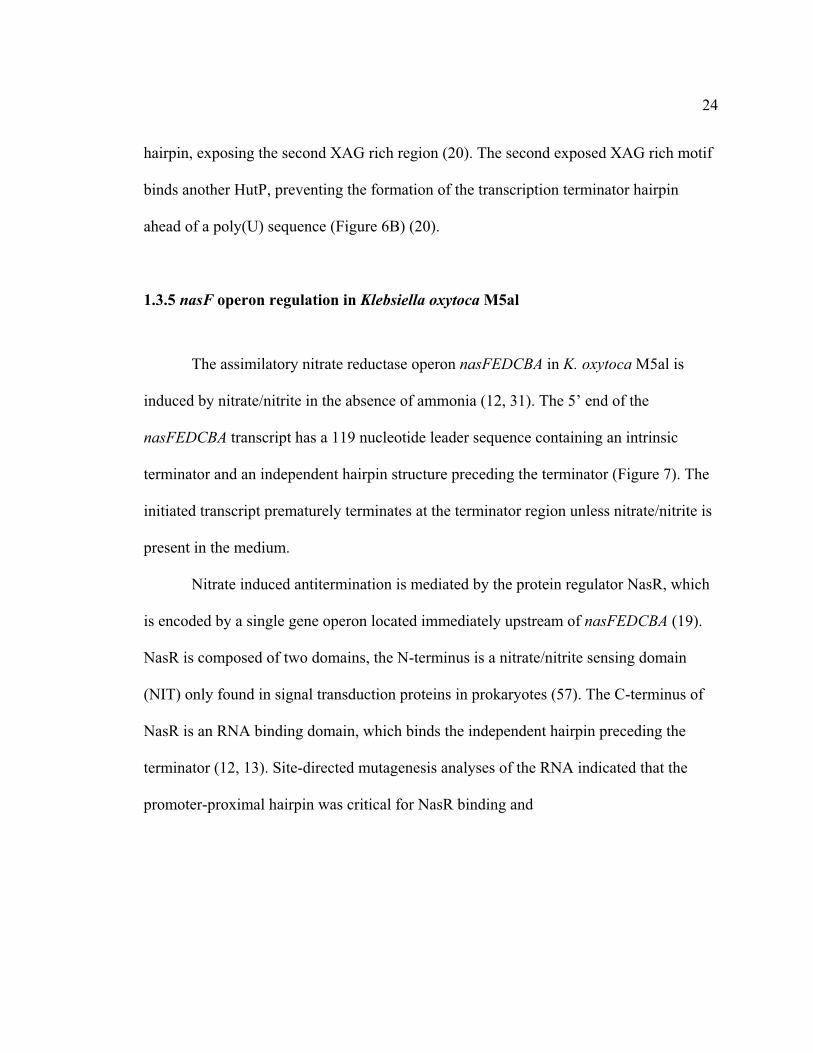
hairpin, exposing the second XAG rich region (20). The second exposed XAG rich motif
binds another HutP, preventing the formation of the transcription terminator hairpin
ahead of a poly(U) sequence (Figure 6B) (20).
1.3.5 nasF operon regulation in Klebsiella oxytoca M5al
The assimilatory nitrate reductase operon nasFEDCBA in K. oxytoca M5al is
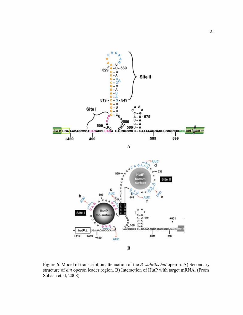
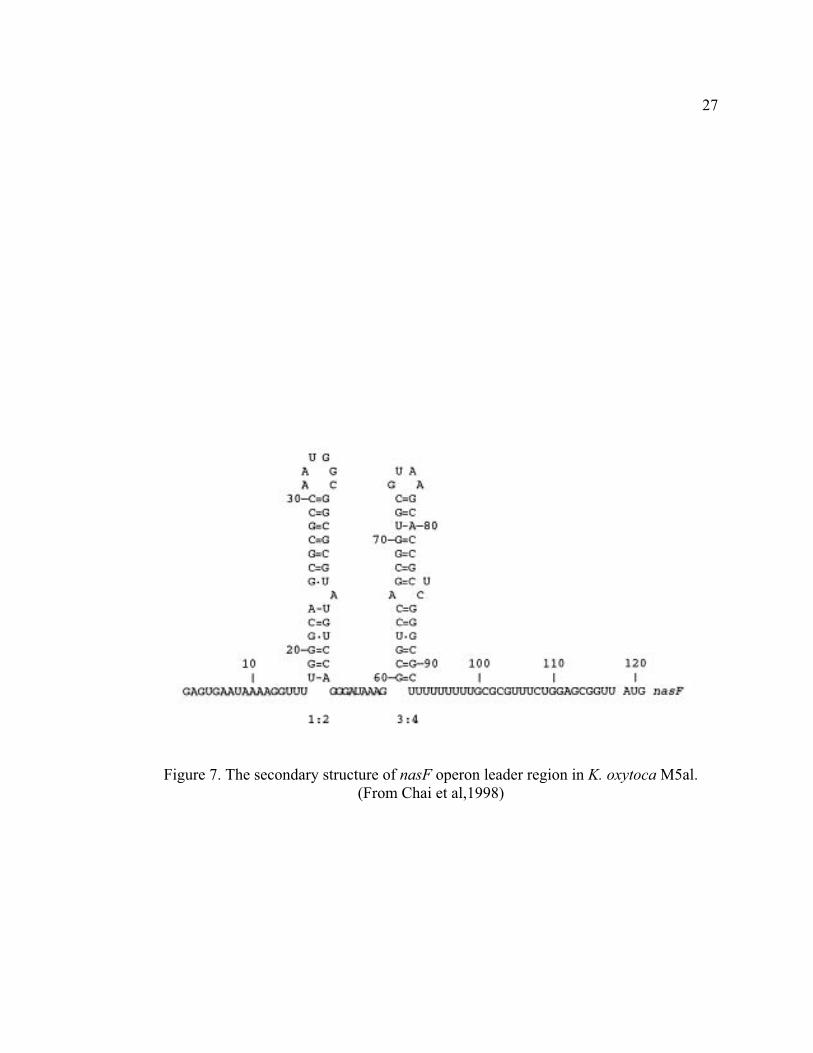
induced by nitrate/nitrite in the absence of ammonia (12, 31). The 5’ end of the
nasFEDCBA transcript has a 119 nucleotide leader sequence containing an intrinsic
terminator and an independent hairpin structure preceding the terminator (Figure 7). The
initiated transcript prematurely terminates at the terminator region unless nitrate/nitrite is
present in the medium.
Nitrate induced antitermination is mediated by the protein regulator NasR, which
is encoded by a single gene operon located immediately upstream of nasFEDCBA (19).
NasR is composed of two domains, the N-terminus is a nitrate/nitrite sensing domain
(NIT) only found in signal transduction proteins in prokaryotes (57). The C-terminus of
NasR is an RNA binding domain, which binds the independent hairpin preceding the
terminator (12, 13). Site-directed mutagenesis analyses of the RNA indicated that the
promoter-proximal hairpin was critical for NasR binding and
25
A
B
Figure 6. Model of transcription attenuation of the B. subtilis hut operon. A) Secondary structure of hut operon leader region. B) Interaction of HutP with target mRNA. (From Subash et al, 2008)

26
antitermination function (12). Although NasR can bind to the leader RNA, the activation
of NasR requires the binding of nitrate/nitrite at the NIT domain (13). The exact
molecular mechanism of NasR mediated antitermination is not clear, available evidence
suggests that the formation of alternative hairpin structures is not involved (13).
1.3.6 AmiR-directed antitermination in Pseudomonas aeruginosa
P. aeruginosa PAC1 can grow on short-chain aliphatic amides by virtue of an
amidase encoded by the amiEBCRS operon (62, 63). The protein products of amiCR are
not involved in amide catabolism, but constitute an amide sensor-antiterminator two-
component regulatory system that autoregulates ami operon expression.
The ami transcript leader region contains a 5’ end hairpin and a 3’end intrinsic
terminator (Figure 8) (63). Nascent mRNA elongation aborts at the terminator region
when there is no amide source in the medium. Premature transcription termination is
prevented by the protein regulator AmiR when amide is supplied. AmiR consists of a C-
terminal RNA-binding domain and an N-terminal domain homologous to receiver
domains of the response regulator of two-component regulatory systems (46). The results
of titration assays suggest that AmiR binds at 35-74 residues of the ami leader region
(Figure 8) (64). Whether AmiR-mediated antitermination involves alternative secondary
structure formation is unknown.
27
Figure 7. The secondary structure of nasF operon leader region in K. oxytoca M5al.
(From Chai et al,1998)

28
Signal transduction between AmiC and AmiR occurs through steric hindrance
(44). In the absence of amide, AmiC and AmiR form a tetrameric complex composed of
an AmiC dimer and an AmiR dimer. AmiR in the complex has no regulatory function.
When amide is available, AmiC binds to the amide and dissociates from the AmiC·AmiR
complex. The released AmiR dimer acts within the ami leader region to help the
elongation polymerase read through the intrinsic terminator structure (44).

29
A
Figure 8. The secondary structure of ami operon leader region in P. aeruginosa. (From Wilson et al, 1996)
30
1. 4. Global nitrogen regulation in proteobacteria
In proteobacteria, the global nitrogen regulation (Ntr) system coordinates cellular
nitrogen metabolism (39). The Ntr system is composed of a PII signal transduction protein
(GlnB), an uridylyltransferase/UMP-removing enzyme (GlnD), and a two-component
regulatory system NtrB/NtrC. NtrB is a sensor kinase protein, and NtrC is a �54-
dependent transcription activator. NtrB regulates NtrC activity in response to a signal
from the PII protein.
PII protein regulates NtrB activity through protein-protein interaction (50). PII
itself is regulated by GlnD through covalent modification (23). GlnD is an intracellular
nitrogen sensor that senses glutamine levels. When nitrogen is depleted, cellular
glutamine levels become low, and PII is uridylylated by GlnD. PII-UMP is not able to
bind to NtrB. Under such conditions, NtrB phosphorylates NtrC and activates its
regulatory function. Phosphorylated NtrC (NrC-P) activates transcription initiation of its
target operons, including those related to nitrate assimilation. When nitrogen is sufficient,
glutamine binds GlnD and triggers its UMP-removing activity, leading to PII-UMP
deuridylylation. PII interacts with NtrB and promotes NtrB to dephosphorylate NtrC-P.
NtrC without phosphate modification does not have activator function.
NtrC recognizes a conserved dyad symmetry sequence 5’-
TGCACCA(N)3TGGTGCA-3’ sequence 100 to 120 nucleotides upstream of the
regulated promoter (31). NtrC-mediated activation, however, needs direct interaction
between NtrC and �54-RNA polymerase holoenzyme. NtrC is brought to the holoenzyme

31
through a DNA-loop structure (59). When in contact, NtrC converts the DNA-RNA
polymerase complex from a closed state to an open state in which DNA strands at the
transcription initiation region melt, allowing transcription to initiate (4).
NtrC is composed of three domains (52). The N-terminus is a signal receiver
domain, which has a set of conserved amino acids involved signal transduction and
phosphorylation. The central domain is an AAA+ domain involved in ATP binding,
hydrolysis, and interaction with RNA polymerase. The C-terminus is a typical helix-turn-
helix DNA-binding domain.
A. vinelandii harbors glnD; ntrB and ntrC; and glnK, a homolog of glnB (25). Our
understanding of the functions of GlnK and GlnD comes from studies of nitrogen fixation
regulation. GlnK is de-uridylylated by GlnD when nitrogen is sufficient, and uridylylated
when nitrogen is depleted. Although the signal transduction from GlnK to NtrB/NtrC has
not been studied thoroughly, it is assumed that NtrC in A. vinelandii is regulated in an
analogous fashion to its regulation in enteric bacteria (21).

32
.
Figure 9. Ntr system in enteric bacteria. (Adapted from Masepohl and Forchhammer,
2007)

33
1. 5 NifL and NifA in A. vinelandii
The nitrogen fixation (nif) operons in A. vinelandii are regulated by a non-typical
two-component regulatory system, NifL/NifA. The NifL/NifA system regulates nif
transcription in response to cellular redox, nitrogen, and carbohydrate status (55). Both
NifL and NifA have modular structures with domains sensing specific signals (Figure 10)
(37).
NifL has an N-terminal PAS (period circadian protein, Ah receptor nuclear
translocator protein, single-minded protein) domain with FAD as the prosthetic group
that senses cellular redox status (58, 68). The NifL C-terminal domain is homologous to
the autophosphorylation domain of sensor kinases of two-component regulatory system
with the conserved histidine residue where phosphate is attached. In contrast with other
sensor kinases, substitution mutation of the histidine residue does not affect its regulatory
function. This domain can bind ADP or ATP, the affinity for ADP is 10-fold higher than
for ATP, suggesting that this domain senses cellular ADP: ATP ratios (58).
NifA has a central AAA+ domain and C-terminal helix-turn-helix DNA binding
domain, similar to the corresponding parts of NtrC (11, 40). The N-terminus of NifA is a
GAF domain (cGMP-specific and –stimulated phosphodiesterases, Anabaena adenylate
cyclases and E. coli FhlA) which can bind 2-oxoglutarate, sensing cellular carbohydrate
status (3, 55).
NifA is a �54-dependent transcription activator, and recognizes a consensus
sequence 5’-TCT-N10-AGA-3’ located 100 to 200 nucleotides upstream of NifA-

34
regulated promoters (9). Like NtrC, NifA contacts �54-RNA polymerase holoenzyme
through a DNA-loop (10).
NifL regulates NifA activity through protein-protein interactions (4). When bound
by NifL, NifA has no activator function. In A. vinelandii, NifL-NifA interactions are
promoted by GlnK (34) . Thus, the Ntr global nitrogen system regulates nif expression
through the NifL/NifA system (Figure 10)

35
NifL
NifA
NifL
PAS domain Kinase-like domain
GAF domain Catalytic domain DNA-binding domain
N
N
C
C
Figure 10. Modular structures of NifL and NifA. (Adapted from Dixon, 2002)

36
1.6 Nitrate transporters and the regulation of assimilatory nitrate reduction in
bacteria
Nitrate needs to be transported into the cell as assimilatory nitrate reduction
occurs in the cytoplasm. There are two types of nitrate transporters in bacteria (41). The
first type is the ABC-type nitrate transporters, which import nitrate at the expense of ATP
hydrolysis. This type of nitrate transporter has been identified in Klebsiella (32), and the
cyanobacteria Synechococcus (47), Synechocystis (24), and Anabaena (17, 18). The
second type of nitrate transporters is permeases belonging to the major facilitator
superfamily (MFS). These MFS nitrate permeases rely on transmembrane proton motive
force (PMF) for their activity. One example of this type transporter is the Bacillus subtilis
nitrate transporter (45).
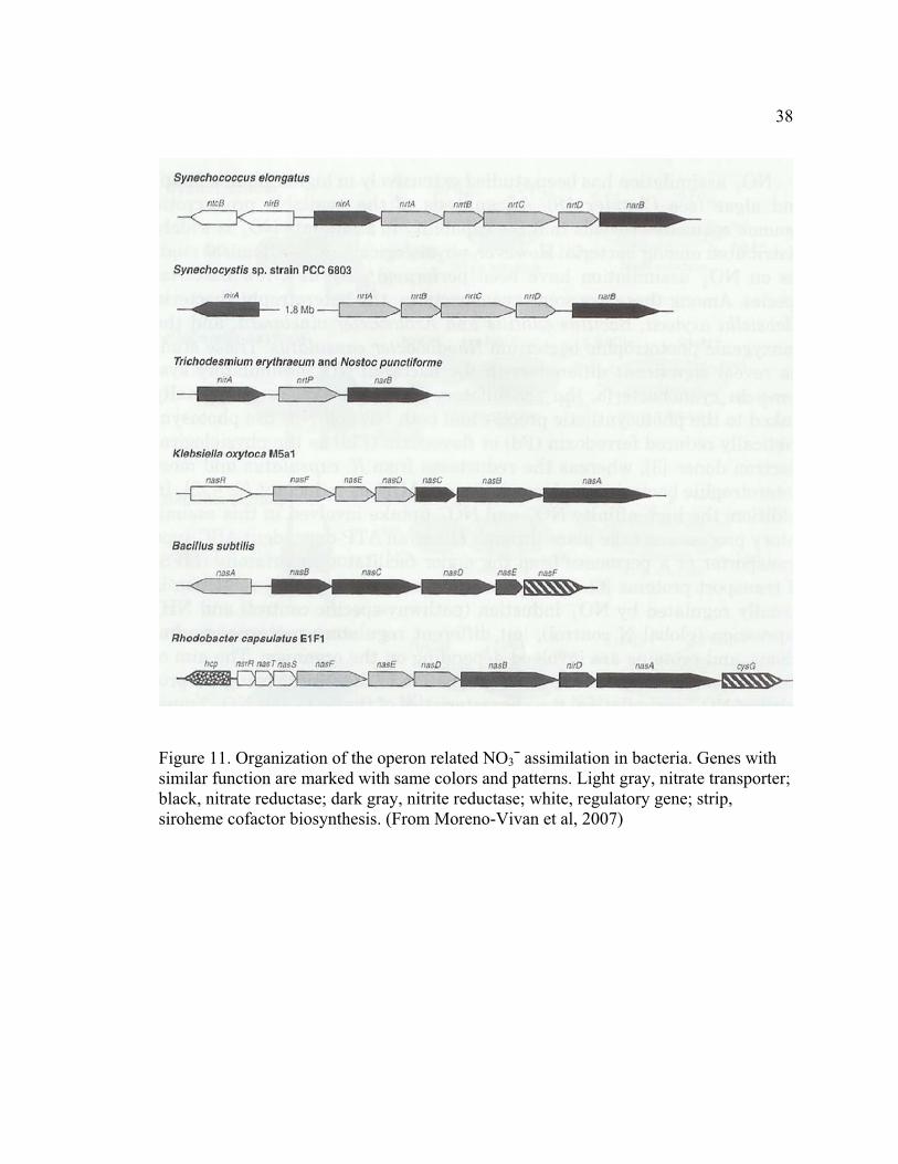
Genes related to nitrate transport, reduction, and their regulations are clustered on
the bacterial genome (Figure 11) (42). Expression of assimilatory nitrate reductases are
subject to global nitrogen regulation (NH3 repression) and operon-specific induction
(nitrate/nitrite induction) (30). For each operon, the regulatory proteins and molecular
mechanisms involved in its regulation may be unique.
In the enteric bacterium K. oxytoca 51, the assimilatory nitrate transporter genes,
nitrate reductase, and nitrite reductase genes are encoded by the operon nasFEDCBA
(32). The nasFED genes encode an ABC-type nitrate transporter, while nasC and nasA
encode nitrate reductase, and nasB encodes nitrate reductase.

37
The nasFEDCBA operon is subject to two levels of control. The general nitrogen
regulation system regulates operon transcription initiation through NtrC. NtrC regulates
operon transcription in response to intracellular nitrogen status. In the presence of
ammonia, the promoter of nasFEDCBA is inhibited due to the inactivation of NtrC; in the
absence of ammonia, NtrC is phosphorylated by NtrB and activates transcription
initiation of the operon. Expression of nasFEDCBA requires the presence of nitrate or
nitrite in the medium. Nitrate/nitrite induction on nasFEDCBA is mediated by an
antiterminator protein NasR which acts positively within the operon leader region (The
details of nitrate/nitrite induction are discussed at section 1.3.5).
38
Figure 11. Organization of the operon related NO3¯ assimilation in bacteria. Genes with similar function are marked with same colors and patterns. Light gray, nitrate transporter; black, nitrate reductase; dark gray, nitrite reductase; white, regulatory gene; strip, siroheme cofactor biosynthesis. (From Moreno-Vivan et al, 2007)
39
1.7 Regulation of the A. vinelandii Assimilatory Nitrate Reductase Operon nasAB
In addition to atmospheric nitrogen (N2), A. vinelandii can use ammonia (NH3),
nitrite (NO2¯), and nitrate (NO3¯) as nitrogen sources. Nitrate is converted into nitrite, and
then into ammonia before being incorporated into glutamate by glutamine synthetase
(GS). Assimilatory nitrate reduction is the only nitrate reduction pathway in A. vinelandii.
The assimilatory nitrate reductase and nitrite reductase are encoded by the nasB and nasA
genes of the nasAB operon, respectively (51). Expression of nasAB is affected by
multiple factors. Ammonia was shown to repress nasAB expression (35). It is assumed
that ammonia inhibition of nasAB expression was a type of global nitrogen regulation,
mediated by NtrC. This assumption is based on the observation that mutation of either
ntrC or the alternate sigma factor gene ntrA (�54) abolished nasAB expression (51, 53,
60), suggesting that nasAB may be driven by an NtrC-dependent promoter.
Nitrate or nitrite induces nasAB expression in the absence of ammonia (35).
Nitrate and nitrite induction on nasAB expression also involve the products of the nasST
operon (22). The nasST operon is located 12,130 bp upstream of nasAB. The deduced
protein NasS shares sequence homology with nitrate transporters in Klebsiella spp. and
Synechococcus spp. (22). However, deletion of nasS had no effect on nitrate uptake by A.
vinelandii. Mutation of nasS without affecting the expression of downstream nasT led to
constitutive expression of the nasAB operon independent of nitrogen source, suggesting
that NasS negatively affected nasAB expression.
40
NasT is a 192-amino-acid polypeptide consisting of two domains (22, 56). The C-
terminal domain of NasT is highly homologous to an RNA-binding domain, first
identified in the C-termini of AmiR and NasR, antitermination regulators present in
Pseudomonas aeruginosa and Klebsiella spp. respectively (56). The NasT N-terminal
domain is homologous to the receiver domain of the response regulator of classic two-
component regulatory systems (22, 43). Mutation of nasT abolished nasAB expression
even in the presence of nitrate and nitrite, indicating that a functional NasT was required
for nasAB expression (22). Possible regulatory roles for NasT in nasAB expression and
the role of the N-terminal domain in NasT activation have been hypothesized. However,
these hypotheses have not been examined in detail and the regulation of nasAB is unclear
(30, 43, 49). One of the goals of this research was to shed further insight into this
regulation.

41
Figure 12. Features known or hypothesized to be involved in the regulation of nasAB
operon in A. vinelandii.

42
Figure 13. Alignment of NasT, AmiR, and NasR
43
1.8 Dissertation Format
All of the research pertaining to this dissertation is presented in two manuscripts
submitted for publication. The goal of the research was to elucidate the molecular
mechanisms controlling the A. vinelandii assimilatory nitrate reductase operon nasAB.
1.8.1 Antitermination Regulation of the Azotobacter vinelandii Assimilatory Nitrate
Reductase Operon nasAB (Appendix A)
This first manuscript focuses on the identification of the nasAB promoter and
testing whether antitermination is involved in nasAB regulation. The results of
bioinformatic analysis suggest a putative �54 recognition site and two NtrC binding sites
are separated from the structural gene nasA by a potential intrinsic terminator structure.
To dissect the roles of these sequences in nasAB regulation, I constructed a
transcriptional lacZ fusion vector and a translational lacZ fusion vector. The translational
probe vector was used to test the effects of sequence mutations on nasAB expression; the
transcriptional probe vector was used to test nasAB promoter activity altered in aspects of
nitrogen regulation.

44
1.8.2 NasS/NasT, a non-canonical two-component regulatory system regulates the
assimilatory nitrate reductase operon (nasAB) in Azotobacter vinelandii via
antitermination (Appendix B)
The second manuscript focuses on the regulatory role of NasT and signal
transduction between NasT and its cognate partner NasS. NasT is homologous to AmiR,
an experimentally confirmed RNA-binding antiterminator. I constructed a bacterial two-
vector system to test whether NasT regulates within the nasAB leader region, and used a
yeast two-hybrid system to test whether NasT forms a dimer, analogous to AmiR. At the
same time, I analyzed the effect of NasS: NasT ratio alterations on nasAB expression.

45
2. PRESENT STUDY
The methods, results, and conclusions of this dissertation are presented in the
attached appendices. The following sections summarize the important findings of each
appendix.
2.1 Antitermination Regulation of the Azotobacter vinelandii Assimilatory Nitrate
Reductase Operon nasAB (Appendix A).
1. A potential �54 recognition site, two potential NtrC recognition sites, and a putative Rho-independent terminator structure were identified upstream of the nasA gene. 2. Secondary structure analysis of the nasAB leader sequence suggests an intrinsic transcriptional terminator structure upstream of nasA. 3. The potential �54 recognition site is essential for nasAB expression. 4. Both NtrC recognition sites are essential for nasAB expression. 5. The putative terminator structure upstream of nasA plays a negative role in nasAB expression. 6. The promoter proximal hairpin upstream of the terminator plays a positive role in nasAB expression. 7. nasAB regulation is subject to global nitrogen regulation. 8. Mutation of nifA affects nasAB transcription.

46
2.2 NasS/NasT, a non-canonical two-component regulatory system regulates nitrate
reductase operon (nasAB) in Azotobacter vinelandii via antitermination
1. Mutation of nasST in A. vinelandii is not lethal.
2. NasT acts within the nasAB leader region as an antiterminator.
3. NasT exists as a dimer or oligomer.
4. Proper stoichiometry of NasS and NasT is essential for nasAB regulation.
5. nasS and nasT are translationally coupled.
6. The assimilatory nitrate reductase operons in A. vinelandii and K. oxytoca
(pneumomiae) M5al use different signal circuits to regulate antitermination.
2.3 Concluding Remarks and Future Directions
A. vinelandii has served as a paradigm for the genetic study of nitrogen fixation in
free-living bacteria for more than thirty years. The molecular mechanism that A.
vinelandii uses to sense the intracellular nitrogen status has been characterized; the signal
cascades that the bacterium uses to regulate nif gene expression in response to nitrogen
status change have been elucidated. Despite these achievements, N-regulation other than
the nitrogen fixation pathway is largely unknown.
Results from this work confirmed that regulation of the assimilatory nitrate
reductase operon nasAB in A. vinelandii is subject to both general nitrogen regulation and
nitrate/nitrite induction (Figure 14). The global nitrogen regulatory system regulates

47
nasAB transcription via NtrC, a �54-dependent activator, in response to the availability of
ammonia in the medium. This regulation was observed in the Nif – strain UW1. In the
Nif + strain UW136, the activity of the nasAB promoter, however, is inhibited even in the
absence of ammonia, suggesting that the activity of NtrC is modulated in UW136 or NtrC
may not be the only regulator acting at the nasAB promoter region.
Strain UW1 differs from wild type strain UW136 in that it harbors a mutation in
the nifA structural gene, indicating that NifA may be involved in nasAB repression.
However, the conserved NifA recognition sequence (TGT-N10-ACA) was not identified
within the nasAB promoter region. One question still to be addressed is whether NifA
acts directly within the nasAB promoter region. An approach to test this could use an E.
coli (lacZ –) two-vector system: one vector contains an inducible nifA gene, and the other
vector has the nasAB promoter–lacZ transcriptional fusion.
Further elucidation of the role of NtrC in A. vinelandii nitrogen regulation would
clarify the role of the general nitrogen regulatory system in nitrate assimilation. NtrC is
known to play an essential role in nitrogen regulation in enteric bacteria. Although NtrC
is not involved in nitrogen fixation regulation, it is assumed that A. vinelandii NtrC plays
an essential role in nitrogen metabolisms other than nitrogen fixation. Since atmospheric
nitrogen represents a more accessible and sustainable nitrogen source than other nitrogen
sources for A. vinelandii, the activity of NtrC may be modulated in response to the status
of nitrogen fixation. The phosphorylation/dephosphorylation of NtrC under different
cellular metabolism conditions can be identified using two-dimensional gel
48
electrophoresis. The results of works may shine light on the still unknown signal
transduction cascades in A. vinelandii.
Nitrate/nitrite regulation involves antitermination within the nasAB leader region.
The leader terminator structure regulates initiated transcripts negatively in the absence of
nitrate/nitrite. Results from my research (Appendix B) confirmed that NasT is required to
overcome the negative role of the leader terminator. My results and the modular structure
of NasT suggest that it may act as an RNA-binding antiterminator. However, the
interaction of NasT with the nasAB leader mRNA needs to be demonstrated directly
using RNA electrophoretic mobility shift assays.
The results of secondary structure analysis suggest that antitermination may not
involve the formation of alternative secondary structures at the nasAB leader region
(Appendix A). One possibility is that NasT interacts with hairpins I and II within the
leader region and modulates the activity of RNA polymerase to overcome the negative
role of the terminator hairpin structure. A strategy to test this hypothesis is to analyze
whether hairpins I and II cause RNA polymerase to read through heterologous terminator
structures fused on the same sequence.
NasS appears to regulate the activity of NasT in response to the availability of
nitrate or nitrite. The structures of the two proteins suggest that signal transduction
between them is not via phosphorylation. The requirement of conserved stoichiometry of
the two proteins for nasAB regulation supports a model that NasS regulates NasT activity
through protein-protein interactions. However, a direct demonstration of this interaction

49
is still needed. Co-immunoprecipitation assays of the two proteins or a yeast two-hybrid
system approach would provide direct evidence of this interaction.

50
Figure 14. Proposed model for nasAB regulation in A. vinelandii.

51
REFERENCES
1. Antson, A. A., E. J. Dodson, G. Dodson, R. B. Greaves, X. Chen, and P. Gollnick. 1999. Structure of the trp RNA-binding attenuation protein, TRAP, bound to RNA. Nature 401:235-242.
2. Antson, A. A., J. Otridge, A. M. Brzozowski, E. J. Dodson, G. G. Dodson, K. S. Wilson, T. M. Smith, M. Yang, T. Kurecki, and P. Gollnick. 1995. The structure of trp RNA-binding attenuation protein. Nature 374:693-700.
3. Aravind, L. and C. P. Ponting. 1997. The GAF domain: an evolutionary link between diverse phototransducing proteins. Trends Biochem. Sci. 22:458-459.
4. Austin, S., M. Buck, W. Cannon, T. Eydmann, and R. Dixon. 1994. Purification and in vitro activities of the native nitrogen fixation control proteins NifA and NifL. J. Bacteriol. 176:3460-3465.
5. Aymerich, S. and M. Steinmetz. 1992. Specificity determinants and structural features in the RNA target of the bacterial antiterminator proteins of the BglG/SacY family. Proc. Natl. Acad. Sci. U. S. A. 89:10410-10414.
6. Babitzke, P. and C. Yanofsky. 1993. Reconstitution of Bacillus subtilis trp attenuation in vitro with TRAP, the trp RNA-binding attenuation protein. Proc. Natl. Acad. Sci. U. S. A. 90:133-137.
7. Babitzke, P., J. T. Stults, S. J. Shire, and C. Yanofsky. 1994. TRAP, the trp RNA-binding attenuation protein of Bacillus subtilis, is a multisubunit complex that appears to recognize G/UAG repeats in the trpEDCFBA and trpG transcripts. J. Biol. Chem. 269:16597-16604.
8. Barbolina, M. V., X. Li, and P. Gollnick. 2005. Bacillus subtilis TRAP binds to its RNA target by a 5' to 3' directional mechanism. J. Mol. Biol. 345:667-679.
9. Buck, M., W. Cannon, and J. Woodcock. 1987. Mutational analysis of upstream sequences required for transcriptional activation of the Klebsiella pneumoniae nifH promoter. Nucleic Acids Res. 15:9945-9956.
10. Buck, M., W. Cannon, and J. Woodcock. 1987. Transcriptional activation of the Klebsiella pneumoniae nitrogenase promoter may involve DNA loop formation. Mol. Microbiol. 1:243-249.

52
11. Buck, M., M. T. Gallegos, D. J. Studholme, Y. Guo, and J. D. Gralla. 2000. The bacterial enhancer-dependent sigma(54) (sigma(N)) transcription factor. J. Bacteriol. 182:4129-4136.
12. Chai, W. and V. Stewart. 1999. RNA sequence requirements for NasR-mediated, nitrate-responsive transcription antitermination of the Klebsiella oxytoca M5al nasF operon leader. J. Mol. Biol. 292:203-216.
13. Chai, W. and V. Stewart. 1998. NasR, a novel RNA-binding protein, mediates nitrate-responsive transcription antitermination of the Klebsiella oxytoca M5al nasF operon leader in vitro. J. Mol. Biol. 283:339-351.
14. Chen, X., A. A. Antson, M. Yang, P. Li, C. Baumann, E. J. Dodson, G. G. Dodson, and P. Gollnick. 1999. Regulatory features of the trp operon and the crystal structure of the trp RNA-binding attenuation protein from Bacillus stearothermophilus. J. Mol. Biol. 289:1003-1016.
15. Declerck, N., H. Dutartre, V. Receveur, V. Dubois, C. Royer, S. Aymerich, and H. van Tilbeurgh. 2001. Dimer stabilization upon activation of the transcriptional antiterminator LicT. J. Mol. Biol. 314:671-681.
16. Efuet, E. T., L. Pulakat, and N. Gavini. 1996. Investigations on the cell volumes of Azotobacter vinelandii by scanning electron microscopy. J. Basic Microbiol. 36:229-234.
17. Frias, J. E., A. Herrero, and E. Flores. 2003. Open reading frame all0601 from Anabaena sp. strain PCC 7120 represents a novel gene, cnaT, required for expression of the nitrate assimilation nir operon. J. Bacteriol. 185:5037-5044.
18. Frias, J. E., E. Flores, and A. Herrero. 1997. Nitrate assimilation gene cluster from the heterocyst-forming cyanobacterium Anabaena sp. strain PCC 7120. J. Bacteriol. 179:477-486.
19. Goldman, B. S., J. T. Lin, and V. Stewart. 1994. Identification and structure of the nasR gene encoding a nitrate- and nitrite-responsive positive regulator of nasFEDCBA (nitrate assimilation) operon expression in Klebsiella pneumoniae M5al. J. Bacteriol. 176:5077-5085.
20. Gopinath, S. C., D. Balasundaresan, T. Kumarevel, T. S. Misono, H. Mizuno, and P. K. Kumar. 2008. Insights into anti-termination regulation of the hut operon in Bacillus subtilis: importance of the dual RNA-binding surfaces of HutP. Nucleic Acids Res.

53
21. Gutierrez, J. C., E. Santero, and M. Tortolero. 1997. Ammonium repression of the nitrite-nitrate (nasAB) assimilatory operon of Azotobacter vinelandii is enhanced in mutants expressing the nifO gene at high levels. Mol. Gen. Genet. 255:172-179.
22. Gutierrez, J. C., F. Ramos, L. Ortner, and M. Tortolero. 1995. nasST, two genes involved in the induction of the assimilatory nitrite-nitrate reductase operon (nasAB) of Azotobacter vinelandii. Mol. Microbiol. 18:579-591.
23. Kamberov, E. S., M. R. Atkinson, P. Chandran, and A. J. Ninfa. 1994. Effect of mutations in Escherichia coli glnL (ntrB), encoding nitrogen regulator II (NRII or NtrB), on the phosphatase activity involved in bacterial nitrogen regulation. J. Biol. Chem. 269:28294-28299.
24. Kaneko, T., S. Sato, H. Kotani, A. Tanaka, E. Asamizu, Y. Nakamura, N. Miyajima, M. Hirosawa, M. Sugiura, S. Sasamoto, T. Kimura, T. Hosouchi, A. Matsuno, A. Muraki, N. Nakazaki, K. Naruo, S. Okumura, S. Shimpo, C. Takeuchi, T. Wada, A. Watanabe, M. Yamada, M. Yasuda, and S. Tabata. 1996. Sequence analysis of the genome of the unicellular cyanobacterium Synechocystis sp. strain PCC6803. II. Sequence determination of the entire genome and assignment of potential protein-coding regions. DNA Res. 3:109-136.
25. Kennedy, C. and P. E. Bishop. 2004. Genetics of Nitrogen Fixation and Related Aspects of Metabolism in Species of Azotobacter: History and Current Status, p. 27-46. In W. Klipp, B. Masepohl, J. R. Gallon and W. E. Newton (ed.), Genetics and Regulation of Nitrogen Fixation in Free-Living Bacteria, vol. 2. Springer Netherlands, Netherland.
26. Kumar, P. K., T. Kumarevel, and H. Mizuno. 2006. Structural basis of HutP-mediated transcription anti-termination. Curr. Opin. Struct. Biol. 16:18-26.
27. Kumarevel, T., H. Mizuno, and P. K. Kumar. 2005. Structural basis of HutP-mediated anti-termination and roles of the Mg2+ ion and L-histidine ligand. Nature 434:183-191.
28. Kumarevel, T., Z. Fujimoto, P. Karthe, M. Oda, H. Mizuno, and P. K. Kumar. 2004. Crystal structure of activated HutP; an RNA binding protein that regulates transcription of the hut operon in Bacillus subtilis. Structure 12:1269-1280.
29. Kuroda, M. I., D. Henner, and C. Yanofsky. 1988. cis-acting sites in the transcript of the Bacillus subtilis trp operon regulate expression of the operon. J. Bacteriol. 170:3080-3088.
30. Lin, J. T. and V. Stewart. 1998. Nitrate assimilation by bacteria. Adv. Microb. Physiol. 39:1-30, 379.

54
31. Lin, J. T. and V. Stewart. 1996. Nitrate and nitrite-mediated transcription antitermination control of nasF (nitrate assimilation) operon expression in Klebsiella pheumoniae M5al. J. Mol. Biol. 256:423-435.
32. Lin, J. T., B. S. Goldman, and V. Stewart. 1994. The nasFEDCBA operon for nitrate and nitrite assimilation in Klebsiella pneumoniae M5al. J. Bacteriol. 176:2551-2559.
33. Lindner, C., A. Galinier, M. Hecker, and J. Deutscher. 1999. Regulation of the activity of the Bacillus subtilis antiterminator LicT by multiple PEP-dependent, enzyme I- and HPr-catalysed phosphorylation. Mol. Microbiol. 31:995-1006.
34. Little, R., I. Martinez-Argudo, and R. Dixon. 2006. Role of the central region of NifL in conformational switches that regulate nitrogen fixation. Biochem. Soc. Trans. 34:162-164.
35. Luque, F., E. Santero, J. R. Medina, and M. Tortolero. 1987. Mutants of Azotobacter vinelandii altered in the regulation of nitrate. Arch. Microbiol. 148:231-235.
36. Maldonado, R., J. Jimenez, and J. Casadesus. 1994. Changes of ploidy during the Azotobacter vinelandii growth cycle. J. Bacteriol. 176:3911-3919.
37. Martinez-Argudo, I., R. Little, N. Shearer, P. Johnson, and R. Dixon. 2004. The NifL-NifA System: a multidomain transcriptional regulatory complex that integrates environmental signals. J. Bacteriol. 186:601-610.
38. McElroy, C., A. Manfredo, A. Wendt, P. Gollnick, and M. Foster. 2002. TROSY-NMR studies of the 91kDa TRAP protein reveal allosteric control of a gene regulatory protein by ligand-altered flexibility. J. Mol. Biol. 323:463-473.
39. Merrick, M. J. and R. A. Edwards. 1995. Nitrogen control in bacteria. Microbiol. Rev. 59:604-622.
40. Missaillidis, S., M. Jaseja, P. Ray, R. Chittock, C. W. Wharton, A. F. Drake, M. Buck, and E. I. Hyde. 1999. Secondary structure of the C-terminal DNA-binding domain of the transcriptional activator NifA from Klebsiella pneumoniae: spectroscopic analyses. Arch. Biochem. Biophys. 361:173-182.
41. Moir, J. W. and N. J. Wood. 2001. Nitrate and nitrite transport in bacteria. Cell Mol. Life Sci. 58:215-224.
42. Moreno-Vivian, C. and E. Flores. 2007. Nitrate Assimilation in Bacteria, p. 263-282. In H. Bothe, S. J. Ferguson and W. E. Newton (ed.), Biology of the nitrogen cycle. Elsevier, Amsterdam ; Boston.

55
43. Morth, J. P., V. Feng, L. J. Perry, D. I. Svergun, and P. A. Tucker. 2004. The crystal and solution structure of a putative transcriptional antiterminator from Mycobacterium tuberculosis. Structure 12:1595-1605.
44. Norman, R. A., C. L. Poh, L. H. Pearl, B. P. O'Hara, and R. E. Drew. 2000. Steric hindrance regulation of the Pseudomonas aeruginosa amidase operon. J. Biol. Chem. 275:30660-30667.
45. Ogawa, K., E. Akagawa, K. Yamane, Z. W. Sun, M. LaCelle, P. Zuber, and M. M. Nakano. 1995. The nasB operon and nasA gene are required for nitrate/nitrite assimilation in Bacillus subtilis. J. Bacteriol. 177:1409-1413.
46. O'Hara, B. P., R. A. Norman, P. T. Wan, S. M. Roe, T. E. Barrett, R. E. Drew, and L. H. Pearl. 1999. Crystal structure and induction mechanism of AmiC-AmiR: a ligand-regulated transcription antitermination complex. EMBO J. 18:5175-5186.
47. Omata, T. 1995. Structure, function and regulation of the nitrate transport system of the cyanobacterium Synechococcus sp. PCC7942. Plant Cell Physiol. 36:207-213.
48. Pau, R. N., L. A. Mitchenall, and R. L. Robson. 1989. Genetic evidence for an Azotobacter vinelandii nitrogenase lacking molybdenum and vanadium. J. Bacteriol. 171:124-129.
49. Pino, C., F. Olmo-Mira, P. Cabello, M. Martinez-Luque, F. Castillo, M. D. Roldan, and C. Moreno-Vivian. 2006. The assimilatory nitrate reduction system of the phototrophic bacterium Rhodobacter capsulatus E1F1. Biochem. Soc. Trans. 34:127-129.
50. Pioszak, A. A., P. Jiang, and A. J. Ninfa. 2000. The Escherichia coli PII signal transduction protein regulates the activities of the two-component system transmitter protein NRII by direct interaction with the kinase domain of the transmitter module. Biochemistry 39:13450-13461.
51. Ramos, F., G. Blanco, J. C. Gutierrez, F. Luque, and M. Tortolero. 1993. Identification of an operon involved in the assimilatory nitrate-reducing system of Azotobacter vinelandii. Mol. Microbiol. 8:1145-1153.
52. Rombel, I., A. North, I. Hwang, C. Wyman, and S. Kustu. 1998. The bacterial enhancer-binding protein NtrC as a molecular machine. Cold Spring Harb. Symp. Quant. Biol. 63:157-166.
53. Santero, E., F. Luque, J. R. Medina, and M. Tortolero. 1986. Isolation of ntrA-like mutants of Azotobacter vinelandii. J. Bacteriol. 166:541-544.
56
54. Schilling, O., C. Herzberg, T. Hertrich, H. Vorsmann, D. Jessen, S. Hubner, F. Titgemeyer, and J. Stulke. 2006. Keeping signals straight in transcription regulation: specificity determinants for the interaction of a family of conserved bacterial RNA-protein couples. Nucleic Acids Res. 34:6102-6115.
55. Schmitz, R. A., K. Klopprogge, and R. Grabbe. 2002. Regulation of nitrogen fixation in Klebsiella pneumoniae and Azotobacter vinelandii: NifL, transducing two environmental signals to the nif transcriptional activator NifA. J. Mol. Microbiol. Biotechnol. 4:235-242.
56. Shu, C. J. and I. B. Zhulin. 2002. ANTAR: an RNA-binding domain in transcription antitermination regulatory proteins. Trends Biochem. Sci. 27:3-5.
57. Shu, C. J., L. E. Ulrich, and I. B. Zhulin. 2003. The NIT domain: a predicted nitrate-responsive module in bacterial sensory receptors. Trends Biochem. Sci. 28:121-124.
58. Soderback, E., F. Reyes-Ramirez, T. Eydmann, S. Austin, S. Hill, and R. Dixon. 1998. The redox- and fixed nitrogen-responsive regulatory protein NIFL from Azotobacter vinelandii comprises discrete flavin and nucleotide-binding domains. Mol. Microbiol. 28:179-192.
59. Su, W., S. Porter, S. Kustu, and H. Echols. 1990. DNA-looping and enhancer activity: association between DNA-bound NtrC activator and RNA polymerase at the bacterial glnA promoter. Proc. Natl. Acad. Sci. U. S. A. 87:5504-5508.
60. Toukdarian, A. and C. Kennedy. 1986. Regulation of nitrogen metabolism in Azotobacter vinelandii: isolation of ntr and glnA genes and construction of ntr mutants. EMBO J. 5:399-407.
61. van Tilbeurgh, H. and N. Declerck. 2001. Structural insights into the regulation of bacterial signalling proteins containing PRDs. Curr. Opin. Struct. Biol. 11:685-693.
62. Wilson, S. and R. Drew. 1991. Cloning and DNA sequence of amiC, a new gene regulating expression of the Pseudomonas aeruginosa aliphatic amidase, and purification of the amiC product. J. Bacteriol. 173:4914-4921.
63. Wilson, S. A. and R. E. Drew. 1995. Transcriptional analysis of the amidase operon from Pseudomonas aeruginosa. J. Bacteriol. 177:3052-3057.
64. Wilson, S. A., S. J. Wachira, R. A. Norman, L. H. Pearl, and R. E. Drew. 1996. Transcription antitermination regulation of the Pseudomonas aeruginosa amidase operon. EMBO J. 15:5907-5916.
57
65. Yang, Y., N. Declerck, X. Manival, S. Aymerich, and M. Kochoyan. 2002. Solution structure of the LicT-RNA antitermination complex: CAT clamping RAT. EMBO J. 21:1987-1997.
66. Yanofsky, C. 2000. Transcription attenuation: once viewed as a novel regulatory strategy. J. Bacteriol. 182:1-8.
67. Yanofsky, C. 1981. Attenuation in the control of expression of bacterial operons. Nature 289:751-758.
68. Zhulin, I. B., B. L. Taylor, and R. Dixon. 1997. PAS domain S-boxes in Archaea, Bacteria and sensors for oxygen and redox. Trends Biochem. Sci. 22:331-333.

58
APPENDIX A. ANTITERMINATION REGULATION OF THE AZOTOBACTER
VINELANDII ASSIMILATORY NITRATE REDUCTASE OPERON NASAB

59
Antitermination regulation of the Azotobacter vinelandii assimilatory nitrate
reductase operon nasAB
Baomin Wang1†, Leland S. Pierson III1, Sandra Greive2, Malkanthi K. Gunatilaka1, and
Christina Kennedy1
1Division of Plant Pathology and Microbiology, Department of Plant Sciences, The
University of Arizona, Tucson, Arizona 85721, USA
2Department of Biological Chemistry, John Innes Centre, Norwich NR4 7UH, UK
† Corresponding author. Mailing address: Department of Plant Sciences, Forbes 303,
University of Arizona, Tucson, AZ 85721. Phone: (520) 621-9835 Fax: (520) 621-7186.
E-mail: [email protected]
Dedication (in memoriam): This manuscript is dedicated to the memory of Christina
Kennedy.

60
ABSTRACT
Azotobacter vinelandii can use nitrate or nitrite as its sole nitrogen source. Nitrate is
reduced into nitrite by nitrate reductase and subsequently into ammonia by nitrite
reductase. The assimilatory nitrate and nitrite reductases are encoded by the nasAB
operon. Previous genetic studies showed that nasAB expression is subject to multiple
regulation factors. However, the molecular details of this regulation are largely unknown.
Several sequences identified upstream of the NasA open reading frame may play roles in
nasAB operon expression. These sequences include a potential �54 binding site, two
putative NtrC binding sites, and several potential hairpin structures, one of which is
located adjacent to a poly (U) region which resembles an intrinsic mRNA terminator. To
further determine the role of these sequences, two lacZ fusion probe vectors for A.
vinelandii were developed. The vectors can be stably maintained in A. vinelandii by
integration into the vnf locus. Site-directed and deletion mutational analyses confirmed
that nasAB contains a �54-dependent promoter that requires NtrC for activation. The
nasAB promoter is repressed to basal levels in wild-type A. vinelandii regardless of the
presence of ammonia. In addition, analyses of a series of deletion mutations in the nasAB
leader region confirm that the putative transcription terminator plays a negative role in
operon expression. A model for nasAB regulation is proposed based on the data presented
here and the current understanding of NasS/NasT two component regulation.

61
INTRODUCTION
Azotobacter vinelandii is a gram-negative, aerobic diazotroph with multiple nitrogen
fixation systems. A. vinelandii harbors three genetically distinct but related nitrogenase
enzymes that utilize different metal cofactors – molybdenum, vanadium, or iron (19). .
Multiple nitrogenases provide A. vinelandii with the advantage of using atmospheric
nitrogen as a nitrogen source under different environmental conditions.
In the absence of atmospheric nitrogen, A. vinelandii can use nitrate or nitrite as sole
nitrogen sources. Nitrate is reduced sequentially into nitrite and ammonium, which is
incorporated into glutamate by glutamine synthetase (41). The enzymes responsible for
A. vinelandii assimilatory nitrate reduction are encoded by the nasAB operon (31). The
nasB gene encodes a nitrate reductase and nasA encodes a nitrite reductase.
Expression of nasAB requires multiple factors. Transcription of nasAB requires
functional ntrA and ntrC genes. Inactivation of either ntrA or ntrC abolished nasAB
expression (24, 31, 34, 40). The ntrA gene encodes the alternate sigma factor �54 that
recognizes promoter regions characterized by conserved GG/GC dinucleotide pairs
located at positions -24/-12 (2). NtrC is a �54-dependent transcriptional activator and
binds to a consensus sequence -100 to -120 nucleotides (nt) upstream of the promoter
(33). The regulatory role of NtrC in enteric bacteria has been well studied. NtrC tightly
regulates the transcription of nitrogen operons in response to intracellular nitrogen levels

62
(29). Although not studied directly, it was assumed that NtrC in A. vinelandii plays a
similar regulatory role as in enteric bacteria (15).
The nasAB operon is induced by the presence of nitrate or nitrite (24). The molecular
mechanisms of nitrate and nitrite induction in A. vinelandii are not clear, and are further
complicated by the finding that nitrate and nitrite induction require the products of the
nasST operon, located 12-kb upstream of nasAB (16). NasS is homologous to the
substrate-binding component of nitrate ABC-type transporters. The results of mutational
analyses, however, suggest that NasS is not required for nitrate import, but instead plays
a negative role in nasAB expression. NasT is a two-domain positive regulator and is
homologous to the RNA-binding antiterminator AmiR of Pseudomonas aeruginosa (16,
36).
Inspection of the A. vinelandii genome sequence identified a putative �54 recognition
sequence, two putative NtrC binding sites, and several potential hairpin structures, one of
which is located adjacent to a poly(T) sequence preceding the nasA structural gene. In the
mRNA, the hairpin and poly(U) structure resembles a Rho-independent transcription
terminator, suggesting that attenuation may be involved in nasAB operon regulation. In
this study, we constructed lacZ translational and lacZ transcriptional fusion probe
plasmids to test the regulatory role of the sequences upstream of nasA. The results of
mutation analysis confirmed that nasAB has an NtrC-dependent promoter that is subject
to nitrogen regulation. Activation of the nasAB promoter is repressed in wild-type A.

63
vinelandii regardless of the nitrogen source. The expression profile of lacZ fusions with
mutations in the nasAB leader region suggests that the putative transcription terminator
plays a negative role in nasAB regulation. In addition, the results of deletion assays show
that a promoter-proximal hairpin is required for induction of nasAB by nitrate/nitrite.
Based on available data, a model for nasAB regulation is proposed.
MATERIALS AND METHODS
Bioinformatics. DNA secondary structure analysis was performed using Mfold (46); the
putative �54 binding site and NtrC binding site were detected by PromScan
(http://www.promscan.uklinux.net). The sequence of the A. vinelandii genome is
available at the Department of Energy Joint Genome Institute (JGI) website
(http://genome.jgi-psf.org/draft_microbes/azovi/azovi.home.html).
Strains, plasmids, and growth conditions. The strains and plasmids used in this study
are listed in Table 1. A. vinelandii UW136 and its derivatives were grown at 30°C in
modified Burk's nitrogen-free salts (BS) medium supplemented with 1% sucrose (39).
When needed, BS was supplemented with the following fixed nitrogen sources:
ammonium acetate, 15 mM; urea, 10 mM; NaNO2, 5 mM; KNO3 10 mM; or NaNO3, 10
mM. E. coli DH5� was grown on Luria-Bertani agar medium or in LB liquid at 37°C.
Media were supplemented with antibiotics where appropriate: for A. vinelandii,
carbenicillin (20 �g/ml) and gentamicin (0.05 �g/ml); for E. coli, carbenicillin (50 �g/ml)
and gentamicin (15 �g/ml).
64
Oligonucleotides. Oligonucleotides used in this study were purchased from Integrated
DNA Technologies, Inc. (Coralville, IA) (Table 2).
DNA manipulation. Plasmid isolations were carried out using the GeneJET Plasmid
miniprep kit (Fermentas, Glen Burnie, MD). Restriction enzyme digestions, ligations,
cloning, and DNA electrophoresis were conducted following standard protocols (25).
DNA fragments were purified from agarose gels using the QIAquick Gel Extraction Kit
(QIAGEN, Valencia, CA).
Transformations. For general cloning, DNAs were transformed into chemically
competent E. coli DH5� (25). A. vinelandii transformations were performed on
competence medium as described previously (1).
Cloning of sequences around the promoter of nitrate reductase gene. A. vinelandii
UW136 genomic DNA was isolated and purified as described previously (32). Purified
DNA was digested with XhoI and separated by agarose gel electrophoresis. DNA
fragments of approximately 7.8-kb were excised from the gel, purified, and integrated
into the XhoI site of pBluescript II KS(+). After transformation into E. coli DH5�,
positive clones were screened by PCR using primers WB30screenf and WB30screenr.
The plasmid pWB30 from one positive colony was sequenced using primers M13f and
M13R to confirm the cloned sequence.

65
Construction of a ntrC deletion mutant. A 2.4-kb region of the A. vinelandii
chromosome carrying ntrC was amplified using primers 103NtrCf1 and 106NtrCr1 and
cloned into the pGEM-T vector (Promega, Madison, WI). The resulting plasmid was
linearized with SacI, blunt-ended using T4 DNA polymerase, and self-ligated to yield
pWB680. Using pWB680, PCR was performed using primer pair 107NtrCSacIf and
108NtrCSacIr. The PCR product was digested with SacI and ligated with the SacI-Gm-
SacI cassette from pTnMod-OGm, giving rise to pWB685. Sequence analysis (LMSE
sequencing facility, University of Arizona) confirmed that the Gm cassette was correctly
inserted in the middle region of ntrC in pWB685.
r
The plasmid pWB685 was transformed into A. vinelandii, and a carbenicillin sensitive
and gentamicin resistance (15 �g/ml) transformant was selected on BSN medium. The
�ntrC::Gm allelic replacement mutation in the transformant was confirmed by colony
PCR using the primer pair 103NtrCf1and 106NtrCr1.
Construction of lacZ fusion vectors. A transcriptional and a translational lacZ fusion
vector for A. vinelandii were constructed. The two plasmids differ mainly in the multiple
cloning sites (MCSs) preceding the lacZ start codon (Figure 2).
The �-lactamase (bla) gene and the pMB1 replicon were amplified from the plasmid
pBluescript II KS(+) using the primer pair 73pblursalIhindIIIrf and 74bpluesacIr. The
amplicon was gel purified and self-ligated, giving rise to the plasmid pWhite.

66
A 2.8-kb vnf sequence was amplified from the plasmid pJW1 using primer pair vnff and
vnfr. The amplicon was digested with SacI and XhoI and cloned into the SacI and SalI
sites of pWhite, giving rise to the plasmid pWvnf.
A fragment containing four tandem copies of the E. coli terminator rrnB1 was amplified
from pPROBE-NT using primer 54T4f and 55T4r and cloned into the SphI site of
pIC20H using blunt end ligation. The resulting plasmid was designated pICT4.
Plasmid pSUP102::Tn5-B21 was digested with BamHI and XhoI and the 3.0 kb BamHI-
lacZ-XhoI was cloned into the BamHI-XhoI sites of pBluescript II KS(+), yielding the
plasmid pBlue-lacZ. The XbaI-lacZ-XhoI fragment was then removed from pBlue-lacZ
and cloned into the XbaI-XhoI sites of pICT4, resulting in pIC-lacZ. pIC-lacZ was
digested by HindIII and a ~4-kb HindIII-rrnB1-lacZ-HindIII fragment was cloned into
the HindIII site of pWvnf, leading to the probe plasmid pVnflacZa.
Plasmid pKT2lacZ was digested with BamHI and SalI and the BamHI-lacZ-SalI fragment
was cloned into the BamHI-XhoI site of pVnflacZa, leading to the probe plasmid
pVnflacZb,
The insertion of a foreign sequence in both probe plasmids can be verified by PCR using
primers 65Indenf and lacZr. 65Indenf recognizes the sequence immediately downstream
67
of the rrnB1 and upstream of the XbaI site, while lacZr recognizes the sequence close to
the 70th bp in the lacZ open reading frame (ORF) and reads towards the lacZ start codon.
Construction of site-directed and deletion mutations within the DNA sequences. Site
specific substitution and deletion mutations were constructed using the overlap extension
method based on two rounds of PCR (11). During the first round, two fragments were
amplified from the wild-type sequence using two pairs of primers: one of each primer
pair contained the desired mutation. The two mutagenizing primers were complementary.
In the second round of PCR, the two products of the first reaction were used as templates.
The amplification was performed with two outsider primers. The resulting PCR product
is the merger of two fragments from the first round of PCR containing the desired
mutation.
�-galactosidase assay. A 10 ml overnight culture of A. vinelandii was concentrated by
centrifugation. The pellet was rinsed twice with 5 ml BS solution, diluted in BS solution
and distributed into 10 ml fresh medium for growth and �-galactosidase activity was
assayed as described previously and reported as Miller Units (42).
RESULTS
The nasAB promoter region. NtrC and NtrA (�54) are required for nasAB expression in
A. vinelandii (24, 35, 40), suggesting that the nasAB operon may be driven by a �54-
dependent promoter that requires NtrC for activation. �54-dependent promoters are unique
in containing a consensus GG/GC pattern, which is located 24/12 base pairs upstream of

68
the transcript initiation site (28). Sequence inspection identified a GG/GC pattern (5’-
CTGGCACAGCCCCTGCA-3’) 146 nucleotides upstream from the nasA start codon. In
addition, there are two regions of dyad symmetry homologous to the consensus NtrC
binding sequence 5’-TGCACCNNNNNTGGTGCA-3’ (12, 22) located approximately 80
nucleotides upstream of the putative �54 binding site (Figure 1A). The arrangements of
the putative NtrC and �54 binding sites resemble a typical NtrC-regulated promoter (44).
Attempts to identify the transcription initiation nucleotide using primer extension were
unsuccessful. We tentatively designated the G, 12 nucleotides downstream of the GC, as
+1 (Figure 1A).
The putative mRNA sequence between the putative �54 recognition sequence and the
nasA start codon was subject to secondary structure analysis. Three potential hairpin
structures were identified using Mfold. The promoter proximal hairpin (I) is located
within the +16 to +41 region, with a 10 base-pair stem and a hexanucleotide AACGTG
loop (�G=-13.5 kcal/mol). The second hairpin (II) is separated from hairpin I by a single
T, comprising a 6 base-pair stem and a hexanucleotide ACAGAA loop (�G=-8.5
kcal/mol). The third hairpin (III) has a high GC content and contains a conserved GNRA
(N is any nucleotide; R is a purine) tetraloop (�G=-28 kcal/mol) (18). This hairpin is
separated from the hairpin II by six nucleotides and followed by a poly(T) sequence
(Figure 1B). The GC-rich hairpin in combination with the poly(U) sequence in the
mRNA resembles an intrinsic transcription terminator structure. In addition to the hairpin
structures, a small potential ORF, which might encode an eight amino acid polypeptide,

69
was identified. The small ORF starts at the last base of the middle stem and ends within
the third stemloop (Figure 1B).
A. vinelandii nasAB expression probes. To elucidate functions of the sequences
upstream of the nasA ORF in operon expression, two lacZ fusion plasmids were
constructed. These two plasmids have similar vector backbones. They contain a pMB1
replicon for propagation in E. coli, a bla gene encoding �-lactamase for ampicillin or
carbenicillin resistance, a tandem terminator rrnB1 sequence T1(4) upstream of the MCSs
to eliminate external transcription effects.
Both plasmids cannot replicate in A. vinelandii, but can be integrated into the vnf region
of the genome through a single-crossover event (Figure 2). The vnf locus is not essential
for bacterial viability and its expression is inhibited in the presence of molybdenum (5).
Both plasmids are highly stable after integration into the A. vinelandii genome and are
maintained without selection for more than 30 generations (data not shown).
The two probe plasmids differ mainly in the MCS and translation initiation region of
lacZ. The plasmid pVnflacZa, a translational fusion probe, has the lacZ gene beginning
within the BamHI of the MCS. The tested DNA sequences with a Shine-Dalgarno (SD)
sequence can be cloned into the MCS to drive lacZ expression. The plasmid pVnflacZb, a
transcriptional fusion probe, has a SD sequence eight nucleotides upstream of the lacZ
start codon. Promoter sequences can be cloned into the MCS for lacZ expression assays.
70
To test whether the tandem terminator rrnB1 sequence T1(4) upstream of the MCS can
block expression from external promoter, a DNA sequence containing the nasA 5’ end
162 nucleotides and its preceding SD sequence was cloned into the XbaI-BamHI region
of pVnflacZa, giving rise to the plasmid pWB674. Transformant AVW674 with pWB674
integrated into the A. vinelandii vnf region gave insignificant levels of �-galactosidase
activity seven hours after growth in BS media supplemented with ammonium, dinitrogen,
nitrite, or nitrate (Table 3), suggesting that T1(4) in probe vectors efficiently insulated the
downstream lacZ sequence from external transcription.
Time course assay of �(nasA-lacZ) fusion in A. vinelandii. A DNA sequence covering
the -316 to +297 region of nasAB operon was fused to lacZ in pVnflacZa, giving rise to
the plasmid pWB552. The transformant AVWB552 with an integrated copy of pWB552
showed increasing �-galactosidase activity as a function of time when supplemented with
nitrate or nitrite and reached a plateau at six hr. When the nitrogen source was replaced
with ammonia, the �-galactosidase activity showed a slight increase during the first hour
but declined afterwards (Figure 3). These results are consistent with previous
observations that nitrate and nitrite induce, and ammonia represses nitrate reductase
expression in A. vinelandii (24, 31, 40). They also indicate that the reporter plasmids are
suitable for further regulatory analyses.

71
The roles of nasAB leader sequences in nitrate and nitrite induction. To dissect the
regulatory role of the nasAB leader hairpin structures, three mutations with deletions of
individual hairpins were constructed. One mutation lacked the 5’ side of the hairpin I; the
second one lacked the 3’ side stem of the hairpin II; and the third one has the hairpin III
of the putative transcriptional terminator deleted (Figure 4). Bioinformatic analysis
indicated that each of the individual hairpin deletions would not significantly affect the
adjacent hairpin structures (data not shown).
The mutated DNA fragments were translationally fused with lacZ in pVnflacZa and
transformed in to A. vinelandii for �-galactosidase assays (Figure 4). The deletion of the
putative terminator hairpin led to constitutive �(nasA-lacZ) expression under all nitrogen
conditions tested, confirming that the structure plays a negative role in nasAB expression.
High expression of �(nasA-lacZ) in the presence of ammonia, however, was unexpected.
It was assumed that nasAB transcription is subject to general nitrogen regulation
mediated by NtrC and repressed when ammonia is supplied in the medium.
Disruption of hairpin I reduced �(nasA-lacZ) expression to background levels, indicating
that this hairpin is required for nasAB expression. Disruption of hairpin II did not appear
to affect �(nasA-lacZ) expression. Thus, a regulatory role for the middle hairpin was not
apparent.

72
For many amino acid biosynthetic operons in bacteria, small ORFs located in the 5’ end
leader region of the mRNAs play a pivotal role in antitermination (14, 17). To test
whether the putative nasAB leader ORF behaved in an analogous manner, the ORF start
codon ATG was changed to a TAG stop codon. The mutant fusion had an expression
profile similar to the wild-type control (Figure 4), suggesting that the putative small ORF
has no role in nasAB regulation.
The nasAB promoter is subject to nitrogen regulation. To analyze the effect of
nitrogen sources on nasAB promoter activity, a transcriptional fusion of the nasAB
promoter (PnasA) and lacZ was constructed. The region from -316 to +16 containing the
nasAB promoter was cloned into the SpeI-BamHI region of pVnflacZb. Since lacZ in
pVnflacZb has its own SD sequence, �-galactosidase activity reflects directly the activity
of PnasA. The �(PnasA-lacZ) fusion was expressed constitutively under the nitrogen
conditions tested: ammonia, dinitrogen, urea, and nitrate. This is in consistent with the
expression profile of �(nasA-lacZ) translational fusion derivative in which the leader
terminator hairpin III deleted.
In strain UW1, a Nif- derivative of UW136, the �(PnasA-lacZ) fusion showed a different
expression pattern. In the presence of ammonia, �-galactosidase activity was similar to
the wild-type strain. In contrast, �-galactosidase activity increased four fold or more in
the mutant as compared to the wild type strain when ammonia was replaced with urea,

73
nitrate, or nitrate. These observations suggest that the nasAB promoter is subject to
nitrogen regulation (29).
Integrity of �54 and NtrC binding sites are essential for nasAB expression. To
determine if the presumptive �54-binding site is required for nasAB operon expression,
the dinucleotide pairs GG/GC in the promoter region were substituted with AA/AT. The
mutation reduced translational �(nasA-lacZ) expression to background levels under all
nitrogen conditions tested (Table 3), indicating that the GG/GC motif was required for
nasAB expression.
Two putative NtrC binding sites were identified upstream of the �54 binding site,
centering at -100 and -118 nt regions (Figure 1A). To verify the roles of these sequences,
a series of 5’ deletions in the nasAB promoter region were constructed (Figure 5A).
These constructs contain the 5’ end of nasA to nucleotide 162 and lack the leader
terminator hairpin III.
The constructs deleted to bases –316, –168, and –131, which contained the intact putative
NtrC binding sites exhibited levels of �-galactosidase activity comparable to the intact
control (Figure 5). The construct deleted to base -108 exhibited a 10-fold reduction in �-
galactosidase activity while the construct deleted to base -43 exhibited a further
reduction. These results indicate that the putative NtrC binding sites are required for
nasAB expression.
74
DISCUSSION
The nasAB operon in A. vinelandii encodes assimilatory nitrate reductase and nitrite
reductase. The results of previous studies showed that the synthesis of nitrate reductase
requires both NtrA and NtrC. The results presented here confirm that the nasAB operon is
driven by a �54-dependent promoter that requires NtrC for activation.
The regulatory role of NtrC in enteric bacteria has been well studied (29). The
transcriptional activator NtrC and its cognate sensor NtrB constitute a two-component
regulatory system, regulating the expression of nitrogen-related operons. NtrB and NtrC
are components of the global nitrogen regulatory system (Ntr), which coordinates cellular
nitrogen metabolism in response to the intracellular nitrogen status. In nitrogen-limited
environments, NtrC is phosphorylated by its cognate sensor partner NtrB. Phosphorylated
NtrC (NtrC-P) activates transcription of target operons to synthesize enzymes that
assimilate alternative nitrogen sources, including nitrate and nitrite. In ammonia-
sufficient environments, NtrC-P is dephosphorylated by NtrB, and genes under its control
are not activated.
Although the regulatory role of NtrC in A. vinelandii had not been well studied, it was
assumed that the A. vinelandii NtrC functions similarly to its counterpart in enteric
bacteria (15). Expression profiles of lacZ transcriptional fusion with the nasAB promoter
in the Nif – strain UW1 confirm that nasAB transcription is subject to nitrogen regulation

75
(Table 4). Nitrogen inhibition of the nasAB promoter (PnasA) is not complete. This is in
agreement with the previous observation that it is quite difficult to cause complete
nitrogen repression in A. vinelandii, as nitrate and nitrite reductase activities in A.
vinelandii can not be eliminated even when the concentration of ammonia reaches 1 g/L
(R. Dixon, person communication)(24).
The nasAB promoter is repressed in the wild-type strain UW136 in the presence of
atmospheric N2. Although the reason for this is unclear, one possibility is that A.
vinelandii has multiple nitrogen fixation systems and that atmospheric nitrogen represents
a more accessible and sustainable nitrogen source than nitrate and nitrite in natural
environments. As a result, nasAB transcription is repressed unless nitrogen fixation
becomes dysfunctional.
Strain UW1 differs from UW136 by a mutation in nifA, the gene that encodes NifA, a
transcriptional activator of all the other nitrogen fixation (nif) operons (20). NifA and its
partner protein NifL regulate nif operon activation in response to nitrogen, redox, and
carbonate levels (27). Sequence analysis failed to identify a region with similarity to the
NifA binding signature (5’-TCT-N10-ACA-3’) (6) within the nasAB promoter region.
How nifA affects nasAB transcription remains unclear.
The 135-nucleotide nasAB leader region is involved in both the positive and negative
regulation of nasAB. The intrinsic terminator structure in the region plays a negative role

76
in nasAB expression in the absence of nitrate/nitrite. Deletion of the terminator hairpin
led to constitutive expression of the operon regardless of the nitrogen source present.
There are two other hairpins upstream of the terminator structure. The promoter-proximal
hairpin I has a positive role in nasAB expression in that deletion of part of it resulted in
the loss of nasAB expression. However, the mechanism by which this hairpin activates
nasAB expression is currently unknown. Secondary structure analyses failed to reveal a
credible alternative structure formation involving both hairpin I and terminator hairpin III
(Want et al, unpublished data). Partial deletion of the center hairpin (II) had no significant
effect on �(nasA-lacZ) expression, suggesting that the formation of an alternative hairpin
may be not required for antitermination.
In other work, we used a heterologous expression system to confirm that NasT acts upon
the nasAB leader region and plays a positive role in nasAB expression (Wang et al,
submitted for publication). Although the molecular mechanism of NasT-mediated
antitermination is unclear, the presence of a putative RNA-binding domain ANTAR
homolog at the C-terminal end of NasT suggests that NasT might interact with the nasAB
leader region directly (27).
Many amino acid biosynthetic operons use translation of a small ORF to influence
transcription termination within the leader region (45). The leader region of the nasAB
operon in A. vinelandii contains a small putative eight peptide ORF, analogous to known

77
amino acid biosynthetic operon. Mutational analysis, however, suggests that this ORF is
not involved in the antitermination. The involvement of NasT in nasAB antitermination
also suggests that the molecular mechanism of nasAB antitermination is different from
those of amino acid biosynthetic operons.
Antitermination is also involved in nitrate and nitrite induction of the assimilatory nitrate
reductase operon nasFEDCBA in Klebsiella oxytoca (pneumomiae) M5al (21). The
operon consists of six structural genes, designated nasFEDCBA(23). The nasFED genes
encode an ABC-type nitrate transporter system, nasA and nasC encode nitrate reductase,
and nasB encodes a nitrite reductase. Transcription of nasFEDCBA is subject to Ntr
regulation; transcription is inhibited when ammonia is generated by anaerobic
(respiratory) nitrate reduction or supplied as the nitrogen source (7, 22). No nif
interference of nasFEDCBA transcription has been observed.
The 119-nucleotide leader region of the nasFEDCBA operon contains an intrinsic
transcriptional terminator (8). Antitermination is mediated by a trans-acting protein NasR
encoded by a single gene operon located immediately upstream of nasFEDCBA (9, 13).
NasR has a nitrate- and nitrite-sensing (NIT) domain and a C-terminal ANTAR domain
(36, 37). It was demonstrated that NasR interacts with a promoter-proximal hairpin
structure in the leader region (8, 9). Thus, A. vinelandii is the second diazotroph to utilize
antitermination to regulate assimilatory nitrate reductase activity. The difference in

78
operon organizations, signal cascades, and leader sequences suggests that these two
antitermination systems evolved independently.
Based on the available data, a model for nasAB regulation is proposed (Figure 6).
Transcription of nasAB in A. vinelandii is attenuated within the operon leader region in
the absence of nitrate or nitrite. Transcription attenuation is removed when nitrate or
nitrite is available in the environment, possibly via antitermination mediated by the
NasS/NasT system.
ACKNOWLEDGMENTS
We thank Bentley A. Fane and Eduardo Santero for discussion, advice, and critical review of the manuscript.

79
TABLE 1. Bacterial strains and plasmids used in this study Strain or plasmid Relevant characteristic(s)a Source or
reference A. vinelandii strains
UW136 Rifr derived from strain UW (ATCC 13705) (3) UW1 nifA - (20) AVW552 UW136 vnf::�(nasA-316 to +297-lacZ) This study AVW627 UW136 vnf::�(nasA-316 to +297-lacZ) with
+60 ATG�TAG mutation This study
AVW643 UW136 vnf::�(nasA-316 to +297-lacZ) with +13 to +81 nt deletion
This study
AVW650 UW136 vnf::�(nasA-316 to +297-lacZ) with +14 to +25 nt deletion
This study
AVW663 UW136 vnf::�(nasA-316 to +297-lacZ) with -24/-12 GG/GC � AA/AT mutation
This study
AVW664 UW136 vnf::�(nasA-316 to +297-lacZ) with +53 to +58 nt deletion
This study
AVW672 UW136 vnf::�(nasA-131 to +297-lacZ) This study AVW674 UW136 vnf::�(nasA+89 to +297-lacZ) This study AVW677 UW136 vnf::�(nasA-43 to +297-lacZ) This study AVW678 UW136 vnf::� (nasA-108 to +297-lacZ) This study AVW693 UW136 vnf::�(nasA-168 to+297-lacZ) This study AVW853 UW136 nasC::Gmr This study AVW816 AVW853 vnf::�( nasA-316 to +297-lacZ) This study AVW908 UW136 vnf::�(nasA-316 to +297-lacZ) with
+68 to +100 nt deletion This study
AVW960 UW136 vnf::�(PnasA-lacZ) This study AVW961 AVW853 vnf::�(PnasA-lacZ) This study AVW962 UW1 vnf::�(PnasA-lacZ) This study
E. coli strain DH5� supE44 �lacU169 hsdR17 recA1 endA1
gyrA96 hi-1 relA1 GIBCO-BRL
Plasmids pBluescript II KS(+)
Carr, cloning vector Stratagene
pBlue-lacZ Carr; pBluescript II KS(+) containing lacZ from pSUP102::Tn5-B21 at BamHI-XhoI region
This study
pGEM-TT
Carr; cloning vector Promega pIC20H Carr; cloning vectors (26)

80
pIC-lacZ Carr; pICT4 carrying XbaI-lacZ-XhoI from pBlue-lacZ
This study
pICT4 Carr; pIC20H carrying four tandem copies of rrnB1 cloned at the SphI site
This study
pJW1 Carr; pTZ19 carrying vnf sequence (43) pKT2-lacZ Knr, expression vector (Pierson,
unpublished data) pPROBE-NT Promoter-probe vector (30) pSUP102::Tn5-B21
Cmr Tetr, Transposable promoter probe; B21 in pSUP102
(38)
pTnMod-OGm Gmr; pMB1, mob_ Tn5 tnp (10) pVnflacZa Carr p15A; translational lacZ fusion probe
vector This study
pVnflacZb Carr p15A; transcriptional lacZ fusion probe vector
This study
pWB30 Carr; pBluescript II KS(+) carrying 7.8-kb XhoI-XhoI DNA fragment containing the 5’ end of nasA and its upstream sequence
This study
pWB552 Carr; pVnflacZa carrying nasA-316 to +297-lacZ
This study
pWB627 Carr; pWB552 with +60 ATG�TAG mutation
This study
pWB643 Carr; pWB552 with +13 to +81 nt deletion This study pWB650 Carr; pWB552 with +14 to +25 nt deletion
at the nasA leader region This study
pWB663 Carr; pWB552 with -24/-12 GG/GC � AA/AT mutation
This study
pWB664 Carr; pWB552 with +53 to +58 nt deletion at the nasA leader region
This study
pWB672 Carr; pVnflacZa carrying nasA-131 to +297-lacZ
This study
pWB674 Carr; pVnflacZa carrying nasA+89 to +297-lacZ
This study
pWB677 Carr; pVnflacZa carrying nasA-43 to +297-lacZ This study
pWB678 Carr; pVnflacZa carrying nasA-108 to +297-lacZ
This study
pWB680 Carr; pGEM-T carrying the ntrC gene This study pWB685 GmrCarr; pWB680 with �ntrC::Gmr This study pWB693 Carr; pVnflacZa carrying nasA-168 to+297-
lacZ This study
pWB908 Carr; pWB552 with +68 to +100 nt deletion
This study
pWB960 Carr; pVnflacZb carrying nasA-316 to +16-lacZ This study

81
pWhite Carr; cloning vector This study pWvnf Carr; pWhite carrying 2.8-kb vnf sequence This study
a Abbreviations: Car, carbenicillin; Cm, chloramphenicol; Gm, gentamicin; Km, kanamycin; Rif,
rifampicin; Tet, tetracycline.

82
TABLE 2. Primers used in this study
Primer
Designation Sequencea (5’-- 3’) Restriction enzymeb and use
1 103NtrCf1 GTTGCCGTCCCTGGTATGG PCR ntrC from the A. vinelandii genome
2 106NtrCr1 GTGGACACGGCATAGATTTC PCR ntrC from the A. vinelandii genome
3 107NtrCSacIf AGAGCTCAGCCATGCCCAGGAG SacI; ntrC deletion mutation construction
4 108NtrCSacIr AGAGCTCGGCTTGGGCAGGTATTC SacI; ntrC deletion mutation construction
5 12XbaI-nasf1 AAATCTAGA AGCAGCGCTTCGAAAC
XbaI; PCR nasA promoter region
6 1BamH162innasA TGGATCCATGCCGGCCAGCACCGGAGAG
BamHI; PCR nasA promoter region
7 43pnasApolyT ATCTAGAGGCGGGACGCCTTTTTTG
XbaI; PCR Shine-Dalgarno region of nasA
8 54 T4f
AGCTGGGGCCGCAATTC PCR tandem rrnB1 from pPROBE-NT
9 55T4r ATTCAGCTTCCGATC PCR tandem rrnB1 from pPROBE-NT
10 56lead-1-f1 CCGACAGAACGGGATAGGACAAAGGCGTC
nasA leader small ORF start codon mutation
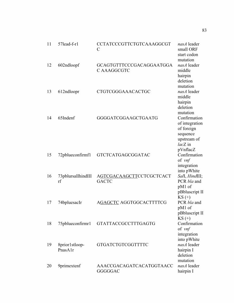
83
11 57lead-f-r1 CCTATCCCGTTCTGTCAAAGGCGTC
nasA leader small ORF start codon mutation
12 602ndloopf GCAGTGTTTCCCGACAGGAATGGAC AAAGGCGTC
nasA leader middle hairpin deletion mutation
13 612ndloopr CTGTCGGGAAACACTGC nasA leader middle hairpin deletion mutation
14 65Indenf GGGGATCGGAAGCTGAATG Confirmation of integration of foreign sequence upstream of lacZ in pVnflacZ
15 72pblueconfirmf1 GTCTCATGAGCGGATAC Confirmation of vnf integration into pWhite
16 73pblursalIhindIIIrf
AGTCGACAAGCTTCCTCGCTCACTGACTC
SalI, HindIII; PCR bla and pM1 of pBbluscript II KS (+)
17 74bpluesacIr AGAGCTC AGGTGGCACTTTTCG PCR bla and pM1 of pBbluscript II KS (+)
18 75pblueconfirmr1 GTATTACCGCCTTTGAGTG Confirmation of vnf integration into pWhite
19 8prior1stloop-PnasA1r
GTGATCTGTCGGTTTTC nasA leader hairpin I deletion mutation
20 9primextenf AAACCGACAGATCACATGGTAACCGGGGGAC
nasA leader hairpin I

84
deletion mutation
21 Deletion-108 AAATCTAGAACCAAGACAGTGCAAG
XbaI; Promoter deletion construction
22 Deletion-131 AAATCTAGAAACCCCATA AGAGG XbaI; Promoter deletion construction
23 Deletion-168 AAATCTAGAGCTCTGCTCGCCCTGTTG
XbaI; Promoter deletion construction
24 Deletion-43 AAATCTAGAGT CATCCGTGAA AC XbaI; Promoter deletion construction
25 lacZr CAAGGCGATTAAGTTGGGTAAC
Confirmation of integration of foreign sequence upstream of lacZ in pVnflacZ
26 M13f GTAAAACGACGGCCAGTG Cloning confirmation
27 M13r CAGGAAACAGCTATGAC Cloning confirmation
28 Nasasigma54f TAACACAGCCCCTATATCGAGAAAACCGAC
�54 binding site mutation
29 Nasasigma54r TATAGGGGCTGTGTTAGTTTCACGGATGACTG
�54 binding site mutation
30 O31Pnasaf CAGAACGGGAATGGACAATTTTTGTTTTCCGCTTTC
Terminator hairpin deletion mutation
31 O32pnasar AATTGTCCATTCCCGTTCTG Terminator hairpin deletion mutation
32 P92 AAGGATCCGCTTATGTGATCTGTC BamHI;

85
G Transcriptional fusion construction
33 vnff AAAGAGCTCATCATCTCGCCCAGTTC
SacI; PCR vnf sequence
34 vnfr AAACGCCTCGAGAAGAGCATG XhoI; PCR vnf sequence
35 WB30screenf CGCCTGGCTCGACGAATG nasA promoter region cloning confirmation
36 WB30screenr GTCCATTCCCGTTCTGTC nasA promoter region cloning confirmation
a The underlined sequences are the restriction sites.
b Restriction enzymes that can digest the underlined sequences.

86
TABLE 3. Expression of �(nasA-lacZ) translational fusion in A. vinelandii and its mutants
�-galactosidase activity (Miller units)a
Nitrogen source Promoter region within the lacZ
fusion
Strain genotype NH4
+ N2 NO2- NO3
-
nasA-316 - +297 Wild type 69 ± 1 96 ± 4 682 ± 9 478 ± 6 nasA-316 - +297 nif - 32 ± 32 NTb 942 ± 16 1389 ± 26
nasA-316 - +297 with �54 binding site
GG/GC�AA/AT mutation
Wild type 3 ± 1 5 ± 2 7 ± 1 5 ± 1
nasA-316 - +297 �ntrC::Gmr 2 ± 0 2 ± 0 23 ± 1 2 ± 0 nasA+89 - +297 Wild type 2 ± 1 3 ± 2 3 ± 1 3 ± 0
a Cultures were grown in BS medium supplemented with the indicated nitrogen sources. After seven hours, �-galactosidase activity was measured. All data are presented as the mean values of triplicates ± standard deviations from a representative experiment. b NT, not tested.

87
TABLE 4. Expression of �(PnasA-lacZ) in A. vinelandii and its mutants
�-galactosidase activity (Miller units)a
Nitrogen source Strain genotype NH4
+ N2 U NO2- NO3
-
Wild type 594 ± 15 307 ± 14 324 ± 10 439 ± 10 394 ± 25 ntrC mutant 2 ± 1 8 ± 1 4 ± 1 2 ± 1 3 ± 1
nif - 559 ± 17 NTb 3470 ± 34 2087 ±142 2677 ± 32
a Cultures were grown in BS medium supplemented with the indicated nitrogen sources. After seven hours, �-galactosidase activity was measured. All data are presented as the mean values of triplicates ± standard deviations from a representative experiment. b NT, not tested.
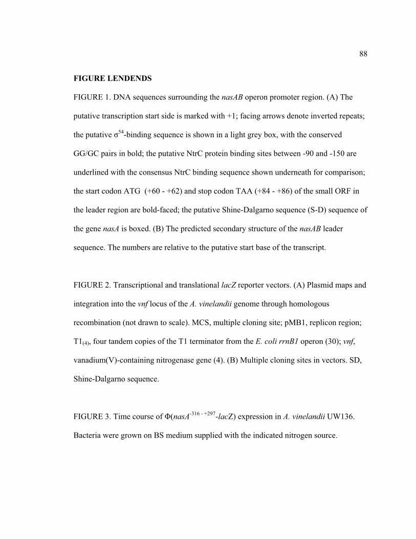
88
FIGURE LENDENDS FIGURE 1. DNA sequences surrounding the nasAB operon promoter region. (A) The
putative transcription start side is marked with +1; facing arrows denote inverted repeats;
the putative �54-binding sequence is shown in a light grey box, with the conserved
GG/GC pairs in bold; the putative NtrC protein binding sites between -90 and -150 are
underlined with the consensus NtrC binding sequence shown underneath for comparison;
the start codon ATG (+60 - +62) and stop codon TAA (+84 - +86) of the small ORF in
the leader region are bold-faced; the putative Shine-Dalgarno sequence (S-D) sequence of
the gene nasA is boxed. (B) The predicted secondary structure of the nasAB leader
sequence. The numbers are relative to the putative start base of the transcript.
FIGURE 2. Transcriptional and translational lacZ reporter vectors. (A) Plasmid maps and
integration into the vnf locus of the A. vinelandii genome through homologous
recombination (not drawn to scale). MCS, multiple cloning site; pMB1, replicon region;
T1(4), four tandem copies of the T1 terminator from the E. coli rrnB1 operon (30); vnf,
vanadium(V)-containing nitrogenase gene (4). (B) Multiple cloning sites in vectors. SD,
Shine-Dalgarno sequence.
FIGURE 3. Time course of �(nasA-316 - +297-lacZ) expression in A. vinelandii UW136.
Bacteria were grown on BS medium supplied with the indicated nitrogen source.

89
FIGURE 4. Expression of the �(nasA-lacZ) translational fusions with different nasAB
leader alterations (not drawn to scale). The top line represents the sequence ranges from -
316 nt upstream of the nasAB transcription start site to the +297 nt (base 162 of the NasA
ORF). The bent arrow represents the direction of transcription, +1 represents the first
nucleotide of the transcript; facing arrows (designated I, II, and III) represent the three
inverted sequences in the nasA leader region. The empty rectangles represent the regions
deleted; represents the ATG � TAG mutation. Values on the right show �(nasA-lacZ)
expression in A. vinelandii. Cultures were grown in BS medium supplemented with the
indicated nitrogen sources and �-galactosidase activity was measured after seven hours.
All data are presented as the mean values of three replicates ± standard deviations from a
representative experiment.
FIGURE 5. Expression of �(nasA-lacZ) transcriptional fusion constructs bearing
deletions within the promoter region in A. vinelandii (not drawn to scale). All deletions
were constructed based on the sequence around the nasA promoter region in the plasmid
pWB908, which has the terminator hairpin in the leader region deleted. (A) The nasA
promoter region from -316 to +1 is shown, the putative tandem NtrC binding sites are
underlined; the 5’ ends (-168, -131, -108, -43) of nasAB promoter deletion mutations are
shown. �-Galactosidase activity of the reporter constructs was measured seven hours
after cultures were grown on BS medium supplemented with ammonium (B) or nitrate
(C). The activity of pWB908 (with 5’end at -316) was arbitrarily set to 100%, and the

90
activities of other fusions are represented as a percentage of that value. This experiment
has been repeated three times with similar results.
FIGURE 6. Model for nasAB regulation. The nasAB operon in A. vinelandii has an NtrC-
dependent promoter, which is subject to global nitrogen regulation. In the wild-type
strain, nasAB transcription initiation is repressed at basal levels regardless of the presence
of ammonia. The terminator structure at the operon leader region regulates transcription
negatively. Nitrate/nitrite induces antitermination, which is mediated by NasS/NasT, a
nitrate sensor-antiterminator system. NasS regulates the activity of NasT directly through
steric hindrance in the absence of nitrate and nitrite.
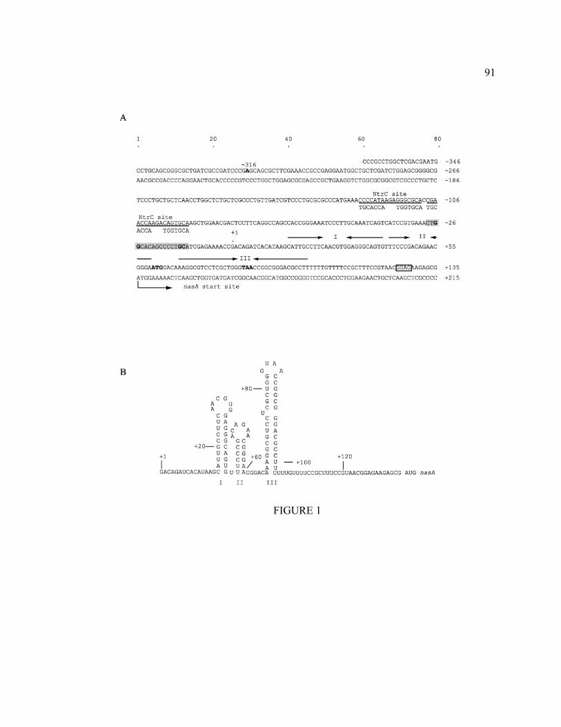
91
FIGURE 1

92
A
B pVnflacZa: T1(4)-XbaI-BcuI-BamHI-lacZ
pVnflacZb: T1(4)-XbaI-BcuI-BamHI-XbaI-SD-lacZ
FIGURE 2

93
0
100
200
0 2 4 6 8
Time (hr)
�-ga
lact
osid
ase
300
400
500
600
700
800
10 12
act
ivity
(Mill
er u
nits
)
AmmoniumNitriteNitrate
FIGURE 3
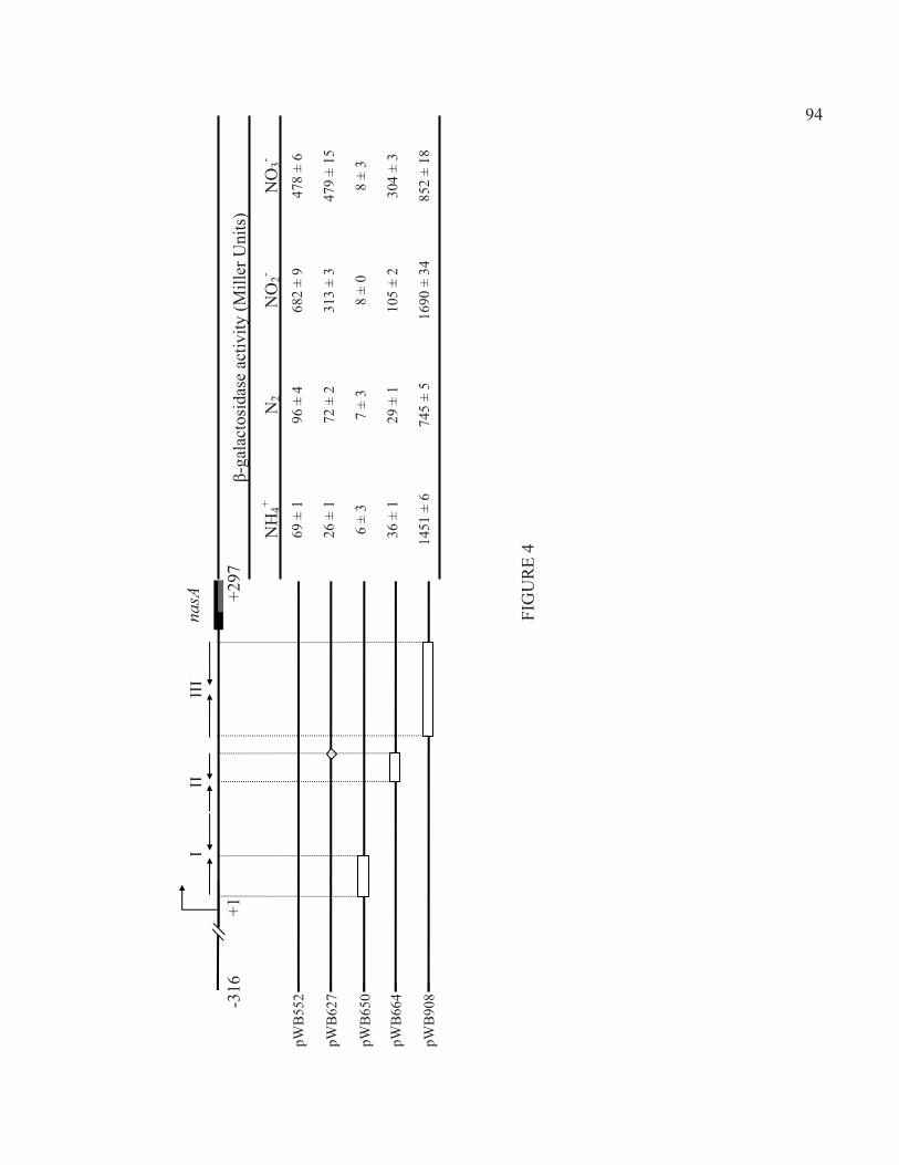
94 94
FIG
UR
E 4
�-ga
lact
osid
ase
activ
ity (M
iller
Uni
ts)
NH
4+N
2N
O2-
NO
3-
69 ±
1
96 ±
4
682
± 9
478
± 6
26 ±
1
72 ±
2
313
± 3
479
± 15
6 ±
3 7
± 3
8 ±
0 8
± 3
36 ±
1
29 ±
1
105
± 2
304
± 3
1451
± 6
74
5 ±
5 16
90 ±
34
852
± 18
pWB
552
pWB
627
pWB
650
pWB
664
pWB
908
nasA
II
III
I
+1
-316
+2
97

95
-131 1 -108 -43 +1 -316 -168 A
NtrC binding sites
B
100.0
78.3 81.5
7.7 4.0
0
20
40
60
80
100
120
-316 -168 -131 -108 -43
5' end
�-ga
lact
osid
ase
activ
ity (%
)
C
100.0
81.071.9
8.91.8
0
20
40
60
80
100
120
-316 -168 -131 -108 -43
5' end
�-ga
lact
osid
ase
activ
ity (%
)
FIGURE 5

96
FIGURE 6

97
References
1. Bali, A., G. Blanco, S. Hill, and C. Kennedy. 1992. Excretion of ammonium by a nifL mutant of Azotobacter vinelandii fixing nitrogen. Appl. Environ. Microbiol. 58:1711-1718.
2. Barrios, H., B. Valderrama, and E. Morett. 1999. Compilation and analysis of sigma(54)-dependent promoter sequences. Nucleic Acids Res. 27:4305-4313.
3. Bishop, P. E. and W. J. Brill. 1977. Genetic analysis of Azotobacter vinelandii mutant strains unable to fix nitrogen. May 132:954-956.
4. Bishop, P. E., D. M. Jarlenski, and D. R. Hetherington. 1982. Expression of an alternative nitrogen fixation system in Azotobacter vinelandii. J. Bacteriol. 150:1244-1251.
5. Bishop, P. E., D. M. Jarlenski, and D. R. Hetherington. 1980. Evidence for an alternative nitrogen fixation system in Azotobacter vinelandii. Proc. Natl. Acad. Sci. U. S. A. 77:7342-7346.
6. Buck, M., M. T. Gallegos, D. J. Studholme, Y. Guo, and J. D. Gralla. 2000. The bacterial enhancer-dependent sigma(54) (sigma(N)) transcription factor. J. Bacteriol. 182:4129-4136.
7. Cali, B. M., J. L. Micca, and V. Stewart. 1989. Genetic regulation of nitrate assimilation in Klebsiella pneumoniae M5al. J. Bacteriol. 171:2666-2672.
8. Chai, W. and V. Stewart. 1999. RNA sequence requirements for NasR-mediated, nitrate-responsive transcription antitermination of the Klebsiella oxytoca M5al nasF operon leader. J. Mol. Biol. 292:203-216.
9. Chai, W. and V. Stewart. 1998. NasR, a novel RNA-binding protein, mediates nitrate-responsive transcription antitermination of the Klebsiella oxytoca M5al nasF operon leader in vitro. J. Mol. Biol. 283:339-351.
10. Dennis, J. J. and G. J. Zylstra. 1998. Plasposons: modular self-cloning minitransposon derivatives for rapid genetic analysis of gram-negative bacterial genomes. Appl. Environ. Microbiol. 64:2710-2715.
11. Dieffenbach, C. W. and G. S. Dveksler. 2003. PCR primer : a laboratory manual. Cold Spring Harbor Laboratory Press, Cold Spring Harbor, N.Y.
12. Ferro-Luzzi Ames, G. and K. Nikaido. 1985. Nitrogen regulation in Salmonella typhimurium. Identification of an ntrC protein-binding site and definition of a consensus binding sequence. EMBO J. 4:539-547.

98
13. Goldman, B. S., J. T. Lin, and V. Stewart. 1994. Identification and structure of the nasR gene encoding a nitrate- and nitrite-responsive positive regulator of nasFEDCBA (nitrate assimilation) operon expression in Klebsiella pneumoniae M5al. J. Bacteriol. 176:5077-5085.
14. Gollnick, P. and P. Babitzke. 2002. Transcription attenuation. Biochim. Biophys. Acta 1577:240-250.
15. Gutierrez, J. C., E. Santero, and M. Tortolero. 1997. Ammonium repression of the nitrite-nitrate (nasAB) assimilatory operon of Azotobacter vinelandii is enhanced in mutants expressing the nifO gene at high levels. Mol. Gen. Genet. 255:172-179.
16. Gutierrez, J. C., F. Ramos, L. Ortner, and M. Tortolero. 1995. nasST, two genes involved in the induction of the assimilatory nitrite-nitrate reductase operon (nasAB) of Azotobacter vinelandii. Mol. Microbiol. 18:579-591.
17. Henkin, T. M. and C. Yanofsky. 2002. Regulation by transcription attenuation in bacteria: how RNA provides instructions for transcription termination/antitermination decisions. Bioessays 24:700-707.
18. Heus, H. A. and A. Pardi. 1991. Structural features that give rise to the unusual stability of RNA hairpins containing GNRA loops. Science 253:191-194.
19. Joerger, R. D. and P. E. Bishop. 1988. Bacterial alternative nitrogen fixation systems. Crit. Rev. Microbiol. 16:1-14.
20. Kennedy, C. and R. L. Robson. 1983. Activation of nif gene expression in Azotobacter by the nifA gene product of Klebsiella pneumoniae. Nature 301:626-628.
21. Lin, J. T. and V. Stewart. 1998. Nitrate assimilation by bacteria. Adv. Microb. Physiol. 39:1-30, 379.
22. Lin, J. T. and V. Stewart. 1996. Nitrate and nitrite-mediated transcription antitermination control of nasF (nitrate assimilation) operon expression in Klebsiella pneumoniae M5al. J. Mol. Biol. 256:423-435.
23. Lin, J. T., B. S. Goldman, and V. Stewart. 1994. The nasFEDCBA operon for nitrate and nitrite assimilation in Klebsiella pneumoniae M5al. J. Bacteriol. 176:2551-2559.
24. Luque, F., E. Santero, J. R. Medina, and M. Tortolero. 1987. Mutants of Azotobacter vinelandii altered in the regulation of nitrate. Arch. Microbiol. 148:231-235.
25. Maniatis, T., E. F. Fritsch, and J. Sambrook. 1982. Molecular cloning: a laboratory manual. Cold Spring Harbor Laboratory Press,U.S, Cold Spring Harbor, N.Y.

99
26. Marsh, J. L., M. Erfle, and E. J. Wykes. 1984. The pIC plasmid and phage vectors with versatile cloning sites for recombinant selection by insertional inactivation. Gene 32:481-485.
27. Martinez-Argudo, I., R. Little, N. Shearer, P. Johnson, and R. Dixon. 2004. The NifL-NifA System: a multidomain transcriptional regulatory complex that integrates environmental signals. J. Bacteriol. 186:601-610.
28. Merrick, M. J. 1993. In a class of its own--the RNA polymerase sigma factor sigma 54 (sigma N). Mol. Microbiol. 10:903-909.
29. Merrick, M. J. and R. A. Edwards. 1995. Nitrogen control in bacteria. Microbiol. Rev. 59:604-622.
30. Miller, W. G., J. H. Leveau, and S. E. Lindow. 2000. Improved gfp and inaZ broad-host-range promoter-probe vectors. Mol. Plant Microbe Interact. 13:1243-1250.
31. Ramos, F., G. Blanco, J. C. Gutierrez, F. Luque, and M. Tortolero. 1993. Identification of an operon involved in the assimilatory nitrate-reducing system of Azotobacter vinelandii. Mol. Microbiol. 8:1145-1153.
32. Robson, R. L., J. A. Chesshyre, C. Wheeler, R. Jones, P. R. Woodley, and J. R. Postgate. 1984. Genome size and complexity in Azotobacter chroococcum. J. Gen. Microbiol. 130:1603-1612.
33. Rombel, I., A. North, I. Hwang, C. Wyman, and S. Kustu. 1998. The bacterial enhancer-binding protein NtrC as a molecular machine. Cold Spring Harb. Symp. Quant. Biol. 63:157-166.
34. Santero, E., A. Toukdarian, R. Humphrey, and C. Kennedy. 1988. Identification and characterization of two nitrogen fixation regulatory regions, nifA and nfrX, in Azotobacter vinelandii and Azotobacter chroococcum. Mol. Microbiol. 2:303-314.
35. Santero, E., F. Luque, J. R. Medina, and M. Tortolero. 1986. Isolation of ntrA-like mutants of Azotobacter vinelandii. J. Bacteriol. 166:541-544.
36. Shu, C. J. and I. B. Zhulin. 2002. ANTAR: an RNA-binding domain in transcription antitermination regulatory proteins. Trends Biochem. Sci. 27:3-5.
37. Shu, C. J., L. E. Ulrich, and I. B. Zhulin. 2003. The NIT domain: a predicted nitrate-responsive module in bacterial sensory receptors. Trends Biochem. Sci. 28:121-124.
38. Simon, R., J. Quandt, and W. Klipp. 1989. New derivatives of transposon Tn5 suitable for mobilization of replicons, generation of operon fusions and induction of genes in gram-negative bacteria. Gene 80:161-169.

100
39. Strandberg, G. W. and P. W. Wilson. 1968. Formation of the nitrogen-fixing enzyme system in Azotobacter vinelandii. Can. J. Microbiol. 14:25-31.
40. Toukdarian, A. and C. Kennedy. 1986. Regulation of nitrogen metabolism in Azotobacter vinelandii: isolation of ntr and glnA genes and construction of ntr mutants. EMBO J. 5:399-407.
41. Toukdarian, A., G. Saunders, G. Selman-Sosa, E. Santero, P. Woodley, and C. Kennedy. 1990. Molecular analysis of the Azotobacter vinelandii glnA gene encoding glutamine synthetase. J. Bacteriol. 172:6529-6539.
42. Walmsley, J. and C. Kennedy. 1991. Temperature-Dependent Regulation by Molybdenum and Vanadium of Expression of the Structural Genes Encoding Three Nitrogenases in Azotobacter vinelandii. Appl. Environ. Microbiol. 57:622-624.
43. Walmsley, J., A. Toukdarian, and C. Kennedy. 1994. The role of regulatory genes nifA, vnfA, anfA, nfrX, ntrC, and rpoN in expression of genes encoding the three nitrogenases of Azotobacter vinelandii. Arch. Microbiol. 162:422-429.
44. Wedel, A. and S. Kustu. 1995. The bacterial enhancer-binding protein NTRC is a molecular machine: ATP hydrolysis is coupled to transcriptional activation. Genes Dev. 9:2042-2052.
45. Yanofsky, C. 2007. RNA-based regulation of genes of tryptophan synthesis and degradation, in bacteria. RNA 13:1141-1154.
46. Zuker, M. 2003. Mfold web server for nucleic acid folding and hybridization prediction. Nucleic Acids Res. 31:3406-3415.

101
APPENDIX B. NASS/NAST, A NON-CANONICAL TWO-COMPONENT REGULATORY SYSTEM REGULATES THE ASSIMILATORY NITRATE
REDUCTASE OPERON (NASAB) IN AZOTOBACTER VINELANDII VIA ANTITERMINATION

102
NasS/NasT, a non-canonical two-component regulatory system regulates the nitrate reductase operon in Azotobacter vinelandii via antitermination
Baomin Wang1†, Christopher Rensing 2 and Christina Kennedy1
1. Division of Plant Pathology and Microbiology, Department of Plant Sciences,
University of Arizona, Tucson, Arizona 85721
2. Department of Soil, Water, and Environmental Science, University of Arizona,
Tucson, Arizona 85721
† Corresponding author. Mailing address: Department of Plant Sciences, Forbes 303,
University of Arizona, Tucson, AZ 85721. Phone: (520) 621-9835. Fax: (520) 621-9290.
E-mail: [email protected]
Dedication (in memoriam): This manuscript is dedicated to the memory of Christina
Kennedy.

103
ABSTRACT
Expression of the assimilatory nitrate reductase operon nasAB in Azotobacter vinelandii
is induced by nitrate and nitrite. This operon-specific induction involves antitermination
of an intrinsic transcription terminator within the leader region of the nasA. Nitrate and
nitrite induction also requires the products of the nasST operon. NasT is homologous to
the antiterminator AmiR in Pseudomonas aeruginosa. Mutation of nasT abolishes nasAB
induction. NasS is homologous to the substrate-binding component of ABC-type
transporters, but is not required for nitrate transport. Mutation of nasS without affecting
downstream nasT expression leads to constitutive expression of nasAB. In this study, a
bacterial two-vector system assay demonstrated that NasT has a positive effect on nasAB
expression and acted within the nasAB leader region. A yeast two-hybrid system analysis
indicated that NasT formed a dimer or higher level complex, resembling the structure of
AmiR. These results suggest nasT and amiR may represent orthologous genes encoding a
family of antitermination regulators. Over-expression of nasT and mutational analyses of
nasS or nasT demonstrated that nasAB regulation required the maintenance of the
endogenous NasS and NasT stoichiometry, which may be achieved through translational
coupling. The results presented here support a model that NasS and NasT constitute a
nitrate sensor-antiterminator regulatory system, with NasS stringently controlling the
activity of NasT by a mechanism requiring stoichiometric interaction between the
proteins.

104
INTRODUCTION
Azotobacter vinelandii strain UW136 is a free-living diazotroph with three genetically
distinct nitrogenase systems: the conventional molybdenum-containing nitrogenase
system (Nif), a vanadium-containing nitrogenase system (Vnf), and a iron-containing
system (Anf) (11, 27). A. vinelandii can also use nitrate or nitrite as its sole nitrogen
source. Nitrate is sequentially reduced to nitrite and to ammonium before being
incorporated into glutamine by glutamine synthetase (35). The assimilatory nitrate
reductase (NasB) and nitrite reductase (NasA) are encoded by a two-gene nasAB operon
(29).
The nasAB operon is subject to complex transcriptional regulation. nasAB has a NtrC-
dependent promoter, which is regulated in response to intracellular nitrogen status. In the
wild type strain with a functional Nif system, nasAB transcription initiation is repressed
to basal levels regardless the presence of ammonia (Wang et al, submitted for
publication). The nasAB operon is also subject to post-transcriptional attenuation
regulation. The 135-nucleotide nasAB leader region contains an intrinsic transcriptional
terminator that blocks transcription of nasAB in the absence of nitrate/nitrite (Wang et al,
submitted for publication).
The presence of nitrate or nitrite in the absence of ammonium stimulates antitermination
within the nasAB leader region. This relief of termination requires the products of the
nasST operon, which is located 12-kb upstream of the nasAB (8). The deduced protein

105
NasS shares sequence homology with the substrate binding component of ABC-type
nitrate transporters in Klebsiella pneumoniae and Synechococcus sp. Deletion of nasS,
however, has no effect on nitrate uptake by A. vinelandii (8). Insertion mutagenesis of
nasS without affecting downstream nasT expression led to constitutive expression of
nasAB, suggesting that NasS plays a negative role in nasAB expression (8).
NasT is a 192 amino acid polypeptide with a modular structure homologous to AmiR, an
mRNA- binding antiterminator in Pseudomonas aeruginosa (8, 32). Both proteins have a
C-terminal AmiR and NasR Transcription Antitermination Regulator (ANTAR) domain
and an N-terminal domain homologous to the receiver domain REC of the response
regulator of classic two-component regulatory systems (8, 24, 32). Mutation of nasT
abolished nasAB expression even in the presence of nitrate and nitrite, indicating that it is
required for nasAB expression (8).
The NasT domain structure and the phenotype of nasT mutants suggested that NasS and
NasT may mediate nitrate- and nitrite-inducted antitermination within the nasAB leader
region (18). In this work, a bacterial two-vector system was used to test the regulatory
role of NasT. The results confirmed that NasT acts positively within the nasAB leader
region. In addition, the results of over-expression analyses indicated that the maintenance
of correct stoichiometry between NasS and NasT is required for nasAB regulation.
Furthermore, the results of mutational analyses suggested that translational coupling was
used to coordinate NasS and NasT synthesis. The results presented here support the

106
model that NasS and NasT constitute a sensor-antiterminator system; signal transduction
from NasS to NasT is conveyed through protein-protein interactions.
MATERIALS AND METHODS
Strains and plasmids. Strains and plasmids used in this study are listed in Table 1. A.
vinelandii and its derivatives were grown on Burk’s nitrogen-free salts (BS) medium
supplemented with 1% sucrose (34). When needed, BS was supplemented with
ammonium acetate, 15 mM; urea, 10 mM; KNO3, 10 mM; and NaNO2, 2.8 mM.
Antibiotics were added where appropriate: Carbenicillin (Car) at 20 �g/ml, Gentamicin
(Gm) at 0.05 �g/ml, and Kanamycin (Km) at 2.0 �g/ml. E. coli was grown in LB (0.5%
NaCl, 1% Tryptone, 0.5% yeast extract) broth or on LB agar ( 1.5%) at 37oC and
antibiotic concentrations were as follows: Chloramphenicol (Cam) at 25 �g/ml, Km at 25
�g/ml. Saccharomyces cerevisiae AH109 was grown in YPDA (yeast extract-peptone-
dextrose-adenine) or SD (synthetic dropout) media as described by the manufacturer
manual for the Matchmaker two-hybrid system (Clontech,).
Bioinformatics. Protein sequence BLAST against the Conserved Domain Database
(CDD) was performed at the NCBI website
http://www.ncbi.nlm.nih.gov/Structure/cdd/wrpsb.cgi. Coil-coil structure prediction was
performed at http://www.russell.embl-heidelberg.de/cgi-bin/coils-svr.pl.

107
DNA manipulation. DNA digestions, ligations, and cloning were conducted following
standard protocols (20). For blunt end ligation, DNA fragments were treated with T4
DNA polymerase following the manufacturer instructions (Fermentas, Glen Burnie, MD).
All enzymes and the plasmid isolation kit GeneJET plasmid miniprep kit were purchased
from Fermentas. DNA purification from agarose gels was performed using the
NucleoSpin Extract II (Clontech, Mountain View, CA). A. vinelandii transformations
were carried out as described previously (30). E. coli transformations were performed
using the CaCl2 chemical method (20). Yeast transformations were carried out using the
LiAc method following the manual for the Matchmaker two-hybrid system (Clontech).
Oligonucleotides used in this study are listed in Table 2. Oligonucleotides were
purchased from IDT Inc. (Coralville, IA). DNA sequencing was performed at the
University of Arizona Genomics and Technology Core (GATC) DNA Sequencing
Facility.
Construction of a A. vinelandii nasST deletion mutant. The 4.0-kb EcoRI fragment
carrying nasST from the plasmid pMAS20 was subcloned into the HindIII-HindIII region
of pIC20H via blunt end ligation. The resulting plasmid was cut with SalI and ligated
with a SacI-Gmr-SacI cassette from pTnMod-OGm through blunt end ligation. The
resulting plasmid pWB296 was then transformed into the nif – derivative UW1.
Transformants were plated on selective BSN media supplemented with gentamicin.
Transformants (Gmr) were patched onto BSN plates supplemented with carbenicillin.

108
GmrCars clones were screened and �nasST::Gmr allelic replacement was confirmed by
colony PCR using the primers nasSf and nasSr.
Construction of PscrX-nasS and PscrX-nasT fusions. The nasS gene was amplified
using primer pairs PciI-nasS and BglII-nasS from pMAS20. The PCR product was
treated with PciI and BglII and cloned into the PciI-BamHI site downstream of the PscrX
promoter in the plasmid pDB1332 (12). The resulting plasmid was restricted with KpnI,
blunt-ended, and ligated with PCR fragments containing the kanamycin resistance gene
from the plasmid pBBR1MCS-2, giving rise to the plasmid pDB1332-S. Using a similar
strategy, PscrX-nasT fusion was constructed, and the resulting plasmids was designated
pDB1332-T.
Construction of plasmids for bacteria two-vector system. To construct a nasS
expression plasmid, nasS was amplified from pMAS20 using primer pairs EcoRI-nasS
and BglII-nasS. The PCR product was treated with EcoRI and BglII and cloned into the
EcoRI-BamHI site of the plasmid pDK6, giving rise to the plasmid pDK6-S. Using a
similar strategy, nasT was amplified from pMAS20 using primer pairs EcoRI-nasT and
BglII-nasT and cloned into the pDK6, giving rise to the plasmid pDK6-T.
The plasmid carrying a PlacUV5-nasA-lacZ fusion was engineered from the plasmid pBT.
The DNA fragment containing chloramphenicol resistance gene, p15A replicon, and
lacUV5 promoter was amplified from pBT using primer pairs BTr and 76pBTNotIr, and

109
digested with NotI and XhoI. The resulting fragment was linked with the NotI-lacZ-XhoI
cassette from the plasmid pblue-lacZ, giving rise to plasmid pBTW.
The DNA sequence containing the nasA leader and nasA 5’ end162 nucleotides or its
deletion derivatives was amplified from plasmid pWB552, pWB643, pWB650, pWB664,
and pWB908 using primer pairs 013NotI77 and 1BamH162innasA, and cloned into the
NotI-BamHI region of pBTW, giving rise to a series of heterologous fusions.
Construction of plasmids for Yeast two-hybrid system. nasT or a truncated nasT
fragment without C-terminal the ANTAR domain were amplified from pRM19. The PCR
products were double digested with NdeI and EcoRI, and cloned into NdeI-EcoRI regions
of the plasmids pGADT7 and pGBKT7.
Construction of lacUV5-nasST’-lacZ and lacUV5-nasT’-lacZ fusions. Two lacUV5-
nasST’-lacZ fusions were constructed. The nasST’ fragments in both fusions start at the
ninth nucleotide of the nasS leader and end at the 62nd codon of nasT with an nasS central
in-frame (codon 56 — 388) deletion. One fusion has the original 389th codon CCC
(Proline) in nasS substituted with a TGA stop codon. The deletion was constructed using
the overlap extension method based on two rounds of PCRs (5). Briefly, the first round
PCR generated two fragments, which are complementary at the ends with the desired
mutation. The second round PCR uses the two fragments from the first round as
templates, generating a merged sequence with the desired mutation. The nasST’

110
fragments were digested with NotI and BamHI, and cloned into the NotI-BamHI region of
pBTW. Then, The XbaI-lacUV5-nasST’-BamHI fragment was subcloned into XbaI-
BamHI region of pVnflacZ, giving rise to plasmids pWB923 and pWB924. Plasmid
pWB924 contains the CCC� TGA nonsense mutation.
The lacUV5-nasT’-lacZ fusions were constructed by the amplification of the nasT’
fragments from pMAS20 using primer pairs O27NotIinS and 026BamHIinT and
subsequent insertion into the NotI-BamHI region downstream of lacUV5 in pBTW. The
XbaI-lacUV5-nasT’-BamHI fragments were subcloned into the XbaI-BamHI region of
pVnflacZa resulting in plasmid pWB903.
�-galactosidase assay. �-galatosidase activities of the fusion plasmids was determined in
E. coli using the method by Miller (1972) (23). �-galactosidase activity was measured in
the yeast two-hybrid systems using the freeze/thaw method following the manual for the
Matchmaker two-hybrid system (Clontech).
RESULTS AND DISCUSSION A homologous nasST mutation in A. vinelandii could be stably maintained. A.
vinelandii is unique in that the chromosome number can vary from 2 to 80, depending on
growth conditions (19). The multiplicity of A. vinelandii genomes results in challenges in
creating mutations in essential genes (13, 35). Since essential gene mutations are lethal to
the bacterium, A. vinelandii will maintain a mixture of chromosomes containing the wild-

111
type allele and mutated allele marked by an antibiotic resistance gene under the antibiotic
selection pressure. Removal of the antibiotic from the medium leads to the rapid
elimination of the mutant allele. For non-essential genes, selection for the mutant allele
results in full replacement of all wild-type copies.
.
In this project, a nasST deletion mutant in the Nif - strain UW1 was constructed. Strain
UW1 can not fix atmospheric nitrogen, but can use nitrate or nitrite as its sole nitrogen
source (14). The nasST deletion mutant has the central SalI-SalI region replaced with a
Gmr cassette, removing 68% of nasS from the 3’ end and 15% of nasT from the 5’ end.
The mutant cannot grow on medium with nitrate or nitrite as the sole nitrogen source
(Figure 1), in agreement with previous observations that nasST mutation in wild type A.
vinelandii represses nasAB expression. Colony PCR verified that the mutant contains
only the mutant allele (data not shown). These results confirm the previous observation of
the inhibition of nasAB expression in a nasST deletion background is not a result of
nasST heterozygosity.
To complement the nasST mutation, nasS or nasT were placed under the control of the
sucrose-inducible PscrX promoter by introduction into the sucrose catabolic regulon (scr)
region of A. vinelandii through homologous recombination. PscrX is active in Burk’s
medium supplemented with 1% sucrose (BS) (12). Expression of nasT in the nasST
deletion mutant restored the strain’s ability to grow in BS broth supplemented with
nitrate or nitrite as sole nitrogen source while expression of nasS had no obvious effect

112
on mutant growth (Figure 1C and D). These results suggest that NasT is produced in an
active form.
NasT acts positively within the nasAB leader region. To test whether NasT is involved
in regulation within the nasAB leader region, a bacterial two-vector system was
constructed. The system is composed of a protein expression plasmid and a heterologous
fusion plasmid under lacUV5 promoter control. The protein expression plasmid was
derived from pDK6 that has a tac promoter followed by a multiple cloning site (MCS).
For protein expression, nasS or nasT was cloned into the EcoRI-BamHI region of the
MCS. The plasmid carrying lacUV5-nasA-lacZ fusion was engineered from the plasmid
pBT, which contains a lacUV5 promoter upstream of the bacteriophage � cI gene. To
construct the lacUV5-nasA-lacZ fusion, the � cI gene was replaced with the nasA-lacZ
cassette. The cassette has the nasA leader sequence extending into nasA 5’ end 162
nucleotides fused with lacZ. The pair of plasmids for both protein expression and lacZ
fusion were co-transformed into E. coli MC1061 for �-galactosidase assays.
The lacUV5-nasA-lacZ fusion had low level expression of lacZ in E. coli. Deletion of the
terminator hairpin III resulted in a 15-fold increase in �-galactosidase activity (Figure 2).
Co-expression of nasT with lacUV5-nasA-lacZ increased nasA gene expression more
than 30-fold. However, the effect of NasT on lacZ expression was partially reduced when
hairpin II was disrupted and virtually abolished when part of the promoter-proximal
hairpin I was deleted. The results presented here are consistent with the observations of
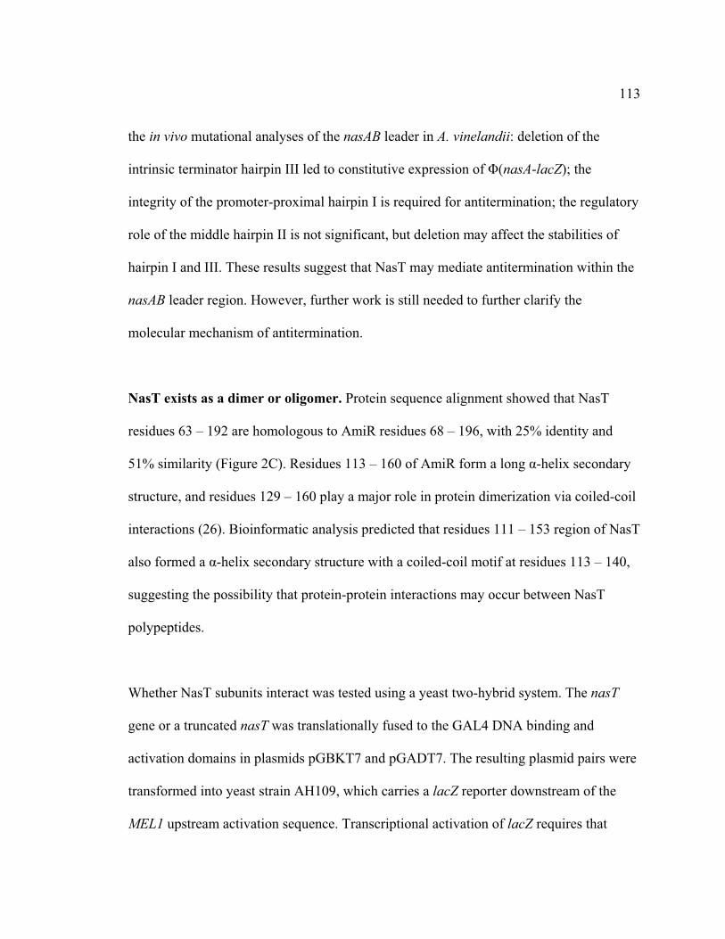
113
the in vivo mutational analyses of the nasAB leader in A. vinelandii: deletion of the
intrinsic terminator hairpin III led to constitutive expression of �(nasA-lacZ); the
integrity of the promoter-proximal hairpin I is required for antitermination; the regulatory
role of the middle hairpin II is not significant, but deletion may affect the stabilities of
hairpin I and III. These results suggest that NasT may mediate antitermination within the
nasAB leader region. However, further work is still needed to further clarify the
molecular mechanism of antitermination.
NasT exists as a dimer or oligomer. Protein sequence alignment showed that NasT
residues 63 – 192 are homologous to AmiR residues 68 – 196, with 25% identity and
51% similarity (Figure 2C). Residues 113 – 160 of AmiR form a long �-helix secondary
structure, and residues 129 – 160 play a major role in protein dimerization via coiled-coil
interactions (26). Bioinformatic analysis predicted that residues 111 – 153 region of NasT
also formed a �-helix secondary structure with a coiled-coil motif at residues 113 – 140,
suggesting the possibility that protein-protein interactions may occur between NasT
polypeptides.
Whether NasT subunits interact was tested using a yeast two-hybrid system. The nasT
gene or a truncated nasT was translationally fused to the GAL4 DNA binding and
activation domains in plasmids pGBKT7 and pGADT7. The resulting plasmid pairs were
transformed into yeast strain AH109, which carries a lacZ reporter downstream of the
MEL1 upstream activation sequence. Transcriptional activation of lacZ requires that

114
GAL4 DNA binding and activation domains come into close proximity, a function that is
dependent on the interaction of the fused target proteins.
The results indicate that the two fused NasT peptides have a strong interaction (Table 3).
Expression of �-galactosidase activity is higher than the positive control interaction
between murine p53 and SV40 large T-antigen (9, 17), indicating NasT peptides have
strong affinity. The N-terminal REC domain showed weak interaction when separated
from the rest of NasT. The difference of affinity strength between the NasT-NasT and
REC-REC interaction suggests that the predicted coiled-coil region in NasT plays a role
in NasT-NasT interaction.
NasT and AmiR share similar predicted overall structural conformations and both have
been shown to play a positive role in antitermination. They may represent a family of
regulator proteins, derived from a common ancestor – a fused protein of the domains
REC and ANTAR. A bacterial genome database search identified that NasT homologues
are widespread among alpha-, beta, and gamma-proteobacteria groups (data not shown),
suggesting regulation mediated by this family of regulators may be common in bacteria.
Modular structure of NasS. NasS has a negative effect on nasAB expression (8).
Genome sequence annotation confirms that nasS is 1,248 bases in length. Protein
sequence BLAST searches against the NCBI conserved domain database showed that
NasS has a conserved central region homologous to TauA (Figure 2A), a periplasmic

115
component of bacterial ABC-type sulfonate transporters (10). The central domain of
NasS is flanked by sequences that have no homology to the databases. NasS is probably a
cytoplasmic protein, as no N-terminal membrane signal peptide or transmembrane
structure was identified in the predicted NasS amino acid sequence based on
bioinformatic analysis.
The lack of DNA or RNA binding domains and the presence of a chemical binding
domain in NasS suggest that NasS may act as a nitrate/nitrite sensor instead of an
expression regulator. It was predicted that NasS and NasT constitute a sensor –
antiterminator system. (18). However, the mechanism of signal transduction between
NasS and NasT is unclear. The presence of an N-terminus REC in NasT suggests that
phosphorylation may be involved in signal transduction. However, the NasT REC
domain lacks the conserved Lysine residue essential for REC phosphorylation (8). In
addition, NasS lacks the kinase domain for auto-phosphorylation in response to substrate
signal. Combined data suggest that signal transduction between NasS and NasT uses a
mechanism other than phosphorylation.
Stoichiometry of NasS and NasT is essential for regulation of nasAB. To test whether
alterations in the ratio of NasS and NasT affects nasAB expression, the translational
fusion of nasA-lacZ which contains the -316 to +297 region around the nasA promoter
region was introduced into the wild type strain containing PscrX – nasS or PscrX – nasT
in the scr region. The results of �-galactosidase assays show that induction of PscrX -
nasT led to constitutive expression of �(nasA-lacZ) fusion independent of nitrogen

116
source; Induction of PscrX -nasS led to lower expression of �(nasA-lacZ) under
conditions of ammonium, N2, or nitrite, but not nitrate (Table 4).
The results presented here suggest that maintenance of the correct stoichiometry between
NasS and NasT is required for nasAB regulation. Over-expression of NasT leads to
constitutive nasAB expression under all conditions, even those in which NasA/NasB
expression is unnecessary.
Translation of nasS and nasT is coupled. The ORFs of NasS and NasT overlap by 19
bases (Figure 4A). mRNA secondary structure analysis predicted that both the NasT start
codon and NasS stop codon are part of an mRNA secondary structure (Figure 4B),
resembling the intergenic regions of translationally coupled cistrons (6, 7, 25).
Translational coupling is a type of regulation that ensures the adjacent genes in a
polycistronic mRNA expressed in a balanced manner. For two genes involved in
translational coupling, translation of the terminal end of the upstream gene is a
prerequisite for the translation of the downstream gene, which lacks the ability to recruit
ribosomes efficiently. Translational coupling has been demonstrated to be used widely by
viruses and bacteria to keep functionally related proteins at consistent ratios (33). For
example, AmiC/AmiR and NifL/NifA are two sensor-regulator pairs that use protein-
protein interactions to convey signals between them (22, 36) Both signal transduction

117
systems employ translational coupling to ensure that the sensors and regulators are
produced at similar stoichiometric ratios (6, 7, 25).
A lacUV5-nasT’-lacZ fusion was constructed to test whether nasT has its own SD. In this
fusion, the NasT ORF start region spanning from 18 nucleotides upstream of the NasT
ATG codon to base 186 was fused to lacZ (Figure 5). The plasmids carrying the fusions
were integrated into the vnf locus of the A. vinelandii genome using single-crossover
recombination (Wang et al, submitted for publication). The lacUV5-nasT’-lacZ fusion
displayed constitutive expression, confirming that nasT has its own SD sequence. We
suspect this SD is the AGGA sequence eight nucleotides upstream of the ATG start
codon (Figure 3B).
To investigate whether translational coupling is involved in nasST expression, two
lacUV5- nasST’-lacZ fusions were constructed (Figure 5). The nasST’ fragments in the
fusions range from the nasS leader sequence to 186 nucleotides in the 5’ end of nasT with
a deletion of the central region (nts 169-1167) of nasS. In addition to deletion of the nasS
central region, one of the fusions has the original Proline CCC codon (nt 1168-1170) in
nasS replaced with the translation stop codon TGA. The TGA null mutation in the nasS
central region should lead to premature termination of translation of nasS mRNA and the
dissociation of the ribosome before reaching the genuine nasS stop codon; In comparsion,
a fusion with the nasS central deletion but no CCC replacement should result in a
shortened polypeptide, but should not affect the translating ribosome from reaching the 3’

118
end of the ORF. �-galactosidase assays show that premature termination of nasS
translation reduced lacZ fusion expression by three-fold (Figure 4), suggesting that the in
vivo translation of nasT is regulated in response to NasS translation.
CONCLUDING REMARKS
Previous work demonstrated that the products of nasST are involved in nasAB regulation
(8), and potential roles of NasS and NasT in this regulation have been postulated (2, 18,
24, 28). The results presented here provide direct evidence that NasT is produced in an
active form and acts within the nasAB leader region, possibly as an antiterminator. The
modular characteristic of NasS suggests that it functions as a nitrate sensor. The
requirement of consistent NasS and NasT stoichiometry for nasAB regulation suggests
that NasS regulates the activity of NasT using a steric hindrance mechanism.
ACKNOWLEDGEMENTS
We thank Leland S. Pierson III, and Eduardo Santero for discussion, advice, and critical review of the manuscript.
REFERENCES
1. Bishop, P. E. and W. J. Brill. 1977. Genetic analysis of Azotobacter vinelandii mutant strains unable to fix nitrogen. May 132:954-956.
2. Caballero, A., A. Esteve-Nunez, G. J. Zylstra, and J. L. Ramos. 2005. Assimilation of Nitrogen from Nitrite and Trinitrotoluene in Pseudomonas putida JLR11. J. Bacteriol. 187:396-399.
119
3. Casadaban, M. J., A. Martinez-Arias, S. K. Shapira, and J. Chou. 1983. Beta-galactosidase gene fusions for analyzing gene expression in Escherichia coli and yeast. Methods Enzymol. 100:293-308.
4. Dennis, J. J. and G. J. Zylstra. 1998. Plasposons: modular self-cloning minitransposon derivatives for rapid genetic analysis of gram-negative bacterial genomes. Appl. Environ. Microbiol. 64:2710-2715.
5. Dieffenbach, C. W. and G. S. Dveksler. 2003. PCR primer : a laboratory manual. Cold Spring Harbor Laboratory Press, Cold Spring Harbor, N.Y.
6. Govantes, F., E. Andujar, and E. Santero. 1998. Mechanism of translational coupling in the nifLA operon of Klebsiella pneumoniae. EMBO J. 17:2368-2377.
7. Govantes, F., J. A. Molina-Lopez, and E. Santero. 1996. Mechanism of coordinated synthesis of the antagonistic regulatory proteins NifL and NifA of Klebsiella pneumoniae. J. Bacteriol. 178:6817-6823.
8. Gutierrez, J. C., F. Ramos, L. Ortner, and M. Tortolero. 1995. nasST, two genes involved in the induction of the assimilatory nitrite-nitrate reductase operon (nasAB) of Azotobacter vinelandii. Mol. Microbiol. 18:579-591.
9. Iwabuchi, K., B. Li, P. Bartel, and S. Fields. 1993. Use of the two-hybrid system to identify the domain of p53 involved in oligomerization. Oncogene 8:1693-1696.
10. Javaux, C., B. Joris, and P. De Witte. 2007. Functional characteristics of TauA binding protein from TauABC Escherichia coli system. Protein J. 26:231-238.
11. Joerger, R. D. and P. E. Bishop. 1988. Bacterial alternative nitrogen fixation systems. Crit. Rev. Microbiol. 16:1-14.
12. Johnson, D. C., M. C. Unciuleac, and D. R. Dean. 2006. Controlled expression and functional analysis of iron-sulfur cluster biosynthetic components within Azotobacter vinelandii. J. Bacteriol. 188:7551-7561.
13. Kennedy, C. and P. E. Bishop. 2004. Genetics of Nitrogen Fixation and Related Aspects of Metabolism in Species of Azotobacter: History and Current Status, p. 27-46. In W. Klipp, B. Masepohl, J. R. Gallon and W. E. Newton (ed.), Genetics and Regulation of Nitrogen Fixation in Free-Living Bacteria, vol. 2. Springer Netherlands, Netherland.
14. Kennedy, C. and R. L. Robson. 1983. Activation of nif gene expression in Azotobacter by the nifA gene product of Klebsiella pneumoniae. Nature 301:626-628.

120
15. Kleiner, D., W. Paul, and M. J. Merrick. 1988. Construction of multicopy expression vectors for regulated over-production of proteins in Klebsiella pneumoniae and other enteric bacteria. J. Gen. Microbiol. 134:1779-1784.
16. Kovach, M. E., P. H. Elzer, D. S. Hill, G. T. Robertson, M. A. Farris, R. M. Roop 2nd, and K. M. Peterson. 1995. Four new derivatives of the broad-host-range cloning vector pBBR1MCS, carrying different antibiotic-resistance cassettes. Gene 166:175-176.
17. Li, B. and S. Fields. 1993. Identification of mutations in p53 that affect its binding to SV40 large T antigen by using the yeast two-hybrid system. FASEB J. 7:957-963.
18. Lin, J. T. and V. Stewart. 1998. Nitrate assimilation by bacteria. Adv. Microb. Physiol. 39:1-30, 379.
19. Maldonado, R., J. Jimenez, and J. Casadesus. 1994. Changes of ploidy during the Azotobacter vinelandii growth cycle. J. Bacteriol. 176:3911-3919.
20. Maniatis, T., E. F. Fritsch, and J. Sambrook. 1982. Molecular cloning: a laboratory manual. Cold Spring Harbor Laboratory Press,U.S, Cold Spring Harbor, N.Y.
21. Marsh, J. L., M. Erfle, and E. J. Wykes. 1984. The pIC plasmid and phage vectors with versatile cloning sites for recombinant selection by insertional inactivation. Gene 32:481-485.
22. Martinez-Argudo, I., R. Little, N. Shearer, P. Johnson, and R. Dixon. 2004. The NifL-NifA System: a multidomain transcriptional regulatory complex that integrates environmental signals. J. Bacteriol. 186:601-610.
23. Miller, J. H. 1972. Experiments in molecular genetics. Cold Spring Harbor Laboratory, Cold Spring Harbor, N.Y.
24. Morth, J. P., V. Feng, L. J. Perry, D. I. Svergun, and P. A. Tucker. 2004. The crystal and solution structure of a putative transcriptional antiterminator from Mycobacterium tuberculosis. Structure 12:1595-1605.
25. Norman, R. A., C. L. Poh, L. H. Pearl, B. P. O'Hara, and R. E. Drew. 2000. Steric hindrance regulation of the Pseudomonas aeruginosa amidase operon. J. Biol. Chem. 275:30660-30667.
26. O'Hara, B. P., R. A. Norman, P. T. Wan, S. M. Roe, T. E. Barrett, R. E. Drew, and L. H. Pearl. 1999. Crystal structure and induction mechanism of AmiC-AmiR: a ligand-regulated transcription antitermination complex. EMBO J. 18:5175-5186.
121
27. Pau, R. N., L. A. Mitchenall, and R. L. Robson. 1989. Genetic evidence for an Azotobacter vinelandii nitrogenase lacking molybdenum and vanadium. J. Bacteriol. 171:124-129.
28. Pino, C., F. Olmo-Mira, P. Cabello, M. Martinez-Luque, F. Castillo, M. D. Roldan, and C. Moreno-Vivian. 2006. The assimilatory nitrate reduction system of the phototrophic bacterium Rhodobacter capsulatus E1F1. Biochem. Soc. Trans. 34:127-129.
29. Ramos, F., G. Blanco, J. C. Gutierrez, F. Luque, and M. Tortolero. 1993. Identification of an operon involved in the assimilatory nitrate-reducing system of Azotobacter vinelandii. Mol. Microbiol. 8:1145-1153.
30. Rudnick, P., C. Kunz, M. K. Gunatilaka, E. R. Hines, and C. Kennedy. 2002. Role of GlnK in NifL-mediated regulation of NifA activity in Azotobacter vinelandii. J. Bacteriol. 184:812-820.
31. Shah, V. K., I. C. Davis, J. K. Gordon, W. H. Orme-Johnson, and W. J. Brill. 1973. Nitrogenase. 3. Nitrogenaseless mutants of Azotobacter vinelandii: activities, cross-reactions and EPR spectra. Biochim. Biophys. Acta 292:246-255.
32. Shu, C. J. and I. B. Zhulin. 2002. ANTAR: an RNA-binding domain in transcription antitermination regulatory proteins. Trends Biochem. Sci. 27:3-5.
33. Spirin, A. S. 2000. Translational Control in Prokaryotes, p. 309-338. In Anonymous Ribosomes. Springer US,
34. Toukdarian, A. and C. Kennedy. 1986. Regulation of nitrogen metabolism in Azotobacter vinelandii: isolation of ntr and glnA genes and construction of ntr mutants. EMBO J. 5:399-407.
35. Toukdarian, A., G. Saunders, G. Selman-Sosa, E. Santero, P. Woodley, and C. Kennedy. 1990. Molecular analysis of the Azotobacter vinelandii glnA gene encoding glutamine synthetase. J. Bacteriol. 172:6529-6539.
36. Wilson, S. A., S. J. Wachira, R. A. Norman, L. H. Pearl, and R. E. Drew. 1996. Transcription antitermination regulation of the Pseudomonas aeruginosa amidase operon. EMBO J. 15:5907-5916.

122
TABLE 1. Bacterial strains and plasmids used in this study
Strain or Plasmid Relevant Characteristic(s)a Source or reference
A. vinelandii UW136 Rif r derived from strain UW (ATCC 13705) (1) UW1 Nif - (31) AVW362 Nif - �nasST::Gm This work AVW599 �scr:: nasS-Kmr This work AVW600 �scr:: nasT-Kmr This work AVW618 �scr:: nasS-Kmr vnf::�(nasA-lacZ) This work AVW620 �scr:: nasT-Kmr vnf::�(nasA-lacZ) This work AVW718 nif -�nasST::Gmr �scr:: nasS-Kmr This work AVW719 nif -�nasST::Gmr �scr:: nasT-Kmr This work AVW903 UW136 derivative, vnf::�(lacUV5-nasT’-
lacZ ) This work
AVW923 UW136 derivative, vnf::�(lacUV5-nasST’-lacZ ) with nasS (nt 169—1167) deletion
This work
AVW924 UW136 derivative, vnf::�(lacUV5-nasST’-lacZ ) with nasS (nt 169—1167) deletion and nt 1168-1170 CCC�TGA mutation
This work
This work E. coli DH5� supE44 �lacU169 hsdR17 recA1 endA1
gyrA96 hi-1 relA1 GIBCO-BRL
MC1061 F- �(lacI-lacY)74 galE15 galK16 relAl rpsL150 spoTl hsdR2 �-
(3)
Yeast S. cerevisiae AH109
MATa, trp1-901, leu2-3, 112, ura3-52, his3-200, gal4�, gal80�, LYS2:: GAL1UAS-GAL1TATA-HIS3, GAL2UAS-GAL2TATA-ADE2, URA3::MEL1UAS-MEL1TATA-lacZ
Clontech
Plasmids pBBR1MCS-2 Kmr; cloning vector (16) pBlue-lacZ Carr; pBluescript II KS(+) containing lacZ
from pSUP102::Tn5-B21 at BamHI-XhoI region
(Wang et al, submitted for publication)
pBT Camr; bait vector of BacterioMatch® II two-hybrid system
Stratagene
pBTW Camr; derivative of pBT, expression vector This work
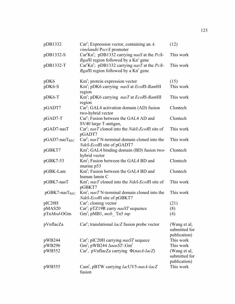
123
pDB1332 Carr; Expression vector, containing an A. vinelandii PscrX promoter
(12)
pDB1332-S CarrKnr; pDB1332 carrying nasS at the PciI-BgalII region followed by a Knr gene
This work
pDB1332-T CarrKnr; pDB1332 carrying nasT at the PciI-BgalII region followed by a Knr gene
This work
pDK6 Kmr; protein expression vector (15) pDK6-S Kmr; pDK6 carrying nasS at EcoRI-BamHI
region This work
pDK6-T Kmr; pDK6 carrying nasT at EcoRI-BamHI region
This work
pGADT7 Carr; GAL4 activation domain (AD) fusion two-hybrid vector
Clontech
pGAD7-T Carr; Fusion between the GAL4 AD and SV40 large T-antigen,
Clontech
pGAD7-nasT Carr; nasT cloned into the NdeI-EcoRI site of pGADT7
This work
pGAD7-nasTREC Carr; nasT N-terminal domain cloned into the NdeI-EcoRI site of pGADT7
This work
pGBKT7 Kmr; GAL4 binding domain (BD) fusion two-hybrid vector
Clontech
pGBK7-53 Kmr; Fusion between the GAL4 BD and murine p53
Clontech
pGBK-Lam Kmr; Fusion between the GAL4 BD and human lamin C
Clontech
pGBK7-nasT Kmr; nasT cloned into the NdeI-EcoRI site of pGBKT7
This work
pGBK7-nasTREC Kmr; nasT N-terminal domain cloned into the NdeI-EcoRI site of pGBKT7
This work
pIC20H Carr; cloning vector (21) pMAS20 Carr; pTZ19R carry nasST sequence (8) pTnMod-OGm Gmr; pMB1, mob_ Tn5 tnp
(4)
pVnflacZa Carr; translational lacZ fusion probe vector (Wang et al, submitted for publication)
pWB244 Carr; pIC20H carrying nasST sequece This work pWB296 Gmr; pWB244 �nasST::Gmr This work pWB552 Carr, pVnflacZa carrying �(nasA-lacZ) (Wang et al,
submitted for publication)
pWB555 Camr, pBTW carrying lacUV5-nasA-lacZ fusion
This work

124
pWB643 Carr; pVnflacZa carrying �(nasA-lacZ) with all leader hairpins deleted
(Wang et al, submitted for publication)
pWB650 Carr; pVnflacZa carrying �(nasA-lacZ) with the 5’ stem of the leader the antiterminator hairpin deleted
(Wang et al, submitted for publication)
pWB664 Carr; pVnflacZa carrying �(nasA-lacZ) with the 3’ stem of leader middle hairpin deleted
(Wang et al, submitted for publication)
pWB692 Camr; pBTW carrying lacUV5-nasA-lacZ fusion with all leader hairpins deleted
This work
pWB901 Camr; pBTW carrying lacUV5-nasA-lacZ fusion with the 5’ stem of the leader the antiterminator hairpin deleted
This work
pWB902 Camr; pBTW carrying lacUV5-nasA-lacZ fusion with the 3’ stem of leader middle hairpin deleted
This work
pWB903 Carr; pVnflacZa carrying lacUV5-T’-lacZ fusion
This work
pWB908 Carr; pVnflacZa carrying �(nasA-lacZ) with the terminator hairpin deleted
(Wang et al, submitted for publication)
pWB910 Camr; pBTW carrying lacUV5-nasA-lacZ fusion with the terminator hairpin deleted
This work
pWB923 Carr; pVnflacZa carrying lacUV5-nasST’-lacZ fusion with nasS codon 56 — 388 deletion
This work
pWB924 Carr; pVnflacZa carrying lacUV5-nasST’-lacZ fusion with nasS codon 56 — 388 deletion and 389 CCC� TGA mutation
This work
a Abbreviations: Car, carbenicillin; Cam, Chloramphenicol; Gm, Gentamicin; Km,
Kanamycin; Rif, rifampicin.

125
TABLE 2. Primers used in this study
Name Sequence (5’-3’)a Restriction enzymeb and use
1BamH162innasA
TGGATCCATGCCGGCCAGCACCGGAGAG
NotI; Clone nasA-lacZ fragment
75pblueconfirmr1
GTATTACCGCCTTTGAGTG
Clone nasA-lacZ fragment
76pBTNotIr AGCGGCCGCTCACAATTCCACAC PCR pBT BglII-nasS ATAAGATCTTTAGGAGGATGCGCAG BglII; Clone nasS
into pDB1332, pDK6
BglII-nasT ATAGATCTCAGCTTCCCAGCATGTCGTGCATG
BglII; Clone nasT into pDB1332, pDK6
BTr TCCGTTGTGGGGAAAGTTATC PCR pBT EcoRI-nasS ATAGAATTCCATGACAGACCACCACG
CA ACTTC EcoRI; Clone nasS into pDK6
EcoRI-nasT TCGAATTCCATGCTGCGCATCCTCCTG EcoRI; Clone nasT into pDK6
EcoRITr AGAATTCTCAGCTTCCC AACATG EcoRI; Clone nasT into pGAD7 or pGBK7
EcoRITr2 TTGAATTCTCAGCGCTGCGCCTGGATGCCCTC
EcoRI; Clone nasT N-terminal domain into pGAD or pBGK
KninBBR1f GGGCTTACATGGCGATAG Amply Knr cassette from pBBR1MCS2
KninBBR1r CCGAAGCCCAACCTTTC
Amply Knr cassette from pBBR1MCS2
nasSf CCAGGGACTGTACGGACTGATC nasST deletion confirmation
nasSr GGCGCATCAG GGTATAG nasST deletion confirmation
NdeITf ACATATGCTGCGCATCCTCCTGAT C NdeI; Clone nasT into pGAD7 or pGBK7
O13NotII+77 AGAGCGGCCGCCGACAGATCACATAAG
NotI; Clone nasA-lacZ fragment
O20NotI-Sf AGAGCGGCCGCGGTCCCAACGGCGGGG
lacUV5-nasST’-lacZ construction
O21nasS GATCAACCTGGGCTTCATGCCCGCCA lacUV5-nasST’-
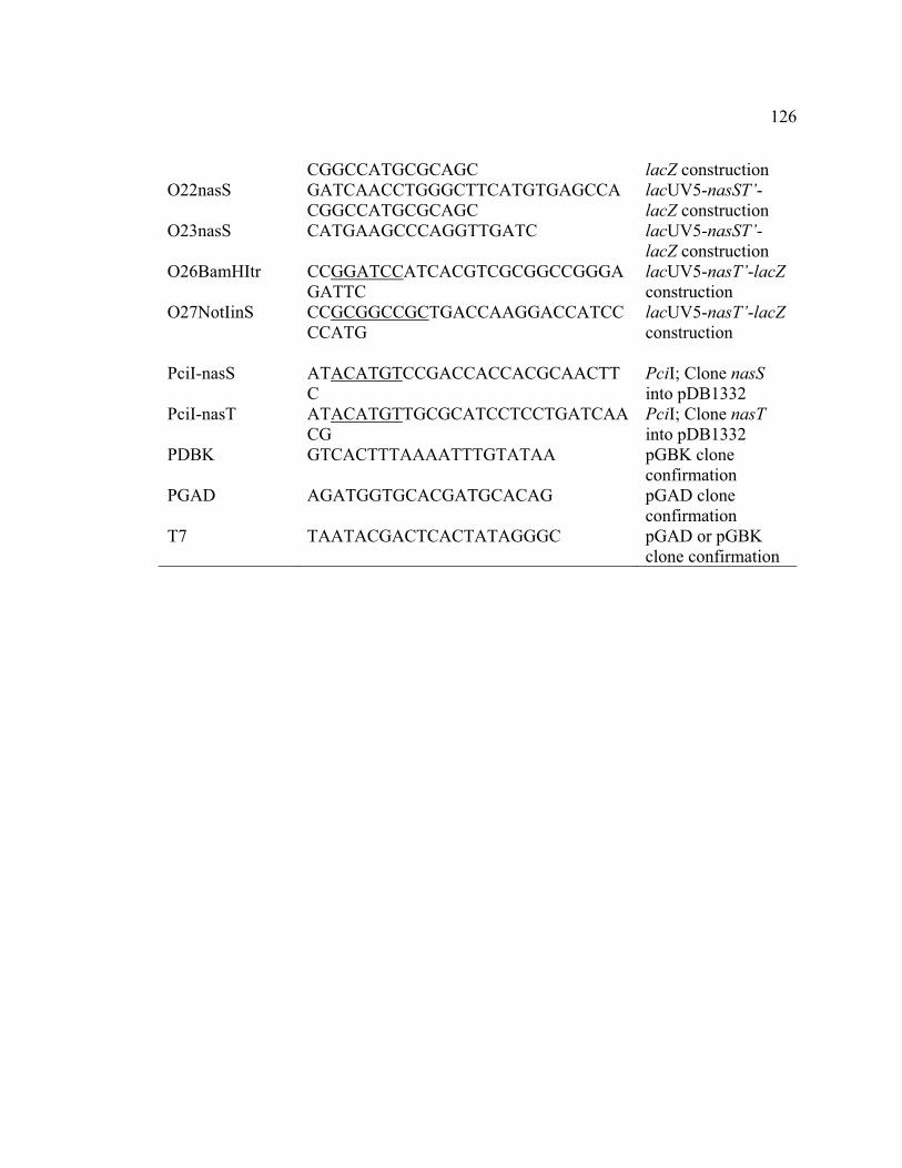
126
CGGCCATGCGCAGC lacZ construction O22nasS GATCAACCTGGGCTTCATGTGAGCCA
CGGCCATGCGCAGC lacUV5-nasST’-lacZ construction
O23nasS CATGAAGCCCAGGTTGATC lacUV5-nasST’-lacZ construction
O26BamHItr CCGGATCCATCACGTCGCGGCCGGGAGATTC
lacUV5-nasT’-lacZ construction
O27NotIinS CCGCGGCCGCTGACCAAGGACCATCCCCATG
lacUV5-nasT’-lacZ construction
PciI-nasS ATACATGTCCGACCACCACGCAACTT
C PciI; Clone nasS into pDB1332
PciI-nasT ATACATGTTGCGCATCCTCCTGATCAACG
PciI; Clone nasT into pDB1332
PDBK GTCACTTTAAAATTTGTATAA pGBK clone confirmation
PGAD AGATGGTGCACGATGCACAG pGAD clone confirmation
T7 TAATACGACTCACTATAGGGC
pGAD or pGBK clone confirmation

127
Table 3. NasT dimerization assay in a yeast two-hybrid systema
Fusionb
BD AD �-Galactosidase activity (Miller units)c
NasT NasT 35 ± 3 TREC
f TREC 2 ± 0 NasT Vector only <1 Vector only NasT <1 TREC Vector only <1 Vector only TREC <1
Control (+) 12 ± 0d Control (-) 1 ± 0e
a All strains are derivatives of yeast strain AH109 carrying two corresponding plasmids.
b BD, fusion to GAL4 DNA-binding domain of plasmid pGADT7; AD, fusion to GAL4
transcriptional activation domain of the plasmid pGBKT7.
c �-Galactosidase assays of yeast strains were performed by the protocols supplied by the
manufacturer (Clontech). All data are presented as the mean values of three replicates ±
standard deviations from a representative experiment.
de Positive are plasmids pGBKT7-53 +pGADT7-RecT; negative control are pGBKT7-
Lam + pGADT7-RecT.
f TREC N-terminal REC domain of NasT.
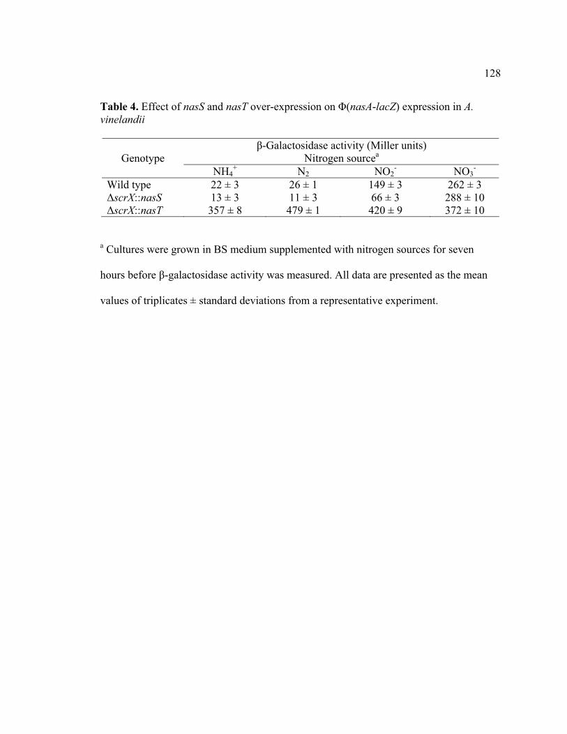
128
Table 4. Effect of nasS and nasT over-expression on �(nasA-lacZ) expression in A. vinelandii
�-Galactosidase activity (Miller units) Nitrogen sourceaGenotype
NH4+ N2 NO2
- NO3-
Wild type 22 ± 3 26 ± 1 149 ± 3 262 ± 3 �scrX::nasS 13 ± 3 11 ± 3 66 ± 3 288 ± 10 �scrX::nasT 357 ± 8 479 ± 1 420 ± 9 372 ± 10
a Cultures were grown in BS medium supplemented with nitrogen sources for seven
hours before �-galactosidase activity was measured. All data are presented as the mean
values of triplicates ± standard deviations from a representative experiment.

129
Figure Legends
Figure 1. nasST mutation and complementation test in A. vinelandii UW1. Cultures were
grown in BS medium supplemented with nitrogen sources. (A) UW1 (nif -). (B)
AVW362 (nif -nasST::Gmr). (C) AVW718 (nif -nasST::Gmr scr:: nasS-Knr). (D)
AVW719 (nif -nasST::Gmr scr:: nasT-Knr).
Figure 2. Test of NasT regulation at the nasAB leader region in E. coli 1061. The nasA-
lacZ cassette is fused the downstream of lacUV5 promoter. The nasS or nasT is induced
by tac promoter in the plasmid pDK6. The top line represents the sequence ranges from
the nasAB transcription start site +1 to the +297 nt (the 162nd nt within the nasA ORF).
The bend arrow represents the transcription direction, facing arrows (designated I, II, and
III) represent inverted sequences in the nasA leader region. The arrows represent the
lacUV5 promoter; the empty rectangles represent the deletion regions. The plasmid pairs
were co-transformed into E. coli M1061 for �-galactosidase activity assays. Two hundred
microliter transformant overnight cultures grown in LB broth supplied with kanamycin
and chloramphenicol were sequentially pelleted, rinsed with LB broth twice, and diluted
in 10 ml of fresh LB broth supplied with kanamycin, chloramphenicol, and 10 �M of
IPTG (isopropyl-1-thio-�-D-galactopyranoside), shake for three hours. Two hundred
microliter cultures were used for �-galactosidase activity. NT, not tested. All data are
presented as the means values for triplicate samples ± standard deviations from a
representative experiment.
130
Figure 3. Modular structures of NasS, NasT, and protein sequence alignment of NasT
with AmiR. The numbers above the bar or sequence indicate amino acid residue
positions. (A) NasS has a central conserved region homologous to TauA (Shaded area).
(B) NasT has an N-Terminal REC domain and C-terminal ANTAR domain (32). (C)
Sequence alignment of AmiR and NasT. The coiled-coil regions are underlined with
dish-lines. For AmiR, coiled-coil region has been experimentally confirmed (26).
Figure 4. Overlap region of nasS and nasT open reading frames. The nasT start codon
ATG and nasS stop codon TGA are bold-faced; the presumptive Shine-Dalgarno
sequence of nasT is underlined. (A) Letters above the DNA sequence represents C-
terminus of NasS; letters below the DNA sequence represents N-terminus of NasT. (B)
The predicted secondary structure around the nasS and nasT overlap region.
Figure 5. Translational coupling of nasS and nasT in A. vinelandii. The top line
represents the NasS and NasT ORFs with numbers above the bar indicating the
nucleotide residues. 186nasT is the 186th nt of nasT. The arrows represent the lacUV5
promoter; the empty rectangles represent the deletion regions; TGA represents the codon
CCC�TGA replacement. Values on the right show �-galactosidase activity. Cultures
were grown in BS medium supplemented with the indicated nitrogen sources for seven
hours before �-galactosidase activity was measured. All data are presented as the mean
values of three replicates ± standard deviations from a representative experiment.
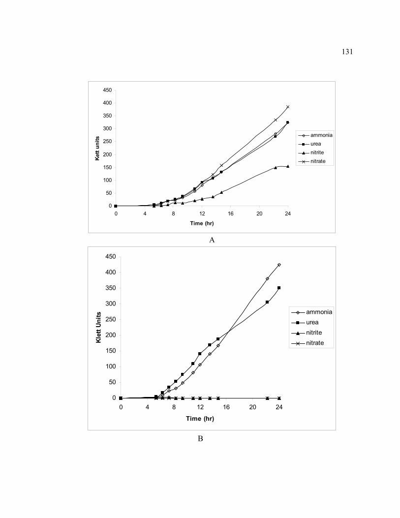
131
0
50
100
150
200
250
300
350
400
450
0 4 8 12 16 20 24
Time (hr)
Ket
t uni
ts
ammoniaureanitritenitrate
A
0
50
100
150
200
250
300
350
400
450
0 4 8 12 16 20 24
Time (hr)
Kle
tt U
nits ammonia
ureanitritenitrate
B

0
50
100
150
200
250
300
350
0 4 8 12 16
Time (hr)
Klet
t uni
ts
20 24
ammoniaureanitritenitrate
0
50
100
150
200
250
300
350
400
450
0 4 8 12 16 20 24
Time (hr)
Kle
tt un
its
ammoniaureanitritenitrate
D
Figure 1.
C
132

133
Figu
re 2
.
�-G
alac
tosi
dase
act
ivity
(Mill
er U
nits
)
pDK
6
(vec
tor a
lone
) N
asS
Nas
T
�-ga
lact
osid
ase
ratio
(+na
sT/-
nasT
)
235
± 2
234
±5
7917
±
233
33.7
341
± 18
N
T 53
2 ±
14
1.6
512
± 4
NT
1788
± 5
3.
5
3524
± 5
6 N
T 29
95 ±
9
0.9
133
+1
+297
nasA
II
III
I

134
1 117 192
REC ANTAR
10 338 416
TauA
1A
B
Figure 3

135
NasS D L H A L S E L P S S A D D Q G P NasS D L H A L S E L P S S A D D Q G P …CGCAGCTTCGACCTCCACGCCCTGAGCGAGCTGCCCTCCTCGGCCGATGACCAAGGA …CGCAGCTTCGACCTCCACGCCCTGAGCGAGCTGCCCTCCTCGGCCGATGACCAAGGACCA S P C A S S C * TCCCCATGCTGCGCATCCTCCTGATCAACGACACGCCGAAAAAGGTCGGCCGCCTGAAAAGC NasT M L R I L L I N D T P K K V G R L K S
A
B
Figure 4
A G G A A C C A G T A G C
T C C T T G A T C A A C G
C C
C G T
T C A
T C G
C C T
C
C
A
C
C
A
GCAGCTGCCCTCCTCGGC
C G ACACGCCGA

136
Figu
re 5
�-ga
lact
osid
ase
activ
ity (M
iller
uni
tes)
N
O3-
NH
4+
461
± 4
51
7 ±
11
3
92 ±
4
3
54 ±
4
1
34 ±
1
1
24 ±
2
nasS
nasT
TGA
1 16
9
11
67 1
280
186 n
asT
136