studies origin - gut.bmj.comgut.bmj.com/content/gutjnl/11/7/600.full.pdf · gut, 1970, 11, 600-609...
TRANSCRIPT
Gut, 1970, 11, 600-609
Studies on the origin of faecal amino acidsin cystic fibrosis1
J. W. T. SEAKINS, R. S. ERSSER, AND I. S. E. GIBBONS2From the Department of Chemical Pathology, Hospital for Sick Children and the Institute of ChildHealth, London
SUMMARY Evidence is presented which demonstrates that excess faecal amino acids incystic fibrosis are derived from unabsorbed dietary protein. Changes in the absorption of fatand nitrogen have little effect on the amino-acid patterns.
In an earlier paper (Gibbons, Seakins, andErsser, 1967) it was shown that the faecal amino-acid patterns of children with cystic fibrosis wereheavier than those of normal children and adults.Most of the amino acids found in proteins wereinvolved, together with some of their decarbo-xylation products, amines'and 'non-alpha aminoacids. These findings were in contrast to normalfaecal patterns found in an infant with Hartnupdisease (Seakins and Ersser, 1967) and threechildren with cystinuria (unpublished observa-tions).
Previously protein absorption in patients withcystic fibrosis and the effect of pancreatin havebeen studied by the balance technique (Harris,Norman, and Payne, 1955) or by the change inblood amino acids following loading tests (West,Wilson, and Eyles, 1946; Anfanger and Heaven-rich, 1949). There is, however, little informationon the composition of faeces and none on thepossible interaction between unabsorbed con-stituents. In this paper, the composition of in-dividual stools from different patients with cysticfibrosis are compared with those from normalchildren and adults. Further information wassought on the sources of faecal amino acids byvarying the dietary protein and fat and by partialsterilization of the gut.'A preliminary account of this work was presented at the 5thInternational Cystic Fibrosis Conference, Cambridge in Sep-tember 1969. Please address reprint requests to J. W. T. Seakins,Institute ofChild Health, 30 Guilford Street, London WC1W 1EH.'Present address: The Hahnemann Medical College, Philadelphia,USA.Received for publication 5 December 1969.
Methods
COLLECTION OF FAECESSpecimens were collected into containers, thetime and presence or absence of marker noted.The specimens were either stored on solid carbondioxide or at -20°C, which prevented furtherbacterial transformations, before analysis.Carmine or Edicol supra blue EG (ICI Ltd) wereused as markers and were given with the firstmeal of the day.
PREPARATION OF SAMPLESPreliminary investigations had shown that somefaecal specimens possessed considerable proteo-lytic activity which resulted in a significantincrease in the amino-acid pattern during thepreparation of the sample for analysis. This wasprevented by using dilute hydrochloric acid (3 %)to make an accurate dilution (3:1 w/w) of theindividual weighed faecal specimen in a Silversonhomogenizer. The final pH was about 2, and thepresence of the mineral acid did not interfere withthe subsequent analyses.
CHROMATOGRAPHY OF AMINO ACIDSAND AMINESA sample of the faecal homogenate (30 g) wasdiluted to 60 ml with acetone, shaken thoroughly,centrifuged, and the supernatant either stored at-20°C or processed immediately in the followingway.
on 13 August 2018 by guest. P
rotected by copyright.http://gut.bm
j.com/
Gut: first published as 10.1136/gut.11.7.600 on 1 July 1970. D
ownloaded from

Studies on the origin offaecal amino acids in cystic fibrosis
A volume of the supernatant equivalent to3 g faeces was diluted with an equal volume ofaqueous acetone (1:1), filtered if necessary, andpassed down a column of Zeo-Karb 225 (SRC10 H+, 1 x 10 cm) washed successively withwater, ethanol, and water to remove absorbedpigments, etc, and the amino acids and aminesdisplaced with 5N-ammonia (25 ml). The am-moniacal effluent was taken to dryness in a rotaryevaporator (a few millilitres of butanol wereadded to stop frothing) and the residue takenup in 10% isopropanol-water (0-3 ml).
All specimens were examined by one-waydescending chromatography for 16 hours, and alength of run of 45 cm, on Whatman no. 3paper with nBuOH-AcOH-H20 (120:30:50) andby two-way electrochromatography in 25-5 cmsquare sheets of Whatman no. 3 paper, pH 2,followed by ascending chromatography in theabove solvent system using an amount equivalentto 100 mg wet weight faeces (Seakins and Ersser,1967). Amino acids and amines were visualizedby ninhydrin/pyridine. The identity of the aminoacids and amines was confirmed by their electro-phoretic and chromatographic behaviour usingauthentic specimens, by their characteristiccolours with the ninhydrin/pyridine and /B-naphthaquinone-4-sulphonate (Folin's) reagents,and by specific location reagents (Table I); inaddition diazotized sulphanilic acid (Pauly'sreagent) was used to confirm the identity oftyrosine, tyramine, histidine, and histamine.Further confirmation where necessary wasobtained by fractionation of the ammoniacaleffluent on successive ion-exchange resins: (a)Zeo-Karb 226 (SRC 43, pH 7-0 pyridine acetate)which retained amines, basic amino acids, andbasic peptides (Blau, 1961) which were elutedwith acetic acid; (b) Zeo-Karb 225 (pyridineform) which retained the aromatic and non-ox-
Amino Compound Separation Location Reagent
Glu, Asp Electrophoresis, pH 4-4 0 2% ninhydrin and 5 % pyridinepyridine acetate in acetone'
Tyr, Phe Electrochromatography 0-2% ninhydrin and 5 % pyridine(see text) in acetone'
Leu, Ileu, Val, Test-pentanol-methyl ethyl 0-2% ninhydrin and 5%/ pyridineTyr NH2, (Met) ketone-water (60:20:20) in in acetone'
atmosphere of diethylamine'Ala n-butanol-acetic acid-water 0-2% ninhydrin and 5% pyridine
(120:30:50)l in acetone'Pro, Hypro n-butanol-acetic acid-water Isatin'
(120:30:50)lMet, (CySj) n-butanol-acetic acid-water lodoplatinate'
(120:30:50)'Try, Try-NH2 n-butanol-acetic acid-water Ehrlich's reagent'
(120:30:50)His, Gly n-butanol-acetic acid-water o-Phthalaldehyde'
(120:30:50)
Table I Separation methods for the semi-quantitationoffaecal amino acids and amines'Smith (1969)'Munier and Sarrazin (1964)'05% solution in acetone, 10 min at 40°C.
amino acids (Kakimoto and Armstrong, 1961);(c) De-Acidite FF (SRA 62, acetate) which re-tained aspartic and glutamic acids (Kakimoto andArmstrong, 1961).Amino acids were quantitated by visual com-
parison with standards of 2, 5, 10, and 20 gg,and where appropriate the samples were diluted sothat the concentration fell within the lower partof this range. Table I summarizes the proceduresemployed. Satisfactory methods could not befound for lysine, threonine, serine, and arginine,though the last three were rarely noted. Recoveryexperiments on authentic amino acids and acasein hydrolysate indicated an overall accuracyof between 5 and 10%.
AMINO NITROGENThe sample was prepared as described above forchromatography of amino acids and the driedammoniacal effluent stored overnight over con-centrated sulphuric acid in a vacuum dessicatorto remove residual ammonia. The residue wasdissolved in a suitable volume of 0 1M potassiumtetraborate, pH 9-4, to give an amino-nitrogenconcentration between 0-02 and 015 mM, usingthe chromatogram as a guide. The amino nitrogenwas determined by a trinitrobenzene sulphonicacid method (Mokrasch, 1967) as modified byPrenton and London (1967) for use on theTechnicon AutoAnalyzer, but omitting thedialyzer unit. Glycine (0-02-0-15 mM) was usedas a standard.
Total nitrogen was determined by Kjeldahl'smethod using selenium dioxide as catalyst, after'black ashing' faecal homogenates with concen-trated sulphuric acid. The method of Fowweatherand Anderson (1946) to evaluate faecal fat wasslightly modified. Equal weights (10 g) of the acidfaecal homogenate and plaster of paris weremixed, dried, and pulverized before extractionwith petroleum spirit (boiling point, 60-80°C) ina Soxhlet apparatus. A sample of this acid homo-genate (1 g) was dried at 105°C for 24 hr and re-weighed to measure water content. Urinaryphenolic acids were isolated and chromato-graphed according to the methods described bySmith, Seakins, and Dayman (1969).
Clinical Material
NORMAL SUBJECTSThe three normal adults are the authors ofthis paper. A total of 48 children (6 months-15years) who were inpatients at the Hospital forSick Children served as normal controls. Theywere either awaiting or recovering from minorsurgical operations or were mentally retardedchildren admitted for assessment. All were receiv-ing normal ward diets, had normal bowel motionsat the time of collection, had no history of
on 13 August 2018 by guest. P
rotected by copyright.http://gut.bm
j.com/
Gut: first published as 10.1136/gut.11.7.600 on 1 July 1970. D
ownloaded from
J. W. T. Seakins, R. S. Ersser, and L S. E. Gibbons
malabsorption or metabolic disease, and were notreceiving antibiotics. The son of one of theauthors (J.S.) acted as a further control, andspecimens were collected at home.
PATIENTSA total of 55 patients (3 months-15 years) witha proven diagnosis of cystic fibrosis of thepancreas were studied. Of these, eight patients(3 months-2 years) were newly diagnosed andhad not received any therapy. Where possible at
Amino Acid Adults' Children'(mg/10O g)
Mean J SD Range Mean ± SD Range
Gly 21 + 15 0- 5 51 ± 3-1 2-14Ala 3-4 1.1 2- 5 6-3 3-8 3-10Val 1.1 i03 1- 2 3-4 ± 28 1-10lieu 1-2 ± 04 1- 2 3-8 i 38 1-15Leu 1.1 ± 0-3 1- 2 3-8 3-6 1-15Met 0 - 0 05 - 0- 2Pro 1-8 +06 1- 3 2-8 2-2 1- 8Phe 0 - 0 1 - 0- 1Tyr 0 - 0- 1 1 - 0- 1Try 0 - 0- 1 1 - 0- 2His 0 - - 2 - 0- 2Asp 7-0 + 2-8 2-10 10-6 i 67 5-35Glu 25-1 ± 7-9 16-40 24-5 ± 10-4 12-30Tyr NH, 0 - 0 1 - 0- 2N-NH, 27-1 i 8-5 13-50 29-5 d 76 16-48
Total N(g/100 g) 1-39 i 033 0.71-1.99 1-11 + 0-34 0-71- 2-06Fat(g/100g) 4-69 ± 1-47 2-51-7-49 3-88 i 243 1.02-l105Water (%) 743 i 54 65-85 76-7 ± 6-9 60-86
Table II Faecal amino acids in normal subjects'Three adults, 16 specimens.'Fifteen children, 21 specimens.
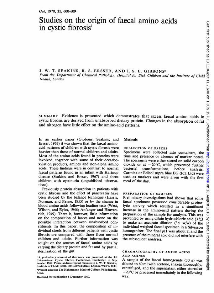
Fig. 1 Two-way electrochromatogram from anormal subject.
least three separate specimens were examined.Pancreatin supplements were given as eitherPancrex V powder or capsules, 0-5-2 g four timesdaily with meals, or as an enteric-coated prepara-tion, Pancrex V Forte tablets, 2-6 tablets fourtimes daily before meals (Paines and Byrne Ltd,Greenford).
Results
The detailed faecal analyses on normal subjectsare presented in Table II. Figure 1 is a typicaltwo-way electrochromatogram. A further 51specimens from 34 children were examined byone-way chromatography and by two-way electro-chromatography only. Qualitatively, all patternswere similar. Lysine was present in most speci-mens, but was not quantitated. No change inthe pattern of amino acids was observed whentwo adults took methyl cellulose (10 g/day forfour days) to increase faecal bulk, nor when anormal boy was given pancreatin.
Figure 2 is a typical two-way electrochromato-gram from a patient with cystic fibrosis. Thedetailed analyses on the 15 patients with cysticfibrosis are summarized in Table III and inFigures 3 and 4. The variations in isoleucine andvaline content approximately paralleled that ofleucine; aspartic acid was always within thenormal range, in contrast to glutamic acid;tryptophan, proline, and methionine were ele-vated in about one third of the patients; excesslysine was present in most patients. Additionallyvariable amounts of a-aminovaleric, ox- and y-amino-n-butyric acids, ,B-alanine, ethanolamine,cadaverine, and less frequently histamine andputrescine were observed. One-way chromato-grams and two-way electrochromatograms fromthe remaining 40 children with cystic fibrosis gavea similar range in faecal amino-acid patterns.The water content of faeces from the 15 patients
with cystic fibrosis was in the normal range(overall mean 75-1 ± 3-7%; Fig. 5) with theexception of three specimens from three patientswhich had a very high fat content (25-30%) and alow water content (approximately 50%/Y). Thesespecimens were not included in the above meanvalue. For a given patient, it was noted that ahigher fat content was associated with a lowerwater content (see also Table IV).The faecal nitrogen (1-28 ± 0-33 g/100 g) was
not significantly different from normal subjects,although the actual daily loss of nitrogen wassignificantly higher in most patients because ofthe increased faecal bulk. The fat content variedconsiderably (Fig. 5) and no relationship betweenfaecal amino nitrogen, total nitrogen, fat, and--bulk -eould -be discerned. This is- exemplified bycomparing the detailed figures in two patients.In the first patient (no. 6) faecal fat (3-4 ± 0-8g/100 g) and nitrogen (0-82 ± 0-08 g/100 g) were
602 on 13 A
ugust 2018 by guest. Protected by copyright.
http://gut.bmj.com
/G
ut: first published as 10.1136/gut.11.7.600 on 1 July 1970. Dow
nloaded from

Studies on the origin offaecal amino acids in cystic fibrosis
Fig. 2 Two-way electrochromatogram from a patientwith cystic fibrosis.
normal as were the corresponding daily faecaloutputs, 4.9 g fat and 1 11 g nitrogen, but therewas a heavy amino-acid pattern in all specimens.In the second patient (no. 15), the faecal nitrogenwas normal (1-67 0-10 g/100 g), the faecal fatwas elevated (15.4 77 g/100 g), and the corre-sponding daily outputs were grossly increased,being respectively 4.8 g and 47-7 g, but theamino nitrogen was just outside the normal range.Over two-thirds of the patients studied gave an
elevated urinary excretion of p-hydroxyphenyl-acetic acid, but there was no clear correlationbetween faecal tyramine and the excretion of itsurinary metabolite.
CHANGE IN NATURE AND QUANTITY OFDIETARY FATFour variations in diet were investigated inpatient no. 5 (Table III). As far as possible dietswere isonitrogenous and isocaloric. Faecalanalyses were performed for each period on sixconsecutive specimens collected at least one weekafter the change in diet. Table IV summarizesthe effects of these changes.The transit time remained unchanged through-
out at approximately 24 hours, although the
Case No.
1 2 3 4 5 6 7 8 9 10 11 12 13 14 15
Age (yr) 0.3 1-5 3 3 3-8 4 4 8 9 10 11 11 12 12 16Sex M F M F F M M F M F F M M M MMean nitrogen (g/100 g) 0-96 - 1-86 1-52 2-07 0-82 1.19 1-16 1-04 1-45 - 1-32 1.54 1-41 1,67Pancreatin' P 0 0 P 0 P P P P P 0 P P P PTransit time (hr) 22 - - 25 24 23 25 6 - 96 35 - 22 - 14
26 32 25 29 26 45No. of specimens 3 2 2 6 6 5 4 9 3 6 3 3 3 5 4
Table III Data on children with cystic fibrosis
'P, receiving pancreatin; 0, off pancreatin.
Amino Acid Period' Significance of DWerences between Periods(mg/ICC g)
I II llI IV
Gly 87 + 16' 70 ± 21 69 + 10 14 ± 13 (>005) I > II, III, >IV(<0001)Ala 60 ± 12 60 ± 28 88 + 10 104 + 18 I, II <III, IV (0 001)lleu 14 ± 5 18 ± 17 33 ± 12 30 ± 9 NotsignificantLeu 15 + 5 23 ±16 36 ±15 36 ± 13 Not significantVal 17 ± 8 27 ±13 35 ±13 36 ± 9 Not significantPro 3 + 1 2 1 4 3 2 ±1 Not significantPhe 5 + 1 12 7 1 1 ±5 1 1 ± 8 Not significantTyr 3 ± 1 2 ±2 3 + 3 5 ± 2 Not significantTry 1 1 1 7 ± 2 I, II, III, <IV (0.002)His 5 ±1 5 1 18 ± 13 11 ± 5 I, II, <III, IV (0 005)Asp 8 ±1 8 ± 1 8 ± 2 8 ± 1 Not significantGlu 57 ± 14 33 ± 13 57 ± 7 24 ± 10 I, III, >II, IV (0 001)Tyr-NH,' 10 ± 5 6 ± 5 14 ± 6 32 ± 11 I, II, III, <IV (0 001)N-NH,' 48 ± 21 71 ± 44 94 ± 15 103 ± 23 I, <II, III, IV (0O005)N(g/100 g) 2-07 ± 0-06 2-33 ± 0-13 2-40 ± 0-26 1-16 ± 0.11 1,II,IIJ, >IV(0.001)Fat (ng/100 g) 9.9 ± 51 6 5 ± 1-3 5-5 ± 1-4 7-1 ± 3 I, >II, III, IV (0 001)Water (%) 64 ± 6 74 ± 4 74 ± 5 82 ± 7 I, <II, III, <IV (0 01)
Table IV Changes in faecal composition with dietary fat'Period I, normal diet; II, low long-chain triglyceride (20 g/day); III, low long-chain triglyceride (20 g/day) with medium-chain triglyceride(40 g/day); IV, same triglyceride intake as Ill with pancreatin added. 'Mean ± 1 SD. 'Tyr-NHs, tyramine; 'N-NH,, amino nitrogen(TNBS method); 'N, nitrogen.
603 on 13 A
ugust 2018 by guest. Protected by copyright.
http://gut.bmj.com
/G
ut: first published as 10.1136/gut.11.7.600 on 1 July 1970. Dow
nloaded from
t~~~~~~~~~
I IL
5 1 0 1 5 5
Glutamic
I
- 10
Acid
T
E...
1 ± I10
Phenylalanine
.110
1540±-
20-._1
151 O0 -
Histidine 100-
5
Tyramine
80 ±
60±
40±
i II
8 9 10 11 12 13 14 15
20+ I'lll l l l
10Tyrosine
' . 'I ITI0I
l
1 2 3 4 5 6 7 8 9 10 1 1 13 1415
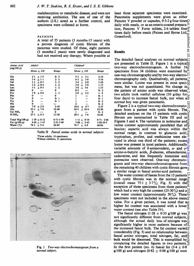
Patient numberFig. 3 Faecal amino acids and tyramine in cystic fibrosis (mean J+ 1 SD mg/100 ml). - - - Upper limit of normal (mean 2 SD).For phenylalanine, tyrosine, and histidine the upper limit ofnormal is about 2 mg/100 g, but tyramine is negligible.
GlycineI140 -
I120+
loo+
80-+
60-+
Alanine
I40-
20- .1
Leucine140 -
120 -
15
T I.J1
80-
60 --
4'1_0 '40-
i- -_-- w-- -- 1
1-
I-
1-
T
5
I' II-F-~5
III15
15
FI
1 34
0
5 6 7
09 W
1 :;= 5
1 . 1t
on 13 August 2018 by guest. P
rotected by copyright.http://gut.bm
j.com/
Gut: first published as 10.1136/gut.11.7.600 on 1 July 1970. D
ownloaded from
Studies on the origin offaecal amino acids in cystic fibrosis
11T 1
1 2 3 4 5 6
I I 2 I cx32
'Ii-L L ge40
-LC
JcI
7 8 9 10 1112131415
Patient number
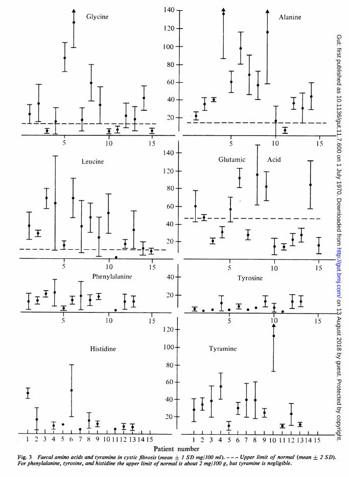
Fig. 4 Faecal amino nitrogen (mean + 1 SD) inpatients with cystic fibrosis.
frequency of defaecation diminished when lowlong-chain triglyceride (20 g/day) with medium-chain triglyceride (40 g/day) was introduced withadded pancreatin. The daily fat output fell from44-2 g to 11.0 g/24 hour when long-chain tri-glyceride was restricted and was further dim-inished to 6.0 g/24 hour when pancreatin supple-ments were added (p < 0.02). Gas-liquid chroma-tography of a saponified petroleum ether extractof acidified faeces when low long-chain tri-cylceride and medium-chain triglyceride weregiven demonstrated the absence of C1o and lowerfatty acids (Leyland, Fosbrooke, Lloyd, Segall,Tamin, Tomkins, and Wolff, 1969).
In addition to the changes in amino acidcomposition noted in Table IV, &-amino-valeric
Protein (g/lO0 g) Amino Acid Faeces (mg/100 g) (mean ± 1 SD)
Casein Gelatin Normal Gelatin pDiet (n = 4) Diet (n =5)
2 27 Gly 28 1 18 145 i37 <0-0013 10 Ala 38 ± 5 20 ±10 <0-0256 2 Val 17 5 2 ±05 <00016 2 Ileu 12 ± 6 02 i 06 <00059 4 Leu 18 ± 4 1 0.7 <0 00110 14 Pro 4 - 1 - Not significant5 2 Phe 10 ± 7 1 + 1 0O056 1 Tyr 11 + 8 1 ± 1 0053 1 His 9 ± 3 6 ± 2 Not significant6 7 Asp 3 i 1 1 - Not significant20 11 Glu 23 ± 6 4 ± 1 <0-0010 13 Hypro 0 01 0 Try 0 03 1 Met 0 0
Tyr-NH, 23 ± 5 3 ± 3 <0 001N-NH, 71 ± 8 135 ± 8 <0 001
N (g7100 g) 1-32 ± 0-26 1-32 ± 0-18 Not significantFat (g/100 g) 12 6 ± 3.0 12-9 ± 2-2 Not significantWater (%/) 76 ± 1 74 ± 4 Not significant
Table V Composition of casein and gelatin and theeffect ofgelatin diet on faecal amino acids
Fig. 5 Water andfat content (mean +t 1 SD) inpatients with cystic fibrosis.
acid was prominent on the electrochromatogramsthroughout the four periods, whilst lysine, whichwas virtually absent when the patient was on anormal diet or was given low long-chain tri-glyceride, or when she was given low long-chaintriglyceride with medium-chain triglyceride, wasvery heavy when pancreatin was also given. Thisincrease in lysine would account for the increasein amino nitrogen. The excretion of p-hydroxy-phenylacetic acid varied from normal to grosslyabnormal Similar results were obtained in twoother patients studied in less detail.
GELATIN DIET
In three patients with cystic fibrosis, one normaladult, and one infantwith normalgastrointestinalfunction, first-class protein was replaced as faras practicable with gelatin for two days. Thefaecal amino-acid patterns of the two normalsubjects did not change. In one patient (no. 12)who received 43 g gelatin plus 19 g first-classprotein during the test period as against 60 gfirst-class protein in the control period there wasa very significant and prompt change in the faecalamino-acid pattern, which quickly returned tothe pattern observed in the control period when anormal diet was resumed. Table V summarizesthe results in this patient. Unexpectedly, nohydroxyproline was detected in the test period,y-amino-n-butyric acid and fi-alanine decreased,whilst 8-amino-n-valeric acid increased. In theother two patients with cystic fibrosis. the resultswere not as clear cut as in the first, although thechanges in the amino-acid patterns of the firstmarked stools were of the same order as in thefirst patient, but subsequent faecal patternsshowed greater variation. The following signi-ficant changes were noted. In the second patient
160 r 90
120 F
80k
40k
v)W
nc)
00
aon0
0a_
Et
0Patient number
1 1 1 1 1 1 1 1 1 1 1 -L-1
605 on 13 A
ugust 2018 by guest. Protected by copyright.
http://gut.bmj.com
/G
ut: first published as 10.1136/gut.11.7.600 on 1 July 1970. Dow
nloaded from
J. W. T. Seakins, R. S. Ersser, and L. S. E. Gibbons
(no. 14) the glycine content increased from40 ± 13 to 100 ± 5 mg/100 g (P, 0.001) whileglutamic acid fell from 83 ± 28 to 15 ± 5 (P,0.005), alanine from 47 ± 22 to 15 ± 5 (P, 0.05),and tryptophan was no longer detectable. In thethird patient (no. 13) there was an immediateincrease in glycine from 18 ± 12 mg/100 g (5-30)to 200 ± 70 (150-300) (P, 0.02). As with the firstpatient, no hydroxyproline was detected in thesetwo test periods. The effect of pancreatin supple-ments was also investigated in these two patients:no consistent changes in the pattern wereobserved, except that hydroxyproline was stillnot detected.
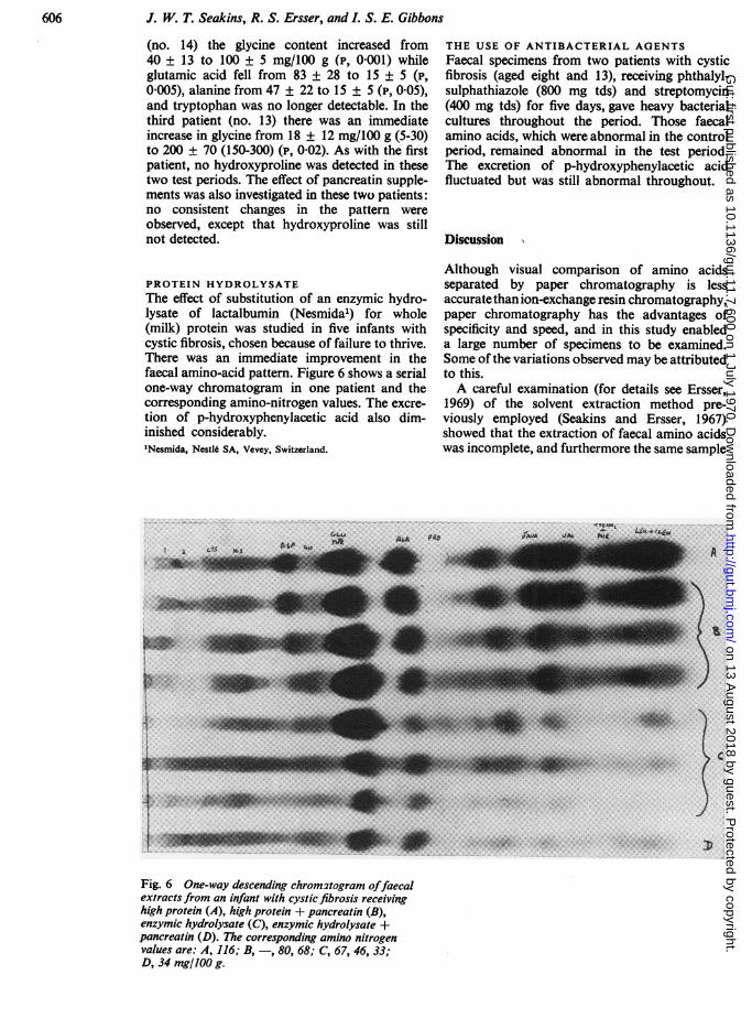
PROTEIN HYDROLYSATEThe effect of substitution of an enzymic hydro-lysate of lactalbumin (Nesmida1) for whole(milk) protein was studied in five infants withcystic fibrosis, chosen because of failure to thrive.There was an immediate improvement in thefaecal amino-acid pattern. Figure 6 shows a serialone-way chromatogram in one patient and thecorresponding amino-nitrogen values. The excre-tion of p-hydroxyphenylacetic acid also dim-inished considerably.'Nesmida, NestI6 SA, Vevey, Switzerland.
THE USE OF ANTIBACTERIAL AGENTSFaecal specimens from two patients with cysticfibrosis (aged eight and 13), receiving phthalyl-sulphathiazole (800 mg tds) and streptomycin(400 mg tds) for five days, gave heavy bacterialcultures throughout the period. Those faecalamino acids, which were abnormal in the controlperiod, remained abnormal in the test period.The excretion of p-hydroxyphenylacetic acidfluctuated but was still abnormal throughout.
Discussion
Although visual comparison of amino acidsseparated by paper chromatography is lessaccuratethan ion-exchange resin chromatography,paper chromatography has the advantages ofspecificity and speed, and in this study enableda large number of specimens to be examined.Some of the variations observed may be attributedto this.A careful examination (for details see Ersser,
1969) of the solvent extraction method pre-viously employed (Seakins and Ersser, 1967)showed that the extraction of faecal amino acidswas incomplete, and furthermore the same sample
C:
Fig. 6 One-way descending chromatogram offaecalextracts from an infant with cystic fibrosis receivinghigh protein (A), high protein + pancreatin (B),enzymic hydrolysate (C), enzymic hydrolysate +pancreatin (D). The corresponding amino nitrogenvalues are: A, 116; B, -, 80, 68; C, 67, 46, 33;D, 34 mg/100 g.
606nu on 13 A
ugust 2018 by guest. Protected by copyright.
http://gut.bmj.com
/G
ut: first published as 10.1136/gut.11.7.600 on 1 July 1970. Dow
nloaded from
Studies on the origin offaecal amino acids in cystic fibrosis
could not be used for other analyses. Aqueousextraction methods (Bickel, 1964; Hooft, Carton,Snoeck, Timmermans, Antener, van den Hende,and Oyaert, 1968) gave variable increases inamino acids, traced to continued enzymic andbacterial proteolytic activity during homo-genization. Homogenization with phosphatebuffer followed by extraction into butanol(Yarbro and Anderson, 1966) gave a very poorrecovery of amino acids. By using dilute hydro-chloric acid, as detailed in the methods section,proteolytic activity was stopped, and an excellentextraction of faecal amino acids was achieved,with the exception of taurine and cysteic acidwhich are not retained on Zeo-Karb 225 (H+).This method of extraction does not distinguishbetween intracellular and extracellular aminoacids. These observations in part explain thehigher values reported by Hooft et al (1968) andthe lower x-amino-nitrogen values given byAsatoor, Chamberlain, Emmerson, Johnson,Levi, and Milne (1967).The trinitrobenzenesulphonic-acid method for
amino nitrogen (Mokrasch, 1967) was preferredto the specific a-amino-nitrogen method of vanSlyke, since it was readily automated, is lesssensitive to ammonia, and gives a better measureof amino acids (both of and non-ox) and aminesthan the latter, although oligopeptides retainedon the resin and present as a streak with lowmobility and Rr on the electrochromatogramswill also contribute to the colour yield.The gravimetric method of Fowweather and
Anderson (1946) for faecal fats was chosen ratherthan van der Kamer's since some children werereceiving medium-chain triglyceride (cf Leylandet al, 1969).The results in normal subjects when the dietary
protein was radically altered, earlier work onprotein amino-acid loads (Milne, Crawford,Girao, and Loughridge, 1960; Milne, Asatoor,Edwards, and Loughridge, 1961; Seakins andErsser, 1967), and the study of the fate of cyclo-leucine (Christensen and Clifford, 1962) showthat faecal amino acids in normal subjects arepredominantly of bacterial origin or arise throughthe bacterial action on unabsorbed endogenousprotein (Nasset, 1964) rather than by secretioninto the large bowel. Likewise, Sheffner, Kirsner,and Palmer (1948) found that the compositionof normal faecal protein was little changed whendietary protein was varied.
In contrast to the consistent patterns given bynormal subjects. the majority of children withcystic fibrosis gave heavy patterns which variedsomewhat from sample to sample in an individualand from patient to patient. It is therefore un-likely that a defect in amino-acid transportanalogous to that found in Hartnup disease(Milne et al. 1960) or cystinuria (Milne et al,1961) operates in cystic fibrosis or excess secretionof amino acids into the large bowel occurs.
In spite of the increased faecal bulk and
reduced transit time observed in cystic fibrosis,the water content of faeces was in the same rangeobserved in normal subjects, indicating efficientwater absorption from the large bowel.The total faecal nitrogen comprises proteins,
substances such as ammonia and volatile amineswhich are lost in the column preparation, andamino compounds which are measured by theTNBS method. In view of the high faecal amino-nitrogen content found in cystic fibrosis, thefinding of the same nitrogen content as in normalsubjects was unexpected. This implies that theprotein and/or volatile amine content is lower incases of cystic fibrosis, which is consistent withthe observation that these faecal specimens havethe same water content but higher fat contentthan normal specimens. Furthermore, the longertransit time in normal subjects would allow moreextensive degradation of any amino acid presentto substances such as ammonia. No relationshipbetween total nitrogen, amino-nitrogen, fat, bulk,or water content and the amino-acid pattern ofindividual specimens was noted in patientsreceiving a free-choice diet.
Pancreatin was not the major source of faecalamino acids, for heavy patterns were observed ineight newly diagnosed patients before any therapyhad been instituted, although there was someevidence to suggest that pancreatin given withordinary protein further increased the abnormalpattern.
Controlled dietary studies indicated thatsteatorrhoea was not the cause of the amino-acidorrhoea, although reduction in faecal fat byreplacing long-chain triglyceride with medium-chain triglyceride and subsequently adding pan-creatin did produce significant changes in someamino acids, which may reflect altered bacterialmetabolism.
Direct evidence for the dietary origin of faecalamino acids in cystic fibrosis was obtained frominvestigations in which first-class protein wasreplaced by second class. Previous workers(Anfanger and Heavenrich, 1949; Christensenand Schwachman, 1949; Gould and Shwachman,1956) showed that gelatin, in common with otherproteins, is poorly absorbed in untreated cysticfibrosis. With the exception of alanine, proline,and hydroxyproline, the prompt changes infaecal amino acids were in the direction anti-cipated from the difference in composition offirst-class protein (eg, casein) and gelatin. It isknown that proline. hydroxyproline, and alanineoccur in gelatin (collagen) in sequences such asGly-Pro-Ala, Gly-Pro-Hyp, Gly-Pro-Gly, whichare more resistant to degradation by the usualproteolytic enzymes (Grassmann, Hannig, andSchleyer, 1960; Grassman, Nordwig, and Hor-mann. 1961) and this is the most likely explana-tion for the low values found for alanine andproline and the absence of hydroxyproline, ratherthan degradation of these amino acids.The improvement in amino-acid pattern follow-
607 on 13 A
ugust 2018 by guest. Protected by copyright.
http://gut.bmj.com
/G
ut: first published as 10.1136/gut.11.7.600 on 1 July 1970. Dow
nloaded from
608 J. W. T. Seakins, R. S. Ersser, and I. S. E. Gibbons
ing the introduction of an enzymic proteinhydrolysate indicated that absorption of (free)amino acids in cystic fibrosis is not seriouslyimpaired, and this is in agreement with earlierobservations on the blood amino-acid responsefollowing loading tests of casein hydrolysates orglycine (West et al, 1946; Anfanger and Heaven-rich, 1949; Rossi and Menano, 1953).Although the tryptic and chymotryptic activity
of faeces from patients with cystic fibrosis notreceiving pancreatin is normally negligible(Haverback, Dyce, Gutentag, and Montgomery,1963) the evidence presented points to bacterialaction on unabsorbed (partially digested) dietaryprotein as an important source of faecal aminoacids in these patients. Many bacteria are cap-able of forming peptidases and proteinases whichare more active against denatured proteins thanagainst native proteins (Sokatch, 1969). However,attempts to sterilize the gut were not successful,so that direct evidence for bacterial proteolysiscould not be obtained; neomycin cannot be usedsince it itself produces a malabsorption syndrome(Asatoor et al, 1967).Non-protein amino acids found in abnormal
amounts in faecal specimens from children withcystic fibrosis are most likely derived by bacterialaction from protein amino-acid precursors(Meister, 1965); ,B-alanine from aspartic acid(also found in babies by Bickel, 1964); y-amino-n-butyric acid from glutamic acid (in babies,Bickel, 1964; in adults, Asatoor et al, 1967) andfirst tentatively identified by (Ross 1951), andS-amino-n-valeric acid (not previously reported)from lysine and proline (Rodwell, 1969). Theidentity of this last amino acid was confirmed bythe procedures outlined in the section onmethods. The diamines cadaverine (from lysine)and putrescine (from ornithine) were found in anumber of specimens from patients with cysticfibrosis; cadaverine in trace amounts was foundin a few normal adults and children in agreementwith Abraham, Radonich, and Jones (1968), butputrescine was absent in all normal specimens.Tyramine was a very common constituent in
faecal specimens from children with cysticfibrosis, and is the source of urinary p-hydroxy-phenylacetic acid in these patients (Gibbons et al,1967; Gjessing and Lindeman, 1967), but un-expectedly phenylethylamine and tryptamine werenever found in any specimen, although manycontained abnormal amounts of phenylalanineand tryptophan, and the tyrosine decarboxylaseof S. faecalis has considerable phenylalaninedecarboxylase activity (Meister, 1965).
The authors wish to thank the Cystic FibrosisResearch Foundation Trust, the Nuffield Founda-tion, the Joint Research Board of the Hospital forSick Children, and the Institute of Child Healthfor financial help; Dr A. P. Norman for permis-
sion to study his patients, Dr Barbara E. Claytonfor valuable discussion, and Mrs W. Bell andMrs S. Evans for technical assistance. The authorsalso thank the Nestle Company for a gift ofNesmida, and Miss A. Fosbrooke for the gaschromatography.References
Abraham, A., Radonich, Z., and Jones, C. T. (1968). Lysineabsorption in the small intestine. The relevance of faecalcadaverikie as an index of lysine malabsorption. Clin. chim.Acta, 22, 619-622.
Anfanger, H., and Heavenrich, R. M. (1949). Amino acid toler-ance tests in children. Amer. J. Dis. Childh, 77, 425-436.
Asatoor, A. M., Chamberlain, M. J., Emmerson, B. T., Johnson,J. R., Levi, A. J., and Milne, M. D. (1967). Metaboliceffects of oral neomycin. Clin. Sci., 33, 111-124.
Bickel, H. (1964). OJber Zucker- und Aminosiurengehalt vonSauglingsstuhlen: Physiologische Grundlagen zur Diag-nose von Resorptionsstorungen. Mschr. Kinderheilk.,112, 173-176.
Blau, K. (1961). Chromatographic methods for the study ofamines from biological material. Biochem. J., 80, 193-200.
Christensen, H. N., and Clifford, J. A. (1962). Excretion of 1-amino-cyclopentanecarboxylic acid in man and the rat.Biochim. biophys. Acta(Amst), 62, 160-162.
Christensen, H. N., and Shwachman, H. (1949). Determinationof the plasma glycine after gelatin-feeding as a diagnosticprocedure for pancreatic fibrosis. J. clin. Invest., 28, 319-321.
Ersser, R. S. (1969). Ninhydrin positive substances in humanfaeces. Thesis for Fellowship, Institute of Medical Labora-tory Technology.
Fowweather, F. S., and Anderson, W. N. (1946). A method forthe determination of fat in faeces. Biochem. J., 40, 350-351.
Gibbons, 1. S. E., Seakins, J. W. T., and Ersser, R. S. (1967).Tyrosine metabolism and faecal aminoacids in cysticfibrosis of the pancreas. Lancet, 1, 877-878.
Gjessing, L. R., and Lindeman, R. (1967). p-Hydroxyphenylaceticacid in cystic fibrosis. Lancet, 2, 47-48.
Gould, B. S., and Shwachman, H. (1956). Studies in cystic fibrosis:determination of plasma proline following protein feedingas a diagnostic test for pancreatic insufficiency. Amer. J.Dis. Childh., 91, 584-587.
Grassmann, W. Hannig, K., and Schleyer, M. (1960). Zur Amino-saurensequenzen des Kollagens. II. Hoppe-Seylers Z.physiol. Chem., 322, 71-79.
Grassman, W. Nordwig, A., and Hormann, H. (1961). Amino-saurensequenzen des Kollagens. III. Hoppe-Seylers Z.physiol. Chem., 323, 48-60.
Harris, R., Norman, A. P., and Payne, W. W. (1955). The effectof pancreatin therapy on fat absorption and nitrogenretentionin children with fibrocystic disease of thepancreas.Arch. Dis. Childh., 30, 424-427.
Haverback, B. J., Dyce, B. J., Gutentag, P. J., and Montgomery,D. W. (1963). Measurement of trypsin and chymotrypsinin stool. A diagnostic test for pancreatic exocrine in-sufficiency. Gastroenterology, 44, 588-597.
Hooft, C., Carton, D., Snoeck, J., Timmermans, J., Antener, I.,van den Hende, C., and Oyaert, W. (1968). Further inves-tigations in the methionine malabsorption syndrome.Helv. paediat. Acta, 23, 334-349.
Kakimoto, Y., and Armstrong, M. D. (1961). The preparationand isolation of D-(-)-ig-aminoisobutyric acid. J. biol.Chem., 236, 3283-3286.
Leyland, F. C., Fosbrooke, A. S., Lloyd, J. K., Segall, M. M.,Tamin, I., Tomkins, R., and Wolff, 0. H. (1969). Use ofmedium-chain triglyceride diets in children with mal-absorption. Arch. Dis. Childh., 44, 170-179.
Meister, A. (1965). Decarboxylation. In Biochemistry ofthe AminoAcids, 2nd ed., vol. I, pp. 325-338. Academic Press, NewYork and London.
Milne, M. D., Asatoor, A. M., Edwards, K. D. G., and Lough-ridge, L. W. (1961). The intestinal absorption defect incystinuria. Gat, 2, 323-337.
Milne, M. D., Crawford, M. A., Girlo, C. B., and Loughridge,L. W. W. (1960). The metabolic disorder in Hartnupdisease. Quart. J. Med., 29, 407-421.
Mokrasch, L. C. (1967). Use of 2,4,6-trinitrobenzenesulfonicacid for the coestimation of amines, aminoacids andproteins in mixtures. Analyt. Biochem., 18, 64-71.
Munier, R. L., and Sarrazin, G. (1964). Amelioration d'unsysteme solvant a haut pouvoir s6parateur permettantl'analyse chromatographique sur papier a deux dimensionsdes m6langes d'amino-acides de grande mobilit6. J.Chromatogr., 13, 143-147.
on 13 August 2018 by guest. P
rotected by copyright.http://gut.bm
j.com/
Gut: first published as 10.1136/gut.11.7.600 on 1 July 1970. D
ownloaded from
609 Studies on the origin offaecal amino acids in cystic fibrosisNasset, E. S. (1964). The nutritional significance of endogenous
nitrogen secretion in non-ruminants. In The Role of theGastrointestinal Tract in Protein Metabolism, edited byH. N. Munro, pp. 83-96, Blackwell, Oxford.
Prenton, M. A., and London, D. R. (1967). The continuous in vivomonitoring of plasma amino-nitrogen. In 5th Colloquiumon Amino Acid Analysis, pp. 70-77. Technicon InternationalDivision, Domont, France.
Rodwell, V. W. (1969). Proline metabolism and lysine metabolism.In Metabolic Pathways, 3rd ed., edited by D. M. Green-berg, Vol. III, pp. 210-211 and 217-218. Academic Press,New York and London.
Ross, C. A. C. (1951). Faecal excretion of amino acids ininfants. Lancet, 2, 190-194.
Rossi, E., and Menano, H. P. (1953). Etude sur les aminoacidemiesprovoquees dans la fibrose pancr6atique avec bronchiec-tasies. Helv. paediat. Acta, 8, 530-543.
Seakins, J. W. T., and Ersser, R. S. (1967). Effects of amino acidloads on a healthy infant with the biochemical features ofHartnup disease. Arch. Dis. Childh., 42, 682-688.
Sheffner, A. L., Kirsner, J. B., and Palmer, W. L. (1948). Studieson amino acid excretion in man: 2. Amino acids in feces.J. biol. Chem., 176, 89-93.
Smith, I. (1969). Aminoacids, amines and related compounds. InChromatographic and Electrophoretic Techniques. 3rd ed.,edited by I. Smith, vol. 1. pp. 104-169. Heinemann,London.
Smith, I., Seakins, J. W. T., and Dayman, J. (1969). Phenolicacids. In Chromatographic and Electrophoretic Techniques.3rd ed., edited by I. Smith, Vol. 1., pp. 364-389.Heinemann, London.
Sokatch, J. R. (1969). In Bacterial Physiology and Metabolism,pp. 165-166. Academic Press, London and New York.
West, C. D., Wilson, J. L., and Eyles, R. (1946). Bloodamino nitrogen levels: changes in bloodamino nitrogen levelsfollowing ingestion of proteins and ofa protein hydrolysatein infants with normal and with deficient pancreaticfunction. Amer. J. Dis. Childh., 72, 251-273.
Yarbro, M. T., and Anderson, J. A. (1966). L-tryptophan meta-bolism in phenylketonuria. J. Pediat., 68, 895-904.
The June 1970 IssueTHE JUNE 1970 ISSUE CONTAINS THE FOLLOWING PAPERS
A psychiatric study of patients with diseases ofthe small intestine DAVID GOLDBERG
Permeability of the small intestine to substancesof different molecular weight C. A. LOEHRY,A. T. R. AXON, P. J. HILTON, R. C. HIDER, ANDB. CREAMER
Scanning and transmission electron microscopicstudies of human intestinal mucosa PETER G.TONER, KATHARINE E. CARR, ANNE FERGUSON, ANDCOLIN MACKAY
The effect of diet on ileostomy function T. J.THOMSON, J. RUNCIE, AND A. KHAN
Morphological changes of the small-intestinalmucosa of guinea pig and hamster followingincubation in vitro and perfusion in vivo withunconjugated bile salts THOMAS S. LOW-BEER,ROBERTO E. SCHNEIDER, AND WILLIAM 0. DOBBINS
Small intestinal mucosal abnormalities in relativesof patients with dermatitis herpetiformis JANETMARKS, DAVID BIRKETIT, SAM SHUSTER, AND D. F.ROBERTS
The influence of age on the xylose absorptiontest MARTIN J. KENDALL
Supersensitivity and gastric emptying aftervagotomy J. TINKER, N. KOCAK, T. JONES, H. I.GLASS, AND ALAN G. COX
Inflammatory bowel disease in ankylosingspondylitis M. I. V. JAYSON, P. R. SALMON, ANDW. J. HARRISON
Ligation of the hepatic artery in the treatment ofheart failure due to hepatic haemangiomatosisM. 0. RAKE, M. M. LIBERMAN, J. L. DAWSON,RACHEL EVANS, E. B. RAFTERY, L. LAWS, ANDROGER WILLIAMS
A clinical and histochemical study of cholestasisV. J. DESMET, A.-M. BULLENS, AND J. DE GROOTE
Plasma insulin in pancreatic disease M. F.ANDERSON, S. H. H. DAVISON, A. P. DICK, C. N.HALES, AND J. OWENS
TechniqueA modified cannula for sampling pancreatic juicein the dog A. N. FAWCETT
Progress reportGlucocorticoids and the gastrointestinal tract:current status PAUL CUSHMAN, JR.
Progress reportVagotomy for gastric ulcer H. L. DUTHIE
Notes and activities
Copies are still available and may be obtained from the PUBLISHING MANAGER,BRMSH MEDICAL ASSOCIATION, TAVISTOCK SQUARE, WC 1H 9JR price 17s. 6D.
on 13 August 2018 by guest. P
rotected by copyright.http://gut.bm
j.com/
Gut: first published as 10.1136/gut.11.7.600 on 1 July 1970. D
ownloaded from