study of the cytotoxicity of raw materials of cosmetic and ... · valiosa ajuda nos testes...
TRANSCRIPT

Study of the cytotoxicity
of raw materials of
cosmetic and topical
pharmaceutical
formulations Verónica Patrícia Moreira da Rocha
Mestrado em Bioquímica Departamento de Química e Bioquímica 2015
Orientadora
Isabel Almeida, Professora assistente, FFUP Co-orientadora
Cláudia Marques, Doutorada, FFUP

Todas as correções determinadas pelo júri, e só essas, foram efetuadas.
O Presidente do Júri,
Porto,
______/______/_________

FCUP/ICBAS Study of the cytotoxicity of raw materials of cosmetic and topical pharmaceutical formulations
III
ACKNOWLEDGEMENTS
Com a finalização de mais uma etapa não posso deixar de agradecer a algumas
pessoas que me apoiaram incondicionalmente.
Agradeço à minha orientadora Professora Doutora Isabel Filipa Almeida pela sua
constante dedicação, compreensão, supervisão, sabedoria, disponibilidade, paciência
e apoio prestado ao longo deste trabalho e à minha co-orientadora Doutora Cláudia
Marques pela partilha dos seus vastos conhecimentos, paciência, empenhamento e
disponibilidade que sempre evidenciou ao longo de todo o processo. Agradeço a
ambas a revisão da dissertação.
Agradeço a todos os Professores e técnicos do laboratório de Tecnologia
Farmacêutica da Faculdade de Farmácia da Universidade do Porto por me acolherem
da melhor forma. Um obrigado especial ao Professor Doutor Paulo Costa pelos
conhecimentos científicos que me facultou e pela constante disponibilidade
demonstrada e ao Professor Doutor Domingos Ferreira agradeço pela sua boa
disposição e motivação.
À Professora Doutora Emília Sousa e à Professora Doutora Marta Correia-da-Silva
agradeço a cedência dos compostos obtidos por síntese química. À Doutora Sara
Cravo agradeço toda a ajuda prestada no evaporador rotativo.
Um muito obrigada às meninas do laboratório: Ana Cláudia, Ana Rita, Bárbara,
Gabriela, Isabel, Joana, Karoll, Mariana, Marlene, Rachel, Rita e Sara por todos os
momentos de boa disposição e inter-ajuda. Um obrigado especial à Marlene pela
valiosa ajuda nos testes estatísticos e à Isabel por toda a ajuda e força que me deu
nesta difícil etapa final.
Pela amizade incondicional, apoio, carinho, por compreenderem as minhas
ausências e por me ajudarem a passar à frente em alguns momentos maus e por
festejarem comigo nos momentos bons, agradeço a todos os meus grandes Amigos,
não citando nomes porque são muitos, mas sabem quem são e são especiais.
Aos meus pais, avó, irmão, cunhada e sobrinha quero agradecer pelo amor
incondicional, carinho e força que me dão todos os dias. Agradeço ao meu irmão por
ser um exemplo para mim e mostrar-me sempre que desistir não é opção.
O meu muito obrigada a todos que, direta ou indiretamente, me apoiaram ou
prestaram colaboração nesta aventura enriquecedora tanto ao nível científico como
pessoal.
IV FCUP/ICBAS
Study of the cytotoxicity of raw materials of cosmetic and topical pharmaceutical formulations
ABSTRACT
The assessment of the toxicological profile of the raw materials of cosmetics and
pharmaceutical formulations is a regulatory requirement. This evaluation should take
into account the route of administration. Considering the specific case of topical
administration, the preliminary toxicological evaluation of new ingredients can be
conducted in skin cell lines, in particular keratinocytes and fibroblasts. The objective of
this work was to study the cytotoxicity of ingredients with potential interest for skin
application in human keratinocytes (HaCaT cell line). The tested compounds included
novel substances obtained by chemical synthesis: 1,2-Dihydroxyxanthone (1,2-DHX),
ascorbic acid persulfated (ASS), chlorogenic acid persulfated (ACS) and resveratrol
glicoside sulfate (RGS) synthesized in Pharmaceutical Chemistry Laboratory, FFUP.
Resveratrol (RSV), resveratrol glicoside (RSV-Gli) and a Castanea sativa leaf extract
were also tested. The study also included nano and micro particles containing RSV. In
an initial step solid lipid nanoparticles (SLN) and nanostructured lipid carriers (NLC)
were prepared using a homogenization/sonication technique, and polymeric
microparticles produced with a piezoelectric atomizing method. The particles were
characterized in relation to size using a laser diffraction technique. To obtain the C.
sativa extract, dry leaves were extracted with a solution of ethanol:water (7:3). The final
dry extract was obtained by lyophilization. Cytotoxicity assays were conducted with an
immortalized cell line of human keratinocytes (HaCaT). For further characterization of
cytotoxicity, different tests were selected, namely: MTT and AlamarBlue® reduction
assays (assessment of metabolic activity), neutral red (NR) uptake (evaluation of
lysosomal integrity), exclusion of propidium iodide (PI) and trypan blue (assessment of
membrane integrity). The results showed that RSV and its derivatives may be
considered non toxic in future studies for concentrations up to 100 μM for RSV, 500 μM
RSV-Gli and 1000 μM to RGS. The botanical extract and 1,2-DHX can be safely used
up to 100 μg/mL and 50 μM, respectively. The lipid nanoparticles prepared and tested
in this work were not considered as promising topical ingredients. Polymeric particles
showed deposition on the surface of the cell monolayer and aggregation which led to a
great variability of the results. The particles present several challenges for toxicological
testing. Other toxicological tests should be performed in order to meet regulatory
requirements before considering the use of the compounds tested for cosmetic or
pharmaceutical products.
Keywords: raw materials, keratinocytes, nanomaterials, cytotoxicity assays
FCUP/ICBAS Study of the cytotoxicity of raw materials of cosmetic and topical pharmaceutical formulations
V
RESUMO
A avaliação do perfil toxicológico das matérias-primas dos produtos cosméticos e
das formas farmacêuticas é uma exigência regulamentar. Esta avaliação deve ter em
perspetiva a via de administração. No caso particular da administração cutânea, uma
preliminar avaliação toxicológica de novos ingredientes deve ser conduzida em linhas
celulares cutâneas nomeadamente, queratinócitos e fibroblastos. O objetivo deste
trabalho foi o estudo da citotoxicidade de ingredientes com potencial interesse para
aplicação na pele em queratinócitos humanos (linha celular HaCaT). Os compostos
testados incluíram substâncias inovadoras obtidas por síntese química: 1,2-di-
hidroxixantona (1,2-DHX), ácido ascórbico sulfatado (AAS), ácido clorogénico
sulfatado (ACS) e o resveratrol glicosilado sulfatado (RGS), sintetizados no laboratório
de Química Farmacêutica, FFUP. Resveratrol (RSV), resveratrol glicosilado (RSV-Gli)
e um extrato de folhas de Castanea sativa também foram estudados. O estudo incluiu
também nano e micro partículas contendo RSV. Numa etapa inicial foram preparadas
nanopartículas lipídicas sólidas (SLN) e transportadores lipídicos nanoestruturados
(NLC) recorrendo a uma técnica de homogeneização/sonicação, e micropartículas
poliméricas produzidas com um método de atomização piezoelétrica. As partículas
foram caraterizadas relativamente ao tamanho utilizando uma técnica de difração a
laser. Para obtenção do extrato de C. sativa, as folhas secas foram extraídas com uma
solução de etanol:água (7:3). O extrato final seco foi obtido por liofilização. Os ensaios
de citotoxicidade foram realizados com uma linha celular imortalizada de
queratinócitos humanos (HaCaT). Para uma caracterização mais aprofundada da
citotoxicidade foram selecionados diferentes ensaios, nomeadamente: ensaio de
redução do MTT e do AlamarBlue® (avaliação da atividade metabólica), incorporação
do vermelho neutro (avaliação da integridade lisossomal), exclusão do iodeto de
propídeo (PI) e do azul de tripano (avaliação da integridade da membrana plasmática).
Os resultados mostraram que o RSV e os seus derivados, podem ser considerados
não tóxicos para estudos futuros em concentrações até 100 μM para o RSV, 500 μM
para o RSV-Gli e 1000 μM para o RGS. O extrato botânico e a 1,2-DHX podem ser
considerados seguros até 100 μg/mL e 50 μM, respetivamente. As nanopartículas
lípidas preparadas e avaliadas neste trabalho não mostraram ser um ingrediente
promissor para aplicação tópica. As partículas poliméricas depositaram e agregaram
sobre a superfície da monocamada de células, o que conduziu a uma grande
variabilidade dos resultados. As partículas apresentam vários desafios para testes
toxicológicos. Outros testes toxicológicos devem ser realizados de forma a cumprir os

VI FCUP/ICBAS
Study of the cytotoxicity of raw materials of cosmetic and topical pharmaceutical formulations
requesitos regulamentares antes de considerar a utilização dos compostos testados
em produtos cosméticos ou farmacêuticos.
Palavras-chave: matérias-primas, queratinócitos, nanomateriais, ensaios de
citotoxicidade
FCUP/ICBAS Study of the cytotoxicity of raw materials of cosmetic and topical pharmaceutical formulations
VII
INDEX OF CONTENTS
ACKNOWLEDGEMENTS ...................................................................................... III
ABSTRACT ........................................................................................................... IV
RESUMO ................................................................................................................V
INDEX OF CONTENTS ......................................................................................... VII
INDEX OF FIGURES...............................................................................................X
INDEX OF TABLES ............................................................................................ XIV
ABBREVIATIONS ................................................................................................ XV
1. INTRODUCTION ............................................................................................ 1
1.1 Safety assessment of raw materials for cosmetic and topical pharmaceutical
formulations............................................................................................................... 1
1.1.1 Toxicological testing of raw materials for topical formulations ............... 5
1.2 In vitro cytotoxicity assays ........................................................................... 8
1.2.1 Evaluation of cell membrane permeability ............................................ 9
1.2.1.1 Calcein-Acetoxymethyl assay ....................................................... 10
1.2.1.2 Fluorescein Diacetate uptake assay ............................................. 11
1.2.1.3 Lactate Dehydrogenase leakage assay ........................................ 11
1.2.1.4 Neutral Red uptake assay ............................................................ 12
1.2.1.5 Propidium Iodide exclusion assay ................................................. 13
1.2.1.6 GF-ACF assay .............................................................................. 13
1.2.1.7 Trypan Blue exclusion assay ........................................................ 14
1.2.2 Evaluation of metabolic activity .......................................................... 14
1.2.2.1 AlamarBlue® reduction assay ....................................................... 14
1.2.2.2 Tetrazolium Salts reduction assay ................................................ 15
1.2.3 Evaluation of adenosine triphosphate content .................................... 16
1.2.3.1 Bioluminescent assay ................................................................... 17
1.2.4 Evaluation of protein content .............................................................. 17
1.2.4.1 Bicinchoninic Acid assay .............................................................. 17
1.2.4.2 Sulphorhodamine assay ............................................................... 18
VIII FCUP/ICBAS
Study of the cytotoxicity of raw materials of cosmetic and topical pharmaceutical formulations
1.3 Use of cytotoxicity assays in safety assessment of raw material for cosmetic
and topical pharmaceutical formulations .................................................................. 20
1.3.1 Analyzed antioxidants raw materials .................................................. 23
2. AIM ............................................................................................................... 28
3. MATERIALS AND METHODS ..................................................................... 29
3.1 Materials ................................................................................................... 29
3.2 Methods .................................................................................................... 29
3.2.1 Preparation of the C. sativa leaf extract .............................................. 29
3.2.1.1 Qualitative analysis of the phenolic composition of C. sativa leaf
extract 30
3.2.2 Preparation of nano and microparticles .............................................. 31
3.2.2.1 Particle size measurements .......................................................... 33
3.2.3 Cell Culture ........................................................................................ 33
3.2.3.1 Characterization of the HaCaT cell line ......................................... 34
3.2.3.2 In vitro cytotoxicity assays ............................................................ 34
3.2.3.2.1 MTT reduction assay .............................................................. 35
3.2.3.2.2 AlamarBlue® reduction assay ................................................. 37
3.2.3.2.3 Neutral Red uptake assay ...................................................... 38
3.2.3.2.4 Trypan Blue exclusion assay .................................................. 40
3.2.3.2.5 Propidium Iodide exclusion assay .......................................... 41
3.2.4 Statistical analysis .............................................................................. 41
4. RESULTS ..................................................................................................... 43
4.1 Qualitative analysis of the phenolic composition of .................................... 43
C. sativa leaf extract .......................................................................................... 43
4.2 Particle size measurements ...................................................................... 44
4.3 Characterization of the HaCaT cell line ..................................................... 45
4.4 In vitro cytotoxicity assays ......................................................................... 45
4.4.1 MTT reduction assay .......................................................................... 46
4.4.2 AlamarBlue® reduction assay ............................................................. 52
4.4.3 Neutral Red uptake assay .................................................................. 56
4.4.4 Trypan Blue exclusion assay .............................................................. 61
4.4.5 Propidium Iodide exclusion assay ...................................................... 66

FCUP/ICBAS Study of the cytotoxicity of raw materials of cosmetic and topical pharmaceutical formulations
IX
4.4.6 Comparison between the results of the different in vitro cytotoxicity
assays…… .......................................................................................................... 67
5. DISCUSSION ............................................................................................... 69
6. CONCLUSIONS ........................................................................................... 74
7. BIBLIOGRAPHIC REFERENCES ................................................................ 76
X FCUP/ICBAS
Study of the cytotoxicity of raw materials of cosmetic and topical pharmaceutical formulations
INDEX OF FIGURES
Figure 1: Main requirements for pharmaceutical formulations. Adapted from [11]. ... 3
Figure 2: Schematic illustration of the three basic principles to assess cell
membrane integrity. Adapted from [24]. ........................................................................ 9
Figure 3: Mechanism of conversion of Calcein-AM to Calcein by esterases. Adapted
from [31]. .................................................................................................................... 10
Figure 4: Mechanism of conversion of FDA to Fluorecein by esterases. Adapted
from [35]. .................................................................................................................... 11
Figure 5: Mechanism of conversion of GF-AFC to AFC by cytoplasmic
aminopeptidase activity. Adapted from [47]. ................................................................ 13
Figure 6: Mechanism of reduction of resazurin to resorufin [50]. ............................ 15
Figure 7: Structures of MTT and colored formazan product [47]. ........................... 16
Figure 8: Structure of skin tissue [70]. ................................................................... 20
Figure 9: Structure of the epidermis. Adapted from [75]. ........................................ 21
Figure 10: Chemical structure of RSV and its derivatives studied in this work. ...... 24
Figure 11: Lipid nanoparticles [104]. ...................................................................... 25
Figure 12: Chemical structure of ACS and ASS studied in this work. ..................... 26
Figure 13: Chemical structure of 1,2-dihydroxyxanthone studied in this work. ....... 26
Figure 14: Overview of basic steps of C. sativa leaf extract preparation. ............... 30
Figure 15: Operating diagram of Nano Spray Dryer B-90. Adapted from [115]. ..... 32
Figure 16: HPLC phenolic profile of C. sativa leaf extract. Detection at 350 nm. (1)
chlorogenic acid; (2) hyperoside; (3) rutin; (4) isoquercitrin; and (5) ellagic acid. Based
on [113]. ...................................................................................................................... 43
Figure 17: Determination of HaCaT cells doubling time by linear regression
analysis. ...................................................................................................................... 45
Figure 18: Evaluation of the linearity of the MTT reduction assay in function of
HaCaT cell density. ..................................................................................................... 46
Figure 19: Cell viability of HaCaT cell line exposed to (A) RSV, (B) RSV-Gli and (C)
RGS, determined by the MTT reduction assay. Results were calculated as percentage
of negative control (cells treated with the solvent). Data are presented as mean ± SD
(n=3-9). Data were analyzed using One-way ANOVA with Dunnett post hoc test. *
P<0.05 data are significantly different from the control value. ..................................... 47
Figure 20: Cell viability of HaCaT cell line exposed to (A) SLN empty and SLN-RSV
and (B) NLC empty and NLC-RSV, determined by the MTT reduction assay. Results
were calculated as percentage of negative control (cells treated with the solvent). Data

FCUP/ICBAS Study of the cytotoxicity of raw materials of cosmetic and topical pharmaceutical formulations
XI
are presented as mean ± SD (n=3-9). Data were analyzed using One-way ANOVA with
Dunnett post hoc test. * P<0.05 data are significantly different from the control value.
Comparisons between loaded and empty nanoparticles were performed with the Tukey
post hoc test. ** P<0.05 data are statistically different between them. ......................... 48
Figure 21: Observation under inverted microscope of HaCaT cell line with 0.01%
polymeric microparticles after 24 hours incubation (10X magnification). ..................... 48
Figure 22: Cell viability of HaCaT cell line exposed to ASS, determined by the MTT
reduction assay. Results were calculated as percentage of negative control (cells
treated with the solvent). Data are presented as mean ± SD (n=3-9). Data were
analyzed using One-way ANOVA with Dunnett post hoc test. * P<0.05 data are
significantly different from the control value................................................................. 49
Figure 23: Cell viability of HaCaT cell line exposed to ACS, determined by the MTT
reduction assay. Results were calculated as percentage of negative control (cells
treated with the solvent). Data are presented as mean ± SD (n=3-9). Data were
analyzed using One-way ANOVA with Dunnett post hoc test. * P<0.05 data are
significantly different from the control value................................................................. 50
Figure 24: Cell viability of HaCaT cell line exposed to 1,2-DHX, determined by the
MTT reduction assay. Results were calculated as percentage of negative control (cells
treated with the solvent). Data are presented as mean ± SD (n=3-9). Data were
analyzed using One-way ANOVA with Dunnett post hoc test. * P<0.05 data are
significantly different from the control value................................................................. 50
Figure 25: Cell viability of HaCaT cell line exposed to C.sativa leaf extract,
determined by the MTT reduction assay. Results were calculated as percentage of
negative control (cells treated with the solvent). Data are presented as mean ± SD
(n=3-9). Data were analyzed using One-way ANOVA with Dunnett post hoc test. *
P<0.05 data are significantly different from the control value. ..................................... 51
Figure 26: Graph of % reduced of AlamarBlue® obtained for HaCaT cell line. (A) %
reduction of AlamarBlue® in function of the cell density and (B) % reduction of
AlamarBlue® in function of the incubation time with AlamarBlue®. ............................... 52
Figure 27: Cell viability of HaCaT cell line exposed to (A) RSV, (B) RSV-Gli and (C)
RGS, determined by the AlamarBlue® reduction assay. Results were calculated as
percentage of negative control (cells treated with the solvent). Data are presented as
mean ± SD (n=3-9). Data were analyzed using One-way ANOVA with Dunnett post hoc
test. * P<0.05 data are significantly different from the control value. ........................... 53
Figure 28: Cell viability of HaCaT cell line exposed to (A) SLN empty and SLN-RSV
and (B) NLC empty and NLC-RSV, determined by the AlamarBlue® reduction assay.
Results were calculated as percentage of negative control (cells treated with the
XII FCUP/ICBAS
Study of the cytotoxicity of raw materials of cosmetic and topical pharmaceutical formulations
solvent). Data are presented as mean ± SD (n=3-9). Data were analyzed using One-
way ANOVA with Dunnett post hoc test. * P<0.05 data are significantly different from
the control value. Comparisons between loaded and empty nanoparticles were
performed with the Tukey post hoc test. ** P<0.05 data are statistically different
between them. ............................................................................................................ 54
Figure 29: Cell viability of HaCaT cell line exposed to 1,2-DHX, determined by the
AlamarBlue® reduction assay. Results were calculated as percentage of negative
control (cells treated with the solvent). Data are presented as mean ± SD (n=3-9). Data
were analyzed using One-way ANOVA with Dunnett post hoc test. * P<0.05 data are
significantly different from the control value................................................................. 55
Figure 30: Cell viability of HaCaT cell line exposed to C.sativa leaf extract,
determined by the AlamarBlue® reduction assay. Results were calculated as
percentage of negative control (cells treated with the solvent). Data are presented as
mean ± SD (n=3-9). Data were analyzed using One-way ANOVA with Dunnett post hoc
test. * P<0.05 data are significantly different from the control value. ........................... 56
Figure 31: Observation under inverted microscope of HaCaT cell line in different
tested concentrations the NR solution (10X magnification). ........................................ 57
Figure 32: Graph of absorbance values obtained for different tested cell density,
final concentration of the Neutral Red and incubation time with NR solution. (A) NR 33
μg/mL (B) NR 50 μg/mL (C) NR 100 μg/mL and (D) NR 330 μg/mL. ........................... 57
Figure 33: Cell viability of HaCaT cell line exposed to (A) RSV, (B) RSV-Gli and (C)
RGS, determined by the NR uptake assay. Results were calculated as percentage of
negative control (cells treated with the solvent). Data are presented as mean ± SD
(n=3-9). Data were analyzed using One-way ANOVA with Dunnett post hoc test. *
P<0.05 data are significantly different from the control value. ..................................... 58
Figure 34: Cell viability of HaCaT cell line exposed to (A) SLN empty and SLN-RSV
and (B) NLC empty and NLC-RSV, determined by the NR uptake assay. Results were
calculated as percentage of negative control (cells treated with the solvent). Data are
presented as mean ± SD (n=3-9). Data were analyzed using One-way ANOVA with
Dunnett post hoc test. * P<0.05 data are significantly different from the control value.
Comparisons between loaded and empty nanoparticles were performed with the Tukey
post hoc test. ** P<0.05 data are statistically different between them. ......................... 59
Figure 35: Cell viability of HaCaT cell line exposed to 1,2-DHX, determined by the
NR uptake assay. Results were calculated as percentage of negative control (cells
treated with the solvent). Data are presented as mean ± SD (n=3-9). Data were
analyzed using One-way ANOVA with Dunnett post hoc test. * P<0.05 data are
significantly different from the control value................................................................. 60

FCUP/ICBAS Study of the cytotoxicity of raw materials of cosmetic and topical pharmaceutical formulations
XIII
Figure 36: Cell viability of HaCaT cell line exposed to C.sativa leaft extract,
determined by the NR uptake assay. Results were calculated as percentage of
negative control (cells treated with the solvent). Data are presented as mean ± SD
(n=3-9). Data were analyzed using One-way ANOVA with Dunnett post hoc test. *
P<0.05 data are significantly different from the control value. ..................................... 61
Figure 37: Observation under inverted microscope of HaCaT cell line in different
tested cell densities, after 48 hours incubation (10X magnification). ........................... 62
Figure 38: Observation under inverted microscope of HaCaT cell line with 10%
DMSO after 24 hours incubation (10X magnification). ................................................. 62
Figure 39: Cell viability of HaCaT cell line exposed to (A) RSV, (B) RSV-Gli and (C)
RGS, determined by the Trypan Blue exclusion. Data are presented as mean ± SD
(n=3-9). Data were analyzed using One-way ANOVA with Dunnett post hoc test. *
P<0.05 data are significantly different from the control value. ..................................... 63
Figure 40: Cell viability of HaCaT cell line exposed to (A) SLN and SLN-RSV and
(B) NLC and NLC-RSV, determined by the Trypan Blue exclusion assay. Data is
presented as mean ± SD (n=3-9). Data were analyzed using One-way ANOVA with
Dunnett post hoc test. * P<0.05 data are significantly different from the control value.
Comparisons between loaded and empty nanoparticles were performed with the Tukey
post hoc test. ** P<0.05 data are statistically different. ................................................ 64
Figure 41: Cell viability of HaCaT cell line exposed to 1,2-DHX, determined by the
Trypan Blue exclusion assay. Data are presented as mean ± SD (n=3-9). Data were
analyzed using One-way ANOVA with Dunnett post hoc test. * P<0.05 data are
significantly different from the control value................................................................. 65
Figure 42: Cell viability of HaCaT cell line exposed to C.sativa leaf extract,
determined by the Trypan Blue exclusion assay. Data are presented as mean ± SD
(n=3-9). Data were analyzed using One-way ANOVA with Dunnett post hoc test. *
P<0.05 data are significantly different from the control value. ..................................... 66
Figure 43: Representative histograms of unstained cells (A) PI stained cells (B)
obtained by flow cytometry (BD ACCURI C6 software). .............................................. 67

XIV FCUP/ICBAS
Study of the cytotoxicity of raw materials of cosmetic and topical pharmaceutical formulations
INDEX OF TABLES
Table 1: Scientific guidelines applied to cosmetic and pharmaceutical formulations
regarding non-clinical safety assessment ...................................................................... 4
Table 2: General toxicological requirements for cosmetic ingredients or active
substances of topical medicines.................................................................................... 7
Table 3: Summary of the most used in vitro cytotoxicity assays ............................ 19
Table 4: Gradient program used for identification of phenolic compounds of C.
sativa EtOH:water (7:3) leaf extract ............................................................................ 31
Table 5: The formulation parameters of empty and RES-loaded SLN/NLC ............ 32
Table 6: Compounds and their concentrations tested in this work ......................... 35
Table 7: Lipid nanoparticles size measurements after preparation. The results are
expressed as mean ± SD of different batches (n=14-21) ............................................ 44
Table 8: Polymeric microparticles size measurements after preparation. The results
are expressed as mean ± SD of different batches (n=2) ............................................. 44
Table 9: IC50 values and the values of the lowest concentration that promote a
significance statistically decrease to the initially cell viability for different cytotoxicity
assays used in HaCaT cell line ................................................................................... 68
FCUP/ICBAS Study of the cytotoxicity of raw materials of cosmetic and topical pharmaceutical formulations
XV
ABBREVIATIONS
LDH Lactate Dehydrogenase
MTS 3-(4,5-dimethylthiazol-2-
yl)-5-(3-
carboxymethoxyphenyl)-2-
(4-sulfophenyl)-2H-
tetrazolium
MTT 3-(4,5-dimethylthiazol-2-
yl)-2,5-diphenyltetrazolium
bromide
NLC Nanostructured Lipid
Carriers
NMs Nanomaterials
NR Neutral Red
PI Propidium Iodide
Poly Polymeric particles
Poly-RSV RSV loaded polymeric
particles
RGS Resveratrol Glicoside
Sulfate
RSV Resveratrol
RSV-Gli Resveratrol Glicoside
SCCS Scientific Committee on
Consumer Safety
SLN Solid Lipid Nanoparticles
SRB Sulphorhodamine
WST-1 4-[3-(4-iodophenyl)-2-(4-
nitrophenyl)-2H-5-
tetrazolio]-1,3-benzene
disulfonate
XTT sodium 3′-[1-phenylamino)-carbonyl]-3,4-tetrazolium]-bis(4-methoxy-6-nitrobenzene) sulfonic acid hydrate
1,2-DHX 1,2-Dihidroxyxanthone
ACS Chlorogenic acid persulfate
ADME Absorption, Distribution,
Metabolism and Excretion
AFC Aminofluorocoumarin
AM Acetoxymethyl
API Active pharmaceutical
ingredients
ASS Ascorbic acid persulfate
ATP Adenosine Triphosphate
BCA Bicinchoninic acid
C. sativa Castanea sativa
DMEM Dulbecco’s Modified
Eagles’s Medium
DMSO Dimethyl sulfoxide
DPBS Dulbecco’s Phosphate
Buffered Saline
EDTA EthyleneDiamineTetraacetic
Acid
EtOH Ethanol
FBS Fetal Bovine serum
FDA Fluorescein Diacetate
GF-AFC Glycylphenylalanyl-
Aminofluorocoumerin
HPLC High Performance Liquid
Chromatography
HTS High Throughput Screening
ICH International Conference on Harmonization of Technical Requirements for Registration of Pharmaceuticals for Human Use

FCUP/ICBAS Study of the cytotoxicity of raw materials of cosmetic and topical pharmaceutical formulations
1
1. INTRODUCTION
1.1 Safety assessment of raw materials for cosmetic and
topical pharmaceutical formulations
Millions of consumers use cosmetic and pharmaceutical products on a daily basis.
The raw materials of cosmetics or pharmaceutical products may be of plant, mineral or
animal source or obtained by chemical synthesis. The determination of their safety
profile is of great importance not only for new ingredients but also when a well-known
and generally recognized as safe ingredient is used in a higher concentration or with a
different application [1]. Regulatory requirements diverge between cosmetics and
pharmaceutical products.
The term cosmetic has a broad definition, according to the Cosmetics Regulation,
cosmetic product “means any substance or mixture intended to be placed in contact
with the external parts of the human body (…) or with the teeth and the mucous
membranes of the oral cavity with a view exclusively or mainly to cleaning them,
perfuming them, changing their appearance, protecting them, keeping them in good
condition or correcting body odours” and includes skin care products, hair care
products, nail and lip care products, and sunscreens [2].
European Regulations regarding the safety of cosmetics were introduced in 1976
[3] by the European Union’s (EU) Cosmetic Directive which has been periodically
updated. In 2009, the legislative recast transforms the Cosmetic Directive 76/768/EEC
into a Regulation. According to the Cosmetics Regulation (EC) No 1223/2009, which
became fully applicable from 11 July 2013, “a cosmetic product made available on the
market shall be safe for human health when used under normal or reasonably
foreseeable conditions of use”. The most significant changes introduced by the new
Cosmetics Regulation include: strengthened safety requirements for cosmetic
products (safety report), introduction of the notion of “responsible person”, centralized
notification of all cosmetic products placed on the EU market, introduction of reporting
of serious undesirable effects and new rules for the use of nanomaterials in cosmetic
products [4].
2 FCUP/ICBAS Study of the cytotoxicity of raw materials of cosmetic and topical pharmaceutical formulations
Cosmetics do not require a pre-marketing approval, being the safety of cosmetics
and their ingredients the responsibility of the “responsible person” who must ensure
that they undergo an expert scientific safety assessment before they are sold. Each
cosmetic product is considered as an individual combination of cosmetic substances. It
is generally accepted that the safety evaluation can be done by ascertaining the toxicity
of its substances [5]. The safety of substances that cause some concern with respect
to human health, are evaluated at the Commission level by a scientific committee,
presently called the Scientific Committee on Consumer Safety (SCCS). The SCCS
evaluates the safety of ingredients such as colorants, preservatives, and UV filters [6].
The cosmetic product safety report should contain the toxicological profile of the
substances included in the cosmetic product for all relevant toxicological endpoints, a
particular focus on local toxicity evaluation (skin and eye irritation), skin sensitization,
and in the case of UV absorption photo-induced toxicity shall be made [2]. It is
noteworthy, the ban on animal testing for cosmetic purposes promoted the
development of alternative in vitro methodologies for safety evaluation.
Pharmaceutical products, also known as medicines, are a fundamental component
of both modern and traditional medicine and are defined as any substance or
combination of substances presented as having properties for treating or preventing
disease in human beings [7]. Unlike cosmetics a marketing authorization for medicinal
products has to be issued by the competent authorities.
The medicine consists of active pharmaceutical ingredients (API) and excipients.
An API is defined by the International Conference on Harmonization of Technical
Requirements for Registration of Pharmaceuticals for Human Use (ICH) guidance as
any substance or mixture of substances intended to be used in the manufacture of a
drug product and that, when used in the production of a drug, becomes an active
ingredient in the drug product. Such substances are intended to exert a
pharmacological activity or other direct effect in the diagnosis, cure, mitigation,
treatment or prevention of disease or to affect the structure and function of the body [8].
In turn, excipients are any constituent of a medicinal product other than the active
substance and the packaging material. The excipients may have several functions, for
example in the manufacturing process (e.g., glidants, lubricants and binders), the
release of the drug from the dosage form (e.g., disintegrants) or simply to improve
handling and dosing uniformity (e.g., fillers or diluents), to provide drug stability (e.g.,
antioxidants), good taste (e.g., sweetening agents and flavours), or appearance (e.g.,
colorant and coating components) [9].
FCUP/ICBAS Study of the cytotoxicity of raw materials of cosmetic and topical pharmaceutical formulations
3
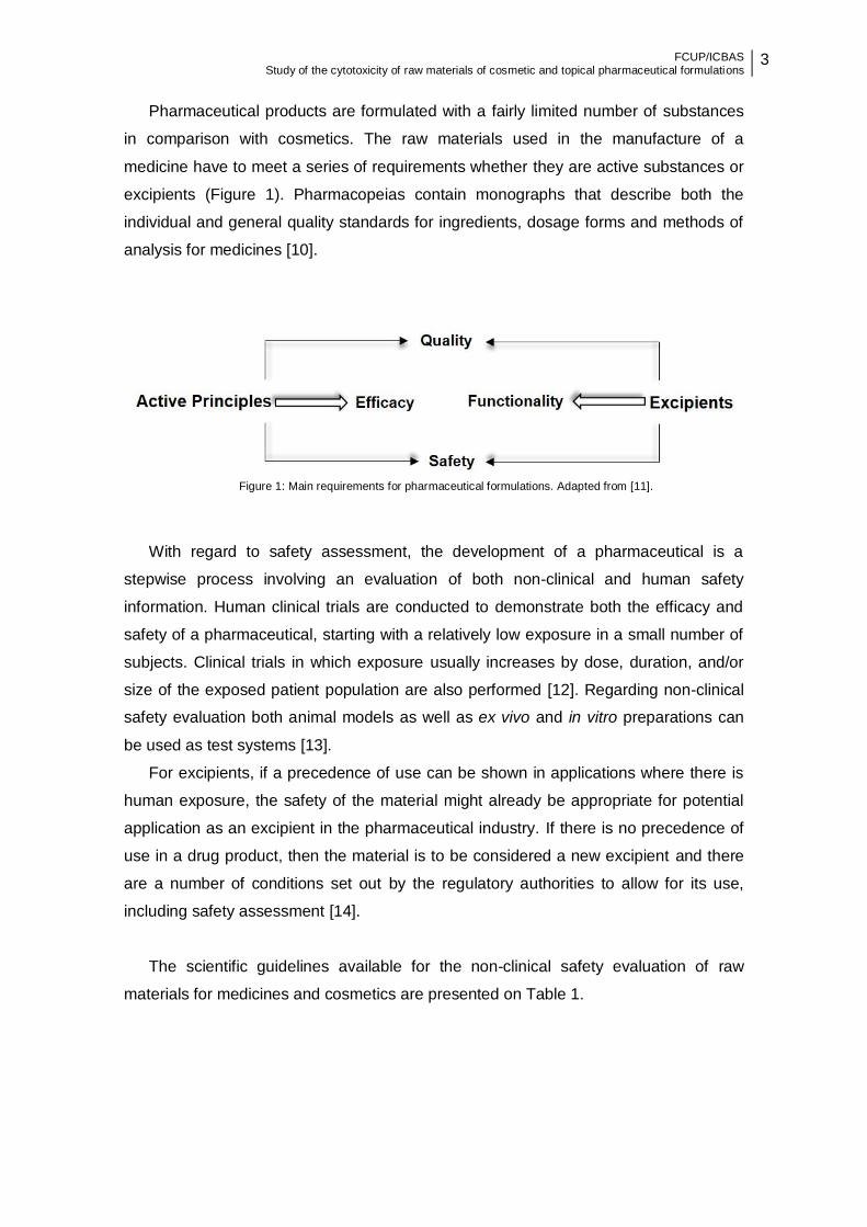
Pharmaceutical products are formulated with a fairly limited number of substances
in comparison with cosmetics. The raw materials used in the manufacture of a
medicine have to meet a series of requirements whether they are active substances or
excipients (Figure 1). Pharmacopeias contain monographs that describe both the
individual and general quality standards for ingredients, dosage forms and methods of
analysis for medicines [10].
Figure 1: Main requirements for pharmaceutical formulations. Adapted from [11].
With regard to safety assessment, the development of a pharmaceutical is a
stepwise process involving an evaluation of both non-clinical and human safety
information. Human clinical trials are conducted to demonstrate both the efficacy and
safety of a pharmaceutical, starting with a relatively low exposure in a small number of
subjects. Clinical trials in which exposure usually increases by dose, duration, and/or
size of the exposed patient population are also performed [12]. Regarding non-clinical
safety evaluation both animal models as well as ex vivo and in vitro preparations can
be used as test systems [13].
For excipients, if a precedence of use can be shown in applications where there is
human exposure, the safety of the material might already be appropriate for potential
application as an excipient in the pharmaceutical industry. If there is no precedence of
use in a drug product, then the material is to be considered a new excipient and there
are a number of conditions set out by the regulatory authorities to allow for its use,
including safety assessment [14].
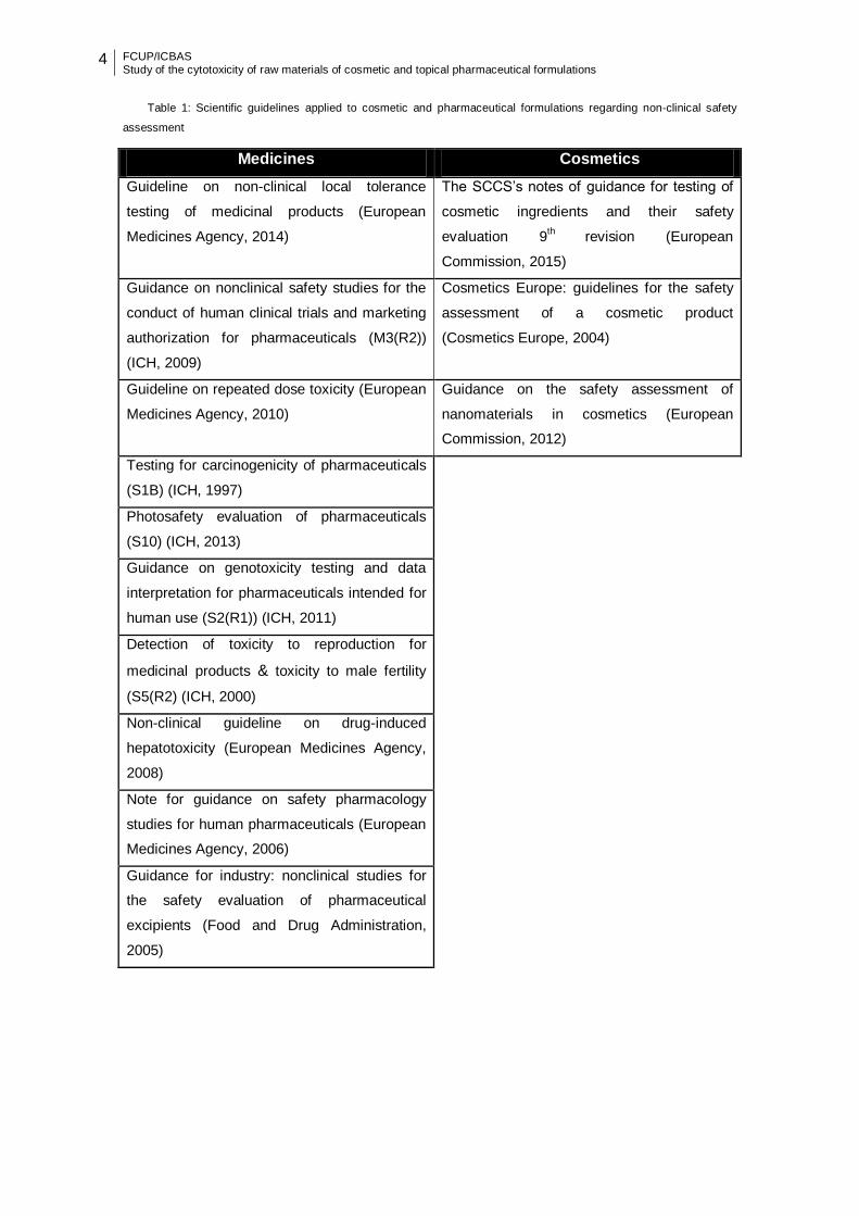
The scientific guidelines available for the non-clinical safety evaluation of raw
materials for medicines and cosmetics are presented on Table 1.
4 FCUP/ICBAS Study of the cytotoxicity of raw materials of cosmetic and topical pharmaceutical formulations
Table 1: Scientific guidelines applied to cosmetic and pharmaceutical formulations regarding non-clinical safety
assessment
Medicines Cosmetics
Guideline on non-clinical local tolerance
testing of medicinal products (European
Medicines Agency, 2014)
The SCCS’s notes of guidance for testing of
cosmetic ingredients and their safety
evaluation 9th
revision (European
Commission, 2015)
Guidance on nonclinical safety studies for the
conduct of human clinical trials and marketing
authorization for pharmaceuticals (M3(R2))
(ICH, 2009)
Cosmetics Europe: guidelines for the safety
assessment of a cosmetic product
(Cosmetics Europe, 2004)
Guideline on repeated dose toxicity (European
Medicines Agency, 2010)
Guidance on the safety assessment of
nanomaterials in cosmetics (European
Commission, 2012)
Testing for carcinogenicity of pharmaceuticals
(S1B) (ICH, 1997)
Photosafety evaluation of pharmaceuticals
(S10) (ICH, 2013)
Guidance on genotoxicity testing and data
interpretation for pharmaceuticals intended for
human use (S2(R1)) (ICH, 2011)
Detection of toxicity to reproduction for
medicinal products & toxicity to male fertility
(S5(R2) (ICH, 2000)
Non-clinical guideline on drug-induced
hepatotoxicity (European Medicines Agency,
2008)
Note for guidance on safety pharmacology
studies for human pharmaceuticals (European
Medicines Agency, 2006)
Guidance for industry: nonclinical studies for
the safety evaluation of pharmaceutical
excipients (Food and Drug Administration,
2005)
FCUP/ICBAS Study of the cytotoxicity of raw materials of cosmetic and topical pharmaceutical formulations
5
1.1.1 Toxicological testing of raw materials for topical
formulations
Procedures for the skin testing of new chemicals and finished products have been
evolving in the face of technological advancements and political pressure. The
evaluation of the skin irritation potential is such an example. It was usually performed
with the method of John Draize [15]. This method consisted in the application under
occlusion of test compounds on rabbit skin and subsequent evaluation of irritant effects
[16]. Recently, measures such as prohibition of execution of cosmetics animal testing
led to the development and validation of alternative in vitro methods using
reconstructed human epidermis. Currently, several in vitro skin irritation tests are
officially validated, namely EpiSkinTM, Modified EpidermTM Skin Irritation Test and
SkinEthicTM Reconstructed Human Epidermis [5]. These models use normal human
keratinocytes that, during culturing, form a multi-layered epidermis including a stratum
corneum at the top, functioning as a barrier [17].
Nanomaterials (NMs) are a particular case of cosmetic ingredients.
Nanotechnologies are a booming business and include the development and
production of nanosized engineered particles, fibers, coatings, etc., collectively referred
to as NMs [18]. According to Regulation (EC) No 1223/2009 nanomaterial means an
insoluble or biopersistent and intentionally manufactured material with one or more
external dimensions, or an internal structure, on the scale from 1 to 100 nm [2].
Currently, these nanosystems can be found in many cosmetic products such as
sunscreens, creams, tooth pastes, shampoos and cleansing agents. Additionally,
nanotechnology has been widely studied for medical and pharmaceutical applications
[19, 20]. Safety data with special considerations to the properties of a specific
nanomaterial is required for its risk assessment. A guidance document on the safety
assessment of nanomaterials in cosmetics was published [21].
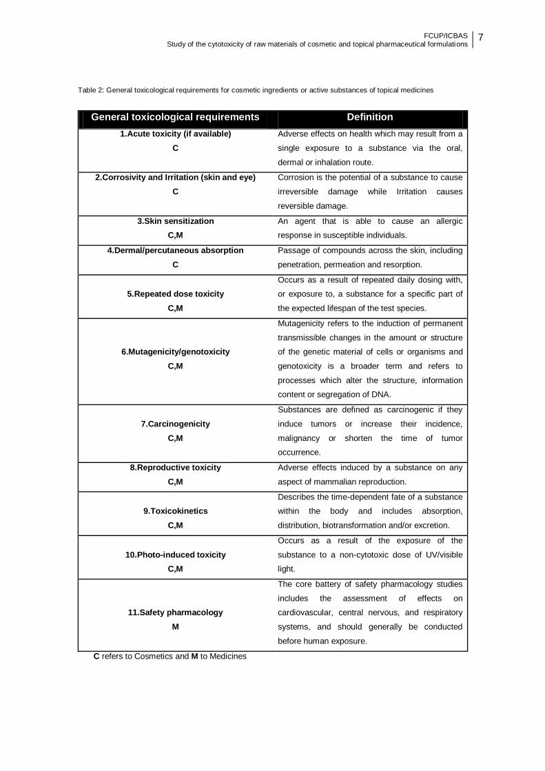
On Table 2 a set of toxicological requirements and their scope of application
(cosmetics or medicines) are listed. Safety requirements for cosmetic ingredients are
addressed in the “Notes of Guidance for Testing of Cosmetic Ingredients and their
Safety Evaluation” [5]. Basic requirements for cosmetic substances present in finished
cosmetic products include acute toxicity (oral, dermal or inhalation), skin and eye
irritation, sensitization and mutagenicity data. For inclusion of a substance in one of the
annexes to Regulation (EC) No 1223/2009, in general, points 1. to 6. are considered
the minimal base set requirements. However, when considerable oral intake is
6 FCUP/ICBAS Study of the cytotoxicity of raw materials of cosmetic and topical pharmaceutical formulations
expected or when the data on dermal/percutaneous absorption indicate a considerable
penetration of the substances through the skin, points 7., 8. and 9. may become
necessary, as well as specific additional genotoxicity and/or mutagenicity data. Photo-
induced toxicity data are specifically required when the substance is present in a
cosmetic product that is expected or intended to being used on sunlight-exposed skin
[5].
The nonclinical safety study recommendations for the marketing approval of a
pharmaceutical usually include repeated dose toxicity studies, reproduction toxicity
studies, genotoxicity studies, local tolerance studies, and for drugs that have special
cause for concern or are intended for a long duration of use, an assessment of
carcinogenic potential. Other nonclinical studies include pharmacologic studies for
safety assessment (safety pharmacology) and pharmacokinetic (absorption,
distribution, metabolism, and excretion-ADME) studies. The complete evaluation of
dermal tolerance for pharmaceutical products intended for administration to the skin
requires a repeated dose dermal tolerance test, and evaluation of the sensitizing
potential, a photosafety evaluation should also be undertaken [13]. As a general rule,
the formulation that is intended to be used clinically should be used in all tests.
FCUP/ICBAS Study of the cytotoxicity of raw materials of cosmetic and topical pharmaceutical formulations
7
Table 2: General toxicological requirements for cosmetic ingredients or active substances of topical medicines
C refers to Cosmetics and M to Medicines
General toxicological requirements Definition
1.Acute toxicity (if available)
C
Adverse effects on health which may result from a
single exposure to a substance via the oral,
dermal or inhalation route.
2.Corrosivity and Irritation (skin and eye)
C
Corrosion is the potential of a substance to cause
irreversible damage while Irritation causes
reversible damage.
3.Skin sensitization
C,M
An agent that is able to cause an allergic
response in susceptible individuals.
4.Dermal/percutaneous absorption
C
Passage of compounds across the skin, including
penetration, permeation and resorption.
5.Repeated dose toxicity
C,M
Occurs as a result of repeated daily dosing with,
or exposure to, a substance for a specific part of
the expected lifespan of the test species.
6.Mutagenicity/genotoxicity
C,M
Mutagenicity refers to the induction of permanent
transmissible changes in the amount or structure
of the genetic material of cells or organisms and
genotoxicity is a broader term and refers to
processes which alter the structure, information
content or segregation of DNA.
7.Carcinogenicity
C,M
Substances are defined as carcinogenic if they
induce tumors or increase their incidence,
malignancy or shorten the time of tumor
occurrence.
8.Reproductive toxicity
C,M
Adverse effects induced by a substance on any
aspect of mammalian reproduction.
9.Toxicokinetics
C,M
Describes the time-dependent fate of a substance
within the body and includes absorption,
distribution, biotransformation and/or excretion.
10.Photo-induced toxicity
C,M
Occurs as a result of the exposure of the
substance to a non-cytotoxic dose of UV/visible
light.
11.Safety pharmacology
M
The core battery of safety pharmacology studies
includes the assessment of effects on
cardiovascular, central nervous, and respiratory
systems, and should generally be conducted
before human exposure.

8 FCUP/ICBAS Study of the cytotoxicity of raw materials of cosmetic and topical pharmaceutical formulations
Dermal tolerance is an important requirement for topical formulations. Animal
models or in vitro tests using reconstructed human epidermis have been used for non-
clinical dermal tolerance assessment of raw materials for the cosmetic and
pharmaceutical industry [13]. Simple cytotoxicity tests can be of value early in the
discovery process of new drugs or active substances for the pharmaceutical industry.
These assays can be added to the battery of information (pharmacologic efficacy,
selectivity, and ADME properties) that is used to differentiate between chemical series
or aid in compound selection for safety tolerance assessment of drugs in animal
studies [22].
Numerous in vitro screening assays have been developed to measure specific
biological activities of chemicals in specific organs or cell types with an eye to
elucidating mechanisms of action. For the purposes of hazard assessment, biological
activity in an in vitro system can identify a mechanism of action or response that could
be extrapolated to an in vivo end point [23].
In the following section an approach to in vitro cytotoxicity assays is done.
1.2 In vitro cytotoxicity assays
The detection of cytotoxicity is crucial in many biological fields, e.g. in toxicology
and in pharmaceutical and cosmetic fields for the assessment of toxic effects elicited
by chemicals, drugs or cosmetic ingredients, respectively, before they are release for
use by the public.
A variety of assays have been developed and used for the measurement of cell
viability or toxicity and the in vitro proliferation. It is important to make a distinction
between cytotoxicity or cell viability and proliferation. Cell proliferation is the
measurement of the number of cells that are dividing in a culture. One way of
measuring this parameter is by performing clonogenic assays, a defined number of
cells are plated onto the appropriate matrix and the number of colonies that are formed
after a period of growth are enumerated or measurement of DNA synthesis as a
marker for proliferation (e.g., 3 [3H]-thymidine or 5-bromo-2’-deoxyuridine) [24].
Definitions of cytotoxicity or cell viability vary depending on the nature of the study.
Cytotoxicity is the cell-killing property of a chemical (e.g., cosmetic or pharmaceutical)
or a mediator cell (e.g., cytotoxic T cell), independent from the mechanisms of death,
while cell viability can be taken as the number of live cells [24].
The need for reliable, easy to handle and fast cytotoxicity tests led to the
development of several assays which are now routinely used and available to detect
FCUP/ICBAS Study of the cytotoxicity of raw materials of cosmetic and topical pharmaceutical formulations
9
cytotoxic effects in in vitro cellular systems [25]. These assays have been devised to
examine and measure cessation of a broad variety of parameters associated with
biochemical events necessary for sustaining viability and/or evidence of changes in
membrane integrity leading to cellular disintegration [26].
To choose an appropriate in vitro cytotoxicity assay, different parameters as: the
cell type, culture conditions applied, test compounds, detection mechanism, specificity,
and sensitivity have to be considered. Cell-toxicity may be assessed by morphological
changes or by alterations in membrane permeability and/or physiological state inferred
from the exclusion of certain dyes or the uptake and retention of others [26, 27].
In the following topic, a brief description, as well as, the advantages and
disadvantages of the most used cytotoxicity assays will be described.
1.2.1 Evaluation of cell membrane permeability
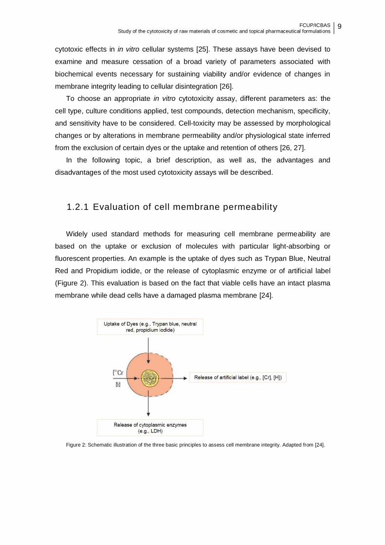
Widely used standard methods for measuring cell membrane permeability are
based on the uptake or exclusion of molecules with particular light-absorbing or
fluorescent properties. An example is the uptake of dyes such as Trypan Blue, Neutral
Red and Propidium iodide, or the release of cytoplasmic enzyme or of artificial label
(Figure 2). This evaluation is based on the fact that viable cells have an intact plasma
membrane while dead cells have a damaged plasma membrane [24].
Figure 2: Schematic illustration of the three basic principles to assess cell membrane integrity. Adapted from [24].
10 FCUP/ICBAS Study of the cytotoxicity of raw materials of cosmetic and topical pharmaceutical formulations
The advantages of such permeability assays are that they are performed easily and
with use of multi-wells plates, many dilutions and/or many compounds can be tested
rapidly [28]. On the other hand, a serious disadvantage of these assays is that the
initial sites of damage of many cytotoxic agents are intracellular. Consequently, cells
may be irreversibly damaged and committed to die and the plasma membrane is still
intact. Thus, these assays tend to underestimate cellular damage when compared to
other methods. Despite this fact, some permeability assays have been widely accepted
for the measurement of cytotoxicity [24].
1.2.1.1 Calcein-Acetoxymethyl assay
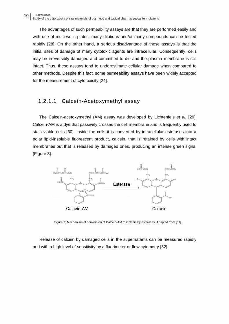
The Calcein-acetoxymethyl (AM) assay was developed by Lichtenfels et al. [29].
Calcein-AM is a dye that passively crosses the cell membrane and is frequently used to
stain viable cells [30]. Inside the cells it is converted by intracellular esterases into a
polar lipid-insoluble fluorescent product, calcein, that is retained by cells with intact
membranes but that is released by damaged ones, producing an intense green signal
(Figure 3).
Figure 3: Mechanism of conversion of Calcein-AM to Calcein by esterases. Adapted from [31].
Release of calcein by damaged cells in the supernatants can be measured rapidly
and with a high level of sensitivity by a fluorimeter or flow cytometry [32].
FCUP/ICBAS Study of the cytotoxicity of raw materials of cosmetic and topical pharmaceutical formulations
11
1.2.1.2 Fluorescein Diacetate uptake assay
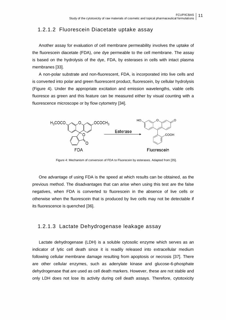
Another assay for evaluation of cell membrane permeability involves the uptake of
the fluorescein diacetate (FDA), one dye permeable to the cell membrane. The assay
is based on the hydrolysis of the dye, FDA, by esterases in cells with intact plasma
membranes [33].
A non-polar substrate and non-fluorescent, FDA, is incorporated into live cells and
is converted into polar and green fluorescent product, fluorescein, by cellular hydrolysis
(Figure 4). Under the appropriate excitation and emission wavelengths, viable cells
fluoresce as green and this feature can be measured either by visual counting with a
fluorescence microscope or by flow cytometry [34].
Figure 4: Mechanism of conversion of FDA to Fluorecein by esterases. Adapted from [35].
One advantage of using FDA is the speed at which results can be obtained, as the
previous method. The disadvantages that can arise when using this test are the false
negatives, when FDA is converted to fluorescein in the absence of live cells or
otherwise when the fluorescein that is produced by live cells may not be detectable if
its fluorescence is quenched [36].
1.2.1.3 Lactate Dehydrogenase leakage assay
Lactate dehydrogenase (LDH) is a soluble cytosolic enzyme which serves as an
indicator of lytic cell death since it is readily released into extracellular medium
following cellular membrane damage resulting from apoptosis or necrosis [37]. There
are other cellular enzymes, such as adenylate kinase and glucose-6-phosphate
dehydrogenase that are used as cell death markers. However, these are not stable and
only LDH does not lose its activity during cell death assays. Therefore, cytotoxicity

12 FCUP/ICBAS Study of the cytotoxicity of raw materials of cosmetic and topical pharmaceutical formulations
assays based on LDH activity are more reliable than other enzyme-based cell death
assays [24].
The LDH leakage assay is based on the conversion of lactate to pyruvate in the
presence of LDH with parallel reduction of NAD, by the following reaction:
The formation of NADH from the above reaction leads to a change in absorbance at
340 nm. LDH is rapidly released into the cell culture supernatant upon damage of the
cell membrane, an indicator of irreversible cell death [38, 39].
Reliability, speed and simple evaluation are some of characteristics of this assay
[40]. Although widely accepted as a marker of cell death, it should be noted that this
test is simply an index of cell membrane integrity, and in certain circumstances can be
positive even when the cell count is not significantly modified [37].
1.2.1.4 Neutral Red uptake assay
The neutral red (3-amino-m-dimethylamino-2-methlphenazine hydrochloride) is a
weak cationic dye that has been used previously for the identification of the viable cells
in cultures [41].
The neutral red (NR) assay is based on the initial protocol described by
Borenfreund and Puerner [42]. This assay focuses on the uptake and subsequent
lysosomal accumulation of the supravital dye NR. Cellular uptake of the dye is
accomplished by passive transport across the plasma membrane. Accumulation of NR
within lysosomes occurs either from the binding of NR to fixed acidic charges, such as
those of polysaccharides, within the lysosomal matrix, or from the trapping of the
protonated form of NR within the acid milieu of the lysosomes [43].
In damaged or dead cells NR is no longer retained in the cytoplasmic vacuoles and
the plasma membrane does not act as a barrier to retain the NR within the cells. It
quantifies the number of viable, uninjured cells after their exposure with test agents [39,
44].
This assay does not measure the total number of cells, but it does show a reduction
in the absorbance related to loss of viable cells [43].
LDH

FCUP/ICBAS Study of the cytotoxicity of raw materials of cosmetic and topical pharmaceutical formulations
13
1.2.1.5 Propidium Iodide exclusion assay
Propidium iodide (PI) is a fluorescent molecule that can only penetrate cell
membrane of dying or dead cells and thus identifies dead cells within a population.
Upon entering dead cells, PI binds to double-stranded DNA by intercalating between
base pairs. As this intercalation is mediated by non-covalent forces, these dyes must
remain present in the buffer used to resuspend cells for data acquisition so that dead
cells will remain labeled [45]. The uptake of PI by the cells results in orange
fluorescence that can be assessed by fluorescence and flow cytometry [46].
1.2.1.6 GF-ACF assay
This assay utilizes the cell-permeant protease substrate glycylphenylalanyl-
aminofluorocoumerin (GF-AFC) that penetrates live cells where cytoplasmic
aminopeptidase activity removes the gly and phe aminoacids to release
aminofluorocoumarin (AFC) and generate a fluorescent signal proportional to the
number of viable cells (Figure 5) [47, 48].
Figure 5: Mechanism of conversion of GF-AFC to AFC by cytoplasmic aminopeptidase activity. Adapted from [47].
The selective detection of viable cells by this method is possible because the
proteolytic activity towards the GF-AFC substrate is dependent upon the continued
maintenance of membrane integrity. Viability can be measured using a fluorometer.
The proteolytic activity decays within seconds after a cytotoxic event, so non-viable
cells do not contribute appreciably to fluorescence generation [48].
One of the advantages of the GF-AFC substrate is that it is relatively non-toxic to
cells in culture.
14 FCUP/ICBAS Study of the cytotoxicity of raw materials of cosmetic and topical pharmaceutical formulations
1.2.1.7 Trypan Blue exclusion assay
One of the earliest methods for assessing cytotoxicity was trypan blue exclusion
assay, which is still widely used today.
The trypan blue is a vital stain used to stain dead cells or tissues. When membrane
integrity of the cells is compromised, there is an uptake of the dye into the cells so that
viable cells, which are unstained, appear clear with a refractile ring around them and
nonviable cells appear dark blue colored [25, 28]. This traditional method involves
manual staining and use of a hemocytometer for counting. Recent advances in
instrumentation have led to a number of semi- or fully automated systems that can
increase the throughput and accuracy of this technique [49].
This method is simple, quick, inexpensive, and requires only a small fraction of total
cells from a cell population. However, trypan blue staining cannot be used to
distinguish between the healthy cells and the cells that are alive but with impaired cell
functions.
1.2.2 Evaluation of metabolic activity
One parameter used as the basis for cytotoxicity assays is the evaluation of
metabolic activity of the viable cells including assays based on tetrazolium and
resazurin reduction [26]. The metabolic assay do not provide direct information about
total cell numbers, but measure the viability of a cell population relative to control. One
potential disadvantage of these metabolic assays is that there is no differentiation
between cells that are actively dividing and those that are quiescent which may result
in an over-estimation of cell number [27].
1.2.2.1 AlamarBlue® reduction assay
AlamarBlue® is an important and sensitive oxidation-reduction indicator that is used
to evaluate metabolic function and cellular health. The resazurin (oxidized form) is a
blue weakly fluorescent indicator dye that changes into highly fluorescent pink resorufin
(reduction form) in response to irreversible chemical reduction (Figure 6) [50].

FCUP/ICBAS Study of the cytotoxicity of raw materials of cosmetic and topical pharmaceutical formulations
15
Figure 6: Mechanism of reduction of resazurin to resorufin [50].
Inside the cell, AlamarBlue® undergoes enzymatic reduction in mitochondria due to
the activity of enzymes such as flavin mononucleotide dehydrogenase, flavin adenine
dinucleotide dehydrogenase, nicotinamide adenine dehydrogenase, and cytochrome
[51]. It was also noted that cytosolic and microsomal enzymes have abilities to reduce
resazurin [52]. The pink resorufin is secreted outside the cells to the medium which
results in visible colour change from blue to pink. The rate of reduction based on colour
change can be quantified colorimetrically or fluorometrically and reflects the number of
viable cells [50].
AlamarBlue® is considered as very sensitive technique, non-toxic to cell cultures
and is less likely to affect normal metabolism by not interfering with the electron chain
[50, 53].
1.2.2.2 Tetrazolium Salts reduction assay
A variety of tetrazolium compounds have been used to detect viable cells. The most
frequently used tetrazolium salt is 3-(4,5-dimethylthiazol-2-yl)-2,5-diphenyltetrazolium
bromide (MTT) [27].
The MTT reduction assay was the first homogeneous cell viability assay developed
for a 96-well format that was suitable for high throughput screening (HTS) [54]. This
assay is used to determine the level of metabolic activity in eukaryotic cells. It is based
on the conversion of soluble tetrazolium into insoluble formazan crystals reduction
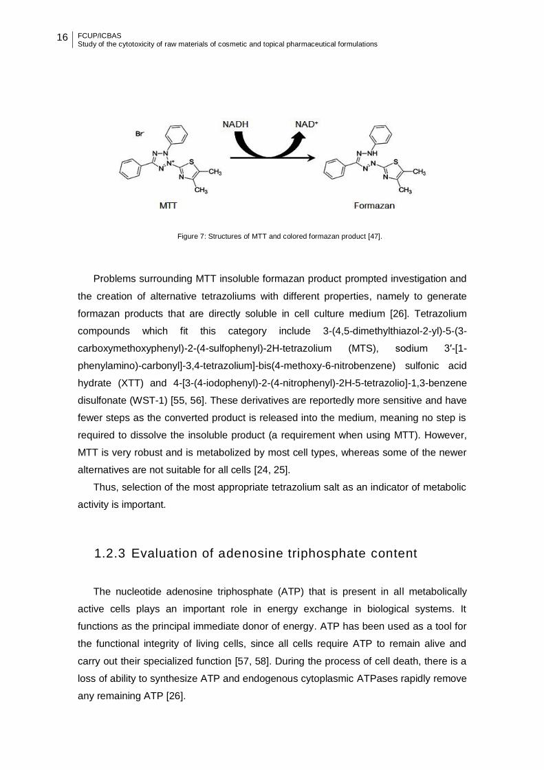
(Figure 7) by cellular mitochondrial and cytosolic enzymes of actively growing cells [25,
26]. The formazan product is impermeable to the cell membranes and requires
solubilization of product.
16 FCUP/ICBAS Study of the cytotoxicity of raw materials of cosmetic and topical pharmaceutical formulations
Figure 7: Structures of MTT and colored formazan product [47].
Problems surrounding MTT insoluble formazan product prompted investigation and
the creation of alternative tetrazoliums with different properties, namely to generate
formazan products that are directly soluble in cell culture medium [26]. Tetrazolium
compounds which fit this category include 3-(4,5-dimethylthiazol-2-yl)-5-(3-
carboxymethoxyphenyl)-2-(4-sulfophenyl)-2H-tetrazolium (MTS), sodium 3′-[1-
phenylamino)-carbonyl]-3,4-tetrazolium]-bis(4-methoxy-6-nitrobenzene) sulfonic acid
hydrate (XTT) and 4-[3-(4-iodophenyl)-2-(4-nitrophenyl)-2H-5-tetrazolio]-1,3-benzene
disulfonate (WST-1) [55, 56]. These derivatives are reportedly more sensitive and have
fewer steps as the converted product is released into the medium, meaning no step is
required to dissolve the insoluble product (a requirement when using MTT). However,
MTT is very robust and is metabolized by most cell types, whereas some of the newer
alternatives are not suitable for all cells [24, 25].
Thus, selection of the most appropriate tetrazolium salt as an indicator of metabolic
activity is important.
1.2.3 Evaluation of adenosine triphosphate content
The nucleotide adenosine triphosphate (ATP) that is present in all metabolically
active cells plays an important role in energy exchange in biological systems. It
functions as the principal immediate donor of energy. ATP has been used as a tool for
the functional integrity of living cells, since all cells require ATP to remain alive and
carry out their specialized function [57, 58]. During the process of cell death, there is a
loss of ability to synthesize ATP and endogenous cytoplasmic ATPases rapidly remove
any remaining ATP [26].

FCUP/ICBAS Study of the cytotoxicity of raw materials of cosmetic and topical pharmaceutical formulations
17
Measuring the amount of ATP from samples of cells in culture has been widely
accepted as a valid marker of the number of viable cells present under most
experimental conditions [26]. Many methods have been used for evaluation of ATP
content, but the most successful technique is the bioluminescent assay, because of its
high sensitivity and wide dynamic range [57]. The disadvantage of this method is the
luminescence-readout, which could be influenced by quenching side effects in the
samples [59].
1.2.3.1 Bioluminescent assay
In the bioluminescent assay, the enzyme, luciferase catalyzes the formation of light
from ATP and luciferin [44], by the following reaction:
ATP + D-Luciferin + O2 → Oxyluciferin + AMP + PPi + CO2 + Light (560nm)
Cellular ATP can be measured by direct lysis of the cells with a suitable detergent,
the released ATP is then free to react with the luciferin-luciferase, leading to light
emission. The emitted light intensity is linearly related to the cell ATP concentration
and can be measured using a luminometer [58].
The principal advantages of ATP detection were shown to be its sensitivity and
reproducibility [60]. The disadvantages of this assay include that it kills the cells and so
the sample generally cannot be used for other purposes after treatment with the
reagent.
1.2.4 Evaluation of protein content
The evaluation of protein content is an indirect measurement of cell viability. The
most used assays are: bicinchoninic acid and sulphorhodamine assays.
1.2.4.1 Bicinchoninic Acid assay
The bicinchoninic acid (BCA) assay also know Smith assay was described by Smith
[61]. The principle this assay is similar to the formation of a Cu2+-protein complex
under alkaline conditions, followed by reduction of the Cu2+ to Cu1+ [62].

18 FCUP/ICBAS Study of the cytotoxicity of raw materials of cosmetic and topical pharmaceutical formulations
In this assay, the amount of reduction is proportional to the protein present,
exhibited by a color change of the sample solution from green to purple in proportion to
protein concentration, which can then be measured using colorimetric techniques or
absorbance. The major advantage of the BCA is that it is stable under alkali conditions
[63]. One disadvantage of the BCA assay is that it is susceptible to interference by
some chemicals present in protein samples, including reducing agents or copper
chelators [64].
1.2.4.2 Sulphorhodamine assay
The Sulphorhodamine (SRB) assay is a popular in vitro cytotoxicity assay
developed by Skehan et. al. [65]. This assay is based on binding of the SRB to basic
aminoacids of cellular proteins and colorimetric evaluation provides an estimate of total
protein mass, which is related to cell number [66].
The advantages of this test include better linearity, higher sensitivity, a stable end
point that does not require time-sensitive measurement and lower cost [67].
Cytotoxicity assays described herein are just a few of the commonly used assays.
These can be used as a prediction of the adverse effects of test compounds on living
systems, for the detection of toxic thresholds, and expansion of experimental data sets
to include multiple toxicity end-point analysis, which are required for any robust
screening regime [27].
In Table 3, the cytotoxicity assays described in this topic are presented, grouped by
the parameter evaluated, as well as the principle of detection of each test.
FCUP/ICBAS Study of the cytotoxicity of raw materials of cosmetic and topical pharmaceutical formulations
19
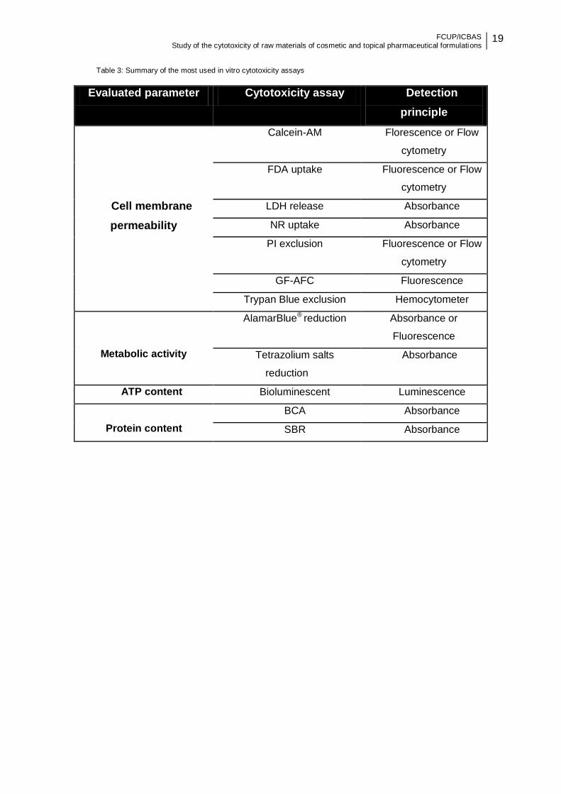
Table 3: Summary of the most used in vitro cytotoxicity assays
Evaluated parameter Cytotoxicity assay Detection
principle
Cell membrane
permeability
Calcein-AM Florescence or Flow
cytometry
FDA uptake Fluorescence or Flow
cytometry
LDH release Absorbance
NR uptake Absorbance
PI exclusion Fluorescence or Flow
cytometry
GF-AFC Fluorescence
Trypan Blue exclusion Hemocytometer
Metabolic activity
AlamarBlue® reduction Absorbance or
Fluorescence
Tetrazolium salts
reduction
Absorbance
ATP content Bioluminescent Luminescence
Protein content
BCA Absorbance
SBR Absorbance
20 FCUP/ICBAS Study of the cytotoxicity of raw materials of cosmetic and topical pharmaceutical formulations
1.3 Use of cytotoxicity assays in safety assessment of
raw material for cosmetic and topical pharmaceutical
formulations
The skin is the first site of contact for many compounds, including ingredients of
cosmetics or dermal pharmaceuticals. Skin, the major human organ, is a
heterogeneous membrane: lipophilic on its surface and hydrophilic in its deeper layers.
It forms a remarkable protective barrier against the external environment, helping to
regulate temperature and fluid balance, keeping out harmful microbes or chemicals and
offering some protection against sunlight and oxidation [68, 69].
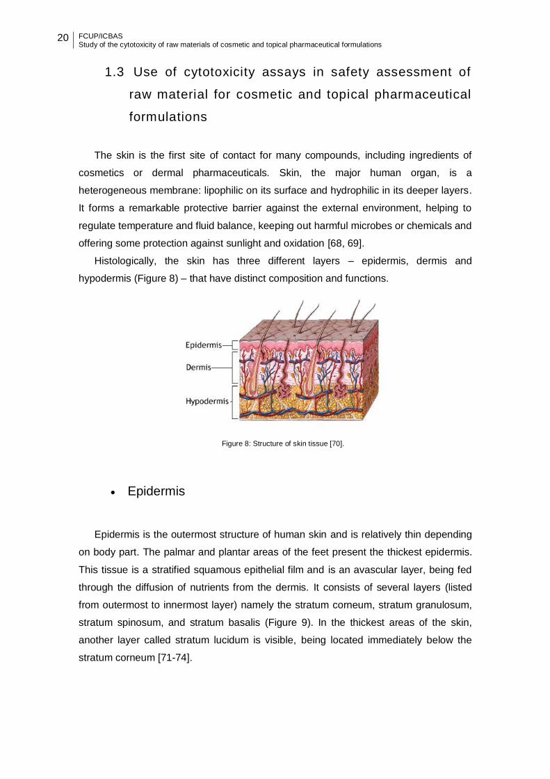
Histologically, the skin has three different layers – epidermis, dermis and
hypodermis (Figure 8) – that have distinct composition and functions.
Figure 8: Structure of skin tissue [70].
Epidermis
Epidermis is the outermost structure of human skin and is relatively thin depending
on body part. The palmar and plantar areas of the feet present the thickest epidermis.
This tissue is a stratified squamous epithelial film and is an avascular layer, being fed
through the diffusion of nutrients from the dermis. It consists of several layers (listed
from outermost to innermost layer) namely the stratum corneum, stratum granulosum,
stratum spinosum, and stratum basalis (Figure 9). In the thickest areas of the skin,
another layer called stratum lucidum is visible, being located immediately below the
stratum corneum [71-74].
FCUP/ICBAS Study of the cytotoxicity of raw materials of cosmetic and topical pharmaceutical formulations
21
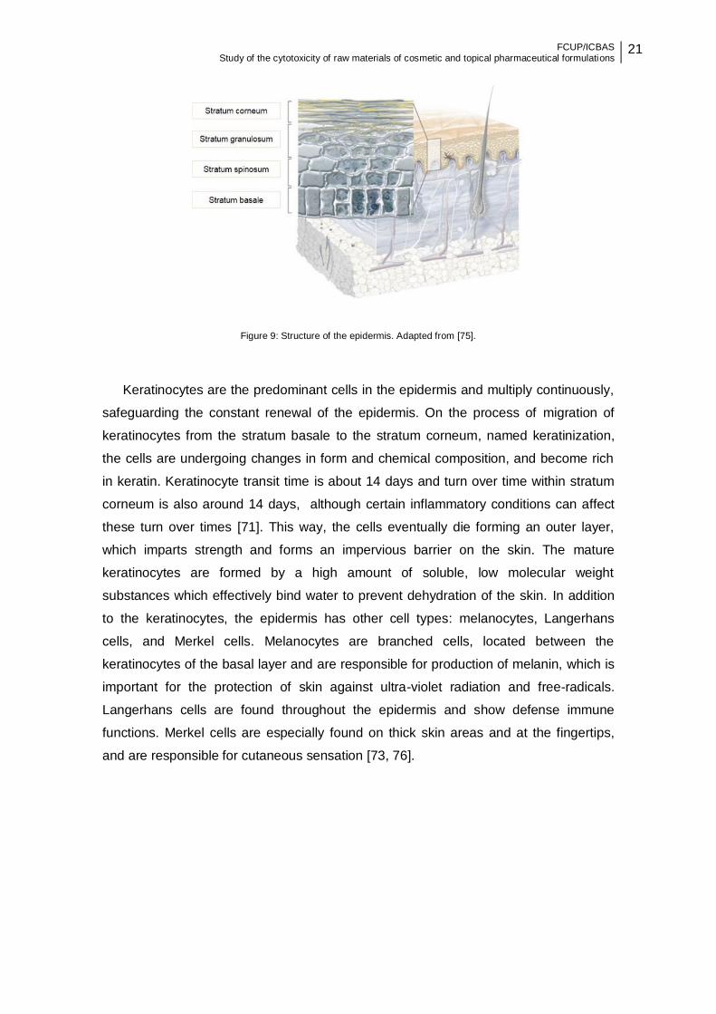
Figure 9: Structure of the epidermis. Adapted from [75].
Keratinocytes are the predominant cells in the epidermis and multiply continuously,
safeguarding the constant renewal of the epidermis. On the process of migration of
keratinocytes from the stratum basale to the stratum corneum, named keratinization,
the cells are undergoing changes in form and chemical composition, and become rich
in keratin. Keratinocyte transit time is about 14 days and turn over time within stratum
corneum is also around 14 days, although certain inflammatory conditions can affect
these turn over times [71]. This way, the cells eventually die forming an outer layer,
which imparts strength and forms an impervious barrier on the skin. The mature
keratinocytes are formed by a high amount of soluble, low molecular weight
substances which effectively bind water to prevent dehydration of the skin. In addition
to the keratinocytes, the epidermis has other cell types: melanocytes, Langerhans
cells, and Merkel cells. Melanocytes are branched cells, located between the
keratinocytes of the basal layer and are responsible for production of melanin, which is
important for the protection of skin against ultra-violet radiation and free-radicals.
Langerhans cells are found throughout the epidermis and show defense immune
functions. Merkel cells are especially found on thick skin areas and at the fingertips,
and are responsible for cutaneous sensation [73, 76].
22 FCUP/ICBAS Study of the cytotoxicity of raw materials of cosmetic and topical pharmaceutical formulations
Dermis
The dermis is a connective tissue layer and contains a collagen- and elastin-
containing extracellular matrix, primarily composed of type I collagen, as well as an
abundance of blood vessels and specialized nerve endings, thus providing structural
and nutritional support to the skin [68, 72, 77]. The dermis contains four major resident
cell types: fibroblasts, dermal dendritic cells, macrophages, and mast cells. Various
infiltrative inflammatory cells can also be found in the dermis under different conditions
[72].
The dermis has two distinct layers: the reticular dermis and papillary dermis. The
reticular dermis is the main layer of the dermis and is continuous with the hypodermis
and has a predominance of elastic and collagen fibers. The papillary dermis is
composed of papillae extending towards the epidermis. Compared with the first, the
papillary dermis has fewer fibers and more cells. This layer of skin also contains
numerous blood vessels, to provide nutrients to the epidermis, removing waste
products and helps to regulate the body temperature [73].
Hypodermis
The hypodermis or subcutaneous fat layer is situated beneath the dermis and
connects this with the underlying organs. It is primarily composed of well-vascularized
adipose tissue and loose connective tissue, which insulates the body and provides both
thermoregulatory and mechanical functions [71-73, 78].
The assessment of cytotoxicity in skin cells can be taken as a measure of the skin
irritation potential. The evaluation of the skin irritation potential is essential to ensuring
the safety of human in contact with a wide variety of cosmetic and/or pharmaceutical
substances.
Traditionally, the assessment of skin irritation involved the use of laboratory animals
[79, 80]. However, since the Regulation (EC) 1223/2009 has come into force, animal
testing of cosmetic and ingredients was banned and the study of their safety has
moved towards in vitro methodologies. Cultured human skin cells are a potentially
useful model for skin irritation testing. In vitro cultures of the monolayer using primary
FCUP/ICBAS Study of the cytotoxicity of raw materials of cosmetic and topical pharmaceutical formulations
23
human keratinocytes, dermal fibroblasts or immortalized epidermal cell lines have been
used in various in vitro irritation assays [81].
In vitro cytotoxicity assays have the advantage of being relatively inexpensive,
standardized, sensitive and allow for rapid screening of compounds [82].
The most common in vitro cytotoxicity assay is MTT [83]. This assay is used in
primary human keratinocytes [84, 85], immortalized keratinocytes [86], melanocytes
[87] and fibroblasts [88]. Reconstructed human epidermal skin models are being used
increasingly for safety testing of chemicals. These models have the advantage of
allowing topical application of the compounds, evaluation of formulations and poorly
soluble compounds, as well as measurement of local toxicological effects in target cells
[89]. MTT reduction assay also is very used in reconstructed human epidermis [90-93].
1.3.1 Analyzed antioxidants raw materials
The skin has an antioxidant defense system that prevents UV-induced oxidative
stress. Nevertheless chronic and excessive exposure to UV-radiation leads to oxidative
stress and oxidative damage [94]. The topical application of antioxidants has achieved
great expression, owing to their ability to prevent or minimize the UV induced-
deleterious effects of reactive species on the skin [95].
In this work were analyzed several compounds: resveratrol and its derivatives;
resveratrol-loaded lipid and polymeric particles, xanthone and plant extract.
Resveratrol and its derivatives
The Resveratrol - RSV (3, 5, 4’ -trihydroidroxystilbene) a natural antioxidant
compound is a non flavonoid polyphenol, present in different plants (peanuts, pistachio
nuts, red grape seeds and skins, red wine, and other plant-derived food products) [96].
Several beneficial effects have been attributed to RSV and its use has been proposed
in the treatment or prevention of different diseases [97].
The resveratrol glicoside – RSV-Gli (3, 5, 4’ -trihydroidroxystilbene-3-β-D-
glucopyranoside) is an natural precursor of RSV and known for its favorable
therapeutic properties: cardiovascular effects, neuroprotection, anti-inflammatory and
immunoregulatory effects, anti-oxidation, anti-tumor, liver and lung protection [98].

24 FCUP/ICBAS Study of the cytotoxicity of raw materials of cosmetic and topical pharmaceutical formulations
The resveratrol glicoside sulfate - RGS (3, 5, 4’ -trihydroidroxystilbene-3-β-D-
glucopyranoside persulfate) was obtained by chemical synthesis and demonstrate in
vitro anticoagulant and antiplatelet activities [99, 100]. In docking study, RGS binds
more stably to sirtuin (lower binding free energy) than the RSV. RGS establishes polar
interactions with 5 sirtuin residues and stacking interactions with the substrate peptide,
whereas RSV establishes only one polar interaction (unpublished results). These
results support the interest of this derivative for skin care formulations.
The chemical structure of RSV and its derivatives (RSV-Gli and RGS) is showed in
Figure 10.
Figure 10: Chemical structure of RSV and its derivatives studied in this work.

FCUP/ICBAS Study of the cytotoxicity of raw materials of cosmetic and topical pharmaceutical formulations
25
Lipid and polymeric particles
Due to its low chemical stability, poor solubility, inefficient systemic delivery, and
low bioavailability [101], RSV was encapsulated in lipid and polymeric particles.
Lipid nanoparticles can be solid lipid nanoparticle (SLN) or nanostructured lipid
carriers (NLC) (Figure 11). The first ones, were invented 20 years ago and are
prepared from a lipid matrix that is solid at body and room temperature and stabilized
by suitable surfactants [102] and NLC are 2nd generation of solid lipid-based colloidal
carriers and are composed of a mixture of a solid and a liquid lipid which lipid matrix is
solid at room and body temperature [103].
Figure 11: Lipid nanoparticles [104].
The benefits of the lipid nanoparticles include the employment of biocompatible and
biodegradable materials, which lead to a low or total absence of toxicity and the ability
to transport lipophilic drugs [104].
On the other hand, polymeric particles are solid colloidal particles that are
dispersed in the liquid and upon water evaporation, they get well organized on the
dosage form solid surface to form close-packed arrays. Eudragit® RS 30D was the
utilized polymer. It is the synthetic homopolymer of poly(acrylate) and
poly(methacrylate) [105].

26 FCUP/ICBAS Study of the cytotoxicity of raw materials of cosmetic and topical pharmaceutical formulations
Chlorogenic acid persulfate and Ascorbic acid persulfate
Chlorogenic acid persulfate (ACS) and ascorbic acid persulfate (ASS) (Figure 12)
are compounds obtained by chemical synthesis with in vitro anticoagulant, antiplatelet
and antioxidant activities [99, 100].
Figure 12: Chemical structure of ACS and ASS studied in this work.
Xanthone
Chemically, xanthones are a class of oxygenated heterocyclic compounds with a
dibenzo--pyrone scaffold [106]. Xanthones can be considered as “privileged
structures”, considering that their scaffold contains a rigid ring system allowing the
insertion of a variety of substituent’s capable of interacting with several biological
targets. Many xanthones with phenolic groups have been described for their
antioxidant properties [107]. These properties have been implicated also with their anti-
inflammatory and cancer chemopreventive activities [108]. In the present work, a 1,2-
dihydroxyxanthone (Figure 13) was used.
Figure 13: Chemical structure of 1,2-dihydroxyxanthone studied in this work.
FCUP/ICBAS Study of the cytotoxicity of raw materials of cosmetic and topical pharmaceutical formulations
27
Castanea sativa leaf extract
Many natural compounds have a wide range of biological activities including
antioxidant, chemopreventive, anti-inflammatory, neuroprotective, and cardioprotective
effects [109].
Castanea sativa (C. sativa) is a species of the flowering plant family Fagaceae,
mainly cultivated in temperate regions like Asia, North of Africa and Southern Europe.
Its leaves are used in folk medicine to treat cough, diarrhea and rheumatic conditions,
lower back pain, and stiff joints or muscles [110].
As in other medicinal plants, some of the active substances have been identified as
phenolic compounds. The scavenging ability has been reported in several studies for
different ROS, such as superoxide radical (O2•-), hydroxyl radical (HO•), peroxyl radical
(ROO•), hydrogen peroxide (H2O2) and singlet oxygen (1O2), which are implicated in
skin aging [111]. The utilization of hydroalcoholic extracts from C. sativa leaves (which
contain rutin, ellagic acid, hyperoside, isoquercitrin and chlorogenic acid) in cosmetic
compositions has been considered safe and suitable for preventing and treating
photoaging [112].
28 FCUP/ICBAS Study of the cytotoxicity of raw materials of cosmetic and topical pharmaceutical formulations
2. AIM
The aim of this Master’s dissertation was to study the cytotoxicity of a series of
ingredients with potential interest for topical application and determine the
concentration range for each compound that can be used in future assays to confirm
the potentialities of these raw materials for application on the skin. For this purpose we
used ingredients of natural origin, ingredients obtained by chemical synthesis and also
the ingredient loaded in lipid nanoparticles and polymeric microparticles and five
distinct cytotoxicity assays were employed.
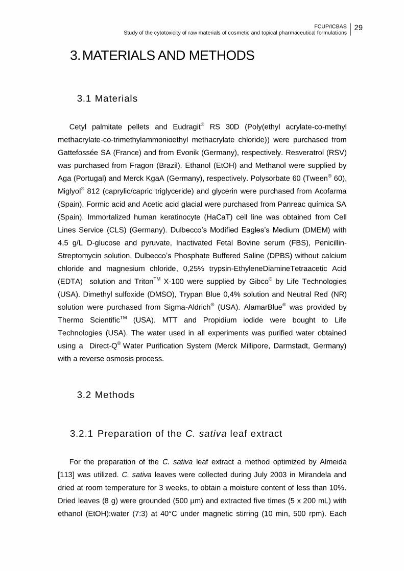
FCUP/ICBAS Study of the cytotoxicity of raw materials of cosmetic and topical pharmaceutical formulations
29
3. MATERIALS AND METHODS
3.1 Materials
Cetyl palmitate pellets and Eudragit® RS 30D (Poly(ethyl acrylate-co-methyl
methacrylate-co-trimethylammonioethyl methacrylate chloride)) were purchased from
Gattefossée SA (France) and from Evonik (Germany), respectively. Resveratrol (RSV)
was purchased from Fragon (Brazil). Ethanol (EtOH) and Methanol were supplied by
Aga (Portugal) and Merck KgaA (Germany), respectively. Polysorbate 60 (Tween® 60),
Miglyol® 812 (caprylic/capric triglyceride) and glycerin were purchased from Acofarma
(Spain). Formic acid and Acetic acid glacial were purchased from Panreac química SA
(Spain). Immortalized human keratinocyte (HaCaT) cell line was obtained from Cell
Lines Service (CLS) (Germany). Dulbecco’s Modified Eagles’s Medium (DMEM) with
4,5 g/L D-glucose and pyruvate, Inactivated Fetal Bovine serum (FBS), Penicillin-
Streptomycin solution, Dulbecco’s Phosphate Buffered Saline (DPBS) without calcium
chloride and magnesium chloride, 0,25% trypsin-EthyleneDiamineTetraacetic Acid
(EDTA) solution and TritonTM X-100 were supplied by Gibco® by Life Technologies
(USA). Dimethyl sulfoxide (DMSO), Trypan Blue 0,4% solution and Neutral Red (NR)
solution were purchased from Sigma-Aldrich® (USA). AlamarBlue® was provided by
Thermo ScientificTM (USA). MTT and Propidium iodide were bought to Life
Technologies (USA). The water used in all experiments was purified water obtained
using a Direct-Q® Water Purification System (Merck Millipore, Darmstadt, Germany)
with a reverse osmosis process.
3.2 Methods
3.2.1 Preparation of the C. sativa leaf extract
For the preparation of the C. sativa leaf extract a method optimized by Almeida
[113] was utilized. C. sativa leaves were collected during July 2003 in Mirandela and
dried at room temperature for 3 weeks, to obtain a moisture content of less than 10%.
Dried leaves (8 g) were grounded (500 µm) and extracted five times (5 x 200 mL) with
ethanol (EtOH):water (7:3) at 40°C under magnetic stirring (10 min, 500 rpm). Each
30 FCUP/ICBAS Study of the cytotoxicity of raw materials of cosmetic and topical pharmaceutical formulations
alcoholic extract was filtered using a glass filter G4 funnel (5–15 μm porosity, Schott,
Germany), and gathered in a kitasato flask. Ethanol was evaporated at 40°C under
vacuum with a rotary evaporator (R-114, Büchi, Switzerland). Finally, the resulting
aqueous mixtures were lyophilized (SP Scientific, Virtis wizard 2.0, USA) for 24 hours,
generating the final dry extract, which was kept in an amber flask at room temperature
in a desiccator. The basic steps of this preparation method are described in Figure 14.
Figure 14: Overview of basic steps of C. sativa leaf extract preparation.
3.2.1.1 Qualitative analysis of the phenolic composition of
C. sativa leaf extract
In order to confirm the presence of phenolic compounds of interest, the C. sativa
EtOH:water (7:3) leaf extract was analysed using High Performance Liquid
Chromatography (HPLC) (Dionex Corporation, Sunnyvale, USA) by comparing
retention times of compounds present in the extract with retention times of standards
previously analysed with the same chromatographic conditions.
The lyophilized extract was dissolved in EtOH:water (7:3) and filtered (0.22 μm)
before being analysed. The sample was injected on a C18 Waters Spherisorb® ODS2
0.46x25x5μm column equipped with a Waters Spherisorb® pre-column (Waters
Corporation, EUA) at 20°C, with detection at 280 nm, 320 nm and 350 nm. Organic
phase was methanol and aqueous phase was water/formic acid (19:1 (v/v)). The
separation was obtained at a flow rate of 0.9 mL/min with a gradient program described
on Table 4.

FCUP/ICBAS Study of the cytotoxicity of raw materials of cosmetic and topical pharmaceutical formulations
31
Table 4: Gradient program used for identification of phenolic compounds of C. sativa EtOH:water (7:3) leaf extract
Time (min) Aqueous phase
Organic phase
0 95 5
3 85 15
13 75 25
25 70 30
39 55 45
42 55 45
44 50 50
47 45 55
50 30 70
56 25 75
60 20 80
3.2.2 Preparation of nano and microparticles
For the preparation of lipid nanoparticles (SLN and NLC) a high shear
homogenization technique was used based on previous studies performed in our lab
[104, 114]. The composition of the SLN and NLC produced in this work are described
on Table 5.
The lipid phase with the surfactant and the aqueous phase were heated to 70ºC
(lipid’s melting point) separately. Then, RSV was dissolved in the lipid phase and the
aqueous phase was added to lipid phase. The emulsion was submitted to a
homogenization process (Ultra-Turrax T25, IKA®-Labortechnik, Germany) at a stirring
speed of 13500 rpm for 2 minutes followed by sonication with a Ultrasonic Processor
(VCX 130, Sonics Vibracell, USA) with intensity of 80% for 2 minutes. Finally, the
nanoparticles underwent a rapid cooling process with running tap water. Empty SLN
and empty NLC were prepared in a similar way, without adding RSV.
The lipid nanoparticles were prepared extemporaneously for all cytotoxicity assays.

32 FCUP/ICBAS Study of the cytotoxicity of raw materials of cosmetic and topical pharmaceutical formulations
Table 5: The formulation parameters of empty and RES-loaded SLN/NLC
SLN empty SLN-RSV NLC empty NLC-RSV
Cetyl Palmitate (solid lipid at room and
physiological temperature)
10% 9.9% 7% 6.9%
Tween 60 (surfactant)
2% 2% 2% 2%
Mygliol 812 (liquid lipid at room and
physiological temperature)
- - 3% 3%
RSV (to be encapsuled)
- 0.1% - 0.1%
Milli-Q Water q.s. 100% q.s. 100% q.s. 100% q.s 100%
For the preparation of polymeric microparticles a piezodriven spray generation
apparatus (Nano Spray Dryer B-90, Büchi, Switzerland) (Figure 15) was used.
Polymeric microparticles containing RSV (Poly-RSV) and empty particles (Poly) were
produced. RSV was dissolved in EtOH:water solution (20:80) until saturation and mixed
with Eudragit® RS 30D (0.5%). The sample was atomized by piezoelectrically driven
membrane vibrations in the small spray cap and millions of precisely sized droplets
were ejected each second by the vibrating actuator. These droplets passed through the
chamber to form dried solid particles that were electrostatically charged and collected
at the collecting electrode. The spray drying experiments were executed using the
following settings: spray cap (hole size): 7 μm; inlet temperature: 120 ºC, compressed
air flow: 100 mL/min; pressure: 33-35 mbar and spray: 80%.
Figure 15: Operating diagram of Nano Spray Dryer B-90. Adapted from [115].

FCUP/ICBAS Study of the cytotoxicity of raw materials of cosmetic and topical pharmaceutical formulations
33
3.2.2.1 Particle size measurements
Particle size of nano and microparticles was analyzed by a laser diffractometry
technique (Mastersizer 3000, Malvern, UK). With this technique the intensity of
scattered light is measured as a laser beam passes through a dispersed particulate
sample. The range of this apparatus is from 0.01 to 3500 μm.
The determination was performed at room temperature using water as a dispersant
with a refractive index of 1.33. The refractive index and absorption index were 1.1 and
0.01 for lipid nanoparticles and 1.48 and 0.1 for polymeric microparticles. The sample
was added to the dispersant until 5-10% of obscuration (preferably 6%) is reached and
tested five times at 1000 rpm. The Dv10, Dv50, Dv90 and Span parameters were
obtained by calculating the average of five runs. Only the measurements with residuals
and weighted residuals near and below 2% were considered.
3.2.3 Cell Culture
The in vitro cytotoxicity was studied using HaCaT cells. The HaCaT adherent and
monolayer cell line is an immortal non-cancerous human keratinocyte cell line, which
was devised from skin of a Caucasian male with 62 years old.
HaCaT cells were maintained at 37°C in a humidified atmosphere of 95% air and
5% CO2 (Heraeus HeraCell 150 Air-Jacketed CO2 Incubator) in DMEM with 10% FBS
and 1% antibiotics (10 000 units/mL penicillin and 10 000 µg/mL streptomycin).
Using an inverted microscope (Motic®, AE2000 Inverted Microscope), cell
confluence was assessed. When cells reached 70-80% confluence, subculture was
done to prevent cell death. Initially, the culture medium was aspirated and the cells
were washed twice with DPBS, then the cells were trypsinized by adding trypsin-EDTA
and incubated for 5 to 8 minutes at 37°C in a 5% CO2 atmosphere. After cell detaching,
medium was added in order to block the action of trypsin and cell suspension was
centrifuged at 416g for 5 minutes. The supernatant was discarded, the pellet
ressuspended in culture medium and cells were counted. For cell counting: 10 µL of
cell suspension was added to 90 µL of 0.4% trypan blue vital dye (1:10), ressuspended
and placed in a Neubauer chamber (Marienfeld Superior, Germany). Using inverted
microscope viable cells (cells that do not incorporate the dye trypan blue) were then
counted in a Neubauer chamber. The number of cells in the total volume was
calculated with the following equation:
34 FCUP/ICBAS Study of the cytotoxicity of raw materials of cosmetic and topical pharmaceutical formulations
(equation 1)
where, n is the number of viable cells counted and d is the dilution coefficient.
For cell freezing, DMSO (5% v/v) was used a cryo-preservative to prevent the
formation of crystals during the storage phase.
3.2.3.1 Characterization of the HaCaT cell line
One of the key parameters to characterize a cell line is the generation time, i.e. how
long it takes for a population of cells to duplicate. For this purpose, 1×10⁵ cells were
seeded in five 25 cm2 flasks and left to adhere for 24 hours at 37°C in a 5% CO2
atmosphere. In the following two days the flasks were trypsinized and cells were
counted using 50 µL of cell suspension added to 50 µL of 0.4% trypan blue vital dye
(1:1). The number of cells in the total volume was calculated using equation 1. The
results were plotted in a graphic (cell number versus time), from which the generation
time was calculated using linear regression analysis.
3.2.3.2 In vitro cytotoxicity assays
For the elaboration of cytotoxicity assays, a series of compounds were chosen,
including novel compounds obtained by chemical synthesis with potential interest for
topical application: 1,2-dihydroxyxanthone (1,2-DHX), Ascorbic acid persulfate (ASS),
Chlorogenic acid persulfate (ACS) and Resveratrol Glicoside Sulfate (RGS)
synthesized in the Pharmaceutical Chemistry Laboratory, FFUP. Resveratrol (RSV),
Resveratrol Glicoside (RSV-Gli) and C. sativa leaf extract were also characterized.
RSV loaded nano and micro particles were also analyzed in this work. The
concentrations tested and solvents are described in Table 6.
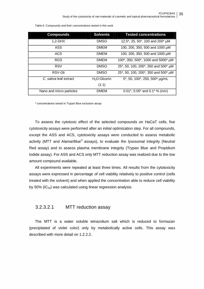
FCUP/ICBAS Study of the cytotoxicity of raw materials of cosmetic and topical pharmaceutical formulations
35
Table 6: Compounds and their concentrations tested in this work
* concentrations tested in Trypan Blue exclusion assay
To assess the cytotoxic effect of the selected compounds on HaCaT cells, five
cytotoxicity assays were performed after an initial optimization step. For all compounds,
except the ASS and ACS, cytotoxicity assays were conducted to assess metabolic
activity (MTT and AlamarBlue® assays), to evaluate the lysosomal integrity (Neutral
Red assay) and to assess plasma membrane integrity (Trypan Blue and Propidium
Iodide assay). For ASS and ACS only MTT reduction assay was realized due to the low
amount compound available.
All experiments were repeated at least three times. All results from the cytotoxicity
assays were expressed in percentage of cell viability relatively to positive control (cells
treated with the solvent) and when applied the concentration able to reduce cell viability
by 50% (IC50) was calculated using linear regression analysis.
3.2.3.2.1 MTT reduction assay
The MTT is a water soluble tetrazolium salt which is reduced to formazan
(precipitated of violet color) only by metabolically active cells. This assay was
described with more detail on 1.2.2.2.
Compounds Solvents Tested concentrations
1,2-DHX DMSO 12.5*, 25, 50*, 100 and 200* µM
ASS DMEM 100, 200, 350, 500 and 1000 µM
ACS DMEM 100, 200, 350, 500 and 1000 µM
RGS DMEM 100*, 350, 500*, 1000 and 5000* µM
RSV DMSO 25*, 50, 100, 200*, 350 and 500* µM
RSV-Gli DMSO 25*, 50, 100, 200*, 350 and 500* µM
C. sativa leaf extract H2O:Glicerin
(1:1)
5*, 50, 100*, 250, 500* µg/mL
Nano and micro particles DMEM 0.01*, 0.05* and 0.1* % (m/v)

36 FCUP/ICBAS Study of the cytotoxicity of raw materials of cosmetic and topical pharmaceutical formulations
Optimization of the MTT reduction assay conditions
For MTT reduction assay, the optimal cell density was previously selected. For this
end, cell densities of 2.5×10³, 5×10³, 1×10⁴, 2×10⁴ and 5×10⁴ cells/well were seeded on
96 well tissue culture plates (150 µL/well) and incubated at 37°C in a 5% CO2
atmosphere for 24 hours. The next day, the medium was removed, cells were washed
one time with DPBS, and fresh culture medium was added and cells were incubated at
37°C in a 5% CO2 atmosphere for another 24 hours. On the third day, a 5 mg/mL MTT
stock solution was prepared in DPBS and mixed by vortex until dissolved and then
diluted 1:10 in serum free DMEM. This solution was protected from light and prepared
on the day of use. The medium was removed, cells were washed one time with DPBS
and 150 μL of serum free DMEM containing 0.5 mg/mL MTT was added. After 2 hours
of incubation incubated at 37°C in a 5% CO2 atmosphere the reagent was removed and
followed by the addition of 150 μL DMSO to dissolve the formazan crystals. Finally, the
absorbance of formazan was determined at 570 nm using a plate reader (Biotek
Instruments, Synergy HT multi-mode microplate reader).
Study of the cytotoxic effect of the tested compound’s using
MTT reduction assay
After optimization of the method, 1×10⁴ cells/well (optimal cell density) were seeded
and incubated at 37°C in a 5% CO2 atmosphere. After 24 hours of incubation, the
medium was removed, cells were washed one time with DPBS and different test
compounds were added and incubated at 37°C in a 5% CO2 atmosphere for another 24
hours. Ten percent DMSO was used as positive control. On the third day, the
procedure was the same as described above.
The results were expressed as the absorbance ratio of treated to control cells:
(equation 2)
where As is the absorbance of treated cells and Ac the absorbance of control cells
(untreated cells).
FCUP/ICBAS Study of the cytotoxicity of raw materials of cosmetic and topical pharmaceutical formulations
37
In order to choose the maximum concentration that does not induce cytotoxicity,
DMSO was tested at different concentrations in this assay. Up to 1% had no influence
on cell viability in the methods used and so the DMSO concentration did not exceed
1%.
3.2.3.2.2 AlamarBlue® reduction assay
The AlamarBlue® is an oxidized form of redox indicator that is blue in color and non-
fluorescent. When incubated with viable cells, the reagent changes color from blue to
pink and become fluorescent. This assay was described with more detail on 1.2.2.1.
Optimization of AlamarBlue® reduction assay conditions
For AlamarBlue® reduction assay, the optimal cell density and incubation time with
AlamarBlue® were previously optimized. For this purpose, cell densities of 5×10³,
1×10⁴, 2×10⁴ and 4×10⁴ cells/well were seeded on 96 well tissue culture plates (150
µL/well) and incubated at 37°C in a 5% CO2 atmosphere for 24 hours. The next day,
the medium was removed, cells were washed one time with DPBS, and fresh culture
medium was added and cells were incubated 37°C in a 5% CO2 atmosphere for
another 24 hours. Ten percent AlamarBlue® of the volume in the well was added to
each well and incubated at 37°C in a 5% CO2 atmosphere. AlamarBlue® incubation
times of 1, 2, 3 and 4 hours were tested. Finally, the absorbances at 570 nm (reduced
form) and 600 nm (oxidized form) were determined using a plate reader.
The % reduction of AlamarBlue® was calculated using the following equation:
–
– (equation 3)
where:
Eoxi570 = molar extinction coefficient (E) of oxidized AlamarBlue® at 570nm = 80586
Eoxi600 = E of oxidized AlamarBlue® at 600nm = 117216
A570 = absorbance of test wells at 570nm
38 FCUP/ICBAS Study of the cytotoxicity of raw materials of cosmetic and topical pharmaceutical formulations
A600 = absorbance of test wells at 600nm
Ered570 = E of reduced AlamarBlue® at 570nm = 155677
Ered600 = E of reduced AlamarBlue® at 600nm = 14652
C570 = absorbance of negative control well (medium, AlamarBlue®, no cells) at
570nm
C600 = absorbance of negative control well (medium, AlamarBlue®, no cells) at
600nm
Study of the cytotoxic effect of the tested compound’s using
AlamarBlue® reduction assay
After optimization of the method, 2×10⁴ cells/well (optimal cell density) were seeded
incubated at 37°C in a 5% CO2 atmosphere. After 24 hours of incubation, the medium
was removed, cells were washed one time with DPBS and the different test
compounds were added and incubated at 37°C in a 5% CO2 atmosphere for another 24
hours. Ten percent DMSO was used as positive control. Ten percent AlamarBlue® of
the volume in the well was added to each well and incubated at 37°C for 4 hours
(optimal incubation time). Finally, the absorbance at 570 nm and 600 nm was
determined using a plate reader.
The % reduction of AlamarBlue® was calculated according to equation 3.
3.2.3.2.3 Neutral Red uptake assay
The amount of NR dye incorporated in the cells represents lysosomal functionality,
as this dye easily penetrates viable cell membranes and accumulates intracellularly in
lysosomes. This assay was described with more detail on 1.2.1.4.
Optimization of NR uptake assay conditions
For NR uptake assay, the optimal cell density, the concentration of the NR solution
and the incubation time with NR were previously optimized. For this purpose, 2.5×10³,
5×10³, 1×10⁴, 2×10⁴ and 5×10⁴ cells/well were seeded on 96 tissue culture well plates
(150 µL/well) and incubated at 37°C in a 5% CO2 atmosphere for 24 hours. The next
day, the medium was removed, cells were washed one time with DPBS, and fresh
culture medium was added and incubated at 37°C in a 5% CO2 atmosphere for another
FCUP/ICBAS Study of the cytotoxicity of raw materials of cosmetic and topical pharmaceutical formulations
39
24 hours. The medium was removed, cells were washed one time with DPBS and 150
μL of culture medium containing 33, 50, 100 and 330 µg/mL NR solution were added to
each well and incubated at 37°C in a 5% CO2 atmosphere. NR incubation times of 2, 3
and 4 hours were tested. NR tends to precipitate, so the medium with stain was
incubated at 37°C in a 5% CO2 atmosphere protected from light overnight, centrifuged
at 1500 g for 10 minutes and filtered (5 μm) before use. After incubation time, NR
solution was removed, cells were washed one time with DPBS at 37°C and 150 μL/well
of the solution formed by 50% ethanol/1% acetic acid was added to extract the NR dye
meanwhile captured within the cells. The plates were then placed in a microplate
shaker for 10 minutes, at room temperature and protected from light. Finally,
absorbance was measured at 540 nm.
Study of the cytotoxic effect of the tested compound’s using
NR uptake assay
After optimization of the method, 2×10⁴ cells/well (optimal cell density) were seeded
and incubated at 37°C in a 5% CO2 atmosphere. After 24 hours of incubation, the
medium was removed, cells were washed one time DPBS and different test
compounds were added and incubated at 37°C in a 5% CO2 atmosphere for another 24
hours. Ten percent DMSO was used as positive control. The medium was removed,
cells were washed one time with DPBS at 37°C and 150 μL of complete DMEM
containing 50 µg/mL NR (optimal concentration) was added to each well and incubated
for 3 hours (optimal incubation time) protected from light. After incubation time, NR
solution was removed, cells were washed one time with DPBS and 150 μL/well of the
solution formed by 50% ethanol/1% acetic acid was added to extract the NR dye
meanwhile captured within the cells. The plates were then placed in a microplate
shaker for 10 min, at room temperature and protected from light. Finally absorbance
was measured at 540 nm.
The results were expressed as the absorbance ratio of treated to control cells,
using Equation 2.
40 FCUP/ICBAS Study of the cytotoxicity of raw materials of cosmetic and topical pharmaceutical formulations
3.2.3.2.4 Trypan Blue exclusion assay
The trypan blue is a vital stain used to stain dead cells. This assay was described
with more detail on 1.2.1.6.
Optimization of Trypan Blue exclusion assay conditions
For Trypan Blue dye exclusion assay, the optimal cell density was previously
selected. For this end, cell densities of 1×10⁴, 2×10⁴, 5×10⁴, 1×10⁵, 2×10⁵ and 3.5×10⁵
cells/well were seeded on 12 well culture plates (1 mL/well) and incubated at 37°C in a
5% CO2 atmosphere for 24 hours. The next day, the medium was removed, cells were
washed one time with DPBS, and fresh culture medium was added and cells were
incubated at 37°C in a 5% CO2 atmosphere for another 24h. Then the contents of the
wells were transferred into conical tubes, wells were washed one time with DPBS and
also transferred into conical tubes. Then, the cells were detached from the bottom of
the wells by the action of trypsin after 5-12 minutes and were centrifuged at 416 g for 5
minutes at room temperature. Cells were counted using 50 µL of cell suspension added
to 50 µL of 0.4% trypan blue vital dye (1:1).
Study of the cytotoxic effect of the tested compound’s using
Trypan Blue exclusion assay
After optimization of the method, 1×10⁵ cells/well (optimal cell density) were seeded
and incubated at 37°C in a 5% CO2 atmosphere. After 24 hours of incubation, the
medium was removed, cells were washed one time DPBS and different test
compounds were added and incubated at 37°C for another 24 hours. Ten percent
DMSO and 15% ethanol 70º were tested as positive control. On the third day, the
procedure was the same as described above.
The % cell viability was calculated according to:
(equation 4)
FCUP/ICBAS Study of the cytotoxicity of raw materials of cosmetic and topical pharmaceutical formulations
41
3.2.3.2.5 Propidium Iodide exclusion assay
PI is a dye that does not pass through intact cell membranes. This assay was
described with more detail on 1.2.1.5.
Study of the cytotoxic effect of the tested compound’s using
Propidium Iodide exclusion assay
Using the cell density previously optimized for Trypan Blue dye exclusion assay,
1×10⁵ cells/well were seeded and incubated at 37°C in a 5% CO2 atmosphere. After 24
hours of incubation at 37°C in a 5% CO2 atmosphere, the medium was removed, cells
were washed one time DPBS and different test compounds were added and incubated
at 37°C in a 5% CO2 atmosphere for another 24 hours. Ten percent DMSO and 0.1%
Triton X-100 were tested as positive control. On the third day, the contents of the wells
were transferred into conical tubes, wells were washed one time with DPBS and also
transferred into conical tubes. Then, the cells were detached from the bottom of the
wells by the action of trypsin after 8 minutes (optimized incubation time), complete
DMEM was added to stop the action of trypsin and the content was centrifuged at 416
g for 5 minutes at room temperature. The supernatant was discarded and the cells
washed two times by addition of cold 5 mL of DPBS + 2% FBS, centrifuging at 416 g
for 6 minutes at a temperature of 4°C. Then, the supernatant was aspirated and the
cells were ressuspended in 100 μL of cold DPBS. Content was transferred to 2 mL
tubes and, finally, stained with 5 μL of PI working solution [10 μL of stock solution (10
mg of PI + 10 mL of Milli Q water) in 1 mL of DPBS] 5 minutes before starting the
analysis. Cells were acquired on the flow cytometer BD AccuriTM C6 and at least ten
thousand events were analyzed on the basis of size, complexity and emission of
orange fluorescence, by setting up instrument settings for FSC, SSC and FL-2 and by
using stained and unstained cells. Data were analyzed by using the BD AccuriTM C6
software version 1.0.264.21.
3.2.4 Statistical analysis
The data is presented as mean ± standard deviation (SD) of at least three
independent experiments. Statistical analysis was carried out using One-way analysis

42 FCUP/ICBAS Study of the cytotoxicity of raw materials of cosmetic and topical pharmaceutical formulations
of variance (ANOVA) followed by the Dunnett post hoc test (comparison to negative
control), after confirming normality and homogeneity of variance with Shapiro-Wilk and
Levene tests, respectively. Comparisons between loaded and empty nanoparticles
were performed with the Tukey post hoc test. Statistical significance was considered
when p < 0.05.
All statistical analyses were performed using the SPSS software (v 23.0; IBM,
Armonk, NY, USA) and graphs were generated by GraphPad Prism for Windows
(version 6.0; GraphPad Software, Inc., USA).
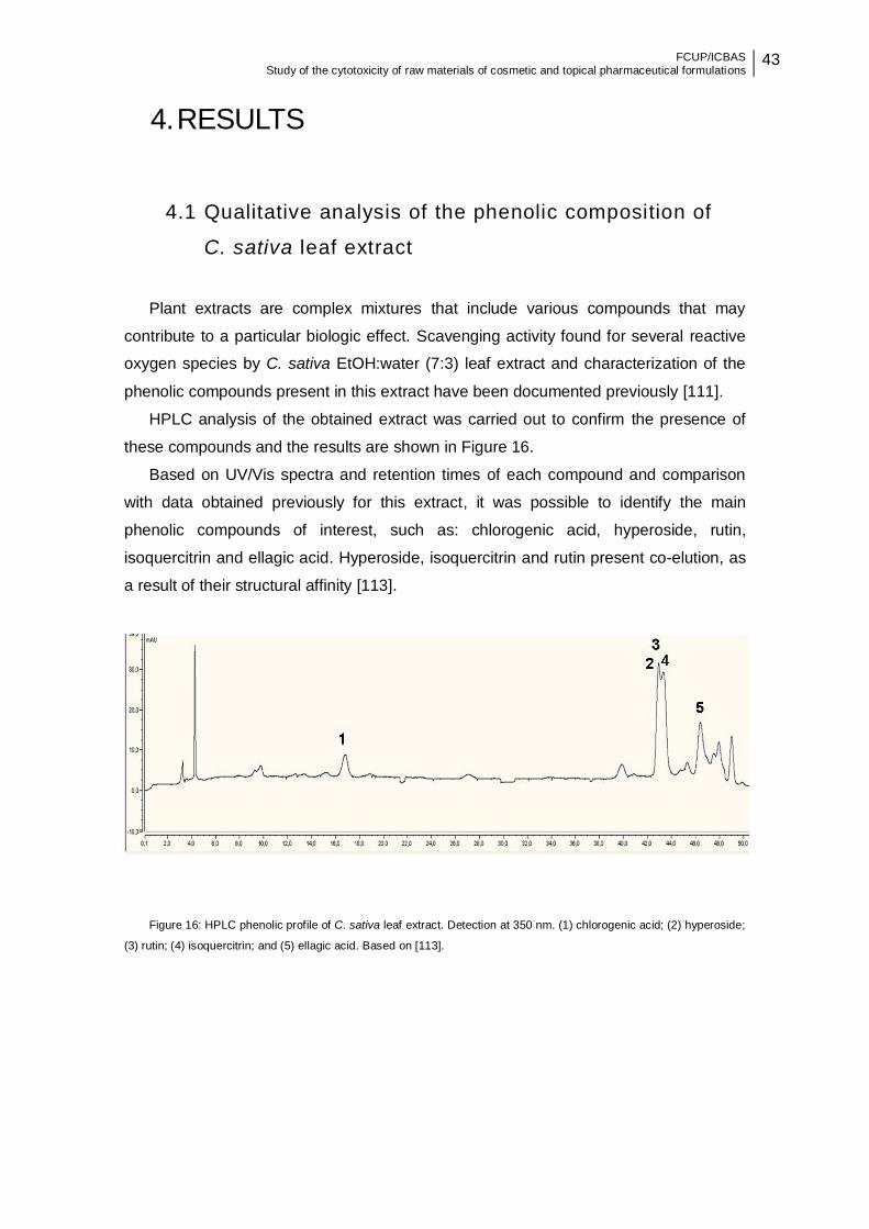
FCUP/ICBAS Study of the cytotoxicity of raw materials of cosmetic and topical pharmaceutical formulations
43
4. RESULTS
4.1 Qualitative analysis of the phenolic composition of
C. sativa leaf extract
Plant extracts are complex mixtures that include various compounds that may
contribute to a particular biologic effect. Scavenging activity found for several reactive
oxygen species by C. sativa EtOH:water (7:3) leaf extract and characterization of the
phenolic compounds present in this extract have been documented previously [111].
HPLC analysis of the obtained extract was carried out to confirm the presence of
these compounds and the results are shown in Figure 16.
Based on UV/Vis spectra and retention times of each compound and comparison
with data obtained previously for this extract, it was possible to identify the main
phenolic compounds of interest, such as: chlorogenic acid, hyperoside, rutin,
isoquercitrin and ellagic acid. Hyperoside, isoquercitrin and rutin present co-elution, as
a result of their structural affinity [113].
Figure 16: HPLC phenolic profile of C. sativa leaf extract. Detection at 350 nm. (1) chlorogenic acid; (2) hyperoside;
(3) rutin; (4) isoquercitrin; and (5) ellagic acid. Based on [113].
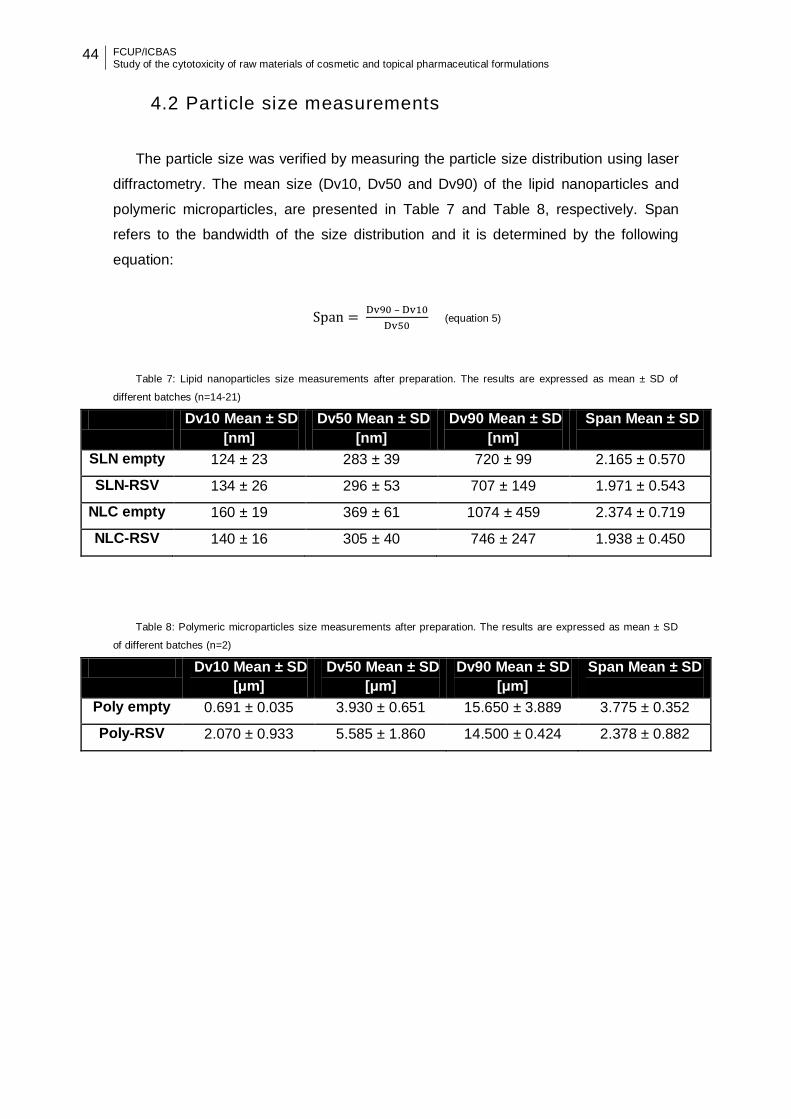
44 FCUP/ICBAS Study of the cytotoxicity of raw materials of cosmetic and topical pharmaceutical formulations
4.2 Particle size measurements
The particle size was verified by measuring the particle size distribution using laser
diffractometry. The mean size (Dv10, Dv50 and Dv90) of the lipid nanoparticles and
polymeric microparticles, are presented in Table 7 and Table 8, respectively. Span
refers to the bandwidth of the size distribution and it is determined by the following
equation:
–
(equation 5)
Table 7: Lipid nanoparticles size measurements after preparation. The results are expressed as mean ± SD of
different batches (n=14-21)
Dv10 Mean ± SD
[nm]
Dv50 Mean ± SD
[nm]
Dv90 Mean ± SD
[nm]
Span Mean ± SD
SLN empty 124 ± 23 283 ± 39 720 ± 99 2.165 ± 0.570
SLN-RSV 134 ± 26 296 ± 53 707 ± 149 1.971 ± 0.543
NLC empty 160 ± 19 369 ± 61 1074 ± 459 2.374 ± 0.719
NLC-RSV 140 ± 16 305 ± 40 746 ± 247 1.938 ± 0.450
Table 8: Polymeric microparticles size measurements after preparation. The results are expressed as mean ± SD
of different batches (n=2)
Dv10 Mean ± SD
[μm]
Dv50 Mean ± SD
[μm]
Dv90 Mean ± SD
[μm]
Span Mean ± SD
Poly empty 0.691 ± 0.035 3.930 ± 0.651 15.650 ± 3.889 3.775 ± 0.352
Poly-RSV 2.070 ± 0.933 5.585 ± 1.860 14.500 ± 0.424 2.378 ± 0.882
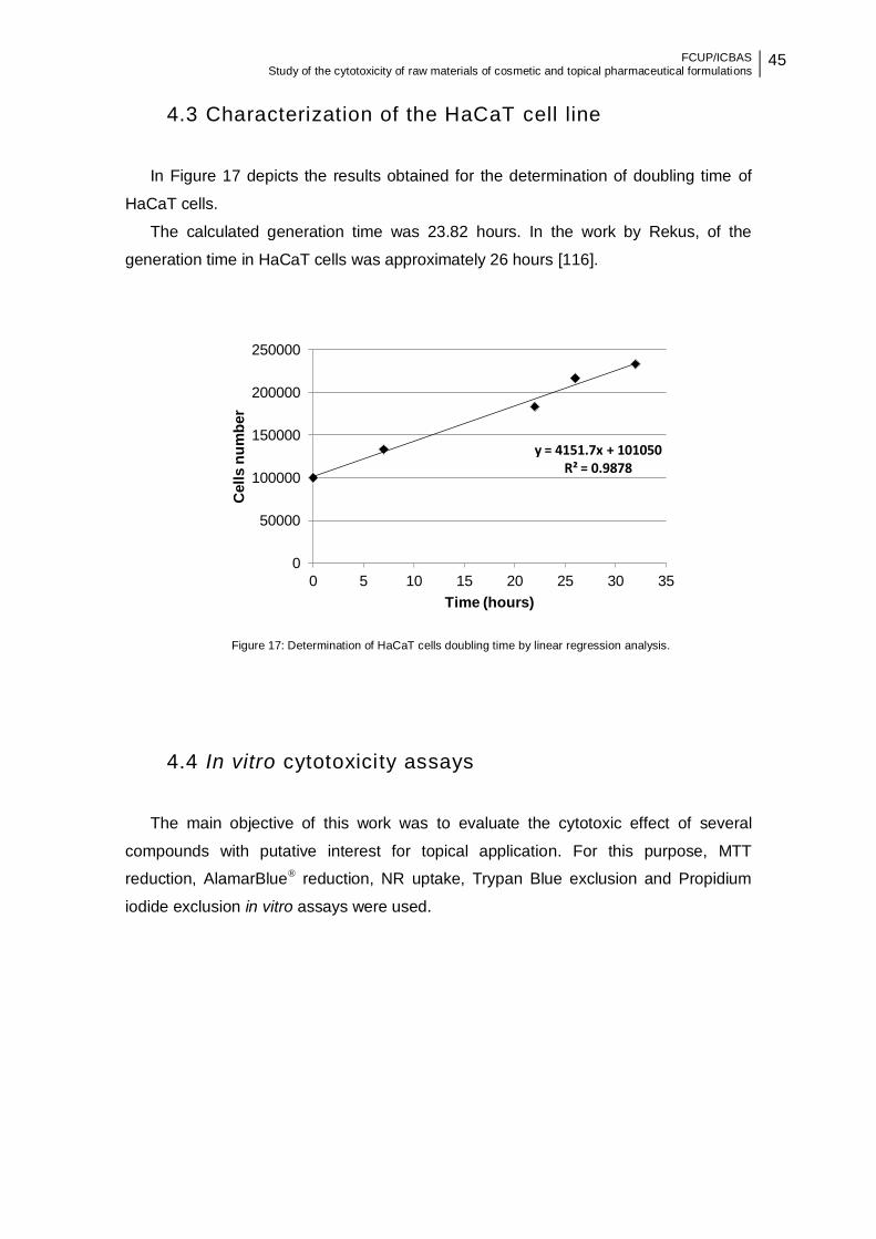
FCUP/ICBAS Study of the cytotoxicity of raw materials of cosmetic and topical pharmaceutical formulations
45
4.3 Characterization of the HaCaT cell line
In Figure 17 depicts the results obtained for the determination of doubling time of
HaCaT cells.
The calculated generation time was 23.82 hours. In the work by Rekus, of the
generation time in HaCaT cells was approximately 26 hours [116].
Figure 17: Determination of HaCaT cells doubling time by linear regression analysis.
4.4 In vitro cytotoxicity assays
The main objective of this work was to evaluate the cytotoxic effect of several
compounds with putative interest for topical application. For this purpose, MTT
reduction, AlamarBlue® reduction, NR uptake, Trypan Blue exclusion and Propidium
iodide exclusion in vitro assays were used.
y = 4151.7x + 101050 R² = 0.9878
0
50000
100000
150000
200000
250000
0 5 10 15 20 25 30 35
Cell
s n
um
ber
Time (hours)
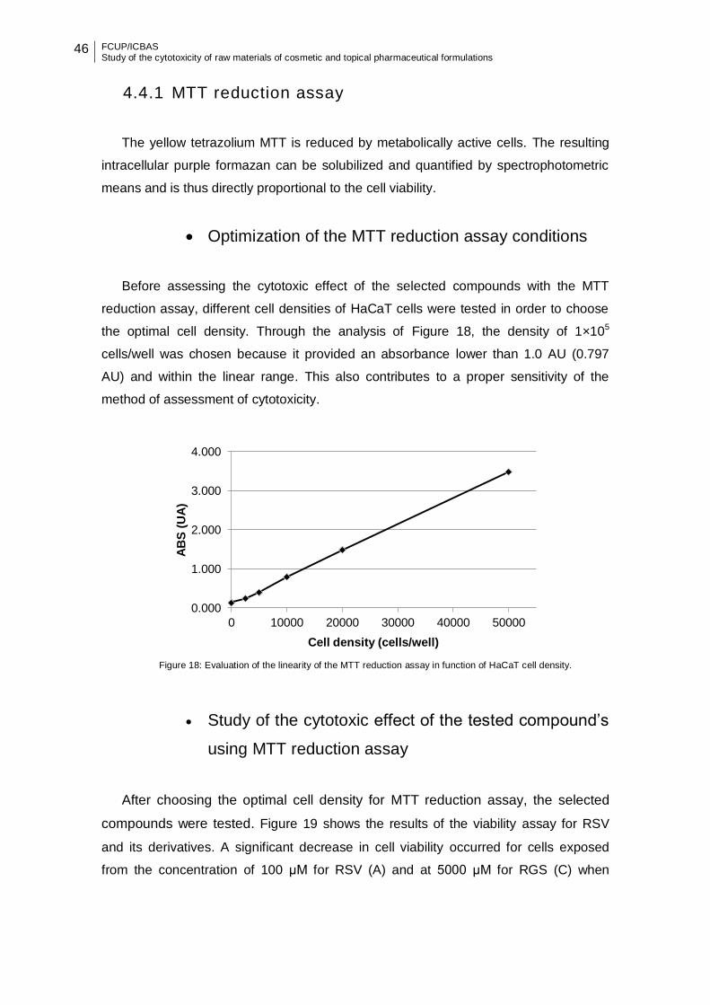
46 FCUP/ICBAS Study of the cytotoxicity of raw materials of cosmetic and topical pharmaceutical formulations
4.4.1 MTT reduction assay
The yellow tetrazolium MTT is reduced by metabolically active cells. The resulting
intracellular purple formazan can be solubilized and quantified by spectrophotometric
means and is thus directly proportional to the cell viability.
Optimization of the MTT reduction assay conditions
Before assessing the cytotoxic effect of the selected compounds with the MTT
reduction assay, different cell densities of HaCaT cells were tested in order to choose
the optimal cell density. Through the analysis of Figure 18, the density of 1×10⁵
cells/well was chosen because it provided an absorbance lower than 1.0 AU (0.797
AU) and within the linear range. This also contributes to a proper sensitivity of the
method of assessment of cytotoxicity.
Figure 18: Evaluation of the linearity of the MTT reduction assay in function of HaCaT cell density.
Study of the cytotoxic effect of the tested compound’s
using MTT reduction assay
After choosing the optimal cell density for MTT reduction assay, the selected
compounds were tested. Figure 19 shows the results of the viability assay for RSV
and its derivatives. A significant decrease in cell viability occurred for cells exposed
from the concentration of 100 μM for RSV (A) and at 5000 μM for RGS (C) when
0.000
1.000
2.000
3.000
4.000
0 10000 20000 30000 40000 50000
AB
S (
UA
)
Cell density (cells/well)
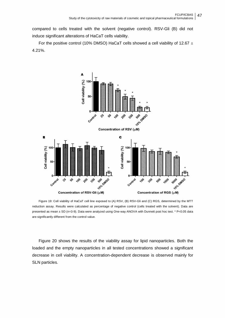
FCUP/ICBAS Study of the cytotoxicity of raw materials of cosmetic and topical pharmaceutical formulations
47
compared to cells treated with the solvent (negative control). RSV-Gli (B) did not
induce significant alterations of HaCaT cells viability.
For the positive control (10% DMSO) HaCaT cells showed a cell viability of 12.67 ±
4.21%.
Figure 19: Cell viability of HaCaT cell line exposed to (A) RSV, (B) RSV-Gli and (C) RGS, determined by the MTT
reduction assay. Results were calculated as percentage of negative control (cells treated with the solvent). Data are
presented as mean ± SD (n=3-9). Data were analyzed using One-way ANOVA with Dunnett post hoc test. * P<0.05 data
are significantly different from the control value.
Figure 20 shows the results of the viability assay for lipid nanoparticles. Both the
loaded and the empty nanoparticles in all tested concentrations showed a significant
decrease in cell viability. A concentration-dependent decrease is observed mainly for
SLN particles.

48 FCUP/ICBAS Study of the cytotoxicity of raw materials of cosmetic and topical pharmaceutical formulations
Figure 20: Cell viability of HaCaT cell line exposed to (A) SLN empty and SLN-RSV and (B) NLC empty and NLC-
RSV, determined by the MTT reduction assay. Results were calculated as percentage of negative control (cells treated
with the solvent). Data are presented as mean ± SD (n=3-9). Data were analyzed using One-way ANOVA with Dunnett
post hoc test. * P<0.05 data are significantly different from the control value. Comparisons between loaded and empty
nanoparticles were performed with the Tukey post hoc test. ** P<0.05 data are statistically different.
In addition to the lipid nanoparticles, polymeric microparticles were also tested with
the MTT reduction assay. However, the deposition of the microparticles on the surface
of the cell monolayer and microparticle aggregation (as shown in Figure 21) led to a
great variability of the results. For this reason, these particles were not included on
subsequent cytotoxicity tests.
Figure 21: Observation under inverted microscope of HaCaT cell line with
0.01% polymeric microparticles after 24 hours incubation (10X magnification).
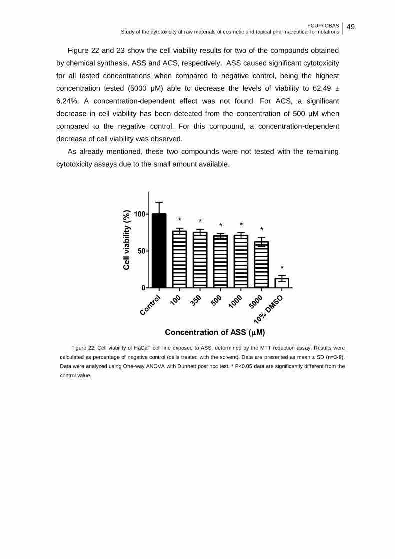
FCUP/ICBAS Study of the cytotoxicity of raw materials of cosmetic and topical pharmaceutical formulations
49
Figure 22 and 23 show the cell viability results for two of the compounds obtained
by chemical synthesis, ASS and ACS, respectively. ASS caused significant cytotoxicity
for all tested concentrations when compared to negative control, being the highest
concentration tested (5000 μM) able to decrease the levels of viability to 62.49 ±
6.24%. A concentration-dependent effect was not found. For ACS, a significant
decrease in cell viability has been detected from the concentration of 500 μM when
compared to the negative control. For this compound, a concentration-dependent
decrease of cell viability was observed.
As already mentioned, these two compounds were not tested with the remaining
cytotoxicity assays due to the small amount available.
Figure 22: Cell viability of HaCaT cell line exposed to ASS, determined by the MTT reduction assay. Results were
calculated as percentage of negative control (cells treated with the solvent). Data are presented as mean ± SD (n=3-9).
Data were analyzed using One-way ANOVA with Dunnett post hoc test. * P<0.05 data are significantly different from the
control value.
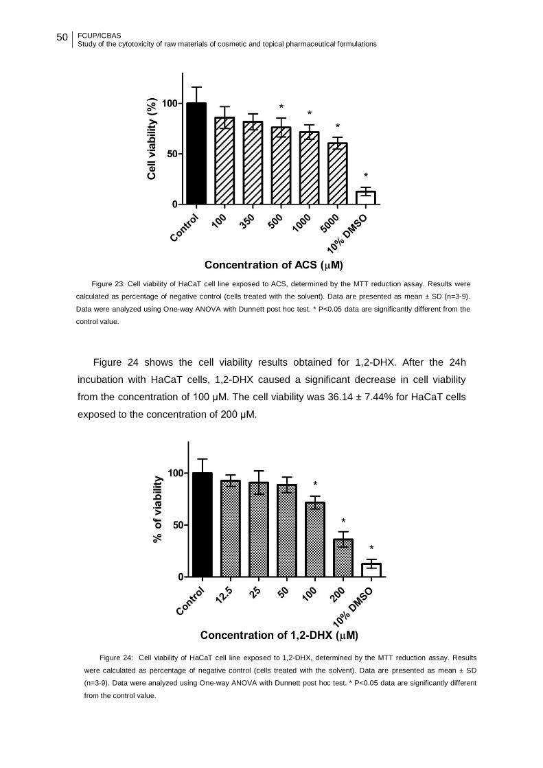
50 FCUP/ICBAS Study of the cytotoxicity of raw materials of cosmetic and topical pharmaceutical formulations
Figure 24 shows the cell viability results obtained for 1,2-DHX. After the 24h
incubation with HaCaT cells, 1,2-DHX caused a significant decrease in cell viability
from the concentration of 100 μM. The cell viability was 36.14 ± 7.44% for HaCaT cells
exposed to the concentration of 200 μM.
Figure 23: Cell viability of HaCaT cell line exposed to ACS, determined by the MTT reduction assay. Results were
calculated as percentage of negative control (cells treated with the solvent). Data are presented as mean ± SD (n=3-9).
Data were analyzed using One-way ANOVA with Dunnett post hoc test. * P<0.05 data are significantly different from the
control value.
Figure 24: Cell viability of HaCaT cell line exposed to 1,2-DHX, determined by the MTT reduction assay. Results
were calculated as percentage of negative control (cells treated with the solvent). Data are presented as mean ± SD
(n=3-9). Data were analyzed using One-way ANOVA with Dunnett post hoc test. * P<0.05 data are significantly different
from the control value.

FCUP/ICBAS Study of the cytotoxicity of raw materials of cosmetic and topical pharmaceutical formulations
51
The ability of C.sativa EtOH:water (7:3) leaf extract to interfere with cell viability of
the HaCaT cells after 24 hours incubation was evaluated. The Figure 25 shows that
C.sativa extract caused significant cytotoxicity when compared to negative control only
at the concentration of 500 μg/mL.
Figure 25: Cell viability of HaCaT cell line exposed to C.sativa leaf extract, determined by the MTT reduction assay.
Results were calculated as percentage of negative control (cells treated with the solvent). Data are presented as mean ± SD
(n=3-9). Data were analyzed using One-way ANOVA with Dunnett post hoc test. * P<0.05 data are significantly different from
the control value.
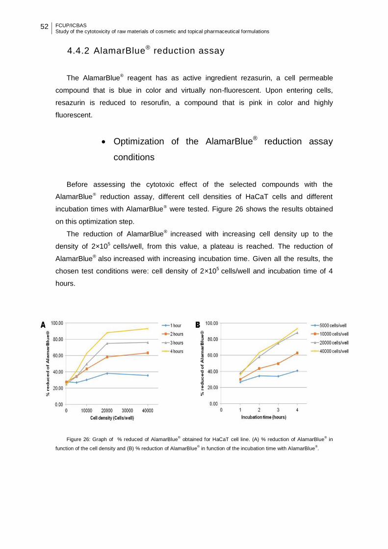
52 FCUP/ICBAS Study of the cytotoxicity of raw materials of cosmetic and topical pharmaceutical formulations
4.4.2 AlamarBlue® reduction assay
The AlamarBlue® reagent has as active ingredient rezasurin, a cell permeable
compound that is blue in color and virtually non-fluorescent. Upon entering cells,
resazurin is reduced to resorufin, a compound that is pink in color and highly
fluorescent.
Optimization of the AlamarBlue® reduction assay
conditions
Before assessing the cytotoxic effect of the selected compounds with the
AlamarBlue® reduction assay, different cell densities of HaCaT cells and different
incubation times with AlamarBlue® were tested. Figure 26 shows the results obtained
on this optimization step.
The reduction of AlamarBlue® increased with increasing cell density up to the
density of 2×10⁵ cells/well, from this value, a plateau is reached. The reduction of
AlamarBlue® also increased with increasing incubation time. Given all the results, the
chosen test conditions were: cell density of 2×10⁵ cells/well and incubation time of 4
hours.
Figure 26: Graph of % reduced of AlamarBlue® obtained for HaCaT cell line. (A) % reduction of AlamarBlue
® in
function of the cell density and (B) % reduction of AlamarBlue® in function of the incubation time with AlamarBlue
®.
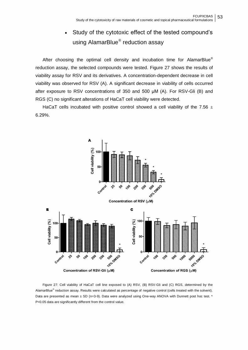
FCUP/ICBAS Study of the cytotoxicity of raw materials of cosmetic and topical pharmaceutical formulations
53
Study of the cytotoxic effect of the tested compound’s
using AlamarBlue® reduction assay
After choosing the optimal cell density and incubation time for AlamarBlue®
reduction assay, the selected compounds were tested. Figure 27 shows the results of
viability assay for RSV and its derivatives. A concentration-dependent decrease in cell
viability was observed for RSV (A). A significant decrease in viability of cells occurred
after exposure to RSV concentrations of 350 and 500 μM (A). For RSV-Gli (B) and
RGS (C) no significant alterations of HaCaT cell viability were detected.
HaCaT cells incubated with positive control showed a cell viability of the 7.56 ±
6.29%.
Figure 27: Cell viability of HaCaT cell line exposed to (A) RSV, (B) RSV-Gli and (C) RGS, determined by the
AlamarBlue® reduction assay. Results were calculated as percentage of negative control (cells treated with the solvent).
Data are presented as mean ± SD (n=3-9). Data were analyzed using One-way ANOVA with Dunnett post hoc test. *
P<0.05 data are significantly different from the control value.
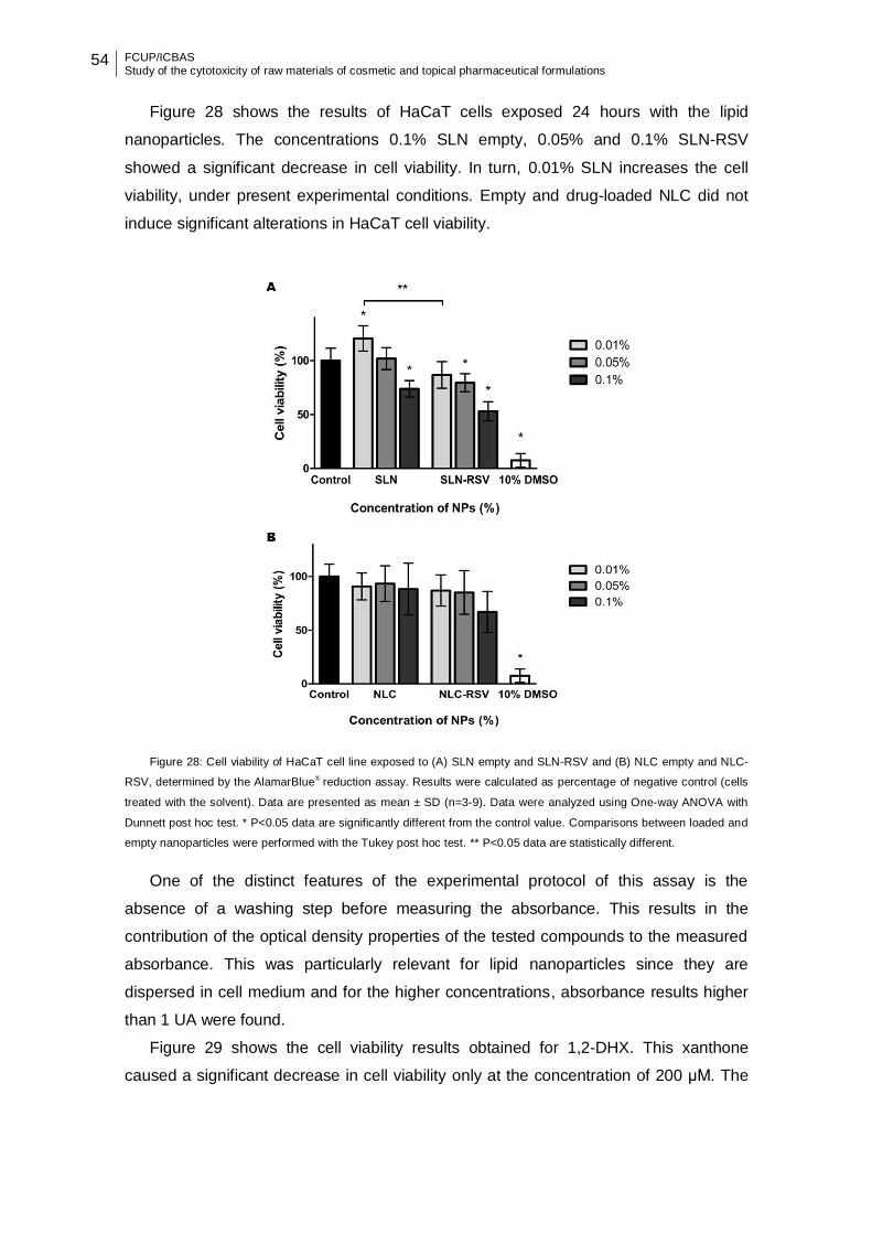
54 FCUP/ICBAS Study of the cytotoxicity of raw materials of cosmetic and topical pharmaceutical formulations
Figure 28 shows the results of HaCaT cells exposed 24 hours with the lipid
nanoparticles. The concentrations 0.1% SLN empty, 0.05% and 0.1% SLN-RSV
showed a significant decrease in cell viability. In turn, 0.01% SLN increases the cell
viability, under present experimental conditions. Empty and drug-loaded NLC did not
induce significant alterations in HaCaT cell viability.
Figure 28: Cell viability of HaCaT cell line exposed to (A) SLN empty and SLN-RSV and (B) NLC empty and NLC-
RSV, determined by the AlamarBlue® reduction assay. Results were calculated as percentage of negative control (cells
treated with the solvent). Data are presented as mean ± SD (n=3-9). Data were analyzed using One-way ANOVA with
Dunnett post hoc test. * P<0.05 data are significantly different from the control value. Comparisons between loaded and
empty nanoparticles were performed with the Tukey post hoc test. ** P<0.05 data are statistically different.
One of the distinct features of the experimental protocol of this assay is the
absence of a washing step before measuring the absorbance. This results in the
contribution of the optical density properties of the tested compounds to the measured
absorbance. This was particularly relevant for lipid nanoparticles since they are
dispersed in cell medium and for the higher concentrations, absorbance results higher
than 1 UA were found.
Figure 29 shows the cell viability results obtained for 1,2-DHX. This xanthone
caused a significant decrease in cell viability only at the concentration of 200 μM. The

FCUP/ICBAS Study of the cytotoxicity of raw materials of cosmetic and topical pharmaceutical formulations
55
cell viability was 54.51 ± 13.53% for HaCaT cells exposed to the concentration of 200
μM.
The Figure 30 shows the results obtained for extract plant. In general, C. sativa leaf
extract did not induce significant alterations of HaCaT cells, when available with
AlamarBlue® reduction assay.
Figure 29: Cell viability of HaCaT cell line exposed to 1,2-DHX, determined by the AlamarBlue® reduction assay.
Results were calculated as percentage of negative control (cells treated with the solvent). Data are presented as mean
± SD (n=3-9). Data were analyzed using One-way ANOVA with Dunnett post hoc test. * P<0.05 data are significantly
different from the control value.
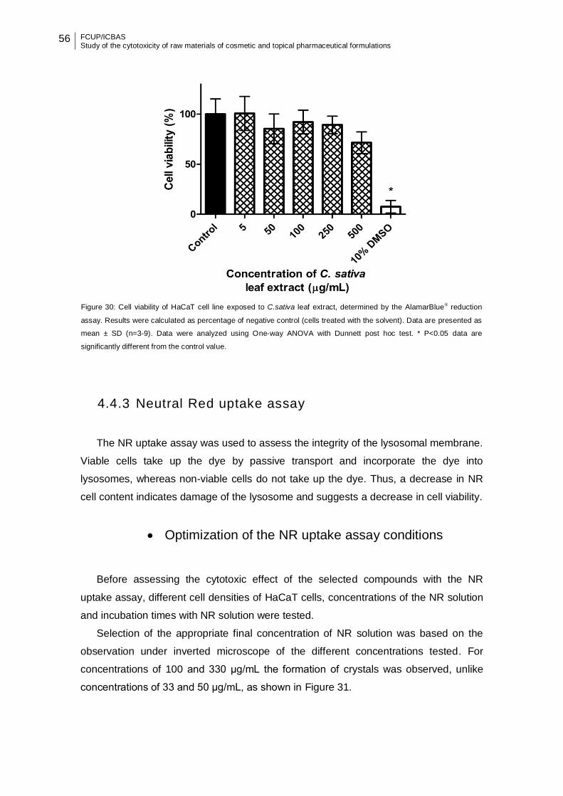
56 FCUP/ICBAS Study of the cytotoxicity of raw materials of cosmetic and topical pharmaceutical formulations
4.4.3 Neutral Red uptake assay
The NR uptake assay was used to assess the integrity of the lysosomal membrane.
Viable cells take up the dye by passive transport and incorporate the dye into
lysosomes, whereas non-viable cells do not take up the dye. Thus, a decrease in NR
cell content indicates damage of the lysosome and suggests a decrease in cell viability.
Optimization of the NR uptake assay conditions
Before assessing the cytotoxic effect of the selected compounds with the NR
uptake assay, different cell densities of HaCaT cells, concentrations of the NR solution
and incubation times with NR solution were tested.
Selection of the appropriate final concentration of NR solution was based on the
observation under inverted microscope of the different concentrations tested. For
concentrations of 100 and 330 μg/mL the formation of crystals was observed, unlike
concentrations of 33 and 50 μg/mL, as shown in Figure 31.
Figure 30: Cell viability of HaCaT cell line exposed to C.sativa leaf extract, determined by the AlamarBlue® reduction
assay. Results were calculated as percentage of negative control (cells treated with the solvent). Data are presented as
mean ± SD (n=3-9). Data were analyzed using One-way ANOVA with Dunnett post hoc test. * P<0.05 data are
significantly different from the control value.
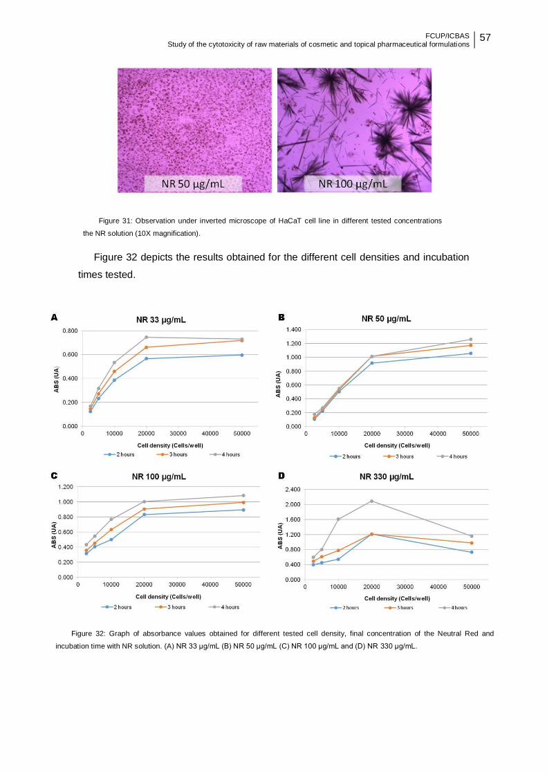
FCUP/ICBAS Study of the cytotoxicity of raw materials of cosmetic and topical pharmaceutical formulations
57
Figure 32 depicts the results obtained for the different cell densities and incubation
times tested.
Figure 31: Observation under inverted microscope of HaCaT cell line in different tested concentrations
the NR solution (10X magnification).
Figure 32: Graph of absorbance values obtained for different tested cell density, final concentration of the Neutral Red and
incubation time with NR solution. (A) NR 33 μg/mL (B) NR 50 μg/mL (C) NR 100 μg/mL and (D) NR 330 μg/mL.
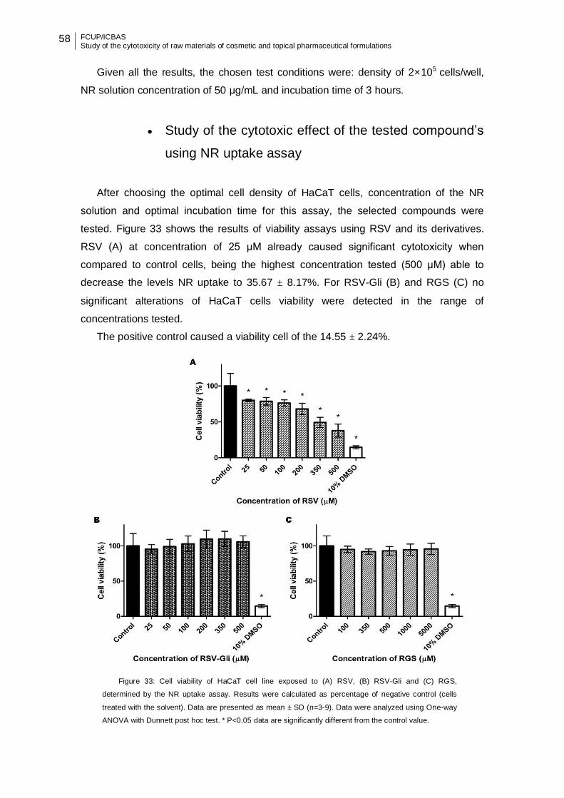
58 FCUP/ICBAS Study of the cytotoxicity of raw materials of cosmetic and topical pharmaceutical formulations
Given all the results, the chosen test conditions were: density of 2×10⁵ cells/well,
NR solution concentration of 50 μg/mL and incubation time of 3 hours.
Study of the cytotoxic effect of the tested compound’s
using NR uptake assay
After choosing the optimal cell density of HaCaT cells, concentration of the NR
solution and optimal incubation time for this assay, the selected compounds were
tested. Figure 33 shows the results of viability assays using RSV and its derivatives.
RSV (A) at concentration of 25 μM already caused significant cytotoxicity when
compared to control cells, being the highest concentration tested (500 μM) able to
decrease the levels NR uptake to 35.67 ± 8.17%. For RSV-Gli (B) and RGS (C) no
significant alterations of HaCaT cells viability were detected in the range of
concentrations tested.
The positive control caused a viability cell of the 14.55 ± 2.24%.
Figure 33: Cell viability of HaCaT cell line exposed to (A) RSV, (B) RSV-Gli and (C) RGS,
determined by the NR uptake assay. Results were calculated as percentage of negative control (cells
treated with the solvent). Data are presented as mean ± SD (n=3-9). Data were analyzed using One-way
ANOVA with Dunnett post hoc test. * P<0.05 data are significantly different from the control value.

FCUP/ICBAS Study of the cytotoxicity of raw materials of cosmetic and topical pharmaceutical formulations
59
Figure 34 shows the results of cell viability obtained with HaCaT cells exposed 24
hours with lipid nanoparticles. Regarding the SLN, there was a significant decrease in
cell viability to 0.05% and 0.1% concentrations. For these lipid nanoparticles, a
concentration-dependent decrease of cell viability was observed. Already the NLC
showed significant decrease in cell viability for 0.1% NLC empty, 0.05% and 0.1%
NLC-RSV. It is also important to underline the significant difference between the empty
NLC and loaded-NLC with RSV for concentrations of 0.05% and 0.1%.
Figure 34: Cell viability of HaCaT cell line exposed to (A) SLN empty and SLN-RSV and (B) NLC empty and NLC-
RSV, determined by the NR uptake assay. Results were calculated as percentage of negative control (cells treated with
the solvent). Data are presented as mean ± SD (n=3-9). Data were analyzed using One-way ANOVA with Dunnett post
hoc test. * P<0.05 data are significantly different from the control value. Comparisons between loaded and empty
nanoparticles were performed with the Tukey post hoc test. ** P<0.05 data are statistically different.
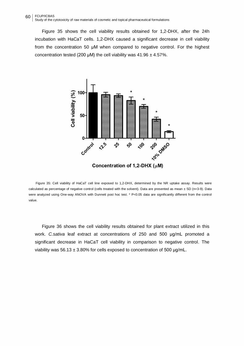
60 FCUP/ICBAS Study of the cytotoxicity of raw materials of cosmetic and topical pharmaceutical formulations
Figure 35 shows the cell viability results obtained for 1,2-DHX, after the 24h
incubation with HaCaT cells. 1,2-DHX caused a significant decrease in cell viability
from the concentration 50 μM when compared to negative control. For the highest
concentration tested (200 μM) the cell viability was 41.96 ± 4.57%.
Figure 36 shows the cell viability results obtained for plant extract utilized in this
work. C.sativa leaf extract at concentrations of 250 and 500 μg/mL promoted a
significant decrease in HaCaT cell viability in comparison to negative control. The
viability was 56.13 ± 3.80% for cells exposed to concentration of 500 μg/mL.
Figure 35: Cell viability of HaCaT cell line exposed to 1,2-DHX, determined by the NR uptake assay. Results were
calculated as percentage of negative control (cells treated with the solvent). Data are presented as mean ± SD (n=3-9). Data
were analyzed using One-way ANOVA with Dunnett post hoc test. * P<0.05 data are significantly different from the control
value.

FCUP/ICBAS Study of the cytotoxicity of raw materials of cosmetic and topical pharmaceutical formulations
61
4.4.4 Trypan Blue exclusion assay
The Trypan Blue exclusion assay was used to assess the integrity of the plasma
membrane. This assay is based on the principle that the intact plasma membranes of
live cells exclude trypan blue, whereas the dead cells do not.
Optimization of the Trypan Blue exclusion assay
conditions
Before assessing the cytotoxic effect of the selected compounds with the Trypan
Blue exclusion assay, different cell densities of HaCaT cells were tested in order to
choose the optimal cell density. Selection of the appropriate cell density for this assay
was based on the observation under inverted microscope of the different cell densities
tested.
Through the analysis of the Figure 37, with regard to confluence obtained for each
cell density, 1×10⁵ cells/well was chosen.
Figure 36: Cell viability of HaCaT cell line exposed to C.sativa leaft extract, determined by the NR uptake assay.
Results were calculated as percentage of negative control (cells treated with the solvent). Data are presented as mean ±
SD (n=3-9). Data were analyzed using One-way ANOVA with Dunnett post hoc test. * P<0.05 data are significantly different
from the control value.
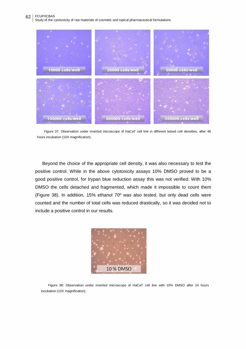
62 FCUP/ICBAS Study of the cytotoxicity of raw materials of cosmetic and topical pharmaceutical formulations
Beyond the choice of the appropriate cell density, it was also necessary to test the
positive control. While in the above cytotoxicity assays 10% DMSO proved to be a
good positive control, for trypan blue reduction assay this was not verified. With 10%
DMSO the cells detached and fragmented, which made it impossible to count them
(Figure 38). In addition, 15% ethanol 70º was also tested, but only dead cells were
counted and the number of total cells was reduced drastically, so it was decided not to
include a positive control in our results.
Figure 37: Observation under inverted microscope of HaCaT cell line in different tested cell densities, after 48
hours incubation (10X magnification).
Figure 38: Observation under inverted microscope of HaCaT cell line with 10% DMSO after 24 hours
incubation (10X magnification).

FCUP/ICBAS Study of the cytotoxicity of raw materials of cosmetic and topical pharmaceutical formulations
63
Study of the cytotoxic effect of the tested compound’s
using Trypan Blue exclusion assay
After choosing the optimal cell density for trypan blue exclusion assay, the
selected compounds were tested. Figure 39 shows the results of the viability assay
for RSV and its derivatives. A significant decrease in cell viability occurred for HaCaT
cells exposed from the concentration 200 μM for RSV (A) and at 5000 μM for RGS (C).
RSV-Gli (B) did not induce significant alterations of HaCaT cells viability.
Figure 39: Cell viability of HaCaT cell line exposed to (A) RSV, (B) RSV-Gli and (C) RGS, determined by the
Trypan Blue exclusion. Data are presented as mean ± SD (n=3-9). Data were analyzed using One-way ANOVA
with Dunnett post hoc test. * P<0.05 data are significantly different from the control value.

64 FCUP/ICBAS Study of the cytotoxicity of raw materials of cosmetic and topical pharmaceutical formulations
Figure 40 show the results of cell viability of HaCaT cells after 24 hours incubation
with lipid nanoparticles. Regarding the SLN, a significant decrease in cell viability to
concentration 0.1% SLN empty, 0.05% and 0.1% SLN-RSV was observed. Already the
NLC showed significant decrease in cell viability for concentration 0.1%.
Figure 40: Cell viability of HaCaT cell line exposed to (A) SLN and SLN-RSV and (B) NLC and NLC-RSV,
determined by the Trypan Blue exclusion assay. Data is presented as mean ± SD (n=3-9). Data were analyzed using
One-way ANOVA with Dunnett post hoc test. * P<0.05 data are significantly different from the control value.
Comparisons between loaded and empty nanoparticles were performed with the Tukey post hoc test. ** P<0.05 data are
statistically different.
Results of the 1,2-DHX after 24 hours of incubation with HaCaT cells are shown in
Figure 41. A significant decrease in the cell viability is detected with 200 μM
concentration. For this concentration the cell viability was 74.29 ± 12.54%.

FCUP/ICBAS Study of the cytotoxicity of raw materials of cosmetic and topical pharmaceutical formulations
65
Results of cell viability of HaCaT cells after 24 hours incubation with C.sativa leaf
extract are shown in Figure 42 and no significant alterations of cells viability were
detected in the range of concentrations tested. There were some difficulties in evaluate
the cytotoxic effects of this compound by Trypan blue exclusion assay. Indeed, after
microscope analysis without the dye addition, dead cells were observed. Probably the
solvent used, glycerin, may interfere with the density of the suspension leading to
unsuitable centrifugation time and g-force that may damage the cells.
Figure 41: Cell viability of HaCaT cell line exposed to 1,2-DHX, determined by the Trypan Blue exclusion assay.
Data are presented as mean ± SD (n=3-9). Data were analyzed using One-way ANOVA with Dunnett post hoc test. *
P<0.05 data are significantly different from the control value.
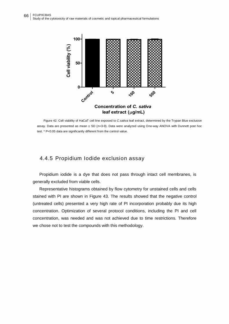
66 FCUP/ICBAS Study of the cytotoxicity of raw materials of cosmetic and topical pharmaceutical formulations
4.4.5 Propidium Iodide exclusion assay
Propidium iodide is a dye that does not pass through intact cell membranes, is
generally excluded from viable cells.
Representative histograms obtained by flow cytometry for unstained cells and cells
stained with PI are shown in Figure 43. The results showed that the negative control
(untreated cells) presented a very high rate of PI incorporation probably due its high
concentration. Optimization of several protocol conditions, including the PI and cell
concentration, was needed and was not achieved due to time restrictions. Therefore
we chose not to test the compounds with this methodology.
Figure 42: Cell viability of HaCaT cell line exposed to C.sativa leaf extract, determined by the Trypan Blue exclusion
assay. Data are presented as mean ± SD (n=3-9). Data were analyzed using One-way ANOVA with Dunnett post hoc
test. * P<0.05 data are significantly different from the control value.

FCUP/ICBAS Study of the cytotoxicity of raw materials of cosmetic and topical pharmaceutical formulations
67
4.4.6 Comparison between the results of the different in
vitro cytotoxicity assays
The IC50 values for cytotoxic effect and values of the lowest concentration that
promote a significance statistically decrease to the initially cell viability, depending on
the assay used are represented in Table 9.
For the chemicals and the plant extract tested on this work, MTT reduction and NR
uptake assays presented higher sensitivity compared to the other assays. The toxicity
was observed with NR uptake assays at lower concentrations, except for RGS and
lower IC50 values were found with the MTT reduction assay. It should be noticed
however that these assays were performed with different conditions such as different
cell densities according to the results of the optimization step.
A wide variability was observed for all lipid nanoparticles which is a manifestation of
the difficulties of analyzing their cytotoxicity in vitro.
Figure 43: Representative histograms of unstained cells (A) PI stained cells (B) obtained by flow
cytometry (BD ACCURI C6 software).
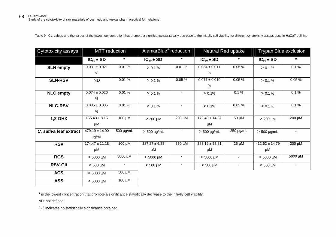
68 FCUP/ICBAS Study of the cytotoxicity of raw materials of cosmetic and topical pharmaceutical formulations
Table 9: IC50 values and the values of the lowest concentration that promote a significance statistically decrease to the initially cell viability for different cytotoxicity assays used in HaCaT cell line
Cytotoxicity assays MTT reduction AlamarBlue® reduction Neutral Red uptake Trypan Blue exclusion
IC50 ± SD * IC50 ± SD * IC50 ± SD * IC50 ± SD *
SLN empty 0.031 ± 0.021
%
0.01 % > 0.1 % 0.01 % 0.084 ± 0.011
%
0.05 % > 0.1 % 0.1 %
SLN-RSV ND 0.01 % > 0.1 % 0.05 % 0.077 ± 0.010
%
0.05 % > 0.1 % 0.05 %
NLC empty 0.074 ± 0.020
%
0.01 % > 0.1 % - > 0.1% 0.1 % > 0.1 % 0.1 %
NLC-RSV 0.085 ± 0.005
%
0.01 % > 0.1 % - > 0.1% 0.05 % > 0.1 % 0.1 %
1,2-DHX 155.43 ± 8.15
µM
100 µM > 200 µM 200 µM 172.40 ± 14.37
µM
50 µM > 200 µM 200 µM
C. sativa leaf extract 479.19 ± 14.90
µg/mL
500 µg/mL > 500 µg/mL - > 500 µg/mL 250 µg/mL > 500 µg/mL -
RSV 174.47 ± 11.18
µM
100 µM 387.27 ± 6.88
µM
350 µM 383.19 ± 53.81
µM
25 µM 412.62 ± 14.79
µM
200 µM
RGS > 5000 µM 5000 µM > 5000 µM - > 5000 µM - > 5000 µM 5000 µM
RSV-Gli > 500 µM - > 500 µM - > 500 µM - > 500 µM -
ACS > 5000 µM 500 µM
ASS > 5000 µM 100 µM
* is the lowest concentration that promote a significance statistically decrease to the initially cell viability.
ND: not defined
( - ) indicates no statistically significance obtained.

FCUP/ICBAS Study of the cytotoxicity of raw materials of cosmetic and topical pharmaceutical formulations
69
5. DISCUSSION
In the present work, the cytotoxicity of a series of ingredients with potential interest
for topical application was studied. This study was performed in HaCaT cells, an
immortal non-cancerous human keratinocyte cell line. Keratinocytes are the
predominant cells in the first layers of the skin, the epidermis. Considering the intended
route of administration for these materials, using a human keratinocyte cell line for their
preliminary safety evaluation is a logical choice.
Toxicological testing is a regulatory requirement for cosmetic and pharmaceutical
products. For cosmetics, each ingredient included in the cosmetic product must
undergo toxicological evaluation, such as, local toxicity and corrosivity evaluation (skin
and eye irritation), skin sensitization, dermal/percutaneous absorption, among others.
In the case of the pharmaceutical products, beyond toxicological requirements, other
nonclinical studies include pharmacology studies (e.g. cardiovascular, central nervous,
respiratory systems) are required.
To investigate the potential cytotoxic effects of a series of ingredients on HaCaT
cells, different cytotoxicity assays were performed (MTT reduction, AlamarBlue®
reduction, NR uptake and Trypan Blue exclusion). Cytotoxicity assays are widely used
in preliminary investigation of the toxicity, since they provide an important advantage in
identification of the cytotoxic compounds [83]. These assays allow determining whether
a compound or extract contains significant quantities of biologically harmful chemicals,
therefore are useful for screening as they serve to separate toxic from nontoxic
materials, providing predictive evidence of compound safety [82]. Toxicity assays are
essentials for a process of screening large chemical libraries easier, however the
selection of one cytotoxicity assay over another can be troublesome due to the
availability of several assays that rely on different physiological mechanisms and
endpoints [117].
When comparing the four cytotoxicity assays employed in this work to assess a
series of ingredients with potential interest for topical application, it is obvious that the
results obtained are not in close agreement. This can be explained by the different
mechanism assessed, except in case of the MTT and AlamarBlue® assays which both
measure the metabolic activity. In turn, NR and trypan blue assess the lysosomal
integrity and cell membrane damage, respectively. It has been previously reported that
different cytotoxicity assays can give different results depending on the test agent used
and the cytotoxicity assay employed [59]. Kim and co-workers showed no correlation

70 FCUP/ICBAS Study of the cytotoxicity of raw materials of cosmetic and topical pharmaceutical formulations
between MTT reduction and trypan Blue exclusion assays for the tested compounds
[118].
In this work, NR and MTT assays were the most sensitive in detecting toxic effects
compared to the other assays utilized since the toxicity was observed with NR assay at
lower concentrations, except for RGS, and lower IC50 values were found with the MTT
assay. These differences among cytotoxicity assays may suggest that the intracellular
effects due to exposure to compounds occur before any permanent cell membrane
damage occur. There are evidences in which these two assays were also more
sensitive when compared with LDH leakage and protein assays [38]. MTT reduction
assay also was more sensitive in a study performed in HaCaT cells [119]. However in
this study, the cell concentration used to perform the MTT test was lower than the one
used for the other tests, thus we cannot exclude the fact that the inferior IC50 values
obtained for the MTT assay might be due to the cells being exposed to higher levels of
the compounds. This evidence emphasizes the need to be careful when comparing
different cytotoxicity tests. In the case of NR assay, this dye uptake by cells occurs
through a process requiring energy, being sensitive to substances that interfere with
cell membrane and lysosomal permeability as well as the process of energy-dependent
endocytosis [120], therefore expectable to be one of the most sensitive test.
RSV a compound know as a strong antioxidant [121], anti-inflammatory [122] and a
cardioprotective agent [123], showed in general a significant decrease in cell viability
from the concentration of 100 μM, except in the NR reduction assay. In this assay,
exposure to RSV at 25 μM already caused a significant decrease in cell viability
comparing to control, which was not dependent on the concentration up to 100 μM.
Noteworthy, cell viability found in such cases was around 80%, which can be
considered non-cytotoxic [124]. A study performed in a primary keratinocyte line
showed that RSV did not induce significant cytotoxicity up to 50 μM [125], which is
consistent with the majority of our results. Other studies have revealed that RSV
inhibits the proliferation of normal human keratinocytes in vitro [125, 126]. Although it
was not the aim of our study, in the Trypan Blue exclusion assay it was also found that
after exposure to RSV at 25 μM the number of viable cells was lower than the one
found for the control, even if no decrease in cell viability was observed. This putative
anti-proliferative effect could be a confounding factor in the analysis of cytotoxicity
results, as is the case of NR reduction assay.
Two RSV derivatives were also studied in this work. RSV-Gli was the only
compound studied in this work that showed no cytotoxicity for the tested concentrations
(25-500 μM). The concentration range was limited by the maximum concentration of
the solvent (DMSO) that doesn’t affect cell viability (1%). In this case, it would be

FCUP/ICBAS Study of the cytotoxicity of raw materials of cosmetic and topical pharmaceutical formulations
71
interesting to extend the concentration range to detect toxic effects. For the RGS, a
significant reduction of HaCaT viability was observed at the highest concentration
tested (5000 μM) and was around 67% with MTT reduction assay.
With regard to the results obtained from cytotoxicity assays of lipid nanoparticles a
greater variability was observed. In some cases significant differences were observed
between RSV-loaded and empty nanoparticles which were not consistent across the
assays. We can generally assume that the toxicity observed is due to the particles
rather than RSV. Indeed, the theoretical calculation of the concentration of RSV
present in the highest concentration tested (0.1%), based on drug loading analysis
previously performed, was 36.5 μM. But when free RSV was tested up 100 μM no
significant evidence of cytotoxicity was detected, except for the NR assay. This
suggests that the excipients used in the matrix of lipid nanoparticles or their physico-
chemical features (e.g. size and charge) could be responsible for the cell death. There
is evidence that only the size of the nanoparticles is not sufficient to explain the
cytotoxic effects [102]. It would be relevant to study the cytotoxicity of the ingredients
alone to assess their contribution to the observed toxic effects. In the work by Ridolfi
and co-workers, they showed that up to 0.5 mg/mL cetyl palmitate-based SLN caused
a decrease in HaCaT cell viability [127]. The type, amount and interaction of the
surfactant with the lipid core on nanoparticles also affect the performance of cells in
culture [102]. Moreover there is evidence that the binding of surfactant to the
nanoparticles surface decrease its availability to interact with cells and consequently
decreases its toxicity [128]. So it is essential testing of the unprocessed starting
material present in the same quantities and ratios as in the final formulation to clarify if
the observed toxicity effects are result of that particular combination of materials per se
or of the nanoparticles in question [102].
On the other hand, the lipid nanoparticles may form aggregates in cell culture
media or suffer changes to its structure, for example desorption of surfactants [102].
Therefore it is important to take into account the stability of the nanoparticles in cell
medium [21, 102]. Studies performed in our laboratory with the same lipid
nanoparticles, showed the zeta potential varied between 27 and 40 mV, which
accounts for a good colloidal stability in aqueous dispersion [129].
The majority of cell viability studies of SLN/NLC were performed by MTT assay
[102]. The greater variability obtained in this study for lipid nanoparticles can also be
explained by possible interactions between test reagents (e.g. MTT) and the
nanoparticles, inducing false positives [21]. Furthermore, there is also evidence that
SLN and NLC in the dispersion themselves absorb visible light [102]. To avoid the
possibility of this lipid interaction with the assay, a washing step before adding the test
72 FCUP/ICBAS Study of the cytotoxicity of raw materials of cosmetic and topical pharmaceutical formulations
reagent was included in the protocol of cytotoxicity assays employed in this work,
except for AlamarBlue® reduction assay. In this case, as is a water soluble dye, the
blank measurement was included in this assay. But in blank for lipid nanoparticles
contributed to optical density properties, which suggests that this cytotoxicity assay is
not suitable for these nanoparticles.
Regarding the polymeric microparticles the MTT reduction assay was the the only
test used, and the aggregation and deposition of the microparticles on the surface of
the cell monolayer led to a great variability of the results.
Two of the compounds obtained by chemical synthesis, ACS and ASS were only
tested with the MTT reduction assay, due to the reduced amount obtained in the
synthesis process. ASS promoted a significant decrease in cell viability at the lowest
concentration studied (100 μM). However this effect was not concentration-dependent,
up to 1000 μM and cell viability was around 75%, which can be considered non-
cytotoxic [124], so this reduction in cell viability could be due to other effects such as an
antiproliferative activity. Elucidation of the cytotoxicity should be further ascertained
with other assays. ACS showed significant cytotoxicity only at the concentration of 500
μM.
1,2-DHX, another compound obtained by chemical synthesis, showed significant
cytotoxicity at concentrations of 50 μM and higher. In the work by Silva and co-workers
1,2-DHX was tested at several concentrations up to 50 μM with MTT reduction and
Neutral Red uptake assays using Caco-2 cells. Silva et al. found no significant
cytotoxicity within the tested concentration range [130].
In the case of the plant extract tested, no significant alterations of HaCaT cells
viability were detected after incubation with concentrations lower than 250 μg/mL. In
the work by Almeida and co-workers, this extract was tested with the MTT reduction
assay, using HaCaT cells. Almeida et al. observed that the extract-induced cytotoxicity
at concentrations equal to or higher than 50 μg/mL [111]. The dissimilarities found in
both studies in terms of results could be explained by the different conditions used on
the assay, such as different cell densities that restrains data comparison.
As for the positive control used (10% DMSO), this proved to be a good cytotoxic
compound, except for Trypan Blue exclusion assay. Indeed, microscopic observation of
the cells incubated with DMSO in the trypan blue test, indicates that probably this
reagent significantly compromised the cell membrane integrity, leading to cell burst and
consequently unable the trypan blue dye to be up taken by dead cells.
Regardless of the used assay, well defined protocols are needed to obtain
meaningful results. In any cytotoxicity assays to be used, optimization must be carried
out for every cell model [27]. Assays must be performed at a uniform temperature and

FCUP/ICBAS Study of the cytotoxicity of raw materials of cosmetic and topical pharmaceutical formulations
73
controlled pH to ensure reproducibility of all wells and across a plate. The optimum
incubation time and cell density must be empirically determined [27]. All the tests used
in this study for measure cytotoxicity of the different compounds were optimized. In
particular, NR uptake and Trypan Blue exclusion assays showed more challenges for
its proper implementation. NR assay was very dependent on temperature, forming
crystals when there was a variation in temperature. Trypan blue is a manual test
involving several cares, such as, collection of all cells (live and dead) from the well and
the correct count of all cells is required. During assay optimization and development,
microscopic observations are necessary for confirmation of cellular toxicity. Some of
the observations include for example, cell detachment or presence of cell fragments
[83]. This was particularly important for Trypan Blue exclusion assay, where it was
found that 10% DMSO is not a good positive control.
74 FCUP/ICBAS Study of the cytotoxicity of raw materials of cosmetic and topical pharmaceutical formulations
6. CONCLUSIONS
The aim of this work was to study the cytotoxicity of several ingredients with
potential interest for topical application. Cytotoxicity assays are widely used in the
preliminary investigation of the toxicity. From the study carried out in this Master
dissertation we were able to determine the concentration range for each compound
that can be used in future assays to confirm the potentialities of these raw materials for
application on the skin. The results obtained are also relevant for other toxicological
studies that are mandatory to meet regulatory requirements.
The optimization of each cytotoxicity assay allowed the establishment of the most
appropriate experimental conditions, considering the cell line used. The optimal cell
density and incubation time with the test reagent were selected as follows: MTT
reduction assay 1×10⁴ cells/well and 2 h; AlamarBlue® reduction assay 2×10⁴ cells/well
and 4 h; NR uptake assay 2×10⁴ cells/well and 4 h and Trypan Blue exclusion assay
1×10⁵ cells/well. The concentration of the NR solution used was 50 μg/mL.
In the case of the RSV and its derivatives, they can be generally regarded as safe
at concentrations up to 100 μM for RSV, 500 μM for RSV-Gli and 1000 μM for RGS.
C. sativa leaf extract and 1,2-DHX can be safely used in future studies at
concentrations up to 100 μg/mL and 50 μM, respectively. In turn, it would be necessary
to complement the basic toxicity evaluation of ACS and ASS using more cytotoxicity
studies. In particular, the absence of a concentration-dependent effect observed for
ASS should be elucidated in future studies.
The lipid nanoparticles prepared and tested in this work were not considered as
promising topical ingredients due to the marked in vitro toxicity presented. Our study
also demonstrated that the nanoparticles present several challenges for toxicological
testing. Improvement of the methodologies used should take into account the
interaction between test reagents and nanoparticles and light dispersion or absorption
effects that can interfere with the cytotoxicity assays. It would also be relevant to study
the cytotoxicity of the ingredients alone to assess their contribution to the observed
toxic effects.
Polymeric particles showed deposition on the surface of the cell monolayer and
aggregation which led to a great variability of the results. Following these observations,

FCUP/ICBAS Study of the cytotoxicity of raw materials of cosmetic and topical pharmaceutical formulations
75
it would be more relevant to test the in vitro toxicity of these particles using for
example, reconstructed human epidermis.
When comparing the cytotoxicity assays used, some differences were noted, being
the MTT reduction and NR uptake assays the most sensitive tests. These remarks
further reinforce the importance of using different in vitro cytotoxicity tests, which rely
on different mechanisms of cell death.
Further toxicological tests should be carried out to meet regulatory requirements
before considering the use of the tested compounds in cosmetic or pharmaceutical
products.
76 FCUP/ICBAS Study of the cytotoxicity of raw materials of cosmetic and topical pharmaceutical formulations
7. BIBLIOGRAPHIC REFERENCES
1. Nohynek GJ, Antignac E, Re T, Toutain H. Safety assessment of personal care products/cosmetics and their ingredients. Toxicology and applied pharmacology. 2010; 243(2): 239-259. 2. Regulation (EC) No 1223/2009 of the European Parliament and of the Council of 30 November 2009 on cosmetic products, L 342 (2009). 3. Council Directive of 27 July 1976 on the approximation of the laws of the Member States relating to cosmetic products (76/768/EEC), (1976). 4. European Commission. Cosmetics: Legislation. [cited 2015 17-10-2015]; Available from: http://ec.europa.eu/growth/sectors/cosmetics/legislation/index_en.htm. 5. SCCS. The SCCS'S notes of Guidance for the testing of cosmetic substances and their safety evaluation, 8th revision, adopted by the SCCS during the 17th plenary meeting of 11 December 2012. 2012. 6. Vinardell MP. The use of non-animal alternatives in the safety evaluations of cosmetics ingredients by the Scientific Committee on Consumer Safety (SCCS). Regulatory Toxicology and Pharmacology. 2015; 71(2): 198-204. 7. Directive 2001/83/ec of the European Parliament and of the Council, (2001). 8. ICH. Good Manufacturing Practice Guide for Active Pharmaceutical Ingredients Q7, (2000). 9. García-Arieta A. Interactions between active pharmaceutical ingredients and excipients affecting bioavailability: Impact on bioequivalence. European Journal of Pharmaceutical Sciences. 2014; 65: 89-97. 10. Infarmed. Farmacopeia Portuguesa 9. Lisboa, 2008. 11. Pifferi G, Restani P. The safety of pharmaceutical excipients. Il Farmaco. 2003; 58(8): 541-550. 12. ICH. Guidance Nonclinical Safety Studies for the Conduct of Human Clinical Trials and Marketing Authorization for Pharmaceuticals M3(R2), (2009). 13. European Medicines Agency. Guideline on non-clinical local tolerance testing of medicinal products, (2014). 14. Moore I. Excipients: Safety, toxicological, and precedence of use issues. Maas & Peither AG - GMP; 2013. 15. Draize JH, Woodard, G., Calvery, H.O. Methods for the study of irritation and toxicity of substances applied topically to the skin and mucous membranes. Journal of Pharmacology and Experimental Therapeutics. 1944; 82: 377-390. 16. Robinson MK, Cohen C, de Fraissinette AdB, Ponec M, Whittle E, Fentem JH. Non-animal testing strategies for assessment of the skin corrosion and skin irritation potential of ingredients and finished products. Food and Chemical Toxicology. 2002; 40(5): 573-592. 17. European Commission. Topical toxicity: Skin Irritation. [cited 2015 09-11-2015]; Available from: https://eurl-ecvam.jrc.ec.europa.eu/validation-regulatory-acceptance/topical-toxicity/skin-irritation. 18. Bleeker EAJ, de Jong WH, Geertsma RE, Groenewold M, Heugens EHW, Koers-Jacquemijns M, et al. Considerations on the EU definition of a nanomaterial: Science to support policy making. Regulatory Toxicology and Pharmacology. 2013; 65(1): 119-125. 19. Crosera M, Bovenzi M, Maina G, Adami G, Zanette C, Florio C, et al. Nanoparticle dermal absorption and toxicity: a review of the literature. International archives of occupational and environmental health. 2009; 82(9): 1043-1055. 20. Nasir A. Nanotechnology and dermatology: part I--potential of nanotechnology. Clinics in dermatology. 2010; 28(4): 458-466.

FCUP/ICBAS Study of the cytotoxicity of raw materials of cosmetic and topical pharmaceutical formulations
77
21. SCCS. Guidance on the Safety Assessment of Nanomaterials in cosmetics. 2012. 22. Benbow JW, Aubrecht J, Banker MJ, Nettleton D, Aleo MD. Predicting safety toleration of pharmaceutical chemical leads: cytotoxicity correlations to exploratory toxicity studies. Toxicol Lett. 2010; 197(3): 175-182. 23. Committee on Predictive et. al., 4- Assays for Predicting Acute Toxicity. In:
Application of Modern Toxicology Approaches for Predicting Acute Toxicity for Chemical Defense.: Washington (DC): National Academies Press (US); 2015. p. 51-79. 24. Roche. Apoptosis, Cell Death and Cell Proliferation- 4th edition. 2008. 186 p. 25. Stoddart MJ. Cell viability assays: introduction. Methods in molecular biology. 2011; 740: 1-6. 26. Niles AL, Moravec RA, Riss TL. Update on in vitro cytotoxicity assays for drug development. Expert opinion on drug discovery. 2008; 3(6): 655-669. 27. Rampersad SN. Multiple applications of Alamar Blue as an indicator of metabolic function and cellular health in cell viability bioassays. Sensors. 2012; 12(9): 12347-12360. 28. Longo-Sorbello GSA, Saydam G, Banerjee D, Bertino JR. Chapter 38 - Cytotoxicity and Cell Growth Assays. In Cell Biology (Third Edition). Academic Press; 2006. p. 315-324. 29. Lichtenfels R, Biddison WE, Schulz H, Vogt AB, Martin R. CARE-LASS (calcein-release-assay), an improved fluorescence-based test system to measure cytotoxic T lymphocyte activity. Journal of immunological methods. 1994; 172(2): 227-239. 30. Bratosin D, Mitrofan L, Palii C, Estaquier J, Montreuil J. Novel fluorescence assay using calcein-AM for the determination of human erythrocyte viability and aging. Cytometry Part A : the journal of the International Society for Analytical Cytology. 2005; 66(1): 78-84. 31. BIO530. Calcein-AM. [cited 2015 21-11-2015]; Available from: https://bio530.wikispaces.com/Calcein+AM. 32. Neri S, Mariani E, Meneghetti A, Cattini L, Facchini A. Calcein-Acetyoxymethyl Cytotoxicity Assay: Standardization of a Method Allowing Additional Analyses on Recovered Effector Cells and Supernatants. Clinical and Diagnostic Laboratory Immunology. 2001; 8(6): 1131-1135. 33. Lindhagen E, Nygren P, Larsson R. The fluorometric microculture cytotoxicity assay. Nature protocols. 2008; 3(8): 1364-1369. 34. Atala A, Lanza RP. Section I: Methods for Cell and Tissue Culture. In: Methods of tissue Engineering. Academic Press; 2002. p. 19-30. 35. Dojindo Molecular Technologies. Cell stain: FDA. [cited 2015 10-11-2015]; Available from: http://www.dojindo.com/store/p/168-Cellstain-FDA.html. 36. Clarke JM, Gillings MR, Altavilla N, Beattie AJ. Potential problems with fluorescein diacetate assays of cell viability when testing natural products for antimicrobial activity. Journal of microbiological methods. 2001; 46(3): 261-267. 37. Hillegass JM, Shukla A, Lathrop SA, MacPherson MB, Fukagawa NK, Mossman BT. Assessing nanotoxicity in cells in vitro. Wiley interdisciplinary reviews Nanomedicine and nanobiotechnology. 2010; 2(3): 219-231. 38. Fotakis G, Timbrell JA. In vitro cytotoxicity assays: Comparison of LDH, neutral red, MTT and protein assay in hepatoma cell lines following exposure to cadmium chloride. Toxicology Letters. 2006; 160(2): 171-177. 39. Doyle A, Griffiths JB. Chapter 2: Cell Quantification. In: Cell and Tissue Culture: Laboratory Procedures in Biotechnology.: Wiley; 1999. p. 53-80. 40. Decker T, Lohmann-Matthes ML. A quick and simple method for the quantitation of lactate dehydrogenase release in measurements of cellular cytotoxicity and tumor necrosis factor (TNF) activity. Journal of immunological methods. 1988; 115(1): 61-69.

78 FCUP/ICBAS Study of the cytotoxicity of raw materials of cosmetic and topical pharmaceutical formulations
41. DeRenzis FA, Schechtman A. Staining by neutral red and trypan blue in sequence for assaying vital and nonvital cultured cells. Stain technology. 1973; 48(3): 135-136. 42. Borenfreund E, Puerner JA. Toxicity determined in vitro by morphological alterations and neutral red absorption. Toxicology Letters. 1985; 24(2–3): 119-124. 43. Freshney RI. Culture of Animal Cells: A Manual of Basic Technique. Sixth ed. USA, 2005. 44. Weyermann J, Lochmann D, Zimmer A. A practical note on the use of cytotoxicity assays. International journal of pharmaceutics. 2005; 288(2): 369-376. Epub 2004/12/29. 45. Wållberg F. Flow Cytometry for Bioprocess Control. Stockholm, Sweden: Royal Institute of Technology; 2004. 46. Clothier R, Gomez-Lechon MJ, Kinsner-Ovaskainen A, Kopp-Schneider A, O'Connor JE, Prieto P, et al. Comparative analysis of eight cytotoxicity assays evaluated within the ACuteTox Project. Toxicology in vitro : an international journal published in association with BIBRA. 2013; 27(4): 1347-1356. 47. Riss TL MR, Niles AL et al. Assay Guidance Manual- Cell Viability Assays. 2013. 48. Niles AL, Moravec RA, Riss TL. In Vitro Viability and Cytotoxicity Testing and Same-Well Multi-Parametric Combinations for High Throughput Screening. Current Chemical Genomics. 2009; 3: 33-41. 49. Louis KS, Siegel AC. Cell viability analysis using trypan blue: manual and automated methods. Methods in molecular biology. 2011; 740: 7-12. 50. Czekanska EM. Assessment of cell proliferation with resazurin-based fluorescent dye. Methods in molecular biology. 2011; 740: 27-32. 51. O'Brien J, Wilson I, Orton T, Pognan F. Investigation of the Alamar Blue (resazurin) fluorescent dye for the assessment of mammalian cell cytotoxicity. European Journal of Biochemistry. 2000; 267(17): 5421-5426. 52. Gonzalez RJ, Tarloff JB. Evaluation of hepatic subcellular fractions for Alamar blue and MTT reductase activity. Toxicology in vitro. 2001; 15(3): 257-259. 53. Page B, Page M, Noel C. A new fluorometric assay for cytotoxicity measurements in-vitro. International journal of oncology. 1993; 3(3): 473-476. 54. Mosmann T. Rapid colorimetric assay for cellular growth and survival: application to proliferation and cytotoxicity assays. Journal of immunological methods. 1983; 65(1-2): 55-63. 55. Barltrop JA, Owen TC, Cory AH, Cory JG. 5-(3-carboxymethoxyphenyl)-2-(4,5-dimethylthiazolyl)-3-(4-sulfophenyl)tetrazolium, inner salt (MTS) and related analogs of 3-(4,5-dimethylthiazolyl)-2,5-diphenyltetrazolium bromide (MTT) reducing to purple water-soluble formazans As cell-viability indicators. Bioorganic & Medicinal Chemistry Letters. 1991; 1(11): 611-614. 56. Cory AH, Owen TC, Barltrop JA, Cory JG. Use of an aqueous soluble tetrazolium/formazan assay for cell growth assays in culture. Cancer communications. 1991; 3(7): 207-212. 57. Wiegand C, Hipler UC. Evaluation of biocompatibility and cytotoxicity using keratinocyte and fibroblast cultures. Skin pharmacology and physiology. 2009; 22(2): 74-82. 58. Crouch SPM, Kozlowski R, Slater KJ, Fletcher J. The use of ATP bioluminescence as a measure of cell proliferation and cytotoxicity. Journal of immunological methods. 1993; 160(1): 81-88. 59. Weyermann J, Lochmann D, Zimmer A. A practical note on the use of cytotoxicity assays. International journal of pharmaceutics. 2005; 288(2): 369-376. 60. Crouch SPM, Slater KJ. High-throughput cytotoxicity screening: hit and miss. Drug Discovery Today. 2001; 6, Supplement 1: 48-53.
FCUP/ICBAS Study of the cytotoxicity of raw materials of cosmetic and topical pharmaceutical formulations
79
61. Smith PK, Krohn RI, Hermanson GT, Mallia AK, Gartner FH, Provenzano MD, et al. Measurement of protein using bicinchoninic acid. Analytical biochemistry. 1985; 150(1): 76-85. 62. Sigma-Aldrich®. Bicinchoninic Acid Kit. [cited 2015 23-11-2015]; Available from: http://www.sigmaaldrich.com/life-science/proteomics/protein-quantitation/bicinchoninic-acid-kit.html. 63. Walker JM. The bicinchoninic acid (BCA) assay for protein quantitation. Methods in molecular biology. 1994; 32: 5-8. 64. He F. BCA (Bicinchoninic Acid) Protein Assay. 2011 [cited 2015 23-11-2015]; Available from: http://www.bio-protocol.org/e44. 65. Skehan P, Storeng R, Scudiero D, Monks A, McMahon J, Vistica D, et al. New colorimetric cytotoxicity assay for anticancer-drug screening. Journal of the National Cancer Institute. 1990; 82(13): 1107-1112. 66. Vichai V, Kirtikara K. Sulforhodamine B colorimetric assay for cytotoxicity screening. Nature protocols. 2006; 1(3): 1112-1116. 67. Voigt W. Sulforhodamine B assay and chemosensitivity. Methods in molecular medicine. 2005; 110: 39-48. 68. Aburjai T, Natsheh FM. Plants used in cosmetics. Phytotherapy Research. 2003; 17(9): 987-1000. 69. Forster M, Bolzinger MA, Fessi H, Briancon S. Topical delivery of cosmetics and drugs. Molecular aspects of percutaneous absorption and delivery. Europe Journal of Dermatology. 2009; 19(4): 309-323. 70. A.D.A.M. Skin (Integumentary System). [cited 2015 02-04-2015]; Available from: http://owh.adam.com/pages/guide/reftext/html/skin_sys_fin.html. 71. Honari G, Maibach H. Chapter 1 - Skin Structure and Function. In: Honari HM, editor. Applied Dermatotoxicology. Boston: Academic Press; 2014. p. 1-10. 72. Barbieri JS, Wanat K, Seykora J. Skin: Basic Structure and Function. Pathobiology of Human Disease. San Diego: Academic Press; 2014. p. 1134-1144. 73. Seeley R, Stephens T, Tate P. Anatomia e Fisiologia, 6ed: Lusociência; 2005. 74. El Maghraby GM, Barry BW, Williams AC. Liposomes and skin: From drug delivery to model membranes. European Journal of Pharmaceutical Sciences. 2008; 34(4–5): 203-222. 75. Eucerin. Understanding skin: Skin structure and function. [cited 2015 17-08-2015]; Available from: http://www.eucerin.co.uk/about-skin/basic-skin-knowledge/skin-structure-and-function. 76. Prista L, Bahia M, Vilar E. Dermofarmácia e Cosmética. Maia: Gráfica Maiadouro para Associação Nacional das Farmácias; 1992. 77. Souto EB, Muller RH. Cosmetic features and applications of lipid nanoparticles (SLN, NLC). International journal of cosmetic science. 2008; 30(3): 157-165. 78. Gurtner GC, Werner S, Barrandon Y, Longaker MT. Wound repair and regeneration. Nature. 2008; 453(7193): 314-321. 79. Lee JK, Kim DB, Kim JI, Kim PY. In vitro cytotoxicity tests on cultured human skin fibroblasts to predict skin irritation potential of surfactants. Toxicology in vitro. 2000; 14(4): 345-349. 80. Shukla SJ, Huang R, Austin CP, Xia M. The Future of Toxicity Testing: A Focus on In Vitro Methods Using a Quantitative High Throughput Screening Platform. Drug Discovery Today. 2010; 15(23-24): 997-1007. 81. Varani J, Perone P, Spahlinger DM, Singer LM, Diegel KL, Bobrowski WF, et al. Human skin in organ culture and human skin cells (keratinocytes and fibroblasts) in monolayer culture for assessment of chemically induced skin damage. Toxicologic pathology. 2007; 35(5): 693-701. 82. Miret S, De Groene EM, Klaffke W. Comparison of in vitro assays of cellular toxicity in the human hepatic cell line HepG2. Journal of biomolecular screening. 2006; 11(2): 184-193.
80 FCUP/ICBAS Study of the cytotoxicity of raw materials of cosmetic and topical pharmaceutical formulations
83. Hamid R, Rotshteyn Y, Rabadi L, Parikh R, Bullock P. Comparison of alamar blue and MTT assays for high through-put screening. Toxicology in vitro. 2004; 18(5): 703-710. 84. Lakatos P, Szabó É, Hegedűs C, Haskó G, Gergely P, Bai P, et al. 3-Aminobenzamide protects primary human keratinocytes from UV-induced cell death by a poly(ADP-ribosyl)ation independent mechanism. Biochimica et Biophysica Acta (BBA) - Molecular Cell Research. 2013; 1833(3): 743-751. 85. Pastore S, Lulli D, Pascarella A, Maurelli R, Dellambra E, Potapovich A, et al. Resveratrol enhances solar UV-induced responses in normal human epidermal keratinocytes. Photochemistry and photobiology. 2012; 88(6): 1522-1530. 86. Pathakoti K, Hwang H-M, Xu H, Aguilar ZP, Wang A. In vitro cytotoxicity of CdSe/ZnS quantum dots with different surface coatings to human keratinocytes HaCaT cells. Journal of Environmental Sciences. 2013; 25(1): 163-171. 87. Tang J, Li Q, Cheng B, Huang C, Chen K. Effects of diethylstilbestrol on the proliferation and tyrosinase activity of cultured human melanocytes. Biomedical reports. 2015; 3(4): 499-502. 88. Gokce EH, Korkmaz E, Dellera E, Sandri G, Bonferoni MC, Ozer O. Resveratrol-loaded solid lipid nanoparticles versus nanostructured lipid carriers: evaluation of antioxidant potential for dermal applications. International journal of nanomedicine. 2012; 7: 1841-1850. 89. Reus AA, Reisinger K, Downs TR, Carr GJ, Zeller A, Corvi R, et al. Comet assay in reconstructed 3D human epidermal skin models--investigation of intra- and inter-laboratory reproducibility with coded chemicals. Mutagenesis. 2013; 28(6): 709-720. 90. Faller C, Bracher M, Dami N, Roguet R. Predictive ability of reconstructed human epidermis equivalents for the assessment of skin irritation of cosmetics. Toxicology in vitro. 2002; 16(5): 557-572. 91. Yamaguchi F, Watanabe S, Harada F, Miyake M, Yoshida M, Okano T. In vitro analysis of the effect of alkyl-chain length of anionic surfactants on the skin by using a reconstructed human epidermal model. Journal of oleo science. 2014; 63(10): 995-1004. 92. Casas JW, Lewerenz GM, Rankin EA, Willoughby JA, Sr., Blakeman LC, McKim JM, Jr., et al. In vitro human skin irritation test for evaluation of medical device extracts. Toxicology in vitro. 2013; 27(8): 2175-2183. 93. Murthy PB, Kishore AS, Surekha P. Assessment of in vitro skin irritation potential of nanoparticles: RHE model. Methods in molecular biology. 2012; 926: 219-234. 94. Nichols JA, Katiyar SK. Skin photoprotection by natural polyphenols: anti-inflammatory, antioxidant and DNA repair mechanisms. Archives of dermatological research. 2010; 302(2): 71-83. 95. Bogdan Allemann I, Baumann L. Antioxidants used in skin care formulations. Skin therapy letter. 2008; 13(7): 5-9. 96. Scognamiglio I, De Stefano D, Campani V, Mayol L, Carnuccio R, Fabbrocini G, et al. Nanocarriers for topical administration of resveratrol: A comparative study. International journal of pharmaceutics. 2013; 440(2): 179-187. 97. Baur JA, Sinclair DA. Therapeutic potential of resveratrol: the in vivo evidence. Nature reviews Drug discovery. 2006; 5(6): 493-506. 98. Du QH, Peng C, Zhang H. Polydatin: a review of pharmacology and pharmacokinetics. Pharmaceutical biology. 2013; 51(11): 1347-1354. 99. Correia-da-Silva M, Cidade H, Sousa E, Pinto M. Searching for Small Molecules with Antioxidant and Anticoagulant Properties to Fight Thrombosis. Columbia International Publishing. 2014; 1(1): 43-50. 100. Correia-da-Silva M, Sousa E, Duarte B, Marques F, Cunha-Ribeiro LM, Pinto MM. Dual anticoagulant/antiplatelet persulfated small molecules. European journal of medicinal chemistry. 2011; 46(6): 2347-2358.
FCUP/ICBAS Study of the cytotoxicity of raw materials of cosmetic and topical pharmaceutical formulations
81
101. Istenič K, Balanč BD, Djordjević VB, Bele M, Nedović VA, Bugarski BM, et al. Encapsulation of resveratrol into Ca-alginate submicron particles. Journal of Food Engineering. 2015; 167, Part B: 196-203. 102. Doktorovova S, Souto EB, Silva AM. Nanotoxicology applied to solid lipid nanoparticles and nanostructured lipid carriers – A systematic review of in vitro data. European Journal of Pharmaceutics and Biopharmaceutics. 2014; 87(1): 1-18. 103. Müller RH, Radtke M, Wissing SA. Nanostructured lipid matrices for improved microencapsulation of drugs. International journal of pharmaceutics. 2002; 242(1–2): 121-128. 104. Silva AC. Solid lipid nanoparticles (SLN) for oral delivery of Risperidone: Faculdade de Farmácia da Universidade do Porto; 2012. 105. Vauthier C, Bouchemal K. Methods for the preparation and manufacture of polymeric nanoparticles. Pharmaceutical research. 2009; 26(5): 1025-1058. 106. Pinto MM, Sousa ME, Nascimento MS. Xanthone derivatives: new insights in biological activities. Current medicinal chemistry. 2005; 12(21): 2517-2538. 107. Panda SS, Chand M, Sakhuja R, Jain SC. Xanthones as potential antioxidants. Current medicinal chemistry. 2013; 20(36): 4481-4507. 108. Pedro M, Cerqueira F, Sousa ME, Nascimento MS, Pinto M. Xanthones as inhibitors of growth of human cancer cell lines and their effects on the proliferation of human lymphocytes in vitro. Bioorganic & medicinal chemistry. 2002; 10(12): 3725-3730. 109. Chiarini A, Micucci M, Malaguti M, Budriesi R, Ioan P, Lenzi M, et al. Sweet Chestnut (Castanea sativa Mill.) Bark Extract: Cardiovascular Activity and Myocyte Protection against Oxidative Damage. Oxidative Medicine and Cellular Longevity. 2013; 2013: 10. 110. Díaz Reinoso B, Couto D, Moure A, Fernandes E, Domínguez H, Parajó JC. Optimization of antioxidants – Extraction from Castanea sativa leaves. Chemical Engineering Journal. 2012; 203: 101-109. 111. Almeida IF, Fernandes E, Lima JL, Costa PC, Bahia MF. Protective effect of Castanea sativa and Quercus robur leaf extracts against oxygen and nitrogen reactive species. Journal of photochemistry and photobiology B, Biology. 2008; 91(2-3): 87-95. 112. Díaz-Reinoso B, Moure A, Domínguez H, Parajó JC. Membrane concentration of antioxidants from Castanea sativa leaves aqueous extracts. Chemical Engineering Journal. 2011; 175: 95-102. 113. Almeida IF. Desenvolvimento de um sistema semi-sólido contendo fitocompostos captadores de espécies reactivas: Faculdade de Farmácia da Universidade do Porto; 2009. 114. Martins S. Drug delivery across blood-brain barrier by means of intravenous administration of lipid nanoparticles: Faculdade de Farmácia da Universidade do Porto; 2012. 115. Baba K, Nishida K. Calpain inhibitor nanocrystals prepared using Nano Spray Dryer B-90. Nanoscale Research Letters. 2012; 7(1): 436-436. 116. Rekus MT. Characterization of growth and differentiation of a spontaneously immortalized keratinocyte cell line (HaCaT) in a defined, serum-free culture system: Heinrich-Heine-Universität Düsseldorf; 2013. 117. Shum D, Radu C, Kim E, Cajuste M, Shao Y, Seshan VE, et al. A high density assay format for the detection of novel cytotoxicagents in large chemical libraries. Journal of enzyme inhibition and medicinal chemistry. 2008; 23(6): 931-945. 118. Kim H, Yoon SC, Lee TY, Jeong D. Discriminative cytotoxicity assessment based on various cellular damages. Toxicology letters. 2009; 184(1): 13-17. 119. Mukherjee SG, O'Claonadh N, Casey A, Chambers G. Comparative in vitro cytotoxicity study of silver nanoparticle on two mammalian cell lines. Toxicology in vitro. 2012; 26(2): 238-251. 120. Repetto G, del Peso A, Zurita JL. Neutral red uptake assay for the estimation of cell viability/cytotoxicity. Nature protocols. 2008; 3(7): 1125-1131.
82 FCUP/ICBAS Study of the cytotoxicity of raw materials of cosmetic and topical pharmaceutical formulations
121. Csuk R, Albert S, Siewert B. Synthesis and radical scavenging activities of resveratrol analogs. Archiv der Pharmazie. 2013; 346(7): 504-510. 122. Martinez J, Moreno JJ. Effect of resveratrol, a natural polyphenolic compound, on reactive oxygen species and prostaglandin production. Biochemical pharmacology. 2000; 59(7): 865-870. 123. Tang PC, Ng YF, Ho S, Gyda M, Chan SW. Resveratrol and cardiovascular health--promising therapeutic or hopeless illusion? Pharmacological research. 2014; 90: 88-115. 124. International Organization for Standardization, ISO 10993-5:2009 Biological evaluation of medical devices — Part 5: Tests for in vitro cytotoxicity, (2009). 125. Wu Z, Uchi H, Morino-Koga S, Shi W, Furue M. Resveratrol inhibition of human keratinocyte proliferation via SIRT1/ARNT/ERK dependent downregulation of aquaporin 3. Journal of Dermatological Science. 2014; 75(1): 16-23. 126. Holian O, Walter RJ. Resveratrol inhibits the proliferation of normal human keratinocytes in vitro. Journal of cellular biochemistry Supplement. 2001; Suppl 36: 55-62. 127. Ridolfi DM, Marcato PD, Machado D, Silva RA, Justo GZ, Durán N. In vitro cytotoxicity assays of solid lipid nanoparticles in epithelial and dermal cells Journal of Physics. 2011; 304. 128. Müller R, Rühl D, Runge S, Schulze-Forster K, Mehnert W. Cytotoxicity of Solid Lipid Nanoparticles as a Function of the Lipid Matrix and the Surfactant. Pharmaceutical research. 1997; 14(4): 458-462. 129. Fernandes M. Preparation and characterization of nano and microparticles loaded with Resveratrol: Faculdade de Farmácia da Universidade do Porto; 2014. 130. Silva R, Sousa E, Carmo H, Palmeira A, Barbosa DJ, Gameiro M, et al. Induction and activation of P-glycoprotein by dihydroxylated xanthones protect against the cytotoxicity of the P-glycoprotein substrate paraquat. Arch Toxicol. 2014; 88(4): 937-951.