stuttgarter beiträge zur · pdf filestuttgarter beiträge zur naturkunde serie a...
TRANSCRIPT
Stuttgarter Beiträge zur NaturkundeSerie A (Biologie)
Herausgeber:
Staatliches Museum für Naturkunde, Rosenstein 1, D-70191 Stuttgart
The Life Cycle of Lemanniella minotauri n. sp.and the Erection of the New FamilyLemanniellidae (Acari: Astigmata)
By Eberhard Wurst , Stuttgart
With 76 figures and 1 table
Summary
All instars of Lemanniella minotauri n. sp. are described from nests of the ant Lasius brun-neus (Hymenoptera: Formicidae) in central Europe. This is the first time non-hypopal instarsof the genus Lemanniella are described. The mites feed on an unidentified fungus growing onthe wood in the nest. Because of the exceptional body shape, peculiarities in setation, and thebehaviour it is proposed to remove the genus Lemanniella from the Acaridae and to establishthe new family Lemanniellidae.
Zusammenfassung
Sämtliche Entwicklungsstadien von Lemanniella minotauri n. sp. werden aus Nestern derAmeise Lasius brunneus (Hymenoptera: Formicidae) beschrieben. Erstmals werden damitnicht-hypopiale Stadien dieser Gattung vorgestellt. Die Milben ernähren sich von einem nichtidentifizierten Pilz, der auf dem Holz im Ameisennest wächst. Aufgrund der außergewöhnli-chen Körperform, Besonderheiten der Borstenkonfiguration und dem Verhalten der Milbenwird vorgeschlagen, für die Gattung Lemanniella, die bisher in die Acaridae gestellt wurde,die neue Familie Lemanniellidae einzurichten.
Contents1. Introduction . . . . . . . . . . . . . . . . . . . . . . . . . . . . . . . . . . . . . . . . . . . . . . . . . . . . . . . . . . . . . 22. Materials, methods and acknowledgements . . . . . . . . . . . . . . . . . . . . . . . . . . . . . . . . . . . . 2
2.1. Materials . . . . . . . . . . . . . . . . . . . . . . . . . . . . . . . . . . . . . . . . . . . . . . . . . . . . . . . . . . . . 22.2. Methods . . . . . . . . . . . . . . . . . . . . . . . . . . . . . . . . . . . . . . . . . . . . . . . . . . . . . . . . . . . . . 62.3. Acknowledgements . . . . . . . . . . . . . . . . . . . . . . . . . . . . . . . . . . . . . . . . . . . . . . . . . . . 8
3. Lemanniellidae n. fam. . . . . . . . . . . . . . . . . . . . . . . . . . . . . . . . . . . . . . . . . . . . . . . . . . . . . 103.1. Definition . . . . . . . . . . . . . . . . . . . . . . . . . . . . . . . . . . . . . . . . . . . . . . . . . . . . . . . . . . . 103.2. Remarks . . . . . . . . . . . . . . . . . . . . . . . . . . . . . . . . . . . . . . . . . . . . . . . . . . . . . . . . . . . . . 10
4. Description of Lemanniella minotauri n. sp. . . . . . . . . . . . . . . . . . . . . . . . . . . . . . . . . . . 124.1. Adults . . . . . . . . . . . . . . . . . . . . . . . . . . . . . . . . . . . . . . . . . . . . . . . . . . . . . . . . . . . . . . 144.2. Tritonymph . . . . . . . . . . . . . . . . . . . . . . . . . . . . . . . . . . . . . . . . . . . . . . . . . . . . . . . . . . 234.3. Deutonymph . . . . . . . . . . . . . . . . . . . . . . . . . . . . . . . . . . . . . . . . . . . . . . . . . . . . . . . . . 234.4. Protonymph . . . . . . . . . . . . . . . . . . . . . . . . . . . . . . . . . . . . . . . . . . . . . . . . . . . . . . . . . 264.5. Larva and egg . . . . . . . . . . . . . . . . . . . . . . . . . . . . . . . . . . . . . . . . . . . . . . . . . . . . . . . . 26
Stuttgarter Beitr. Naturk. Ser. A Nr. 621 34 S. Stuttgart, 1. 6. 2001
5. Observations on the biology of L. minotauri n. sp. . . . . . . . . . . . . . . . . . . . . . . . . . . . . . 326. References . . . . . . . . . . . . . . . . . . . . . . . . . . . . . . . . . . . . . . . . . . . . . . . . . . . . . . . . . . . . . . . 33
1. Introduction
Ants’ nests serve as habitats for a considerable number of mite species. Most ofthese species are adapted to and depend on one or only a few ant species. Among themyrmecophilous astigmatic mites are some unusual forms with a hitherto enigmaticsystematic position. An example is Myrmolichus greimae occurring in the nests ofthe palearctic ant Lasius fuliginosus and feeding on the fungus Cladosporiummyrmecophilum (TÜRK & TÜRK 1957) which is cultivated by the ants and reinforcesthe carton nest material (SEIFERT 1996).
Another representative of the Astigmata exhibiting an unusual morphology wasdescribed by MAHUNKA (1977) from deutonymphs having been collected from antsof the species Myrmica sabuleti in Switzerland. The exceptional morphology of thenew deutonymphs prompted MAHUNKA (1977) to establish the new genus Leman-niella (after the Latin name of the Lake Geneva, Lacus Lemannus). MAHUNKA
(1977) named the new mite species L. reducta, the species name apparently referringto the reduction of numerous characters (e.g. palposoma, leg setae) in the deu-tonymph. MAHUNKA (1977) included the genus Lemanniella in the Acaridae but headded the remark “that his very specialized form adopts a very isolated positionwithin the family Acaridae”.
Since then nothing was added to our knowledge of Lemanniella. In the course ofexaminations of wooden nest materials of ants in central Europe over the last tenyears I succeeded in elucidating the complete life cycle of another species of thegenus Lemanniella occurring in the nests of the ant Lasius brunneus.
This paper describes the new Lemanniella species and for the first time presentsnon-hypopal instars of this genus. First observations on the biology of the newspecies are reported. Because of the exceptional body shape, peculiarities in setation,and the behaviour I propose to remove the genus Lemanniella from the Acaridaeand to establish a new family for this aberrant astigmatic mite genus.
2. Materials, Methods and Acknowledgements
2.1. Materials
Material from ants’ nests in fallen or logged trees was collected. Localities where mites andants were taken are the following (all materials were collected by E. WURST):
Germany – 25. 03. 90. Untermberg near Bietigheim-Bissingen (south-west Germany); in awood nest of Lasius brunneus. – 09. 02. 95. Pasture at Korntal-Münchingen (near Stuttgart,south-west Germany); in a wood nest of Lasius brunneus. – 08. 04. 00 Garden between Un-terriexingen and Oberriexingen (near Bietigheim-Bissingen, south-west Germany); in a woodnest of Lasius brunneus.
Austria – 26. 02. 98 Vienna, Wienerwald between Leopoldsberg and Kahlenberg (Jose-finenhütte); in an abandoned wood nest of an unidentified ant.
2 stuttgarter beiträge zur naturkunde Ser. A, Nr. 621
wurst, lemanniellidae n. fam.; l. minotauri n. sp. 3
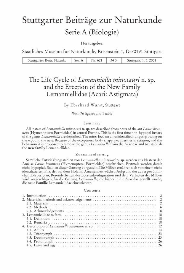
Fig
. 1.
Lem
anni
ella
min
otau
rin.
sp.
, mal
e; la
tera
l vie
w. –
Sca
le b
ar: 2
0 µm
.
4 stuttgarter beiträge zur naturkunde Ser. A, Nr. 621
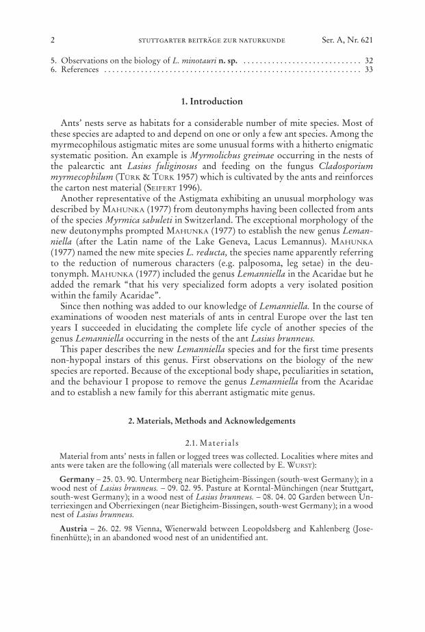
Fig. 2. Lemanniella minotauri n. sp., male; ventral view, left legs partly omitted. – Scalebar: 25 µm.
wurst, lemanniellidae n. fam.; l. minotauri n. sp. 5
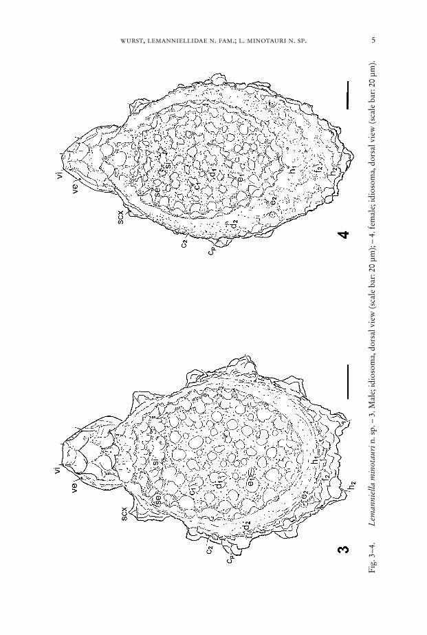
Fig
. 3–4
.L
eman
niel
la m
inot
auri
n. s
p. –
3. M
ale;
idio
som
a, d
orsa
l vie
w (s
cale
bar
: 20
µm);
– 4.
fem
ale;
idio
som
a, d
orsa
l vie
w (s
cale
bar
: 20
µm).
6 stuttgarter beiträge zur naturkunde Ser. A, Nr. 621
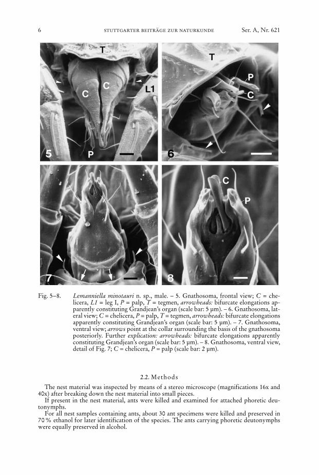
Fig. 5–8. Lemanniella minotauri n. sp., male. – 5. Gnathosoma, frontal view; C = che-licera, L1 = leg I, P = palp, T = tegmen, arrowheads: bifurcate elongations ap-parently constituting Grandjean’s organ (scale bar: 5 µm). – 6. Gnathosoma, lat-eral view; C = chelicera, P = palp, T = tegmen, arrowheads: bifurcate elongationsapparently constituting Grandjean’s organ (scale bar: 5 µm). – 7. Gnathosoma,ventral view; arrows point at the collar surrounding the basis of the gnathosomaposteriorly. Further explication: arrowheads: bifurcate elongations apparentlyconstituting Grandjean’s organ (scale bar: 5 µm). – 8. Gnathosoma, ventral view,detail of Fig. 7; C = chelicera, P = palp (scale bar: 2 µm).
2.2. Methods
The nest material was inspected by means of a stereo microscope (magnifications 16x and40x) after breaking down the nest material into small pieces.
If present in the nest material, ants were killed and examined for attached phoretic deu-tonymphs.
For all nest samples containing ants, about 30 ant specimens were killed and preserved in70 % ethanol for later identification of the species. The ants carrying phoretic deutonymphswere equally preserved in alcohol.
From all localities mites were prepared for light microscopy. In order to clarify which in-stars belong to the same species, mites in moulting torpor were separated just before hatchingand prepared for microscopic examination. Eggs in different phases of development (after for-mation of the prelarva and after formation of the larva, but before hatching) were equally dealtwith.
For light microscopy, the mites were mounted in Hoyer’s fluid. Drawings were made witha Zeiss drawing apparatus. Light micrographs were produced by using the Zeiss photomicro-scope “Axiophot”.
wurst, lemanniellidae n. fam.; l. minotauri n. sp. 7
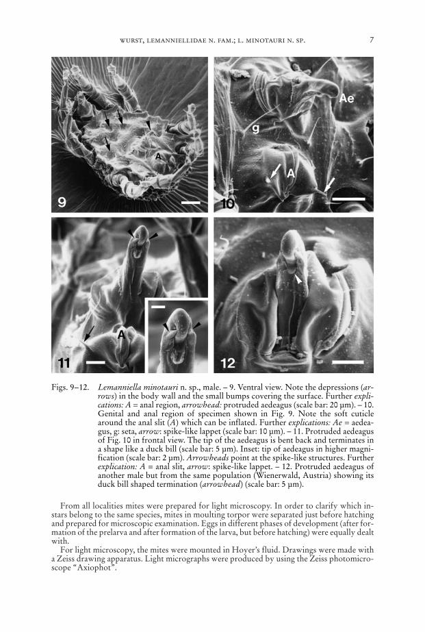
Figs. 9–12. Lemanniella minotauri n. sp., male. – 9. Ventral view. Note the depressions (ar-rows) in the body wall and the small bumps covering the surface. Further expli-cations: A = anal region, arrowhead: protruded aedeagus (scale bar: 20 µm). – 10.Genital and anal region of specimen shown in Fig. 9. Note the soft cuticlearound the anal slit (A) which can be inflated. Further explications: Ae = aedea-gus, g: seta, arrow: spike-like lappet (scale bar: 10 µm). – 11. Protruded aedeagusof Fig. 10 in frontal view. The tip of the aedeagus is bent back and terminates ina shape like a duck bill (scale bar: 5 µm). Inset: tip of aedeagus in higher magni-fication (scale bar: 2 µm). Arrowheads point at the spike-like structures. Furtherexplication: A = anal slit, arrow: spike-like lappet. – 12. Protruded aedeagus ofanother male but from the same population (Wienerwald, Austria) showing itsduck bill shaped termination (arrowhead) (scale bar: 5 µm).
For scanning electron microscope (SEM) investigations, the mites were killed by freezingand were cleaned by washing with a surfactant. Further preparation was performed afterBOCK (1987) in five steps: 1) fixation by a modified Carnoy (acetic acid : chloroforme :ethanol = 1 : 1 : 3) for at least 4 hrs, 2) ethanol (5–10 min), 3) hexamethyldisilazane (5 min), 4)air-drying, 5) sputtering with gold. A few ants with attached deutonymphs were fixed with-out prior washing in order to guarantee that the mites maintain their position. The mites andthe mite loaded ants were examined by using the SEM DSM 940 (Zeiss).
The species concept follows the “biological species concept” sensu MAYR (1963). Thenomenclature of idiosomal chaetotaxy follows GRIFFITHS et alii (1990), the nomenclature ofleg chaetotaxy is according to GRANDJEAN (1939).
2.3. Acknowledgements
The first complete description of a mite of the genus Lemanniella was made possible by fi-nancial support of the “Stiftung Natur und Umwelt” of the Landesbank Baden-Württemberg(Stuttgart).
For their most generous help I thank the following persons: Prof. Dr. BRIGITTE FRANK
(Stuttgart) and Prof. Dr. A. FAIN (Brussels) supported this study from its beginning. I amgrateful to Prof. Dr. B. M. OCONNOR (Ann Arbor) for discussions on the taxonomic statusof Lemanniella. Dr. K.-F. RAQUÉ (Heidelberg), Dr. B. SEIFERT (Görlitz), and D. VEILE (Ober-sulm) determinated the species identity of the ants and gave expert advice about the buildersof the nest materials. I also thank B. CURTH and E. RÜCKER (Stuttgart) for assistance in theSEM preparations. Dr. F. WOOG (Stuttgart) critically read the manuscript and gave valuablecomments.
8 stuttgarter beiträge zur naturkunde Ser. A, Nr. 621
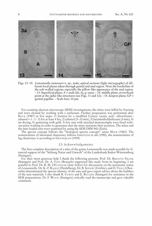
Figs. 13–15. Lemanniella minotauri n. sp., male; optical sections (light micrographs) of dif-ferent focal planes taken through genital and anal region. Note the borderline ofthe soft-walled regions, especially the pillow-like appearance of the anal region.– 13. Superficial plane; A = analt slit, 3a, g: setae; – 14. middle plane; arrowheadspoint at the spike-like structures (see Figs. 11 and 12); – 15. deepest plane; GP =genital papillae. – Scale bars: 10 µm.
wurst, lemanniellidae n. fam.; l. minotauri n. sp. 9
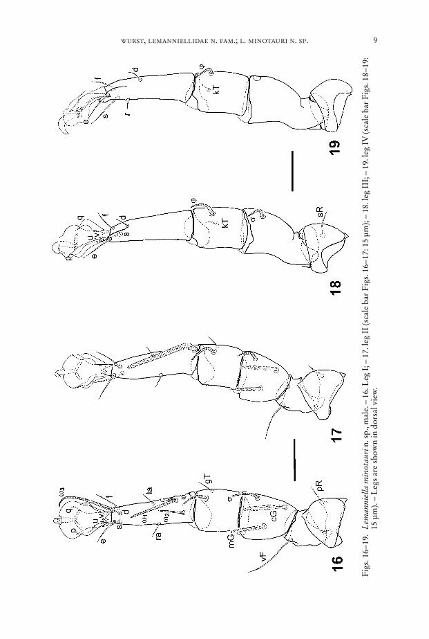
Fig
s. 1
6–19
.L
eman
niel
la m
inot
auri
n. s
p., m
ale.
– 1
6. L
eg I
; – 1
7. le
g II
(sca
le b
ar F
igs.
16–
17: 1
5 µm
); –
18. l
eg I
II; –
19.
leg
IV (s
cale
bar
Fig
s. 1
8–19
:15
µm
). –
Leg
s ar
e sh
own
in d
orsa
l vie
w.
3. Lemanniellidae n. fam.
Type genus: Lemanniella Mahunka, 1977.
3.1. Definit ion3.1.1. Non-hypopal instars
Dorsum without sejugal furrow. Adults with a dome-shaped elevation delimitedlaterally and posteriorly by a band of cuticle decorated with tiny tubercles. Entiredorsum of adults of honeycomb appearance, conspicuously regular on the dome, ir-regular in front of it and beneath the band. Dorsum of non-hypopal juvenile instarscovered with mushroom-like protuberances with a jagged cap. Ventral body wall ofall non-hypopal instars with depressions. Femur and genu of leg III and IV fused inall non-hypopal instars. Pretarsi with pronounced ambulacrum and strong claw. Se-tae p and q foliate in all legs. Males lack adanal suckers and sucker-like setae on legIV. Retroconjugate mating mode.
3.1.2. DeutonymphPalposoma absent. Scapular setae lacking. Setae 1a, 3a, 3b, 4a reduced (only the
sockets are present). Genital opening integrated into the sucker plate. Tarsus I and IIwith weak claw and five (leg I) and six (leg II) setae. Solenidion sigma of genu I andII reduced (only the sockets are present). Leg III and IV without claw and solenidia.Tibia and tarsus of leg III and IV fused.
3.2. RemarksSince our knowledge on Lemanniella is based on the life cycle of only one species,
the family definition necessarily has to be provisonal.
10 stuttgarter beiträge zur naturkunde Ser. A, Nr. 621
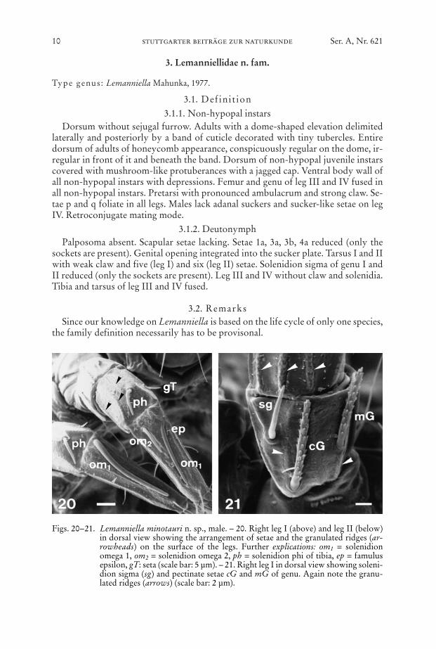
Figs. 20–21. Lemanniella minotauri n. sp., male. – 20. Right leg I (above) and leg II (below)in dorsal view showing the arrangement of setae and the granulated ridges (ar-rowheads) on the surface of the legs. Further explications: om1 = solenidionomega 1, om2 = solenidion omega 2, ph = solenidion phi of tibia, ep = famulusepsilon, gT: seta (scale bar: 5 µm). – 21. Right leg I in dorsal view showing soleni-dion sigma (sg) and pectinate setae cG and mG of genu. Again note the granu-lated ridges (arrows) (scale bar: 2 µm).

wurst, lemanniellidae n. fam.; l. minotauri n. sp. 11
Fig. 22. Lemanniella minotauri n. sp., female; ventral view, left legs partly omitted. –Scale bar: 25 µm.
A definition of non-hypopal instars for the genus Lemanniella is not given be-cause our knowledge is at present restricted to only one species.
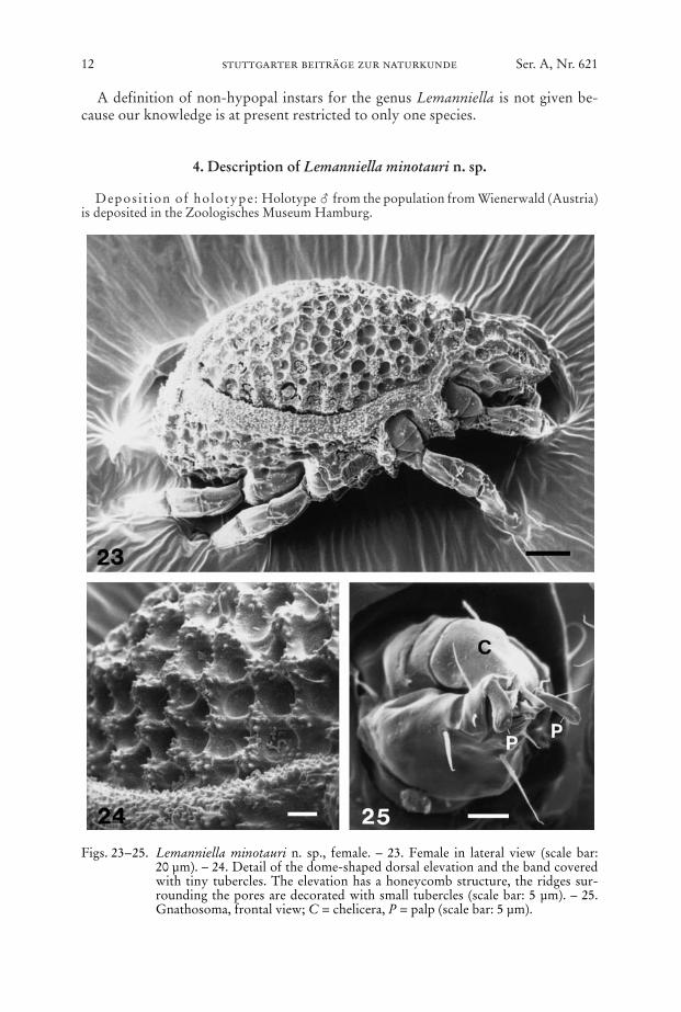
4. Description of Lemanniella minotauri n. sp.
Deposit ion of holotype: Holotype � from the population from Wienerwald (Austria)is deposited in the Zoologisches Museum Hamburg.
12 stuttgarter beiträge zur naturkunde Ser. A, Nr. 621
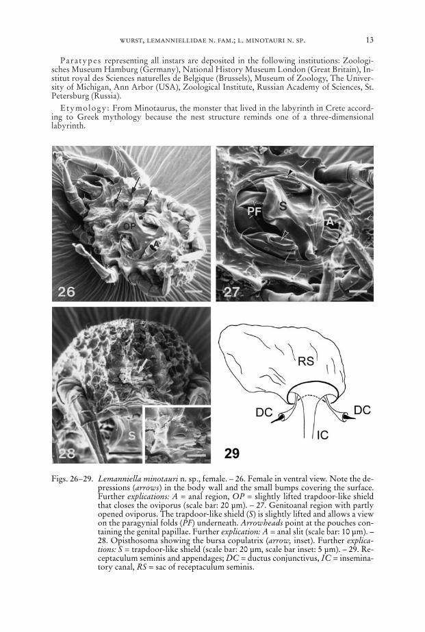
Figs. 23–25. Lemanniella minotauri n. sp., female. – 23. Female in lateral view (scale bar:20 µm). – 24. Detail of the dome-shaped dorsal elevation and the band coveredwith tiny tubercles. The elevation has a honeycomb structure, the ridges sur-rounding the pores are decorated with small tubercles (scale bar: 5 µm). – 25.Gnathosoma, frontal view; C = chelicera, P = palp (scale bar: 5 µm).
Paratypes representing all instars are deposited in the following institutions: Zoologi-sches Museum Hamburg (Germany), National History Museum London (Great Britain), In-stitut royal des Sciences naturelles de Belgique (Brussels), Museum of Zoology, The Univer-sity of Michigan, Ann Arbor (USA), Zoological Institute, Russian Academy of Sciences, St.Petersburg (Russia).
Etymology: From Minotaurus, the monster that lived in the labyrinth in Crete accord-ing to Greek mythology because the nest structure reminds one of a three-dimensionallabyrinth.
wurst, lemanniellidae n. fam.; l. minotauri n. sp. 13
Figs. 26–29. Lemanniella minotauri n. sp., female. – 26. Female in ventral view. Note the de-pressions (arrows) in the body wall and the small bumps covering the surface.Further explications: A = anal region, OP = slightly lifted trapdoor-like shieldthat closes the oviporus (scale bar: 20 µm). – 27. Genitoanal region with partlyopened oviporus. The trapdoor-like shield (S) is slightly lifted and allows a viewon the paragynial folds (PF) underneath. Arrowheads point at the pouches con-taining the genital papillae. Further explication: A = anal slit (scale bar: 10 µm). –28. Opisthosoma showing the bursa copulatrix (arrow, inset). Further explica-tions: S = trapdoor-like shield (scale bar: 20 µm, scale bar inset: 5 µm). – 29. Re-ceptaculum seminis and appendages; DC = ductus conjunctivus, IC = insemina-tory canal, RS = sac of receptaculum seminis.
14 stuttgarter beiträge zur naturkunde Ser. A, Nr. 621
Differential diagnosis : At the date of publishing Lemanniella minotauri canbe distinguished from the only other known species L. reducta only by differencesin the characters of the deutonymphs. The most prominent differences are 1) theenormous length of solenidion omega 3 in L. reducta and 2) the shape of seta e in legII which is formed as a spoonlike adhesive seta in L. reducta whereas in L. minotau-ri it is reduced to a short spine.
Description: Retroconjugate mites with facultative hypopody. All instars oc-cur in nests of the ant Lasius brunneus. All idiosomal setae smooth. All non-hypopalinstars share the following characters: legs bear their normal number of solenidia ontarsi, setae u and v in all legs short spines (Fig. 35), setae p and q partly fused with thepretarsus (Figs. 34–35), tibia I and II with one smooth seta (gT) and solenidion phi(Fig. 20), genu I and II with two pectinate setae (cG, mG) and solenidion sigma (Fig.21), femur I and II with one smooth seta, all legs with longitudinal granulated ridges(Figs. 20–21, 34).
4.1. Adults
4.1.1. MaleBody ovoid with truncated posterior end, anterior part elongated forming a
tegmen covering the gnathosoma (Fig. 1), length of idiosoma between 170 µm and180 µm, colour: brown.
Dorsum (Figs. 1, 3): Idiosomal chaetome: vi, ve, scx, si, se, c1, c2, cp, d1, d2, e1, e2,h1, h2, f2. – Ridges of honeycomb-like dome-shaped part decorated with small tu-bercles (see Fig. 24). All setae short.
Venter (Fig. 2, 9): Idiosomal chaetome: 1a, c3, 3a, 3b, g, 4a, ps1, ps2, ps3, h3. –Gnathosoma with chelate chelicerae, palp tarsus with three button-like chetae dis-tally and one dorsal filiform seta (Figs. 5–8).
Epimera I fused medially forming a sternum, epimera II–IV ending freely. Patternof deep depressions in the body wall follows the course of leg apodemes underneath:depressions occur along the apodemes and at their medial end. Venter behind thegnathosoma a sclerotized plate with lobed margin, partly bordering the gnathosomawith a “collar” (Fig. 7). This “collar” bears two thin bifurcate elongations pointingat the gnathosoma and apparently constituting the Grandjean’s organ (Figs. 5–7).Ventral plate with a soft walled region harbouring the aedeagus and another soft re-gion around the anal slit (Fig. 10). All sclerotized regions covered with small bumps.
Aedeagus (Figs. 10–15) finger-like, the tip of it bends back and terminates in ashape like a duck bill. At each side a spike-like structure parallels the tip of the aedea-gus.
Anal s l i t (Figs. 10–11, 13) situated in an area of soft cuticle forming a pillow.Lateral to the posterior end of the anal slit two spike-like lappets originate in thisarea.
Legs (Figs. 16–21): Chaetome see Table 1. Solenidion omega 1 of tarsus II con-siderably larger than omega 1 of tarsus I (Figs. 16–17, 20).
4.1.2. FemaleBody ovoid, anterior part forming a tegmen covering the gnathosoma (Fig. 23),
length of idiosoma between 195 µm and 210 µm, colour: brown.
wurst, lemanniellidae n. fam.; l. minotauri n. sp. 15
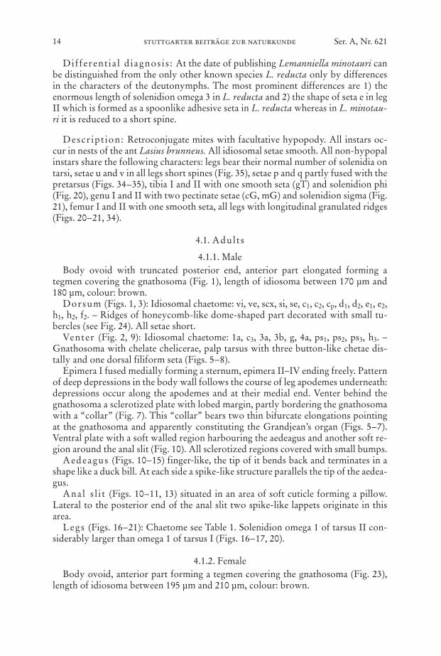
Fig
s. 3
0–33
.L
eman
niel
la m
inot
auri
n. s
p., f
emal
e. –
30.
Leg
I; –
31.
leg
II (
scal
e ba
r F
igs.
30–
31: 1
5 µm
); –
32. l
eg I
II; –
33.
leg
IV (
scal
e ba
r F
igs.
32–3
3: 1
0 µm
). –
Leg
s ar
e sh
own
in d
orsa
l vie
w.
16 stuttgarter beiträge zur naturkunde Ser. A, Nr. 621
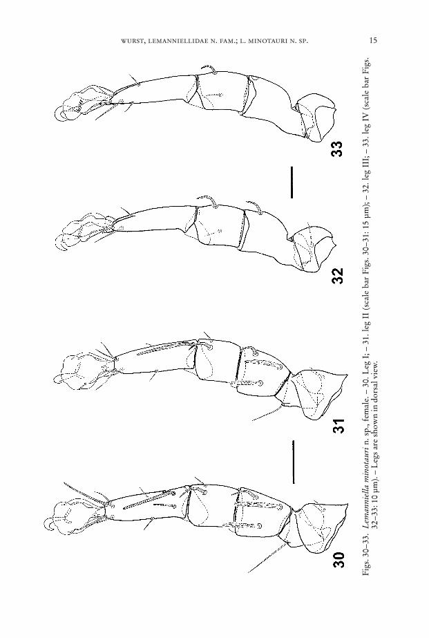
Fig. 36. Lemanniella minotauri n. sp.; mating position.
Figs. 34–35. Lemanniella minotauri n. sp., female. – 34. Right leg III, dorsal view, showingleaf-like setae p and q which are adjacent to the pretarsus. The arrowhead pointsat the connection between the seta and the pretarsal cuticle. Note the granulatedridges on tarsus. Further explications: d, e, f: setae (scale bar: 5 µm); – 35. legs Ishowing the cuticular connection (arrowhead) between seta q and the pretarsus.Further explications: u, v, f, s: setae (scale bar: 5 µm).
wurst, lemanniellidae n. fam.; l. minotauri n. sp. 17
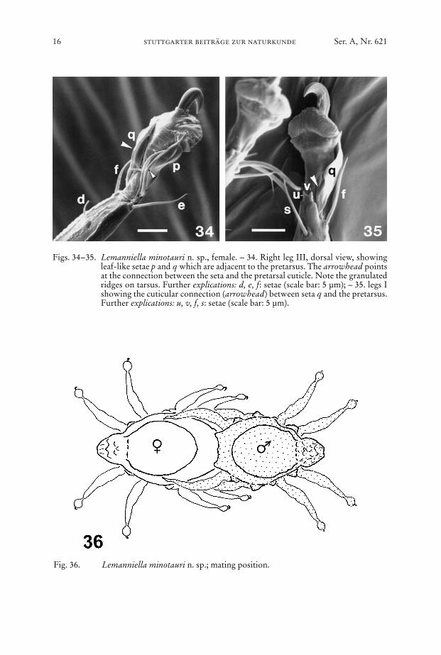
Fig. 37. Lemanniella minotauri n. sp., tritonymph; ventral view, left legs partly omitted.– Scale bar: 20 µm.
18 stuttgarter beiträge zur naturkunde Ser. A, Nr. 621
Fig
s. 3
8–39
.L
eman
niel
la m
inot
auri
n. s
p. –
38.
Tri
tony
mph
; idi
osom
a, d
orsa
l vi
ew (
scal
e ba
r: 2
0 µm
); –
39. d
euto
nym
ph; i
dios
oma,
dor
sal
view
(sca
le b
ar: 2
0 µm
).
wurst, lemanniellidae n. fam.; l. minotauri n. sp. 19
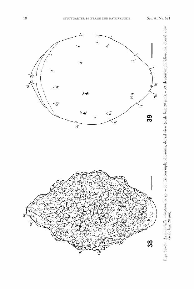
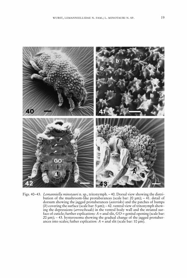
Figs. 40–43. Lemanniella minotauri n. sp., tritonymph. – 40. Dorsal view showing the distri-bution of the mushroom-like protuberances (scale bar: 20 µm); – 41. detail ofdorsum showing the jagged protuberances (asterisks) and the patches of bumps(b) covering the surface (scale bar: 5 µm); – 42. ventral view of tritonymph show-ing the depressions (arrowheads) in the ventral body wall and the striated sur-face of cuticle; further explications: A = anal slit, GO = genital opening (scale bar:20 µm); – 43. hysterosoma showing the gradual change of the jagged protuber-ances into scales; futher explication: A = anal slit (scale bar: 10 µm).
20 stuttgarter beiträge zur naturkunde Ser. A, Nr. 621
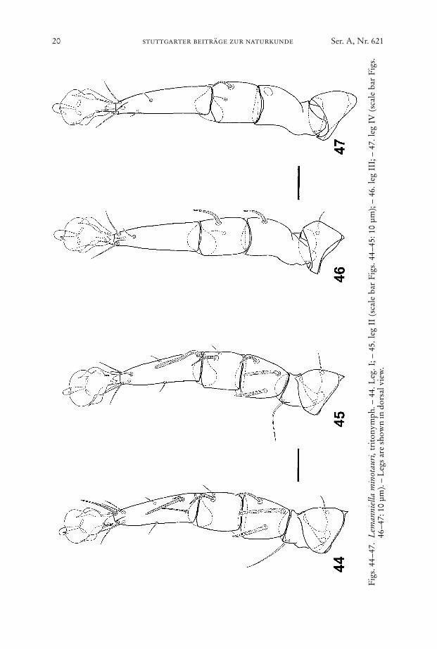
Fig
s. 4
4–47
. L
eman
niel
la m
inot
auri
,tri
tony
mph
. – 4
4. L
eg. I
; – 4
5. le
g II
(sc
ale
bar
Fig
s. 4
4–45
: 10
µm);
– 46
. leg
III
; – 4
7. le
g IV
(sc
ale
bar
Fig
s.46
–47:
10
µm).
– L
egs
are
show
n in
dor
sal v
iew
.
wurst, lemanniellidae n. fam.; l. minotauri n. sp. 21
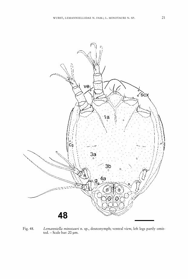
Fig. 48. Lemanniella minotauri n. sp., deutonymph; ventral view, left legs partly omit-ted. – Scale bar: 20 µm.
22 stuttgarter beiträge zur naturkunde Ser. A, Nr. 621
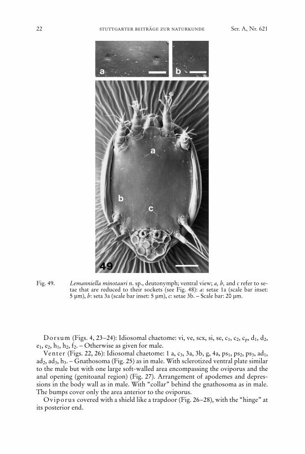
Fig. 49. Lemanniella minotauri n. sp., deutonymph; ventral view; a, b, and c refer to se-tae that are reduced to their sockets (see Fig. 48): a: setae 1a (scale bar inset:5 µm), b: seta 3a (scale bar inset: 5 µm), c: setae 3b. – Scale bar: 20 µm.
Dorsum (Figs. 4, 23–24): Idiosomal chaetome: vi, ve, scx, si, se, c1, c2, cp, d1, d2,e1, e2, h1, h2, f2. – Otherwise as given for male.
Venter (Figs. 22, 26): Idiosomal chaetome: 1 a, c3, 3a, 3b, g, 4a, ps1, ps2, ps3, ad1,ad2, ad3, h3. – Gnathosoma (Fig. 25) as in male. With sclerotized ventral plate similarto the male but with one large soft-walled area encompassing the oviporus and theanal opening (genitoanal region) (Fig. 27). Arrangement of apodemes and depres-sions in the body wall as in male. With “collar” behind the gnathosoma as in male.The bumps cover only the area anterior to the oviporus.
Oviporus covered with a shield like a trapdoor (Fig. 26–28), with the “hinge” atits posterior end.
Bursa copulatr ix (Fig. 28) a small slit without accessory structures situated atthe hind end of opisthosoma. For receptaculum seminis see Fig. 29.
Legs (Figs. 30–35): Chaetome see Table 1.
4.2. Tritonymph
Body ellipsoid, anterior part forming a tegmen covering the gnathosoma (Fig. 40),if directly developed from protonymph length of idiosoma between 180 µm and200 µm, colour: white.
Dorsum (Figs. 38, 40): Idiosomal chaetome: vi, ve, scx, si, se, c1, c2, cp, d1, d2, e1,e2, h1, h2, f2. – Small patches of bumps dispersed between the jagged mushroom-likestructures covering the surface (Fig. 41). The mushroom-like structures graduallychange into scales laterally and posteriorly and into jagged ridges anteriorly (Fig.43). All setae short.
Venter (Figs. 37, 42): Idiosomal chaetome: 1a, c3, 3a, 3b, g, 4a, ps1, ps2, ps3, h3, –Gnathosoma as in male. Epimera I not fused, epimera II ending freely, epimera IIIand IV not discernible, with small sejugal apodemes. Pattern of depressions followsthe arrangement of the apodemes. “Collar” behind the gnathosoma as in male. Gen-ital and anal aperture anteriorly and posteriorly accompanied by sclerotized bands(Fig. 37). Entire surface striated and without bumps (Fig. 42).
Legs (Figs. 44–47): Chaetome see Table 1.
4.3. Deutonymph
Body ovoid with tapered posterior end, length of idiosoma approximately135 µm, colour: beige.
wurst, lemanniellidae n. fam.; l. minotauri n. sp. 23
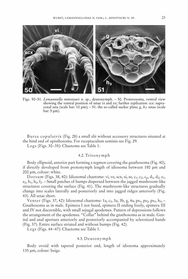
Figs. 50–51. Lemanniella minotauri n. sp., deutonymph. – 50. Proterosoma, ventral viewshowing the ventral position of setae vi and ve; further explication: scx: supra-coxal seta (scale bar: 10 µm); – 51. the so-called sucker plate; g, h3: setae (scalebar: 5 µm).
24 stuttgarter beiträge zur naturkunde Ser. A, Nr. 621
Fig
s. 5
2–55
.L
eman
niel
la m
inot
auri
n. s
p., d
euto
nym
ph. –
52.
Leg
I; –
53.
leg
II (
scal
e ba
r F
igs.
52–
53: 1
0 µm
); –
54. l
eg I
II; –
55.
leg
IV (
scal
e ba
rF
igs.
54–
55: 5
µm
). –
Leg
s ar
e sh
own
in d
orsa
l vie
w.
Dorsum (Fig. 39): Idiosomal chaetome: c1, c2, cp, d1, d2, e1, e2, h1, h2, h3, f2.Venter (Figs. 48–50, 60): Idiosomal chaetome: vi, ve, scx, c3, g; setae 1a, 3a, 3b, 4a
only as sockets. – Two palpal solenidia. Epimeres I fused medially forming a ster-num, epimeres II ending freely, apodemes of leg III and IV strongly reduced. So-called sucker plate (Fig. 51) with complete set of modified setae, central “sucker” rel-atively small, the radius of it only half the radius of the anterior “sucker”.
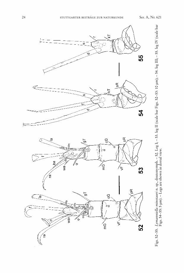
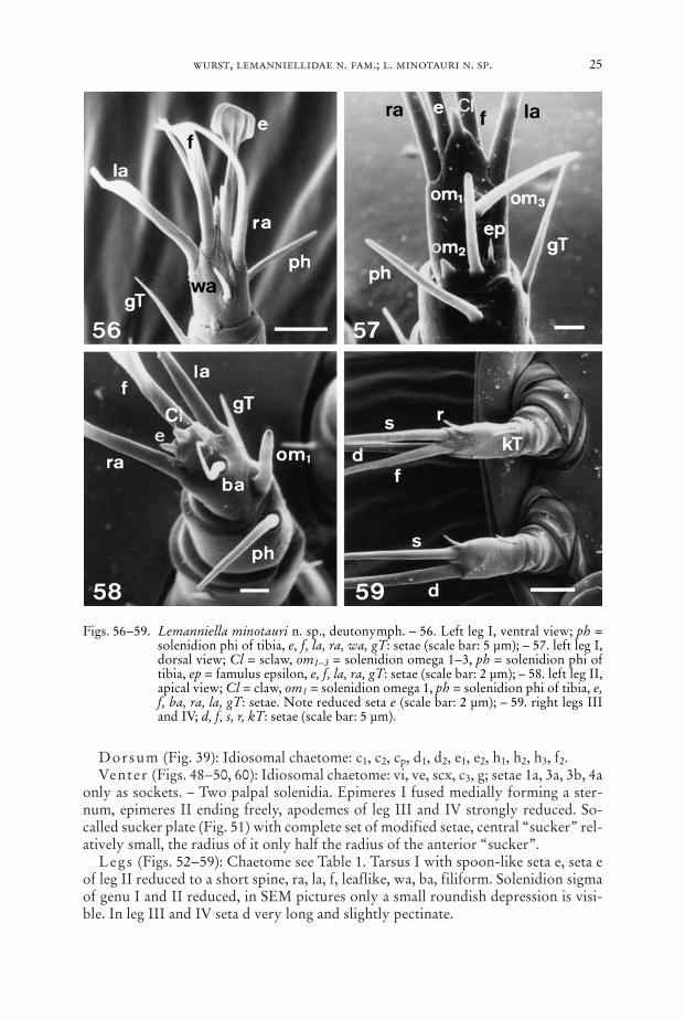
Legs (Figs. 52–59): Chaetome see Table 1. Tarsus I with spoon-like seta e, seta eof leg II reduced to a short spine, ra, la, f, leaflike, wa, ba, filiform. Solenidion sigmaof genu I and II reduced, in SEM pictures only a small roundish depression is visi-ble. In leg III and IV seta d very long and slightly pectinate.
wurst, lemanniellidae n. fam.; l. minotauri n. sp. 25
Figs. 56–59. Lemanniella minotauri n. sp., deutonymph. – 56. Left leg I, ventral view; ph =solenidion phi of tibia, e, f, la, ra, wa, gT: setae (scale bar: 5 µm); – 57. left leg I,dorsal view; Cl = sclaw, om1–3 = solenidion omega 1–3, ph = solenidion phi oftibia, ep = famulus epsilon, e, f, la, ra, gT: setae (scale bar: 2 µm); – 58. left leg II,apical view; Cl = claw, om1 = solenidion omega 1, ph = solenidion phi of tibia, e,f, ba, ra, la, gT: setae. Note reduced seta e (scale bar: 2 µm); – 59. right legs IIIand IV; d, f, s, r, kT: setae (scale bar: 5 µm).
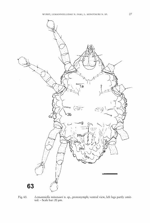
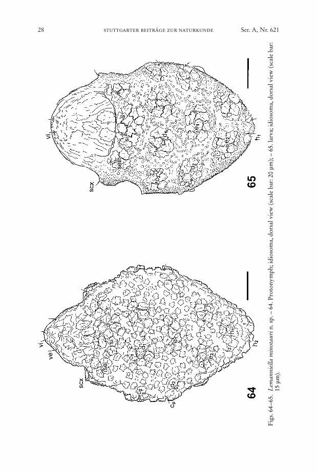
4.4. Protonymph
Body ellipsoid, anterior part forming a tegmen covering the gnathosoma, lengthof idiosoma between 135 µm and 160 µm, colour: white.
Dorsum (Fig. 64): Idiosomal chaetome. vi, ve, scx, si, se, c1, c2, cp, d1, d2, e1, e2, h1,h2, f2. – As given for tritonymph but with lower density of mushroom-like struc-tures.
Venter (Fig. 63): Idiosomal chaetome: 1a, c3, 3b, g, ps1, ps2, ps3, h3. – As given fortritonymph.
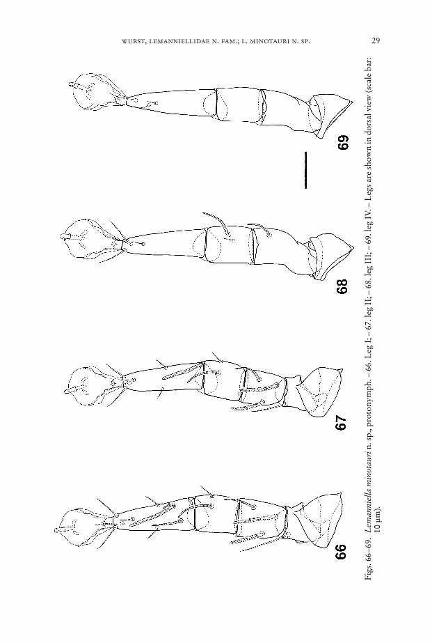
Legs (Figs. 66–69): Chaetome see Table 1.
4.5. Larva and egg
Body ellipsoid, length of idiosoma between 90 µm and 110 µm, colour; white.Dorsum (Fig. 65): Idiosomal chaetome: vi, ve, scx, si, se, c1, c2, cp, d1, d2, e1, e2, h1.
– The mushroom-like structures are clustered in patches of five to ten elements each.The area between these patches is covered with numerous bumps. With cuticularscales at the flanks and at the opisthosomal end. All setae short.
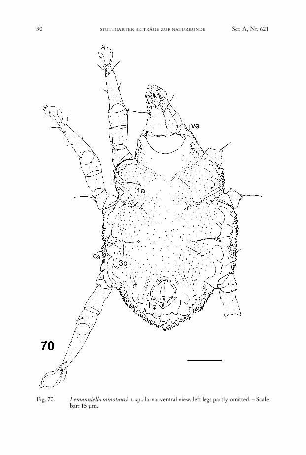
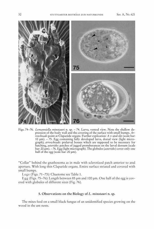
Venter (Figs. 70, 74): Idiosomal chaetome: 1a, c3, 3b, h2. – Gnathosoma as inmale. Epimera I not fused, epimera II ending freely, sejugal apodemes short. Bodywall with only shallow depressions near the medial termination of the apodemes.
26 stuttgarter beiträge zur naturkunde Ser. A, Nr. 621
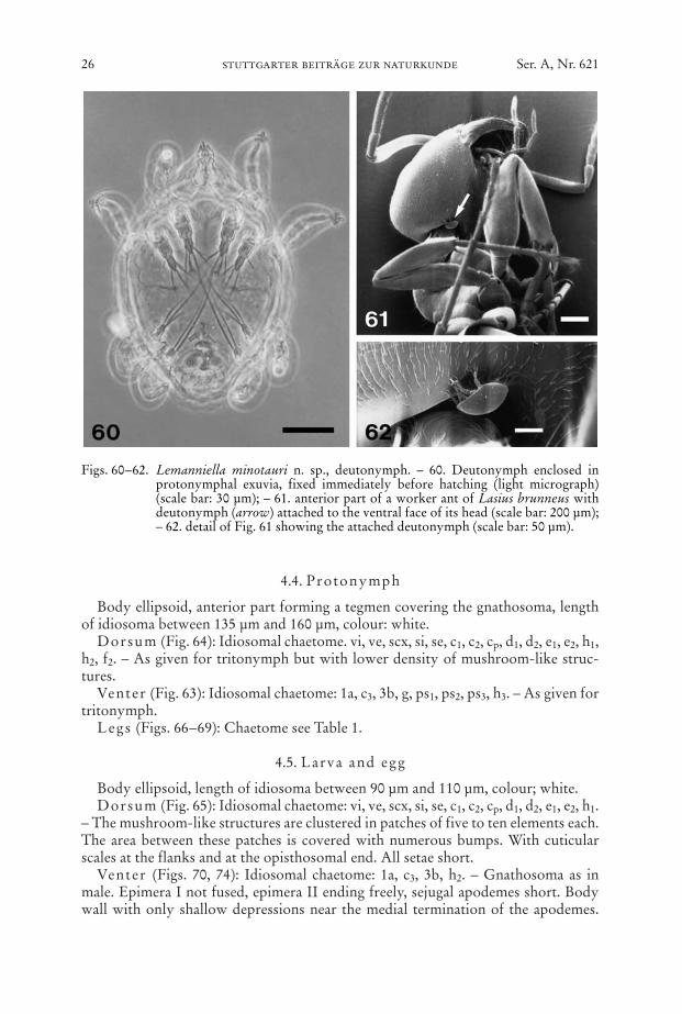
Figs. 60–62. Lemanniella minotauri n. sp., deutonymph. – 60. Deutonymph enclosed inprotonymphal exuvia, fixed immediately before hatching (light micrograph)(scale bar: 30 µm); – 61. anterior part of a worker ant of Lasius brunneus withdeutonymph (arrow) attached to the ventral face of its head (scale bar: 200 µm);– 62. detail of Fig. 61 showing the attached deutonymph (scale bar: 50 µm).
wurst, lemanniellidae n. fam.; l. minotauri n. sp. 27
Fig. 63. Lemanniella minotauri n. sp., protonymph; ventral view, left legs partly omit-ted. – Scale bar: 20 µm.
28 stuttgarter beiträge zur naturkunde Ser. A, Nr. 621
Fig
s. 6
4–65
.L
eman
niel
la m
inot
auri
n. s
p. –
64.
Pro
tony
mph
; idi
osom
a, d
orsa
l vie
w (s
cale
bar
: 20
µm);
– 65
. lar
va; i
dios
oma,
dor
sal v
iew
(sca
le b
ar:
15 µ
m).
wurst, lemanniellidae n. fam.; l. minotauri n. sp. 29
Fig
s. 6
6–69
.L
eman
niel
la m
inot
auri
n. s
p., p
roto
nym
ph. –
66.
Leg
I; –
67.
leg
II; –
68.
leg
III;
– 6
9. le
g IV
. – L
egs
are
show
n in
dor
sal v
iew
(sca
le b
ar:
10 µ
m).
30 stuttgarter beiträge zur naturkunde Ser. A, Nr. 621
Fig. 70. Lemanniella minotauri n. sp., larva; ventral view, left legs partly omitted. – Scalebar: 15 µm.
wurst, lemanniellidae n. fam.; l. minotauri n. sp. 31
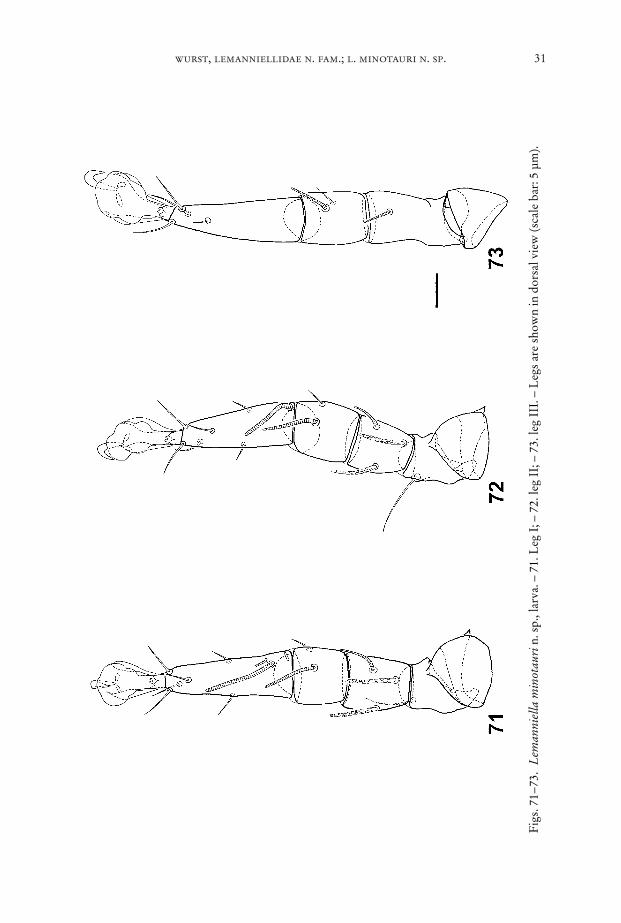
Fig
s. 7
1–73
.L
eman
niel
la m
inot
auri
n. s
p., l
arva
. – 7
1. L
eg I
; – 7
2. le
g II
; – 7
3. le
g II
I. –
Leg
s ar
e sh
own
in d
orsa
l vie
w (s
cale
bar
: 5 µ
m).
“Collar” behind the gnathosoma as in male with sclerotized patch anterior to analaperture. With long thin Claparède organs. Entire surface striated and covered withsmall bumps.
Legs (Figs. 71–73): Chaetome see Table 1.Egg (Figs. 75–76): Length between 85 µm and 100 µm. One half of the egg is cov-
ered with globules of different sizes (Fig. 76).
5. Observations on the Biology of L. minotauri n. sp.
The mites feed on a small black fungus of an unidentified species growing on thewood in the ant nests.
32 stuttgarter beiträge zur naturkunde Ser. A, Nr. 621
Figs. 74–76. Lemanniella minotauri n. sp. – 74. Larva, ventral view. Note the shallow de-pression of the body wall and the covering of the surface with small bumps. Ar-rowheads point at Claparède organs. Further explication: A = anal slit (scale bar:10 µm). – 75. Egg containing fully developed larva, dorsal view (light micro-graph); arrowheads: prelarval bosses which are supposed to be necessary forhatching, asterisks: patches of jagged protuberances on the larval dorsum (scalebar: 20 µm). – 76. Egg (light micrograph). The globules (asterisks) cover only onehalf of the egg (scale bar: 20 µm).
Mating occurred in the retroconjugate mode (Fig. 36). In this position the en-larged and inflated region of the male anal region apparently serves as an attachmentorgan that helps in maintaining the connection with the female by adhesion. Thismight be supported by fluid discharged from the anus. If this interpretation of therole of the specialized male anal region is true, the question must be raised if the so-called adanal (or any other) suckers of astigmatic mites actually function as suckers(based on a pressure-difference principle and necessitating clean surfaces for thetightness) or rather as adhesive devices (based on the exploitation of surface forcesand tolerating impurities on the surfaces involved).
The eggs were glued on the substrate by the globules covering one half of the egg.Only a few of the inspected worker ants carried deutonymphs. In most cases on-
ly one deutonymph was attached, I never found more than two mites on one ant. Asattachment site the deutonymphs preferred the head of the ant (Figs. 61–62). Herethe hypopi were located at the posterior surface of the head or the ventral region ofthe head posterior to the mouth parts. In very rare cases deutonymphs were attachedto the mesosoma.
6. References
BOCK, C. (1987): Einfache Schnellpräparationsmethode mit Carnoy und Hexamethyldisil-azan für das REM. – Optik (Suppl. 3) 77: 7; Stuttgart
GRANDJEAN, F. (1939): La chaetotaxie des pattes chez les Acaridiae. – Bull. Soc. zool. Fr. 64:50–60; Paris.
GRIFFITHS, D. A., ATYEO, W. T., NORTON R. A. & LYNCH, C. A. (1990): The idiosomalchaetotaxy of astigmatid mites. – J. Zool. (Lond.) 220: 1–32; London.
MAHUNKA, S. (1977): Neue und interessante Milben aus dem Genfer Museum XIX. EinigeAngaben zur Kenntnis der Milbenfauna der Ameisen-Nester (Acari: Acarida, Tarsone-mida). – Archs Sci. (Geneva) 30 (1): 91–106; Geneva.
wurst, lemanniellidae n. fam.; l. minotauri n. sp. 33
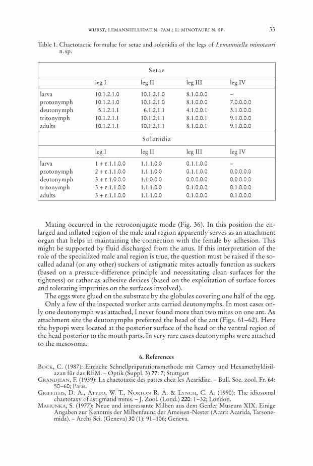
Setae
leg I leg II leg III leg IV
larva 10.1.2.1.0 10.1.2.1.0 8.1.0.0.0 –protonymph 10.1.2.1.0 10.1.2.1.0 8.1.0.0.0 7.0.0.0.0deutonymph 5.1.2.1.1 6.1.2.1.1 4.1.0.0.1 3.1.0.0.0tritonymph 10.1.2.1.1 10.1.2.1.1 8.1.0.0.1 9.1.0.0.0adults 10.1.2.1.1 10.1.2.1.1 8.1.0.0.1 9.1.0.0.0
Solenidia
leg I leg II leg III leg IV
larva 1 + ε.1.1.0.0 1.1.1.0.0 0.1.1.0.0 –protonymph 2 + ε.1.1.0.0 1.1.1.0.0 0.1.1.0.0 0.0.0.0.0deutonymph 3 + ε.1.0.0.0 1.1.0.0.0 0.0.0.0.0 0.0.0.0.0tritonymph 3 + ε.1.1.0.0 1.1.1.0.0 0.1.0.0.0 0.1.0.0.0adults 3 + ε.1.1.0.0 1.1.1.0.0 0.1.0.0.0 0.1.0.0.0
Table 1. Chaetotactic formulae for setae and solenidia of the legs of Lemanniella minotaurin. sp.
MAYR, E. (1963): Animal species and evolution. I–XIV. – 797 pp.; Cambridge, Mass.SEIFERT, B. (1996): Ameisen: beobachten, bestimmen. – 352 pp.; Augsburg.TÜRK, E. & TÜRK, F. (1957): Systematik und Ökologie der Tyroglyphiden Mitteleuropas. –
In: STAMMER, H.-J. (ed.): Beiträge zur Systematik und Ökologie mitteleuropäischerAcarina. – Vol. 1 Tyroglyphidae und Tarsonemini, pp. 1–231; Leipzig.
Author’s address:
Dipl.-Biol. EBERHARD WURST, Institut für Zoologie (Fachgebiet Parasitologie), UniversitätStuttgart, Emil-Wolff-Str. 34, D-70599 Stuttgart, F. R. Germany.
34 stuttgarter beiträge zur naturkunde Ser. A, Nr. 621

ISSN 0341-0145
Schriftleitung: Dr. Wolfgang Seeger, Rosenstein 1, D-70191 StuttgartGesamtherstellung: Gulde-Druck GmbH, D-72072 Tübingen