sub-diffraction-limit imaging by stochastic optical reconstruction microscopy (storm) michael j....
Post on 21-Dec-2015
297 views
TRANSCRIPT

Sub-diffraction-limit imaging by Stochastic Optical Reconstruction
Microscopy (STORM)
Michael J. Rust, Mark Bates, Xiaowei ZhuangHarvard University
Published Online August 9, 2006Nature Methods Vol.3 No.10
Presented by Artie Wu

STORM
• High-resolution fluorescence microscopy method based on high-accuracy localization of photoswitchable fluorophores
• Imaging resolution of 20nm

Outline
• Background– fluorescence microscopy– diffraction
• Motivation• Fluorescence Microscopy Alternatives• STORM• Results• Conclusions
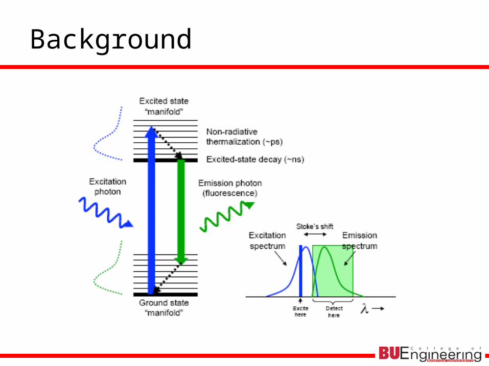
Background

Background

Motivation I
• Resolution limit set by diffraction of light
• Fluorescence microscopy widely used in molecular and cell biology

Fluorescence Microscopy Alternatives
• Lateral resolution of 10s of nanometers– Near-field scanning optical microscopy (NSOM)– Multiphoton fluorscence– Stimulated emission depletion (STED)– Saturated structured-illumination microscopy (SSIM)

Near-field scanning optical microscopy (NSOM)
• Image at interface due to evanescent field
• Study what goes on near membrane– Exocytosis & endocytosis
• Build up point by point• Drawback: low imaging
depth

Motivation II
• Single-molecule detection leads to sub-diffraction-limit spatial resolution
• Stochastic optical reconstruction microscopy (STORM)– Fluorescence image constructed from high-accuracy
localization of individual fluorescent molecules– Imaging resolution: ~20nm using TIRF and
photoswitchable cyanine dye, Cy5

STORM
•Cy5: fluorescent and dark state using different λ•Cy3: secondary dye•Series of imaging cycle•In each cycle
•Only 1-3 switches in FOV are switched ON•Stochastically different subset of fluorophores are ON
•Red: 633nm, 30W/cm2, 2s•Green: 532nm, 1W/cm2, 0.5s•Photobleaching: 230s

Resolution
• Limited by accuracy of localization of switches
• 2d Gaussian fit to PSF used to find centroid position of switch

Centroid position
• Fit to pixelated Gaussian function
A: background fluorescence level
Io: amplitude of peak
a,b: widths of Gaussian distribution
xo,yo: center coordinates of peak
δ: fixed half-width of pixel in object plane
b
yyerf
b
yyerf
a
xxerf
a
xxerf
abIA
eIdYdXAyxI
ooooo
b
yY
a
xX
o
y
y
x
x
o
4
,2/0

Results I
• Linear, dsDNA with 2 switches separated by 135 bps (46nm)
• Theoretical dist = 40nm• Experimental dist = 41nm

Results II
• Longer DNA with 4 switches spaced 46 nm apart
• Localize large number of switches within diffraction-limited spot by cycling switches on/off

Conclusions
• STORM capable of imaging biological structures with sub-diffraction-limit resolution
• Resolution limited by # photons emitted per switch cycle– Cyanine switch ~3000 photons/cycle
• Theoretical localization accuracy of 4nm
• Corresponds to imaging resolution of ~20nm
– Imaging speed improved by increasing switching rate• Stronger excitation or fluorophores with faster switching kinetics
• Valuable tool for high-resolution in situ hybridization and immunofluorescence imaging