subassemblies and asymmetry in assembly of herpes simplex ... · cryo-electron microscopy suggests...
TRANSCRIPT

Subassemblies and Asymmetry in Assembly of Herpes Simplex VirusProcapsid
Anastasia A. Aksyuk,* William W. Newcomb, Naiqian Cheng, Dennis C. Winkler, Juan Fontana, J. Bernard Heymann,Alasdair C. Steven
Laboratory of Structural Biology Research, National Institute of Arthritis, Musculoskeletal and Skin Diseases, National Institutes of Health, Bethesda, Maryland, USA
* Present address: Anastasia A. Aksyuk, Meso Scale Discovery, Rockville, Maryland, USA.
ABSTRACT The herpes simplex virus 1 (HSV-1) capsid is a massive particle (~200 MDa; 1,250-Å diameter) with T�16 icosahe-dral symmetry. It initially assembles as a procapsid with ~4,000 protein subunits of 11 different kinds. The procapsid undergoesmajor changes in structure and composition as it matures, a process driven by proteolysis and expulsion of the internal scaffold-ing protein. Assembly also relies on an external scaffolding protein, the triplex, an �2� heterotrimer that coordinates neighbor-ing capsomers in the procapsid and becomes a stabilizing clamp in the mature capsid. To investigate the mechanisms that regu-late its assembly, we developed a novel isolation procedure for the metastable procapsid and collected a large set of cryo-electronmicroscopy data. In addition to procapsids, these preparations contain maturation intermediates, which were distinguished byclassifying the images and calculating a three-dimensional reconstruction for each class. Appraisal of the procapsid structure ledto a new model for assembly; in it, the protomer (assembly unit) consists of one triplex, surrounded by three major capsid pro-tein (MCP) subunits. The model exploits the triplexes’ departure from 3-fold symmetry to explain the highly skewed MCP hex-amers, the triplex orientations at each 3-fold site, and the T�16 architecture. These observations also yielded new insights intomaturation.
IMPORTANCE This paper addresses the molecular mechanisms that govern the self-assembly of large, structurally complex, mac-romolecular particles, such as the capsids of double-stranded DNA viruses. Although they may consist of thousands of proteinsubunits of many different kinds, their assembly is precise, ranking them among the largest entities in the biosphere whosestructures are uniquely defined to the atomic level. Assembly proceeds in two stages: formation of a precursor particle (procap-sid) and maturation, during which major changes in structure and composition take place. Our analysis of the HSV procapsid bycryo-electron microscopy suggests a hierarchical pathway in which multisubunit “protomers” are the building blocks of the pro-capsid but their subunits are redistributed into different subcomplexes upon being incorporated into a nascent procapsid andare redistributed again in maturation. Assembly is a highly virus-specific process, making it a potential target for antiviral inter-vention.
Received 10 September 2015 Accepted 11 September 2015 Published 6 October 2015
Citation Aksyuk AA, Newcomb WW, Cheng N, Winkler DC, Fontana J, Heymann JB, Steven AC. 2015. Subassemblies and asymmetry in assembly of herpes simplex virusprocapsid. mBio 6(5):e01525-15. doi:10.1128/mBio.01525-15.
Editor Terence S. Dermody, Vanderbilt University School of Medicine
Copyright © 2015, Aksyuk et al. This is an open-access article distributed under the terms of the Creative Commons Attribution-Noncommercial-ShareAlike 3.0 Unportedlicense, which permits unrestricted noncommercial use, distribution, and reproduction in any medium, provided the original author and source are credited.
Address correspondence to Alasdair C. Steven, [email protected].
This article is a direct contribution from a Fellow of the American Academy of Microbiology.
As with other herpesviruses, the virion of herpes simplex virus1 (HSV-1) is composed of a nucleocapsid that is surrounded
by an amorphous layer of proteins called the tegument and en-closed in an envelope studded with glycoproteins (1, 2). The cap-sid is first assembled as a precursor particle or procapsid that un-dergoes irreversible changes in structure and composition as itmatures. In this, as in other distinctive features, herpesvirus capsidassembly resembles that of tailed double-stranded DNA (dsDNA)bacteriophages, suggesting common evolutionary origins (3–7).
The HSV-1 procapsid has a spherical shell that consists of 150hexamers and 11 pentamers of the major capsid protein (MCP),320 triplexes, and a single dodecamer of the portal protein. Itoverlies a thick-walled (~250-Å) inner shell (8, 9) made up of~1,900 copies of the scaffolding protein (10) (see Fig. S1 [inset] in
the supplemental material). About 10% of the scaffolding sub-units have the viral protease and a linker fused to their N terminus.In maturation, the protease is activated and processes its polypro-tein and the scaffolding protein. As DNA is packaged, the pro-cessed scaffolding protein is expelled and the capsid morphologyconverts from spherical to polyhedral (i.e., its facets flatten). Al-though the protease is required if a procapsid is to mature, and theportal and terminase complex (the DNA packaging motor) areessential if DNA is to be packaged, four proteins suffice to producea correctly formed T�16 procapsid shell. These proteins are theMCP, the two triplex subunits, and the scaffolding protein. Theirmorphogenetic mechanism is the main focus of the present study.
Triplexes consist of two UL18 subunits (34 kDa) and one UL38subunit (50 kDa), and they occupy all 3-fold positions (local and
RESEARCH ARTICLE crossmark
September/October 2015 Volume 6 Issue 5 e01525-15 ® mbio.asm.org 1
on March 5, 2020 by guest
http://mbio.asm
.org/D
ownloaded from

global) on the icosahedral surface lattice, where they coordinateinteractions between hexamers and pentamers of the MCP. Thesecapsomers are separated by 20-Å gaps (9, 11), but in maturation,direct contacts are established between them as the contiguouscapsid “floor” is established. In the mature capsid, the triplexesremain associated with the 3-fold lattice sites, where they nowserve as stabilizing clamps, similarly to gpD of phage lambda (12,13) or gp.soc of phage T4 (14–16).
Recently, several herpesvirus capsid reconstructions have beenreported at resolutions below 10 Å (17–19). These results wereachieved by imaging mature, DNA-filled capsids inside intact vi-rions. However, mature capsid structures provide little direct in-formation on assembly, on account of the structural changes thattake place during maturation. Earlier reconstructions of the pro-capsid and maturation intermediates have been limited to resolu-tions of 20 to 30 Å (11, 20). In large part, this barrier has beenattributable to difficulty in isolating procapsids, which are ex-tremely labile; moreover, they are metastable and tend to embarkon maturation, even in the absence of proteolytic activity (21, 22).This gives rise to structural heterogeneity that limits the resolutionof cryo-electron microscopy (cryo-EM) reconstructions.
In the present study, we sought to achieve a more detailedstructural account of the procapsid and maturation intermedi-ates. To this end, we developed a new isolation procedure andexploited recent technical developments in cryo-EM to collect alarge data set of 12,000 micrographs (~100,000 particles) on amicroscope equipped with a “direct detector” camera and auto-mated data collection software (see Materials and Methods). Asanalysis proceeded, it became apparent that many of the particlesexhibited visible departures from icosahedral symmetry. The re-maining particles were classified. In addition to the naive procap-sid, four other states were sufficiently populated to yield recon-structions at resolutions of 11 Å to 16 Å. In addition, cryo-electrontomography was used to investigate the structure of the internalscaffolding shell. These data have led to a new model of procapsidassembly based on a protomer (assembly unit) consisting of onetriplex plus three MCP subunits. As assembly proceeds, the threeMCP subunits from a given protomer are incorporated into threeneighboring capsomers (each a hexamer or pentamer of theMCP). The scaffolding shell guides the curvature of the growingsurface shell. This model rationalizes certain departures fromsymmetry in the procapsid structure—viz., the intrinsic asymme-try of the triplexes and the distortions of the MCP hexamers—andassigns them roles in specifying the T�16 geometry of the pro-capsid.
RESULTSA gentle protocol for procapsid purification. Procapsids wereproduced using the m100 virus mutant, which lacks the viral pro-tease (23). Alternatively, the temperature-sensitive protease mu-tant ts.Prot.A can be used (24). With circumspect handling, pro-capsids can be extracted from infected cells, but they are extremelylabile and do not withstand any gradient purification. Previously,in order to obtain adequate yields of procapsids, a monoclonalantibody against the MCP was used to concentrate the particles(24). However, this procedure results in aggregation in three di-mensions and a low yield of particles suitably distributed oncryo-EM grids. To tackle this problem, we developed a new puri-fication protocol (see Materials and Methods); in brief, we opti-mized a differential centrifugation procedure using a combination
of gentle pelleting and filtering to obtain a concentrated suspen-sion of procapsids from nuclear lysates (see Materials and Meth-ods). The sample was harvested at 12 to 14 h postinfection andfrozen immediately after purification, at about 18 to 20 h postin-fection.
Five relatively long lived maturation intermediates. Whenprepared for cryo-EM, these isolates gave monolayer distributionsof procapsids suitable for automated data collection. However,many (40 to 50%) of the particles were visibly distorted (see Fig. S1in the supplemental material). Discarding them left a total of~50,000 particles, and these were subjected to iterative classifica-tion. In this procedure, not only must different views be identifiedbut also different conformers, each represented by a current den-sity map. In a given cycle, each particle is assigned to the referencemap with which it has the highest correlation. A new set of refer-ence maps is then calculated, and the procedure is repeated untilconvergence. We started with the 17 previously reported interme-diates (11, 25) as reference maps. However, after three cycles ofclassification and reconstruction, most classes were sparsely pop-ulated, limiting these reconstructions to low resolution. Accord-ingly, the number of classes was reduced to 10 and then furtherreduced to five. In each class, the top 50% of particles, as ranked bycorrelation coefficients, were used to calculate the reconstruction.This strategy led to a distribution with 3,000 to 6,000 particles perclass and reconstructions with resolutions of 11 Å to 16 Å (Ta-ble 1). It is likely that the number of distinct intermediates isgreater than five, but the density maps described here (Fig. 1)represent relatively stable (i.e., long-lived) staging posts on thematuration pathway. The sequential ordering of the five recon-structions is based on the time course experiment previously re-ported (11) in which the waxing and waning of the various classesof procapsid were monitored. The improved resolution made itpossible to segment the maps into their molecular constituentsand to place the crystal structure of the HK97 capsid protein in theMCP floor domains. This analysis yielded new information aboutthe movements of the capsid proteins during maturation (see be-low).
Shifts in the positions and orientations of MCP subunitsduring procapsid maturation. Maturation of the HSV-1 capsid isvery similar to that of dsDNA bacteriophages, including HK97,T4, and P22, among others (reviewed in reference 26). All of theseviruses have capsid proteins based on the same distinctive fold,termed the HK97 fold after the system in which it was discovered(27). This fold is embellished with an N-terminal scaffolding do-main in HK97, with insertion domains in T4 (28) and P22 (29)and with a large (120-kDa) C-terminal appendage in HSV-1 (30)(Fig. 1 to 3). In the HSV-1 MCP, the “floor” domain has the HK97fold while the “tower” appendage, with its middle and tip do-mains, protrudes outward (Fig. 2). A crystal structure has beendetermined for the tip domain (604 amino acids [aa] out of 1,374total) (31).
TABLE 1 Statistics for the five density maps
Map no. Resolution (Å) No. of particles
1 (procapsid) 14 3,8812 16 6,2863 15 4,1754 12 5,0965 (capsid) 11 3,530
Aksyuk et al.
2 ® mbio.asm.org September/October 2015 Volume 6 Issue 5 e01525-15
on March 5, 2020 by guest
http://mbio.asm
.org/D
ownloaded from

In the procapsid, the six MCP subunits per hexamer are dis-tributed asymmetrically around its central axis. There are threedifferent kinds of hexamers, distinguished by their positions onthe icosahedral surface lattice—P (peripentonal), E (edge), and C
(central)—and by their structures (Fig. 4). P-hexamers have aparticularly distorted appearance (Fig. 1 to 3). With the currentdensity maps, it was possible to segment capsomers into individ-ual subunits and show that, despite the asymmetry of a given hex-
FIG 1 Structures of five intermediates (1 to 5) in HSV-1 procapsid maturation. Map 1 represents the earliest procapsid captured, and map 5 shows analmost-mature capsid. The capsids are viewed along a 2-fold axis. (Left) Rendering of the outer surface, color-coded radially from yellow to blue. The procapsidis 1,250 Å in diameter. The blow-up at bottom right is centered on the P-hexamer, i.e., the hexamer closest to the vertex. (Middle) Central sections. The blow-upsare centered on the pentamer (top) and the E-hexamer (edge hexamer) and P-hexamer (bottom). The E-hexamer is centered on a 2-fold axis at the middle of anedge. The two arrows point to a region where substantial changes take place in the transcapsomer pore (left) and the initially empty region which is filled in by“floor” density in the mature capsid. (Right) Concentric spherical layers of density inside the capsid correspond to regions occupied by different segments of thescaffolding protein. (Right) Spherical sections at a radius of 600 Å, illustrating how the major departures from 6-fold symmetry in the earliest hexamers areresolved (i.e., become symmetric) in the mature capsid.
Assembly of Herpes Simplex Virus Procapsid
September/October 2015 Volume 6 Issue 5 e01525-15 ® mbio.asm.org 3
on March 5, 2020 by guest
http://mbio.asm
.org/D
ownloaded from

amer, its six MCP subunits are consistent in shape, and it is theirpositioning, i.e., rigid body-like shifts and tilts relative to a 6-fold-symmetric ring, that is responsible for the observed departuresfrom 6-fold symmetry (Fig. 2 and 3).
In state 1 (of 5), putatively the earliest procapsid, the majorityof intersubunit contacts within a capsomer are between adjacentfloor domains. During maturation, MCP subunits swivel so thatsome contacts between neighboring floor domains within a hex-
amer are broken and some new interactions are engaged (Fig. 2and 3). These rearrangements also result in formation of the con-tiguous floor (Fig. 4C), in which adjacent capsomers are con-nected. In contrast to the hexamers, the pentamers do not changeduring maturation and the disposition of their floor domains re-mains essentially the same throughout the transition. However,pushed by reorganization of the surrounding hexamers, the pen-tamers move radially, as the particle transforms into the angular(polyhedral) form of the mature capsid (see Movie S1 in the sup-plemental material).
In summary, several striking changes take place in the matur-ing surface shell. In addition to formation of the floor, the hexam-ers become 6-fold symmetric, the axial pores through the capsom-ers narrow down, and the capsid angularizes. Movements of the“drawbridge” domains constrict the pore (the drawbridge domainis an outcrop of the middle domain [Fig. 2B, arrow]), and theoutermost “tip” domains move closer together, narrowing thispart of the pore (Fig. 1 and 2). Both of the latter changes takeplace relatively late in maturation and are most apparent inmaps 4 and 5.
As the procapsid matures, the triplexes change their interac-tions but not their shape. Despite the many maturation-relatedbehaviors that it shares with phages, there is no overall expansionof the HSV-1 procapsid. In effect, this procapsid is preexpanded,with the triplexes acting as spacers between capsomers (Fig. 4C).Just as the increase in size of maturing phage capsids is primarilydue to rotations of the MCP subunits (32), those of the maturingHSV-1 procapsid also undergo substantial rotations as the floordomains move into the spaces underlying the triplexes (Fig. 4C).
There are six quasiequivalent triplexes per asymmetric unit ofthe T�16 surface lattice, labeled Ta to Tf in the schematic insert inFig. 4A. Due to the imposition of icosahedral symmetry in thereconstructions, the Tf triplex centered on the 3-fold axis is (arti-factually) 3-fold symmetric. The other five triplexes exhibit a pro-nounced directionality, i.e., departure from 3-fold symmetry, andhave defined orientations relative to the frame of reference givenby an icosahedral facet, marked with arrows in Fig. 4A (left panel).An identical pattern of triplex directionality is observed in themature capsid (Fig. 4A, right panel). (Although, to our knowl-edge, this pattern has not been previously described, it has also
FIG 2 Conformational changes in the P-hexamer during maturation. (A)Image from map 1. (B) Image from map 5. Radial color coding goes from red(inner surface) to blue (outer surface). The crystal structure of the tip domain(PDB 1N07) fits well into both density maps, indicating that the tip and middledomains rotate as a single rigid body during this transition. HK97 capsidprotein structures have been fitted into the floor domains. Prohead I (theHK97 procapsid; PDB 3QPR) was used for map 1, and head II (mature capsid;PDB 2FT1) was used for map 5. The middle panels show cutaway views downthe 6-fold axis and highlight the rotation and outward movement of the floordomains. At bottom are side views of the MCP subunit, giving its domainorganization. The black arrow points to the “drawbridge” domain, an outcropof density from the middle domain. The asterisk points out a protrusion in thetip domain, an MCP-MCP interaction region in a mature capsomer.
FIG 3 Rearrangement of the P-hexamer during maturation. The panels showsegmented hexamers from maps 1 to 5. (A) View from the outside. (B) Viewfrom inside the capsid. (C) Side view. The radius-dependent color coding is asin Fig. 2. The initial major departure from 6-fold symmetry in the externalprotrusions (blue [A]) is less evident in the floor domains (red [B]).
Aksyuk et al.
4 ® mbio.asm.org September/October 2015 Volume 6 Issue 5 e01525-15
on March 5, 2020 by guest
http://mbio.asm
.org/D
ownloaded from

been observed independently by J. F. Conway [personal commu-nication].) It follows that triplex directionality is specified duringprocapsid assembly and does not change upon maturation. Wepropose below (Discussion) an assembly model in which the di-rectionality of the triplexes plays a key role.
At the current resolution, triplexes Ta to Te are closely similarin structure (they can be superimposed pairwise with correlationcoefficients of �0.97). Moreover, they do not change perceptiblyas the procapsid matures (Fig. 4A [bottom row] and 4B). In over-all morphology, the triplex resembles a “gorilla,” with two similar
“arms” that we assign to the two UL18 subunits and a “back” and“head” that we assign to UL38 (Fig. 4B). Otherwise described, thetriplex has the form of an asymmetric tripod. We are not yet ableto delineate the connection between the two UL18 subunits,which, on their own, have been shown to dimerize in vitro (33).However, the UL18 subunits probably extend from the “arms”into the “head” region and make contact with each other. Thisassignment is consistent with previous triplex segmentations inmature-state reconstructions of several different herpesviruses(17–19). Moreover, the UL38 homologs are smaller in cytomega-
FIG 4 Structure and orientations of the triplexes in the procapsid and the mature capsid. (A) An icosahedral facet from map 1 is shown at left, and one frommap 5 is shown at right. The six quasiequivalent triplexes, Ta through Tf (nomenclature according to Heymann et al. [11]), segmented out from the respectivemaps, are aligned in a row underneath. Morphologically, the triplex resembles a gorilla. In the upper panels, an arrow indicates the directionality of each triplex.(B) Several views of the averaged procapsid triplex (left) and the mature capsid triplex (right). At the current resolutions, the various triplexes in each capsid areclose to isomorphous, so they could be averaged together without loss of information. Similarly, they change little during maturation. The averaged triplexes weresegmented into three regions that were assigned to two UL18 subunits (yellow and red) and one UL38 subunit (orange) (see text). (C) Monochrome view of thesame triangular facets as in panel A but with the triplexes excised, highlighting the gaps between adjacent capsomers in the procapsid and the extent of floorreorganization during maturation.
Assembly of Herpes Simplex Virus Procapsid
September/October 2015 Volume 6 Issue 5 e01525-15 ® mbio.asm.org 5
on March 5, 2020 by guest
http://mbio.asm
.org/D
ownloaded from

lovirus (CMV) and Kaposi’s sarcoma-associated herpesvirus(KSHV) than in HSV-1 (~35 kDa versus ~50 kDa), and the triplexdensity in those capsids lacks the “head” of the HSV-1 “gorilla,”consistent with the assignment given here.
Although there is little variation in triplex structure, there aresubstantial changes in their interactions with the surroundingMCPs as the procapsid matures. Appraisal of the points of contactbetween triplexes and MCPs reveals interaction areas in additionto the four previously described (11) (Fig. 5). In the procapsid,these are c1 to c3 (named consistently with Fig. 6 of reference 11)between the triplex and the middle domain of neighboring MCPs.During maturation, the c2 connection is severed due to the in-creased separation of the MCPs, and a new connection, c4, isformed next to c3 on the same MCP. In addition, as maturationproceeds, the three subunits of a given triplex contact the floordomains of the three surrounding MCPs, establishing connec-tions c5 to c7 (Fig. 5).
A periodicity in the scaffolding shell. Formation of the pro-capsid is also guided by coassembly of the outer shell with an innershell of scaffolding proteins. (Parenthetically, we note that bacte-riophage �X174, a much smaller [T�1] particle, also has innerand outer scaffolding proteins [34], but there is little reason tosuppose that they operate as in the HSV-1 system.) The protease,located at the N-terminal end of the polyprotein, protrudes intothe cavity inside the inner shell (10). The inner shell is thick-walled(~250 Å; we equate this dimension with the length of the scaffold-ing protein), and it appears in the reconstructions as a set of con-centric spherical shells (Fig. 1, middle column). The densest shellpeaks at a radius of about 280 Å, and this is likely to be the site ofpredominant nearest-neighbor interactions. However, the shellsdisclose no information about the in-plane packing of scaffoldingprotein protomers.
To explore the possibility that offsets in register between theouter and inner shells may be responsible for lateral (in-plane)smearing of inner shell density, we addressed this problem bycryo-electron tomography, which affords density maps of individ-ual particles (1). Some 700 procapsids were extracted from 34tomograms, their icosahedral orientations were determined, andthe maps were averaged. However, no substructure was observedin the inner shell, other than the observed spherical stratification
(Fig. 6, top). Since these alignments were dominated by the MCP/triplex shell (as in the cryo-EM reconstructions), we masked it outand aligned the remaining inner shells relative to each other, with-out applying symmetry. The top correlation-ranked 50% of thesereconstructions were combined to give a final rendering of theinner shell (Fig. 6, bottom). As in the cryo-EM reconstructions,the densest feature is a layer at a radius of ~250 Å, but again, thereis no structural differentiation within this layer. However, the re-gion outside this layer is resolved into two strata: in the outer one,at a radius of ~450 Å, an 80-Å in-plane repeat is resolved (arrowsin Fig. 6, bottom). The possible significance of this repeat is con-sidered further in the Discussion.
DISCUSSIONParticle stability and conformational diversity. In this study, weproduced preparations of HSV-1 procapsids that made suitablespecimens for high-throughput cryo-EM. However, a substantialfraction turned out to be distorted and could not be used forreconstruction. These distortions may reflect the response of frag-ile particles to physical stresses (e.g., surface tension), and/or theymay represent particles in which icosahedral symmetry is notmaintained throughout maturation but the transformation prop-agates out from an initiation site or sites. (Despite the absence ofprotease, structural transformation initiates spontaneously over aperiod of hours to days in a stochastic process. It is likely that DNApackaging in vivo causes capsid maturation to proceed more rap-idly.) In our earlier work in which an antibody was used to con-centrate the procapsids, there was a lower incidence of distortedparticles and late-stage intermediates (9, 24). In retrospect, it maybe that the antibody had a beneficial stabilizing effect on the pro-capsids and may also have restrained them from embarking onmaturation.
In an earlier analysis, 5,000 procapsid images were divided into17 classes (11). However, most of these classes were sparsely pop-ulated and could yield only low-resolution reconstructions. In thepresent analysis, we started with ~100,000 procapsids and the 17previously reported models. As before, the exact number of dis-tinct classes could not be rigorously determined. As a pragmaticmeasure intended to maximize resolution, we reduced the num-
FIG 5 Contacts between the Tc triplex and surrounding MCPs. MCPs are ingray, and the triplexes are segmented into the two UL18 subunits (yellow andred) and one UL38 subunit (orange). The top views (A), side views (B), andcutaway views (C) of the triplex with surrounding MCPs are shown for theprocapsid (map 1, top) and the nearly mature capsid (map 5, bottom). c1through c7 denote intermolecular contacts (see text).
FIG 6 Tomographic reconstruction of the HSV-1 procapsid with and with-out symmetrization. Left, central section of procapsid map obtained by aver-aging and symmetrizing multiple tomograms. A schematic model of an elon-gated scaffolding protein dimer is drawn. Right, central section through thescaffolding core reconstruction obtained by averaging without applying sym-metry. The arrows indicate 8-nm repeats in the outer part of the scaffoldingshell. Bars, 20 nm.
Aksyuk et al.
6 ® mbio.asm.org September/October 2015 Volume 6 Issue 5 e01525-15
on March 5, 2020 by guest
http://mbio.asm
.org/D
ownloaded from

ber of classes and increased the numbers of particles in them,recognizing that this may concede some heterogeneity on a scaletoo small to be evident from visual inspection. We finished upwith five classes and resolutions of 11 to 16 Å, approximately twiceas high as in previous work. In the following discussion, we as-sume that state 1 represents the naive procapsid, state 5 representsthe nearly mature state, and states 2 to 4 represent relatively long-lived intermediates, in exploring their implications for the assem-bly mechanism and maturation dynamics.
A model for HSV-1 procapsid assembly. Assembly of anHSV-1 procapsid entails the ordered aggregation of some 4,000protein subunits (reviewed in reference 35). For a fully fledgedprocapsid, these are of 11 different kinds, but just four gene prod-ucts—the MCP, the two triplex subunits, and a scaffolding pro-tein—suffice to produce a basic procapsid with a geometricallycorrect T�16 shell. With complex assembly systems, an efficientstrategy is first to produce subassemblies (protomers), which thenassemble into higher-order structures. This modus operandi hasbeen demonstrated in the relatively simple case of the HK97 pro-capsid (a T�7 particle), where the protomers are hexamers andpentamers of the same protein, an MCP-scaffolding protein fu-sion (36, 37). The proposition that a scenario of this kind appliesto the HSV-1 procapsid raises the following questions. (i) Howmany different kinds of protomers are involved? (ii) How manyconformational variants of each protomer are there in the preas-sembly pool? (iii) In what order are the protomers incorporatedinto nascent particles? (In principle, the fewer the protomers, theless the need for regulatory supervision.) While hexamers andpentamers of the MCP are natural candidates, this model is un-dermined by two considerations: the fact that these capsomers donot make nearest-neighbor contacts in the procapsid (Fig. 4C)and the extreme variations in structure exhibited by assembledhexamers. On the other hand, several considerations suggest thatthe triplexes play an important role: they are essential for correctassembly (21, 22), they occupy strategic positions in the procapsidsurface lattice, and the quasiequivalent versions are all closely sim-ilar in structure (Fig. 4A).
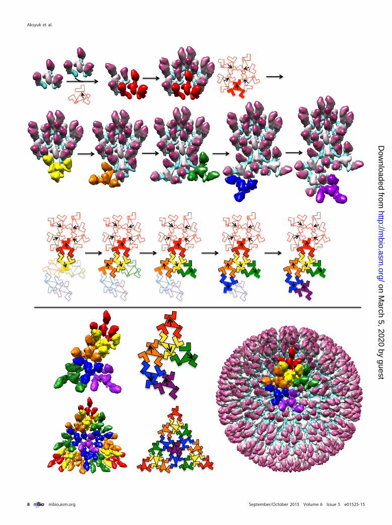
Drawing primarily on observed features of the procapsid, wepropose that the assembly unit (protomer) consists of a triplexsurrounded by three MCP subunits and that the T�16 procapsidshell is built from 320 copies of this protomer (Fig. 7). To date,such a protomer has not been isolated outside the context of acapsid. Each icosahedral facet accommodates 16 protomers of sixquasiequivalent kinds: three each for Ta to Te and one for Tf. (Wename the protomers according to the triplexes that they contain.)Despite little sequence similarity, we infer that there is enoughstructural similarity between UL38 and UL18 that both proteinscan bind an MCP subunit (hence, three per triplex) but also suf-ficient difference between them for the protomers to depart sig-nificantly from 3-fold symmetry. Strikingly, four protomers (Ta,Tb, Tc, and Td) are essentially superimposable (they align withpairwise correlations of �0.95 [see Fig. S2 in the supplementalmaterial]). As the Te protomer includes an MCP that is part of the2-fold-symmetrized E-hexamer, it is also compromised by the ap-plication of icosahedral symmetry.
We envisage that procapsid assembly starts with five protomersclustering with Ta directionality around a 5-fold axis (Fig. 7, firstrow). The next step involves adding a ring of protomers orientedso as to have Tc triplexes, as shown in Fig. 7 (second row, leftmostpanel). Due to its pseudo-3-fold nature, there are three different
settings in which a Tc protomer could be added. On examining theprocapsid structure, we noticed that one of these possibilities sup-ports more MCP-MCP interactions between the two interactingprotomers (Fig. 8). We suppose that it may be selected on thatbasis. Continuing, the rest of the asymmetric unit can be built upin similar fashion (Fig. 7, second row, second to fifth panels). Thisoutgrowth to complete an asymmetric unit, starting from an ini-tial pentamer of protomers, is illustrated schematically in Fig. 7,third row. Further addition of appropriately oriented protomersleads to a complete procapsid (Fig. 7, fourth row). The essentialfeatures of this model are that it explains both the observed direc-tionality pattern of the triplexes and the skewed/distorted natureof MCP hexamers. Also noteworthy is that each of the three MCPsubunits in a given protomer becomes incorporated into a differ-ent capsomer (hexamer or pentamer) in the nascent procapsid.
In the mature capsid, the four protomer-equivalents (a triplexplus three MCPs) that can be distinguished without compromisefrom imposed icosahedral symmetry are markedly different fromeach other (see Fig. S2 in the supplemental material). These dif-ferences come about when the protruding MCP towers (middleplus tip domains) of a given protomer move away from their tri-plex, and six or five of these protrusions (from different protom-ers) cluster into hexameric or pentameric rings, respectively.
Generalization of the model to accommodate a scaffold and aportal. Thus far, we have not considered the internal scaffold. Ithas been observed that the inner (scaffold) and outer (MCP/tri-plex) shells coassemble approximately in unison, i.e., incompleteparticles appear to have about the same amount of inner and outershell (24). This suggests that the scaffolding shell guides the cur-vature of the growing procapsid via a network of interactions be-tween the MCP floor domains and the underlying scaffolding sub-units. That these interactions are weak is suggested by thenonregistration of the inner and outer shells (Fig. 6 and relatedDiscussion). Transient oligomers have been shown to play a rolein the assembly of other capsids (38, 39). The HSV-1 procapsidhas 960 MCP subunits. The most recent value reported for thecopy number of the scaffolding proteins is 1,918 � 170 (10).Given evidence that the scaffolding protein forms dimers (40),this would be consistent with one dimer per MCP subunit. Thus,one could imagine expanding the protomer by adding three scaf-folding protein dimers, one to each MCP. (There are also indica-tions that the scaffolding protein forms a complex with MCP at anearlier stage of assembly in the cytoplasm and escorts it into thenucleus [41].) We suspect that the ~80-Å repeat detected in thescaffolding may reflect the average spacing between protomers atthat radial level in the inner shell of the procapsid.
In herpesviruses and dsDNA phages alike, the procapsid has adodecameric ring at the portal vertex. Although the HSV-1 portalprotein UL6 is not essential for assembly (hence, a procapsid lack-ing a portal is possible), it is likely to be involved in initiatingprocapsid assembly (42). In the absence of a specific nucleus, suchas the portal, several starting points of assembly are possible, butsome of these might lead to geometric barriers, resulting in a fail-ure to incorporate the next protomer. The presence of portalmight serve a nucleus that provides a direction for growth, limit-ing other nonproductive assemblies. Despite the absence of directexperimental evidence, it is nevertheless noteworthy that simplyomitting one MCP subunit from each of five Ta protomers andorganizing these five reduced protomers around the portal ringwould afford a nucleation complex from which outgrowth of the
Assembly of Herpes Simplex Virus Procapsid
September/October 2015 Volume 6 Issue 5 e01525-15 ® mbio.asm.org 7
on March 5, 2020 by guest
http://mbio.asm
.org/D
ownloaded from
Aksyuk et al.
8 ® mbio.asm.org September/October 2015 Volume 6 Issue 5 e01525-15
on March 5, 2020 by guest
http://mbio.asm
.org/D
ownloaded from

procapsid surface lattice could then proceed as outlined above. Inthis complex, the portal ring would essentially replace an MCPpentamer (see Fig. S3 in the supplemental material).
MATERIALS AND METHODSPropagation of mutant virus. Stocks of M100 virus (a gift of M. Gao,Bristol-Myers Squibb) were generated by infecting monolayers of thecomplementary cell line F3 (a gift of P. Desai, Johns Hopkins University).F3 monolayers were grown to 75% confluency and infected at a multiplic-ity of infection (MOI) of 0.1 PFU/cell. At 36 h postinfection, infected-cellmedium was clarified and mixed with polyethylene glycol 8000 (PEG8000) and NaCl to give final concentrations of 7% and 2.3%, respectively.After gentle overnight stirring at 4°C, the PEG precipitate was pelleted andresuspended in small volumes of PBS, aliquoted, and stored frozen at�80°C. The titers of these stocks were determined on both complemen-tary F3 cells and noncomplementary Vero cells. Stocks having a titer ratio
of 104-fold or greater between the two cell types were used to produceprocapsids.
Procapsid production. One 75-cm2 flask of Vero cells was infectedwith M100 stock at an MOI of 5 PFU/cell. Attachment was allowed to takeplace for 45 min at room temperature. Unattached virus was then re-moved by washing the monolayer with 10 ml of PBS and replacing it with15 ml of overlay medium (minimum essential medium [MEM] supple-mented with 1% fetal bovine serum [FBS] and penicillin-streptomycin[Pen-Strep]). Infection was allowed to proceed for 12 to 14 hours at 37°C,at which time the infected monolayer was scraped off and pelleted at~200 � g in a clinical centrifuge for 5 min at room temperature. Beyondthis point, the entire procedure was conducted at room temperature, us-ing phosphate-buffered saline (PBS) supplemented with complete pro-tease inhibitor plus EDTA (Roche Diagnostics). The pellet was resus-pended in 10 ml of PBS, repelleted, and then gently resuspended in 1.0 mlof PBS and transferred to a 1.5-ml microcentrifuge tube. One hundredmilliliters of 10% Triton X-100 was added to the cell suspension, mixed byinversion, and incubated for 20 min, promoting the release of procapsidsfrom infected cell nuclei. After nuclei were removed by centrifugation at850 � g for 4 min, the supernatant was transferred to a 1.5-ml microcen-trifuge tube. Procapsids were purified in four pelleting steps in an Eppen-dorf microcentrifuge (model 5424), followed by use of a spin filter. Thefirst pelleting step was at 5,000 rpm for 4 min. After the supernatant wastransferred to a new tube, the tube containing the pellet was inverted,allowed to drain, and then gently resuspended with 100 �l of PBS. Thesupernatant from this step was recentrifuged at 6,000 rpm for 5 min: thissupernatant was transferred to a fresh tube, and the pellet was drained andresuspended as in the previous step. This process was repeated two moretimes for speeds of 8,000 rpm and 10,000 rpm. Pellets that appeared tur-quoise were combined, sonicated in a bath sonicator (Branson; modelHD-50) for 1 s, and centrifuged at 845 � g for 3 min to remove aggregates.This supernatant was centrifuged in a 300,000-molecular-weight (MW)-cutoff Nanosep centrifugal filter (Pall) at 5,000 rpm until the volume wasreduced to 50 �l. Three hundred microliters of PBS were added to the spinfilter, and the sample was recentrifuged until the volume was reduced to50 �l. This, the final procapsid preparation, was then processed forcryo-EM within 18 to 20 hours postinfection.
Cryo-electron microscopy. Typically, a 3-�l drop of sample was ap-plied to a glow-discharged Quantifoil holey grid which had been overlaidwith a thin layer of carbon, incubated for 40 s, and then blotted andflash-frozen using a Leica EM GP PlungeFreezer. About 12,000 micro-graphs were collected on a Titan Krios microscope operated at 300 kV atthe FEI Nanoport (Acht, Netherlands), using EPU automation software tooperate a Falcon II camera. Images were recorded at 2.3 Å/pixel. The finalset of particles was picked manually from 9,945 micrographs.
Image processing and reconstruction. Boxing, defocus determina-tion, and computational processing were carried out using the Bsoft pack-age (43). The computational selection and classification procedures uti-lized were based on correlation coefficients calculated in reciprocal spacebetween fast Fourier transforms (FFTs) of the images and reference mapprojections. Seventeen previously reported intermediates (11) were usedas initial reference maps. Thereafter, newly calculated reconstructionswere used as references for the next cycle. The particles were allowed toundergo redistribution in successive cycles. After the first three iterations,it was observed that only 10 out of 17 classes contained more than 300particles. Accordingly, the other seven reference maps were omitted and
FIG 7 An assembly pathway for the HSV-1 procapsid. The top panel shows assembly of the procapsid asymmetric unit from protomers, each consisting of 3MCPs surrounding a triplex. The final asymmetric unit consists of six protomers, each colored according to the respective triplex, as in Fig. 4. Ta, red; Tb, orange;Tc, yellow; Td, green; Te, blue; Tf, magenta. The pathway shown starts with five protomers associating around a 5-fold axis. The orientations of the triplexes areshown on the schematic (bottom row) with arrows, as in Fig. 4. The schematic illustrates the sequential addition of protomers, as shown in the surface renderings(above). Upon addition of each protomer, two new MCP-MCP contacts are formed and highlighted as black outlines on the schematic. The colors in theschematic correspond to the density colors shown as surface rendering. The lower panel shows the asymmetric unit, the icosahedral triangle, and the outer surfaceof the complete procapsid. The sequence illustrated here is not exclusive; some other starting assemblies and sequences of protomer addition could also lead intoproductive assembly pathways.
FIG 8 Directionality of triplex association. Two triplex-MCP protomers—Taand Tc—are shown, associating in three different ways. The Ta-Tc association,observed in the density maps, is shown on the left; two alternative 3-fold-related positions are shown in the middle and on the right. Middle row, cut-away views, showing MCP floor domain associations. The arrows indicateinteractions made between the neighboring MCPs. The arrangement (at left)best supports the formation of new MCP-MCP contacts. Bottom row, sche-matic diagram of the three potential modes of interaction.
Assembly of Herpes Simplex Virus Procapsid
September/October 2015 Volume 6 Issue 5 e01525-15 ® mbio.asm.org 9
on March 5, 2020 by guest
http://mbio.asm
.org/D
ownloaded from

particles from these classes were allowed to undergo redistribution amongthe remaining 10 reference maps (classes). After 10 cycles of reconstruc-tion and refinement, the resolutions of 5 out of 10 reconstructions hadsignificantly improved, whereas the resolutions of the other five classeswere still limited to 20 Å. Thus, the number of references was furtherreduced to five, and the selection criteria allowed only 50% of the top-scoring particles, as ranked by correlation coefficients, to be used in thereconstructions. The resolutions of the five reconstructions (Table 1) werecalculated using the Fourier shell correlation criterion with a thresholdof 0.5.
Structure fitting and figure preparation. HK97 capsid protein struc-tures as found in prohead I (PDB 3QPR) and head II (PDB 2FT1) werefitted into maps 1 and 5, respectively, using UCSF Chimera (44). SEG-GER, implemented in UCSF Chimera, was used for map segmentation.Figures were prepared using UCSF Chimera.
Cryo-electron tomography. Grids prepared as described above wereimaged in an FEI Titan Krios microscope operated at 300 kV. Tilt serieswere collected using FEI Tomography 4 operated in batch mode at�29,000 magnification, giving a final pixel size of 0.96 nm after 2-foldbinning. In each tilt series, images were recorded at 1.5° increments overan angular range of approximately �55° to �55°. The electron dose was~1 e�/Å2 per image, for a cumulative dose of ~70 e�/Å2 per tilt series. Tiltseries target defocus ranged from �4 �m to �8 �m, corresponding tofirst contrast transfer function zeros from (28 Å)�1 to (40 Å)�1. Tomo-gram reconstruction was done using IMOD (45). Subtomogram align-ment and averaging used Bsoft routines (43), modified as needed andwrapped into Python scripts. The top 75% of subvolumes, ranked accord-ing to cross-correlation coefficient, were used to calculate the final densitymap of the scaffolding core. Although classification of the particles wasattempted, limited resolution and particle numbers made it difficult toachieve a stable result, and the averaged structure shown represents asuperposition of all classes.
SUPPLEMENTAL MATERIALSupplemental material for this article may be found at http://mbio.asm.org/lookup/suppl/doi:10.1128/mBio.01525-15/-/DCSupplemental.
Figure S1, PDF file, 0.7 MB.Figure S2, PDF file, 0.1 MB.Figure S3, PDF file, 0.2 MB.Movie S1, MOV file, 7.9 MB.
ACKNOWLEDGMENTS
We thank Kasim Sader of the FEI Nanoport facility in Acht, Netherlands,for data collection and James Conway for helpful discussions.
This work was supported by the Intramural Research Program ofNIAMS, with additional support from an NIGMS PRAT fellowship (toA.A.A.).
REFERENCES1. Grünewald K, Desai P, Winkler DC, Heymann JB, Belnap DM,
Baumeister W, Steven AC. 2003. Three-dimensional structure of herpessimplex virus from cryo-electron tomography. Science 302:1396 –1398.http://dx.doi.org/10.1126/science.1090284.
2. Mettenleiter TC, Klupp BG, Granzow H. 2009. Herpesvirus assembly: anupdate. Virus Res 143:222–234. http://dx.doi .org/10.1016/j.virusres.2009.03.018.
3. Fokine A, Rossmann MG. 2014. Molecular architecture of tailed double-stranded DNA phages. Bacteriophage 4:e28281. http://dx.doi.org/10.4161/bact.28281.
4. Hendrix RW, Johnson JE. 2012. Bacteriophage HK97 capsid assemblyand maturation. Adv Exp Med Biol 726:351–363. http://dx.doi.org/10.1007/978-1-4614-0980-9_15.
5. Rixon FJ, Schmid MF. 2014. Structural similarities in DNA packagingand delivery apparatuses in herpesvirus and dsDNA bacteriophages. CurrOpin Virol 5:105–110. http://dx.doi.org/10.1016/j.coviro.2014.02.003.
6. Steven AC, Heymann JB, Cheng N, Trus BL, Conway JF. 2005. Virusmaturation: dynamics and mechanism of a stabilizing structural transi-
tion that leads to infectivity. Curr Opin Struct Biol 15:227–236. http://dx.doi.org/10.1016/j.sbi.2005.03.008.
7. Steven AC, Spear PG. 1997. Herpesvirus capsid assembly and envelop-ment, p 312–351. In Chiu W, Burnett RM, Garcea RL (ed), Structuralbiology of viruses. Oxford University Press, New York, NY.
8. Newcomb WW, Cockrell SK, Homa FL, Brown JC. 2009. Polarized DNAejection from the herpesvirus capsid. J Mol Biol 392:885– 894. http://dx.doi.org/10.1016/j.jmb.2009.07.052.
9. Trus BL, Booy FP, Newcomb WW, Brown JC, Homa FL, Thomsen DR,Steven AC. 1996. The herpes simplex virus procapsid: structure, confor-mational changes upon maturation, and roles of the triplex proteinsVP19c and VP23 in assembly. J Mol Biol 263:447– 462. http://dx.doi.org/10.1016/S0022-2836(96)80018-0.
10. Newcomb WW, Trus BL, Cheng N, Steven AC, Sheaffer AK, TenneyDJ, Weller SK, Brown JC. 2000. Isolation of herpes simplex virus procap-sids from cells infected with a protease-deficient mutant virus. J Virol74:1663–1673. http://dx.doi.org/10.1128/JVI.74.4.1663-1673.2000.
11. Heymann JB, Cheng N, Newcomb WW, Trus BL, Brown JC, Steven AC.2003. Dynamics of herpes simplex virus capsid maturation visualized bytime-lapse cryo-electron microscopy. Nat Struct Biol 10:334 –341. http://dx.doi.org/10.1038/nsb922.
12. Lander GC, Evilevitch A, Jeembaeva M, Potter CS, Carragher B, John-son JE. 2008. Bacteriophage lambda stabilization by auxiliary proteingpD: timing, location, and mechanism of attachment determined by cryo-EM. Structure 16:1399 –1406. http://dx.doi.org/10.1016/j.str.2008.05.016.
13. Yang F, Forrer P, Dauter Z, Conway JF, Cheng N, Cerritelli ME, StevenAC, Plückthün A, Wlodawer A. 2000. Novel fold and capsid-bindingproperties of the lambda-phage display platform protein gpD. Nat StructBiol 7:230 –237. http://dx.doi.org/10.1038/73347.
14. Ishii T, Yamaguchi Y, Yanagida M. 1978. Binding of the structuralprotein soc to the head shell of bacteriophage T4. J Mol Biol 120:533–544.http://dx.doi.org/10.1016/0022-2836(78)90352-2.
15. Qin L, Fokine A, O’Donnell E, Rao VB, Rossmann MG. 2010. Structureof the small outer capsid protein, Soc: a clamp for stabilizing capsids ofT4-like phages. J Mol Biol 395:728 –741. http://dx.doi.org/10.1016/j.jmb.2009.10.007.
16. Steven AC, Greenstone HL, Booy FP, Black LW, Ross PD. 1992. Con-formational changes of a viral capsid protein. Thermodynamic rationalefor proteolytic regulation of bacteriophage T4 capsid expansion, co-operativity, and super-stabilization by soc binding. J Mol Biol 228:870 – 884. http://dx.doi.org/10.1016/0022-2836(92)90871-G.
17. Dai X, Gong D, Wu TT, Sun R, Zhou ZH. 2014. Organization ofcapsid-associated tegument components in Kaposi’s sarcoma-associatedherpesvirus. J Virol 88:12694 –12702. http://dx.doi.org/10.1128/JVI.01509-14.
18. Homa FL, Huffman JB, Toropova K, Lopez HR, Makhov AM, ConwayJF. 2013. Structure of the pseudorabies virus capsid: comparison withherpes simplex virus type 1 and differential binding of essential minorproteins. J Mol Biol 425:3415–3428. http://dx.doi.org/10.1016/j.jmb.2013.06.034.
19. Zhou ZH, Hui WH, Shah S, Jih J, O’Connor CM, Sherman MB, KedesDH, Schein S. 2014. Four levels of hierarchical organization, includingnoncovalent chainmail, brace the mature tumor herpesvirus capsidagainst pressurization. Structure 22:1385–1398. http://dx.doi.org/10.1016/j.str.2014.05.019.
20. Cheng N, Trus BL, Belnap DM, Newcomb WW, Brown JC, Steven AC.2002. Handedness of the herpes simplex virus capsid and procapsid. JVirol 76:7855–7859.
21. Tatman JD, Preston VG, Nicholson P, Elliott RM, Rixon FJ. 1994.Assembly of herpes simplex virus type 1 capsids using a panel of recom-binant baculoviruses. J Gen Virol 75:1101–1113. http://dx.doi.org/10.1099/0022-1317-75-5-1101.
22. Thomsen DR, Roof LL, Homa FL. 1994. Assembly of herpes simplexvirus (HSV) intermediate capsids in insect cells infected with recombinantbaculoviruses expressing HSV capsid proteins. J Virol 68:2442–2457.
23. Gao M, Matusick-Kumar L, Hurlburt W, DiTusa SF, Newcomb WW,Brown JC, McCann PJ, Deckman I, Colonno RJ. 1994. The protease ofherpes simplex virus type 1 is essential for functional capsid formation andviral growth. J Virol 68:3702–3712.
24. Newcomb WW, Homa FL, Thomsen DR, Booy FP, Trus BL, Steven AC,Spencer JV, Brown JC. 1996. Assembly of the herpes simplex virus capsid:characterization of intermediates observed during cell-free capsid forma-tion. J Mol Biol 263:432– 446. http://dx.doi.org/10.1006/jmbi.1996.0587.
Aksyuk et al.
10 ® mbio.asm.org September/October 2015 Volume 6 Issue 5 e01525-15
on March 5, 2020 by guest
http://mbio.asm
.org/D
ownloaded from

25. Heymann JB, Conway JF, Steven AC. 2004. Molecular dynamics ofprotein complexes from four-dimensional cryo-electron microscopy. JStruct Biol 147:291–301. http://dx.doi.org/10.1016/j.jsb.2004.02.006.
26. Häuser R, Blasche S, Dokland T, Haggård-Ljungquist E, von Brunn A,Salas M, Casjens S, Molineux I, Uetz P. 2012. Bacteriophage protein-protein interactions. Adv Virus Res 83:219 –298. http://dx.doi.org/10.1016/B978-0-12-394438-2.00006-2.
27. Wikoff WR, Liljas L, Duda RL, Tsuruta H, Hendrix RW, Johnson JE.2000. Topologically linked protein rings in the bacteriophage HK97caps id . Science 289:2129 –2133. http://dx.doi .org/10.1126/science.289.5487.2129.
28. Fokine A, Leiman PG, Shneider MM, Ahvazi B, Boeshans KM, StevenAC, Black LW, Mesyanzhinov VV, Rossmann MG. 2005. Structural andfunctional similarities between the capsid proteins of bacteriophages T4and HK97 point to a common ancestry. Proc Natl Acad Sci USA 102:7163–7168. http://dx.doi.org/10.1073/pnas.0502164102.
29. Chen DH, Baker ML, Hryc CF, DiMaio F, Jakana J, Wu W, DoughertyM, Haase-Pettingell C, Schmid MF, Jiang W, Baker D, King JA, ChiuW. 2011. Structural basis for scaffolding-mediated assembly and matura-tion of a dsDNA virus. Proc Natl Acad Sci USA 108:1355–1360. http://dx.doi.org/10.1073/pnas.1015739108.
30. Cardone G, Heymann JB, Cheng N, Trus BL, Steven AC. 2012. Procap-sid assembly, maturation, nuclear exit: dynamic steps in the production ofinfectious herpesvirions, p 423– 439. In Rossmann MG, Rao V (ed), Viralmolecular machines. Springer, New York, NY.
31. Bowman BR, Baker ML, Rixon FJ, Chiu W, Quiocho FA. 2003. Struc-ture of the herpesvirus major capsid protein. EMBO J 22:757–765. http://dx.doi.org/10.1093/emboj/cdg086.
32. Conway JF, Wikoff WR, Cheng N, Duda RL, Hendrix RW, Johnson JE,Steven AC. 2001. Virus maturation involving large subunit rotations andlocal refolding. Science 292:744 –748. http://dx.doi.org/10.1126/science.1058069.
33. Spencer JV, Newcomb WW, Thomsen DR, Homa FL, Brown JC. 1998.Assembly of the herpes simplex virus capsid: preformed triplexes bind tothe nascent capsid. J Virol 72:3944 –3951.
34. Fane BA, Prevelige PE, Jr. 2003. Mechanism of scaffolding-assisted viralassembly. Adv Protein Chem 64:259 –299. http://dx.doi.org/10.1016/S0065-3233(03)01007-6.
35. Baines JD. 2011. Herpes simplex virus capsid assembly and DNA
packaging: a present and future antiviral drug target. Trends Microbiol19:606 – 613. http://dx.doi.org/10.1016/j.tim.2011.09.001.
36. Oh B, Moyer CL, Hendrix RW, Duda RL. 2014. The delta domain of theHK97 major capsid protein is essential for assembly. Virology 456-457:171–178. http://dx.doi.org/10.1016/j.virol.2014.03.022.
37. Xie Z, Hendrix RW. 1995. Assembly in vitro of bacteriophage HK97proheads. J Mol Biol 253:74 – 85. http://dx.doi.org/10.1006/jmbi.1995.0537.
38. Johnson JM, Tang J, Nyame Y, Willits D, Young MJ, Zlotnick A. 2005.Regulating self-assembly of spherical oligomers. Nano Lett 5:765–770.http://dx.doi.org/10.1021/nl050274q.
39. Tresset G, Decouche V, Bryche JF, Charpilienne A, Le Coeur C, Barbier C,Squires G, Zeghal M, Poncet D, Bressanelli S. 2013. Unusual self-assemblyproperties of norovirus Newbury2 virus-like particles. Arch Biochem Biophys537:144765–152. http://dx.doi.org/10.1016/j.abb.2013.07.003.
40. Pelletier A, Dô F, Brisebois JJ, Lagacé L, Cordingley MG. 1997. Self-association of herpes simplex virus type 1 ICP35 is via coiled-coil interac-tions and promotes stable interaction with the major capsid protein. JVirol 71:5197–5208.
41. Nicholson P, Addison C, Cross AM, Kennard J, Preston VG, Rixon FJ.1994. Localization of the herpes simplex virus type 1 major capsid proteinVP5 to the cell nucleus requires the abundant scaffolding protein VP22a. JGen Virol 75:1091–1099. http://dx.doi.org/10.1099/0022-1317-75-5-1091.
42. Newcomb WW, Thomsen DR, Homa FL, Brown JC. 2003. Assembly ofthe herpes simplex virus capsid: identification of soluble scaffold-portalcomplexes and their role in formation of portal-containing capsids. J Virol77:9862–9871. http://dx.doi.org/10.1128/JVI.77.18.9862-9871.2003.
43. Heymann JB, Belnap DM. 2007. Bsoft: image processing and molecularmodeling for electron microscopy. J Struct Biol 157:3–18. http://dx.doi.org/10.1016/j.jsb.2006.06.006.
44. Pettersen EF, Goddard TD, Huang CC, Couch GS, Greenblatt DM,Meng EC, Ferrin TE. 2004. UCSF Chimera—a visualization system forexploratory research and analysis. J Comput Chem 25:1605–1612. http://dx.doi.org/10.1002/jcc.20084.
45. Kremer JR, Mastronarde DN, McIntosh JR. 1996. Computer visualiza-tion of three-dimensional image data using IMOD. J Struct Biol 116:71–76. http://dx.doi.org/10.1006/jsbi.1996.0013.
Assembly of Herpes Simplex Virus Procapsid
September/October 2015 Volume 6 Issue 5 e01525-15 ® mbio.asm.org 11
on March 5, 2020 by guest
http://mbio.asm
.org/D
ownloaded from