subcylindraceae vel obclavatae, hyalinae, laeves, … · subcylindraceae vel obclavatae, hyalinae,...
TRANSCRIPT
subcylindraceae vel obclavatae, hyalinae, laeves, 6.5-10.5 X 3-5 [i = 8.4 X 3.9] J,Un, usque ad ter prolificantes.Conidia elongatoellipsoidea vel obovata, extremitatibusaliquantum rotundatis, dictyoseptata,hyalina. tenuitunicata,laevia, 30-39(-40) X 11-19(-20) [i = 34.5 X 15] J,Un; appendices mucosae, initio irregulares, tandemtentaculiformes; ratione conidii long./lat = 2.3:1.
Foliicolous. Conidiomata pycnidial, hypophyllous, scattered, deeply immersed in tbé host mesophyll with on1ythe ostiole visible in surface view, globose to subglobose in sectional view, 140-160 J,Un wide, 190-250 J,Undeep, unilocular, glabrous, ostiolate; wall up to 25 J,Un thick, of colourless textura angularis in the outermostlayer and of colourless textura prismatica in the inner layers, but around the ostiolar region of almost colourlessto dark brown, thick, lenticular textura angularis or textura globulosa with thick-walled cells; ostiole circularor oval, 30-40 J.Un diam., neck canal lined with unbranched or branched, sparsely septate, smooth-walled,recurved periphyses 10-17 X 1-2.5 J.Un. Conidiophores arising all around the cavity of the conidioma, sparselyseptate, branched on1y at the base and 15-22 J.Un long, or often reduced to conidiogenous cells, invested inmucus. Conidiogenous cells subcylindrical to obclavate, colourless, smooth-walled, 6.5-10.5 X 3-5 [i = 8.4 X3.9] J,Un, with up to 3 prolíferations. Conidia elongate-ellipsoid to obovoid with somewhat rounded ends,dictyoseptate, colourless, thin- and smooth-walled, 30-39(-40) X 11-19(-20) [i = 34.5 X 15] um: appendagesinitially irregular, eventually tentaculiform; mean conidium length/width ratio = 2.3:1.
Habitat: On leaf of Litsea sp.Specimen examined: DAOM 215257 [Holotype], Somwarpet, Coorg, India, 30.VI.1962, T.R.Nag Raj.Known distribution: India.The fungus is associated with leaf spots.
137. ZELANDIOCOELA Nag Raj anam.-gen. novoConidiomata stromatica, pycnidioidea, initio immersa, deinde erumpentia, irregulatim plurilocularia loculis
convolutis, gIabra, atrobrunnea vel nigra, initio clausa, postremo per aliquot foramina circularia vel ovaliaparietis apicalis dehiscentia, gIobulo viscido veI cirrho conidiorum albido vel ebumeo velam; paries e texturaangulari celIulis crassitunicatis, brunneis vel atrobrunneis compositus. Conidiophora circum cavitatem loculorumenascentia, ad cellulas conidiogenas redacta, in muco involuta. CelIulae conidiogenae discretae, subcylindraceaevel late conicae, hyalinae, laeves. Conidiogenesis: ontogenea holoblastica; maturatio conidiorum cum ontogeneasynchrona; delimitatio per septum duplex; secessio schizolytica; proliferatio cellularum conidiogenarumenteroblastica, conidia plura ad locos eosdem aut altiores successive producens; cellulae conidiogenae in zonacollaruli crassitiebus periclinalibus notatis praeditae; regeneratio cellularum conidiogenarum nulla Conidiafusiformia, extremitatibus aliquantum apiculatis, unicellularia, hyalina, tenuitunicata, laevia, utrinque appendicesmucosas ínfundibuliformes vel irregulares ferentia; appendices per gelatisationem zonarum certarum parietis adpolos conidiorum crescentium orientes.
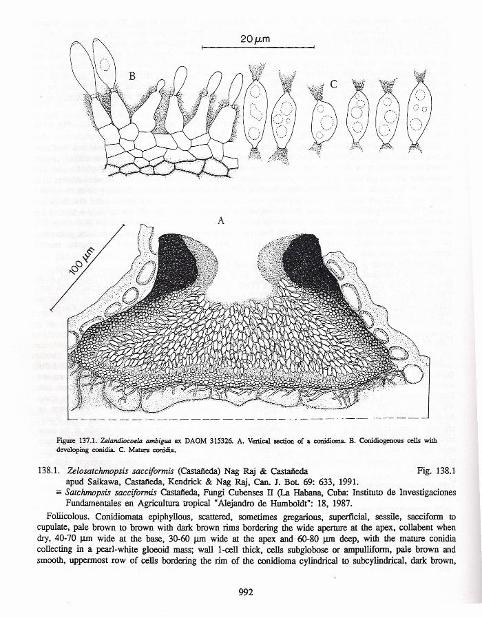
Conidiomata stromatic, pycnidioid, immersed, eventually becoming erumpent, irregularly plurilocular withconvoIuted Iocules, glabrous, dark brown to black, initially closed, ultimately dehiscing by severa! circular tooval openings in the apical wall and covered with a pearl white or cream, gIoeoid gIobule or conidial cirrhi;wall of textura angularis with thick-walled, brown to dark brown cells. Conidiophores lining the locules, reducedto conidiogenous cells and invested in mucus. Conidiogenous cells discrete, subcylindrical to broadly conical,colourless, smooth. Conidiogenesis: ontogeny by apical wall building in the first conidium and by repIacementwall-building in subsequent conidia; maturation by diffuse wall-building synchronous with ontogeny; delimitationby a doubIe septum; secession schizolytic; proliferation of conidiogenous celI enteroblastic-percurrent to produceadditional conidia at the same or slight1yhigher leveI; conidiogenous cells with marked periclinal thickeningsin the collarette zone; regeneration of conidiogenous cells absent Conidia fusiform with somewhat apiculateends, unicellular, colourless, thin-walled, smooth, with funnel-shaped to irregular, mucoid appendages of typeH at both ends, arising by gelatinization of appendage primordia at the poles of the developing conidiaType anamorph-species: Zelandiocoela ambigua (Nag Raj & Kendrick) Nag Raj.TeIeomorph: Unknown.
Zelandiocoela, with its bipolar mucoid conidium appendages, is likeIy to be confused with severalother coelomycete genera. The conidium appendages in Allantophomopsis Petrak, Hymenopsis Saccardo, and
990

Koorchaloma Subramanian are of type C, while the appendages in Mirimyces Nag Raj, Tiarospora Saccardo& Marchal, and Zelandiocoela are of type H. In Mirimyces, the appendage arises from a torus of mucusresulting from gelatinization of appendage primordia - convex lens-shaped wall areas, subapica1 in position, anddelirnited at an early stage of conidium development In Tiarospora and Zelandiocoela; the appendage primordiaon the developing conidia are bipolar, conic and obconic in shape. The differences between Tiarospora andZelandiocoela outweigh the similarity. The conidia in Tiarospora are thick-walled, eventually pigmented andl-septate, while in Zelandiocoela the conidia are thin-walled, colourless and unicellular. Another feature presentin Tiarospora, but absent in Zelandiocoela, is the microconidiogenous cells lining the ostiolar channel in theneck region of the conidioma.
137.1. Zelandiocoela ambígua (Nag Raj & Kendrick) Nag Raj comb. novo Fig. 137.1;: Apostrasseria ambigua Nag Raj & Kendrick, Cano J. Bot 61: 15, 1983.
Foliicolous. Conidiomata stromatic, pycnidioid, amphigenous, scattered to gregarious and confluent,intra-epidermal in origin, immersed, eventually becoming erumpent, elongate-oval to irregular in outline, broadlyconical in section, up to 300 JlII1 long, 170-230 JlII1 wide and 90-140 JlII1 deep, irregularly plurilocular withconvoluted locules, glabrous, dark brown to black, initially c1osed, ultimately with one to three, circular to oval,openings in the apical wall, 30-40 JlII1 diam., covered with a pearl white or cream, gloeoid globule or conidialcirrhi; wall 15-20 JlII1 thick, of textura angularis with thick-walled, brown to dark brown cells. Conidiophoresreduced to conidiogenous cells, lining the locules, and invested in mucus. Conidiogenous cells subcylindricalto broadly conica1, most1y colourless, occasionally darker in the lower part, smooth, 5-8 X 2-4 [x = 6.5 X 2.9]JlII1, with two to three proliferations. Conidia fusiform with somewhat apiculate ends, unicellular, colourless,thin- and smooth-walled, 8.5-14 X 3-4.5 [x = 11.5 X 3.9J J.1I11, with funnel-shaped to irregular, mucoidappendages at both ends.
Habitat: On leaves of Podocarpus hallü, P. totara and Podocarpus sp. in litter.Specimens examined: 1. PDD 41572 [Holotype], on P. totara, Forest Hill Scenic Reserve, 18 mi. N of
Invercargill, New Zealand, 18.N.1974, B.Kendrick (KNZ 707); 2. DAOM 215263, on Podocarpus sp. in litter,Tunnel entrance, Milford, New Zealand, I.N.1980, B.Kendrick; 3. DAOM 315326, on leaves of Podocarpushallii, Governor's Bush Track, Mt Cook National Park, South Island, New Zealand, 3lY.1980, B.Kendrick.
Known distribution: New Zealand.
138. ZEWSATCHMOPSIS Nag Rajapud Saikawa, Castai'ieda, Kendrick & Nag Raj, Cano J. Bot 69: 633, 1991.
Conidiomata sacciform or cupulate, superficial, sessile, unilocular, brown to dark brown; periclinal wall l-cellthick, cells pale brown, smooth, and each cell (except for the top two to three rows of dark brown andsornewhat encrusted cells bordering the wide open apical end) functioning as a conidiogenous cell.Conidiogenous cells discrete, ampulliform, pale brown, smooth. Conidiogenesis: ontogeny holoblastic by apicalwall-building in the f1I'St conidium and by replacement wall-building in subsequent conidia; maturation bymoderate diffuse wall-building asynchronous with ontogeny or somewhat delayed; delirnitation by a doubleseptum; secession schizolytic; proliferation enteroblastic-percurrent to produce additional conidia at the sameleveI; conidiogenous cells with moderate periclinal thickenings in collarette zone; regeneration of conidiogenouscells absent. Conidia fusiform to falcate, unicellular or euseptate, colourless, smooth-walled, with a cellular,unbranched, attenuated, excentric basal appendage of type A, maintaining protoplasmic continuity with theconidium body.Type anamorph-species: Zelosatchmopsis sacciformis (Castai'ieda) Nag Raj & Castai'ieda.Teleomorph: Unknown.
The genus has remained monotypic.
20J.Lm
Figure 137.1. Zelandiocoela ambigua ex DAOM 315326. A. Vertical section of a conidioma. B. Conidiogenous cells withdeveloping conidia. C. Mature conidia.
138.1. Zelosatchmopsis sacciformis (Castaãeda) Nag Raj & Castaãeda Fig. 138.1apud Saikawa, Castaãeda, Kendrick & Nag Raj, Can. J. Bot 69: 633, 1991.
== Satchmopsis sacciformis Castaãeda, Fungi Cubenses II (La Habana, Cuba: Instituto de InvestigacionesFundamentaIes en Agricultura tropical" Alejandro de Humboldt": 18, 1987.
Foliicolous. Conidiomata epiphyllous, scattered, sometimes gregarious, superficial, sessile, sacciform tocupulate, pale brown to brown with dark brown rims bordering the wide aperture at the apex, collabent whendry, 40-70 J..lIIl wide at the base, 30-60 J..lIIl wide at the apex and 60-80 J..lIIl deep, with the mature conidiacollecting in a pearl-white gloeoid mass; wall l-cell thick, cells subglobose or ampulliform, pale brown andsmooth, uppermost row of cells bordering the rim of the conidioma cylindrical to subcylindrical, dark brown,
992