suberoylanilide hydroxamic acid: a potential epigenetic
TRANSCRIPT

Suberoylanilide hydroxamic acid: a
potential epigenetic therapeutic agent for
lung fibrosis?Z. Wang*, C. Chen*, S.N. Finger#, S. d/o Kwajah M.M#, M. Jung", H. Schwarz#,N. Swanson+, R.R. Lareu*,+,1 and M. Raghunath*,e
ABSTRACT: Pulmonary fibrosis represents a fatal stage of interstitial lung diseases of known and
idiopathic aetiology. No effective therapy is currently available. Based on an indication-discovery
approach we present novel in vitro evidence that the histone deacetylases inhibitor
suberoylanilide hydroxamic acid (SAHA), an FDA approved anti-cancer drug, has antifibrotic
and anti-inflammatory potential.
Human lung fibroblasts (fetal, adult and idiopathic adult pulmonary fibrosis) were treated with
transforming growth factor (TGF)-b1 with or without SAHA. Collagen deposition, a-smooth muscle
actin (a-SMA) expression, matrix metalloproteinase (MMP)1 activity, tissue inhibitor of MMP
(TIMP)1 production, apoptosis and cell proliferation were assessed. Pro-inflammatory cytokines
relevant to pulmonary fibrosis were assayed in SAHA-treated human peripheral blood mono-
nuclear cells (PBMC) and its subpopulations.
SAHA abrogated TGF-b1 effects on all the fibroblast lines by preventing their transdifferentia-
tion into a-SMA positive myofibroblasts and increased collagen deposition without inducing
apoptosis. However, MMP1 activity and TIMP1 production was modulated without a clear
fibrolytic effect. SAHA also inhibited serum-induced proliferation of the fibroblast lines and
caused hyperacetylation of a-tubulin and histone. Cytokine secretion was inhibited from PBMC
and lymphocytes at nonapoptotic concentrations.
Taken together, these data demonstrate combined antifibrotic and anti-inflammatory properties
of SAHA, suggesting its therapeutic potential for pulmonary fibrosis.
KEYWORDS: Antifibrotic, collagen, fibroblast, histone deacetylase inhibitor, pulmonary fibrosis,
suberoylanilide hydroxamic acid
Interstitial lung diseases constitute a diverseset of lung disorders with different levels ofinflammation and fibrosis resulting in irre-
versible loss of lung function and ultimatelyrespiratory failure [1]. The most common repre-sentative of interstitial lung diseases is idiopathicpulmonary fibrosis (IPF). The general pathologi-cal feature of IPF is the excessive deposition ofcollagen I which destroys the architecture of thenormal lung parenchyma [2]. Patients with IPFhave a median survival of 2–3 yrs after diagnosis[1]. The current therapy for IPF is based onreducing inflammation with corticosteroids andimmunosuppressive drugs. However, the appar-ent ineffectiveness of this regimen has led todoubts about the initial role of inflammation inthe pathogenesis of IPF [3, 4] and to the conceptthat correcting the activated state of IPF fibro-blasts/myofibroblasts [5] might be more impor-tant than an anti-inflammatory strategy. New
therapeutic strategies, including antioxidantagents, anti-endothelial cell antibody, anti-cyto-kine therapy and antifibrotic agents, such aspirfenidone, have been evaluated in clinical trialswithout showing significant improvement in thetreatment of IPF [6]. Clearly, IPF remains a largetherapeutic challenge and the development ofeffective antifibrotic drugs is urgently required.
To explore new antifibrotic drugs, we have beeninvestigating the therapeutic potential of histonedeacetylase inhibitors (HDACi). HDACi aresmall organic molecules which change geneexpression profiles at the epigenetic level andchange protein function by inhibiting the activityof histone deacetylases. HDACi are of primeinterest in cancer research because they induceapoptosis and cell cycle arrest in malignant cellsand are anti-angiogenic [7]. Recently publishedstudies suggested that HDACi might also inhibit
AFFILIATIONS
*Tissue Modulation Laboratory,
Division of Bioengineering, Faculty of
Engineering,#Dept of Physiology, National
University of Singapore,1NUS Tissue Engineering Program,
Dept of Orthopedic Surgery,eDept of Biochemistry, Yong Loo Lin
School of Medicine, National
University of Singapore, Singapore,"Institute of Pharmaceutical
Sciences, University of Freiburg,
Freiburg, Germany, and+Molecular Hepatology, School of
Medicine and Pharmacology, Faculty
of Medicine, Dentistry and Heath
Sciences, The University of Western
Australia, Perth, Australia.
CORRESPONDENCE
M. Raghunath
Division of Bioengineering, Faculty of
Engineering, and Dept of
Biochemistry
Yong Loo Lin School of Medicine
Division Office Block E3A #04-15
7 Engineering Dr. 1
Singapore 117574
E-mail: [email protected]
Received:
June 05 2008
Accepted after revision:
Jan 25 2009
First published online
Feb 12 2009
European Respiratory Journal
Print ISSN 0903-1936
Online ISSN 1399-3003
EUROPEAN RESPIRATORY JOURNAL VOLUME 34 NUMBER 1 145
Eur Respir J 2009; 34: 145–155
DOI: 10.1183/09031936.00084808
Copyright�ERS Journals Ltd 2009
c
collagen production in different fibroblast types. Phenyl-butyrate decreased basal levels and transforming growth factor(TGF)-b1-stimulated a1(I) collagen messenger RNA (mRNA)and protein levels in the lung fetal fibroblast IMR-90 [8].Trichostatin A (TSA) was reported to inhibit synthesis ofcollagen type I and III and a-smooth muscle actin (a-SMA)both at the protein and mRNA level in primary cultured rathepatic stellate cells [9], and in TGF-b1-stimulated rat skinfibroblasts [10]. In addition, collagen production from systemicsclerosis fibroblasts was inhibited and total collagen depositionwas reduced in bleomycin-induced skin fibrosis in mice by TSA[11].
Based on an indication-discovery approach, we evaluated suber-oylanilide hydroxamic acid (SAHA) (Zolina1 (vorinostat), anFDA-approved HDACi, which is already in clinical use as athird-line drug for the treatment of cutaneous T-cell lymphomaand under evaluation for other types of cancer [12]. Herein, westudied the antifibrotic potential of SAHA in TGF-b1-treatedfetal lung fibroblasts (FLF), adult lung fibroblasts (ALF) and IPFlung fibroblast (ILF) lines, and its potential anti-inflammatoryeffect in peripheral blood mononuclear cells (PBMC), lympho-cytes and peripheral monocytes.
MATERIALS AND METHODSCompounds and reagentsSAHA was synthesised as described previously [13], dissolvedin dimethyl sulfoxide to 50 mM and stored at -80uC. Allreagents were from Sigma Aldrich (St. Louis, MI, USA) unlessstated otherwise.
Treatment of lung fibroblast linesNormal human FLF IMR-90 (CCL-186; ATCC, Manassas, VA,USA), ALF CCD-19 Lu (CCL-210; ATCC) and ILF LL29 (CCL-134; ATCC) were cultured in 10% fetal bovine serum (FBS)Dulbecco’s modified Eagle medium (GIBCO Invitrogen,Singapore). Cells, except for proliferation assays, were seededat 16104?well-1, 56104?well-1 and 2.56105?well-1 in 96-, 24-and 6-well plates, respectively. After 24 h, fibroblasts weretreated with different concentrations of SAHA with or withoutTGF-b1 (5 ng?mL-1; R&D Systems, Minneapolis, MN, USA) inserum-free Dulbecco’s modified Eagle medium plus30 mg?mL-1 ascorbic acid phosphate (Wako Pure ChemicalsIndustries Ltd, Osaka, Japan) in the presence of 100 mg?mL-1
dextran sulfate 500 kDa (pK Chemicals A/S, Koge, Denmark),for the rapid deposition of collagen I within 24 h [14].
Quantitative immunocytochemistryFibroblasts cultured in 96-well Lumox plates with an opticalbottom (Greiner BioOne, Gottingen, Germany) were fixed withabsolute methanol and air dried for 15 min. Cell layer wasblocked with 3% bovine serum albumin in PBS for 30 minfollowed by incubation with primary antibodies for 90 min.Secondary antibodies and nuclear stain 4,6-diamidino-2-phenylindole (DAPI; Invitrogen Molecular Probes, Carlsbad,CA, USA) were stained for 30 min. The cell layer was washedwith PBS three times after each step. Antibodies againstcollagen type I (1:500), a-SMA (1:100; Dako, Glostrup,Denmark) or acetylated a-tubulin (1:500) were from mouseand acetylated histone 3 (1:200; Upstate Biotechnology, NewYork, NY, USA) from rabbit. Secondary antibodies were AlexaFluor (AF) 594 goat anti-mouse and AF488 goat anti-rabbit
(both 1:400; Invitrogen Molecular Probes). The fluorescenceintensity was quantified using the PHERAstar microplatereader with a focusing lens system (BMG Labtech,Mornington, Australia). The ratio of the fluorescence intensityof the AF dye to DAPI was used to normalise the relativeamount of the antigen to cell number. Images were taken withan Olympus LX71 epifluorescence microscope (Olympus,Tokyo, Japan).
Biochemical assay of collagen matrixDeposited collagen was extracted from cell cultures andanalysed as described previously [14]. Briefly, cell layers in24-well plates were digested in situ with 250 mg?mL-1 porcinegastric mucosa pepsin (Roche, Basel, Switzerland) in 0.1 MHCl for 2 h and neutralised with 1 M NaOH. Extracts werevisualised using SDS-PAGE gels with silver staining andanalysed by densitometry.
Western blotWestern blots were performed as previously described [14].Briefly, proteins were extracted with loading buffer (50 mMTris-Cl pH 6.8, 2% SDS, 0.1% bromophenol blue and 10%glycerol) with 5 mM dithiothreitol and separated on 10% SDS-PAGE gels. Primary antibodies against acetylated a-tubulin(1:2500), a-SMA (1:500) and b-actin (1:1000) were from mouse,and acetylated histone 3 (1:3000; Upstate Biotechnology) fromrabbit. Secondary antibodies were goat anti-mouse and goatanti-rabbit horse radish peroxidase (both 1:1000; Pierce,Rockford, IL, UK). Blots were developed with AmershamTM
ECL plus Western blotting detection system (GE Healthcare,Chalfont St Giles, UK) and chemiluminescence was capturedwith a VersaDoc Imaging System model 5000 (Bio-Rad,Hercules, CA, USA).
Flow cytometry analysis of a-SMA expressionFibroblasts in suspension were fixed with 2% paraformalde-hyde in PBS and permeabilised with 0.1% saponin in PBS for15 min. The suspension was incubated with mouse anti-a-SMA (1:1000) and AF488 chicken anti-mouse (1:100; InvitrogenMolecular Probes), each for 30 min at 4uC. The cells werewashed with PBS twice after each step. AF488 was detected byflow cytometry.
Adherent cytometryFor proliferation assay, cells were seeded at 26104?well-1 in 24-well plate and cultured in 10% FBS in the presence or absenceof 5 mM SAHA. After 3 days, fibroblasts were stained withDAPI after methanol fixation. Nine image sites covering 71% ofthe total well area were acquired at 26magnification using aNikon TE600 fluorescence microscope with an automated Ludlstage (BioPrecision 2; Ludl Electronic Products Ltd,Hawthorne, NY, USA) and analysed using the Metamorph1
Imaging System software (Molecular Devices, Downingtown,PA, USA). A nucleus was defined as a fluorescent region witha length of 10–15 mm and a pixel intensity value of 10 unitsabove background.
Carboxyfluoroscein succinimidyl ester labelling anddetectionFibroblasts at 106?mL-1 were pre-labelled with 2 mM carboxy-fluoroscein succinimidyl ester (CFSE; Invitrogen Molecular
INTERSTITIAL LUNG DISEASE Z. WANG ET AL.
146 VOLUME 34 NUMBER 1 EUROPEAN RESPIRATORY JOURNAL

25m)
p)
20
15
10
5 *
*
*
*
0 5 0 5- - + +
Fold
cha
nge
n)
Fold
cha
nge
o)
Fold
cha
nge
a) b) c) d)
e) f) g) h)
i) j) k) l)
0SAHA µMTGF-β1
SAHA µMTGF-β1
5 0 5- - + +
14
12
10
8
6
4
2
00
*
**
*
0 5 0 5- - + +
10
8
6
4
2
0
*
* *
*
0 5 0 5- - + +
0 5
FLF ALF ILF
0 5- - + +
SAHA µMTGF-β1
Acetylatedhistone 3
Acetylatedα-tubulin
β-actin
0 5 0 5- - + +
0 5 0 5- - + +
FIGURE 1. Suberoylanilide hydroxamic acid (SAHA) induces hyperacetylation of histone and a-tubulin. Fetal lung fibroblasts (FLF), adult lung fibroblasts (ALF) and
idiopathic pulmonary fibrosis lung fibroblasts (ILF) were treated with or without transforming growth factor (TGF)-b1 (5 ng?mL-1) and 5 mM SAHA for 24 h. The acetylation of
histone 3 and a-tubulin were quantified by quantitative immunocytochemistry (QICC) and Western blot. a, b) Immunocytochemistry of acetylation of histone 3 (green) and a-
tubulin (red) in a–d) FLF (IMR-90), e–h) ALF (CCD-19 Lu) and i–l) ILF (LL29). Scale bars550 mm. m–o) Average fold change of acetylation of histone 3 (h) and a-tubulin (&)
from quadruplicates were quantified by QICC. m) FLF, n) ALF and o) ILF. *: p,0.05 compared with untreated control. p) Western blots of acetylated histone 3 and acetylated
a-tubulin.
Z. WANG ET AL. INTERSTITIAL LUNG DISEASE
cEUROPEAN RESPIRATORY JOURNAL VOLUME 34 NUMBER 1 147
Probe). With or without 5 mM SAHA, 16105 cells?well-1 wereseeded into 6-well plate and cultured in 10% FBS. After 3 days,fibroblasts were trypsinised, washed twice with PBS and CFSEwas detected by flow cytometry.
Matrix metalloproteinase 1 activity and tissue inhibitor ofmatrix metalloproteinase 1 ELISAFibroblasts were treated with or without TGF-b1 (5 ng?mL-1)and SAHA at 5 mM for 24 h. Matrix metalloproteinase (MMP)1activity and tissue inhibitor of MMP (TIMP)1 production weredetermined using the Fluorokine E kit and the Quantikine
human TIMP1 kit (both R&D Systems) according to themanufacturers protocol. 4-aminophenylmercuric acetate(APMA)-activated MMP1 was detected by cleavage offluorescence-labelled substrate peptide. Concentrations ofactive MMP1 and TIMP1 were calculated from standardcurves obtained from standards run in parallel.
Apoptosis assayBriefly, 106 fibroblasts, PBMC, lymphocytes or monocytes werestained with 100 mL fluorescein isothiocyanate-annexin V andpropidium iodide (PI) labelling solution for 15 min at room
8e)
6
4
2
0
Fold
cha
nge
a)
b)
c)
TGF-β1
-TG
F-β1
+TG
F-β1
-TG
F-β1
+TG
F-β1
-TG
F-β1
+
0 2.5 5 10
SAHA µM
d) 0 5
FLF ALF ILF
0 5- - + +
0 5 0 5- - + +
0 5 0 5- - + +
SAHA µMTGF-β1
α-SMA
β-actin
*
**
FLF
0 05 510 102.5 2.5- +- +- +- +
10
8
6
4
2
0
*
*
ALF
0 05 510 102.5 2.5- +- +- +- +
20
16
8
12
4
0
*
*
ILF
0 05 510 102.5 2.5- +- +- +- +
SAHAµMTGF-β1
36022.70%
180
0
51130.43%
255
0
31679.30%
158
0
27737.09%
138
0
40912.00%
204
0
4117.24%
205
0
20579.26%
102
0
30735.86%
153
0
717
100101102103104 100101102103104 100101102103104 100101102103104
9.52%358
0
47824.79%
239
0
26274.92%
131
0
40052.59%
200
0
SMA-log
Co
un
ts
FLF
ALF
ILF
f) 0 5 0 5- - + +
SAHA µMTGF-β1
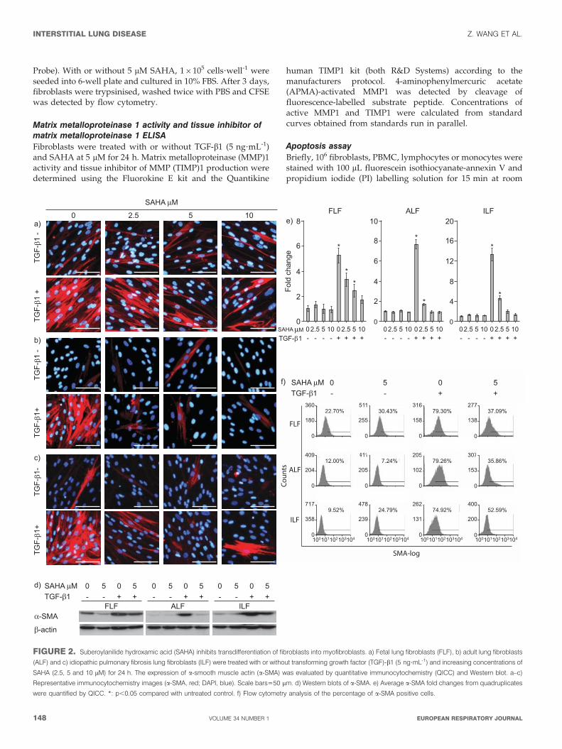
FIGURE 2. Suberoylanilide hydroxamic acid (SAHA) inhibits transdifferentiation of fibroblasts into myofibroblasts. a) Fetal lung fibroblasts (FLF), b) adult lung fibroblasts
(ALF) and c) idiopathic pulmonary fibrosis lung fibroblasts (ILF) were treated with or without transforming growth factor (TGF)-b1 (5 ng?mL-1) and increasing concentrations of
SAHA (2.5, 5 and 10 mM) for 24 h. The expression of a-smooth muscle actin (a-SMA) was evaluated by quantitative immunocytochemistry (QICC) and Western blot. a–c)
Representative immunocytochemistry images (a-SMA, red; DAPI, blue). Scale bars550 mm. d) Western blots of a-SMA. e) Average a-SMA fold changes from quadruplicates
were quantified by QICC. *: p,0.05 compared with untreated control. f) Flow cytometry analysis of the percentage of a-SMA positive cells.
INTERSTITIAL LUNG DISEASE Z. WANG ET AL.
148 VOLUME 34 NUMBER 1 EUROPEAN RESPIRATORY JOURNAL

temperature using Annexin-V-FLUOS staining kit (Roche,Penzberg, Germany) according to the manufacturers protocol.The percentages of annexin-V single positive, annexin-V/PIdouble-positive and annexin-V/PI double-negative cells weredetermined by flow cytometry.
Treatment of PBMC and its subpopulations andcytokines ELISAHuman PBMC were isolated from buffy coats usingHistopaque-1077 (Sigma Aldrich) from blood of healthyvolunteers at the National University Hospital (Singapore)under informed consent and local international review board(National University Hospital) approval. Monocytes werefurther isolated from PBMC with the human MonocyteIsolation Kit II (Miltenyi Biotec, Bergisch Gladbach, Germany).Lymphocytes were isolated as the cells remained on themagnetic column after removal of monocytes. PBMCs, lympo-cytes and monocytes were seeded at 16106?mL-1 in 10% FBSRPMI-1640 and incubated with 0.5 ng?mL-1 phorbol myristateacetate plus 50 nM calcium ionophore A23187 (PMA/CI)alone or with 0.5 or 1 mM SAHA for 24 or 48 h. Supernatantswere collected and frozen at -20uC. The cytokines assayed weretumour necrosis factor (TNF)-a, interleukin (IL)-13, IL-10
(eBioscience, San Diego, CA, USA), IL-6, IL-8 and TGF-b1(R&D Systems) following the manufacturers protocol.
Statistical analysisStatistical analysis was performed using SPSS 12.0. (SPSS Inc.,Chicago, IL,USA)EqualityoferrorvarianceswastestedbyLevene’stest. Data with equal variances were compared by Turkey test anddata with unequal variances by Dunnet’s T3 test. Probability valuesof p,0.05 were accepted as the level of statistical significance.
RESULTSSAHA induces hyperacetylation of histone 3 and a-tubulinBasal acetylation levels of histone and a-tubulin were noted inuntreated FLF, ALF and ILF, but were significantly increasedin the presence of SAHA after 24 h as assessed by quantitativeimmunocytochemistry (QICC; fig. 1a–o) and Western blot(fig. 1p), indicating that SAHA inhibited HDACs activity.TGF-b1 did not change the acetylation of histone 3 and a-tubulin significantly in the presence or absence of SAHA.
SAHA inhibits TGb-1-induced myofibroblasttransdifferentiationUntreated cell cultures of all the fibroblasts lines showed lowlevels of a-SMA expression by immunocytochemistry (fig. 2a–c),
�����
�������
�������
�������
�������
�������
��
������ �� � ��
������������ ���
������������ ���
������������ ���
����
����
��
����������� � � � � � � � � � �
��
�� � �� � � �� � �� �� � � � � � � �� �� � �� � �� � �������
������
�
������
���
�� ��
����
����
��
����������� � � � � � � � � � �
� �� � �� � � �� � �� �� � � � � � � �� �� � �� � �� � �������
������
�
������
���
��
��
����
����
��
����������� � � � � � � � � � �
� �� � �� � � �� � �� �� � � � � � � �� �� � �� � �� � �������
������
�
�
�
��
��
�� ����
�
�!�
�!�
�!�
�!�
�!�
�!�
�!�
�!�
�!�
FIGURE 3. Suberoylanilide hydroxamic acid (SAHA) inhibits transforming growth factor (TGF)-b1-induced collagen production. Fetal lung fibroblasts (FLF), adult lung
fibroblasts (ALF) and idiopathic pulmonary fibrosis lung fibroblasts (ILF) were treated with or without TGF-b1 (5 ng?mL-1) and increasing concentrations of SAHA (2.5, 5, 10
and 20 mM) for 24 h. The expression of collagen I was quantified by quantitative immunocytochemistry (QICC) and SDS-PAGE. a–c) Representative immunocytochemistry
(collagen I, red; DAPI, blue). Scale bars550 mm. d) Representative SDS-PAGE from three independent experiments. e) Average collagen fold changes were quantified from
quadruplicates by QICC and three independent SDS-PAGE gels by biochemical assay. Statistically significant differences in collagen deposition quantified by QICC (h) and
biochemical assay (&).*: p,0.05 compared with untreated control.
Z. WANG ET AL. INTERSTITIAL LUNG DISEASE
cEUROPEAN RESPIRATORY JOURNAL VOLUME 34 NUMBER 1 149
Western blot (fig. 2d), and flow cytometry analyses (fig. 2f).Microscopy revealed that this was due to a few interspersed cellsthat were positive for a-SMA. Although TGF-b1 triggeredtransdifferentiation of the majority of fibroblasts into myofibro-blasts in all three fibroblast lines, ILF were the most responsivewith a 14-fold enhanced a-SMA expression followed by eight-fold in ALF and five-fold in FLF, as assessed by QICC (fig. 2e).SAHA abolished this effect in a dose-dependent manner from 2.5to 10 mM by both reducing the expression of a-SMA andpercentage of a-SMA positive fibroblasts (fig. 2).
SAHA inhibits TGb-1-induced collagen productionBoth QICC (fig. 3a–c) and quantitative gel electrophoresis(fig. 3d) demonstrated increased collagen deposition inresponse to TGF-b1. ILF were the most responsive with anincrease of 3–4-fold in comparison with 1.5–2-fold in FLF andALF, respectively (fig. 3e). SAHA abrogated TGF-b1-inducedcollagen deposition dose-dependently in all the fibroblastslines. At 10 mM, SAHA completely counteracted the effect ofTGF-b1 on ILF by limiting collagen deposition to pre-treatmentlevels, and 5 mM on FLF and ALF. However, SAHA alone didnot significantly affect the basal level of collagen deposition inthe three fibroblast lines.
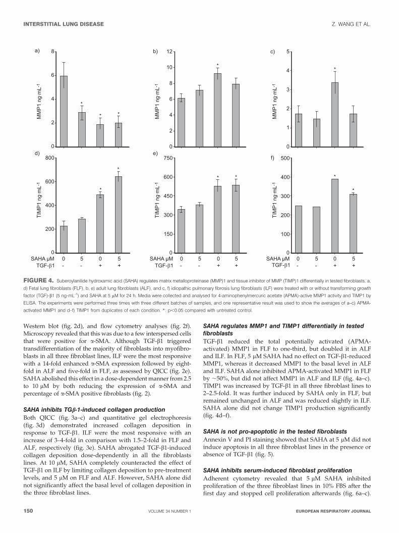
SAHA regulates MMP1 and TIMP1 differentially in testedfibroblastsTGF-b1 reduced the total potentially activated (APMA-activated) MMP1 in FLF to one-third, but doubled it in ALFand ILF. In FLF, 5 mM SAHA had no effect on TGF-b1-reducedMMP1, whereas it decreased MMP1 to the basal level in ALFand ILF. SAHA alone inhibited APMA-activated MMP1 in FLFby ,50%, but did not affect MMP1 in ALF and ILF (fig. 4a–c).TIMP1 was increased by TGF-b1 in all three fibroblast lines to2–2.5-fold. It was further induced by SAHA only in FLF, butremained unchanged in ALF and was reduced slightly in ILF.SAHA alone did not change TIMP1 production significantly(fig. 4d–f).
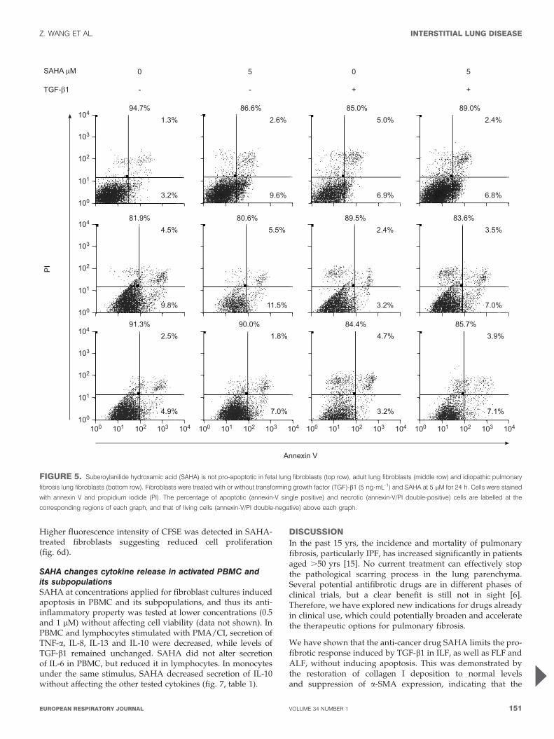
SAHA is not pro-apoptotic in the tested fibroblastsAnnexin V and PI staining showed that SAHA at 5 mM did notinduce apoptosis in all three fibroblast lines in the presence orabsence of TGF-b1 (fig. 5).
SAHA inhibits serum-induced fibroblast proliferationAdherent cytometry revealed that 5 mM SAHA inhibitedproliferation of the three fibroblast lines in 10% FBS after thefirst day and stopped cell proliferation afterwards (fig. 6a–c).
a)
*
* *
**
****
* *
MM
P1
ng·m
L-1
TIM
P1
ng·m
L-1
TIM
P1
ng·m
L-1
TIM
P1
ng·m
L-1
MM
P1
ng·m
L-1
MM
P1
ng·m
L-1
0
2
4
6
8
0
2
4
6
12
10
8
0
1
2
3
5
4
b) c)
d)
00 05 5- +- +
SAHA µMTGF-β1
0 05 5- +- +
SAHA µMTGF-β1
0 05 5- +- +
SAHA µMTGF-β1
200
400
600
800
0
150
300
450
750
600
0
100
200
300
500
400
e)f)
FIGURE 4. Suberoylanilide hydroxamic acid (SAHA) regulates matrix metalloproteinase (MMP)1 and tissue inhibitor of MMP (TIMP)1 differentially in tested fibroblasts. a,
d) Fetal lung fibroblasts (FLF), b, e) adult lung fibroblasts (ALF), and c, f) idiopathic pulmonary fibrosis lung fibroblasts (ILF) were treated with or without transforming growth
factor (TGF)-b1 (5 ng?mL-1) and SAHA at 5 mM for 24 h. Media were collected and analysed for 4-aminophenylmercuric acetate (APMA)-active MMP1 activity and TIMP1 by
ELISA. The experiments were performed three times with three different batches of samples, and one representative result was used to show the averages of a–c) APMA-
activated MMP1 and d–f) TIMP1 from duplicates of each condition. *: p,0.05 compared with untreated control.
INTERSTITIAL LUNG DISEASE Z. WANG ET AL.
150 VOLUME 34 NUMBER 1 EUROPEAN RESPIRATORY JOURNAL
Higher fluorescence intensity of CFSE was detected in SAHA-treated fibroblasts suggesting reduced cell proliferation(fig. 6d).
SAHA changes cytokine release in activated PBMC andits subpopulationsSAHA at concentrations applied for fibroblast cultures inducedapoptosis in PBMC and its subpopulations, and thus its anti-inflammatory property was tested at lower concentrations (0.5and 1 mM) without affecting cell viability (data not shown). InPBMC and lymphocytes stimulated with PMA/CI, secretion ofTNF-a, IL-8, IL-13 and IL-10 were decreased, while levels ofTGF-b1 remained unchanged. SAHA did not alter secretionof IL-6 in PBMC, but reduced it in lymphocytes. In monocytesunder the same stimulus, SAHA decreased secretion of IL-10without affecting the other tested cytokines (fig. 7, table 1).
DISCUSSIONIn the past 15 yrs, the incidence and mortality of pulmonaryfibrosis, particularly IPF, has increased significantly in patientsaged .50 yrs [15]. No current treatment can effectively stopthe pathological scarring process in the lung parenchyma.Several potential antifibrotic drugs are in different phases ofclinical trials, but a clear benefit is still not in sight [6].Therefore, we have explored new indications for drugs alreadyin clinical use, which could potentially broaden and acceleratethe therapeutic options for pulmonary fibrosis.
We have shown that the anti-cancer drug SAHA limits the pro-fibrotic response induced by TGF-b1 in ILF, as well as FLF andALF, without inducing apoptosis. This was demonstrated bythe restoration of collagen I deposition to normal levelsand suppression of a-SMA expression, indicating that the
���
���
��
���
���
���
���
��
���
���
���
���
��
���
���
��� ��� �� ��� ��� ��� ��� �� ��� ��� ��� ��� �� ��� ��� ��� ��� �� ��� ���
"��#$ %&�&$ %���$ %"��$
���$
�� $
�&$
"�&$
���$
&�"$
��$
&�%$
%��"$
���$
"�%$
%��&$
���$
����$
%"��$
��$
�� $
%��&$
���$
#��$
"���$
��$
��"$
"���$
��%$
#��$
%��#$
��"$
#��$
�����
������
�
�
�
�
�
�
�
�
����'(�)
*�
%���$
��#$
�� $
FIGURE 5. Suberoylanilide hydroxamic acid (SAHA) is not pro-apoptotic in fetal lung fibroblasts (top row), adult lung fibroblasts (middle row) and idiopathic pulmonary
fibrosis lung fibroblasts (bottom row). Fibroblasts were treated with or without transforming growth factor (TGF)-b1 (5 ng?mL-1) and SAHA at 5 mM for 24 h. Cells were stained
with annexin V and propidium iodide (PI). The percentage of apoptotic (annexin-V single positive) and necrotic (annexin-V/PI double-positive) cells are labelled at the
corresponding regions of each graph, and that of living cells (annexin-V/PI double-negative) above each graph.
Z. WANG ET AL. INTERSTITIAL LUNG DISEASE
cEUROPEAN RESPIRATORY JOURNAL VOLUME 34 NUMBER 1 151

compound inhibited the transdifferentiation of fibroblasts tomyofibroblasts, the cell type primarily responsible for patho-logical matrix accumulation. We also investigated the potentialfibrolytic properties of SAHA and tested fibroblast collagenase,MMP1 and its inhibitor TIMP1, components responsible forremodeling of extracellular matrix. Surprisingly, the totalpotentially activated-MMP1 was doubled by TGF-b1 in ALFand ILF. SAHA reversed it to the basal level. SAHA has beenreported to inhibit TGF-b1-induced TIMP1 expression inmouse fibroblast [16]. However, in our hands SAHA had no
effect on TGF-b1-induced TIMP1 production in human normaland ILF, and it even induced a further increase of TIMP1 inFLF. Therefore, it is conceivable that the net reduction ofcollagen deposition might be accredited to the effects of SAHAon collagen synthesis and secretion rather than collagenturnover. We have also shown the first evidence that SAHAinhibited fibroblast proliferation in serum.
SAHA is known as a broad-spectrum HDACi [17]. Incomparison with published work on TSA, our data suggest
Cou
nts
CFSE-log
0
97
195
292
390 102.31
100 101 102 1041030
125
251
376
502 67.13
100 101 102 1041030
183
367
551
735 77.20
100 101 102 104103
0
141
283
425
567 271.16
100 101 102 1041030
133
266
399
532 235.83
100 101 102 1041030
28
57
85
114 305.98
100 101 102 104103
a)
d)
Cel
l num
ber ×
1,0
00
00 1 2
Time day3
40
20
60
80
100 b)
0 1 2 3Time day Time day
c)
0 1 2 3
* **
* * ** * *
■
■
■
■ ●●
●
■
■
■
■
● ●●
●● ●
■
■
■
■
FIGURE 6. Suberoylanilide hydroxamic acid (SAHA) inhibits fibroblast proliferation. a) Fetal lung fibroblasts (FLF), b) adult lung fibroblasts (ALF) and c) idiopathic
pulmonary fibrosis lung fibroblasts (ILF) were cultured in 10% fetal bovine serum (FBS) with (#) or without (h) SAHA at 5 mM. Averages of cell numbers from triplicates were
quantified by adherent cytometry at each time-point up to 3 days. *: p,0.05 compared with fibroblasts cultured in 10% FBS at the same time-point. d) Changes of
carboxyfluoroscein succinimidyl ester (CFSE) fluorescence intensity after 3 days without SAHA (top row) or with 5 mM SAHA (bottom row).
INTERSTITIAL LUNG DISEASE Z. WANG ET AL.
152 VOLUME 34 NUMBER 1 EUROPEAN RESPIRATORY JOURNAL

�!��
�+�
,-!�
�
��
�
��
���
���
��
�!��
�+�
,-!�
�
-�
�
��
�
��
��
��
&�
�!��
���
,-!�
�
��
�
�
&
%
��
�����
�+�
,-!�
�
��
�
��
���
���
��
��
� ��� ������
�����
�+�
,-!�
�
+�
�
��
���
���
��
��
� ��� ������
���
�����
�+�
,-!�
�
.�
�
���
��
���
���
���
� ��� ������
�!��
�+�
,-!�
�
/�
�
���
��
���
���
���
�!��
�+�
,-!�
�
0�
�
��
���
#��
�!�&
��,
-!�
�
��
�
��
�
��
��
�!�&
��,
-!�
�
��
�
�
��
��
�!�&
��,
-!�
�
1�
�
���
��
���
�!�%
��,
-!�
�
��
�
��
���
���
��
���
�!�%
��,
-!�
�
��
�
�
��
&�
%�
���
� �
�!�%
��,
-!�
�
(�
�
���
&��
"��
� ��
�2���
���,
-!�
�
��
�
�
&
%
�2���
���,
-!�
�
��
�
�
�
��
� �2���
���,
-!�
�
��
�
�
&
%
� ��� ������ � ��� ������ � ��� ������
FIGURE 7. Suberoylanilide hydroxamic acid (SAHA) changes cytokine release in peripheral blood mononuclear cells (PBMC) and its subpopulations. a–c) tumour
necrosis factor (TNF)-a, d–f) interleukin (IL)-6, g–i) IL-8, j, k) IL-13, l–m) IL-10, and o–q) transforming growth factor (TGF)-b1. SAHA at 0.5 or 1 mM was added to PBMC (a, d, g,
j, l, and o), lymphocytes (b, e, h, k, m and p) or peripheral monocytes (c, f, i, n and q) stimulated by 0.5 ng?mL-1 phorbol myristate acetate plus 50 nM calcium ionophore for
24 h (h) or 48 h (&). The experiments were performed three times with cells from three different donors. The averages of duplicate measurements from one representative
experiment are shown. *: p,0.05 compared with untreated control.
Z. WANG ET AL. INTERSTITIAL LUNG DISEASE
cEUROPEAN RESPIRATORY JOURNAL VOLUME 34 NUMBER 1 153

that inhibiting HDAC activity by SAHA interferes with thepro-fibrotic effects of TGF-b1. Although Smad expression andactivation are not significantly altered in the presence of TSA,TGF-b1-induced expression of Sp1, an essential transcriptionfactor of Smad-dependent collagen synthesis, is inhibited byTSA in foreskin fibroblasts [18]. Another line of evidence is thatHDAC4, and to a lesser extent HDAC6 and HDAC8, arerequired for TGF-b1-induced myofibroblastic differentiation.In particular, the expression of the endogenous repressor of theTGF-b1 pathway, 59-TG-39-interacting factor, is stimulated bysilencing HDAC4 [19]. As further evidence for the broad-spectrum effects of SAHA, we observed hyperacetylated a-tubulin in human lung fibroblasts in response to SAHA. Thisstrongly indicates that HDAC6, a microtubule-associateddeacetylase [20], is inhibited. Of note, fibroblast motility isstrongly compromised with HDAC6 inhibition [21]. Therefore,(cross)inhibition of HDAC6 with SAHA might have anadditional beneficial effect by inhibiting the invasive andcontractile motility of myofibroblasts.
It is well known that inflammation typically precedes fibrosisin fibroproliferative diseases [22]. However, the role ofinflammation in the early pathogenic stage of IPF is underdebate because current anti-inflammatory therapy seems to beineffective in IPF, yet patchy chronic interstitial inflammationis observed in the pathology of IPF [5]. Therefore, we alsoinvestigated the anti-inflammatory potential of SAHA. As theconcentrations applied in fibroblast cultures caused apoptosisin PBMC and its subpopulations, we lowered the concentra-tions by five to 10 times, which is also in accordance with anearlier study [23]. A reduced secretion of cytokines wasobserved in PMA/CI activated lymphocytes and PBMC. Ourobservation of reduced secretion of IL-13 from lymphocytes inresponse to SAHA might be significant, as IL-13 is known toplay a dominant role in pulmonary fibrosis. Overexpression ofIL-13 in the lung generates severe subepithelial airway fibrosis[24]. IL-13 induces the production of TGF-b1 in macrophagesand also indirectly activates latent TGF-b1 by stimulatingMMP production [25]. Furthermore, IL-13 increases theexpression of a-SMA and collagen III in human primary lungfibroblasts, and induces apoptosis in lung epithelia orepithelial cell lines [26]. We also found that IL-10 was reducedin PBMC and its subpopulations. The exact role of this
cytokine in pulmonary fibrosis remains to be established.Although delivery of IL-10 plasmid inhibits bleomycin-induced pulmonary fibrosis [27], transgenic mice over expres-sing IL-10 develop pulmonary inflammation and subepithelialfibrosis with an accumulation of TGF-b1 [28]. In accordance,IL-10 augments the fibrotic responses to inhaled silica particlesas seen in IL-10 deficient mice [29] and over expression of IL-10by adenoviral gene transfer [30]. In vitro, IL-10 also inducesTGF-b1 expression in alveolar macrophages [29]. Hence,inhibition of IL-13 and IL-10 by SAHA may have additionalbeneficial effects in IPF.
Of note, this is the first study evaluating a potential antifibroticdrug under conditions of enhanced matrix formation inducedby macromolecular crowding [14]. We believe that such abiophysical culture environment is better suited than standardconditions in generating a comprehensive and functionalextracellular matrix in a short-time frame, with all biochemicalprocesses in place that represent fibrogenesis and fibrolysis[14]. This should add further weight to the conclusion derivedherein, that SAHA possesses antifibrotic properties and isworth further exploration for a therapeutic option forpulmonary fibrosis.
SUPPORT STATEMENTM. Raghunath recieved funding by a start-up grant from Provost andthe Office of Life Sciences of National University of Singapore (R-397-000-604-101; R-397-000-604-712), the Faculty of Engineering (R-397-000-017-112) and the National Medical Research Council (R397-000-018-213).
STATEMENT OF INTERESTA statement of interest for M. Jung can be found at www.erj.ersjournals.com/misc/statements.dtl
ACKNOWLEDGEMENTSThe authors would like to thank G. Lin (Immunology Program,National University of Singapore, Singapore) for expert technical helpin flow cytometry and the National University of Singapore TissueEngineering Program for strong support.
REFERENCES1 American Thoracic Society/European Respiratory Society
International Multidisciplinary Consensus Classification of theIdiopathic Interstitial Pneumonia. Am J Respir Crit Care Med 2002;165: 277–304.
2 Scotton CJ, Chambers RC. Molecular targets in pulmonary fibrosis:the myofibroblast in focus. Chest 2007; 132: 1311–1321.
3 Gauldie J. Pro: Inflammatory mechanisms are a minor componentof the pathogenesis of idiopathic pulmonary fibrosis. Am J Respir
Crit Care Med 2002; 165: 1205–1206.
4 Strieter RM. Con: Inflammatory mechanisms are not a minorcomponent of the pathogenesis of idiopathic pulmonary fibrosis.Am J Respir Crit Care Med 2002; 165: 1206–1207.
5 Maher TM, Wells AU, Laurent GJ. Idiopathic pulmonary fibrosis:multiple causes and multiple mechanisms? Eur Respir J 2007; 30:835–839.
6 Bhatt N, Baran CP, Allen J, et al. Promising pharmacologicinnovations in treating pulmonary fibrosis. Curr Opin Pharmacol
2006; 6: 284–292.
7 Bolden JE, Peart MJ, Johnstone RW. Anticancer activities of histonedeacetylase inhibitors. Nat Rev Drug Discov 2006; 5: 769–784.
TABLE 1 Suberoylanilide hydroxamic acid changessecretion of cytokines from peripheral bloodmononuclear cells (PBMC) and subpopulations
PBMC Lymphocytes Peripheral
monocytes
Th1 TNF-a Q Q R
Pro-inflammatory IL-6 R Q R
Chemokine IL-8 Q Q R
Th2 IL-13 Q Q ND
IL-10 Q Q Q
Others TGF-b1 R R R
Th; T helper; TNF: tumour necrosis factor; IL; interleukin; TGF: transforming
growth factor; ND: not detected.
INTERSTITIAL LUNG DISEASE Z. WANG ET AL.
154 VOLUME 34 NUMBER 1 EUROPEAN RESPIRATORY JOURNAL

8 Rishikof DC, Ricupero DA, Liu H, et al. Phenylbutyrate decreases
type I collagen production in human lung fibroblasts. J Cell
Biochem 2004; 91: 740–748.
9 Niki T, Rombouts K, De Bleser P, et al. A histone deacetylase inhi-
bitor, trichostatin A, suppresses myofibroblastic differentiation of
rat hepatic stellate cells in primary culture. Hematology 1999; 29:
858–867.
10 Rombouts K, Niki T, Greenwel P, et al. Trichostatin A, a histone
deacetylase inhibitor, suppresses collagen synthesis and prevents
TGF-b(1)-induced fibrogenesis in skin fibroblasts. Exp Cell Res
2002; 278: 184–197.
11 Huber LC, Distler JH, Moritz F, et al. Trichostatin A prevents the
accumulation of extracellular matrix in a mouse model of
bleomycin-induced skin fibrosis. Arthritis Rheum 2007; 56:
2755–2764.
12 Marks PA, Breslow R. Dimethyl sulfoxide to vorinostat: develop-
ment of this histone deacetylase inhibitor as an anticancer drug.
Nat Biotechnol 2007; 25: 84–90.
13 Stowell JC, Huot RI, Van Voast L. The synthesis of N-hydroxy-N’
phenyloctanediamide and its inhibitory effect on proliferation of
AXC rat prostate cancer cells. J Med Chem 1995; 38: 1411–1413.
14 Lareu RR, Subramhanya KH, Peng Y, et al. Collagen matrix
deposition is dramatically enhanced in vitro when crowded with
charged macromolecules: the biological relevance of the excluded
volume effect. FEBS Lett 2007; 581: 2709–2714.
15 Gribbin J, Hubbard RB, Le Jeune I, et al. Incidence and mortality of
idiopathic pulmonary fibrosis and sarcoidosis in the UK. Thorax
2006; 61: 980–985.
16 Young DA, Billingham O, Sampieri CL, et al. Differential effects of
histone deacetylase inhibitors on phorbol ester- and TGF-b1
induced murine tissue inhibitor of metalloproteinases-1 gene
expression. FEBS J 2005; 272: 1912–1926.
17 Arts J, Angibaud P, Marien A, et al. R306465 is a novel potent
inhibitor of class I histone deacetylases with broad-spectrum
antitumoral activity against solid and haematological malignan-
cies. Br J Cancer 2007; 97: 1344–1353.
18 Ghosh AK, Mori Y, Dowling E, et al. Trichostatin A blocks TGF-b-
induced collagen gene expression in skin fibroblasts: involvement
of Sp1. Biochem Biophys Res Commun 2007; 354: 420–426.
19 Glenisson W, Castronovo V, Waltregny D. Histone deacetylase 4 isrequired for TGFb1-induced myofibroblastic differentiation.Biochim Biophys Acta 2007; 1773: 1572–1582.
20 Hubbert C, Guardiola A, Shao R, et al. HDAC6 is a microtubule-associated deacetylase. Nature 2002; 417: 455–458.
21 Tran AD, Marmo TP, Salam AA, et al. HDAC6 deacetylation oftubulin modulates dynamics of cellular adhesions. J Cell Sci 2007;120: 1469–1479.
22 Wynn TA. Common and unique mechanisms regulate fibrosis invarious fibroproliferative diseases. J Clin Invest 2007; 117: 524–529.
23 Bode KA, Schroder K, Hume DA, et al. Histone deacetylaseinhibitors decrease Toll-like receptor-mediated activation ofproinflammatory gene expression by impairing transcriptionfactor recruitment. Immunology 2007; 122: 596–606.
24 Zhu Z, Homer RJ, Wang Z, et al. Pulmonary expression ofinterleukin-13 causes inflammation, mucus hypersecretion, sub-epithelial fibrosis, physiologic abnormalities, and eotaxin produc-tion. J Clin Invest 1999; 103: 779–788.
25 Lanone S, Zheng T, Zhu Z, et al. Overlapping and enzyme-specificcontributions of matrix metalloproteinases-9 and -12 in IL-13-induced inflammation and remodelling. J Clin Invest 2002; 110:463–474.
26 Borowski A, Kuepper M, Horn U, et al. Interleukin-13 acts as anapoptotic effector on lung epithelial cells and induces pro-fibroticgene expression in lung fibroblasts. Clin Exp Allergy 2008; 38:619–628.
27 Nakagome K, Dohi M, Okunishi K, et al. In vivo IL-10 genedelivery attenuates bleomycin induced pulmonary fibrosis byinhibiting the production and activation of TGF-b in the lung.Thorax 2006; 61: 886–894.
28 Lee CG, Homer RJ, Cohn L, et al. Transgenic overexpression ofinterleukin (IL)-10 in the lung causes mucus metaplasia, tissueinflammation, and airway remodeling via IL-13-dependent and-independent pathways. J Biol Chem 2002; 277: 35466–35474.
29 Barbarin V, Arras M, Misson P, et al. Characterization of the effectof interleukin-10 on silica-induced lung fibrosis in mice. Am J
Respir Cell Mol Biol 2004; 31: 78–85.30 Barbarin V, Xing Z, Delos M, et al. Pulmonary overexpression of
IL-10 augments lung fibrosis and Th2 responses induced by silicaparticles. Am J Physiol Lung Cell Mol Physiol 2005; 288: L841–L848.
Z. WANG ET AL. INTERSTITIAL LUNG DISEASE
EUROPEAN RESPIRATORY JOURNAL VOLUME 34 NUMBER 1 155