subtilisin inhibitor like protein ‘pplpi-1’ from leaves of ... (28).pdf(cajanus cajan, cv. bsmr...
TRANSCRIPT

Vol.:(0123456789)1 3
3 Biotech https://doi.org/10.1007/s13205-017-1040-y
ORIGINAL ARTICLE
Subtilisin inhibitor like protein ‘ppLPI‑1’ from leaves of pigeonpea (Cajanus cajan, cv. BSMR 736) exhibits inhibition against Helicoverpa armigera gut proteinases
Faiyaz K. Shaikh1,2 · Prafull P. Gadge1,2 · Manohar V. Padul1 · Manvendra S. Kachole1,2
Received: 13 June 2017 / Accepted: 6 December 2017 © Springer-Verlag GmbH Germany, part of Springer Nature 2017
AbstractHelicoverpa armigera is an orthodox rival of many crop plants affecting agricultural economy. Plant leaves found to accumu-late proteinase inhibitors, although this insect pest chooses leaves for laying eggs. Plant defense response at this juncture is not fully explored. In thiscontext, here we are reporting proteinase inhibitor (ppLPI-1) having significant homology with the I13 family from leaves of pigeonpea (cv. BSMR 736). The isolation of ppLPI-1 was carried out from leaves of field-grown pigeonpea under an outbreak of H. armigera. The acetone precipitated ppLPI-1 (125 µg) displayed substantial inhibition potential towards bovine trypsin (56.5 ± 1.8%) and HaGPs (52.6 ± 1.7%) on solution assay. These results were corroborated with dot-blot analysis. The molecular form of ppLPI-1 was characterized by reverse zymography and GXCP. The optimum condition was found to be pH 8 and temperature in the range of 30–40 °C. The protein identification via MASCOT-PMF and NCBI-BLAST search showed substantial homology with an inducible subtilisin inhibitor of Fabaceae comprising Vigna angularis (96%), Canavalia lineata (78%), Cicer arietinum (76%), Glycine max (75%), Medicago truncatula (73%) and Vicia faba (73%) consists of conserved domain of potato inhibitor I family.
Keywords Proteinase inhibitor · Pigeonpea · ppLPI-1 · HaGPs · Helicoverpa armigera · Subtilisin inhibitor
Introduction
Pigeonpea (Cajanus cajan) is the second and fifth most important pulse crop of India and rest of the world, respec-tively. Being chief source of protein, it proved to be main diet of large section of the vegetarian population in India, eastern Africa and Central America (Srivastava and Joshi 2011). Helicoverpa armigera is devastating insect pest of this crop and invade more than 300 plant species through-out the world (Rajapakse and Walter 2007). The actively growing larvae of H. armigera preferred protein produced in storage and non-storage parts of the plant for feeding. The accumulated variable gut proteinases system comprising
serine (trypsin and chymotrypsin), metallo, aspartic and cysteine proteinases is responsible for digestive functioning (Bown et al. 1997; Emmerling et al. 2001; Bayes et al. 2005; Pauchet et al. 2008).
Host plant defense against insect pests encompasses expressions of proteinase inhibitors (PIs) and supposed to be ecological and economical defense approach. These mol-ecules are responsible for inhibition of proteinases expressed in the gut of insect pests (Lawrence and Koundal 2002). The scarcity of protein-rich diet attributed to the action of PIs leads to underdevelopment or even death of insect pests (Bown et al. 1997). Generally, PIs are found to regulate the activity of endogenous proteinases of plants (Ryan 1990). Induced expression of PIs on onset of insect pests attack makes them promising candidate for insect pest management (Constabel 1999).
The occurrence of PIs has been conferred from the stor-age tissues (seeds and tubers) of many host plants of H. armigera including pigeonpea (C. cajan), Cotton (Gos-sypium hirsutum), chickpea (Cicer arietinum), mungbean (Vigna radiata), etc. (Lawrence and Koundal 2002; Raja-pakse and Walter 2007). However, the gut proteinases
* Faiyaz K. Shaikh [email protected]
1 Department of Biochemistry, Dr. Babasaheb Ambedkar Marathwada University, Aurangabad, Maharashtra, India
2 Department of Biotechnology, MGM’s Institute of Biosciences and Technology, Aurangabad, Maharashtra 431003, India
3 Biotech
1 3
system of H. armigera is found to adapt the action of PIs expressed in most of these plants. The mechanism of adapta-tion could be the synthesis of PIs-insensitive proteinases or expression of proteinases that degrade PIs (Srinivasan et al. 2005; Tabashnik et al. 2008). The expression of about nine PIs was reported from seed extract of pigeonpea (Pichare and Kachole 1996; Chougule et al. 2003; Padul et al. 2012). But all these molecules are found to be feeble in their actions against gut proteinases system of H. armigera.
The storage tissues are sites where on the third and fourth instar larvae generally fed. They have fully functional diges-tive proteinases system and could be the basis of adapta-tion to host plant PIs. Non-storage tissues such as leaves, flowers and roots are primary contact areas for the first and second instar larvae (Padul et al. 2012). Female H. armigera lays its eggs on leaves or tender branches. These larvae start feeding on these tissues and later shift to reproductive parts (Liu et al. 2010). These larvae consume food at five times the rate of third and fourth instar larvae with rapid spread on non-infested areas (Johnson and Zalucki 2007). Hence, restriction of the movement of H. armigera from leaves to reproductive organs will provide the limitation in the fur-ther loss of crops. To achieve this, biochemical interactions between the host plant and H. armigera need to be explored at this juncture. Earlier PIs expression in non-storage tissues was reported from few plants (Ryan 1990; Damle et al. 2005; Padul et al. 2012).
The detailed study of nature, specificity and molecular biochemistry of PIs from non-storage tissues such as leaves is prime concern to exploit PIs as biological agent for insect control. In this regard, here we report the electrophoresis-based preparative isolation, mass spectrometry-based identi-fication and biochemical characterization of novel PI named as ppLPI-1 from field collected matured leaves of pigeonpea cv. BSMR 736 under H. armigera attack.
Materials and methods
Procurement of chemicals
Trypsin (bovine pancreas, E.C. 3.4.21.4), acrylamide, bisacrylamide, tetramethylethylenediamine (TEMED), PVP (polyvinylpyrrolidone) and N-α-benzoyl-dl-arginine-p-nitroanilide (BApNA) were obtained from Sisco Research Laboratories (SRL), Mumbai, India. X-ray films were obtained from Fuji film, USA. All other chemicals used in this study were of the available highest purity.
Procurement of leaves
Dry mature seeds of pigeonpea (cv. BSMR 736) were pro-cured from the Badnapur Agricultural Research Station
of Marathwada Agricultural University, Parbhani (MS) India. The seeds were sowed naturally in the field of the Department, Dr. Babasaheb Ambedkar Marathwada Uni-versity, Aurangabad. Leaves were harvested from fully grown pigeonpea plants which were naturally exposed to H. armigera.
Procurement of insects
Second instar larvae of H. armigera were collected from pigeonpea fields.
Extraction of PIs from seeds and leaves of pigeonpea
The extraction of seed PIs were carried out according to Shaikh et al. (2014). The leaves PIs were extracted accord-ing to the method of Padul et al. (2012). The field-collected matured leaves of pigeonpea were dried and pulverized in acetone using tissue homogenizer. The depigmented powder was finally washed with hexane to remove fat. The resulting powder of leaves was suspended in distilled water contain-ing 1% PVP (1:10 w/v) and kept at 15 °C for overnight to extract the proteins. The suspension was then centrifuged at 12,000g for 20 min at 4 °C. The clear supernatant obtained was used as source of crude PIs.
Extraction of HaGPs
The HaGPs extraction was carried out by removing the mid-gut tissue of the second instar larvae of H. armigera. Fresh or thawed (at – 20 °C) midgut tissue was homogenized in 0.1 M glycine–NaOH buffer (1:3 w/v pH 9.6) for 15 min at 10 °C. The suspension was centrifuged at 12,000g for 20 min at 4 °C. The resulting supernatant was used as source of HaGPs.
Detection of PIs by dot‑blot test
The dot-blot test was carried out to determine the potency of crude leaves PIs against trypsin and HaGPs using gel-atin coated X-ray film (Pichare and Kachole 1994; Padul et al. 2012). Three varied concentrations of the enzyme and inhibitor were prepared: 1 (1:3), 2 (1:1), and 3 (3:1) v/v, respectively. The total volume was made up to 20 µl by the adjusting buffers, 0.1 M Tris–HCl (pH 7.8) for trypsin and 0.1 M glycine–NaOH (pH 9.6) buffer for HaGPs was used. The resulting samples were loaded onto X-ray film. After incubating for 20 min at 37 °C, the film was washed with tap water and dried in air. The varying proportions of enzyme and inhibitor produced different patterns of gelatin hydroly-sis on the X-ray film depending on the efficacy of inhibitor.

3 Biotech
1 3
The inhibition pattern was observed visually and scanned at 300 dpi using an HP digital scanner.
Electrophoretic visualization of PIs
Crude leaves PIs and seed PIs were visualized by gel X-ray film contact print technique (GXCP) and reverse zymog-raphy (Pichare and Kachole 1994; Shaikh et al. 2014). For electrophoresis, 80 μg sample was loaded onto native poly-acrylamide gel and electrophoresis allowed to run under influence of constant current of 20 mA (Davis 1964). For GXCP analysis, after electrophoresis resulting gel was equil-ibrated in 0.1 M Tris–HCl (pH 7.8) for 10 min at 37 °C. Followed by equilibration gel was incubated with 0.1 mg/ml trypsin solution for 10 min at 37 °C. After removing excess trypsin by dipping gel in the above-mentioned buffer, it was overlaid on X-ray film for 5–10 min. The gel was removed from X-ray film, the resulting X-ray film washed with tap water and the PIs bands were visualized as unhydrolyzed gelatin. The X-ray film was scanned at 300 dpi using an HP digital scanner. The gel was then washed and stained with 0.1% coomassie brilliant blue R-250. The same procedure was carried out for HaGPs using 0.1 M glycine–NaOH (pH 9.6) buffer containing 0.3 M CaCl2. The experiment was repeated three times with three replicates each.
For reverse zymography, the gel was equilibrated in 0.1 M Tris–HCl (pH 7.8) and dipped into 1% casein, prepared in 0.1 M Tris–HCl (pH 7.8) for 20 min at 37 °C. After remov-ing excess substrate the gel was incubated in 0.1% bovine trypsin for 10 min at 37 °C. The resulting gel then washed with buffer and stained with 0.1% coomassie brilliant blue R-250.
Preparative electrophoretic purification of ppLPI‑1
The ppLPI-1 was purified by preparative native-PAGE. After electrophoresis, a vertical strip of the gel was cut and processed to detect the PI activity band on X-ray film, as previously described. A horizontal strip of the remaining gel corresponding to the ppLPI-1 activity band of the X-ray film was excised and stored overnight at – 20 °C and then the sample was eluted. Preparative electrophoresis was repeated several times to obtain sufficient amounts of ppLPI-1 for fur-ther characterization. The ppLPI-1 was further run on 10% native-PAGE and excised from the stained gel and dehy-drated with 50% acetonitrile and stored at – 20 °C.
MALDI‑TOF MS analysis
The identification analysis of ppLPI-1 was done by matrix-assisted laser desorption ionization mass spectrometry (MALDI-TOF MS) using UltrafleXtreme (Bruker Dalton-ics, Germany) system at a Proteomics facility of Molecular
Biophysics Unit, IISC (Bangalore, India). Overnight in-gel trypsin digestion of ppLPI-1 band was carried out at 37 °C. Peptide mass fingerprints of tryptic digests were acquired and compared with the existing entries in the global database by using the Mascot search engine (http://www.matrixsci-ence.com), which uses a probability-based scoring system. Viridiplantae (Green Plants) was used as a taxonomy filter for a specific search. The threshold of “good match” was defined as per Chen et al. (2009). The MUSCLE program was used to align the homologous PI sequences obtained after NCBI BLAST of matched internal protein sequence (Edgar 2004).
Assay of trypsin inhibitory activity
Trypsin inhibition was assayed according to the method of Kakade et al. (1969), using BApNA as the substrate and bovine trypsin as the standard enzyme. The different con-centration of crude PIs of leaves and 40 µl trypsin (1 mg/ml, prepared in 0.01 N HCl) were incubated for 10 min in Tris–HCl buffer (0.1 M, pH 7.8 and 0.3 M CaCl2). To this mixture, 0.3 ml BApNA (1 mM, prepared in DMSO) was added. The final volume of the reaction mixture was 1.2 ml. After incubating the reaction mixture at 37 °C for 30 min, the reaction was terminated by adding 0.3 ml of 30% glacial acetic acid (v/v). A blank and a trypsin control were run simultaneously. The absorbance was recorded at 410 nm against the blank. The inhibition of HaGPs was assessed by incubating the inhibitor with HaGPs for 20 min in 0.1 M glycine–NaOH (pH 9.6), which contained 0.3 M CaCl2.
One trypsin activity unit is equal to the release of 1.0 μmol of p-nitroaniline per min under specific assay con-ditions, with one inhibitor unit being defined as the inhibi-tion of one enzyme unit. Inhibitory activity is expressed as the percentage of inhibited enzyme activity out of the total enzyme activity.
Effect of pH on proteinases and crude PIs activities
The effect of pH on the total activity of trypsin and HaGPs in presence of crude PIs of leaves was carried out in differ-ent buffer systems as described previously. The buffer sys-tem utilized were 0.1 M glycine–HCl (pH 2), 0.1 M sodium citrate (pH 4) and (pH 6), 0.1 M Tris–HCl (pH 8), 0.1 M glycine–NaOH (pH 10), and 0.1 M glycine–NaOH (pH 12).
Effect of temperatures on proteinases and crude PIs activities
The effect of temperature on the total activity of trypsin and HaGPs in presence of crude PIs of leaves was studied by performing the reaction at different temperatures (20–80 °C
3 Biotech
1 3
at 10 °C intervals) in a dry water bath. Inhibition assays were assayed according to the method described previously.
Protein determination
Protein content was estimated by Lowry et al. (1951) method using Bovine serum albumin (BSA) as the standard.
Statistical analysis
All experiments were conducted in triplicate. Means and standard deviations were calculated and compared. The analysis was performed using Microsoft Excel 2010.
Results and discussion
The host plant resistance plays important role in insect pest management (Dhillon and Sharma 2012). The plant PIs is a necessary component of host plant resistance and could be a sustainable and natural approach against insect pests. The action of PI is attributed to their binding to the active site of the gut proteinases which may block, alter or hinder access to the substrate (Jongsma and Bolter 1997). This result in the reduction in the availability of essential amino acids required for growth and development of insect pests.
As a valuable component in plant defense the nature, specificity, and stability of these PIs could be explored at various developmental stages. The non-storage tissues such as leaves are more exposed to H. armigera. The PIs response at this site is expected to be very important. In this regard, we are reporting the isolation, preparative purification and identification of PI designated as ppLPI-1 from the field col-lected leaves of pigeonpea (cv. BSMR 736). The molecular aspects like specificity, stability, optimal conditions and
suitability of ppLPI-1 as host plant defense component is discussed.
Solution assay of crude leaves PIs activity
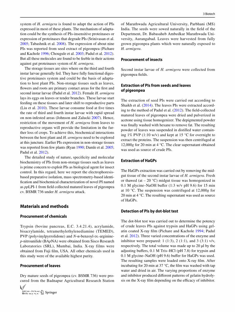
On the solution assay of PIs activity from the acetone pre-cipitated protein of leaves (125 µg), 56.5 ± 1.8% inhibition against bovine trypsin was reported (Fig. 1a). At the same concentration of the crude leaves PIs, 52.6 ± 1.7% inhibi-tion was achieved against the HaGPs (gut of second instar larvae used). The inhibition efficiency of crude leaves PIs was also assessed by dot-blot assay. The substantial inhibi-tion to the trypsin and HaGPs were observed with different concentrations of crude leaves PIs (Fig. 1b). This is the first report regarding significant inhibition potential from leaves of pigeonpea cv. BSMR 736 against HaGPs. Previously sig-nificant inhibition to HaGPs was reported from leaves of pigeonpea cv. BDN-2 (Padul et al. 2012). The leaves are initial contact area for first and second instar larvae and sub-stantial inhibition to HaGPs may attribute to the accumula-tion of inducible PIs in response to H. armigera infestation in the field. Earlier PIs induction in the response to insect infestation is reported in barley seedlings (Casaretto and Corcuera 1998). Induction of PIs occurs via the octadeca-noid pathway, which catalyzes the breakdown of linolenic acid and the formation of jasmonic acid which induces PIs gene expression (Farmer et al. 1992).
Electrophoretic characterization of leaves PIs activity
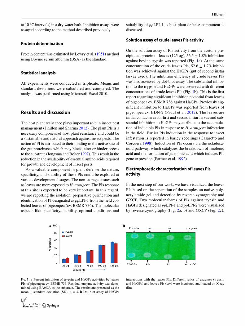
In the next step of our work, we have visualized the leaves PIs based on the separation of the samples on native-poly-acrylamide gel and detection by reverse zymography and GXCP. Two molecular forms of PIs against trypsin and HaGPs designated as ppLPI-1 and ppLPI-2 were visualized by reverse zymography (Fig. 2a, b) and GXCP (Fig. 2c).
Fig. 1 a Percent inhibition of trypsin and HaGPs activities by leaves PIs of pigeonpea cv. BSMR 736. Residual enzyme activity was deter-mined using BApNA as the substrate. The results are presented as the mean ± standard deviation (SD), n = 3. b Dot blot assay of HaGPs
interactions with the leaves PIs. Different ratios of enzymes (trypsin and HaGPs) and leaves PIs (v/v) were incubated and loaded on X-ray film

3 Biotech
1 3
Earlier presence of monomorphic PIs pattern on the gel was reported from the seed extracts of most of the pigeon-pea cultivars (Chougule et al. 2003). The ppLPI-1 and ppLPI-2 reported in this study have resemblance with PI-3 and PI-4 of the crude protein of seed extract of the same plant (Fig. 2a, b). Almost similar PIs pattern was reported by Padul et al. (2012) from leaves of pigeonpea cv. BDN-2 wherein three PIs (i.e. PI-3, PI-4 and PI-5, numbered accord-ing to an occurrence in seed extract) found to be induci-ble. Among them, PI-3 was appeared first in all types of induction used in the study. The ppLPI-1 resembles to PI-3 protein which has expression in the seeds and leaves. The nature, specificity, and stability against H. armigera of this
PI could be a prime concern to design competent PIs-based plant defense strategies.
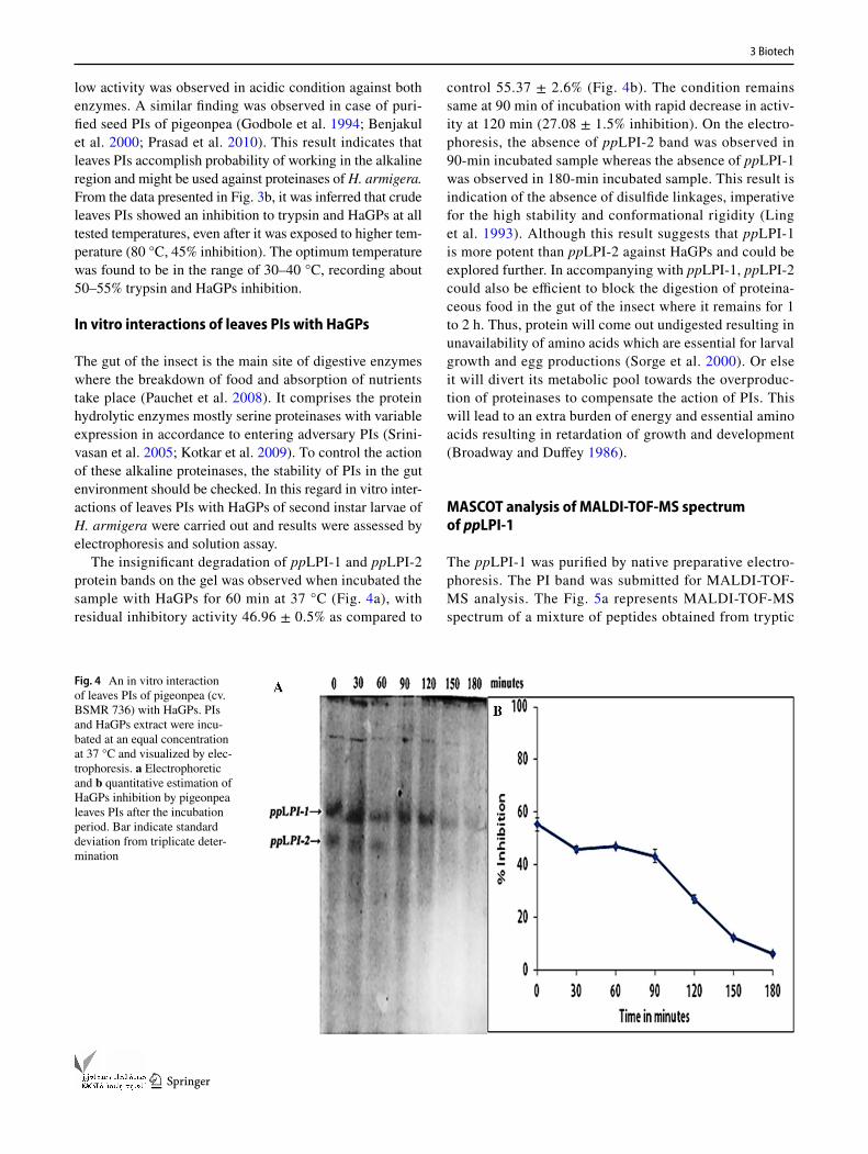
Optimum parameters for leaves PIs activity
The protein requirement of H. armigera accomplishes by the proteolytic action of alkaline proteinases present in the gut. To inhibit these alkaline proteinases, it is necessary to have PI with efficient activity in the alkaline condition. Here, we found that the inhibitory activity of leaves PIs against bovine trypsin and HaGPs was not sensitive to pH over the range 6.0–12.0 with an optimum at pH 8 (Fig. 3a). The reasonable activity was achieved in the alkaline condition whereas very
Fig. 2 Native-PAGE reverse zymography analysis of leaves PIs of pigeonpea cv. BSMR 736: samples were run on a 10% polyacrylamide gel and stained with a coomassie brilliant blue R-250 and b silver staining. (I) Leaves PIs; (II) seed PIs of pigeonpea c GXCP photograph of leaves PIs (PIs of trypsin and HaGPs). The detailed procedure is mentioned in “Materials and methods” section
Fig. 3 a Total trypsin activity in the presence of crude leaves PIs assayed in different pH conditions. b Total HaGPs activity in pres-ence of crude leaves PIs assayed by performing the reaction at differ-
ent temperatures. The assays were performed as mentioned in “Mate-rials and methods”. Bar indicate standard deviation from triplicate determination
3 Biotech
1 3
low activity was observed in acidic condition against both enzymes. A similar finding was observed in case of puri-fied seed PIs of pigeonpea (Godbole et al. 1994; Benjakul et al. 2000; Prasad et al. 2010). This result indicates that leaves PIs accomplish probability of working in the alkaline region and might be used against proteinases of H. armigera. From the data presented in Fig. 3b, it was inferred that crude leaves PIs showed an inhibition to trypsin and HaGPs at all tested temperatures, even after it was exposed to higher tem-perature (80 °C, 45% inhibition). The optimum temperature was found to be in the range of 30–40 °C, recording about 50–55% trypsin and HaGPs inhibition.
In vitro interactions of leaves PIs with HaGPs
The gut of the insect is the main site of digestive enzymes where the breakdown of food and absorption of nutrients take place (Pauchet et al. 2008). It comprises the protein hydrolytic enzymes mostly serine proteinases with variable expression in accordance to entering adversary PIs (Srini-vasan et al. 2005; Kotkar et al. 2009). To control the action of these alkaline proteinases, the stability of PIs in the gut environment should be checked. In this regard in vitro inter-actions of leaves PIs with HaGPs of second instar larvae of H. armigera were carried out and results were assessed by electrophoresis and solution assay.
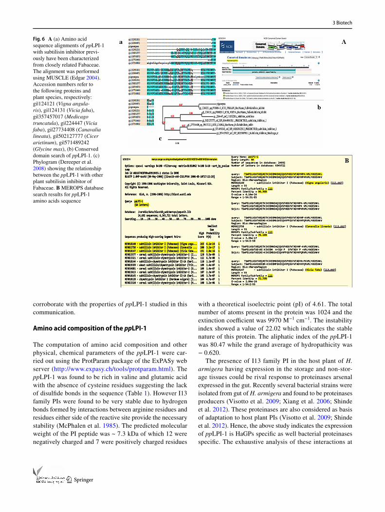
The insignificant degradation of ppLPI-1 and ppLPI-2 protein bands on the gel was observed when incubated the sample with HaGPs for 60 min at 37 °C (Fig. 4a), with residual inhibitory activity 46.96 ± 0.5% as compared to
control 55.37 ± 2.6% (Fig. 4b). The condition remains same at 90 min of incubation with rapid decrease in activ-ity at 120 min (27.08 ± 1.5% inhibition). On the electro-phoresis, the absence of ppLPI-2 band was observed in 90-min incubated sample whereas the absence of ppLPI-1 was observed in 180-min incubated sample. This result is indication of the absence of disulfide linkages, imperative for the high stability and conformational rigidity (Ling et al. 1993). Although this result suggests that ppLPI-1 is more potent than ppLPI-2 against HaGPs and could be explored further. In accompanying with ppLPI-1, ppLPI-2 could also be efficient to block the digestion of proteina-ceous food in the gut of the insect where it remains for 1 to 2 h. Thus, protein will come out undigested resulting in unavailability of amino acids which are essential for larval growth and egg productions (Sorge et al. 2000). Or else it will divert its metabolic pool towards the overproduc-tion of proteinases to compensate the action of PIs. This will lead to an extra burden of energy and essential amino acids resulting in retardation of growth and development (Broadway and Duffey 1986).
MASCOT analysis of MALDI‑TOF‑MS spectrum of ppLPI‑1
The ppLPI-1 was purified by native preparative electro-phoresis. The PI band was submitted for MALDI-TOF-MS analysis. The Fig. 5a represents MALDI-TOF-MS spectrum of a mixture of peptides obtained from tryptic
Fig. 4 An in vitro interaction of leaves PIs of pigeonpea (cv. BSMR 736) with HaGPs. PIs and HaGPs extract were incu-bated at an equal concentration at 37 °C and visualized by elec-trophoresis. a Electrophoretic and b quantitative estimation of HaGPs inhibition by pigeonpea leaves PIs after the incubation period. Bar indicate standard deviation from triplicate deter-mination

3 Biotech
1 3
digestion of ppLPI-1 and their m/z value, respectively. Interpretation of the MALDI-TOF data was carried out using online ‘‘MASCOT search tool’’ (version 2.4.1; http://www.matrixscience.com). The sequence analy-sis of ppLPI-1 using MASCOT-PMF search showed three peptides, K.TSWPELVGVTAEQAETKIK.E, K . E E M V DV Q I Q VS P H D S F V TA DY N P K . R a n d R.LYVDESNKVTR.T, corresponding to mass values (m/z value) 2087.749, 2748.217 and 1324.181 respec-tively matched to that of subtilisin inhibitor of Phaseolus angularis (taxonomy: Vigna angularis) with 60% protein sequence coverage (Fig. 5b, c).
Sequence homology searches by NCBI‑BLAST and MEROPS database
On BLAST similarity search with the NCBI non-redundant database of PIs of Fabaceae, the peptides of ppLPI-1 were found to consist of the conserved domain of potato inhibitor I family (Fig. 6A). These peptides revealed 96% homology with subtilisin inhibitor of V. angularis (105 score, 1e−27 E value), 78% with subtilisin inhibitor of Canavalia lineata (79.3 score, 1e−17 E value), 76% with predicted subtilisin
inhibitor 1-like of C. arietinum (83.2 score, 8e−19 E value), 75% predicted subtilisin inhibitor 1-like of Glycine max (79.0 score, 5e−17 E value), 73% with subtilisin inhibitor of Medicago truncatula (79.0 score, 3e−17 E value), 73% with subtilisin inhibitor of Vicia faba (75.9 score, 2e−16 E value) and 69% with subtilisin inhibitor of V. faba (70.9 score, 2e−14 E value). On comparison in MEROPS data-base of proteinases and proteinase inhibitors, these peptides showed similarity with a subtilisin inhibitor I serine protein-ase inhibitor from the I13 family (Fig. 6B). These peptides showed 96.36% sequence homology with subtilisin inhibitor of V. angularis (MER018140, 265 scores, 1e−24 E value), 78.18% with subtilisin inhibitor of C. lineata (MER022758, 200 scores, 2e−17 E value) and 72.73% with subtilisin inhibitor of V. faba (MER018147, 191 scores, 2.8e−16 E value).
This result indicates that ppLPI-1 belongs to I13 fam-ily with conserved domain of potato inhibitor I family. The PIs belonging to I13 family are found to inhibit serine proteinases (trypsin/chymotrypsin/ subtilisin) by the stand-ard mechanism (Laskowski and Kato 1980; Beuning et al. 1994). The other properties of I13 family PIs like lack of disulfide bonds, stability at pH 2–9 and high temperatures
Fig. 5 a MALDI-TOF-MS spectra (peptide mass finger-prints) of peptides generated from tryptically digested ppLPI-1. b Peptide mass fingerprint (PMF) analysis of ppLPI by MASCOT search tool (version 2.4.1; http://www.matrixsci-ence.com). c Summary of matched peptides of ppLPI-1
3 Biotech
1 3
corroborate with the properties of ppLPI-1 studied in this communication.
Amino acid composition of the ppLPI‑1
The computation of amino acid composition and other physical, chemical parameters of the ppLPI-1 were car-ried out using the ProtParam package of the ExPASy web server (http://www.expasy.ch/tools/protparam.html). The ppLPI-1 was found to be rich in valine and glutamic acid with the absence of cysteine residues suggesting the lack of disulfide bonds in the sequence (Table 1). However I13 family PIs were found to be very stable due to hydrogen bonds formed by interactions between arginine residues and residues either side of the reactive site provide the necessary stability (McPhalen et al. 1985). The predicted molecular weight of the PI peptide was ~ 7.3 kDa of which 12 were negatively charged and 7 were positively charged residues
with a theoretical isoelectric point (pI) of 4.61. The total number of atoms present in the protein was 1024 and the extinction coefficient was 9970 M−1 cm−1. The instability index showed a value of 22.02 which indicates the stable nature of this protein. The aliphatic index of the ppLPI-1 was 80.47 while the grand average of hydropathicity was − 0.620.
The presence of I13 family PI in the host plant of H. armigera having expression in the storage and non-stor-age tissues could be rival response to proteinases arsenal expressed in the gut. Recently several bacterial strains were isolated from gut of H. armigera and found to be proteinases producers (Visotto et al. 2009; Xiang et al. 2006; Shinde et al. 2012). These proteinases are also considered as basis of adaptation to host plant PIs (Visotto et al. 2009; Shinde et al. 2012). Hence, the above study indicates the expression of ppLPI-1 is HaGPs specific as well bacterial proteinases specific. The exhaustive analysis of these interactions at
Fig. 6 A (a) Amino acid sequence alignments of ppLPI-1 with subtilisin inhibitor previ-ously have been characterized from closely related Fabaceae. The alignment was performed using MUSCLE (Edgar 2004). Accession numbers refer to the following proteins and plant species, respectively: gi|124121 (Vigna angula-ris), gi|124131 (Vicia faba), gi|357457017 (Medicago truncatula), gi|224447 (Vicia faba), gi|27734408 (Canavalia lineata), gi|502127777 (Cicer arietinum), gi|571489242 (Glycine max), (b) Conserved domain search of ppLPI-1. (c) Phylogram (Dereeper et al. 2008) showing the relationship between the ppLPI-1 with other plant subtilisin inhibitor of Fabaceae. B MEROPS database search results for ppLPI-1 amino acids sequence

3 Biotech
1 3
molecular level is essential and future prospective of this study.
Acknowledgements The financial assistance in the form of the fellow-ship to Faiyaz K. Shaikh from the University Grant Commission (UGC) and Department of Ministry of Minority Affairs, Government of India, New Delhi is greatly acknowledged.
Author contributions FKS designed, performed, coordinated the main study and responsible for manuscript preparation. PPG helped in elec-trophoresis techniques and manuscript editing. MVP helped in data interpretation and analysis. MSK is responsible for concept and the final approval of the manuscript.
Compliance with ethical standards
Conflict of interest The authors declares that there is no conflict of intrest regarding the publication of this paper.
References
Bayes A, Comellas-Bigler M, Vega MR, Maskos K, Bode W, Aviles MA, Jongsma FX, Beekwilder J, Vendrell J (2005) Structural basis of the resistance of insect carboxypeptidases to plant pro-tease inhibitors. PNAS 102:16602–16607. https://doi.org/10.1073/pnas.0505489102
Benjakul S, Visessanguan W, Thummaratwasik P (2000) Isola-tion and characterization of trypsin inhibitors from some thai legume seeds. J Food Biochem 24:107–127. https://doi.org/10.1111/j.1745-4514.2000.tb00689.x
Beuning LL, Spriggs TW, Christeller JT (1994) Evolution of the pro-teinase inhibitor I family and apparent lack of hypervariability in
the proteinase contact group. J Mol Evol 39:644–654. https://doi.org/10.1111/j.1742-4658.2005.04975
Bown DP, Wilkinson HS, Gatehouse JA (1997) Differentially regulated inhibitor sensitive and insensitive protease genes from the phy-tophagous pest, Helicoverpa armigera, are members of complex multigene families. Insect Biochem Mol Biol 27:625–638
Broadway RM, Duffey SS (1986) Plant proteinase inhibitors: mecha-nism of action and effect on the growth and digestive physiology of larval Heliothis zea and Spodoptera exigua. J Insect Physiol 32:827–833
Casaretto JA, Corcuera LJ (1998) Proteinase inhibitor accumulation in aphid-infested barley leaves. Phytochemistry 49:2279–2286. https://doi.org/10.1016/S0031-9422(98)00332-X
Chen H, Bodulovic G, Hall PJ, Moore A, Higgins TJ, Djordjevic MA, Rolfe BG (2009) Unintended changes in protein expres-sion revealed by proteomic analysis of seeds from transgenic pea expressing a bean alpha-amylase inhibitor gene. Proteomics 9:4406–4415. https://doi.org/10.1002/pmic.200900111
Chougule NP, Hivrale VK, Chhabda PJ, Giri AP, Kachole MS (2003) Differential inhibitions of Helicoverpa armigera gut proteinases by proteinase inhibitors of pigeonpea (Cajanus cajan) and its wild relatives. Phytochemistry 64:681–687
Constabel CP (1999) A survey of herbivore-inducible defensive pro-teins and phytochemicals. In: Agrawal S, Tuzun S, Bent E (eds) Inducible plant defenses against pathogens and herbivores: bio-chemistry, ecology, and agriculture. American Phytopathology Society Press, St Paul, Minnesota, pp 137–166
Damle MS, Giri AP, Sainani MN, Gupta VS (2005) Higher accumu-lation of proteinase inhibitors in flowers than leaves and fruits as a possible basis for differential feeding preference of Heli-coverpa armigera on tomato (Lycopersicon esculentum Mill, Cv. Dhanashree). Phytochemistry 66:2659–2667. https://doi.org/10.1016/j.phytochem.2005.09.006
Davis BJ (1964) Disc electrophoresis II. Method and application to human serum proteins. Ann N Y Acad Sci 121:404–427. https://doi.org/10.1111/j.1749-6632.1964.tb14213
Dereeper A, Guignon V, Blanc G, Audic S, Buffet S, Chevenet F, Dufayard JF, Guindon S, Lefort V, Lescot M, Claverie JM, Gas-cuel O (2008) Phylogeny.fr: robust phylogenetic analysis for the non-specialist. Nucl Acids Res 36:465–469. doi:https://doi.org/10.1093/nar/gkn180
Dhillon MK, Sharma HC (2012) Paradigm shifts in research on host plant resistance to insect pests. Indian J Plant Prot 40:1–11
Edgar RC (2004) MUSCLE: multiple sequence alignment with high accuracy and high throughput. Nucleic Acids Res 32:1792–1797. https://doi.org/10.1093/nar/gkh340
Emmerling M, Chandler D, Sandeman M (2001) Molecular cloning of three cDNA encoding aminopeptidases from the midgut of Helicoverpa armigera, the Australian native budworm. Insect Biochem Mol Biol 31:899–907
Farmer EE, Johnson RR, Ryan CA (1992) Regulation of expression of proteinase inhibitor genes by methyl jasmonate and jasmonic acid. Plant Physiol 98:995–1002
Godbole SA, Krishna TG, Bhatia CR (1994) Purification and char-acterization of protease inhibitors from pigeon pea (Cajanus Cajan) seed. J Sci Food Agric 64:87–93. https://doi.org/10.1002/jsfa.2740640113
Johnson ML, Zalucki MP (2007) Feeding and foraging behaviour of a generalist caterpillar: are third instars just bigger versions of firsts? Bull Entomol Res 97:81–88. https://doi.org/10.1017/S0007485307004750
Jongsma MA, Bolter CJ (1997) The adaptation of insects to plant proteinase inhibitors. J Insect Physiol 43:885–896. https://doi.org/10.1016/S0022-1910(97)00040-1
Table 1 Amino acid composition of the ppLPI-1 protein computed by ProtParam package of the ExPASy web server (http://www.expasy.ch/tools/protparam.html)
Amino acid Number Mol%
Ala (A) 3 4.7Arg (R) 2 3.1Asn (N) 3 4.7Asp (D) 5 7.8Cys (C) 0 0.0Gln (Q) 3 4.7Glu (E) 7 10.9Gly (G) 1 1.6His (H) 1 1.6Ile (I) 2 3.1Leu (L) 3 4.7Lys (K) 5 7.8Met (M) 1 1.6Phe (F) 1 1.6Pro (P) 3 4.7Ser (S) 5 7.8Thr (T) 5 7.8Trp (W) 1 1.6Tyr (Y) 3 4.7Val (V) 10 15.6
3 Biotech
1 3
Kakade ML, Simons N, Liener IE (1969) An evaluation of natural vs. synthetic substrates for measuring the antitryptic activity of soybean samples. Cereal Chem 46:518–521
Kotkar HM, Sarate PJ, Tamhane VA, Gupta VS, Giri AP (2009) Responses of midgut amylases of Helicoverpa armigera to feed-ing on various host plants. J Insect Physiol 55:663–670. https://doi.org/10.1016/j.jinsphys.2009.05.004
Laskowski MJ, Kato I (1980) Protein inhibitors of proteinases. Annu Rev Biochem 49:593–626. https://doi.org/10.1146/annurev.bi.49.070180.003113
Lawrence PK, Koundal KR (2002) Plant protease inhibitors in control of polyphagous insects. Electron J Biotechnol 1:93–109. https://doi.org/10.2225/vol5-issue1-fulltext-3
Ling MH, Qi H, Chi C (1993) Protein, cDNA sequences of the towel gourd trypsin inhibitor. J Biol Chem 268:810–814
Liu F, Xu Z, Zhu YC, Huang F, Wang Y, Li H, Gao C, Zhou W, Shen J (2010) Evidence of field-evolved resistance to Cry1Ac-express-ing Bt cotton in Helicoverpa armigera (Lepidoptera: Noctuidae) in northern China. Pest Manag Sci 66:155–161. https://doi.org/10.1002/ps.1849
Lowry OH, Rosebrough NJ, Farr AL, Randall RJ (1951) Protein meas-urement with Folin phenol reagent. J Biol Chem 193:265–275
McPhalen CA, Schnebli HP, James MN (1985) Crystal and molec-ular structure of the inhibitor eglin from leeches in complex with subtilisin Carlsberg. FEBS Lett 188:55–58. https://doi.org/10.1016/0014-5793(85)80873-5
Padul MV, Tak RD, Kachole MS (2012) Protease inhibitor (PI) medi-ated defense in leaves and flowers of pigeonpea (protease inhibitor mediated defense in pigeonpea). Plant Physiol Biochem 52:77–82. https://doi.org/10.1016/j.plaphy.2011.10.018
Pauchet Y, Muck A, Heckel DG, Preiss S (2008) Mapping the larval midgut lumen proteome of Helicoverpa armigera, a generalist herbivorous insect. J Proteome Res 7:1629–1639. https://doi.org/10.1021/pr7006208
Pichare MM, Kachole MS (1994) Detection of electrophoretically sepa-rated proteinase inhibitors using X-ray film. J Biochem Biophys Methods 28:215–224
Pichare MM, Kachole MS (1996) Protease inhibitors of Pigeon-pea (Cajanus cajan) and its wild derivatives. Physiol Plant 98:845–851
Prasad ER, Merzendorfer H, Madhurarekha C, Dutta-Gupta A, Pad-masree K (2010) Bowman-Birk proteinase inhibitor from Cajanus cajan seeds: purification, characterization, and insecticidal
properties. J Agric Food Chem 58:2838–2847. https://doi.org/10.1021/jf903675d
Rajapakse CNK, Walter GH (2007) Polyphagy and primary host plants: oviposition preference versus larval performance in the lepidopteran pest Helicoverpa armigera. Arthropod Plant Interact 1:17–26. https://doi.org/10.1007/s11829-007-9003-6
Ryan CA (1990) Protease inhibitors in plants: genes for improv-ing defenses against insects and pathogens. Ann Rev Phy-topathol 28:425–449. https://doi.org/10.1146/annurev.py.28.090190.002233
Shaikh FK, Gadge PP, Shinde AA, Padul AA, Kachole MS (2014) Characterization of the AlTI13 protein from Indian siris (Albi-zia lebbeck) that inhibit the growth of cotton bollworm (Heli-coverpa armigera). Asia Pac Entomol 17:319–325. https://doi.org/10.1016/j.aspen.2014.02.002
Shinde AA, Shaikh FK, Padul MV, Kachole MS (2012) Bacillus subtil-lis RTSBA6 6.00, a new strain isolated from gut of Helicoverpa armigera (Lepidoptera: Noctuidae) produces chymotrypsin-like proteases. Saudi J Biol Sci 19:317–323. https://doi.org/10.1016/j.sjbs.2012.03.001
Sorge D, Nauen R, Range S, Hoffmann KH (2000) Regulation of vitelogenesis in the fal armyworm, Spodoptera frugiperda (Lepi-doptera: Noctuidae). J Insect Physiol 46:969–976. https://doi.org/10.1016/S0022-1910(99)00207-3
Srinivasan A, Chougule NP, Giri AP, Gatehouse JA, Gupta VS (2005) Podborer (Helicoverpa armigera Hubn.) does not show specific adaptations in gut proteinases to dietary Cicer arietinum Kunitz proteinase inhibitor. J Insect Physiol 51:1268–1276. https://doi.org/10.1016/j.jinsphys.2005.07.005
Srivastava CP, Joshi N (2011) Insect pest management in pigeon pea in Indian scenario: a critical review. Indian J Entomol 73:63–75
Tabashnik BE, Gassmann AJ, Crowder DW, Carriere Y (2008) Insect resistance to Bt crops: evidence versus theory. Nat Biotechnol 26:199–202. https://doi.org/10.1038/nbt1382
Visotto LE, Oliveira MGA, Ribon AOB, Mares-Guia TR, Guedes RNC (2009) Characterization and identification of proteolytic bacteria from the gut of the Velvet bean caterpillar (Lepidop-tera: Noctuidae). Environ Entomol 38:1078–1085. https://doi.org/10.1603/022.038.0415
Xiang H, Wei G, Jia S, Huang J, Miao X, Zhou Z, Zhao L, Ping Huang Y (2006) Microbial communities in the larval midgut of laboratory and field populations of cotton bollworm (Heli-coverpa armigera). Can J Microbiol 52:1085–1092. https://doi.org/10.1139/w06-064