success and radiological evaluation of dental implants
TRANSCRIPT

Aus der Klinik für Mund, Kiefer und Gesichtschirurgie der Medizinischen Fakultät Charité – Universitätsmedizin Berlin
DISSERTATION
Success and radiological evaluation of dental implants after augmentation with iliac bone:
A long-term study
zur Erlangung des akademischen Grades
Doctor medicinae dentariae (Dr. med. dent.)
vorgelegt der Medizinischen Fakultät Charité – Universitätsmedizin Berlin
von
Mohammed Rasheed Abdulla Al-Ghrairi
aus Bagdad - Irak
Datum der Promotion: 17.09.2021

2

3
Hinweise: Teilergebnisse der vorliegenden Arbeit wurden veröffentlicht in der Publikation: Tobias Fretwurst, Claudia Nack, Mohammed Al-Ghrairi, Jan-Dirk Raguse, Andreas
Stricker, Reiner Schmelzeisen, Katia Nelson, Susanne Nahles. Long-term
retrospective evaluation of the peri-implant bone level in onlay grafted patients
with iliac bone from the anterior superior iliac crest. Journal of Cranio-Maxillo-
Facial Surgery. 2015;43(6):956-960.
Die Anteile an der Publikation sind in der Anteilserklärung ausführlich darzustellen,
welche hinter der Eidesstattlichen Versicherung eingebunden ist.

4
Table of Contents
Table of Contents ................................................................................. 4
List of tables.......................................................................................... 6
Abbreviation .......................................................................................... 7
Abstract (English) ................................................................................. 8
Abstract (Deutsch) .............................................................................. 10
1. Introduction ................................................................................... 12
1.1. Biology of Bone ....................................................................................................... 14
1.1.1. Composition of Bone ..................................................................................... 16
1.1.2. Bone Cells ........................................................................................................ 17
1.2. Basics of bone formation ...................................................................................... 22
1.3. Osteogenesis of defect or fracture healing ..................................................... 26
1.4. Alveolar Bone ........................................................................................................... 29
1.5. Resorptive changes of the alveolar bone ......................................................... 31
1.6. Reconstruction of resorptive changes of the alveolar bone ....................... 32
1.7. Bone augmentation procedure in oral cavity .................................................. 34
1.7.1. General background ........................................................................................ 34
1.7.2. Human bone grafts ......................................................................................... 35
1.7.3. Xenogenic and Synthetic bone replacement materials ............................... 37
1.8. Vascular and Avascular autografts ................................................................... 38
1.8.1. Vascular Autogenous Graft ............................................................................ 38
1.8.2. Avascular Autograft ....................................................................................... 38
1.9. Harvesting of autogenous bone from Intraoral sites ................................... 39
1.9.1. Harvesting bone from the Mandibular symphysis. ....................................... 39
1.9.2. Harvesting bone from the Mandibular retromolar area (Ramus).................. 40
1.10. Harvesting of autogenous bone from Extraoral sites ................................. 42
1.10.1. Harvesting bone from Calvaria ..................................................................... 42
1.10.2. Harvesting bone from the iliac crest ............................................................ 43
2. Aim of Study .................................................................................... 47
3. Methodology ................................................................................... 48
3.1. Study design and sample .................................................................................... 48
3.2. Exclusion criteria ................................................................................................... 48

5
3.3. Surgical procedure ................................................................................................. 48
3.4. Implant placement ................................................................................................... 52
3.5. Radiographic and clinical evaluation ................................................................ 54
3.6. Statistical analysis .................................................................................................. 56
4.Results .............................................................................................. 57
5. Discussion ....................................................................................... 61
6. Conclusion ...................................................................................... 78
7. References ...................................................................................... 79
8. Eidesstattliche Versicherung ......................................................... 96
Anteilserklärung an etwaigen erfolgten Publikationen ........................................ 98
9. Curriculum vitae (Lebenslauf) ..................................................... 100
10. List of Publication ....................................................................... 101
11. Acknowledgements .................................................................... 102
Table of figures
Fig 1 Osteoblastogenesis and fate .............................................................................................. 19
Fig 2 Osteoclastogenesis and apoptosis ..................................................................... 20
Fig 3 Endochondral Ossification .................................................................................... 25
Fig 4 Alveolar bone cross section through the maxilla ............................................ 30
Fig 5 Alveolar bone vertical section through various regions of the mandible . 30
Fig 6 The exposed median cortical plate of the anterior superior rim of iliac
bone ........................................................................................................................................ 50
Fig 7 The mobilized bone graft after horizontal and vertical bone incision with
surgical saw. ........................................................................................................................ 51
Fig 8 The harvested corticocancellous iliac graft with two curved cortical walls
and cancellous internal part before preparation for intraoral fixation. ................ 51

6
Fig 9 Bone graft fixation in the maxilla with multiple microscrews ...................... 52
Fig 10 The exposed maxilla for implant placement after 3 months ...................... 53
Fig 11 The initial implant position directly after implant insertion ....................... 54
Fig 12 Implant position and bone loss value ............................................................... 55
Fig 13 Comparison of bone loss between males and females ............................... 59
List of tables
Table 1 Types of bone graft materials ........................................................................... 36
Table 2 mean crestal bone loss for all implants over 120 months ........................ 58
Table 3 mean crestal bone loss regarding gender for all implants after 120
months ................................................................................................................................... 58
Table 4 lengths, diameters, frequencies, and manufacturers of implant system
used. ....................................................................................................................................... 60
Table 5 mean peri-implant bone resorption according to implant system after
10-years follow – up, T4. ................................................................................................... 60

7
Abbreviation
3D 3 Dimensions ATF-4 Activating transcription factor-4 BMPs Bone morphogenic proteins BSM Bone substituting material Cbfa1 Core-binding factor alpha1 CEJ Cement-enamel junction cm Centimeter CSF-1 CT
Colony stimulating factor 1 Computed tomography
DBBM DCIA
Deproteinized bovine bone matrix Deep circumflex iliac artery bone flap
Dlx Distal less homeobox FGF-23 Fibroblast growth factor-23 Fra Fos-related antigen GBR Guided bone regeneration GTR Guided tissue regeneration ICAM-1 Intercellular adhesion molecule-1 ICC Interclass correlation coefficient IGF-I Insulin-like growth factor I IGF-II Insulin-like growth factor II IL-1 Interleukin 1 Ncm Newton-centimeter M-CSF Macrophage-colony stimulating factor μm Micrometer mm Millimeter MSCs Msx2 NFAT
Mesenchymal stem cells MSCs Homeobox protein/gene nuclear factor for activating T cells,
OPG Osteoprotegerin Osx Osterix PDGF Platelet derived growth factor PGE2 Prostaglandin E2 PTFE Polytetrafluoroethylene PTFE PTH Parathyroid hormone PTHrP PTH-related protein RANKL Receptor activator of nuclear factor-κB ligand
(RANKL) RUNX2 Runt-related transcription factor 2 TGF-β Transforming growth factor-beta TNF family Tumor necrosis factor

8
Abstract (English)
Objective: The aim of the present study was the analysis of the long-term crestal bone
level changes of dental implants after augmentation with an anterior iliac bone graft.
Materials and methods: A total of 32 patients (mean age of 52 years; range 22–70
years) with an atrophied maxillary and mandibular bone volume of less than 5 mm
augmented with an autologous iliac bone graft were involved in this study. The healing
period was 3 months before implant placement. The patients were monitored with
spaced standardised radiological examination at 1, 3, 5 and 10 years for evaluation of
peri-implant crestal bone loss. The statistical evaluation was descriptive, and the
comparative statistics regarded the influencing factors, such as age, gender and
location on the peri-implant resorption rates were analysed with the Mann-Whitney U-
test and the Spearman rank-order correlation coefficient.
Results and Discussion: The augmentation was successfully performed in all
patients. A total of 150 implants inserted 3 months after jaw augmentation were
placed. The mean observation period was 69 months (range 12–165 months; the
success rate for maxilla 96%; success rate for mandible 92%), five implants in the
maxilla and two implants in the mandible were lost. After 1 year, the mean amount of
crestal bone loss was 1 mm, increasing to 1.8 mm at 10 years. There was a significant
difference between gender and the amount of crestal bone loss, no significant
difference was found between bone loss and the implant system, diameter of implant
and patient age. The presented long-term results showed that the peri-implant bone
loss rates in the augmented regions were comparable to the rates reported in the
literature of non-augmented jaws.

9
Conclusion: A successful long-term reconstructive procedure can be performed in
patients with atrophic maxilla and mandible, with merging of the iliac onlay graft and
dental implants. In the long term, the results demonstrated high success rates and
stability in the peri-implant bone level after more than 5 years.

10
Abstract (Deutsch)
Zielstellung: Ziel der vorliegenden Studie war die Auswertung radiologischer
periimplantärer Knochenabbauraten nach Augmentation mit avaskulären
Beckenkammtransplantaten in hoch atrophen Kiefern.
Material und Methode: In die retrospektive Untersuchung wurden 32 Patienten mit
einem Durchschnittsalter von 52 Jahren eingeschlossen, bei denen aufgrund einer
extremen Alveolarkammatrophie und einem Knochenvolumen von weniger als 5mm
im Ober- bzw./oder Unterkiefer eine Augmentation mit avaskulären
Blocktransplantaten durchgeführt worden ist. Nach einer Einheilzeit von 3 Monaten
wurden dann die enossalen Implantate inseriert, die als Basis für implantatgetragenen
Zahnersatz dienten. Anhand von standardisierten Orthopantomogrammen wurden die
periimplantären Knochenabbauraten postoperativ, nach 1, 3, 5 und nach 10 Jahren
ausgewertet und verglichen. Die statistische Auswertung erfolgte deskriptiv, die
vergleichende Statistik hinsichtlich etwaiger Einflussfaktoren wie Alter, Geschlecht
und Lokalisation auf die periimplantären Abbauraten wurden mit dem Mann-Whitney
U-test und Spearman rank-order Korrelationskoeffizient geprüft.
Ergebnis und Diskussion: Bei 32 Patienten wurden insgesamt 150 Implantate
inseriert. Der mittlere Beobachtungszeitraum betrug 69 Monate (12-165 Monate;
Erfolgsrate für den Oberkiefer 96%, Erfolgsrate für den Unterkiefer 92%). Innerhalb
des Nachuntersuchungszeitraums gingen 5 Implantate im Oberkiefer und 2 im
Unterkiefer zu Verlust. Der periimplantäre Knochenabbau lag 1 Jahr post
implantationem durchschnittlich bei 1 mm und nach 10 Jahren bei 1,8 mm. Die
Ergebnisse belegen, dass die periimplantären Knochenabbauraten in den
augmentierten Regionen vergleichbar zu in der Literatur beschriebenen Abbauraten
in nicht augmentierten Kiefern waren.

11
Zusammenfassung: Bei Patienten mit atrophischem Kiefer konnten nach
Augmentation mit kortikospongiösen Beckenkammtransplantaten und zweizeitiger
Implantation nach 3 Monaten gute Langzeitergebnisse erreicht werden. Die
Ergebnisse zeigen eine hohe Erfolgsraten und stabile periimplantäre Verhältnisse
über einen Zeitraum von mehr als 5 Jahren.

12
1. Introduction
An edentulous or partially edentulous ridge due to missing teeth or the ageing process
leads to bone atrophy and significant alteration in the jaw dimensions and morphology
(Atwood & Coy, 1971; Araújo & Lindhe 2005). In the first year after tooth extraction,
the changes in the alveolar ridge are clinically significant, which is associated with a
vertical loss of the alveolar ridge up to 3 mm and up to a 50% width reduction (Schropp
et al., 2003; Heberer et al. 2008; Tan et al. 2012).
The reconstruction of severe atrophy in the maxilla and mandible remains the most
common practice in oral and maxillofacial surgery. However, dental rehabilitation can
successfully be achieved with a dental implant in combination with jawbone
augmentation (Nelson et al., 2006 a & b; Heberer et al., 2009; Chiapasco et al., 2012).
There are different augmentation procedures involving autogenous, allogenic,
xenogeneic and synthetic materials (Vermeeren et al., 1996; Whitmyer et al., 2003;
Reinert et al., 2003; Nelson et al., 2006 a&b; Barone et al., 2011; Sbrodone et al.,
2014).
An autologous bone graft is considered the “gold standard” for the reconstruction of
vertical and horizontal alveolar bone atrophy. It has immunological and biological
advantages over allogenic and synthetic bone substitutes as a result of its outstanding
combination of osteogenic, osteoinductive, as well as osteoconductive properties, and
can be harvested from the intra and extra oral sites (Kao & Scott, 2007; Pape et al.,
2010; Fretwurst et al., 2015b; Sakkas et al., 2017). However, limitations of intraoral
autografts have been reported, including restricted donor sites, morbidity, and limited
availability (Nkenke et al., 2014). All of these drawbacks dictate the choice for using

13
extraoral donor sites, especially when an extended reconstruction of the jaw is planned
and significant amount bone is needed (Zhu et al., 2012; Kilinc et al., 2017).
One of the most common sources of extraoral autogenous bone, mostly
corticocancellous bone, is the iliac crest. Other donor sites involve the fibula, calvarium
and tibia (Mazock et al., 2003; Dimitriou et al., 2001; Dasmah et al., 2012; Sakkas et
al.,2017). Although bone resorption tendency in the graft and morbidity in this donor
site are mentioned (Schwartz-Arad & Dori, 2002; Kessler et al., 2005; Fretwurst et al.,
2015b), the iliac crest remains the most used extraoral donor site because of its
advantages, which include easy access, significant amount and quality of bone
containing a high concentration of osteogenic cells preferable for regeneration by
osteogenesis (Kessler et al., 2005; Khoury et al., 2007; Schaaf et al., 2010). Authors
showed in long-term studies that the reduction in the healing time to 3 months for both
graft and implant is enough for osseointegration of the implant and overcoming the
resorption tendency (Nelson et al. 2006a; Heberer et.al. 2009). Another strategy
suggested is the addition of bone substitutes, may minimize bone resorption after iliac
bone grafting (Wiltfang et al., 2014).
In literature, the survival rates of implants, which placed after onlay iliac bone grafting
have range from 60% to 100% (according to Kaplan & Meier criteria) with most
reported survival rates being at least 90% (Kaplan & Meier 1958; Chiapasco et al.,
2006; Boven et al., 2014; Nguyen et al., 2019). Long-term studies concerning
quantitative survival rates of dental implant in onlay iliac graft are available. However,
a long - term studies for evaluation the qualitative success rate, the peri-implant bone
level changes and related influencing parameters do not exist (Verhoeven et al., 2006;
Chiapasco et al., 2008, 2014; Sbordone et al., 2012; Boven et al., 2014, Hagn, 2018,
Nguyen et al. 2019). This study was aimed to observe the success outcome of

14
implants and the peri-implant bone level changes as well as their suspected
influencing factors in the atrophied jaws after augmentation with onlay grafts which
harvested from anterior superior iliac bone, in long-term. The success rate in the
current study has been evaluated according to the success criteria of Buser et al.
(1990 and 1997).
1.1. Biology of Bone
Bone tissue is a unique type of connective tissue with physiological mineralisation. On
the organic level, it has cartilaginous joints, marrow space, cortical and cancellous
bone structures (mineralised tissues) (Seeman et al., 2006; Burr & Akkus, 2014), while
on a tissue level, it has both mineralised and non-mineralised tissues, the latter known
as osteoid. Additionally, it has three types of cells, osteoblasts (bone-forming cells),
osteocytes (embedded in the mineralising bone matrix) and osteoclasts (bone-
resorbing cells) (Buckwalter et al., 1996; Florencio-Silva et al., 2015).
At the macroscopic level, according to the mechanical and biological requirements,
bone is in two forms, either dense (cortical/compact bone) or a meshwork
(cancellous/trabecular bone) composed of trabecular struts. There is no histological
difference between these two types of bone and they can be distinguished by their
amount and distribution of porosity and solid substances (Marx & Garg, 1998; Seeman
et al., 2006; Osterhoff et al., 2016; Burr 2019).
Cortical bone is the principal component of the shafts or diaphysis in the long bones
of the extremities and on the external sides of flat bone. Compact bone also engulfs
the cancellous bone of the body of vertebrates, on the ends of long bones
(metaphysis), in the iliac crest, and the skull. It supplies support and protection (Burr,
2019), consisting of multiple Haversian systems or osteons, which have a central tube
15
carrying a blood vessel, nerves and lymphatics, and are enclosed by several sheets
of concentric lamellae (Seeman et al., 2006; Osterhoff et al., 2016). The cortical bone
Haversian canals form about 3–5% porosity and increase with ageing and osteoporotic
alterations in the skeleton (Burr, 2019). Cancellous bone is located firstly in the
metaphyses of the long bones, as well as in the vertebrae, ribs, and iliac crest. It
comprises struts and rods of bone, each represents about 200 μm thickness, and form
only about 25–30% of the entire tissue volume, with marrow space accounting for the
remainder (Burr, 2019).
Cancellous bone derives its primary mechanical benefit from its architecture, which
provides structural support without increasing the weight of the entire bone. The gap
between the trabecular struts is filled with red marrow in which produces blood cells.
Bone lineage cells differentiate into adipocytes, and the bone marrow within the
diaphysis will be fattier in nature (i.e., yellow marrow) with ageing. Bone is the primary
blood-forming organ, as red marrow is present throughout life in the ends of the bones,
vertebrae, iliac crests, and ribs (Marx & Garg, 1998; Burr, 2019).
Excluding articular surfaces, the bone surface is covered with periosteum, which is
composed of two sheets of specialised connective tissue. The external fibrous layer
will give periosteal hardness because it is mainly composed of dense collagenous
fibres and fibroblasts, as well as being abundant with nerve fibres and blood supply.
The internal layer is in an intimate direct connection with the bone, contains osteogenic
cells, and is often referred to as the cambium layer (Marx & Garg, 1998). These
osteogenic cells in the cambium layer cause periosteal expansion during growth
because of the mechanical factor, as well as during fracture repair (Chanavaz, 1995;
Marx & Garg, 1998; Allen et al., 2004; seet al., 2017; Serowoky et al., 2020). In normal
situations, these cells are responsible for the production of highly organised lamellar

16
bone. By differentiation into osteoblast and chondrocytes, these mesenchymal cells
located in the cambium will act as bone repair cells (Nakahara et al., 1990; Ito et al.,
2001), whereas during pathological situations and in the primary stages of bone repair,
they form disorganised woven bone (Krane et al.1977; Burr, 1989; Kannus et al., 1995;
Marx & Garg, 1998; Silva & Touhey, 2007; Fuchs et al., 2019).
Bone has a protective function against impact loading, thereby preventing deformities.
Additionally, it can absorb and distribute forces by changing bone form without any
cracks. It also plays a considerable role in haematopoiesis and mineral metabolic
process, hence, is regarded as an endocrine organ (Currey et al., 2003; Burr, 2014).
1.1.1. Composition of Bone
Bone is a complex of organic and inorganic material. The inorganic matrix is composed
of mineral (65%), water (10%) and lipids (1%). The hydroxyapatite crystals are forming
about 90% of minerals while calcium phosphate and carbonate apatite represent the
remaining minerals. The organic part represents 25% of bone composition. It has type
I collagen and non-collagenous proteins in percentages of 90% and 10% respectively.
These bone components will offer both mechanical and metabolic functions (Boskey
2013; Gasser & Kneissal 2017). It is important to know that these constituents vary
with age progression (Boskey & Coleman, 2010), gender, species, and the location
(Donnelly et al., 2012), and can be changed by disease and cure (Boskey 2013;
Mandair & Morris, 2015).

17
1.1.2. Bone Cells
Osteoprogenitor cells differentiate from pluripotent mesenchymal cells and are
division-active precursors of osteoblasts. They are characterised by a flattened
cytoplasm with oval to elongated core and are usually found in the endosteal or
periosteal bone surface or the cambium layer of the periosteum (Liebich, 2010; Fuchs
et al., 2019).
Osteoblasts are involved in bone formation and can synthesise an osteoid which
represents the extracellular matrix. They are specialised basophilic bone cells that can
not to divide, which develop from undifferentiated mesenchymal cells and produce
large proportions of the organic bone matrix, including 90% type I collagen fibres and
about 10% of other proteins (Boskey et al., 1984; Mandair & Morris, 2015). In the
active state, they have a cubic shape with a round nucleus, are about 22–30 microns
in size and are located endosteal or periosteal bone surfaces (Liebich, 2010). The
reduction in metabolic activity flattens the cells, which form a spindle shape. After bone
formation, osteoblasts either undergo apoptosis or become entrapped in the
mineralised bone matrix then differentiate into osteocytes or become inert cells which
can appear on quiescent bone surfaces. The so-called bone lining cells are dormant,
flat, inactive osteoblasts, and can be reactivated within a short time through
osteoinductive signals to contribute to local bone formation processes (Miller & Jee,
1987; Chow et al., 1998; Eriksen, 2010; Kim et al., 2012). However, the bone lining
cells are in intimate relationship with matrix-embedded osteocytes via gap crossing
and take part in calcium exchange between the bone marrow compartment and the
mineralised bone matrix (Dobnig & Turner, 1995; Kim et al., 2012). It has been
demonstrated that these cells, which can be found on the bone surfaces, are positive
for alkaline phosphatase and ICAM-1 (intercellular adhesion molecule-1), also playing

18
a crucial role in bone remodelling (Everts et al. 2002).
Osteoblasts originate from mesenchymal stem cells (MSCs), which can differentiate
into osteoblasts, chondrocyte, myoblasts and adipocytes (Caplan & Bruder, 2001).
The osteoblast lineage cells can differentiate into preosteoblasts, osteoblasts, bone
lining cells, and osteocytes, cells which represent mesenchymal progenitors. There
are multiple cytokines that have an important role in osteoblast differentiation, such as
hedgehogs, bone morphogenic proteins (BMPs), transforming growth factor-beta
(TGF-β), parathyroid hormone (PTH), and WNTs (Lang, 2012; de Gorter & ten Dijke,
2013). During the processes of endochondral and intramembranous ossification, there
is a crucial role of Runx2/Cbfa 1 (see Fig. 1) (Ducy et al., 1997; Komori et al., 1998)
and Osterix/Sp7 in maintaining and controlling these two activities. According to de
Gorter and ten Dijke (2013), osteoblasts establish a mixture of extracellular proteins
osteocalcin, osteopontin, osteonectin, bone sialoprotein, alkaline phosphatase, and a
huge amount of type I collagen. The calcified bone matrix is considered as a storage
cabinet for growth factors, calcium and phosphates. Additionally, these growth factors
have an essential role in controlling osteoblastic differentiation and function. These
growth factors include insulin-like growth factor I (IGF-I), insulin-like growth factor II
(IGF-II), TGF-β, and BMPs (de Gorter & ten Dijke, 2013).
Osteocytes are derived from osteoblasts, have dendritic spurs with
mechanoreceptors and a large, mostly oval nucleus. They become entombed through
matrix deposition in spaces called lacunae. They are involved in the regulation of
phosphate metabolism and secrete FGF-23 (fibroblast growth factor). They are
surrounded by calcified bone substance and flattened between lamellar bone layers
(Bonewald, 2011). They make about 90% of the bone cells compared to 4–6%
osteoblasts and 1–2% osteoclasts, making them the superabundant cell in the bone

19
matrix and surfaces (Bonewald, 2011). Osteocytes are widely spread on the
mineralised bone matrix and by the presence of the dendritic processes, osteoblast
and their neighbouring cells, including bone marrow cells, are connected. These
dendritic projections are occupied by micro-canals, called canaliculi, filled with fluid,
that is, directed toward the surface and blood supply (Knothe Tate et al., 2004;
Bonewald, 2011). Figure 1 shows osteoblastogenesis and the principal transcription
factors that control the proliferation and differentiation of osteoblast precursors. After
bone formation, mature osteoblasts are flattened, forming lining cells over the bone
surface. Their fate is either death by apoptosis or they are surrounded by bone matrix,
then converted into osteocytes. The transcription factors involved are ATF-4 (activating
transcription factor-4), Dlx (distal less homeobox), Fra (Fos-related antigen), Osx
(Osterix), and Runx2 (runt-related transcription factor 2).
Fig 1 Osteoblastogenesis and fate
Runx2: Runt - Related transcription factor 2, Dlx: Distal less homeobox, ATF- 4: Activating transcription factor-4, Fra: Fos-related antigen, NFAT: Nuclear factor for activating T cells,
Msx2: Homeobox protein/gene. (own illustration).

20
Osteoclasts are amoeboid movable multi-nucleated large cells responsible for bone
resorption and they arise from haematopoietic cells in the bone marrow. They are
located on bone surfaces, where they form reaction zones, so-called Howship’s
lacunae, which by their proteolytic enzyme activity enhance the degradation/resorption
of intercellular bone substances (Schell et al., 2006). They are regarded as a member
of the monocyte/macrophage family according to Suda et al. (1999) and can be
produced in vitro from precursors of mononuclear phagocytes. There are two
important cytokines in osteoclastogenesis (see Fig. 2), macrophage-colony
stimulating factor (M-CSF or CSF-1) (Pixley & Stanley, 2004) and receptor activator of
nuclear factor-κB ligand (RANKL) (Suda et al., 1999; Boyle et al., 2003). The latter is
a member of the TNF family, basically behaving as a secretory protein from activated
T cells (Weitzmann et al., 2000) and is important in priming precursor cells in
osteoclastogenesis.
Fig 2 Osteoclastogenesis and apoptosis
M-CSF: Macrophage-Colony stimulating factor, OPG: Osteoprotegerin, RANKL: receptor activator of nuclear factor κappa-B ligand. (own illustration).

21
Osteoclastogenesis can be activated/controlled by multiple factors like PTH, PTH-
related protein (PTHrP), prostaglandin E2 (PGE2), interleukin 1 (IL-1), and 1,25-
(OH)2D3 and these stimulators upregulate RANKL expression. Osteoprotegerin
(OPG) inhibits osteoclastogenesis through acting as a decoy receptor for RANKL.
OPG is a soluble form of the TNF receptor (Kostenuik & Shalhoub, 2001; Liebich,
2010). Activators of OPG include oestrogen, BMP, and TGF-β while it is inhibited by
proinflammatory cytokines (Rosen, 2013). Osteoclastic differentiation and activation
are regulated by the equilibrium between RANKL and OPG in osteoblast lineage cells
(Hofbauer et al., 2000; Boyle et al., 2003; Rosen, 2013). Osteoclast precursor survival,
proliferation, differentiation and cytoskeletal rearrangement are regulated by M-CSF.
The process of building and dismantling the bone (modelling/remodelling) is achieved
by the balanced interaction of osteoblasts and osteoclast activity and by physiological
forces acting on the bone (Frost, 1994). In the absence of stress/loading moments as
a mechanical stimulus, there is an increase in osteoclast activity, which is associated
with increased absorption of the bone, leading to a continuous volume reduction of the
bone. This process has been used as a law of transformation of the bone of the Berlin
anatomist and surgeon, Julius Wolf (1892) (Frost, 1994).
Differentiation of osteoclasts is controlled by RANKL and M-CSF, as well as other
cytokines produced by osteoblasts and osteocytes that control many stages of
osteoclastogenesis, such as precursor proliferation, commitment, differentiation and
maturation. Osteoprotegerin OPG which is also secreted by osteoblasts and
osteocytes acts as a decoy receptor for RANKL and reduces osteoclast differentiation
(Hofbauer et al., 2000; Boyle et al., 2003; Rosen, 2013).

22
1.2. Basics of bone formation
Bone formation can take place according to various mechanisms:
1. Intramembranous ossification (direct ossification)
2. Endochondral ossification (indirect ossification)
Most parts of the skull, the scapula and clavicula, mandible and maxilla are formed
through intramembranous ossification, whereas the remainder of the bones of the
skeleton are formed by endochondral ossification (Amir et al., 2006).
The development of intramembranous bone is characterized by proliferation of
mesenchymal cells via division-active progenitor cells direct to osteoblasts (Franz-
Odendaal, 2011) in the so-called primary ossification centres of the embryonic
connective tissue. Mesenchymal connective tissue cells immigrate into the defect via
the vascular structures, trigger the synthesis of the bone matrix and differentiate along
the osteogenic cell cascade (osteoblast lineage) from osteoprogenitor cells to
metabolically active osteoblasts (Zomorodian et al., 2012; Yang et al., 2013; Allen &
Burr, 2019). They express hydroxyapatite crystals and are responsible for the
formation of osteoids, the synthesis of collagen and the control of mineralization
(Gawlitta et al., 2010).
The extracellularly formed collagen fiber enclose the osteoids and create ossification
nuclei. The ossification centres then fuse to develop bone trabeculae, which connect
to each other and form a templet for later bone matrix mineralization. The mechanical
stability of the vascularization network is essential for direct ossification bone
development (Claes et al., 2002; Bischoff et al., 2008).
The initial collagen matrix produced in the intramembranous ossification is
disorganised and known as woven bone, having an irregular microscopic lamellar

23
structure of collagen fibres and blood vessels, as well as a lower degree of
mineralization. The woven bone is later removed through osteoclastgenesis and
replaced by lamellar bone, finally forming the trabecular structures (Burr et al., 1989;
Hall et al., 1992; Kannus et al., 1995; Silva & Touhey, 2007; Fuchs et al., 2019).
In contrast to intramembranous ossification, endochondral ossification begins with a
condensation of mesenchymal cells which do not develop into osteoblasts but are
differentiated into chondroblasts through a specific transcription factor. In
endochondral ossification, the cartilage matrix is converted from the inside. This form
of ossification plays a role in the emergence of spongy bone inside the long bones and
short bones (see Fig. 3). Chondroblasts initiate cartilage matrix synthesis with some
of the cells becoming embedded in the matrix that contains mainly collagen II, where
these cells differentiate into chondrocytes (Yang, 2013; Allen & Burr, 2019).
The perichondrium is a membranous tissue that encircles the hyaline cartilage and a
group of cells switch to the osteoblastic phenotype that build bone matrix within the
cartilaginous tissue via RUNX2 (Akiyama et al., 2002). The previous process is
responsible for bordering the diaphysis of long bones (primary or diaphyseal
ossification center), consequently forming a lamellar structure known as the bone
collar. Then, the periosteum substitutes the perichondrium, which is considered as the
origin of osteoblasts required for the subperiosteal expansion of the bone collar. Bone
collar synthesis and expansion will reduce the availability of nutrients required by the
primary cartilage structure, leading to calcification and death of chondrocytes, finally,
osteoclasts will eradicate the remnants of the cartilaginous tissue. The bone marrow
space is formed by vascular invasion (Kosher et al., 1986; Ornitz et al., 2002;
Hartmann et al., 2007; Fuchs et al., 2019).

24
The same process makes secondary ossification centers at both ends of the long bone
in the epiphyses over time. Epiphysis, the cartilage layer, separates the central
diaphyseal region that accommodates a bone marrow cavity from the two-secondary
ossification centers. Additionally, longitudinal bone growth occurs through the action
of the epiphysis or growth plate which forms at the interface of the two ossification
centers (see Fig. 3), that is, a primary ossification center in the bone shaft and a
secondary ossification center in the epiphyses (Gawlitta et al., 2010; Ignatius et al.,
2011; Grimes et al., 2011; Gasser & Kneissal, 2017).

25
Fig 3 Endochondral Ossification
A & B Foetal hyaline cartilage model develops, C cartilage calcifies and the periosteal bone collar forms around the diaphysis, D & E primary ossification centres form in the diaphysis, F the secondary ossification centre forms in the epiphysis, G & H bone replaces cartilage except the articular cartilage and epiphysial plates, I & J the epiphyseal plate ossifies and forms the epiphysial line (adapted from Basic and Applied Bone Biology, chapter 5, p.87, Allen & Burr 2013, 2019). (the redistribution is permited from publisher Elsevier / USA, Licence ID:1104248-2, copy rightsholder: Elsevier Science & Technology Journals).
26
1.3. Osteogenesis of defect or fracture healing
The bone has a special property, that is, the ability to regenerate without scarring, with
a complete restoration of structure and function (Cornell & Lane, 1992). The healing
time of defects is characterised by being the same as bone formation and bone
resorption (Einhorn, 1998; Cho et al., 2002; Byrne et al., 2011). There are different
types of the bone healing depending on the size of the defect. In primary healing, there
is sufficient vascularisation for the defect ends and they are in intimate contact or fixed
to each other without dislocation (Marsell & Einhorn, 2011). Primary healing of
fractures occurs by either contact or gap healing, recreating a lamellar bone structure
which has successful anatomical and biomechanical properties. Direct bone healing
can happen in the case of anatomical repair of the fractured pieces with a hard fixation,
leading to a valuable reduction in the interfragmentary force. To achieve mechanical
continuity, bone on one side of cortex should fuse with bone on the other side. The
term contact healing of fusion between bones occurs when the gap between fragments
is less than 0.01 mm and the interfragmentary force is less than 2% (Shapiro, 1988).
Hulse stated that in this situation, cutting cones are established at the ends of the
osteons nearest the fracture site (Slatter et al. 2003). Osteoclasts are found at the tips
of the cutting cones and these cells cross the fracture line to form longitudinal holes
about 50–100 μm/day. These cavities are topped up with bone tissue from osteoblasts
located at the back of the cutting cones. This occurs during the concurrent formation
of bone fusion and reestablishment of Haversian systems formed in an axial direction
(Kaderly, 1991; Sumner-Smith et al. 2002). Osteoblast’s precursors are carried by the
blood vessels passing through the reformed Haversian systems. After that, the
osteons will undergo maturation into lamellar bone by direct remodelling, leading to
healing of the fracture without a periosteal callus (Greenbaum, 1993; Einhorn, 1998).

27
The main difference between gap and contact healing is that in the first, the bony union
and Haversian remodelling do not occur at the same time. It happens when there is
stability with an anatomical reduction, although the gap should be less than 800 μm to
1 mm (Kaderly, 1991). In this procedure, the fracture region is deposited with lamellar
bone which is directed perpendicularly to the long axis, and it needs a secondary
osteonal reconstruction, which differs from contact healing (Schenk, 1994).
The primary bone structure is exchanged by longitudinal revascularized osteons
holding osteoprogenitor cells, which differentiate into osteoblasts and form lamellar
bone no-ball sides of the gap (Shapiro et al., 1988). The formed lamellar bone is
characterised by mechanical weakness and is deposited in a perpendicular direction
to the long axis. This primary process lasts between 3 and 8 weeks, before a
secondary remodelling similar to the contact healing surge with cutting cones occurs.
This process is not as substantial as endochondral remodelling but is important for
complete restoration of both biomechanical and anatomical characteristics of the bone
(Shapiro, 1988).
Secondary fracture healing occurs due to the continuity interruption (more than 0.5
mm fracture gap) and rupture of the surrounding vessels, that lead to the development
of a hematoma, which is characterised by platelet aggregation and activation of the
locally acting coagulation cascade, which enhances stabilisation of the blood clot. The
distribution of molecular mediators, such as platelet derived growth factor (PDGF) and
transforming growth factor-ß (TGF-ß, induces migration of neutrophil granulocytes,
mast cells, macrophages, endothelial cells, fibroblasts and other chemotactic factors
(Fazzalari, 2011). Simultaneously, local inflammatory mediators (IL-1, IL-6) activate
proteolytic enzyme cascades, followed by vasodilation and excessive capillary influx
(Remedios, 1999). In this stage, granulation begins with the immigration of

28
mesenchymal cells and proliferation of fibroblast-rich granulation tissue (Philip et al.,
2005; Bielby et al, 2007). The mesenchymal progenitor cells differentiate according to
their developmental cascade into fibro-, chondro- or osteoblasts and organise the fibrin
giascaffold in the fracture gap (Marsell & Einhorn, 2011). Between two to three weeks,
soft callus arises and after mineralisation of the basic substance, it will be followed by
the formation of woven bone. The woven bone is converted into lamellar bones
through the interaction of osteoblasts and osteoclasts to achieve complete
consolidation (Giannoudis et al., 2011).
The phases of defect healing apply to every bony area in the human skeleton, even to
tooth-bearing structures of the maxillary facial bones. Teeth are lost, either through
traumatic, cariogenic or inflammatory causes, and immediately after tooth extraction,
the healing cascade arises in the alveolar socket. Scientific research has shown that
the dimensional resorptive changes (which will be described in the next sections)
occur in the alveolar ridge after tooth extraction causing a clinical problem in the
prosthetic rehabilitation of the jaw (Schropp et al., 2003; Nahles et al., 2014).

29
1.4. Alveolar Bone
The alveolar bone is that part of the maxilla (upper jaw) or mandible (lower jaw) which
supports the teeth (see Fig. 4 & 5). The alveolar bone grows in combination with the
growth and eruption of the teeth. The alveolar bone process is formed from cells of the
alveolar bone proper (dental follicle) and cells associated with tooth development. The
alveolar bone in combination with the root cementum and periodontal ligament
constitutes the periodontium of the tooth, which has an important role in the distribution
and relief (resorption) of the outer acting forces, such as mastication (Lindhe et al.,
2008).
Alveolar bone is composed of two parts: firstly, the alveolar process of the maxilla and
the mandible, which represents the home of the developing tooth buds, then receives
the root of the tooth after eruption. The alveolar process provides structural stability
for teeth. There is no need for this part of bone if the teeth are lost, then it will be
subjected to resorption. Secondly, the alveolar bone proper represents the bone that
surrounds the tooth socket, which forms the site of attachment of the periodontal tissue
and its related tooth (Chu et al., 2014).
The alveolar process has two layers, the outer layer (cortical bone) and the inner layer
(cancellous bone) (see Fig. 4 & 5). Nerves and blood vessels run through the alveolar
process to supply the bone and teeth. The cortical bone of the alveolar process is
thicker in the mandible than in the maxilla. The alveolar crest at the coronal border of
the socket is formed by the fusion of the alveolar bone proper with the cortical bone of
the process. In healthy people, the alveolar crest is nearly 1–2 mm below the cement-
enamel junction (CEJ) of the tooth. Interdental bone is the alveolar bone between two
teeth and the interradicular septum is the bone between the roots (Carranza &
Newman, 2006; Chu et al., 2014).

30
Fig 4 Alveolar bone cross section through the maxilla
The cross section through the maxilla at the mid root level, the arrows illustrate the wall of the socket which is lined with cortical bone and cancellous bone (adapted from Clinical Periodontology and Implant Dentistry Book, 5th edition, p.35). (The permission is acquired from publisher Blackwell Munksgaard / UK, Licence ID:1104248-1, copy rightsholder: John Wiley & Sons – Books)
Fig 5 Alveolar bone vertical section through various regions of the mandible
The represents a vertical section through various regions of the mandible, B & L indicate buccal and lingual aspects, arrows show linea obliqua (adapted from Clinical Periodontology and Implant Dentistry, 5th edition, p.36). (The permission is acquired from publisher Blackwell Munksgaard / UK, Licence ID:1104248-1, copy rightsholder: John Wiley & Sons – Books).

31
1.5. Resorptive changes of the alveolar bone
Post extraction resorptive changes take place in both horizontal and vertical
dimensions (Atwood & Coy, 1971; Schropp et al., 2003; Tallgren, 2003; Sargolzaie et
al., 2018). After tooth loss, the total height of the alveolar bone can decrease up to
60% after 2 years of being load free (Cawood & Howell, 1988). The dimensional
changes take place differently in the lower and upper jaws, and are particularly
pronounced in the buccal alveolar walls, with 50% occurring in the first three months
(Schropp et al., 2003; Petaibunlue et al., 2019). Tan and colleagues declared that a
horizontal alveolar bone loss of about 29–63% and vertical bone loss of 11–22% occur
within the first three to six months (Tan et al., 2012). Typical jaw atrophy (centripetal
form in the maxilla, centrifugal in the mandibula) in case of non-existent residual tooth
stock leads to a transversal shift of the mandibulo-maxillary relationship (Heberer et
al., 2008). Many conditions can lead to bone resorption before tooth extraction, such
as traumatic lesion of bone and teeth, periapical lesion and progressive periodontal
disease, with traumatic tooth extraction being the most common cause of bone loss
(Jahangiri et al., 1998; Chen et al., 2004; Irinakis & Tabesh, 2007). The actual cause
stands and the extent behind the resorptive changes are still unknown. Systemic
conditions e.g., osteoporosis, renal disease and endocrine disorders may play a role
in enhancing bone loss by changing normal physiological processes and metabolism
(Atwood, 1962, 2001; Irinakis & Tabesh, 2007; AlSheikh et al., 2019). The main goals
of reconstruction and augmentation are the restoration of a sufficient bony situation,
both in the horizontal as well as vertical dimension to the placement of the dental
implant in an ideal prosthetic position (Nahles et al., 2014).

32
1.6. Reconstruction of resorptive changes of the alveolar bone
Depending on the size of the defect, bone regeneration of the alveolar bone and pre-
implant preparation of various concepts, such as onlay technique, Le-Fort osteotomy
technique, distraction osteogenesis, as well as guided tissue regeneration (GTR
procedure) and guided bone regeneration (GBR procedure) with a necessary
combination of surgical intervention. In the case of onlay bone grafting, autologous
grafts are mostly used to repair vertical, horizontal or combined progressively alveolar
bone defects. In the interpositional technique, such as Le-Fort osteotomy or sandwich
technique, the interposition of the autogenous bone graft is to compensate sagittal
discrepancies between the upper and lower jaws. Distraction osteogenesis as further
procedures are also used in the orthognathic surgery, as well as to correct atrophic
changes in the jaws (Hidding et al., 2000; Nahles et al., 2014). Other possibilities for
bone regeneration are GTR and GBR procedures. Originally, GTR procedures were
used in periodontal surgery for the regeneration of the periodontium. Later, the
indication was extended with the possibility of regeneration of bone defects (GBR
procedure) (Rose & Rosenberg, 2001; Bremm et al., 2004). The concept of GTR
involves the application of a membrane to achieve a physical barrier between the soft
tissue and bone tissue, thus preventing the growth of rapidly generating cells of soft
tissue into the placed graft (Zellin et al. 1996; Lindhe et al., 2008). At the same time,
the blood clot is stabilised and the gained space used to support the mineralisation of
the mesenchymal tissue (Zellin et al., 1996; Lindhe et al. 2008). GBR is useful in
reconstruction of the horizontal defect of the alveolar bone or one wall bone defect
around the implant body as in the dehiscence defect and fenestration. It is used in a
one-stage procedure (augmentation and implantation simultaneously) when primary
stability and correct 3D position is acquired. In such cases, GBR will include

33
autogenous bone collected from a neighbouring area with a bone scraper to cover the
exposed implant surface. Bone substituting material (BSM) is utilised to cover these
autogenous bones graft, then a membrane is placed on the top or both mixed to
directly cover this defect. Autogenous bone can also be mixed with BSM and used in
sinus augmentation (Buser et al., 1995; Budihardja & Mücke., 2019).
The barrier function can be achieved through non-resorbable as well as resorbable
membranes. Non-resorbable membranes mainly consist of polytetrafluoroethylene (e-
PTFE-Teflon) and are preferred because of their simple application. They are mostly
used in large bone defects that are augmented (Chiapasco et al. 2006). A significant
drawback of using a non-resorbable membrane is the need for a second surgical
procedure for its removal. Having an additional surgery is mostly associated with
concerns over patient acceptance, time, cost, and possible morbidity associated with
any surgical procedure besides the probability of acquiring an infection which may
consequently result in dehiscence (Tatakis et al., 1999; Chiapasco et al. 2006).
During the last decades, resorbable membranes have been increasingly used in bone
augmentation. The advantage of these membranes is the residue-free metabolism, a
second intervention to remove the membrane is therefore unnecessary (Bunyaratavej
& Wang, 2001; Nahles et al. 2014). Resorbable membranes are tissue-compatible and
permeable for vascular proliferation (Kozlovsky et al., 2009).

34
1.7. Bone augmentation procedure in oral cavity
1.7.1. General background
Human bone grafts or bone substitute materials are used in augmentative procedures
to fulfil various requirements:
- osteogenesis, osteoinduction, osteoconductive
- biocompatibility
- porosity
Osteogenesis is the ability to form new bones, that is, the osteogenic capability of
osteoblasts derived from an autogenous graft and nourished by diffusion to form new
viable bone tissue (Budihardja & Mücke, 2019). Osteoinduction is the procedure of
new bone formation by stimulating mesenchymal cell differentiation into osteogenic
cells (Kenley et al., 1993). Cytokines, such as BMP and TGF, modulate bone matrix
osteoinductive properties to enhance neoangiogenesis (Budihardja & Mücke, 2019),
while osteoinductive proteins (e.g., BMP cytokines) induce the regulatory mechanisms
and guide cell differentiation (Xiao et al., 2007). Osteoconduction is the presence of
a structure as guidance or scaffold to enable the growth of bone tissue. The presence
of these conductive effects enables the neogenesis of the bone through the
proliferation of blood vessels (Davies, 2003; Budihardja & Mücke , 2019).
Autologous bone has a unique property in comparison with other augmentation
materials, that is, it combines osteoinductive, osteoconductive and osteogenic
properties, which is not the case for bone substitutes and composite materials. Hence,
autologous bone grafts are the “gold standard” because of all the previously mentioned
characteristics and lack of immunological response. Accordingly, it is regarded as the

35
most convenient material for bone regeneration procedures (Chiapasco et al., 2006;
Sbordone et al., 2014; Sakkas et al., 2017).
With regard to biocompatibility, bone substitute materials are expected to be non-toxic,
teratogenic or carcinogenic. Furthermore, they should not provoke pro-inflammatory
reactions and there should be no rejection of the bone graft (Nahles et al., 2014). The
porosity of the bone substitute material influences vascularisation, with the size and
interconnection of the pores determining bone reconstruction (Eggli et al., 1988;
Kirmeier et al., 2007).
1.7.2. Human bone grafts
There are different types of human bone grafts, with autologous bone being the gold
standard due to its biological value (Zijderveld et al., 2005; Artzi et al., 2005; Sakkas
et al., 2017). Depending on the extent of the atrophic area, the bone graft can be
selected from the intraoral (chin, retromolar, tuber) or extraoral donor sites (ilium, fibula
calvarial) for transplantation into the defective area (Nelson et al., 2006 a,b; Beck-
Broichsitter et al., 2015). Many studies have demonstrated the successful integration
of autologous grafts as a result of active remodelling (Nelson et al., 2006 a,b).
However, the limited availability, morbidity in the donor site and the resorption of grafts
post augmentation are considered disadvantages of autologous bone grafts
(Lundgren et al., 1997; Sbordone et al., 2011; Guarnieri et al., 2019). (see Table 1)

36
Table 1 Types of bone graft materials
Bone graft Source Advantages Disadvantages
Autografts “Gold standard”
Donor site and recipient site are from the same individual
Osteogenic, osteoinductive and osteoconductive properties; no risk of immunological rejection or infection transmission
Pain and morbidity in the donor site; limited quantity and availability
Allograft Donor and recipient are genetically different but belong to the same species
(Osteoinductive) and osteoconductive properties without donor site morbidity; high availability
Absence of osteogenic properties; risk of an antigenic response and disease transmission
Xenograft Donor and recipient are genetically non-identical and belong to different species
osteoconductive properties; low cost; high availability
Absence of osteogenic properties; risk of an antigenic response and disease transmission
Alloplastic Biological materials synthesised in a laboratory
Osteoconductive properties; low cost; high availability
Minimal risk of rejection

37
Allogeneic graft augmentation is achieved between the same species (donor and
recipient) which are not genetically identical. Furthermore, processing of the allogenic
graft requires certain procedures, such as cryopreservation, lyophilisation and
deproteinization (Misch et al., 1993; AlGhamdi et al. 2010; Sakkas et al., 2017).
Allogenic grafts are highly biocompatible, easily applicable, yield good postoperative
results, with no donor site morbidity and are readily obtainable (Margonar et al., 2010;
Sakkas et al., 2017). They remain critical from an ethical aspect, and there is the
possibility of cellular and humoral rejection and the risk of infection transmission (Glass
et al. 2008). Allogenic grafts providing osteoconductive properties through structural
porosity have advantages, such as their availability and avoidance of morbidity at the
donor site (Zimmermann & Moghaddam, 2011; Oryan et al., 2014).
1.7.3. Xenogenic and Synthetic bone replacement materials
Xenogenic bone substitution materials are derived from animals (bovine, porcine) and
act osteoconductively. According to the literature, xenogenic substitution materials
have a low risk of infection transmission and are similar to allografts, with a low rate of
morbidity because there is no need for a second surgical intervention. These materials
are subjected to demineralisation and deproteinisation via thermal and chemical
treatments using sodium hydroxide (Hönig et al., 1999; Ausenda et al., 2019).
Alloplastic bone substitution materials are biocompatible synthetic materials,
including calcium carbonate, tricalcium phosphate, hydroxyapatite, bioglass and
calcium-coated polymers (Budihardja & Mücke, 2019).

38
1.8. Vascular and Avascular autografts
1.8.1. Vascular Autogenous Graft
The bone graft is taken with its arteries and veins and anastomosed with blood vessels
of the recipient area (usually neck vessels) by microsurgery procedure. There are
considerable benefits by using this method. For instance, there is less cellular
ischaemia and fast growth in the area due to the direct blood nutritional supply. Both
of bone and soft tissue can be gathered in this bone graft (Mitchell, 2006; Steel &
Cope, 2015). Nonetheless, this method has some drawbacks including the need for
highly skilled operator hands, a complicated surgical operation in addition to being a
relatively expensive procedure. Generally, this type of graft is usually used in the
reconstruction of large defects of the maxilla or mandible, post tumour resection and
other malignant lesions. Fibula and iliac bone are the most common areas used for
harvesting the graft (Brown et al., 1996; Mücke et al., 2009, 2013). The consideration
of donor site morbidity is mentioned and discussed intensively in literature, as after
DCIA harvesting, which can be included mobility reduction of hip joint and lumbar
spine as well as persistent sensory disturbance (Rendenbach et al., 2019). A
permanent deficiency in ankle joint motion and persistent pain may occur after fibula
harvesting (Rendenbach et al., 2018).
1.8.2. Avascular Autograft
1. Intraoral: symphysis, retromolar region, tuber maxillae and Crista
zygomaticoalveolaris.
2. Extraoral: iliac crest, calvaria, head of tibia, fibula and ribs.

39
1.9. Harvesting of autogenous bone from Intraoral sites
Intraoral autogenous bone is regarded as main origin for donor bone graft as it
possesses osteogenic, osteoconductive, and osteoinductive properties. Moreover, the
cortical bony nature has an important advantage in providing mechanical resistance.
Intraoral autogenous bone is used to repair alveolar bone defects, either the
horizontal, vertical, or a combination of the two (Neukam & Mosgau, 2004; Clementini
et al., 2011). However, because of the small size of the harvested intraoral boney
piece, the intraoral graft can only be used to reconstruct a small to medium size bony
lesions (Schwartz-Arad & Dori, 2002; Nkenke et al., 2014).
Grafts can be collected in the form of:
• bone graft, including cortical and cancellous bone.
• cancellous bone alone.
Intraoral autogenous bone grafts can be gathered from the following regions:
symphysis mandible, retromolar (ramus) area, the anterior wall of the sinus, tuber
maxillae, edentulous region, mandibular tori, and crista zygomaticoalveolaris.
Generally, both retromolar and symphysis regions can give a large volume of bone,
whereas only a small amount of bone can be obtained from other intraoral bony tissues
(Proussaefs et al., 2002; Neukam & Mosgau, 2004; Zouhary et al., 2010).
1.9.1. Harvesting bone from the Mandibular symphysis.
The mandibular symphysis can provide a reasonable quantity of a graft of both cortical
and cancellous bone characteristics. Accordingly, this is considered as a positive to
promote graft healing. However, harvesting bone from this site should be performed
carefully to avoid sensitivity issues ad compromising the vitality of the adjacent teeth
(Nkenke et al., 2001). Other postoperative complications may occur including ptosis
40
in the chin, which may affect the patient’s aesthetic profile when the bone is collected
from this region (Hunt et al.,1999; Montazem et al., 2000).
1.9.2. Harvesting bone from the Mandibular retromolar area (Ramus)
Generally, retromolar and symphysis regions in the oral cavity can provide a valuable
quantity of bone for grafting (Proussaefs et al., 2002; Zouhary et al., 2010; Clementini
et al. 2011). The external oblique ridge is preferred because it offers a sufficient
quantity of mandibular bone blocks grafts. The close proximity of the donor and graft
sites, the reduction of the required time for anaesthesia and the procedure, minimal
donor site morbidity, the absence of cutaneous scarring and less hospital admission
time, result in favourable conditions for augmentation utilising autogenous bone grafts.
Additionally, intraoral bone grafting is preferable when only limited amounts of bone
are required (Khoury et al., 2007; Klijn et al., 2010; Nkenke et al., 2014; Voss et al.,
2016). In addition to the osteogenic, osteoconductive, and osteoinductive properties
of retromolar bone, the cortical bony structure has outstanding mechanical resistance
features. The horizontal or vertical bony defects, as well as a combination of them,
can be repaired by using a retromolar donor site (Neukam & Mosgau, 2004; Clementini
et al., 2011; Khoury et al., 2007,2009, 2015; Budihardja & Mücke, 2019). However,
the relatively limited amount of bone that could be gained from this donor site means
this approach is only suitable for treating small to moderate bone defects (Schwartz-
Arad & Dori, 2002; Budihardja & Mücke, 2019). Multiple grafting procedures can be
distinguished depending on the size and shape of the bony defect. The bone from this
donor site can be used in different forms, such as block bone grafts or particulate bone,
with both successful for augmentation procedures of alveolar ridge deficiencies (Aloy-
Prósper et al., 2011; Dasmah et al., 2012; Voss et al., 2016).

41
Harvesting bone from the mandibular ramus should be performed by elevating the
outer plate, which consists mainly of cortical bone. The purpose behind this way of the
bone harvesting is that to eradicate the risk of causing trauma to the mandibular canal
or any teeth located in the mandibular cancellous area (Nkenke et al., 2001). However,
because the retromolar bone blocks consist mainly of cortical bone with a small
amount of cancellous bone, this may make it more resistant to revascularisation,
consequently negatively affect its regeneration potential (Khoury et al., 2015).
Accordingly, the harvested thick bone block was divided with a micro saw into two thin
bone blocks as in the shell technique of the grafting procedure (Khoury et al., 2007;
Khoury et al., 2015). The splitting procedure will increase the number of bone blocks,
providing more surfaces in a different form. Moreover, it will enhance revascularisation
and regeneration (Khoury et al., 2007). Bone blocks may be fixated in the form of a
single monocortical block or utilising the bone shell technique (both vestibular and
lingual aspects are rebuilt by applying bone block), in which space is left between the
bone graft and the recipient site, which is then filled with cancellous bone. This
technique allows for more osteoconductivity of the graft (Khoury et al., 2007).
Reconstruction of the atrophied jaw is challenging as vertical bone reconstruction is
more difficult than horizontal bone reconstruction. Intraoral donor sites can provide
bone grafts to achieve augmentation of small to moderate bony defects and the
retromolar bone graft is regarded as a convenient graft for this purpose. Both bone
blocks and particulated bone graft can be gathered from this site and if multiple
cancellous bones are required, it is optimal to use the symphysis area for this purpose.
The success rate of augmentation from intraoral donor sites is high if performed by
experienced surgeons using the correct technique (Khoury et al., 2007; Nkenke et al.,
2014; Voss et al., 2016; Budihardja & Mücke, 2019).

42
1.10. Harvesting of autogenous bone from Extraoral sites
1.10.1. Harvesting bone from Calvaria
Skull cortical bone has the highest mineral density of the human body in comparison
with other extraoral cortical bones, higher than that obtained from the mandible.
(Cordaro & Terheyden 2014, 2019). Therefore, the skull bone is less susceptible to
resorption and this resistance to resorption exceeds that of the other extraoral cortical
bone (Mertens et al., 2012; Chiapasco et al.2013), promoting healing in the intraoral
recipient site. It can be harvested under general anaesthesia from the parietal bone
on both sides of the sagittal suture, which is considered as an area of great thickness
(Cordaro & Terheyden 2014, 2019).
The thickness of blocks harvested from the dipole ranges between 4–5 mm thick and
this includes cortical bone. During osteotomies, bone chips are gathered by a bone
trap. It has been stated by some scientists that using pieces of pericranium is important
as a source of natural collagen membrane (Chiapasco et al., 2013). Authors reported
a high success rate up to 96% of implants placed in augmented jaw with calvarium
bone (Chiapasco et al., 2013). The morbidity of the donor region can be lowered if the
graft is carefully obtained and the resulting donor defects are rapidly refilled with newly
formed bone. However, grafting from the calvarial sites could result in severe
complications, intracerebral haemorrhage which is a life-threatening condition,
intracranial penetration and fracture of the skull (Scheerlinck et al., 2013). Accordingly,
bone collection from the skull region should be conducted by skilled maxillofacial
surgeons who can deal with any probable complications. An advantage of this
procedure is that the scar is disguised by the hair (Condaro & Terheyden, 2014, 2019).

43
1.10.2. Harvesting bone from the iliac crest
The iliac bone, both the anterior and posterior iliac crest, is one of the most commonly
used extraoral donor sites for harvesting avascular autologous cortical and cancellous
bone grafts for bone reconstruction (Kessler et al., 2005; Pape et al., 2010). Bone from
the iliac crest is rich in cancellous material, which contains many vital cells that
promote osteoconductive, osteoinductive and osteogenic/proliferative effects that
result in high healing and regenerative capacity compared to other donor sites
(Springer et al., 2004; Khoury et al., 2007). Moreover, the iliac crest can be utilised in
a variety of clinical situations in the maxilla and mandible, including defects with a large
discontinuity of the mandible because it obtains an adequate bone volume for
prosthetic rehabilitation and facial aesthetic standardisation (Pogrel et al., 1997;
Springer et al., 2004; Chiapasco et al., 2008). Nonetheless, these iliac crest grafts can
be harvested as a bicortical block, monocortical block, or particulate material
composed of cancellous bone and bone marrow. The specificity of the required
surgical procedure and the site to be repaired will determine the shape and size of the
graft. Specific methods are used for the maxilla and mandible and on some occasions,
the same technique can be applied for both jaws. The surgeon should reform the
harvested piece of iliac bone to fit and be suitable according to the required shape and
position considering the prosthetic position of the abutment (Kademani et al., 2006;
Khoury et al., 2007). As mentioned before, a bone graft from the hip donor site can be
harvested from both the anterior and posterior iliac crest (Kessler et al., 2005). In a CT
measurement study, Kilinc et al. suggested that corticocancellous bone, as well as a
more cancellous bone graft, could be harvested with the posterior approach. While
harvesting from the anterior one, it is possible to obtain a larger cortical as well as a
bicortical bone graft (Kilinc et al., 2017), as confirmed by Engelsted and Mores (2010),

44
who reported that the collected cancellous bone graft from the posterior iliac bone was
larger than that from the anterior iliac bone graft. Generally, the iliac bone provides an
adequate volume of cortical, corticocancellous and cancellous bone graft (Kessler et
al., 2005). Regarding the morbidity and complications after bone grafting of both
approaches, the literature has discussed discomfort, pain, functional disorders, gait
disturbances, herniation and sensory disturbances (Banwart et al., 1995; Arrington et
al.,1996; Kalk et al., 1996; Ahlmann et al., 2002; Kessler et al., 2005; Barone et al.,
2011; Dimitriou et al.,2011; Fretwurst et al., 2015b; Ou et al. 2015, Suda et al., 2018),
with some reports of more postoperative complaints after grafting from the anterior
iliac crest compared to the posterior iliac crest (Kessler et al., 2005). In contrast,
Fretwurst et al. reported a high acceptance of up to 95% in patients with harvesting
procedures from the anterior iliac crest, as well as a lower postoperative rate of
complaints (Kalk et al., 1996; Ahlmann et al., 2002; Fretwurst et al., 2015b).
Postoperative complications in the iliac donor site are avoidable, especially as some
authors have described safe surgical techniques for bone harvesting and wound
closure (Arrington et al., 1996; Dimitriou et al., 2011; Fretwurst et al. 2015b). In the
literature, harvesting bone from the posterior iliac crest is associated with fewer
postoperative complications but some authors argue that the posterior approach has
drawbacks, that the harvesting is limited to only monocortical blocks and the patient
should be repositioned to prepare the intraoral recipient site, which elongates the
operation/anaesthesia time and increases the costs (Chan et al., 2001; Kessler et al.,
2005). Although some authors reported that the volume of cancellous bone which is
gained from the posterior approach is larger than that from the anterior (Kessler et al.,
2005; Engelsted & Mores, 2010), other authors prefer the anterior approach which
permits harvesting larger mono and/or bicortical bone grafts, as it is less time-

45
consuming and the patient does not have to be repositioned to prepare the recipient
oral and maxillofacial region (Fretwurst et al., 2015b; Kilinc et al., 2017).
In the 1970s, iliac crest bone grafts were utilised to reconstruct jaw atrophy due to
early missing teeth or the ageing process and to improve prosthetic rehabilitation
without implant insertion, resulting in almost complete resorption of the autogenous
graft (Curtis et al., 1977). The resorption tendency of the iliac bone graft was
intensively described in the literature, especially when the bone graft was not properly
and punctually loaded after augmentation (Vermeeren et al., 1996; Verhoeven et al.,
2006; Cordaro & Terheyden, 2014, 2019). Some studies showed that bone grafts that
have a membranous origin, such as mandibular symphysis and ramus, demonstrated
less resorption tendency than endochondral bone grafts like the iliac crest and tibial
plateau (Smith & Abramson, 1974; Dolanmaz et al., 2015). However, other authors
suggested that the morphology, the ratio of cortical to the cancellous bone and the
microarchitecture of the bone graft are the determinants of bone loss behaviour and
survivability of the bone graft (Ozaki & Buchmann, 1998; Sugg et al., 2013). Authors
reported in the observation time of up to 5 years, there was 50% bone loss (mostly in
the first year) of the graft volume and around implants after augmentation with a one-
step iliac bone graft (Vermeeren et al., 1996; Verhoeven et al., 2006). In the early
1990s, some problems were reported with the one-step technique comprising
insufficient implant positioning for prosthetic rehabilitation and tissue dehiscence, by
which patients suffered from peri-implantitis as well as aggressive bone resorption.
Accordingly, this necessitated a second surgical intervention, so this technique was
not recommended by some authors (Nyström et al., 1993; Vermeeren et al., 1996;
Van der Maij et al., 2004; Verhoeven et al., 2006). Since then, the timing of the implant
placement, whether simultaneous or delayed, has been extensively discussed in the

46
literature (Lundgren et al., 1997; Sjöstrom et al., 2006; Verhoeven et al., 2006). By the
end of the 1990s, the two-stage method was established, with Lundgren et al. (1999)
demonstrating that waiting for bone graft regeneration through revascularisation and
delayed implant placement improved the osseointegration of the implant. Furthermore,
other authors recommended waiting for 4–6 months to perform the second surgical
intervention for implant placement (Schliephake et al., 1997; Lundgren et al., 1997).
However, numerous studies have reported that the initial bone resorption occurred in
the first six months (Nyström et al., 1995). Long-term studies showed that three
months of healing for both the graft and the implants is sufficient for the
revascularisation of graft and the secure insertion of rough-surfaced dental implants,
as well as the loading of the implants, similar to that achieved in non-augmented jaws
(Raghoebar et al., 2003, Nelson et al., 2006 a, b; Heberer et al., 2009).
In the last three decades, many oral and maxillofacial surgery centres, surgeons and
authors have utilised and described this donor site and its indications. The iliac bone
crest is considered the gold standard under the autogenous bone graft applied in
dental implantology and has a wide spectrum of usage. It has also been established
that the two-step technique is the standard approach for the treatment of atrophied
jaws, as well as the waiting before implant placement for 3 months after augmentation
for successful graft healing and revascularisation. However, no long-term study to date
has evaluated this protocol or whether there is an individual influence on the peri-
implant bone level changes and the iliac graft resorption tendency.

47
2. Aim of Study This study aimed to evaluate crestal bone level changes around dental implants
placed in onlay bone grafts harvested from the anterior superior iliac rim in the long
term. Moreover, the present study analysed some parameters like gender, age of the
patients as well as type and diameter of the dental implants that may influence the
peri-implant bone level changes.

48
3. Methodology
This study was approved by the local ethical committee of the Charité Medical
University Berlin, Germany. (Ethics number: EA2/135/13).
3.1. Study design and sample
A total of 32 patients (22 female and 10 male) with a mean age of 52 years (range,
22-70 years) underwent maxillary or mandibular onlay augmentation with iliac bone
grafts and were re-examined. All patients were partially edentulous or edentulous and
showed a severe resorption of the alveolar ridge with a remaining bone volume of > 5
mm in height.
Onlay grafting with corticocancellous bone from the anterior superior iliac crest was
performed in all patients. In this study, corticocancellous bone blocks with a cortical
rim of < 4 mm were harvested from the median margin of the anterior iliac crest.
3.2. Exclusion criteria
The exclusion criteria were age less than 18 years, periodontitis, smoking, history of
immunosuppression, irradiation, or chemotherapy, and participation in other studies.
3.3. Surgical procedure
Bone harvesting and bone augmentation were performed under general anaesthesia
while the implant placement was done under local anaesthesia. The collection of bone
from the iliac crest was conducted by two expert surgeons according to a standardised
protocol as follows: the selected iliac crest was positioned in the ordinary style to form
the anterior part of the crest and to approach the anterior iliac spine. The surgical
incision was made 1 cm behind the anterior iliac spine in a posterior direction following
the iliac crest. An incision was performed in the midcrest of the rim to cleft the
musculotendinous aponeurosis of the tensor muscle of the fascia lata and the oblique

49
abdominal muscles avoiding harming the muscle fibres. The median cortical plate of
the anterior rim was exposed directly by pulling back the iliac muscle subperiosteally.
After raising the iliac muscle, the donor area was exposed with a retractor (see Fig.
6). Two vertical and two horizontal bone incisions were made by an oscillating surgical
saw (Aesculap, B. Braun Melsungen AG, Melsungen, Germany) and osteotomes
(Gebruder Martin GmbH & Co. KG, KLS Martin Group, Tuttlingen, Germany) to
harvest a corticocancellous bone block. A superior horizontal bone incision was made
midcrestally and the inferior horizontal bone incision was achieved with a curved
osteotome (Fig. 7). The iliac bone block was harvested in a way to have two cortical
layers, which were angulated with each other (Fig. 8). Following harvesting of the
corticocancellous bone block from the inner table, extra cancellous bone was
extracted utilizing curettes being cautious to avoid perforating the lateral cortical plate.
The harvested cancellous bone will be used when sinus lifting was planned
simultaneously. The shape and size of the onlay bone grafts were made according to
the needed size and dimensions. Suturing was made in three sheets including the
muscle layer, the subcutaneous and intracutaneous layers (2-0 Vicryl®, 4-0 Vicryl®
and 5-0 Ethilon®, Ethicon, Norderstedt, Germany). The oral part of the operation
began with a crestal incision in the attached gingiva of the edentulous alveolar crest
with three vertical releasing incisions. In the edentulous region of jaw,
corticocancellous bone blocks were fixated on the labial and occlusal part of the
alveolar ridge, so that the two cortical walls were occlusal and vestibular. Microscrews
were applied to fix each bone block (Modus 1.5, Medartis, Umkirch, Germany) (see
Fig. 9). Iliac cancellous bone was applied to fill the gaps between the blocks. The bone
grafts were shielded with periosteum and the passively mobilised mucosa (tension
free) was sutured utilizing a running suture and attached with interrupted sutures (5-0

50
Monocryl, Ethicon, Norderstedt, Germany). The patients were given an intravenous
antibiotic regimen (Unacid 2 g or 600 mg clindamycin when the patient is allergic to
penicillin) during the operation and a postoperative oral antibiotic (Unacid 2 g 2x1 or
300 mg clindamycin three times a day) for seven days after the procedure. All patients
are supplied with an elastic abdominal bandage (Helios® Leibbandage Spezial Typ B,
med.kontex GmbH, Teonisvorst/Krefeld, Germany) for one week and were
hospitalised for 2–3 days. The iliac sutures were removed after ten days.
Fig 6 The exposed median cortical plate of the anterior superior rim of iliac spine.
(From Charité University Medicine of Berlin, CVK- Oral & Maxillofacial Surgery)

51
Fig 7 The mobilized bone graft after horizontal and vertical bone incision with surgical
saw.
(From Charité University Medicine of Berlin, CVK- Oral & Maxillofacial Surgery).
Fig 8 The harvested corticocancellous iliac graft with two curved cortical walls
and cancellous internal part before preparation for intraoral fixation.
(From Charité University Medicine of Berlin, CVK- Oral & Maxillofacial Surgery)

52
Fig 9 Bone graft fixation in the maxilla with multiple microscrews.
(From Charité University Medicine of Berlin, CVK- Oral & Maxillofacial Surgery)
(Modus 1.5; Medartis, Umkirch, Germany)
Postoperative clinical evaluation was performed after 1, 3, 10, and 30 days as well as
after 3 months. The clinical examination included observing complications such as
inflammation, mucosal erythema, wound dehiscence and loss of bone grafts.
Orthopantomograms (standardised radiographic examinations) were performed
before and immediately after the surgical procedure, after 1, 3, 5, and 10 years.
3.4. Implant placement
After three months of healing, the microscrews were removed and the dental implants
placed by making a mucoperiosteal flap in the area of the previous incision line used
in the grafting procedure (see Fig. 10).
Among all 32 patients, a total of 150 implants were placed according to manufacturer’s
surgical protocol; 99 were Camlog RootLine implants (Camlog Biotechnologies,
Wimsheim, Germany), 18 were Straumann Tissue Level implants, 10 were Straumann

53
Bone Level implants (Straumann AG, Basel, Switzerland), and 23 were Steri-Oss
implants (Nobel Biocare Deutschland GmbH, Cologne, Germany). The bone level
Straumann, Camlog and Steri-Oss were inserted equicrestally. All Tissue level
Straumann implants were positioned with the smooth-rough border at the crestal bone
level (see Fig. 12). The mucoperiosteal flaps were closed with running sutures and
secured with randomly interrupted sutures (5-0 Monocryl, Ethicon, Norderstedt,
Germany).
Fig 10 The exposed maxilla for implant placement after 3 months.
(From Charité University Medicine of Berlin, CVK- Oral & Maxillofacial Surgery)

54
Fig 11 The initial implant position directly after implant insertion
(From Charité University Medicine of Berlin, CVK- Oral & Maxillofacial Surgery)
The duration of unloaded healing of the maxillary implants was 12 weeks, 8 weeks for
mandibular implants. At loading, the stability of the implant was re-examined with a
torque ratchet. If the torque of the implant was < 35 Ncm, then the prosthetic treatment
was initialised. All patients are supplied with a splinted prosthetic treatment and were
either restored with a removable denture seated on individually fabricated bars or with
fixed bridges.
3.5. Radiographic and clinical evaluation
All patients received spaced standardised radiological examination for evaluation of
peri-implant crestal bone loss. The quantitative evaluation of the crestal bone loss was
analysed on routine orthopantomographs as described previously (Go’mez-Roman et
al., 1997; Semper et al., 2010; Heberer et al., 2011; Nack et al., 2014). Conventional
radiographs (Orthophos Plus, Sirona, Bensheim, Germany) were not digitised and
were analysed using a previously published method (Semper et al., 2010; Heberer et
al., 2011). Digital orthopantomographs (Orthophos XG 5/Ceph, Sirona, Bensheim,

55
Germany) were analysed with Kodak Dental Imaging Software 6.8 (Carestream
Dental, Stuttgart, Germany). The measurement of the vertical changes in the marginal
bone was performed at the described time points t0 (postoperatively = baseline), t1
(after 1 year), t2 (after 3 years), t3 (after 5 years), and t4 (after 10 years) five times at
mesial (m) and distal (d) sites of the implant (see Fig. 12 A & B). To eliminate
radiographic distortions, the values were adjusted with respect to the original length of
the implant.
The interpretation of the values of the mesial and distal sites was performed
separately, and the mean values for all sites (mesial/distal) were determined as
follows: m1/d1 = value of mesial/distal bone contact from the reference point after 1
year; m2/d2 = value of mesial/distal bone contact from the reference point after 3
years; m3/d3 = value of mesial/distal bone contact after 5 years; and m4/d4 = value of
mesial/distal bone contact from the reference point after 10 years. Bone level changes
were analysed by subtracting the values of bone loss from the initial postoperative
value.
.
A B
Fig 12 Implant position and bone loss value
A: The initial postoperative implant position, B: the measurement of bone loss value (Gómez-Roman et al.,1995) at 1, 3, 5 and 10 years

56
The success outcome of the implants was clinically investigated and analyzed
according to certain success criteria as well described by Buser et al. 1990. The
implants were regarded successful when fulfil the success criteria.
These “criteria are:
- Absence of persistent subjective complaints, such as pain foreign body
sensation and / or dysesthesia.
- Absence of recurrent peri-implant infection with suppuration.
- Absence of mobility.
- Absence of continuous radiolucency around the implant.’’
(Buser et al., 1990; Buser et al., 1997).
3.6. Statistical analysis
The interclass correlation coefficient (ICC) was used to determine the intra observer
reliability using SPSS16.0 software (SPSS Inc., Chicago, IL, USA). Besides the
descriptive evaluation of the data, the correlation between the diameter, age, gender,
and localisation with mesial and distal bone level changes was analysed using the
Mann-Whitney U-test and the Spearman rank-order correlation coefficient. The mesial
and distal site were analysed separately. Statistical analysis was performed using
SPSS 13.0 software (SPSS Inc.) and SAS version 9.1 (SAS Institute Inc., Cary, NC,
USA). A P-value of < 0.05 was considered statistically significant.

57
4.Results A total of 150 implants were placed (23 Steri-Oss, 99 Camlog and 28 Straumann). The
mean observation period of all implants was 69 months (range, 12e165 months). A
total of 88 implants (maxilla, 83, mandible, 5) were followed up for more than 5 years,
and 29 implants were followed up for 13 years. The mean observation period of the
maxilla with 125 implants (23 Steri-Oss, 82 Camlog implants, 20 Straumann) was 75
months (range 12–165 months) and the mandible with 25 implants (17 Camlog
implants, 8 Straumann implants) had a mean follow-up of 42 months (range 12–91
months).
In this current study, the clinical evaluation and success outcome of implants placed
on the augmented maxilla and mandible has been analysed according to the success
criteria of Buser et al. 1990. The implants regarded successful when there is no
mobility, no continues peri-implant radiolucency, no recurrent peri-implant infection
with putrid secretion and absence of persistent symptoms such as pain, sensation of
foreign body or dysesthesia (Buser et al. 1990).
During the observation period, seven implants (five maxillary and two mandibular
implants) were lost. Two implants in the lower jaw were lost prior to loading due to a
failed osseointegration. No second implant insertion was performed. One implant was
lost in the maxilla after 6 years and four maxillary implants after 8 years due to peri-
implantitis. The survival success rate of the implants was 95% (maxilla, 96%;
mandible, 92%).
The mean values of bone level changes over time are listed in table 2 & 3. There was
a mean crestal bone loss of 2 mm (range 0.5–4 mm) in female patients and 1 mm
(range, 0.5–2 mm) in males documented after 10 years.
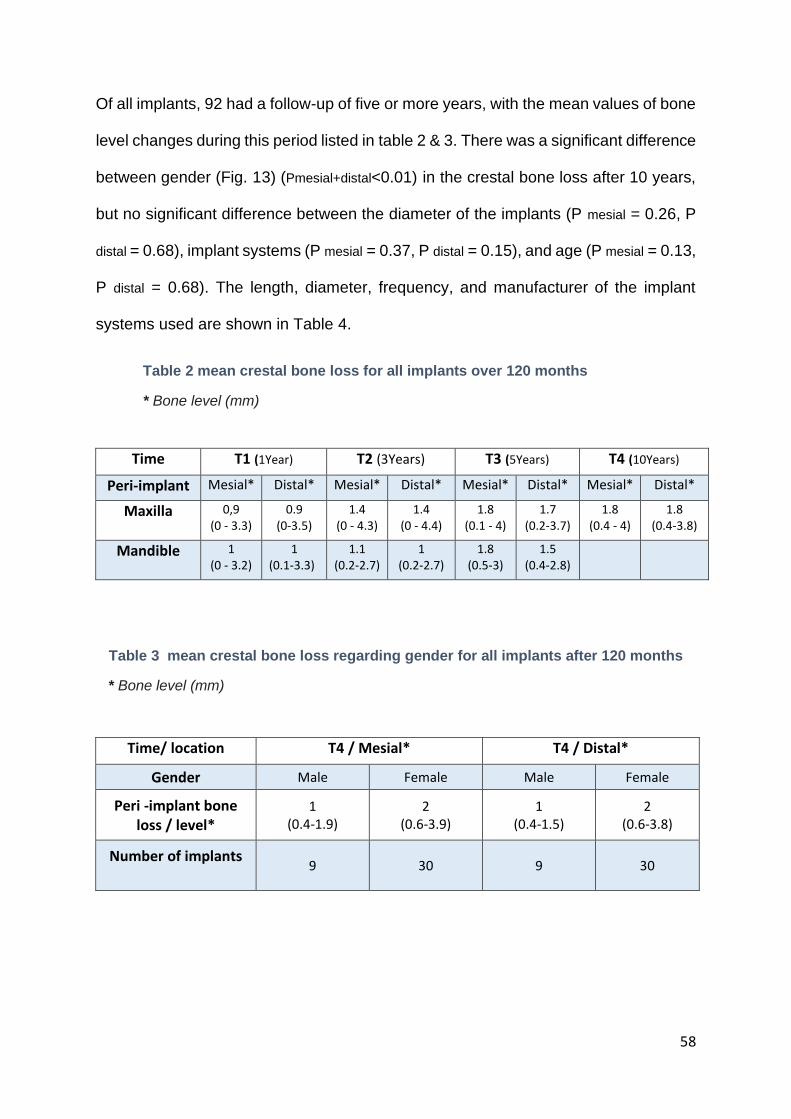
58
Of all implants, 92 had a follow-up of five or more years, with the mean values of bone
level changes during this period listed in table 2 & 3. There was a significant difference
between gender (Fig. 13) (Pmesial+distal<0.01) in the crestal bone loss after 10 years,
but no significant difference between the diameter of the implants (P mesial = 0.26, P
distal = 0.68), implant systems (P mesial = 0.37, P distal = 0.15), and age (P mesial = 0.13,
P distal = 0.68). The length, diameter, frequency, and manufacturer of the implant
systems used are shown in Table 4.
Table 2 mean crestal bone loss for all implants over 120 months
* Bone level (mm)
Time T1 (1Year) T2 (3Years) T3 (5Years) T4 (10Years)
Peri-implant Mesial* Distal* Mesial* Distal* Mesial* Distal* Mesial* Distal*
Maxilla 0,9 (0 - 3.3)
0.9 (0-3.5)
1.4 (0 - 4.3)
1.4 (0 - 4.4)
1.8 (0.1 - 4)
1.7 (0.2-3.7)
1.8 (0.4 - 4)
1.8 (0.4-3.8)
Mandible 1 (0 - 3.2)
1 (0.1-3.3)
1.1 (0.2-2.7)
1 (0.2-2.7)
1.8 (0.5-3)
1.5 (0.4-2.8)
Table 3 mean crestal bone loss regarding gender for all implants after 120 months
* Bone level (mm)
Time/ location T4 / Mesial* T4 / Distal*
Gender Male Female Male Female
Peri -implant bone loss / level*
1 (0.4-1.9)
2 (0.6-3.9)
1 (0.4-1.5)
2 (0.6-3.8)
Number of implants 9 30 9 30

59
Male Female
Fig 13 Comparison of bone loss between males and females
Diagram showing the significant differences of the bone loss between
M-Male and F-Female.

60
Table 4 lengths, diameters, frequencies, and manufacturers of implant system used.
Diameter
Frequency
%
Length
Frequancy %
3.3 4 17.4 10 1 4.3
Steri - Oss 3.8 14 60.9 12 14 60.9
4.5 5 21.7 14 4 17.4
16 4 17.4
Total 23 100 Total 23 100
3.8 63 63.4 9 4 4.0
Camlog 4.3 33 33.3 11 45 45.5
5.0 2 2 13 45 45.5
6.0 1 1 16 5 5.0
total 99 100 Total 99 100
Straumann
3.3
2
7.1
10
17
60.7
4.1 26 92.9 12 11 39.3
total 28 100 Total 28 100
Table 5 mean peri-implant bone resorption according to implant system after 10-years follow – up, T4.
Implant system Steri-Oss Camlog Straumann
Number of implants 23 16 0
T4 / Mesial
Peri -implant bone
loss in mm
0.9
(0.4 – 3.9)
1
(0.5 -3.9)
T4 / Distal Peri-implant bone
loss in mm
0.9 (0.4 – 3.8)
0.9 (0.8 – 3.4)

61
5. Discussion
Population ageing and the overall number of infections, trauma and cancer of the
maxilla and mandible are increasing, which can result in alveolar and basal bone loss
necessitating advanced surgical and prosthetic reconstructive treatment (Maiorana et
al., 2005). Typically, a collapse of the ridge tissue appears within the first three months
after tooth extraction (Schropp et al., 2003) and this alteration in the architecture of the
soft tissue and bone morphology can make the functional and aesthetic rehabilitation
of the oral cavity more difficult, whether via conventional dentures or with an implant-
supported prosthesis. Therefore, an adequate bone volume is needed for the insertion
of dental implants for confirming primary stability (Misch et al., 1994; Castagna et al.,
2013). In cases with severe alveolar atrophy, the treatment strategy demands a
significant reconstruction by utilising autogenous bone grafts harvested from extraoral
donor sites (Maiorana et al., 2005).
The present study evaluated the success rate of implants and radiographical data of
bone level changes in peri-implant regions in thirty-two partially edentulous and
edentulous patients. Initially, all patients had severely atrophied maxilla and mandible
requiring bone augmentation before implant placement. The analysis revealed a high
success rate after ten years, with a mean value of 95%, and a relatively stable peri-
implant bone level of 1–2 mm during the observation period. In jaws that were
augmented with iliac bone grafts, the bone resorption rate around implants was
comparable to areas augmented with autogenous bone grafts harvested from other
donor sites as well as around the implants in native non-augmented bones.

62
The graft quality
In the current study, a strategy was adopted to overcome the well-known tendency of
endochondral bone (iliac bone graft) for resorption. The protocol involved only
harvesting a corticocancellous block containing mainly cortical curvature. As
described in the methodology section, the corticocancellous iliac bone block was
harvested from the medial inner table of the ilium. The outer two cortical layers faced
the periosteum and represented the later occlusal and vestibular aspects of the jaws.
The inner cancellous layer faced the recipient bone after its manipulation to adapt the
jaw surfaces. This concept has been discussed and investigated in numerous studies
(Nelson et al. 2006a; Heberer et al., 2008). With no regard to the available amount of
bone graft, bone density is considered a crucial factor for successful osseointegration
which will improve the primary stability of dental implants (Esposito et al., 1998).
In general, several studies clarified that the embryologic origin of bone plays a major
role in determining the rate and behaviour of bone resorption and survivability. The
superior preservative property of membranous over endochondral bone has been
highlighted in many studies and provides evidence about its favourable clinical use as
an onlay grafting material in the craniofacial skeleton. Ozaki (1998) reported that the
cortical bone is a superior onlay grafting material, regardless of the embryologic origin
of its donor site (Ozaki & Buchmann, 1998). The resorption tendency of iliac crest bone
grafts might be related to the quality and microarchitecture of the graft, as it is now
obvious that the amount of cortical bone is crucial for the maintenance of the graft
volume rather than the native embryologic origin (Ozaki & Buchmann, 1998; Ozaki et
al., 1999; Heberer et al., 2008). Lundgren et al. (1997) stated that both cortical
thickness and density of the donor bone are essential factors that could affect bone
resorption pattern. The corticocancellous iliac grafts should be harvested with a thick

63
resorption-resistant cortical bone to maintain sufficient graft volume after the initial
resorption, thereby enables placement of long implants with optimum primary stability
(Lundgren et al., 1997). Indeed, Heberer et al. (2008) reported that the harvested thick
cortex iliac bone blocks with a large proportion (72%) of cortical bone resulted in a
mean 1.2 mm low bone resorption after 3 months of healing.
Simultaneous or delayed
This study verified the two-step technique which involves augmentation of the
atrophied jaw, followed by implant placement after a primary healing period of 3
months in contrast to the one-step technique allowing insertion of the dental implants
at the same time as jaw augmentation with a bone graft. Many previous studies have
discussed and compared the one and two-step approaches, observing bone-implant
interference when studying its related influence on the successful outcome (Lundgren
et al., 1999; Yerit et al., 2004; Tosun et al., 2018). There are issues associated with
the one-step technique, such as insufficient implant positioning for prosthetic
rehabilitation and tissue dehiscence, which lead to poor primary implant stability and
poor prosthetic orientation (Nyström et al., 1993; Vermeeren et al., 1996; Van der Maij
et al., 2004). Also, a high rate of marginal bone loss is associated with the one-step
approach (Vermeeren et al.,1996; Tosun et al., 2018). Nonetheless, the one-step
method has some advantages, a single operation is more preferable to the patient and
more cost-effective as well as being more time-efficient, thereby accelerating patient’s
rehabilitation (Yerit et al., 2004; van der Meij et al., 2005). However, the simultaneous
method is still controversial given the high prevalence of complications (Vermeeren et
al., 1996; Verhoeven et al. 1997). Dehiscence is the most frequent complication and
can happen directly after augmentation. This wound dehiscence is accompanied by
infection, resulting in comprehensive bone loss in the peri-implant area, which may
64
lead to graft and implants loss. Other challenges include difficulties in angulation and
positioning of the implants as well as unexpected resorption at the augmented sites
(Vermeeren et al., 1996; Schliephake et al., 1997; Verhoeven et al., 1997; van der
Maij et al., 2005; Boven et al., 2014).
Lundgren et al (1999) preferred the second step method and described it in their
histomorphological and morphometric analyses study of ten patients. They placed two
micro-implants in the corticoancellous iliac graft and six months later (at ordinary
implant placement), one micro-implant was retrieved with a trephine drill and a new
one was placed in the graft. At the abutment surgery six months later, the remaining
two micro-implants were removed in the same way and three bone specimens were
histomorphologically analysed in the different healing periods (0–6, 0–12 and 6–12
months). The authors concluded better osseointegration of titanium micro-implants in
the augmented maxilla with an onlay iliac bone graft after 6 months from
augmentation. They showed that during the healing time in the two-step technique,
the graft was revascularised comprising regenerated bone and bone marrow at the
time of implant placement. This is in contrast to the one-step technique when the
implant is placed into virtually non-viable bone. Other studies reported a striking
difference in the survival and success rates between simultaneous and delayed
implant placement in the augmented jaw. Yerit et al. (2004) compared the one and
two-step technique after horseshoe Lefort I osteotomy with an iliac bone graft, finding
that the 5-year survival rate of simultaneous implant placement was 87% in contrast
to the delayed implant placement of 91%. Although their patients were satisfied after
prosthetic rehabilitation, they concluded that the two-step technique was the method
of choice. Sjöstrom et al. (2006) applied radiological examination and resonance
frequency analysis measurement six months after grafting and implantation in a three-

65
year follow-up survey, showing predictable and stable long-term results of 29 atrophic
edentulous maxilla augmented with an anterior iliac bone graft and treated with
delayed implant placement. In the long-term ten-year observational study of
Schliephake et al. (1997), they reported a noticeable difference in the success rate
between primary and secondary implant placement on an augmented jaw with a
combined inlay and onlay iliac bone grafts of 67.6% and 96.6%, respectively. Tosun
et al. (2018) also reported that the delayed approach is a credible method with a high
success rate and less bone resorption.
By contrast, the healed augmented alveolar bone in the two-step approach permits
more adjustment in the alveolar bone before implant placement. The visual inspection
of the graft after primary healing and the initial resorption permits implant placement
within the optimum depth (epicrestal, supracrestal and subcrestal according to implant
system manufacturer requirement). Conversely, implants placed simultaneously to
bone graft, which may undergo a huge bone loss that threatens the graft and implant
success outcome, was also suggested by Lundgren et al. (1997). Furthermore, the
two-step approach enables implant placement with navigation systems utilising 3D
planning and a surgical guide splint. All the above enables good primary stability and
optimum positioning of the dental implant, which are the most important requirements
for successful dental rehabilitation of edentulous or partially edentulous patients (Yerit
et al., 2004). In this study, the two-step technique was performed with implant insertion
three months after grafting, with functional prosthetic loading after another three
months. At loading, the stability of the implants was re-examined utilising a torque
ratchet and the prosthodontic treatment was initialised only when the torque value was
greater than 35 Ncm. Many studies have debated the most appropriate duration of
healing time before implant placement, discussing whether a longer healing time

66
means better bone integration and revascularisation. However, there will be more
bone graft loss when it is not punctually and adequately loaded. It has been shown
that three months of healing for both the graft and the implants are sufficient to ensure
revascularisation of the graft, as well as secure insertion and loading of rough-surfaced
dental implants (Nelson et al., 2006 a, b; Heberer et al., 2008). Nelson et al. (2006a)
concluded that there was an adequate bony structure for secure implant placement
after a healing period of three months in their histomorphometric evaluation, reporting
that the amount of newly formed bone after three months was comparable to that
formed after 4–5 months of healing. Furthermore, it was possible to prevent the onset
of graft resorption by shortening the healing time before implantation. Tosun et al.
(2018) indicated that implant placement three months after onlay iliac bone grafting is
more reliable and results in a higher success rate and less bone resorption compared
with simultaneous implant placement.
The success rate and resorption behaviour reported in these studies are in line with
the present study findings, hence, the two-step technique is regarded as a standard
technique in dealing with the placement of dental implant after jaw augmentation with
an onlay iliac bone graft. Autologous bone blocks can be incorporated in the recipient
site within three months (Heberer et al., 2008), so, it is not useful to wait for more than
six months before implant placement because the graft may undergo resorption at the
periosteal surface if it is not promptly and sufficiently loaded (Nelson et al., 2006 a, b;
Heberer et al., 2008; Cordaro et al., 2011; Cordaro & Terhyeden 2014, 2019).

67
Success rate
It is generally accepted that osseointegration is a basic requirement for successful
implantation. "Osseointegration" is defined as living and organised bone tissue in direct
contact with a functionally loaded implant without any intervening connective tissue
(Brånemark, 1983). It is important to know the consequences if an implant is no longer
fully osseointegrated, therefore, success and survival rates were discussed and
determined in several clinical studies to evaluate dental implant destiny. However,
comparison and definitions of these two terms can differ significantly from one another
The first step in determining a success rate is to define success criteria and many of
the clinical and radiological success criteria for implants to date are negative criteria,
the absence of which indicates success and the occurrence of which indicates failure
(Albrektsson et al., 1986; Buser et al., 1990; Misch et al., 2008).
In this current study, the clinical evaluation of implants placed on the augmented
maxilla and mandible has been done according to the success criteria of Buser et al.
1990. The clinical evaluation was made after 1, 3, 10, and 30 days after augmentation
as well as 3 and 6 months, then annually up to 10 years post-implantation. The
evaluation included the assessment of complications such as inflammation, mucosal
erythema, wound dehiscence, loss of bone grafts and implant mobility. The
radiographical examination (panoramic x-ray) was performed before and directly after
augmentation and implantation, then at 3 months, 6 months, 1 year, 3 years, 5 years
and 10 years. In this observational study, the implants were described as successful
when there was no mobility, no continuous peri-implant radiolucency, no recurrent peri-
implant infection with putrid secretion and absence of persistent symptoms such as
pain, feeling of foreign body or dysesthesia (Buser et al., 1990). In this study, good
bone graft integration in the recipient site, the high implant success rate (mean: 95%)

68
and the mean value of peri-implant bone loss confirmed the success criteria of
Albrektsson et al. (1986) and Buser et al. (1990).
The present study results are in line with several long-term follow-up studies which
have reported a high success rate of implants after augmentation with an iliac bone
graft (Nyström et. al., 2009; Chiapasco et al., 2008; Boven et al., 2014; Nguyen et al.,
2019). The prospective study of Nyström et al. (2009) observed in the long-term the
success outcome of 334 implants in 44 patients who received an onlay and inlay bone
grafts harvested from the anterior iliac bone crest. All patients waited six months for
graft healing before implant placement and another 6 months for osseointegration
before prosthetic rehabilitation. After 11 years of follow-up, the success rate was 90%.
The study of Nyström et al. confirmed the treatment outcome of the current study and
even the current work has a better success rate after treatment of both atrophic jaws.
Chiapasco and colleagues showed an implant success rate of 93.7% and a survival
rate of 96.7% among 60 implants, which were placed after 4–7 months in the
reconstructed mandible with ilium or calvarium bone after benign tumour resection. No
information about the success rates regarding the different augmentation procedure
was given. The sixteen patients were subjected to a follow-up of (mean: 94 months,
range: 36–132 months) after rehabilitation with a fixed prosthesis and 14 of them were
fully satisfied (Chiapasco et al., 2008). Boven et al. reported a survival rate of 98.7%
in a five-year observational study involving forty edentulous patients who received two
implants four months after augmentation with an onlay iliac bone graft in the
intraforaminal area and overdenture for each one. The good results of Boven et al.
included a stable marginal bone loss of 0.6 +/- 0.7 mm, high patient satisfaction and
stable clinical parameters, which correspond with the success rate criteria of
Albrektsson et al. and Buser et al. However, the high survival rate was not regarded

69
as a fully successful outcome, maybe because some patients suffered from sensitivity
disturbances due to mental nerve damage during mandible augmentation, which is
inconsistent with the success criteria of Albrektsson (1986) and Buser (1990). The two
studies are in agreement with the results of the current study, particularly for the
success rate, which included the general prosthetic rehabilitation outcome and patient
acceptance of general complaints regarding donor and recipient sites.
Nguyen and colleagues (2019) achieved a 100% success rate of implants placed in
the augmented maxilla and mandible with an anterior iliac bone graft, followed by a
removable and fixed dental prosthesis. The implant placement was performed 4–6
months after augmentation and implants healing expended to 3–5 months, which was
ended by the initiation of prosthetic rehabilitation. After a mean observation period of
50 months (range, 12–62 months), they concluded that a combination of the iliac onlay
bone graft and dental implants resulted in satisfactory reconstruction and a reliable
long-term prognosis (Nguyen et al., 2019). Although they reported a higher success
rate than that of the current study, the comparison is limited because they utilised a
small sample (only seven patients with 29 implants).
Another study reported a lower success rate of dental implant placed in an augmented
jaw with ilium in the long-term. Nyström et al. (2002) achieved a success rate of 74.6–
85%, which was considered at the date of publication as a high success rate. The
patients gained augmentation of the atrophied maxilla with an iliac bone graft and
placement of dental implants, followed by a fixed prosthesis. All patients in their study
underwent radiological investigation with computer tomography. The low success rate
might be due to all patients undergoing the one-stage approach as well as more than
one graft failure occurred in some patients, which induced inflammation that resulted
70
in dehiscence of the covering soft tissue, consequently leading to a high marginal bone
loss or in some cases, complete loss of the graft.
For comparison, studies have reported implant success rates in the non-augmented
bone of 84% to 98.7% with follow-up periods ranging between 2 and 15 years
(Albrektsson et al., 1986; Buser et al., 1997; Pikner et al., 2009; Mertens et al. 2012;
Degidi et al., 2012). The long-term study of Degidi and colleagues (2012) compared
the survival rate of implants placed in healed and post-extractive sites, with success
rates of 98.05% and 96.52%, respectively. They measured the success of the implant
according to the criteria of Albrektsson et al. (1986) and Misch et al. (2008). The
survival rate of Degidi et al. (2012) is in the line with the current study but is limited to
compare because they utilised the one system (TiUnite implants) and the post-
extractive group demonstrated a low success rate of restoration, which affected the
peri-implant soft and hard tissue. Nonetheless, the current study reported a better
outcome in comparison to Degidi et al., especially as the qualitative success rate was
intensively explored in this study studied. The 11-year study of Mertens et al. (2012)
studied the outcome of patients (15) and implant (94) level after the restoration of the
edentulous maxilla (non-augmented) with an implant-supported fixed prosthesis,
reporting a success rate of 92% according to Albrektsson’s criteria (1986). The current
results indicated a higher mean of success rate even when compared with the success
rate of implants placed in the native non-augmented jaw like that of Mertens et al.
(2012). In the present work, the 150 implants success rate during long-term follow-up
was comparable to the success and survival rate of dental implants placed in the native
non-augmented jawbone. The success rate assessment may not be profound or
significant without an evaluation of the peri-implant bone level changes, therefore, the
bone resorption rate was measured around the implants in this study.

71
Peri-implant bone resorption
The stable peri-implant bone level is regarded as a crucial factor to assess dental
implant outcomes measured by clinical and radiological parameters (Lang et al., 2012;
Lang & Zitzmann, 2012). Marginal bone loss is very important in dental implantology
because the peri-implant bone is necessary for implant stability and plays an important
role in aesthetic outcomes. The presence of sufficient bone in the peri-implant area
has a direct impact on soft tissue integrity, consequently, this will improve both
hygienical and aesthetical aspects (Nisapakultorn et al., 2010; Negri et al., 2014). Only
a few long-term studies have evaluated the peri-implant bone level changes after onlay
iliac crest bone grafting (Sjöstrom et al., 2006; Nyström et al., 2009; Boven et al. 2014).
In the present study, the peri-implant bone level was radiographically measured and
examined regarding the reference point as mentioned before in the methodology
section. In the current work, the insertion level of 23 Steri-Oss Nobel Biocare, 99
RootLine Camlog (machined surface,1.4 mm), 10 Straumann implants (bone level)
was equicrestally and 18 Straumann implants (tissue level) were placed with the
smooth-rough border at the crestal bone level (supracrestally, implant-abutment
interference 2.8 mm above the crestal bone). Frequently, the implant platform was
placed either equicrestally or surpracrestally and sometimes subcrestally according to
the manufacturer’s instructions. A two-year observational study of 120 implants with
various systems, insertion levels and different internal connections reported that the
placement of implant supracrestally revealed low marginal bone resorption compared
to crestally placed implants (Augustin-Panadero et al., 2019). Based on a systematic
review, implants placed equicrestally revealed more peri-implant bone resorption
(Schwarz et al., 2014). Chiapasco and colleagues (2014) conducted a comparative
study of 95 bone level and 97 tissue level Straumann implants, comparing

72
radiographically the peri-implant bone level of the implants in 50 patients within a
follow-up period ranging from 12–68 months. All patients received jaw reconstruction
treatment with a different autogenous bone graft (iliac bone, calvarium and retromolar)
and they demonstrated that regardless of the type of bone graft, the tissue level
implants (supracrestal) indicated lower peri-implant bone resorption. In the present
study, there was no significant difference in peri-implant bone level changes among
the different implant systems used but only 18 tissue level implants were analysed in
the present study.
Therefore, the implant insertion level and the implant-abutment junction are decisive
factors having a direct impact on marginal bone loss. In the current investigation, the
major crestal bone loss appears within the first year, with an average loss of less than
1 mm. Other studies investigated the marginal bone loss around implants that were
placed in augmented jaws with anterior iliac crest bone (Nyström et al., 2009; Boven
et al., 2014; Tosun et al., 2018; Nguyen et al., 2019). In the long-term prospective study
of Nyström et al. (2009), they observed for 11 years the successful outcome and
marginal bone resorption around 334 implants in 44 patients that received onlay and
inlay bone grafts harvested from the anterior iliac bone crest. All patients were treated
with the two-step approach, waiting six months for both implant placement and
prosthetic restoration. By the end of their observation, the mean bone resorption was
2.4 mm after 10 years, 1.8 mm in the first year. Hence, the present study findings are
in agreement in more than one aspect despite the present study having more positive
points, that is, the success rate was higher, the peri-implant bone loss was less over
10 years and both jaws were treated.
Boven et al. (2014) observed the peri-implant bone resorption of implants placed after
4 months from augmentation with an iliac bone graft in the interforaminal area in 40

73
patients, showing that the mean peri-implant bone loss of implants in the augmented
area was comparable with those placed in the non-augmented mandible. They
observed the peri-implant bone level radiologically with OPTG and demonstrated an
accumulative mean radiographic marginal bone loss of 0.6 + 0.7 mm over five years.
Although the authors only reconstructed the mandible, their data agree with those of
the current study regarding the bone loss rate. However, the comparison is still limited
because only bone level changes in the mandible were evaluated and information
about the implant insertion level is missing. Tosun et al. (2018) reported that the mean
resorption values on the buccal, lingual, mesial, and distal sides of the implants were
1.08 mm, 0.36 mm, 0.30 mm, and 0.25 mm respectively, (mean 0.49 mm) in the
delayed implantation after 3 months from augmentation with an anterior iliac bone graft.
Despite this study reporting a lower rate of marginal bone loss, it is difficult to directly
compare to the current study as they only examined 61 implants of 10 patients for 29
months and only inserted one implant system (Straumann Standard Plus, Switzerland),
with the radiological examination performed with a Cone-Beam CT at 29 months after
implantation. Nguyen et al. (2019) measured the peri-implant bone changes mesially
and distally of 29 implants inserted in the maxilla and mandible after augmentation with
anterior iliac crest bone. Although they inserted two implant types, bone level and
tissue level Straumann implants, they found no significant difference in the marginal
bone loss at the end of the observation period (1.12 ± 0.50 mm). The value and nature
of the marginal bone loss in the present study are comparable to that of Nguyen et al.
Furthermore, the observation time in the current study was much longer than that of
Nguyen et al. (mean, 50 months), with a higher number of implants (150) compared.
Duttenhoefer et al. (2015) evaluated and observed the successful outcome of graft and
dental implants, as well as the peri-implant bone level changes in augmented jaws with

74
non-vascular fibula up to 15 years. The 39 dental implants in 8 patients revealed a high
success rate of 97% and a low peri-implant bone reduction of 1.4 mm mesially and
distally after 10 years, with their results fulfilling the success criteria of Buser. The
highest rate of bone resorption occurred in the first year after implant placement (about
0.7 mm), with no subsequent significant marginal bone reduction thereafter according
to radiological examination up to 15 years. The bone loss nature, the low peri-implant
bone resorption and the high success outcome of Duttenhoefer et al. are comparable
to the current work. Although their results are better than these of the current study,
their observations included only eight patients.
Voss et al. (2016) evaluated the long-term (10 years) success of implants after grafting
with particulate bone harvested from the retromolar area and peri-implant bone loss.
They inserted 164 implants in the maxilla and mandible, reporting a mean bone loss of
2.47 mm and 2.50 mm mesially and distally, respectively. They concluded that the
primary stability of the implant has a direct impact on bone resorption, especially as
some implants were inserted simultaneously while other implants were placed 4–6
months after augmentation. These results confirm the two-step technique induces high
primary stability and are in line with the present study, especially as they utilised
several implant systems in both jaws and followed up patients for up to 10 years.
However, the peri-implant bone loss in the current study is lower than that of Voss et
al (2016) considering that a large number of implants were placed simultaneously with
bone grafting in their work. The comparable results of the last two studies in addition
to the current study confirm the suggestion of Ozaki and Buchmann (1998), who
demonstrated that the cortical bone and its 3D microarchitecture is a superior onlay
grafting material, regardless of the donor site and embryologic origin. The comparison
of the current study with the results of the above-mentioned augmentation studies

75
demonstrates that the current results are comparable with the good outcome of long-
term as well as short-term studies (Nelson et al., 2006 a, b; Nystöm et al., 2009; Boven
et al., 2014; Voss et al., 2016; Tosun et al., 2018; Nguyen et al., 2019).
The present study results demonstrate that the implants placed in augmented bone
have similar bone level changes compared to implants inserted in non-augmented
regions. In the 10-year prospective study of Degidi et al. (2012), they surveyed 210
TiUnite implants in 59 patients and radiographically evaluated the accumulated mean
peri-implant bone loss, 1.93 mm and 1.98 mm in healed and post-extractive sites
respectively. The value of marginal bone loss confirmed the results of the current study,
especially as they measured the difference in bone level mesially and distally in the
long term. However, they did not define the level of implant insertion exactly (slightly
above crestal level). In the study of Mertens and colleagues (2012), they performed an
annual radiographical examination of 94 implants in 15 patients and showed relatively
stable mesial and distal bone level loss of 0.88 mm after 11 years, which is comparable
with the success rate of the present study. They reported a lower marginal bone loss
in comparison to the current study but the comparison is still limited especially as they
only utilised one implant system (TiO blast TM, Astra Tech AB, Mölndal, Sweden) in
the non-augmented maxilla and did not state the insertion level of the implants. In the
10-year observation study of Pikner and colleagues (2009), they included 640 patients
treated with 3,462 Brånemark implants and excluded patients with augmentation and
overdentures, finding that the worst bone loss occurred in the first year, then slowed
down thereafter. They recorded a mean marginal bone loss of 2.1 mm in the mesial
and distal aspect, reporting that the implants placed close to the midline of the
mandible in edentulous jaws showed a higher bone loss in comparison to implants in
other positions.

76
The present study findings are in agreement with the above studies regarding the peri-
implant bone loss rate and its nature. The present study showed that there is no effect
of patient age, as confirmed by Mertens et al. (2012), Digidi et al. (2012), Voss et al.
(2016) and Pikner et al. (2009), whereas Hugoson and Laurell (1993) observed an
increased occurrence of marginal bone loss in natural dentation of older age groups.
It has been observed that gender has a significant effect on the treatment outcome, as
there was a notable bone loss in females compared with males after 10 years. This
could be attributed to hormonal factors that may influence bone physiology. Manolagas
et al. (2013) discussed the role of oestrogens and androgens in bone health and
preservation, stating that sex steroid hormones can preserve the balance between
bone accretion and resorption through changes in osteoclastogenesis and
osteoblastogenesis. Osteoporosis and compromised systemic bone metabolism may
affect bone remodelling and osseointegration or maintenance of osseointegration of
implants (Chrcanovic et al., 2015a). Osteoporosis is a common condition in
postmenopausal women due to the reduction in the level of oestrogen which is
responsible for inducing early and late forms of osteoporosis in women after
menopause and elderly men (Riggs, 2000). Theoretically, all the above outweigh the
hypothesis that female patients face more peri-implant bone resorption than male
patients, thereby confirming the findings of the current study. The aforementioned
study of Nyström et al. (2009) found that female patients after 11 years showed a
higher rate of failure implant of 11% than male, while male patients had more marginal
bone resorption. They also demonstrated that smokers had a lower success rate and
more bone resorption.
According to a systematic review and meta-analysis of Chrcanovic et al. (2015a), the
insertion of implants in male patients influenced the marginal bone loss and survival

77
rate of implants. They reviewed 91 publications, including 27, 203 and 25,154 implants
in males and females, respectively, with male patients having a slightly lower survival
rate and more marginal bone loss. Negri and colleagues (2014) concluded after 36
months of clinical and radiographical investigation of 632 implants in 252 patients, that
patient gender and age significantly affected the marginal bone loss around the
implants, with elderly men suffering from more peri-implant bone resorption around
implants placed in the maxilla, in contrast to the present study. However, their
observational period was shorter, and smokers were not excluded from their study.
The clinical trial of Chrcanovic et al. (2015b) further suggested that smoking has a
statistically significant effect on influencing implant failure rates. A survey in the United
States (2005–2014) found that the smoking prevalence within the adult population was
higher among males than females (Jamal et al., 2014), which may account for the
difference between the results of the above two studies and the results of the current
study, especially as smoking was an exclusion criterion in the present work.
It is difficult to anticipate the impact of patient gender on marginal bone loss around
the implants because of the limited number of studies regarding the marginal bone
loss. Although the survival and success outcome of osseointegrated implants are well
discussed and reported extensively, the effects of gender are still unknown
(Chrcanovic et al., 2015a). The impact of gender demonstrated in the present study
cannot be compared to other studies, hence further scientific studies are necessary to
confirm this finding.

78
6. Conclusion
In previous capital, there was a trail to compare the results of this study with other
results. This comparison dealt with studies that observed the outcome and peri-implant
bone level changes in patients who had undergone implant placement after
augmentation with an iliac bone graft, non-vascular fibula and retromolar bone graft or
non-augmented jaws. The treatment outcome and findings were discussed and
compared, as well as their related parameters in both jaws for short-term and long-
term studies, revealing that the results of the present study are comparable with the
good results of the aforementioned studies. At the same time, the patients who
participated in this study suffered from extreme atrophy in both jaws and achieved
successful implant-dependent prosthetic rehabilitation in the long-term. It is important
to note that the assessment of success does not depend only on the presence and
stability of the implants, many parameters defined the success rate.
In summary, this retrospective study demonstrated a high implant success rate (96%
in maxilla and 92% in mandible) of implants that were placed into the augmented
atrophic jaws which have been built up by utilising an onlay bone graft harvested from
the anterior iliac bone crest. The peri-implant bone level changes in augmented jaws
were comparable with those of non-augmented jaws. These results confirm the choice
of the autogenous iliac crest graft as the "gold standard" for the reconstruction of the
extremely atrophied upper and lower jaw.

79
7. References
Ahlmann, E., Patzakis, M., Roidis, N., Shepherd, L. and Holtom, P., Comparison of
anterior and posterior iliac crest bone grafts in terms of harvest-site morbidity and
functional outcomes, J Bone Joint Surg Am, 2002, 84(5):716-720
Akiyama H, Chaboissier MC, Martin JF, Schedl A, de Crombrugghe B. The
transcription factor Sox9 has essential roles in successive steps of the chondrocyte
differentiation pathway and is required for expression of Sox5 and Sox6. Genes Dev.
2002;16(21):2813-2828.
Albrektsson T, Zarb G, Worthington P, Eriksson AR. The long-term efficacy of
currently used dental implants: a review and proposed criteria of success. Int J Oral
Maxillofac Implants. 1986;1(1):11-25.
AlGhamdi AS, Shibly O, Ciancio SG. Osseous grafting part I: autografts and allografts
for periodontal regeneration--a literature review. J Int Acad Periodontol. 2010;12(2):34-
38.
Allen MR, Hock JM, Burr DB. Periosteum: biology, regulation, and response to
osteoporosis therapies. Bone. 2004;35(5):1003-1012.
Allen MR, Burr DB. Chapter 5 - Bone Growth, Modeling, and Remodeling. In: Burr
DB, Allen MR. Basic and Applied Bone Biology (Second Edition): Academic Press;
2019.
Aloy-Prósper A, Maestre-Ferrin L, Peñarrocha-Oltra D, Peñarrocha-Diago M. Bone
regeneration using particulate grafts: an update. Med Oral Patol Oral Cir Bucal.
2011;16(2):e210-214.
AlSheikh HA, AlZain S, Warsy A, AlMukaynizi F, AlThomali A. Mandibular residual
ridge height in relation to age, gender and duration of edentulism in a Saudi population:
A clinical and radiographic study. Saudi Dent J. 2019;31(2):258-264.
Amir LR, Becking AG, Jovanovic A, Perdijk FB, Everts V, Bronckers AL. Formation of
new bone during vertical distraction osteogenesis of the human mandible is related to
the presence of blood vessels. Clin Oral Implants Res. 2006;17(4):410-416.
Araújo MG, Lindhe J. Dimensional ridge alterations following tooth extraction. An
experimental study in the dog. J Clin Periodontol. 2005;32(2):212-218.
Arrington ED, Smith WJ, Chambers HG, Bucknell AL, Davino NA. Complications of
iliac crest bone graft harvesting. Clin Orthop. 1996;329:300-9.
Artzi Z, Kozlovsky A, Nemcovsky CE, Weinreb M. The amount of newly formed bone
in sinus grafting procedures depends on tissue depth as well as the type and residual
amount of the grafted material. J Clin Periodontol. 2005;32(2):193-199.
Atwood DA. Some clinical factors related to rate of resorption of residual ridges. 1962.
J Prosthet Dent. 2001;86(2):119-125.
Atwood DA, Coy WA. Clinical, cephalometric, and densitometric study of reduction of
residual ridges. J Prosthet Dent. 1971;26(3):280-295.

80
Ausenda F, Rasperini G, Acunzo R, Gorbunkova A, Pagni G. New Perspectives in the
Use of Biomaterials for Periodontal Regeneration. Materials (Basel). 2019;12(13).
Banwart JC, Asher MA, Hassanein RS. Iliac crest bone graft harvest donor site
morbidity. A statistical evaluation. Spine. 1995;20: 1055-60.
Barone A, Ricci M, Mangano F, Covani U. Morbidity associated with iliac crest
harvesting in the treatment of maxillary and mandibular atrophies: a 10-year analysis.
J Oral Maxillofac Surg. 2011;69(9):2298-2304.
Beck-Broichsitter, B. E., Garling, A., Koehne, T., Barvencik, F., Smeets, R., Mehl, C.,
Jeschke, A., Wiltfang, J. and Becker, S. T.3D-tracking the regenerative potential of the
mandible with micro-CTs. Oral Maxillofac Surg. 2015;19(1):29-35.
Bielby, R., Jones, E. and McGonagle, D., The role of mesenchymal stem cells in
maintenance and repair of bone, Injury, 2007, 38 Suppl 1:S26-32
Bischoff DS, Zhu JH, Makhijani NS, Kumar A, Yamaguchi DT. Angiogenic CXC
chemokine expression during differentiation of human mesenchymal stem cells
towards the osteoblastic lineage. J Cell Biochem. 2008;103(3):812-824.
Bonewald LF. The amazing osteocyte. J Bone Miner Res. 2011;26(2):229-238.
Boskey AL. Bone composition: relationship to bone fragility and antiosteoporotic drug
effects. Bonekey Rep. 2013; 2:447.
Boskey AL, Coleman R. Aging and bone. J Dent Res. 2010;89(12):1333-1348.
Boskey AL, Posner AS. Bone structure, composition, and mineralization. Orthop Clin
North Am. 1984;15(4):597-612.
Boven GC, Meijer HJ, Vissink A, Raghoebar GM. Reconstruction of the extremely
atrophied mandible with iliac crest onlay grafts followed by two endosteal implants: a
retrospective study with long-term follow-up. Int J Oral Maxillofac Surg.
2014;43(5):626-632.
Boyle WJ, Simonet WS, Lacey DL. Osteoclast differentiation and activation. Nature.
2003;423(6937):337-342.
Branemark P-I. Osseointegration and its experimental background. The Journal of
Prosthetic Dentistry. 1983;50(3):399-410.
Bremm LL, Sallum AW, Casati MZ, Nociti FH, Sallum EA. Guided tissue regeneration
in Class II furcation defects using a resorbable polylactic acid barrier. Am J Dent.
2004;17(6):443-446.
Brown JS. Deep circumflex iliac artery free flap with internal oblique muscle as a new
method of immediate reconstruction of maxillectomy defect. Head Neck.
1996;18(5):412-421.
Buckwalter JA, Glimcher MJ, Cooper RR, Recker R. Bone biology. I: Structure, blood
supply, cells, matrix, and mineralization. Instr Course Lect. 1996;45:371-386.
Budihardja AS, Mücke T. Bone Management in Dental Implantology. Switzerland:
Springer; 2019.

81
Bunyaratavej P, Wang HL. Collagen membranes: a review. J Periodontol.
2001;72(2):215-229.
Burr, D. B., Schaffler, M. B., Yang, K. H., Lukoschek, M., Sivaneri, N., Blaha, J. D. and
Radin, E. L. Skeletal change in response to altered strain environments: is woven bone
a response to elevated strain? Bone. 1989;10(3):223-233.
Burr DB. Chapter 1 - Bone Morphology and Organization. In: Burr DB, Allen MR. Basic
and Applied Bone Biology (Second Edition): Academic Press; 2019.
Burr DB, Akkus O. Chapter 1 - Bone Morphology and Organization: Elsevier Inc;
2014.
Buser D, Dula K, Belser UC, Hirt HP, Berthold H. Localized ridge augmentation using
guided bone regeneration. II. Surgical procedure in the mandible. Int J Periodontics
Restorative Dent. 1995;15(1):10-29.
Buser D, Ingimarsson S, Dula K, Lussi A, Hirt HP, Belser UC. Long-term stability of
osseointegrated implants in augmented bone: a 5-year prospective study in partially
edentulous patients. Int J Periodontics Restorative Dent. 2002;22(2):109-117.
Buser, D., Mericske-Stern, R., Bernard, J. P., Behneke, A., Behneke, N., Hirt, H. P.,
Belser, U. C. and Lang, N. P. Long-term evaluation of non-submerged ITI implants.
Part 1: 8-year life table analysis of a prospective multi-center study with 2359 implants.
Clin Oral Implants Res. 1997;8(3):161-172.
Buser D, Weber HP, Lang NP. Tissue integration of non-submerged implants. 1-year
results of a prospective study with 100 ITI hollow-cylinder and hollow-screw implants.
Clin Oral Implants Res. 1990;1(1):33-40.
Byrne DP, Lacroix D, Prendergast PJ. Simulation of fracture healing in the tibia:
mechanoregulation of cell activity using a lattice modeling approach. J Orthop Res.
2011;29(10):1496-1503.
Caplan AI, Bruder SP. Mesenchymal stem cells: building blocks for molecular
medicine in the 21st century. Trends Mol Med. 2001;7(6):259-264.
Carranza FA, Newman MG, Takei HH, Klokkevold PR. Carranza's clinical
periodontology. St. Louis, Mo.: Saunders Elsevier; 2006.
Castagna L, Polido WD, Soares LG, Tinoco EM. Tomographic evaluation of iliac crest
bone grafting and the use of immediate temporary implants to the atrophic maxilla. Int
J Oral Maxillofac Surg. 2013;42(9):1067-1072.
Cawood JI, Howell RA. A classification of the edentulous jaws. Int J Oral Maxillofac
Surg. 1988;17(4):232-236.
Chanavaz M. Anatomy and histophysiology of the periosteum: quantification of the
periosteal blood supply to the adjacent bone with 85Sr and gamma spectrometry. J
Oral Implantol. 1995;21(3):214-219.
Chen ST, Wilson TG, Jr., Hämmerle CH. Immediate or early placement of implants
following tooth extraction: review of biologic basis, clinical procedures, and outcomes.
Int J Oral Maxillofac Implants. 2004;19 Suppl:12-25.

82
Chiapasco M, Autelitano L, Rabbiosi D, Zaniboni M. The role of pericranium grafts in
the reduction of postoperative dehiscences and bone resorption after reconstruction of
severely deficient edentulous ridges with autogenous onlay bone grafts. Clin Oral
Implants Res. 2013;24(6):679-687.
Chiapasco M, Casentini P, Zaniboni M. Implants in reconstructed bone: a comparative
study on the outcome of Straumann® tissue level and bone level implants placed in
vertically deficient alveolar ridges treated by means of autogenous onlay bone grafts.
Clin Implant Dent Relat Res. 2014;16(1):32-50.
Chiapasco M, Colletti G, Romeo E, Zaniboni M, Brusati R. Long-term results of
mandibular reconstruction with autogenous bone grafts and oral implants after tumor
resection. Clin Oral Implants Res. 2008;19(10):1074-1080.
Chiapasco M, Zaniboni M, Boisco M. Augmentation procedures for the rehabilitation
of deficient edentulous ridges with oral implants. Clin Oral Implants Res. 2006;17 Suppl
2:136-159.
Cho TJ, Gerstenfeld LC, Einhorn TA. Differential temporal expression of members of
the transforming growth factor beta superfamily during murine fracture healing. J Bone
Miner Res. 2002;17(3):513-520.
Chow JW, Wilson AJ, Chambers TJ, Fox SW. Mechanical loading stimulates bone
formation by reactivation of bone lining cells in 13-week-old rats. J Bone Miner Res.
1998;13(11):1760-1767.
Chrcanovic BR, Albrektsson T, Wennerberg A. Dental implants inserted in male
versus female patients: a systematic review and meta-analysis. J Oral Rehabil.
2015;42(9):709-722. A
Chrcanovic BR, Albrektsson T, Wennerberg A. Smoking and dental implants: A
systematic review and meta-analysis. J Dent. 2015;43(5):487-498. B
Chu T-MG, Liu SS-Y, Babler WJ. Chapter 11 - Craniofacial Biology, Orthodontics, and
Implants: Elsevier Inc; 2014.
Claes L, Eckert-Hübner K, Augat P. The effect of mechanical stability on local
vascularization and tissue differentiation in callus healing. J Orthop Res.
2002;20(5):1099-1105.
Clementini M, Morlupi A, Agrestini C, Ottria L. Success rate of dental implants inserted
in autologous bone graft regenerated areas: a systematic review. Oral Implantol
(Rome). 2011;4(3-4):3-10.
Cordaro L, Torsello F, Morcavallo S, di Torresanto VM. Effect of bovine bone and
collagen membranes on healing of mandibular bone blocks: a prospective randomized
controlled study. Clin Oral Implants Res. 2011;22(10):1145-1150.
Cordaro L, Terheyden H, Chen S, Buser D, Wismeijer D. Ridge Augmentation
Procedures in Implant Patients: A Staged Approach. Berlin: Berlin: Quintessenz
Verlag; 2019.
Cornell CN, Lane JM. Newest factors in fracture healing. Clin Orthop Relat Res.
1992(277):297-311.

83
Currey JD. The many adaptations of bone. J Biomech. 2003;36(10):1487-1495.
Curtis TA, Ware WH. Autogenous bone graft procedures for atrophic edentulous
mandibles. J Prosthet Dent. 1977;38(4):366-379.
Dasmah A, Thor A, Ekestubbe A, Sennerby L, Rasmusson L. Particulate vs. block
bone grafts: three-dimensional changes in graft volume after reconstruction of the
atrophic maxilla, a 2-year radiographic follow-up. J Craniomaxillofac Surg.
2012;40(8):654-659.
Davies JE. Understanding peri-implant endosseous healing. J Dent Educ.
2003;67(8):932-949.
De Gorter DJJ TDP. Signal transduction cascades controlling osteoblast
differentiation.In: Rosen CJ, editor. Primer on the metabolic Bone diseases and
disorders of mineral metabolism.
8th ed. New York. Wiley. 2013;8th:15-24.
Degidi M, Nardi D, Piattelli A. 10-year follow-up of immediately loaded implants with
TiUnite porous anodized surface. Clin Implant Dent Relat Res. 2012;14(6):828-838.
Dimitriou R, Mataliotakis GI, Angoules AG, Kanakaris NK, Giannoudis PV.
Complications following autologous bone graft harvesting from the iliac crest and using
the RIA: a systematic review. Injury. 2011;42 Suppl 2:S3-15.
Dobnig H, Turner RT. Evidence that intermittent treatment with parathyroid hormone
increases bone formation in adult rats by activation of bone lining cells. Endocrinology.
1995;136(8):3632-3638.
Dolanmaz D, Esen A, Yıldırım G, İnan Ö. The use of autogeneous mandibular bone
block grafts for reconstruction of alveolar defects. Ann Maxillofac Surg. 2015;5(1):71-
76.
Donnelly E, Meredith DS, Nguyen JT, Boskey AL. Bone tissue composition varies
across anatomic sites in the proximal femur and the iliac crest. J Orthop Res.
2012;30(5):700-706.
Ducy P, Zhang R, Geoffroy V, Ridall AL, Karsenty G. Osf2/Cbfa1: a transcriptional
activator of osteoblast differentiation. Cell. 1997;89(5):747-754.
Duttenhoefer, F., Nack, C., Doll, C., Raguse, J. D., Hell, B., Stricker, A., Nelson, K.
and Nahles, S. Long-term peri-implant bone level changes of non-vascularized fibula
bone grafted edentulous patients. J Craniomaxillofac Surg. 2015;43(5):611-615.
Eggli PS, Müller W, Schenk RK. Porous hydroxyapatite and tricalcium phosphate
cylinders with two different pore size ranges implanted in the cancellous bone of
rabbits. A comparative histomorphometric and histologic study of bony ingrowth and
implant substitution. Clin Orthop Relat Res. 1988(232):127-138.
Einhorn TA. The cell and molecular biology of fracture healing. Clin Orthop Relat Res.
1998(355 Suppl): S7-21.
Engelstad ME, Morse T. Anterior iliac crest, posterior iliac crest, and proximal tibia
donor sites: a comparison of cancellous bone volumes in fresh cadavers. J Oral
Maxillofac Surg. 2010;68(12):3015-3021.

84
Eriksen EF. Cellular mechanisms of bone remodeling. Rev Endocr Metab Disord.
2010;11(4):219-227.
Esposito M, Hirsch JM, Lekholm U, Thomsen P. Biological factors contributing to
failures of osseointegrated oral implants. (II). Etiopathogenesis. Eur J Oral Sci.
1998;106(3):721-764.
Everts, V., Delaissé, J. M., Korper, W., Jansen, D. C., Tigchelaar-Gutter, W., Saftig,
P. and Beertsen, W.The bone lining cell: its role in cleaning Howship's lacunae and
initiating bone formation. J Bone Miner Res. 2002;17(1):77-90.
Fazzalari NL. Bone fracture and bone fracture repair. Osteoporos Int.
2011;22(6):2003-2006.
Florencio-Silva R, Sasso GRdS, Sasso-Cerri E, Simões MJ, Cerri PS. Biology of
Bone Tissue: Structure, Function, and Factors That Influence Bone Cells. BioMed
research international. 2015; 2015:421746-421746.
Franz-Odendaal TA. Induction and patterning of intramembranous bone. Front Biosci
(Landmark Ed). 2011; 16:2734-2746.
Fretwurst, T., Nack, C., Al-Ghrairi, M., Raguse, J. D., Stricker, A., Schmelzeisen, R.,
Nelson, K. and Nahles, S. Long-term retrospective evaluation of the peri-implant bone
level in onlay grafted patients with iliac bone from the anterior superior iliac crest.
Journal of Cranio-Maxillo-Facial Surgery. 2015;43(6):956-960.A
Fretwurst, T., Wanner, L., Nahles, S., Raguse, J. D., Stricker, A., Metzger, M. C. and
Nelson, K. A prospective study of factors influencing morbidity after iliac crest
harvesting for oral onlay grafting. J Craniomaxillofac Surg. 2015;43(5):705-709.B
Frost HM. Wolff's Law and bone's structural adaptations to mechanical usage: an
overview for clinicians. Angle Orthod. 1994;64(3):175-188.
Fuchs RK, Thompson WR, Warden SJ. 2 - Bone biology. In: Pawelec KM, Planell JA.
Bone Repair Biomaterials (Second Edition): Woodhead Publishing; 2019.
Gasser JA, Kneissel M. Bone Physiology and Biology. In: Smith SY, Varela A,
Samadfam R. Bone Toxicology. Cham: Springer International Publishing; 2017.
Gawlitta D, Farrell E, Malda J, Creemers LB, Alblas J, Dhert WJ. Modulating
endochondral ossification of multipotent stromal cells for bone regeneration. Tissue
Eng Part B Rev. 2010;16(4):385-395.
Giannoudis, P. V., Jones, E. and Einhorn, T. A., Fracture healing and bone repair,
Injury, Netherlands, 2011, pp. 549-550.
Glass Y, Eickholz P, Nentwig GH, Dannewitz B. Glossar der Grundbegriffe für die
Praxis: Knochenersatz- und -aufbaumaterialien. Parodontologie. 2008;19(4):465-74.
Gomez-Roman, G., Schulte, W., d'Hoedt, B. and Axman-Krcmar, D., The Frialit-2
implant system: five-year clinical experience in single-tooth and immediately
postextraction applications, Int J Oral Maxillofac Implants, 1997, 12(3):299-309.
Greenbaum MA, Kanat IO. Current concepts in bone healing. Review of the literature.
J Am Podiatr Med Assoc. 1993;83(3):123-129.

85
Grimes R, Jepsen KJ, Fitch JL, Einhorn TA, Gerstenfeld LC. The transcriptome of
fracture healing defines mechanisms of coordination of skeletal and vascular
development during endochondral bone formation. J Bone Miner Res.
2011;26(11):2597-2609.
Guarnieri R, Di Nardo D, Di Giorgio G, Miccoli G, Testarelli L. Effectiveness of
Xenograft and Porcine-Derived Resorbable Membrane in Augmentation of Posterior
Extraction Sockets with a Severe Wall Defect. A Radiographic/Tomographic
Evaluation. J Oral Maxillofac Res. 2019;10(1):e3.
Hagn T. Rekonstruktion des Ober- und / oder Unterkiefer-alveolarfortsatzes mit
kortiko-spongiösen Beckenkammtransplantaten bei Kieferatrophie: Ludwig-
Maximilians-Universität München; 2018.
Hall BK, Miyake T. The membranous skeleton: the role of cell condensations in
vertebrate skeletogenesis. Anat Embryol (Berl). 1992;186(2):107-124.
Hartmann C. Skeletal development--Wnts are in control. Mol Cells. 2007;24(2):177-
184.
Heberer S, Al-Chawaf B, Hildebrand D, Nelson JJ, Nelson K. Histomorphometric
analysis of extraction sockets augmented with Bio-Oss Collagen after a 6-week healing
period: a prospective study. Clin Oral Implants Res. 2008;19(12):1219-1225.
Heberer S, Kilic S, Hossamo J, Raguse JD, Nelson K. Rehabilitation of irradiated
patients with modified and conventional sandblasted acid-etched implants: preliminary
results of a split-mouth study. Clin Oral Implants Res. 2011;22(5):546-551.
Heberer S, Rühe B, Krekeler L, Schink T, Nelson JJ, Nelson K. A prospective
randomized split-mouth study comparing iliac onlay grafts in atrophied edentulous
patients: covered with periosteum or a bioresorbable membrane. Clin Oral Implants
Res. 2009;20(3):319-326.
Hidding J, Zöller JE, Lazar F. [Micro- and macrodistraction of the jaw. A sure method
of adding new bone]. Mund Kiefer Gesichtschir. 2000;4 Suppl 2:S432-437.
Hofbauer LC, Khosla S, Dunstan CR, Lacey DL, Boyle WJ, Riggs BL. The roles of
osteoprotegerin and osteoprotegerin ligand in the paracrine regulation of bone
resorption. J Bone Miner Res. 2000;15(1):2-12.
Hönig JF, Merten HA, Heinemann DE. Risk of transmission of agents associated with
Creutzfeldt-Jakob disease and bovine spongiform encephalopathy. Plast Reconstr
Surg. 1999;103(4):1324-1325.
Hugoson A, Laurell L. A prospective longitudinal study on periodontal bone height
changes in a Swedish population. J Clin Periodontol. 2000;27(9):665-674.
Hunt DR, Jovanovic SA. Autogenous bone harvesting: a chin graft technique for
particulate and monocortical bone blocks. Int J Periodontics Restorative Dent.
1999;19(2):165-173.
Ignatius, A., Ehrnthaller, C., Brenner, R. E., Kreja, L., Schoengraf, P., Lisson, P.,
Blakytny, R., Recknagel, S., Claes, L., Gebhard, F., Lambris, J. D. and Huber-Lang,

86
M. The anaphylatoxin receptor C5aR is present during fracture healing in rats and
mediates osteoblast migration in vitro. J Trauma. 2011;71(4):952-960.
Irinakis T, Tabesh M. Preserving the socket dimensions with bone grafting in single
sites: an esthetic surgical approach when planning delayed implant placement. J Oral
Implantol. 2007;33(3):156-163.
Ito Y, Fitzsimmons JS, Sanyal A, Mello MA, Mukherjee N, O'Driscoll SW. Localization
of chondrocyte precursors in periosteum. Osteoarthritis Cartilage. 2001;9(3):215-223.
Jahangiri L, Devlin H, Ting K, Nishimura I. Current perspectives in residual ridge
remodeling and its clinical implications: a review. J Prosthet Dent. 1998;80(2):224-237.
Jamal A, Agaku IT, O'Connor E, King BA, Kenemer JB, Neff L. Current cigarette
smoking among adults--United States, 2005-2013. MMWR Morb Mortal Wkly Rep.
2014;63(47):1108-1112.
Kademani D, Keller E. Iliac crest grafting for mandibular reconstruction. Atlas Oral
Maxillofac Surg Clin North Am. 2006;14(2):161-170.
Kaderly RE. Primary bone healing. Semin Vet Med Surg Small Anim. 1991;6(1):21-
25.
Kalk WW, Raghoebar GM, Jansma J, Boering G. Morbidity from iliac crest bone
harvesting. J Oral Maxillofac Surg. 1996;54:1424-9; discussion 1430.
Kannus, P., Haapasalo, H., Sankelo, M., Sievänen, H., Pasanen, M., Heinonen, A.,
Oja, P. and Vuori, I. Effect of starting age of physical activity on bone mass in the
dominant arm of tennis and squash players. Ann Intern Med. 1995;123(1):27-31.
Kao ST, Scott DD. A review of bone substitutes. Oral Maxillofac Surg Clin North Am.
2007;19(4):513-521, vi.
Kaplan EL, Meier P. Nonparametric Estimation from Incomplete Observations. Journal
of the American Statistical Association. 1958;53(282):457-481.
Kenley, R. A., Yim, K., Abrams, J., Ron, E., Turek, T., Marden, L. J. and Hollinger, J.
O. Biotechnology and bone graft substitutes. Pharm Res. 1993;10(10):1393-1401.
Kessler P, Thorwarth M, Bloch-Birkholz A, Nkenke E, Neukam FW. Harvesting of bone
from the iliac crest--comparison of the anterior and posterior sites. Br J Oral Maxillofac
Surg. 2005;43(1):51-56.
Khoury F AA, Missika P., ed Bone Augmentation in oral Implantology. Berlin and
London 2007.
Khoury F, Hanser T. Mandibular bone block harvesting from the retromolar region: a
10-year prospective clinical study. Int J Oral Maxillofac Implants. 2015;30(3):688-697.
Khoury F, Hanser T. Augmentative Verfahren in der Implantologie. Berlin [u.a.]: Berlin
u.a. : Quintessenz-Verl.; 2009.
Kilinc A, Korkmaz IH, Kaymaz I, Kilinc Z, Dayi E, Kantarci A. Comprehensive analysis
of the volume of bone for grafting that can be harvested from iliac crest donor sites. Br
J Oral Maxillofac Surg. 2017;55(8):803-808.

87
Kim, S. W., Pajevic, P. D., Selig, M., Barry, K. J., Yang, J. Y., Shin, C. S., Baek, W. Y.,
Kim, J. E. and Kronenberg, H. M. Intermittent parathyroid hormone administration
converts quiescent lining cells to active osteoblasts. J Bone Miner Res.
2012;27(10):2075-2084.
Kirmeier R, Payer M, Lorenzoni M, Wegscheider WA, Seibert FJ, Jakse N. Harvesting
of cancellous bone from the proximal tibia under local anesthesia: donor site morbidity
and patient experience. J Oral Maxillofac Surg. 2007;65(11):2235-2241.
Klijn RJ, Meijer GJ, Bronkhorst EM, Jansen JA. Sinus floor augmentation surgery
using autologous bone grafts from various donor sites: a meta-analysis of the total
bone volume. Tissue Eng Part B Rev. 2010;16(3):295-303.
Knothe Tate ML, Adamson JR, Tami AE, Bauer TW. The osteocyte. Int J Biochem
Cell Biol. 2004;36(1):1-8.
Komori T, Kishimoto T. Cbfa1 in bone development. Curr Opin Genet Dev.
1998;8(4):494-499.
Kosher, R. A., Gay, S. W., Kamanitz, J. R., Kulyk, W. M., Rodgers, B. J., Sai, S.,
Tanaka, T. and Tanzer, M. L.Cartilage proteoglycan core protein gene expression
during limb cartilage differentiation. Dev Biol. 1986;118(1):112-117.
Kostenuik PJ, Shalhoub V. Osteoprotegerin: a physiological and pharmacological
inhibitor of bone resorption. Curr Pharm Des. 2001;7(8):613-635.
Kozlovsky, A., Aboodi, G., Moses, O., Tal, H., Artzi, Z., Weinreb, M. and Nemcovsky,
C. E. Bio-degradation of a resorbable collagen membrane (Bio-Gide) applied in a
double-layer technique in rats. Clin Oral Implants Res. 2009;20(10):1116-1123.
Krane SM. Paget's disease of bone. Clin Orthop Relat Res. 1977(127):24-36.
Lang NP, Pun L, Lau KY, Li KY, Wong MC. A systematic review on survival and
success rates of implants placed immediately into fresh extraction sockets after at least
1 year. Clin Oral Implants Res. 2012;23 Suppl 5:39-66.
Lang NP, Zitzmann NU. Clinical research in implant dentistry: evaluation of implant-
supported restorations, aesthetic and patient-reported outcomes. J Clin Periodontol.
2012;39 Suppl 12:133-138.
Liebich H-G. Funktionelle Histologie der Haussäugetiere und Vögel: Lehrbuch und
Farbatlas für Studium und Praxis. 5. Auflage ed. Stuttgart: Stuttgart: Schattauer GmbH;
2010.
Lindhe J, Lang NP, Karring T. Clinical periodontology and implant dentistry. Vol 1:
Blackwell munksgaard Oxford; 2008.
Lundgren S, Nyström E, Nilson H, Gunne J, Lindhagen O. Bone grafting to the
maxillary sinuses, nasal floor and anterior maxilla in the atrophic edentulous maxilla. A
two-stage technique. Int J Oral Maxillofac Surg. 1997;26(6):428-434.
Lundgren S, Rasmusson L, Sjöström M, Sennerby L. Simultaneous or delayed
placement of titanium implants in free autogenous iliac bone grafts. Histological
analysis of the bone graft-titanium interface in 10 consecutive patients. Int J Oral
Maxillofac Surg. 1999;28(1):31-37.

88
Maiorana C, Beretta M, Salina S, Santoro F. Reduction of autogenous bone graft
resorption by means of bio-oss coverage: a prospective study. Int J Periodontics
Restorative Dent. 2005;25(1):19-25.
Mandair GS, Morris MD. Contributions of Raman spectroscopy to the understanding
of bone strength. Bonekey Rep. 2015; 4:620.
Manolagas SC, O'Brien CA, Almeida M. The role of estrogen and androgen receptors
in bone health and disease. Nat Rev Endocrinol. 2013;9(12):699-712.
Margonar R, dos Santos PL, Queiroz TP, Marcantonio E. Rehabilitation of atrophic
maxilla using the combination of autogenous and allogeneic bone grafts followed by
protocol-type prosthesis. J Craniofac Surg. 2010;21(6):1894-1896.
Marsell R, Einhorn TA. The biology of fracture healing. Injury. 2011;42(6):551-555.
Marx RE, Garg AK. Bone structure, metabolism, and physiology: its impact on dental
implantology. Implant Dent. 1998;7(4):267-276.
Mazock JB, Schow SR, Triplett RG. Posterior iliac crest bone harvest: review of
technique, complications, and use of an epidural catheter for postoperative pain
control. J Oral Maxillofac Surg. 2003;61(12):1497-1503.
Mertens C, Steveling HG, Stucke K, Pretzl B, Meyer-Bäumer A. Fixed implant-retained
rehabilitation of the edentulous maxilla: 11-year results of a prospective study. Clin
Implant Dent Relat Res. 2012;14(6):816-827.
Miller SC, Jee WS. The bone lining cell: a distinct phenotype? Calcif Tissue Int.
1987;41(1):1-5.
Misch CE, Dietsh F. Bone-grafting materials in implant dentistry. Implant Dent.
1993;2(3):158-167.
Misch CE, Dietsh F. Endosteal implants and iliac crest grafts to restore severely
resorbed totally edentulous maxillae--a retrospective study. J Oral Implantol.
1994;20(2):100-110.
Misch, C. E., Perel, M. L., Wang, H. L., Sammartino, G., Galindo-Moreno, P., Trisi, P.,
Steigmann, M., Rebaudi, A., Palti, A., Pikos, M. A., Schwartz-Arad, D., Choukroun, J.,
Gutierrez-Perez, J. L., Marenzi, G. and Valavanis, D. K. Implant success, survival, and
failure: the International Congress of Oral Implantologists (ICOI) Pisa Consensus
Conference. Implant Dent. 2008;17(1):5-15.
Mitchell DA. An Introduction to Oral and Maxillofacial Surgery: Oxford University Press, USA; 2006.
Montazem A, Valauri DV, St-Hilaire H, Buchbinder D. The mandibular symphysis as a
donor site in maxillofacial bone grafting: a quantitative anatomic study. J Oral
Maxillofac Surg. 2000;58(12):1368-1371.
Mücke T, Loeffelbein DJ, Hohlweg-Majert B, Kesting MR, Wolff KD, Hölzle F.
Reconstruction of the maxilla and midface--surgical management, outcome, and
prognostic factors. Oral Oncol. 2009;45(12):1073-1078.

89
Mücke, T., Loeffelbein, D. J., Kolk, A., Wagenpfeil, S., Kanatas, A., Wolff, K. D.,
Mitchell, D. A. and Kesting, M. R. Comparison of outcome of microvascular bony head
and neck reconstructions using the fibular free flap and the iliac crest flap. Br J Oral
Maxillofac Surg. 2013;51(6):514-519.
Nack C, Raguse JD, Stricker A, Nelson K, Nahles S. Rehabilitation of irradiated
patients with chemically modified and conventional SLA implants: five-year follow-up.
J Oral Rehabil. 2015;42(1):57-64.
Nahles S, Nack C, Gratecap K, Lage H, Nelson JJ, Nelson K. Bone physiology in
human grafted and non-grafted extraction sockets--an immunohistochemical study.
Clin Oral Implants Res. 2013;24(7):812-819.
Nahles S. Knochenphysiologische Vorgänge bei der Defektheilung im Alveolarkamm
2014.
Nakahara H, Bruder SP, Goldberg VM, Caplan AI. In vivo osteochondrogenic potential
of cultured cells derived from the periosteum. Clin Orthop Relat Res. 1990(259):223-
232. A
Nakahara, H., Bruder, S. P., Haynesworth, S. E., Holecek, J. J., Baber, M. A.,
Goldberg, V. M. and Caplan, A. I. Bone and cartilage formation in diffusion chambers
by subcultured cells derived from the periosteum. Bone. 1990;11(3):181-188. B
Nakashima, K., Zhou, X., Kunkel, G., Zhang, Z., Deng, J. M., Behringer, R. R. and de
Crombrugghe, B. The novel zinc finger-containing transcription factor osterix is
required for osteoblast differentiation and bone formation. Cell. 2002;108(1):17-29.
Negri, M., Galli, C., Smerieri, A., Macaluso, G. M., Manfredi, E., Ghiacci, G., Toffoli,
A., Bonanini, M. and Lumetti, S. The effect of age, gender, and insertion site on
marginal bone loss around endosseous implants: results from a 3-year trial with
premium implant system. Biomed Res Int. 2014; 2014:369051.
Nelson K, Glatzer C, Hildebrand D, Hell B, Klein M. Clinical evaluation of endosseous
implants in nonvascularized fibula bone grafts for reconstruction of the severely
atrophied mandibular bone. J Oral Maxillofac Surg. 2006;64(9):1427-1432. A
Nelson K, Ozyuvaci H, Bilgic B, Klein M, Hildebrand D. Histomorphometric evaluation
and clinical assessment of endosseous implants in iliac bone grafts with shortened
healing periods. Int J Oral Maxillofac Implants. 2006;21(3):392-398. B
Neukam FW, Mosgau SS. Implantate bei ausgedehnten Knochendefiziten. In: Koeck
B, Wagner W, editors. Implantologie ed; 2004. p. 200–15.
Nguyen TTH, Eo MY, Kuk TS, Myoung H, Kim SM. Rehabilitation of atrophic jaw using
iliac onlay bone graft combined with dental implants. Int J Implant Dent. 2019;5(1):11.
Nisapakultorn K, Suphanantachat S, Silkosessak O, Rattanamongkolgul S. Factors
affecting soft tissue level around anterior maxillary single-tooth implants. Clinical oral
implants research. 2010;21:662-670.
Nkenke E, Neukam FW. Autogenous bone harvesting and grafting in advanced jaw
resorption: morbidity, resorption and implant survival. Eur J Oral Implantol. 2014;7
Suppl 2:S203-217.

90
Nkenke E, Schultze-Mosgau S, Radespiel-Tröger M, Kloss F, Neukam FW. Morbidity
of harvesting of chin grafts: a prospective study. Clin Oral Implants Res.
2001;12(5):495-502.
Nyström E, Ahlqvist J, Gunne J, Kahnberg KE. 10-year follow-up of onlay bone grafts
and implants in severely resorbed maxillae. Int J Oral Maxillofac Surg. 2004;33(3):258-
262.
Nyström E, Ahlqvist J, Legrell PE, Kahnberg KE. Bone graft remodelling and implant
success rate in the treatment of the severely resorbed maxilla: a 5-year longitudinal
study. Int J Oral Maxillofac Surg. 2002;31(2):158-164.
Nystrom E, Kahnberg KE, Gunne J. Bone grafts and Brånemark implants in the
treatment of the severely resorbed maxilla: a 2-year longitudinal study. Int J Oral
Maxillofac Implants. 1993;8(1):45-53.
Nyström E, Legrell PE, Forssell A, Kahnberg KE. Combined use of bone grafts and
implants in the severely resorbed maxilla. Postoperative evaluation by computed
tomography. Int J Oral Maxillofac Surg. 1995;24(1 Pt 1):20-25.
Nyström E, Nilson H, Gunne J, Lundgren S. A 9-14 year follow-up of onlay bone
grafting in the atrophic maxilla. Int J Oral Maxillofac Surg. 2009;38(2):111-116.
Ornitz DM, Marie PJ. FGF signaling pathways in endochondral and intramembranous
bone development and human genetic disease. Genes Dev. 2002;16(12):1446-1465.
Oryan A, Alidadi S, Moshiri A, Maffulli N. Bone regenerative medicine: classic options,
novel strategies, and future directions. J Orthop Surg Res. 2014;9(1):18.
Osterhoff G, Morgan EF, Shefelbine SJ, Karim L, McNamara LM, Augat P. Bone
mechanical properties and changes with osteoporosis. Injury. 2016;47 Suppl 2(Suppl
2):S11-20.
Ou CJ, Sternfeld WC, Stausmire JM. Iliac Crest Herniation Secondary to Autogenous
Bone Grafting Found on Osteopathic Examination. J Am Osteopath Assoc.
2015;115(8):518-521.
Ozaki W, Buchman SR. Volume maintenance of onlay bone grafts in the craniofacial
skeleton: micro-architecture versus embryologic origin. Plast Reconstr Surg.
1998;102(2):291-299.
Ozaki W, Buchman SR, Goldstein SA, Fyhrie DP. A comparative analysis of the
microarchitecture of cortical membranous and cortical endochondral onlay bone grafts
in the craniofacial skeleton. Plast Reconstr Surg. 1999;104(1):139-147.
Pape HC, Evans A, Kobbe P. Autologous bone graft: properties and techniques. J
Orthop Trauma. 2010;24 Suppl 1:S36-40..
Petaibunlue S, Serichetaphongse P, Pimkhaokham A. Influence of the anterior arch
shape and root position on root angulation in the maxillary esthetic area. Imaging Sci
Dent. 2019;49(2):123-130.
Philip SJ, Kumar RJ, Menon KV. Morphological study of rib regeneration following
costectomy in adolescent idiopathic scoliosis. Eur Spine J. 2005;14(8):772-776.
91
Pikner SS, Gröndahl K. Radiographic analyses of "advanced" marginal bone loss
around Brånemark dental implants. Clin Implant Dent Relat Res. 2009;11(2):120-133.
Pixley FJ, Stanley ER. CSF-1 regulation of the wandering macrophage: complexity in
action. Trends Cell Biol. 2004;14(11):628-638.
Pogrel MA, Podlesh S, Anthony JP, Alexander J. A comparison of vascularized and
nonvascularized bone grafts for reconstruction of mandibular continuity defects. J Oral
Maxillofac Surg. 1997;55(11):1200-1206.
Proussaefs P, Lozada J, Kleinman A, Rohrer MD. The use of ramus autogenous block
grafts for vertical alveolar ridge augmentation and implant placement: a pilot study. Int
J Oral Maxillofac Implants. 2002;17(2):238-248.
Raghoebar GM, Schoen P, Meijer HJ, Stellingsma K, Vissink A. Early loading of
endosseous implants in the augmented maxilla: a 1-year prospective study. Clin Oral
Implants Res. 2003;14(6):697-702.
Reinert S, König S, Bremerich A, Eufinger H, Krimmel M. Stability of bone grafting and
placement of implants in the severely atrophic maxilla. Br J Oral Maxillofac Surg.
2003;41(4):249-255.
Remedios A. Bone and bone healing. Vet Clin North Am Small Anim Pract.
1999;29(5):1029-1044, v.
Rendenbach, C., Goehler, F., Hansen, L., Kohlmeier, C., Amling, M., Hanken, H.,
Beck-Broichsitter, B., Heiland, M. and Riecke, B. Evaluation of long-term functional
donor-site morbidity after deep circumflex iliac crest artery bone flap harvest.
Microsurgery. 2019;39(4):304-309.
Rendenbach, C., Rashad, A., Hansen, L., Kohlmeier, C., Dyck, M. L., Suling, A.,
Assaf, A. T., Amling, M., Heiland, M., Wikner, J., Riecke, B. and Kreutzer, K. Functional
donor site morbidity longer than one year after fibula free flap: A prospective
biomechanical analysis. Microsurgery. 2018;38(4):395-401.
Riggs BL. The mechanisms of estrogen regulation of bone resorption. J Clin Invest.
2000;106(10):1203-1204.
Rose LF, Rosenberg E. Bone grafts and growth and differentiation factors for
regenerative therapy: a review. Pract Proced Aesthet Dent. 2001;13(9):725-734; quiz
736, 721-722.
Rosen CJ. Osteoclast Biology and Bone Resorption and Signal Transduction
Cascades Controlling Osteoblast Differentiation. In Primer on the metabolic bone
diseases and disorders of mineral metabolism. 8th ed. New York: Wiley; 2013 p. 25–
33.
Sakkas A, Wilde F, Heufelder M, Winter K, Schramm A. Autogenous bone grafts in
oral implantology-is it still a "gold standard"? A consecutive review of 279 patients with
456 clinical procedures. Int J Implant Dent. 2017;3(1):23.
Sargolzaie N, Rafiee M, Salari Sedigh H, Zare Mahmoudabadi R, Keshavarz H.
Comparison of the effect of hemihydrate calcium sulfate granules and Cerabone on

92
dental socket preservation: An animal experiment. J Dent Res Dent Clin Dent
Prospects. 2018;12(4):238-244.
Sbordone C, Sbordone L, Toti P, Martuscelli R, Califano L, Guidetti F. Volume
changes of grafted autogenous bone in sinus augmentation procedure. J Oral
Maxillofac Surg. 2011;69(6):1633-1641.
Sbordone C, Toti P, Guidetti F, Califano L, Pannone G, Sbordone L. Volumetric
changes after sinus augmentation using blocks of autogenous iliac bone or freeze-
dried allogeneic bone. A non-randomized study. J Craniomaxillofac Surg.
2014;42(2):113-118.
Sbordone C, Toti P, Guidetti F, Califano L, Santoro A, Sbordone L. Volume changes
of iliac crest autogenous bone grafts after vertical and horizontal alveolar ridge
augmentation of atrophic maxillas and mandibles: a 6-year computerized tomographic
follow-up. J Oral Maxillofac Surg. 2012;70(11):2559-2565.
Schaaf H, Lendeckel S, Howaldt HP, Streckbein P. Donor site morbidity after bone
harvesting from the anterior iliac crest. Oral Surg Oral Med Oral Pathol Oral Radiol
Endod. 2010;109(1):52-58.
Scheerlinck LM, Muradin MS, van der Bilt A, Meijer GJ, Koole R, Van Cann EM. Donor
site complications in bone grafting: comparison of iliac crest, calvarial, and mandibular
ramus bone. Int J Oral Maxillofac Implants. 2013;28(1):222-227. scheer
Schell, H., Lienau, J., Epari, D. R., Seebeck, P., Exner, C., Muchow, S., Bragulla, H.,
Haas, N. P. and Duda, G. N. Osteoclastic activity begins early and increases over the
course of bone healing. Bone. 2006;38(4):547-554.
Schenk RK HE. Histologic and ultrastructural features of fracture healing. In: Brighton
CT, Friedlander GE, Lane JM, editors. Bone formation and repair. American
Academy of Orthopedic Surgeons; Rosemont: 1994. pp. 117–145. In: Friedlander GE
LJ. Bone formation and repair. Brighton CT1994.
Schliephake H, Neukam FW, Wichmann M. Survival analysis of endosseous implants
in bone grafts used for the treatment of severe alveolar ridge atrophy. J Oral Maxillofac
Surg. 1997;55(11):1227-1233; discussion 1233-1224.
Schropp L, Wenzel A, Kostopoulos L, Karring T. Bone healing and soft tissue contour
changes following single-tooth extraction: a clinical and radiographic 12-month
prospective study. Int J Periodontics Restorative Dent. 2003;23(4):313-323.
Schwartz-Arad D, Dori S. [Intraoral autogenous onlay block bone grafting for implant
dentistry]. Refuat Hapeh Vehashinayim (1993). 2002;19(2):35-39, 77.
Schwarz F, Hegewald A, Becker J. Impact of implant-abutment connection and
positioning of the machined collar/microgap on crestal bone level changes: a
systematic review. Clin Oral Implants Res. 2014;25(4):417-425.
Seeman E, Delmas PD. Bone quality--the material and structural basis of bone
strength and fragility. N Engl J Med. 2006;354(21):2250-2261.
93
Semper W, Heberer S, Nelson K. Retrospective analysis of bar-retained dentures with
cantilever extension: marginal bone level changes around dental implants over time.
Int J Oral Maxillofac Implants. 2010;25(2):385-393.
Serowoky MA, Arata CE, Crump JG, Mariani FV. Skeletal stem cells: insights into
maintaining and regenerating the skeleton. Development. 2020;147(5).
Shapiro F. Cortical bone repair. The relationship of the lacunar-canalicular system and
intercellular gap junctions to the repair process. J Bone Joint Surg Am.
1988;70(7):1067-1081.
Silva MJ, Touhey DC. Bone formation after damaging in vivo fatigue loading results
in recovery of whole-bone monotonic strength and increased fatigue life. J Orthop Res.
2007;25(2):252-261.
Sjöström M, Lundgren S, Sennerby L. A histomorphometric comparison of the bone
graft-titanium interface between interpositional and onlay/inlay bone grafting
techniques. Int J Oral Maxillofac Implants. 2006;21(1):52-62.
Slatter DH. Textbook of small animal surgery. Vol 1: Elsevier Health Sciences; 2003.
Smith JD, Abramson M. Membranous vs endochondrial bone autografts. Arch
Otolaryngol. 1974;99(3):203-205.
Springer IN, Terheyden H, Geiss S, Härle F, Hedderich J, Açil Y. Particulated bone
grafts--effectiveness of bone cell supply. Clin Oral Implants Res. 2004;15(2):205-212.
Steel BJ, Cope MR. A brief history of vascularized free flaps in the oral and
maxillofacial region. J Oral Maxillofac Surg. 2015;73(4):786.e781-711.
Suda T, Takahashi N, Udagawa N, Jimi E, Gillespie MT, Martin TJ. Modulation of
osteoclast differentiation and function by the new members of the tumor necrosis factor
receptor and ligand families. Endocr Rev. 1999;20(3):345-357.
Sugg KB, Rosenthal AH, Ozaki W, Buchman SR. Quantitative comparison of volume
maintenance between inlay and onlay bone grafts in the craniofacial skeleton. Plast
Reconstr Surg. 2013;131(5):1014-1021.
Sumner-Smith G, Fackelman GE. Bone in clinical orthopedics: Thieme; 2002.
Tallgren A. The continuing reduction of the residual alveolar ridges in complete
denture wearers: a mixed-longitudinal study covering 25 years. 1972. J Prosthet Dent.
2003;89(5):427-435.
Tan WL, Wong TL, Wong MC, Lang NP. A systematic review of post-extractional
alveolar hard and soft tissue dimensional changes in humans. Clin Oral Implants Res.
2012;23 Suppl 5:1-21.
Tatakis DN, Promsudthi A, Wikesjö UM. Devices for periodontal regeneration.
Periodontol 2000. 1999;19:59-73.
Tosun E, Avağ C, Başlarlı Ö, Kiriş S, Özturk A, Akkocaoğlu M. Comparison between
peri-implant bone level changes of implants placed during and 3 months after iliac bone
grafting. Oral Surg Oral Med Oral Pathol Oral Radiol. 2018;125(2):e12-e16.

94
Van der Meij, E. H., Blankestijn, J., Berns, R. M., Bun, R. J., Jovanovic, A., Onland, J.
M. and Schoen, J. The combined use of two endosteal implants and iliac crest onlay
grafts in the severely atrophic mandible by a modified surgical approach. Int J Oral
Maxillofac Surg. 2005;34(2):152-157.
Verhoeven JW, Cune MS, Ruijter J. Permucosal implants combined with iliac crest
onlay grafts used in extreme atrophy of the mandible: long-term results of a prospective
study. Clin Oral Implants Res. 2006;17(1):58-66.
Vermeeren JI, Wismeijer D, van Waas MA. One-step reconstruction of the severely
resorbed mandible with onlay bone grafts and endosteal implants. A 5-year follow-up.
Int J Oral Maxillofac Surg. 1996;25(2):112-115.
Voss, J. O., Dieke, T., Doll, C., Sachse, C., Nelson, K., Raguse, J. D. and Nahles,
S. Retrospective long-term analysis of bone level changes after horizontal alveolar
crest reconstruction with autologous bone grafts harvested from the posterior region of
the mandible. J Periodontal Implant Sci. 2016;46(2):72-83.
Wang T, Zhang X, Bikle DD. Osteogenic Differentiation of Periosteal Cells During
Fracture Healing. J Cell Physiol. 2017;232(5):913-921.
Weitzmann MN, Cenci S, Rifas L, Brown C, Pacifici R. Interleukin-7 stimulates
osteoclast formation by up-regulating the T-cell production of soluble osteoclastogenic
cytokines. Blood. 2000;96(5):1873-1878.
Whitmyer C, Esposito SJ, Alperin S. Longitudinal treatment of a severely atrophic
mandible: a clinical report. J Prosthet Dent. 2003;90(2):116-120.
Wiltfang J, Jätschmann N, Hedderich J, Neukam FW, Schlegel KA, Gierloff M. Effect
of deproteinized bovine bone matrix coverage on the resorption of iliac cortico-
spongeous bone grafts - a prospective study of two cohorts. Clin Oral Implants Res.
2014;25(2):e127-132.
Xiao YT, Xiang LX, Shao JZ. Bone morphogenetic protein. Biochem Biophys Res
Commun. 2007;362(3):550-553.
Yang Y. Skeletal morphogenesis and embryonic development. In: Rosen CJ, editor.
Primer on the metabolic bone diseases and disorders of mineral metabolism. 8th ed.
New York: Wiley; 2013. p. 3–14.
Yerit, K. C., Posch, M., Guserl, U., Turhani, D., Schopper, C., Wanschitz, F., Wagner,
A., Watzinger, F. and Ewers, R. Rehabilitation of the severely atrophied maxilla by
horseshoe Le Fort I osteotomy (HLFO). Oral Surg Oral Med Oral Pathol Oral Radiol
Endod. 2004;97(6):683-692.
Zellin G, Hedner E, Linde A. Bone regeneration by a combination of osteopromotive
membranes with different BMP preparations: a review. Connect Tissue Res.
1996;35(1-4):279-284.
Zhu SS, Feng G, Li JH, Luo E, Hu J. Correction of mandibular deficiency by inverted-
L osteotomy of ramus and iliac crest bone grafting. Int J Oral Sci. 2012;4(4):214-217.
Zijderveld SA, Zerbo IR, van den Bergh JP, Schulten EA, ten Bruggenkate CM.
Maxillary sinus floor augmentation using a beta-tricalcium phosphate (Cerasorb) alone

95
compared to autogenous bone grafts. Int J Oral Maxillofac Implants. 2005;20(3):432-
440.
Zimmermann G, Moghaddam A. Allograft bone matrix versus synthetic bone graft
substitutes. Injury. 2011;42 Suppl 2: S16-21.
Zomorodian E, Baghaban Eslaminejad M. Mesenchymal stem cells as a potent cell
source for bone regeneration. Stem Cells Int. 2012; 2012:980353.
Zouhary KJ. Bone graft harvesting from distant sites: concepts and techniques. Oral Maxillofac Surg Clin North Am. 2010;22(3):301-316, v.
96
8. Eidesstattliche Versicherung „Ich, Mohammed Al-Ghrairi, versichere an Eides statt durch meine eigenhändige
Unterschrift, dass ich die vorgelegte Dissertation mit dem Thema: „Langzeitverlauf
periimplantärer Knochenabbauraten nach Augmentation mit autologen
Beckenkammtransplantaten bei extremer Alveolarkammatrophie.“
„ Success and radiological evaluation of dental implants after augmentation with iliac
bone: A long-term study’’ selbstständig und ohne nicht offengelegte Hilfe Dritter
verfasst und keine anderen als die angegebenen Quellen und Hilfsmittel genutzt habe.
Alle Stellen, die wörtlich oder dem Sinne nach auf Publikationen oder Vorträgen
anderer Autoren/innen beruhen, sind als solche in korrekter Zitierung kenntlich
gemacht. Die Abschnitte zu Methodik (insbesondere praktische Arbeiten,
Laborbestimmungen, statistische Aufarbeitung) und Resultaten (insbesondere
Abbildungen, Graphiken und Tabellen) werden von mir verantwortet.
[Für den Fall, dass Sie die Forschung für Ihre Promotion ganz oder teilweise in
Gruppenarbeit durchgeführt haben:] Ich versichere ferner, dass ich die in
Zusammenarbeit mit anderen Personen generierten Daten, Datenauswertungen und
Schlussfolgerungen korrekt gekennzeichnet und meinen eigenen Beitrag sowie die
Beiträge anderer Personen korrekt kenntlich gemacht habe (siehe Anteilserklärung).
Texte oder Textteile, die gemeinsam mit anderen erstellt oder verwendet wurden, habe
ich korrekt kenntlich gemacht.
Meine Anteile an etwaigen Publikationen zu dieser Dissertation entsprechen denen,
die in der untenstehenden gemeinsamen Erklärung mit dem/der Erstbetreuer/in,

97
angegeben sind. Für sämtliche im Rahmen der Dissertation entstandenen
Publikationen wurden die Richtlinien des ICMJE (International Committee of Medical
Journal Editors; www.icmje.og) zur Autorenschaft eingehalten. Ich erkläre ferner, dass
ich mich zur Einhaltung der Satzung der Charité – Universitätsmedizin Berlin zur
Sicherung Guter Wissenschaftlicher Praxis verpflichte.
Weiterhin versichere ich, dass ich diese Dissertation weder in gleicher noch in
ähnlicher Form bereits an einer anderen Fakultät eingereicht habe.
Die Bedeutung dieser eidesstattlichen Versicherung und die strafrechtlichen Folgen
einer unwahren eidesstattlichen Versicherung (§§156, 161 des Strafgesetzbuches)
sind mir bekannt und bewusst.“
Datum Unterschrift

98
Anteilserklärung an etwaigen erfolgten Publikationen
Mohammed Al-Ghrairi hatte folgenden Anteil an den folgenden Publikationen:
Publikation 1: Fretwurst T, Nack C, Al-Ghrairi M, Jan-Dirk Raguse, Andreas Stricker,
Reiner Schmelzeisen, Katia Nelson, Susanne Nahles. Long-term retrospective
evaluation of the peri-implant bone level in onlay grafted patients with iliac bone from
the anterior superior iliac crest. Journal of Cranio-Maxillo-Facial Surgery.
2015;43(6):956-960.
Beitrag im Einzelnen:
1. Idee, Vorbereitung der Studie:
Herr Mohammed Al-Ghrairi war an der Vorbereitung (Auswahl der zu untersuchenden
Parameter, statistische Analysemöglichkeiten) der publizierten Studie beteiligt. Des
Weiteren erstellte er Einwilligungs- und Aufklärungsbögen für die zu untersuchenden
Patienten.
2. Durchführung der Studie:
Die Untersuchung und Befundung der Patienten, sowie die Aktensichtung- und
Auswertung der Patientendaten.

99
3. Aufbereitung der Daten zur Vorbereitung der statistischen Analyse:
Herr Mohammed Al-Ghrairi bereitete die Daten auf und erstellte die SPSS-Tabelle für
die statistische Analyse.
4. Verfassen der Publikation, Literaturrecherche:
Herr Mohammed Al-Ghrairi führte die Literaturrecherche durch. Die Auswahl,
Beschreibung und Deutung der Ergebnisse erfolgte in Rücksprache mit Herrn PD
Dr.Tobias Fretwurst und Frau Prof. Dr. Susanne Nahles.
____________________________________________________
Unterschrift, Datum und Stempel des/der erstbetreuenden Hochschullehrers/in
_____________________________________
Unterschrift des Doktoranden/der Doktorandin

100
9. Curriculum vitae (Lebenslauf)
Mein Lebenslauf wird aus datenschutzrechtlichen Gründen in der elektronischen
Version meiner Arbeit nicht veröffentlicht.

101
10. List of Publication
- Susanne Nahles, Claudia Nack, Mohammed Al-Ghrairi, Andreas Stricker,
Bodo Hoffmeister, Katia Nelson. Langzeitverlauf periimplantärer
Knochenabbauraten nach Augmentation mit autologen Beckenkamm-
transplantaten bei extremer Alveolarkammatrophie. Vortrag, 62. Kongress der
DGMKG, 31.05 - 02.06.2012 Freiburg.
- Tobias Fretwurst, Claudia Nack, Mohammed Al-Ghrairi, Jan-Dirk Raguse,
Andreas Stricker, Reiner Schmelzeisen, Katia Nelson, Susanne Nahles. Long-
term retrospective evaluation of the peri-implant bone level in onlay
grafted patients with iliac bone from the anterior superior iliac crest.
Journal of Cranio-Maxillo-Facial Surgery. 2015;43(6):956-960.

102
11. Acknowledgements
I would like to convey all my acknowledgment and appreciation to my wonderful
supervisor, Professor Dr. Susanne Nahles for her Patience, kindness and incredible
help during every step of this work as well as her support and suggestions which
providing me the opportunity to fulfil my dissertation. She helped me and gave me the
confidence and self-esteem to deal with most of the challenges during this period.
My special thanks to Dr. Claudia Sachse for her great help and guide with suggestions
during this work through giving me valuable advice and recommendations.
I would also like to thank my best friend Dr. Hasanain Al-Khafaji for his knowledge and
support to the highest standards inspired and motivated me. Without his critical reading
and persistent help this dissertation would not have been possible.
My big thanks to Dr. Ingeborg Al-Abadi & PD. Dr. Hussain Al-Abadi, who supported
and advised me to achieve all my success in Germany, (Herzlichen Dank).
A special thanks to my dear and supportive friends Ziad Oukacha and Hassan Jamil.
Also, I would like to express my deep love with warm thanks to my family especially
my parents Rasheed and Budoor for being the best parents all over the universe and
for devoting everything for me to achieve my dreams.
Also, I would like to extend my thanks to my brothers Muthana and Abdulla, my sister
Ban, my father- in- law Kamil, my mother-in-law Rafeef, my brothers- in- law Ahmed
and Mustafa and my sister - in- law Rwaeda, for their continuous priceless support and
cooperation.
Last but not the least, I would like to extend my heartfelt gratitude to Rusul my lover,
my wife and my best half for all the efforts she has made for me to achieve all the
successes throughout my life. Thank you very much for your patience (I love you).
Deep thanks and love for my three angles Yuser, Ahmed and Ayser, I do everything
for you. You are my whole life.

103