successful extracorporeal membrane oxygenation in four children with malignant disease and...
TRANSCRIPT
Successful Extracorporeal Membrane Oxygenation in Four Children WithMalignant Disease and Severe Pneumocystis carinii Pneumonia
Viveka Linden, MD, PhD,1 Jonas Karlen, MD, PhD,2 Mats Olsson, MD, PhD,3
Kenneth Palmer, MD,1 Henrik Ehren, MD, PhD,4
Jan-Inge Henter, MD, PhD,2* and Mats Kalin, MD, PhD5
Background. Toxic deaths due to intensifiedtreatment are of increasing concern in view ofthe generally improved prognosis for childrenwith malignancies. Pneumocystis carinii pneu-monia (PCP) during immunosuppressive treat-ment in children with malignancies is a severecomplication with a poor prognosis if mechani-cal ventilation is required. Extracorporeal mem-brane oxygenation (ECMO) is a recognizedtechnique that provides temporary respiratorysupport for patients with intractable respiratoryfailure. Procedure. We here report successfulECMO support in four children with PCP, sec-ondary to chemotherapy-induced immunosup-pression, in whom the respiratory situation de-teriorated despite pharmacological treatment
and mechanical ventilation. The duration ofECMO was 7–43 days. Results. Three childrenwith acute lymphoblastic leukemia recoveredfrom their PCP to continue chemotherapy andare now in complete continuous remission.Their pulmonary function normalized com-pletely during 6–12 months after ECMOtherapy. One child with a primitive neuroecto-dermal tumor survived the PCP and was offECMO for 11 days before succumbing to unre-lated complications. Conclusions. We con-clude that ECMO offers an important means ofrespiratory support in children with severe PCPthat can also be adopted during treatment formalignant disease. Med. Pediatr. Oncol. 32:25–31, 1999. © 1999 Wiley-Liss, Inc.
Key words: childhood malignancies; extracorporeal membrane oxygenation;Pneumocystis carinii pneumonia
INTRODUCTION
The long-term survival of children with malignant dis-eases has markedly improved during the last decades astreatment gradually has been intensified [1–4]. The over-all 5-year survival of children with acute lymphoblasticleukemia (ALL) in Sweden has increased from 10% in1970 and 65% in 1980 to 80% in 1990. However, thebenefits of further treatment intensification must beweighed against the increased risks of toxicity. In theearly phases of treatment, toxic complications are pri-marily infectious events.
Though most infections can be successfully managedwith antimicrobial therapy and supportive care, infec-tions still cause a majority of early toxic deaths [5].Pneumocystis cariniipneumonia (PCP) is a rare compli-cation which carries a poor prognosis in children withmalignant diseases and who are under immunosuppres-sion [6]. When these patients require mechanical venti-lation, they most commonly would succumb to their re-spiratory disease.P. carinii is now classified as a funguswith suggested infective forms present in indoor [7] andoutdoor environments [8].
We recently treated four children who developed PCPin association with chemotherapy using extracorporealmembrane oxygenation (ECMO). ECMO is an estab-lished complex life support technique that uses a modi-
fied heart-lung machine to provide temporary respiratorysupport to patients with intractable respiratory failure [9].Since immunosuppression has previously been one of theexclusion criteria for ECMO, very few children with PCPand ECMO treatment have been reported. Three of thefour children had ALL and one had a brain tumor. Allchildren survived their severe PCP; three are long-termsurvivors without respiratory sequelae and are in com-plete continuous remission.
1Department of Pediatric Anesthesia and Intensive Care, KarolinskaHospital, Karolinska Institute, Stockholm, Sweden2Department of Pediatric Hematology and Oncology, Karolinska Hos-pital, Karolinska Institute, Stockholm, Sweden3Swedish Institute for Infectious Disease Control, Stockholm and Mi-crobiology and Tumor Biology Center, MTC, Stockholm, Sweden4Department of Pediatric Surgery, Karolinska Hospital, KarolinskaInstitute, Stockholm, Sweden5Section of Infectious Diseases, Department of Medicine, KarolinskaHospital, Karolinska Institute, Stockholm, Sweden
Presented in part at the XIIth Meeting of the Nordic Organization forPediatric Hematology and Oncology (NOPHO), Lund, Sweden, 1994,and at ELSO’s Sixth Annual Meeting, Detroit, MI, 1994.
Grant sponsor: Children’s Cancer Foundation of Sweden.
*Correspondence to: Jan-Inge Henter, Department of Pediatric Hema-tology/Oncology, Karolinska Hospital, S-171 76 Stockholm, Sweden.
Received 12 March 1998; Accepted 23 June 1998
Medical and Pediatric Oncology 32:25–31 (1999)
© 1999 Wiley-Liss, Inc.
In this article, we summarize our experiences withECMO as a respiratory support in children with malig-nancies and PCP, including indication, duration, and pro-cedures for discontinuation of ECMO as well as subse-quent and long-term follow-up of vital capacity.
MATERIALS AND METHODSPatients
Three of the children had ALL (patients 2, 3, and 4)and one child had a primitive neuroectodermal tumor(PNET) in the cerebellum (patient 1). Since January1992, children with ALL in Sweden are treated accordingto the study protocol NOPHO-92 (Nordic Organizationfor Pediatric Hematology and Oncology), common to allNordic countries. Patients are selected for treatment ac-cording to a standard-risk (SR), intermediate-risk (IR), orhigh-risk (HR) protocol following defined criteria (e.g.,age, initial white blood cell count [WBC], presence ofmediastinal lymphoma, and immunophenotype).
Two children (patients 2 and 4) with ALL were treatedaccording to the HR protocol and one (patient 3) to the IRprotocol. Two of the three children (patients 2 and 3)with ALL developed PCP during the early intensificationphase, while the third patient (no. 4) with ALL developedPCP in the late intensification phase after 40 weeks oftreatment. The child with PNET (no. 1) received postop-erative chemotherapy [10] and radiotherapy (RT) during5 months and had a persistent leukopenia when PCPoccurred 2 months later.
Technical Performance
ECMO with veno-venous access [9] was used in allfour patients. While on ECMO, they were awake with noor minimal sedation, breathing spontaneously with mini-mized ventilator settings. Patient 1 had a tracheostoma,prior to ECMO, and breathed spontaneously. Patients 2,3, and 4 were orally intubated and patient-triggered ven-tilation with inspiratory pressure support (Servo Venti-lator 300, Siemens-Elema AB, Sweden) was used. In thisventilation mode, the patient triggers the pressure supportat a preset level of flow. Gas flows into the lungs at a
constant pressure, reducing the work of breathing. Thefollowing ‘‘lung rest’’ settings were used to minimizeiatrogenic lung damage: positive inspiratory pressure(PIP) 20 cm H2O, positive end-expiratory pressure(PEEP) 8 cm H2O, and fraction of inspired oxygen(FiO2) 0.4. The patients were given total parenteral nu-trition or enteral feeding as appropriate. Prophylactic an-tibiotics were administered to prevent infection of theECMO circuit. When the lungs started to recover, extra-corporeal support was decreased and ECMO was discon-tinued when gas exchange was adequate with medianventilatory settings.
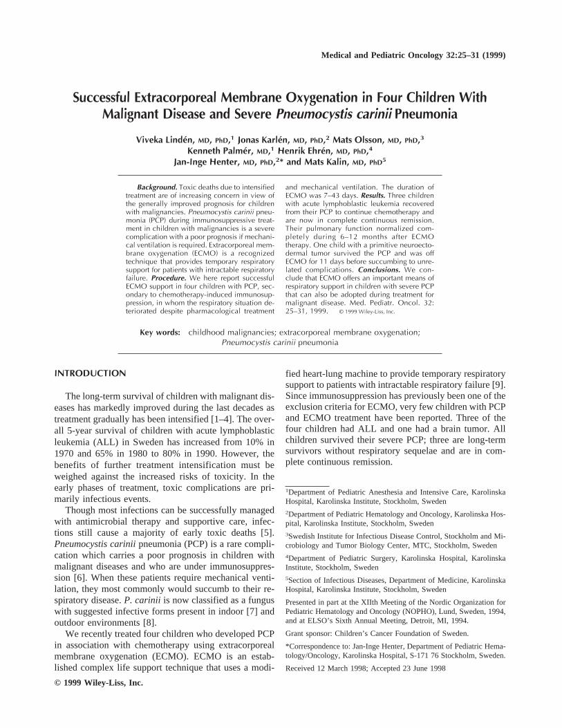
The selection criteria for ECMO were PaO2 (mm Hg)/FiO2 < 150 and intrapulmonary venous admixture (shuntfraction) > 30% [11]. Values for the four patients areshown in Table I together with other relevant ECMOdata.
During ECMO and ventilatory support, the vital ca-pacity was calculated using the Servo Ventilator 300 andthe Ventrakt Respiratory Monitoring System (Med-Science, St. Louis, MO). The follow-up spirometrieswere performed with a Pulmonary Function Laboratory2200 Sensormedics (Infrasonics, San Diego, CA).
Diagnostic MethodsMaterial for microbiological analyses was obtained by
bronchoalveolar lavage (BAL) during bronchoscopy orby aspiration through the endotracheal tube after instil-lation of isotonic sodium chloride. Immunofluorescence(IFL) was performed using a commercially availableP.carinii specific antibody (Dakopatts, Denmark). Twonested polymerase chain reaction (PCR) assays wereused, based on amplification of theP. carinii thymidylatesynthase (TS-PCR) gene [12] and the mitochondriallarge subunit ribosomal (mt-PCR) gene [8] . Indirect im-munofluorescent analysis (IF) of serum samples was per-formed using paraffin sections ofP. carinii infected hu-man lung as an antigen [13].
CASE REPORTSPatient 1
Pre-ECMO. A 13-year-old girl who had surgery for aPNET in the cerebellum received postoperative chemo-
TABLE I. Data Concerning Ventilatory Support and ECMO in Four Patients With Malignant Disease and PCP
Patientno.
Days ofmechanicalventilation
prior to ECMO
Blood gases priorto ECMO
pH;PaO2;PaCO2
(mmHg) PaO2/FiO2
Pulmonaryshunt
fractiona
Ventilatorysettings prior
to ECMOPIP/PEEP;RR;FiO2
bDays onECMO
Days of assistedventilation/CPAP
after ECMO
Totalhospital
stay (days)
1 10 7,4; 82; 53 82 0,32 60/9; 40; 1,0 17 0 (39)c
2 1 7,4; 57; 33 57 0,41 30/5; 28; 1,0 43 34 873 9 7,4; 56; 38 56 0,42 30/4; 34; 1,0 18 6 454 1 7,4; 37; 40 37 0,57 38/8; 28; 1,0 7 6 33
aPulmonary shunt fraction4 normal value < 0.30.bRR, respiratory rate (per minute).cThe patient died before being discharged from the hospital, 12 days after ECMO off.
26 Linden et al.

therapy consisting of four courses of ‘‘eight drugs in oneday’’ [10] and three courses of high- dose methotrexate(3 g/m2). Subsequent RT of the central nervous system(CNS) was prolonged for 2 months due to repeated de-lays because of leukopenia. Two months after comple-tion of RT, the child was admitted to the hospital becauseof fever and coughing. On admission, she was febrile andtachypneac. The physical examination was otherwise un-remarkable. A chest X-ray showed moderate bilateralinterstitial and alveolar infiltrates. Antibiotic treatmentwas started with oral cotrimoxazole. Her condition dete-riorated with increasing radiological pulmonary infil-trates and progressive respiratory insufficiency whichprompted mechanical ventilation on day 2 after admis-sion as well as a change of antibiotic regimen to paren-teral cefuroxime, netilmicin, erythromycin, acyclovir,and pentamidine. High-dose dexamethasone was added.A tracheostomy was performed. Ten days after admis-sion, a bronchoscopic BAL was performed and IFL waspositive for P. carinii. The antibiotic treatment waschanged to monotherapy with parenteral cotrimoxazole.Bacteriological cultures and virological diagnostic stud-ies were negative. Two days later, the fever increased andthe respiratory situation deteriorated, prompting the ad-dition of cefuroxime and acyclovir. The chest X-rayshowed increased bilateral infiltrates, alveolar edema,and pleural effusion bilaterally. She was in severe respi-ratory failure and there was no further conventional treat-ment to be offered. When arterial desaturation occurreddespite FiO2 1.0 and PIP 50–60 cm H2O, the patient wasreferred for ECMO (Table I), which was started 14 daysafter the first symptoms of infection occurred.
ECMO. On day 2 of ECMO, the chest X-ray showedincreased pleural fluid and bilateral pleural drains wereinserted. The patient was kept very slightly sedated,mostly awake and breathing spontaneously with an oxy-gen supply to her tracheostoma. Chest X-ray on day 4showed a slight improvement with increased pulmonaryaeration. The treatment with cotrimoxazole, cefuroxime,and acycclovir continued, with pentamidine being addedlater. She improved slowly and was decannulated after17 days on ECMO. The respiratory rate was 40/min,PaCO2 increased during sleep, but the blood gases wereotherwise stable. She needed supplementary oxygen onher tracheostoma during 3 additional days.
Post-ECMO. Her general condition was stable duringthe first week after ECMO. Oral feeding and mobiliza-tion were started. Antimicrobial treatment was contin-ued. Leukopenia persisted and the CD4+ / CD8+ ratiowas low. Aspirated bronchiolar fluids were negative forP. carinii, bacteria, or fungi. In the second week afterECMO, the patient deteriorated with nausea, increasingserum urea levels, gastrointestinal bleeding, anemia, andthrombocytopenia which prompted multiple transfusions.Eleven days after the discontinuation of ECMO, hyper-
capnea was noted and she needed mechanical ventilationwith high inspiratory pressure. A chest X-ray showednew dense alveolar bilateral infiltrates. She had grandmal seizures and died after a few hours. Diagnostic stud-ies did not yield a diagnosis of the final phase of herillness. Permission for autopsy was not granted.
Patient 2Pre-ECMO. A previously healthy 6-year-old girl with
ALL was treated according to NOPHO-92’s HR arm dueto T-cell phenotype. Seven days after the start of theearly intensification phase, the patient developed cough-ing and fever. On examination, she was febrile with nofocal signs of infection. A chest X-ray showed bilateralinterstitial infiltrates. The day after admission, the coughworsened and tachypnea developed. Antibiotic treatmentwas started with parenteral cotrimoxazole, vancomycin,amikacin, and ganciclovir. Betamethasone was added.On day 3 after admission, increasing tachypnea and re-spiratory difficulties developed and mechanical ventila-tion was started. A BAL was performed and IFL waspositive forP. carinii. Bacteriological cultures and viro-logical studies were negative. The next day, the respira-tory situation deteriorated further, despite maximal ven-tilatory support. Nitric oxide (NO) inhalation was triedwithout effect and ECMO was started 4 days after ad-mission (Table I).
ECMO. From day 2 on ECMO, she was breathingspontaneously with pressure support ventilation. The pa-tient remained intubated during the whole ECMO periodand was kept fully awake. Initially there was a profusesecretion of viscous mucus from the airways. Cotrimoxa-zole and vancomycin were given during the entireECMO period. Amikacin was given on days 2–13 andceftazidim on days 15–27. The chest X-ray showed com-plete consolidation bilaterally with no air present in theparenchyma during 15 days. However, even during thisperiod with a very limited pulmonary gas exchange, shewas awake, playing, drawing pictures, and preferred tobreathe spontaneously with endotracheal continuouspositive airway pressure (CPAP) at 10 cm H2O. Fromday 16 on, her pulmonary function gradually improvedand so did the chest X-rays. On day 19, epistaxis andhematuria occurred and the coagulation parametersshowed fibrinolysis. Due to the coagulatory problemsand the pulmonary improvement, ECMO was discontin-ued on day 19. The cannulas were kept in place and shewas still intubated, breathing spontaneously with CPAPand supplementary oxygen. After a couple of hours, herwork of breathing increased, the arterial saturation fell,and she became dyspneic and anxious. Mechanical ven-tilation was started. Restart of ECMO was necessary af-ter 11 hours. Due to clotting of the cannulas, an acuterecannulation was performed. The patient deterioratedquickly during this operation. She had a circulatory arrest
ECMO for P. carinii Pneumonia 27

and a subsequent successful cardiopulmonary resuscita-tion. After a couple of hours on ECMO, the patient wasawake and mentally alert. During the following days, sheslowly improved both clinically and radiologically andshowed no signs of cerebral ischemia. Repeated bloodcultures showed growth ofStaphylococcus epidermidis,despite vancomycin treatment since the start of ECMOand the replacement of the entire ECMO circuit threetimes. However, there were no clinical or other signs ofsepticemia. ECMO was discontinued after a total of 43days. She received a tracheostoma and breathed sponta-neously with pressure support and CPAP during another34 days.
Post-ECMO. She was discharged from the hospitalafter 92 days. The vital capacity was markedly reduced,23% of predicted value, when ECMO was finally dis-continued. By the time the tracheal cannula was re-moved, the vital capacity had increased to 38%. After 6months it had returned to normal values (Fig. 1). Fol-lowing the successful treatment of the PCP, chemo-therapy treatment was restarted and completed withoutproblems. The girl is doing well in complete continuousremission and without any respiratory problems.
Patient 3Pre-ECMO. A 5-year-old previously healthy boy with
pre-B ALL was treated according to NOPHO-92 IR armdue to his initially elevated WBC. The first phase of theinduction was completed without complications. On day50, at the start of the intensification phase, he developedfever and coughing. Initially, there were no breathingdifficulties or tachypnea. A chest X-ray revealed bilateralinterstitial infiltrates. Parenteral administration of cefu-roxime was started and cotrimoxazole was added the
following day. Breathing difficulties developed on day 3after admission. Ganciclovir and hydrocortisone wereadded. Microbiological cultures were negative. On day 5after admission, increasing respiratory difficultiesprompted the start of mechanical ventilation. After 9days with increasing FiO2 and airway pressures, the pa-tient deteriorated further. Pneumomediastinum and sub-cutaneous emphysema occurred. There was still no etio-logical diagnosis, but PCP was suspected from the clini-cal picture. IFL and PCR forP. carinii were negative inaspirated tracheal fluid, but the material was obtainedafter 12 days of appropriate antibiotic treatment. Therewas a significant increase in antibodies toP. carinii. Thepatient had severe respiratory failure and was referred forECMO 14 days after symptoms of infection started(Table I).
ECMO. During ECMO, he remained intubated,breathing spontaneously with pressure support. He wasfully awake, playing video games and watching TV. Ini-tially he had abundant secretion from the airways. Thechest X-ray showed improved aertation and regression ofthe pneumomediastinum. Cotrimoxazole, vancomycin,and fluconazole were administered during the entireECMO period. On day 7, a pneumothorax was foundwhich was drained with prompt effect. During the fol-lowing days, the patient improved further, although thechest X-ray still showed large and confluent infiltrates.He was decannulated after 18 days on ECMO and extu-bated after another 6 days.
Post-ECMO. He was discharged after a total stay of65 days. The vital capacity was very low, 25% of normalvalue, when ECMO was discontinued. One month later,
Fig. 1. The vital capacity inpatients 2–4 with ALL and PCPduring ECMO treatment andfollow-up after 12 months.
28 Linden et al.

the vital capacity was increased to 65% and returned tonormal values after another 5 months (Fig. 1). The pa-tient has now completed his chemotherapy treatment andis doing well in complete continuous remission. He isfully recovered from his pulmonary disorder.
Patient 4Pre-ECMO. A previously healthy 6-year-old boy with
pre-B ALL was treated according to the NOPHO-92 HRarm due to his initially elevated WBC. He received in-duction treatment followed by CNS prophylaxis and aninterimistic maintenance phase. A late intensificationtreatment including dexamethasone (10 mg/m2) during 4weeks was initiated in week 36 from the start of therapy.Three days after the end of this chemotherapy block, thepatient was admitted with fever, coughing, and tachy-pnea. The lungs were clear on auscultation. A chest X-ray showed bilateral interstitial infiltrates. Parenteral ce-furoxime was started and erythromycin and cotrimoxa-zole were added after 4 days. When the symptomsprogressed and microbiological cultures were negative,antibiotic treatment was changed to gentamicin, piper-acillin, acyclovir, amphotericin B, and flucytosine whilecotrimoxazole was continued. After 1 week, the condi-tion deteriorated and mechanical ventilation was neces-sary. Bacteriological and virological diagnostic studieswere still negative. Aspirated bronchiolar fluid, obtained7 days after the start of cotrimoxazole treatment, wasnegative forP. carinii with IFL and TS-PCR, but posi-tive with the mt-PCR assay. The patient had Fi02 1.0 andhigh ventilatory settings with a peripheral saturation of80%, but no hypercapnea or acidosis. Inhaled NO wastried with no effect. The next day, 9 days after admission,he deteriorated with severe oxygenation problems andECMO was started (Table I).
ECMO. Initially he produced large amounts of mucusin the airways. He rapidly improved both clinically andradiologically. Cotrimoxazole, gentamicin, acyclovir,amphotericin B, and hydrocortisone were given duringthe ECMO period. After 7 days on ECMO, he was de-cannulated. He was breathing with pressure support foranother 4 days, had CPAP during 2 days, and was extu-bated 6 days after the discontinuation of ECMO. Supple-mentary oxygen was needed for another 4 days.
Post-ECMO. He was discharged after a total stay of33 days. His vital capacity was very low, 23% of thepredicted value, at the time of decannulation fromECMO. Five months later, his vital capacity had in-creased to 80%, and it has remained so (Fig. 1). Chemo-therapy for ALL was reinstituted after the successfultreatment of the PCP. The patient has now finished histreatment without problems and is doing well, withoutany respiratory problems and in continuous complete re-mission.
DISCUSSION
To our knowledge, there are no reports in the literaturefrom other centers on using ECMO as respiratory supportin combination with conventional treatment of severePCP. The ELSO registry (Extracorporeal Life SupportOrganisation, Ann Arbor, Michigan) receives data fromover 100 ECMO centers worldwide. Since 1986, only 14patients (as of January 1998) have been reported withPCP and ECMO; all were pediatric cases. Of the fivelong-term surviving patients in the registry, three ofthem, reported here, were treated at the ECMO center inStockholm, Sweden. ECMO is not a curative treatment.It is used to take over the function of the lungs, providing‘‘resting conditions’’ to minimize iatrogenic damageand, above all, time for the damaged lungs to recover.One of the exclusion criteria for ECMO has been immu-nosuppression [11] because of the high risk of life-threatening infections with the ECMO circuit as the portof entry.
The case fatality rate in PCP has remained at or closeto 50% among non-human immunodeficiency virus(HIV)-infected patients during the last decades [6,14].The course is often fulminant. In patients who requiremechanical ventilation, the prognosis is very serious[6,15]. Occasional cases of severe PCP infection are seenin pediatric oncology centers also if prophylaxis is usedand our experiences with ECMO as a respiratory supporttechnique may be important.
Some ECMO-related experiences seem particularlyimportant to emphasize. It is important to start ECMOearly in order to reduce the initial barotrauma to the lung.Eligibility criteria have been described earlier [11]. InPCP, the infectious process in the lungs involves attach-ment of the microorganisms to alveolar cells with focalnecrosis, increased capillary permeability, damage to thebasement membrane, and resulting transudation and exu-dation of plasma components [16]. The deposition of celldebris in the alveoli contributes to a further decrease ofthe gas exchange. In these patients, mechanical ventila-tion with very high positive inspiratory pressure is usu-ally necessary and very soon causes barotrauma to thelung parenchyma. The edema in the parenchyma is ini-tially less pronounced, but will gradually increase as ven-tilation is continued.
ECMO is a supply of sufficient oxygen to meet thetissue demand while the lungs are very gently ventilatedto prevent further barotrauma and promote healing andrepair. The period of radiological consolidation of thepulmonary parenchyma lasted for several weeks in threeof our four patients (nos. 1, 2, and 3) after ECMO wasstarted. With effective ECMO therapy, in contrast to thesituation in patients with mechanical ventilation, theedema will slowly diminish and disappear. This is ac-companied by alveolar regeneration and restitution of the
ECMO for P. carinii Pneumonia 29

lung cell function and gas exchange. The extracorporealblood flow can be reduced and finally discontinued. Theperiod of healing and repair will last for several weeksafter ECMO.
Our findings also suggest that the first conceivableopportunity to discontinue ECMO may be when the cal-culated vital capacity is about 25% of the predicted valuefor age and gender and when the patient is breathingspontaneously with pressure support ventilation. In pa-tient 2, we tried to get off ECMO after 19 days becauseof hemorrhagic and coagulation problems. The bloodgases at first remained stable when the extracorporealflow was decreased and the chest X-ray was improvedseemingly to an acceptable degree. However, the attemptwas unsuccessful, probably because the patient’s lungcapacity was not yet sufficient; we did not calculate hervital capacity prior to discontinuation of the ECMO. Herbreathing became very labored after 9 hours. Due to theincreased breathing work, which per se rapidly mighthave caused cellular edema of the lung parenchyma withdeteriorating gas exchange and further reduced vital ca-pacity, ECMO was restarted. When finally decannulatedat day 43, her vital capacity was calculated to be 23% ofthe predicted value (Fig. 1). She had general anesthesiawith long-acting muscle relaxants. Immediately after theinduction, she desaturated from 100 to 60% and re-mained so, despite high inspiratory pressure (50 cmH2O), FiO2 1.0, and a respiratory rate of 40, until sherecovered. The vital capacity during anesthesia and me-chanical ventilation was calculated to 6%. However, af-ter 1 hour, she was awake, again breathing spontaneouslywith pressure support ventilation, FiO2 0.4 , arterial satu-ration of 92%, and the vital capacity had returned to thepre-decannulation value, 23%. In patients 3 and 4, thevital capacity was calculated before the decannulationand was found to be 23% and 25%, respectively (Fig.1),and ECMO was discontinued successfully. These pa-tients were also under general anesthesia with short-acting muscle relaxants. As patient 2, they both showedsevere desaturation while mechanically ventilated, al-though for a shorter period of time. It is known thatprolonged paralyzation can impede recovery of pulmo-nary function in infants [17]. The last 10 pediatric pa-tients on ECMO at our unit have all been decannulatedsuccessfully while unsedated, awake, and breathingspontaneously with pressure support and with only localanesthetics as analgesia. Thus, according to our experi-ence, it seems important to have the vital capacity re-stored to at least 25% of the predicted value before dis-continuing ECMO and to keep the patients awake andbreathing spontaneously during the decannulation. It maybe speculated whether a second ECMO therapy wouldhave changed the outcome in patient 1. However, at thattime, we had no experience of starting and runningECMO for a second time.
A further observation is the gradual improvement ofvital capacity as shown in Figure 1. After 3 months, thevital capacity in patients 2, 3, and 4 had returned to 60%of the predicted value for age and the vital capacity wasrestored to normal values first after 6 months (Fig. 1).These facts are important to be aware of if the patient forany reason is planned for general anesthesia with musclerelaxation and/or mechanical ventilation, which we rec-ommend should be postponed for the first 3 months afterECMO.
NO was only available for one patient and high fre-quency oscillation ventilation was not available for pe-diatric patients at this time.
Finally, septicemia during ECMO treatment was not aproblem despite the evident immunosuppression in thesepatients. We used prophylactic antibiotics against staph-ylococci and performed blood cultures daily in all fourpatients. In patient 2, repeated blood cultures showedgrowth ofS.epidermidisdespite administration of vanco-mycin. The complete ECMO circuit was changed threetimes, but blood cultures continued to be positive. How-ever, the patient had no fever, was hemodynamicalystable, and C-reactive protein and WBC remained nor-mal. The blood cultures became negative immediatelyafter ECMO had been disconnected. Among the threeother patients, none had a positive blood culture.
CONCLUSIONS
Based on the experiences of ECMO in four patientswith severe PCP, the following conclusions seem justi-fied. 1) Children with chemotherapy-induced immuno-suppression and PCP in whom conventional assisted ven-tilation is insufficient may be rescued with ECMO. 2)Decannulation should probably not be performed untilthe vital capacity has increased to about 25% of the pre-dicted value. 3) Pulmonary function seems to normalizewithin half a year after ECMO. 4) ECMO circuit ac-quired septicemia was not a problem in these immuno-suppressed children. 5) Despite ECMO therapy of sig-nificant length, which precludes prescheduled chemo-therapy, children with delayed ALL therapy may survivewithout relapse. 6) Since the care of these patients con-stitute a challenge, we suggest they be referred to centerswith the experience of pediatric ECMO.
REFERENCES
1. Granowetter L. Ewings sarcoma and extracranial primitive neu-roectodermal tumors. Curr Opin Oncol 1992;4:696–703.
2. Rivera GK, Pinkel D, Simone JV, et al. Treatment of acute lym-phoblastic leukaemia. N Engl J Med 1993;329:1289–1295.
3. Lilleyman JS, Pinkerton CR. Lymphoblastic leukemia and non-Hodgkin’s lymphoma. Br Med Bull 1996;52:742–763.
30 Linden et al.

4. Pappo AS. Rhabdomyosarcoma and other soft tissue sarcomas inchildren. Curr Opin Oncol 1996;8:311–316.
5. Wheeler K, Chessels JM, Bailey CC, et al. Treatment relateddeaths during induction and in first remission in acute lympho-blastic leukaemia: MRC UKALL X. Arch Dis Child 1996;74:101–107.
6. Sepkowitz K.Pneumocystis cariniipneumonia in patients withoutAIDS. Clin Infect Dis 1993;17(suppl 2):S416–422.
7. Olsson M, Sakura A, Lindberg L-A, et al. Detection ofPneumo-cystis carinii DNA filtration of air. Scand J Infect Dis 1996;28:279–282.
8. Wakefield E. DNA sequences identical toPneumocystis cariniif.spcarinii andPneumocystis cariniif.sphominisin samples of airspora. J Clin Microbiol 1996;34:1754–1759.
9. Bartlett RH, Gazzaniga AB, Toomasian J, et al. Extracorporealmembrane oxygenation (ECMO) in neonatal respiratory failure.100 cases. Ann Surg 1986;204:236–245.
10. Bleyer A, Millstein J, Balis F, et al. Eight drugs in 1 day chemo-therapy for brain tumors: A new approach and rationale for pre-radiation chemotherapy. Med Pediatr Oncol 1983;11:213 (abstr).
11. Custer J, Fackler J. ECLS for children with acute respiratorydistress syndrome. In: Zwischenberger JB, Bartlett RH, editors.
ECMO Extracorporeal Cardiopulmonary Support in Critical Care.Ann Arbor: Extra Corporeal Life Support Organization; 1995. p341–349.
12. Olsson M, Elvin K, Lidman C, et al. A rapid and simple nestedPCR assay for the detection ofPneumocystis cariniiin sputumsamples. Scand J Infect Dis 1996;28:597–600.
13. Elvin K, Bjorkman A, Heurlin N, et al. Seroreactivity toPneu-mocystis cariniiin patients with AIDS versus other immunosup-pressed patients. Scand J Infect Dis 1994;26:33–40.
14. Arend SM, Kroon FP, van’t Wout JW.Pneumocystis cariniipneu-monia in patients without AIDS, 1980 through 1993. Arch InternMed 1995;155:2436–2441.
15. Hughes WT, Kuhn S, Chaudhary S, et al. Successful chemopro-phylaxis for Pneumocystis cariniipneumonia. N Engl J Med1977;297:1419–1426.
16. Hughes WT.Pneumocystis cariniipneumonia. In: Patrick CC,editor. Infections in Immunocompromised Infants and Children.New York: Churchill Livingstone; 1992. p. 711–718.
17. Bhutani VK, Abbasi S, Sivieri EM. Continous skeletal muscleparalysis: Effect on neonatal pulmonary mechanics. Pediatrics1988;81:419–422.
ECMO for P. carinii Pneumonia 31