successful treatment of macrorhabdosis in budgerigars ... · sodium benzoate administration via...
TRANSCRIPT

Journal of Mycology Research. Vol 1, No 1: Page 21-27, September 2014
JMR (Sep.2014), 1(1) 21
Successful treatment of macrorhabdosis in budgerigars(Melopsittacus undulatus) using sodium benzoateSeyed Ahmad Madani1*, Amir Ghorbani1, Fatemeh Arabkhazaeli2
1Department of Avian Diseases, Faculty of Veterinary Medicine, University of Tehran, Tehran, Iran.2Department of Parasitology, Faculty of Veterinary Medicine, University of Tehran, Tehran, Iran.
* Corresponding Author: Email: [email protected], Tel: +98 21 61117150
(Received: 7 March 2014,
Macrorhabdosis is a debilitating syndrome in budgerigars (Melopsittacus undulatus) due to theascomycetous yeast Macrorhabdus ornithogaster. In the present study, occurrences of acutemacrorhabdosis resulting in severe mortality in budgerigar fledglings and the effect of differenttreatment regimens for the control of the disease were investigated. The budgerigar (Melopsittacusundulates) flock consisted of over five hundred breeding adults. The morbidity of chicks reached90% with more than 50% mortality. The significant clinical and pathological findings includeddistended abdomen, diarrhoea, ingluvitis, proventriculitis, and mild enteritis. Severe M.ornithogaster infection was diagnosed based on cytologic and histologic investigations. Threeweeks of nystatin medication in the feed and vinegar administration in the drinking water led tomoderate improvement of the flock mortality. After the initial treatment, 500 mg/Lsodium benzoatewas administered in the drinking water for four weeks. The second treatment regimen waspromisingly effective in reducing mortality. However, some sick and retarded birds with M.ornithogaster with positive proventricular smears at necropsy were found in the flock.Consequently, a higher dosage of 1 gr/Lin drinking water for another four weeks was recommended.After the eight weeks of treatment, no new cases were found in the flock and all dropping samplesbecame negative for the presence of M. ornithogaster. Based on these preliminary findings, sodiumbenzoate can be an efficient and inexpensive alternative to the previous labour intensive andexpensive treatment using amphotericin B.Keywords: budgerigar, macrorhabdosis, Macrorhabdus ornithogaster, megabacteriosis, sodium
benzoate.
Introduction
Macrorhabdosis or megabacteriosis is thecondition caused by the so-called"Megabacterium", which is a rod-shaped tofilamentous Gram-positive, periodic acid-Schiff-positive microorganism found in the isthmusbetween the proventriculus and ventriculus ofbirds (Van Herck et al., 1984). It was recentlyestablished that the causative agent is anascomycetous yeast named Macrorhabdusornithogaster (Tomaszewski et al., 2003).Macrorhabdosis was initially reported inbudgerigars (Melopsittacus undulatus) in 1977, as
a debilitating syndrome (Jones & Carroll, 1977).Since then it was reported in many different avianspecies either with or without clinical signs orpathologic lesions (Gerlach, 2001). In 2008,macrorhabdosis was first incidentally detected in abudgerigar in Iran (Madani & Nejati, 2008), andpathology and control of the infection was studiedthoroughly in a budgerigar flock in Kerman, Iran(Kheirandish & Salehi, 2011). While the incidenceof macrorhabdosis, or at least its detection anddiagnosis have significantly increased in recentyears (Marlier et al., 2006; Phalen et al., 2002;Filippich et al., 1993), information about its bio-pathologic characteristics and therapeuticmeasures are scant in the scientific literature.
Abstract:
Accepted: 20 July 2014 )

Control and treatment of M. ornithogasterinfection are challenging for avian clinicians.Because subclinical infections can occur in manybirds without obvious clinical signs, it is almostimpossible to keep large aviaries free from theinfection (Filippich et al., 2004). On the other hand,it has been reported that many antimicrobial andantifungal drugs like iodine preparations,luphenuron, nystatin, fluconazole, ketoconazole,itraconazole, and terbinafine were not effective intreating the infection (Filippich et al., 1993; Phalenet al., 2002; Phalen, 2005). The most effectivetreatment has been reported to be amphotericin B,which is given orally or gavage fed at a dose of 100mg/kg twice a day for a month (Phalen et al., 2002;Phalen, 2005). A water-soluble preparation ofamphotericin B when given for 14 days in drinkingwater was not effective (Phalen et al., 2002).Amphotericin B resistant strains of M.ornithogaster were identified in Australia(Filippich et al., 1993). The sensitivity of M.ornithogaster to sodium benzoate has beenpreviously shown in limited in vitro (Hanafusa etal., 2007b) and in vivo (Hoppes, 2011) studies. Inthe present study the clinicopathologic aspects ofacute macrorhabdosis (so-called Megabacteriosis)in a budgerigar flock were studied and differenttherapeutic regimens, including sodium benzoateadministration, have been investigated.
Materials and Methods
Birds. A breeding budgerigar (Melopsittacusundulates) flock with more than five hundred adultbirds was referred to the Pet Bird Clinic of theDepartment of Avian Diseases, Faculty ofVeterinary Medicine, University of Tehran. Theflock had a six months history of high mortality andpoor growth in fledglings. Previous treatment withketoconazole and blue vitriol were not effective.The morbidity of four-day-old to one-month-oldhatchlings and fledglings had reached 90% withmore than 50% mortality at the referral time. Basedon the history presented by the owner, more than3000 chicks had died or were culled during the lastsix months.
Preliminary diagnostic work-up: Thoroughclinical examination of five submitted livefledglings was performed. Faecal wet smears andfaecal Gram stained smears were investigated. Allfive birds were euthanized and a thoroughnecropsy was performed. In addition two deadchicks were also referred for necropsy. Heartblood, liver, and crop mucosal samples werecultured on blood and MacConkey agar. The distalintestines of chicks were cultured on selenite brothto investigate Salmonella infection. Fresh wetsmears from the crop, proventriculo-ventricularjunction, and distal intestine, and also Giemsastained contact smears from livers, spleens, andlungs of all chicks were microscopicallyinvestigated. Thin sections from the liver, kidney,lung, gonad, adrenal, proventriculus, gizzard,spleen, duodenum, jejunum, crop, thyroid andparathyroid of some birds were studied forhistopathologic lesions using haematoxylin andeosin staining.
First treatment regimen and controlmeasures: In the first step immediately after initialpresumptive diagnosis based on clinical andnecropsy findings, treating the flock with nystatin(JaBer Ebne Hayyan, Tehran, Iran; 500 mg/kgfeed) in addition to the use of vinegar (10 ml/L ofdrinking water) for five to six weeks wasrecommended.
Second treatment regimen: According to theoutcome of first regimen, which was evaluatedthree weeks after its initiation, the second treatmentplan using sodium benzoate (Merck, Darmstadt,Germany; 1 gr/L drinking water) was started.
Diagnostic follow-up: Droppings werecollected from each cage for 24 hours and wereevaluated weekly during the treatment for thepresence of M. ornithogaster. Based on ourinstruction, the owner brought two to three birdsback to the clinic on three different occasions overthe next six months for follow-up. Besides faecalsmears, samples were taken from the crop ofreferred live birds to detect M. ornithogaster usingcrop lavage. Complete post-mortem examinationincluding gross pathology, microbial culture,cytologic investigation of gastrointestinal
Treatment of macrorhabdosis in budgerigars Seyed Ahmad Madani et al.
JMR (Sep.2014), 1(1)22

contents, liver, spleen and lung tissue of fiveadditional moribund or dead birds were performedon three occasions during next six months.
Results
In the clinical examination, the fledglings haddistended abdomens and droppings attached to thevent region. Another sign was accumulation ofdried droppings around the toes of fledglings (Fig.1a). While growth retardation and occasionallycachexia were evident in some birds, no featherdystrophy or follicular degeneration indicatingFrench molt or budgerigar fledgling disease (BFD)could be observed in referred cases during thewhole study.
The wet smear investigation of droppingsshowed the presence of large numbers of large rod-shaped bacilli resembling M. ornithogaster in allspecimens.
Necropsy findings indicated proventriculitisand proventricular dilatation. Typical diphthericingluvitis was obvious in fledglings' cadavers (Fig.1b). The crop mucosa showed pseudomembranousnecrosis. The proventricular walls of all necropsiedchicks seemed to be enormously thickened and tinyhaemorrhages were evident at the mucosal surfacewhile a thick mucoid layer covered the mucosa ofthe proventriculus and some parts of the intestines(Fig. 2). Some chicks had dark brown to blackintestinal contents indicating antemortemhaemorrhagic diathesis in the intestinal tract,which could be a sign of severe debilitation and/oranorexia in small birds. All wet smears preparedfrom the proventriculo-ventricular junction hadtoo many large rods resembling M. ornithogaster.The Gram stained smears from the proventricularmucosa had too many Gram-positive rods of M.ornithogaster and also some Gram-negativebacilli. No bacteria could be isolated from the heartblood and the liver samples of the chicks. Gram-negative coliform bacilli other than Escherichiacoli and also coagulase-negative Staphylococciwere isolated from the crop content of some chicks.All intestinal samples were also negative forSalmonella.
Histologic investigation revealed diffused renaltubular necrosis with some round circular toamorphous eosinophilic spheroid materials insome tubuli indicating antemortem dehydration.Nonspecific small necrotic foci in the liver of somechicks with typical anisokaryosis and bile ducthyperplasia, and severe pancreatic zymogendepletion were occasionally found in some tissuesamples. No viral inclusion bodies could be foundin the studied specimens. No microscopic lesionscould be found in thyroid, parathyroid, and spleentissues. The most significant lesions were found inthe proventriculus and the crop. Too many rod-shaped M. ornithogaster gathered together andformed a broom stick appearance that invaded themucosa of the proventriculus and the crop (Fig. 3and 4). Submucosal cellular infiltration wasminimal but degeneration, necrosis and sloughingof the epithelial cells were present in some sections.Acute macrorhabdosis was confirmed as thedefinitive diagnosis based on the clinical, necropsyand histological findings.
Follow-up necropsies and microscopicinvestigation of the wet smears prepared fromeither proventricular mucosa of die-offs or faecalsamples after three weeks of initial controlmeasures, including the administration of nystatinin the feed and vinegar in the drinking water,showed only a slight improvement while themortality of the remaining chicks continued. So itwas decided to change the treatment regimen tosodium benzoate administration via drinkingwater. The treatment with 1 gr/L of sodiumbenzoate resulted in depression and lethargy insome affected birds and obvious decreased waterconsumption in the flock. Consequently the dose ofmedication decreased to the level of 500 mg/L ofdrinking water and it continued until eight weeks.Interestingly, the droppings examined during thetreatment were negative for the presence of M.ornithogaster from day 20 after medication andmortality of chicks almost ceased. In addition, theoccasional necropsy examination of some birdsafter eight weeks of the treatment showed typicallyreduced lesions and a subjectively decreasednumber of proventricular yeasts. Even after this
Treatment of macrorhabdosis in budgerigarsSeyed Ahmad Madani et al.
JMR (Sep.2014), 1(1) 23
eye catching result in the control and the treatment,there were still birds with droppings attached to thevent region. While faecal samples were negative inthose birds, a few M. ornithogaster could still befound in the wet smears obtained from theproventricular mucosa during post-mortemexamination.
Additionally, weak birds that were still sickdespite the treatment were isolated from the flockand placed in separate cages. Apparently healthypairs were placed back together again and the nests
were returned into the cages. Furthermore, thedosage of sodium benzoate was returned to 1 gr/Lof drinking water for another four weeks and untilthe chicks hatched. Interestingly, necropsyexamination and analysis of the droppings of thebirds after four weeks of treatment with the higherdosage showed highly desirable results and itseemed to be very efficient in the control of M.ornithogaster in the flock, as the last two weakbirds which were euthanized and examined, werenegative for the presence of M. ornithogaster intheir proventriculus, the privilege site of
Treatment of macrorhabdosis in budgerigars Seyed Ahmad Madani et al.
JMR (Sep.2014), 1(1)24
Fig. 1. A) Heavy plantar contamination of the feet in a two-week-old budgerigar affected by severe macrorhabdosis. Diarrhoeaand khaki coloured dropping can be a clinical sign ofmacrorhabdosis in budgerigars. B) Distended abdomen anddiphtheric ingluvitis in budgerigar hatchling. Typical thickeningof crop and mucosal diphtheric lesion can be the result of severemacrorhabdosis.
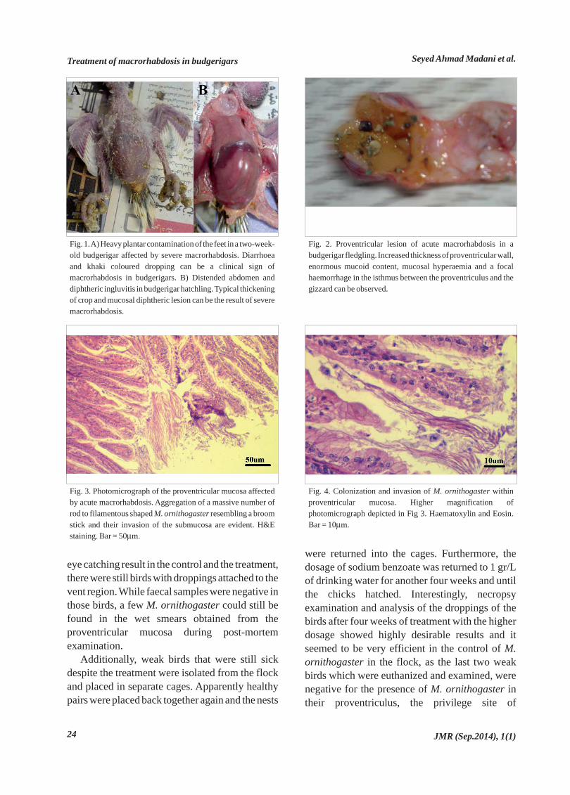
Fig. 2. Proventricular lesion of acute macrorhabdosis in abudgerigar fledgling. Increased thickness of proventricular wall,enormous mucoid content, mucosal hyperaemia and a focalhaemorrhage in the isthmus between the proventriculus and thegizzard can be observed.
Fig. 3. Photomicrograph of the proventricular mucosa affectedby acute macrorhabdosis. Aggregation of a massive number ofrod to filamentous shaped M. ornithogaster resembling a broomstick and their invasion of the submucosa are evident. H&Estaining. Bar = 50μm.
Fig. 4. Colonization and invasion of M. ornithogaster withinproventricular mucosa. Higher magnification ofphotomicrograph depicted in Fig 3. Haematoxylin and Eosin.Bar = 10μm.

subclinical infection.
Discussion and Conclusion
In this study, clinical signs and pathologic findingsof acute macrorhabdosis in budgerigars causingsevere loss were thoroughly investigated. Theefficiency of the administration of sodiumbenzoate in drinking water in the affectedbudgerigar flock, which previously had beenunsuccessfully treated with a variety of differentdrugs, were further clinically evaluated for the firsttime in Iran. Although this study was not conductedunder strictly controlled experimental infectionconditions, the results seem to put forward newscopes of studies on the effects of sodium benzoateon M. ornithogaster infection in budgerigars.
Macrorhabdosis was reported as the leadingcause of illness and death in exhibition budgerigarsin England (Baker, 1992; Baker, 1996). It wasshown that almost one third of healthy budgerigarscould carry M. ornithogaster in their stomachwithout any obvious clinical signs (Baker, 1997),but abundant faecal shedding can be an indicationof clinical disease (Filippich et al., 2004). Due to itsfastidious nature, isolation is not an appropriateprocedure for diagnosis and as a consequence invitro drug sensitivity studies are rare for M.ornithogaster (Hanafusa et al., 2007a; Hanafusa etal., 2007b). M. ornithogaster could be stained bydifferent conventional staining methods, such asGiemsa, Gram, and PAS, but a fresh wet smear isthe most sensitive and of course easiest method forits detection (Gerlach, 2001; Filippich et al., 2004).As a result of misdiagnosis of other materials anddebris, particularly plant fibres, due to aninexperienced microscopist, other more specificmethods, such as Calcofluor-white M2R staining(Moore et al., 2001) and PCR (Tomaszewski et al.,2003) have been used in some studies, but neitherof them have become a routine diagnostic method.
Treatment and control of the infection,especially in large collections, could bechallenging for avian clinicians (Gerlach, 2001;Phalen, 2005). Although in some instances widelyavailable antifungal drugs, such as nystatin, have
been used for treatment both per os (Scullion &Scullion, 2004) and in drinking water (Kheirandish& Salehi, 2011), in the present case theadministration of nystatin in the feed for threeweeks was not successful in reducing morbidityand mortality. It should be mentioned that nystatinis not water-soluble and water medication of thisdrug for large aviaries is questionable. It wasdemonstrated that lowering gastrointestinal pHwith drinking water acidification using differentorganic acids could have some therapeutic effectson M. ornithogaster infection (Gerlach, 2001;Filippich, 2004; Phalen, 2005), but in ourexperience it was not a fully efficient method forthe treatment of heavy infection. Despite the partialefficiency of amphotericin B in drinking water insome experiments (Christensen et al., 1997;Filippich & Hendrikz, 1998), it is now believed thatthe most efficient method for the treatment isgavage feeding of amphotericin B (100 mg/kg)twice a day for 30 days (Filippich et al., 2004;Phalen, 2005). There is no need to elaborate on thedifficulty and demanding nature of performingsuch a treatment in large bird colonies. Inagreement with the results of the present study,successful treatment of the infection in a largebudgerigar aviary using sodium benzoate, which isa less expensive and water-soluble alternative toamphotericin B, has been once reported previously(Hoppes, 2011). Benzoic acid is a naturalingredient in many foodstuffs and plant extracts(Anonymous, 2000; SCCP, 2005). Undissociatedbenzoic acid is responsible for antimicrobialactivity, but as it is only slightly soluble in water, itssalt, sodium benzoate with 200 times more watersolubility, is often used as a preservative andantifungal agent in food, beverages, cosmetics, andpharmaceuticals (Anonymous, 2000). Fungalgrowth was inhibited in a pH dependent manner byconcentrations of sodium benzoate ranging from100-60000 mg/L(Anonymous, 2000). The toxicityof sodium benzoate is rare and its LD50 for rats wasreported to be more than 1700 mg/kg (Anonymous,2000; SCCP, 2005). Sodium benzoate may causeslight skin irritation in healthy human subjects, butin patients with urticaria or asthma, exacerbation of
Treatment of macrorhabdosis in budgerigarsSeyed Ahmad Madani et al.
JMR (Sep.2014), 1(1) 25

symptoms was observed (Anonymous, 2000). Inspite of its safety for use in animals and humans,lethargy, depression and reduced waterconsumption were observed in the flock during theinitiation of the treatment with 1 gr/L of sodiumbenzoate. Unfortunately, the daily waterconsumption of birds was not accurately measuredduring the treatment process. However, accordingto the breeder's claim, except for the higher doseintroduction of sodium benzoate (1 gr/L) in thedrinking water, the treatment did not significantlyreduce the water consumption of the birds and didnot cause abnormal mortality. Reducing the dosageto 500 mg/L alleviated the initial adverse effects.Afterwards, returning the dosage to its former statewas well tolerated by the birds during the final fourweeks of the treatment process, indicating possibleadaptation. It was also indicated in another report,that breeding pairs and chicks were moresusceptible to the adverse effects of sodiumbenzoate, probably due to more water consumption(Hoppes, 2011).
Viral inclusion bodies or other specificmacroscopic and/or microscopic lesions likefeather dystrophy or French molt indicatingcommon viral diseases of budgerigars likepolyomaviruses and circoviruses were notobserved in this budgerigar flock. As long as therewas no attempt in the detection of such virusesusing virological or molecular methods, theirpossible association with the presented situationshould be kept in mind.
According to this study, the use of sodiumbenzoate in the treatment and control of M.ornithogaster infection in budgerigars that are notproducing eggs and rearing their chicks, willachieve acceptable results as compared to the useof other antifungals, such as nystatin. In addition,in the follow-up, there were no occurrences ofabnormal mortality and other adverse effects onreproduction of the birds treated with thissubstance. Even after the subsequent investigationfollowing over two cycles of reproduction, thebirds showed almost complete recovery and theywere returned to their maximum performance afterthe treatment with sodium benzoate. Mortality of
chicks after the treatment regimen was reduced toits acceptable range of less than 0.1% in a week.
Studying the effect of sodium benzoate on M.ornithogaster infection in experimentally infectedbudgerigars is highly recommended to assess itsimpact more accurately.
Treatment of macrorhabdosis in budgerigars Seyed Ahmad Madani et al.
JMR (Sep.2014), 1(1)26
Anonymous, 2000. Benzoic acid and sodiumbenzoate. In: Concise International ChemicalAssessment Document 26. World HealthOrganization, Geneva, Switzerland.Baker, J.R., 1992. Megabacteriosis in exhibitionbudgerigars.Veterinary Record, 131: 12-14.Baker, J.R., 1996. Causes of mortality andmorbidity in exhibition budgerigars in the UnitedKingdom. Veterinary Recod, 139: 156-162.Baker, J.R., 1997. Megabacteria in diseased andhealthy budgerigars. Veterinary Record, 140: 627.Christensen, N.H., Hunter, J.E.B., Alley, M.R.,1997. Megabacteriosis in a flock of budgerigars.New Zealand Veterinary Journal, 45(5): 196-198.Filippich, L.J., Henderikz, J.K., 1998. Prevalenceof magabacteria in budgerigar colonies. AustralianVeterinary Journal, 76(2): 92-95.Filippich, L.J., Perry, R.A., 1993. Drug trialsagainst Megabacteria in budgerigars(Melopsittacus undulatus). Australian VeterinaryPractitioner, 23:184-9.Filippich, L.J., Speer, B., Powers, L.V., Phalen, D.,2004. Diagnosis and treatment options formegabacteria (Macrorhabdus ornithogaster).Journal of Avian Medicine and Surgery, 18(3):189-195.Gerlach, H., 2001. Megabacteriosis. Seminars inAvian and Exotic Pet Medicine, 10(1): 12-19.Hannafusa, Y., Bradley, A., Tomaszewski, E.E.,Libal, M.C., Phalen, D.N., 2007a. Growth andmetabolic characterization of Macrorhabdus
ornithogaster. Journal of Veterinary DiagnosticInvestigation, 19: 256-265.Hanafusa, Y., Costa, E., Bradley, A., Phalen, D.A.,2007b. Further investigation into the biology ofMacrorhabdus ornithogaster. Prodeedings of the
1.
2.
3.
4.
5.
6.
7.
8.
9.
10.
11.
References

Treatment of macrorhabdosis in budgerigarsSeyed Ahmad Madani et al.
JMR (Sep.2014), 1(1) 27
Association of Avian Veterinarians, 28th AnnualConference and Expo, August 6-9, Providence,USA, p277-279.Hoppes, S., 2011. Treatment of Macrorhabdusornithogastor with Sodium Benzoate inBudgerigars (Melopsittacus undulates).Proceedings of the association of avianveterinarians, 32nd Annual Conference & Expo,August 6-12, 2011, Seattle, USA, p67.Jones, D.M., Carroll, C.M.M., 1977. Debilitatingsyndrome in budgerigars (Melopsittacus
undulatus). Veterinary Records, 101(10): 188.Kheirandish, R., Salehi, M., 2011.Megabacteriosis in budgerigars: diagnosis andtreatment. Comparative Clinical Pathology, 20:501-505.Madani, S.A., Nejati, A., 2008. Report ofmegabactriosis in a budgerigar (Melopsittacus
undulatus). Abstracts of 5th Convention of IranianVeterinary Clinicians, 12-14 Feb., Ahvaz, Iran. (inPersian)Marlier, D., Leroy, C., Sturbois, M., Delleur, V.,Poulipoulis, A., Vindevogel, H., 2006. Increasingincidence of megabacteriosis in canaries (Serinuscanarius domesticus). The Veterinary Journal,172:549-552.Phalen, D.N., Tomaszewski, E., Davis, A., 2002.Investigation into the detection, treatment, andpathogenicity of avian gastric yeast. Proceedingsof the 23rd Annual Conference of the Associationof Avian Veterinarians. Monterey, USA, p49-51.Phalen, D., 2005. Diagnosis and management ofMacrorhabdus ornithogaster (formerlyMegabacteria). Veterinary Clinics Exotic AnimalPractice, 8: 299-306.Scientific Committeee on Consumer Products(SCCP), 2005. Opinion on benzoic acid andsodium benzoate. European Commission Health& Consumer Protection Directorate-General.Scullion, F.T., Scullion, M.G., 2004. Successfultreatment of megabacteriosis in a canary (Serinuscanaria) with nystatin. Veterinary Record, 155:528-529.Tomaszewski, E.K., Logan, K.S., Snowden, K.F.,Kurtzman, C.P., Phalen, D.N., 2003. Phylogeneticanalysis identifies the megabacterium of birds as a
12.
13.
14.
15.
16.
17.
18.
19.
20.
21.
novel anamorphic ascomycetous yeast,Macrorhabdus ornithogaster gen. nov., sp. nov.International Journal of Systematic andEvolutionary Microbiology, 53:1201-1205.Van Herck, H., Duijser, T., Zwart, P., Dorrestein,G.M., Buitelaar, M., VanderHage, M.H., 1984. Abacterial proventriculitis in canaries (Serinuscanaria). Avian Pathology, 13:561-572.
22.