succinate dehydrogenase: assembly, regulation and role … · succinate dehydrogenase: assembly,...
TRANSCRIPT
European Journal of Scientific Research ISSN 1450-216X Vol.51 No.1 (2011), pp.133-142 © EuroJournals Publishing, Inc. 2011 http://www.eurojournals.com/ejsr.htm
Succinate Dehydrogenase: Assembly, Regulation and Role in
Human Disease
Omar S. Hajjawi
Department of Biology, Arab American University
P.O. Box 240, Jenin, Israeli Occupied Territories of Palestine
E-mail: [email protected] Tel: 04 2510801-6, Ext.429; Fax: 04 2510810
Abstract
Mitochondrial succinate dehydrogenase (SDH) whose regulation and assembly
requires additional factors that are beginning to be discovered. SDH that has been subject to a focused but significant renaissance consists of four unclearly encoded subunits. Mutations in the structural gene subunits (SDHA, B, C and D) of the complex itself cause a variety of human diseases. SDH-A pathogenic mutations have been reported to cause an encephalomyopathy in childhood, while mutations in the genes encoding the other three subunits have been associated only with tumour formation. It participates in the electron transport in the respiratory chain, and in succinate catabolism in the Krebs cycle. SDH is also called the electron transport chain complex II and it has been the least studied of the mitochondrial respiratory five complexes. It has seen renewed interest, because of its role in human disease. Following a brief description of SDH genes and subunits, we examine the properties and roles of SDH in the mitochondria. The mechanisms underlying the pathogenesis of SDH mutations are beginning to be understood. We stress the importance of SDH in a number of diseases and the need to better delineate the consequences of SDH deficiency in human. Keywords: Succinate dehydrogenase, mitochondria, electron transport chain, complex II,
Krebs cycle, tumour, ubiquinone, flavin adenine dinucleotide, atpenin, malonate, 3-nitro-proprionate, TTFA: 4,4,4,-trifluoro-1-(2-thienyl)1,3-butanedione, paragangliomas; pheochromocytoma; cardiomyopathy.
Introduction Membrane-bound Succinate dehydrogenase [SDH; E.C.1.3.99.1 succinate: (acceptor) oxireductase] or succinate-coenzyme Q reductase (SQR) or Complex II, is present in all aerobic cells. Ever since its discovery (Thunberg, 1909). The enzyme has several particularly interesting properties: (1) SDH is a membrane-bound dehydrogenase linked to the respiratory chain and a member of the tricarboxylic acid (TCA) cycle or Krebs cycle or citric acid cycle (Krebs and Kornberg,1957); (2) its activity is modulated by several activators and inhibitors; and (3) SDH is a complex enzyme, and covalently bound flavin adenine dinucleotide (FAD). There is a considerable knowledge about the composition, enzymology and membrane binding of the enzyme (Ackrell et al, 1978; Ohnishi, 1979), and relatively new discoveries about its genetics and biosynthesis (Bourgeron et al, 1995; Brière et al, 2005; Lodish et al, 2010). SDH catalyzes the oxidation of succinate to fumarate as shown in Fig.1. In this equation,
Succinate Dehydrogenase: Assembly, Regulation and Role in Human Disease 134
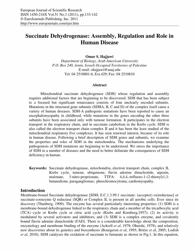
two hydrogen atoms are removed from succinate by flavin adenine dinucleotide (FAD), a prosthetic group that is tightly attached to SDH. Two electrons from the reduced SDH-FADH2 complex are then transferred to ubiquinone (Q), a soluble component of the electron transport system complex II. Ubiquinone is then reduced to ubiquinol ( QH2).
Figure 1: Oxidation of succinate to fumarate by the enzyme succinate dehydrogenase.
In this reaction, two hydrogen atoms are removed from succinate by FAD, a prosthetic group that is tightly attached to succenate dehydrogenase. Two electrons from the reduced SDH-FADH2
complex are then transferred to ubiquinone, a soluble component of the electrons transport system complex II. SDH is unique among the Krebs cycle enzymes in that it is tightly bound to the inner mitochondrial membrane; the other enzymes of the pathway are located in the mitochondrial matrix. Because SDH is bound to the inner membrane, it is easily isolated along with mitochondria by the technique of differential centrifugation.
Fumarate reductase [E.C.4.2.1.2] is often found in anaerobic or facultative organisms, where it reduces fumarate to succinate in the reverse of the SDH reaction (Fig.1). SDH and fumarate reductase catalyze the same reactions, but heir equilibriums are shifted toward succinate oxidation and fumarate reduction, respectively (Bhatt,2009).
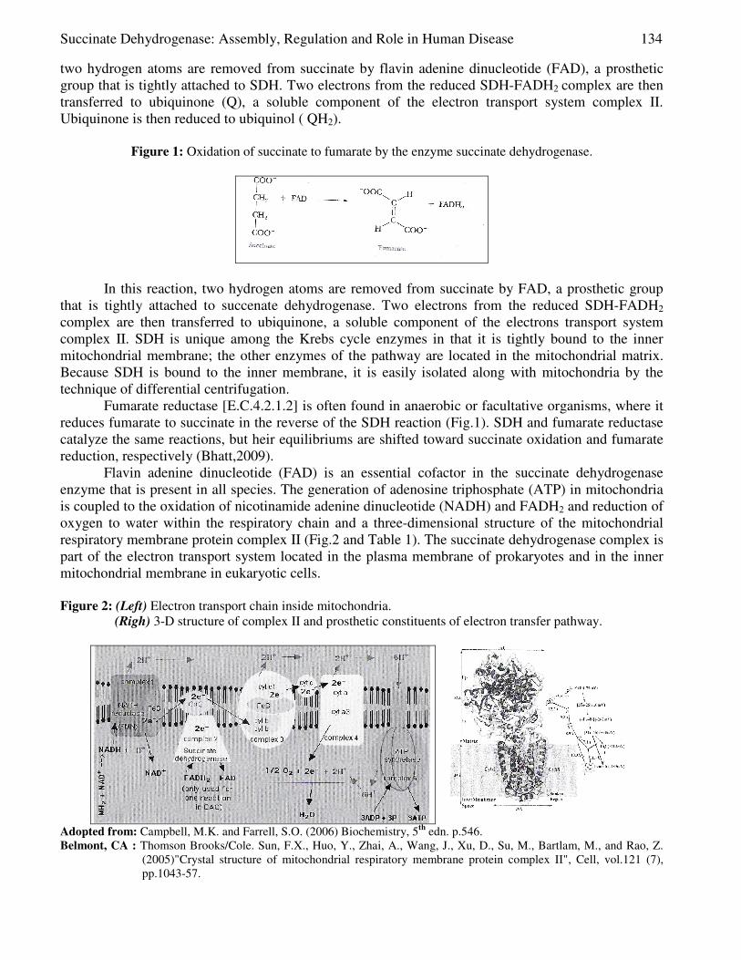
Flavin adenine dinucleotide (FAD) is an essential cofactor in the succinate dehydrogenase enzyme that is present in all species. The generation of adenosine triphosphate (ATP) in mitochondria is coupled to the oxidation of nicotinamide adenine dinucleotide (NADH) and FADH2 and reduction of oxygen to water within the respiratory chain and a three-dimensional structure of the mitochondrial respiratory membrane protein complex II (Fig.2 and Table 1). The succinate dehydrogenase complex is part of the electron transport system located in the plasma membrane of prokaryotes and in the inner mitochondrial membrane in eukaryotic cells. Figure 2: (Left) Electron transport chain inside mitochondria.
(Righ) 3-D structure of complex II and prosthetic constituents of electron transfer pathway.
Adopted from: Campbell, M.K. and Farrell, S.O. (2006) Biochemistry, 5th edn. p.546. Belmont, CA : Thomson Brooks/Cole. Sun, F.X., Huo, Y., Zhai, A., Wang, J., Xu, D., Su, M., Bartlam, M., and Rao, Z.
(2005)"Crystal structure of mitochondrial respiratory membrane protein complex II", Cell, vol.121 (7), pp.1043-57.
135 Omar S. Hajjawi
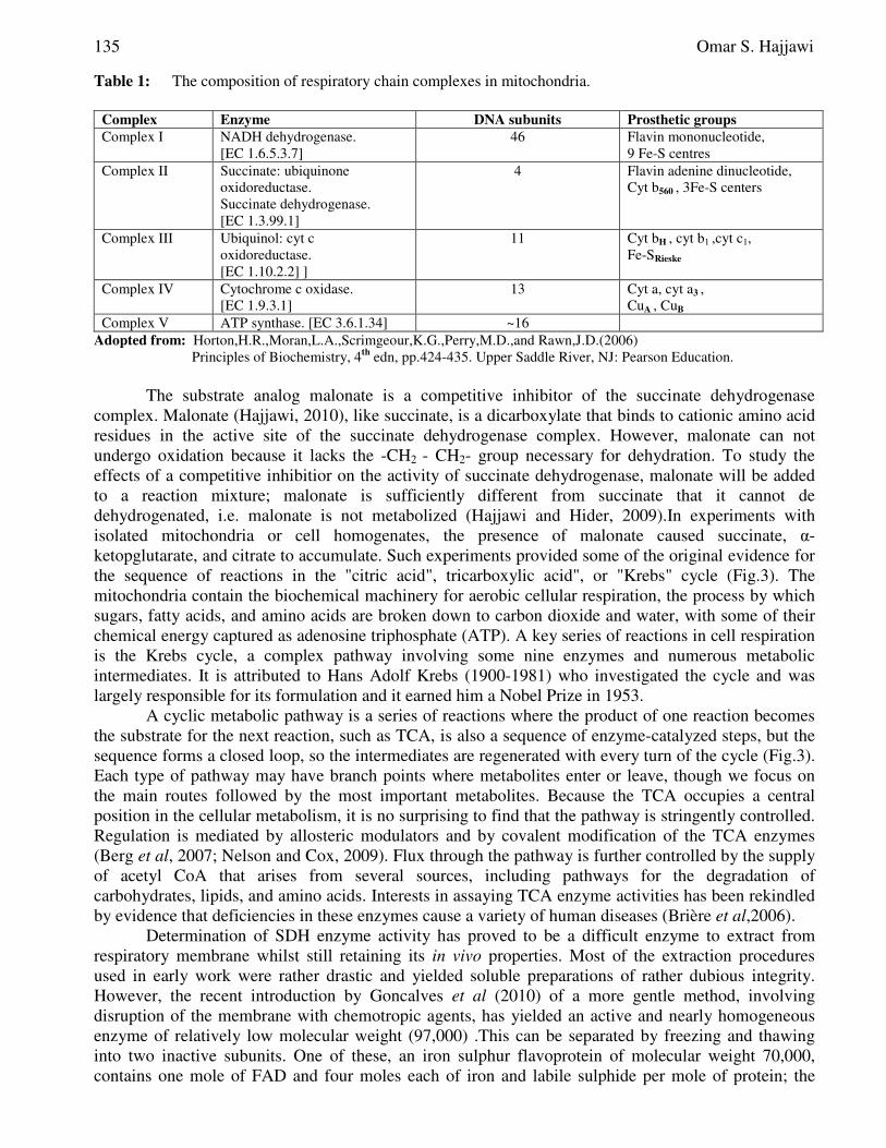
Table 1: The composition of respiratory chain complexes in mitochondria.
Complex Enzyme DNA subunits Prosthetic groups
Complex I NADH dehydrogenase. [EC 1.6.5.3.7]
46 Flavin mononucleotide, 9 Fe-S centres
Complex II Succinate: ubiquinone oxidoreductase. Succinate dehydrogenase. [EC 1.3.99.1]
4 Flavin adenine dinucleotide, Cyt b560 , 3Fe-S centers
Complex III Ubiquinol: cyt c oxidoreductase. [EC 1.10.2.2] ]
11 Cyt bH , cyt b1 ,cyt c1, Fe-SRieske
Complex IV Cytochrome c oxidase. [EC 1.9.3.1]
13 Cyt a, cyt a3 , CuA , CuB
Complex V ATP synthase. [EC 3.6.1.34] ~16 Adopted from: Horton,H.R.,Moran,L.A.,Scrimgeour,K.G.,Perry,M.D.,and Rawn,J.D.(2006)
Principles of Biochemistry, 4th edn, pp.424-435. Upper Saddle River, NJ: Pearson Education.
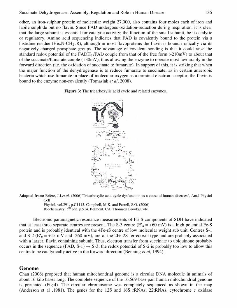
The substrate analog malonate is a competitive inhibitor of the succinate dehydrogenase complex. Malonate (Hajjawi, 2010), like succinate, is a dicarboxylate that binds to cationic amino acid residues in the active site of the succinate dehydrogenase complex. However, malonate can not undergo oxidation because it lacks the -CH2 - CH2- group necessary for dehydration. To study the effects of a competitive inhibitior on the activity of succinate dehydrogenase, malonate will be added to a reaction mixture; malonate is sufficiently different from succinate that it cannot de dehydrogenated, i.e. malonate is not metabolized (Hajjawi and Hider, 2009).In experiments with isolated mitochondria or cell homogenates, the presence of malonate caused succinate, α-ketopglutarate, and citrate to accumulate. Such experiments provided some of the original evidence for the sequence of reactions in the "citric acid", tricarboxylic acid", or "Krebs" cycle (Fig.3). The mitochondria contain the biochemical machinery for aerobic cellular respiration, the process by which sugars, fatty acids, and amino acids are broken down to carbon dioxide and water, with some of their chemical energy captured as adenosine triphosphate (ATP). A key series of reactions in cell respiration is the Krebs cycle, a complex pathway involving some nine enzymes and numerous metabolic intermediates. It is attributed to Hans Adolf Krebs (1900-1981) who investigated the cycle and was largely responsible for its formulation and it earned him a Nobel Prize in 1953.
A cyclic metabolic pathway is a series of reactions where the product of one reaction becomes the substrate for the next reaction, such as TCA, is also a sequence of enzyme-catalyzed steps, but the sequence forms a closed loop, so the intermediates are regenerated with every turn of the cycle (Fig.3). Each type of pathway may have branch points where metabolites enter or leave, though we focus on the main routes followed by the most important metabolites. Because the TCA occupies a central position in the cellular metabolism, it is no surprising to find that the pathway is stringently controlled. Regulation is mediated by allosteric modulators and by covalent modification of the TCA enzymes (Berg et al, 2007; Nelson and Cox, 2009). Flux through the pathway is further controlled by the supply of acetyl CoA that arises from several sources, including pathways for the degradation of carbohydrates, lipids, and amino acids. Interests in assaying TCA enzyme activities has been rekindled by evidence that deficiencies in these enzymes cause a variety of human diseases (Brière et al,2006).
Determination of SDH enzyme activity has proved to be a difficult enzyme to extract from respiratory membrane whilst still retaining its in vivo properties. Most of the extraction procedures used in early work were rather drastic and yielded soluble preparations of rather dubious integrity. However, the recent introduction by Goncalves et al (2010) of a more gentle method, involving disruption of the membrane with chemotropic agents, has yielded an active and nearly homogeneous enzyme of relatively low molecular weight (97,000) .This can be separated by freezing and thawing into two inactive subunits. One of these, an iron sulphur flavoprotein of molecular weight 70,000, contains one mole of FAD and four moles each of iron and labile sulphide per mole of protein; the
Succinate Dehydrogenase: Assembly, Regulation and Role in Human Disease 136
other, an iron-sulphur protein of molecular weight 27,000, also contains four moles each of iron and labile sulphide but no flavin. Since FAD undergoes oxidation-reduction during respiration, it is clear that the large subunit is essential for catalytic activity; the function of the small subunit, be it catalytic or regulatory. Amino acid sequencing indicates that FAD is covalently bound to the protein via a histidine residue (His.N-CH2 .R), although in most flavoproteins the flavin is bound ironically via its negatively charged phosphate groups. The advantage of covalent bonding is that it could raise the standard redox potential of the FADH2 /FAD couple from that of the free form (-210mV) to about that of the succinate/fumarate couple (+30mV), thus allowing the enzyme to operate most favourably in the forward direction (i.e. the oxidation of succinate to fumarate). In support of this, it is striking that when the major function of the dehydrogenase is to reduce fumarate to succinate, as in certain anaerobic bacteria which use fumarate in place of molecular oxygen as a terminal electron acceptor, the flavin is bound to the enzyme non-covalently (Tomasiak et al, 2008).
Figure 3: The tricarboxylic acid cycle and related enzymes.
Adopted from: Brière, J.J.et.al. (2006)"Tricarboxylic acid cycle dysfunction as a cause of human diseases", Am.J.Physiol
Cell Physiol, vol.291, p.C1115. Campbell, M.K. and Farrell, S.O. (2006) Biochemistry, 5th edn. p.514. Belmont, CA: Thomson Brooks/Cole.
Electronic paramagnetic resonance measurements of FE-S components of SDH have indicated
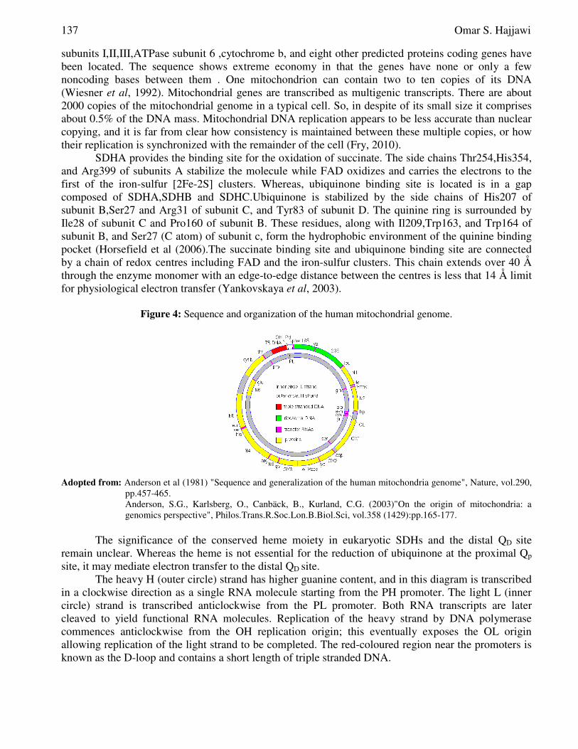
that at least three separate centres are present. The S-3 centre (E'o = +60 mV) is a high potential Fe-S protein and is probably identical with the 4Fe-rS centre of low molecular weight sub unit. Centres S-1 and S-2 (E'o = +15 mV and -260 mV), are of the 2Fe-2S ferredoxin type and are probably associated with a larger, flavin containing subunit. Thus, electron transfer from succinate to ubiquinone probably occurs in the sequence (FAD, S-1) → S-3; the redox potential of S-2 is probably too low to allow this centre to be catalytically active in the forward direction (Benning et al, 1994). Genome Chan (2006) proposed that human mitochondrial genome is a circular DNA molecule in animals of about 16 kilo bases long. The complete sequence of the 16,569-base pair human mitochondrial genome is presented (Fig.4). The circular chromosome was completely sequenced as shown in the map (Anderson et al ,1981). The genes for the 12S and 16S rRNAs, 22tRNAs, cytochrome c oxidase
137 Omar S. Hajjawi
subunits I,II,III,ATPase subunit 6 ,cytochrome b, and eight other predicted proteins coding genes have been located. The sequence shows extreme economy in that the genes have none or only a few noncoding bases between them . One mitochondrion can contain two to ten copies of its DNA (Wiesner et al, 1992). Mitochondrial genes are transcribed as multigenic transcripts. There are about 2000 copies of the mitochondrial genome in a typical cell. So, in despite of its small size it comprises about 0.5% of the DNA mass. Mitochondrial DNA replication appears to be less accurate than nuclear copying, and it is far from clear how consistency is maintained between these multiple copies, or how their replication is synchronized with the remainder of the cell (Fry, 2010).
SDHA provides the binding site for the oxidation of succinate. The side chains Thr254,His354, and Arg399 of subunits A stabilize the molecule while FAD oxidizes and carries the electrons to the first of the iron-sulfur [2Fe-2S] clusters. Whereas, ubiquinone binding site is located is in a gap composed of SDHA,SDHB and SDHC.Ubiquinone is stabilized by the side chains of His207 of subunit B,Ser27 and Arg31 of subunit C, and Tyr83 of subunit D. The quinine ring is surrounded by Ile28 of subunit C and Pro160 of subunit B. These residues, along with Il209,Trp163, and Trp164 of subunit B, and Ser27 (C atom) of subunit c, form the hydrophobic environment of the quinine binding pocket (Horsefield et al (2006).The succinate binding site and ubiquinone binding site are connected by a chain of redox centres including FAD and the iron-sulfur clusters. This chain extends over 40 Å through the enzyme monomer with an edge-to-edge distance between the centres is less that 14 Å limit for physiological electron transfer (Yankovskaya et al, 2003).
Figure 4: Sequence and organization of the human mitochondrial genome.
Adopted from: Anderson et al (1981) "Sequence and generalization of the human mitochondria genome", Nature, vol.290,
pp.457-465. Anderson, S.G., Karlsberg, O., Canbäck, B., Kurland, C.G. (2003)"On the origin of mitochondria: a genomics perspective", Philos.Trans.R.Soc.Lon.B.Biol.Sci, vol.358 (1429):pp.165-177.
The significance of the conserved heme moiety in eukaryotic SDHs and the distal QD site
remain unclear. Whereas the heme is not essential for the reduction of ubiquinone at the proximal Qp
site, it may mediate electron transfer to the distal QD site. The heavy H (outer circle) strand has higher guanine content, and in this diagram is transcribed
in a clockwise direction as a single RNA molecule starting from the PH promoter. The light L (inner circle) strand is transcribed anticlockwise from the PL promoter. Both RNA transcripts are later cleaved to yield functional RNA molecules. Replication of the heavy strand by DNA polymerase commences anticlockwise from the OH replication origin; this eventually exposes the OL origin allowing replication of the light strand to be completed. The red-coloured region near the promoters is known as the D-loop and contains a short length of triple stranded DNA.
Succinate Dehydrogenase: Assembly, Regulation and Role in Human Disease 138
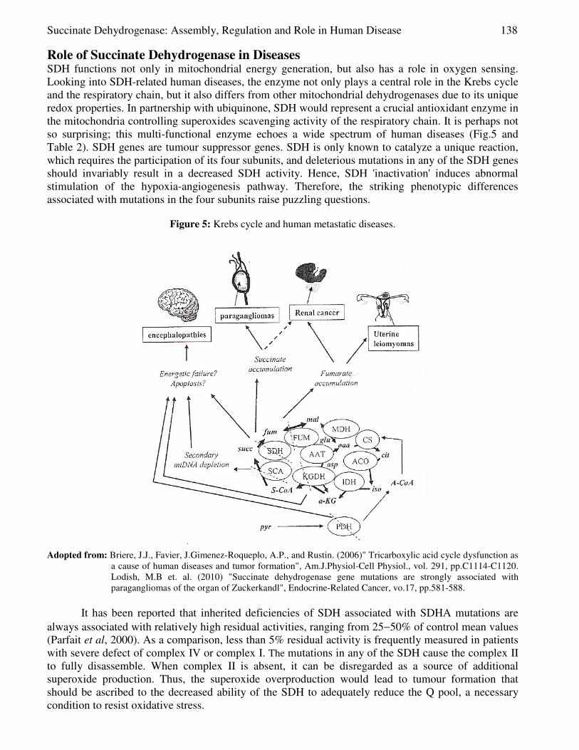
Role of Succinate Dehydrogenase in Diseases SDH functions not only in mitochondrial energy generation, but also has a role in oxygen sensing. Looking into SDH-related human diseases, the enzyme not only plays a central role in the Krebs cycle and the respiratory chain, but it also differs from other mitochondrial dehydrogenases due to its unique redox properties. In partnership with ubiquinone, SDH would represent a crucial antioxidant enzyme in the mitochondria controlling superoxides scavenging activity of the respiratory chain. It is perhaps not so surprising; this multi-functional enzyme echoes a wide spectrum of human diseases (Fig.5 and Table 2). SDH genes are tumour suppressor genes. SDH is only known to catalyze a unique reaction, which requires the participation of its four subunits, and deleterious mutations in any of the SDH genes should invariably result in a decreased SDH activity. Hence, SDH 'inactivation' induces abnormal stimulation of the hypoxia-angiogenesis pathway. Therefore, the striking phenotypic differences associated with mutations in the four subunits raise puzzling questions.
Figure 5: Krebs cycle and human metastatic diseases.
Adopted from: Briere, J.J., Favier, J.Gimenez-Roqueplo, A.P., and Rustin. (2006)" Tricarboxylic acid cycle dysfunction as
a cause of human diseases and tumor formation", Am.J.Physiol-Cell Physiol., vol. 291, pp.C1114-C1120. Lodish, M.B et. al. (2010) "Succinate dehydrogenase gene mutations are strongly associated with paragangliomas of the organ of Zuckerkandl", Endocrine-Related Cancer, vo.17, pp.581-588.
It has been reported that inherited deficiencies of SDH associated with SDHA mutations are
always associated with relatively high residual activities, ranging from 25−50% of control mean values (Parfait et al, 2000). As a comparison, less than 5% residual activity is frequently measured in patients with severe defect of complex IV or complex I. The mutations in any of the SDH cause the complex II to fully disassemble. When complex II is absent, it can be disregarded as a source of additional superoxide production. Thus, the superoxide overproduction would lead to tumour formation that should be ascribed to the decreased ability of the SDH to adequately reduce the Q pool, a necessary condition to resist oxidative stress.
139 Omar S. Hajjawi
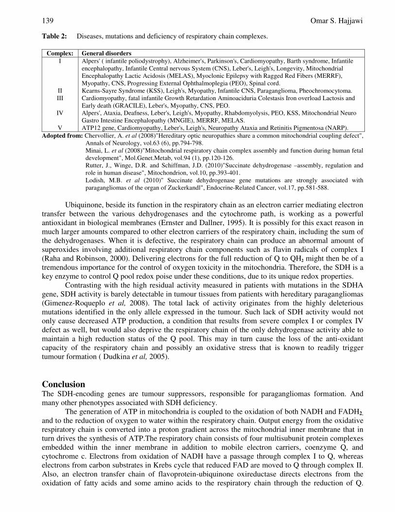
Table 2: Diseases, mutations and deficiency of respiratory chain complexes.
Complex: General disorders
I Alpers' ( infantile poliodystrophy), Alzheimer's, Parkinson's, Cardiomyopathy, Barth syndrome, Infantile encephalopathy, Infantile Central nervous System (CNS), Leber's, Leigh's, Longevity, Mitochondrial Encephalopathy Lactic Acidosis (MELAS), Myoclonic Epilepsy with Ragged Red Fibers (MERRF), Myopathy, CNS, Progressing External Ophthalmoplegia (PEO), Spinal cord.
II Kearns-Sayre Syndrome (KSS), Leigh's, Myopathy, Infantile CNS, Paraganglioma, Pheochromocytoma. III Cardiomyopathy, fatal infantile Growth Retardation Aminoaciduria Colestasis Iron overload Lactosis and
Early death (GRACILE), Leber's, Myopathy, CNS, PEO. IV Alpers', Ataxia, Deafness, Leber's, Leigh's, Myopathy, Rhabdomyolysis, PEO, KSS, Mitochondrial Neuro
Gastro Intestine Encephalopathy (MNGIE), MERRF, MELAS. V ATP12 gene, Cardiomyopathy, Leber's, Leigh's, Neuropathy Ataxia and Retinitis Pigmentosa (NARP).
Adopted from: Chervollier, A. et al (2008)"Hereditary optic neuropathies share a common mitochondrial coupling defect", Annals of Neurology, vol.63 (6), pp.794-798. Minai, L. et al (2008)"Mitochondrial respiratory chain complex assembly and function during human fetal development", Mol.Genet.Metab, vol.94 (1), pp.120-126. Rutter, J., Winge, D.R. and Schiffman, J.D. (2010)"Succinate dehydrogenase –assembly, regulation and role in human disease", Mitochondrion, vol.10, pp.393-401. Lodish, M.B. et al (2010)" Succinate dehydrogenase gene mutations are strongly associated with paragangliomas of the organ of Zuckerkandl", Endocrine-Related Cancer, vol.17, pp.581-588.
Ubiquinone, beside its function in the respiratory chain as an electron carrier mediating electron
transfer between the various dehydrogenases and the cytochrome path, is working as a powerful antioxidant in biological membranes (Ernster and Dallner, 1995). It is possibly for this exact reason in much larger amounts compared to other electron carriers of the respiratory chain, including the sum of the dehydrogenases. When it is defective, the respiratory chain can produce an abnormal amount of superoxides involving additional respiratory chain components such as flavin radicals of complex I (Raha and Robinson, 2000). Delivering electrons for the full reduction of Q to QH2 might then be of a tremendous importance for the control of oxygen toxicity in the mitochondria. Therefore, the SDH is a key enzyme to control Q pool redox poise under these conditions, due to its unique redox properties.
Contrasting with the high residual activity measured in patients with mutations in the SDHA gene, SDH activity is barely detectable in tumour tissues from patients with hereditary paragangliomas (Gimenez-Roqueplo et al, 2008). The total lack of activity originates from the highly deleterious mutations identified in the only allele expressed in the tumour. Such lack of SDH activity would not only cause decreased ATP production, a condition that results from severe complex I or complex IV defect as well, but would also deprive the respiratory chain of the only dehydrogenase activity able to maintain a high reduction status of the Q pool. This may in turn cause the loss of the anti-oxidant capacity of the respiratory chain and possibly an oxidative stress that is known to readily trigger tumour formation ( Dudkina et al, 2005). Conclusion The SDH-encoding genes are tumour suppressors, responsible for paragangliomas formation. And many other phenotypes associated with SDH deficiency.
The generation of ATP in mitochondria is coupled to the oxidation of both NADH and FADH2, and to the reduction of oxygen to water within the respiratory chain. Output energy from the oxidative respiratory chain is converted into a proton gradient across the mitochondrial inner membrane that in turn drives the synthesis of ATP.The respiratory chain consists of four multisubunit protein complexes embedded within the inner membrane in addition to mobile electron carriers, coenzyme Q, and cytochrome c. Electrons from oxidation of NADH have a passage through complex I to Q, whereas electrons from carbon substrates in Krebs cycle that reduced FAD are moved to Q through complex II. Also, an electron transfer chain of flavoprotein-ubiquinone oxireductase directs electrons from the oxidation of fatty acids and some amino acids to the respiratory chain through the reduction of Q.
Succinate Dehydrogenase: Assembly, Regulation and Role in Human Disease 140
Complex III oxidizes reduced obiquinone , and subsequently the electrons have moved on through cytochrome c to complex IV, where molecular oxygen is reduced to water. Complexes I, III, and IV pump protons to generate the electrochemical gradient that complex V utilizes to drive ATP synthesis. The electron transfer path in the oxidation of NADH by complex I involves initial reduction of a FMN cofactor, and the subsequent transport through seven Fe-S centres to Q binding site. The electron transfer path in the oxidation of succinate by complex II involves initial reduction of a FAD cofactor followed by electron transfer through three Fe-S centres to Q. Whereas reduction of Q by flavoprotein-ubiquinone oxireductase links oxidation of nine matrix flavoprotein dehydrogenases with the respiratory chain. Electron transfer by flavoprotein-ubiquinone occurs through a Fe-S centre to a FAD moiety where Q is reduced (Rutter et al, 2010).
Primary mitochondrial diseases are relatively rare, because the major defects in Krebs cycle are incompatible with life and many affected embryos would have died at an early phase (Mani et al, 2008). However, there are about 150 different types of mitochondrial hereditary defects have been reported. Mitochondrial DNA is maternally inherited through the egg cell cytoplasm, but many of the inherited defects map to the nuclear genome since the majority of mitochondrial proteins were inherently imported from the cytosol, the aqueous portion of the cytoplasm minus the subcellular structures. All the copies of the mitochondrial DNA are identical in healthy people (Frye, 2010). Mitochondrial diseases have recently generated great interest in the autoimmune component, and in innate apoptosis programme of cell death. The permeability transition of mitochondrial is believed to be involved in the suicidal process of apoptosis. An elaboration of self-destruction cascade is responsible for the programmed loss of cells during tissue differentiation, the self-destruction of virally - infected cells, and the unwanted cell damage that follows loss of tissue perfusion in cardiovascular disease. Mitochondria that have undergone permeability transition, release a protease from the inter-membrane space which then activates the subsequent nuclear stages of the apoptotic cascade. References [1] Ackrell, B.A.C., Kearney, E.B. and Singer, T.P. (1978)"Mammalian succinate dehydrogenase",
Methods Enzymol,vol.53,pp.466-483. [2] Anderson,S., Bankier,A.T., Barrell,B.G., De Bruijn,M.H.L., Coulson,A.R., Peron,I.C.,
Nierlich,D.P., Roe,B.A., Sanger,F., Schrieier,P.H., Smith,A.J.H., Staden, R., Young, I.G. (1981)" Sequence and organization of the human mitochondrial genome", Nature, vol.290, pp.457-465.
[3] Anderson, S.G., Karlsberg, O., Canbäck, B., Kurland, C.G. (2003)"On the origin of mitochondria: a genomics perspective", Philos. Trans. R. Soc.Lon.B.Biol. Sci, vol.358 (1429):pp.165-177.
[4] Berg, J.M., Tymoczko, J.L., and Stryer, L. (2007) Biochemistry, 6th edn. New York: W.H.Freeman & Co.
[5] Benning, M.M., Meyer, T.F., Rayment, I.and Holden, H.M. (1994)" Molecular structure of the oxidized high potential iron-sulfur protein isolated from Ectothiorhodospira vacuolata", Biochemistry, vol.33, pp.2476-2483.
[6] Bhatt, D.K. (2009) "Modulation of tricarboxylic acid cycle dehydrogenase during hepatocarcinogenesis induced by hexachlorocyclohexane in mice", Experimental and Toxicological Pathology, vol.61 (4), pp.325-332.
[7] Bourgeron, T., Rustin, P., Chretien, D., Birch-Machin, M., Bourgeois, M., Viegas-Péquignot, E., Munnich, A., and Rötig, A. (1995)"Mutation of a nuclear succinate dehydrogenase gene results in mitochondrial respiratory chain deficiency", Nature Generics, vol.11, pp.144-149.
[8] Brière, J.J., Favier, J., Ghouzzi, V. El., Djouadi, F., Bénit, P., Gimenez, A.P., and Rustin. (2005) Succinate dehydrogenase deficiency in human", Cellular and Molecular Life Sciences, vol.62 (19-20), pp.2317-2324).
141 Omar S. Hajjawi
[9] Brière, J.J., Favier, J., Gimenez-Roqueplo, A.P. and Rustin, P. (2006)" Tricarboxylic acid cycle dysfunction as a cause of human diseases and tumor formation", Am.J.Physiol.Cell Physiol., vol.29 (6), pp.C1114-1120.
[10] Campbell, M.K. and Farrell, S.O. (2006) Biochemistry, 5th edn. Belmont, CA: Thomson Brooks/Cole.
[11] Chan, D.C. (2006) "Mitochondria: Dynamic organelles in disease, aging and development", Cell, vol.125 (7), pp.1241-1252.
[12] Chevrollier, A., Guillet, V., Loiseau, D., Gueguen, N., de Rescenzo, M.A.P., Verny,C., Ferre,M,Dollfus,H, Odent,S.,Milea,D,Goizet,C,Amati- Bonneau, D., and Reynier, P. (2008)"Hereditary optic neuropathies share a common mitochondrial coupling defect", Annals of Neurology, vol.63 (6), pp.794-798.
[13] Dudkina , N.V., Eubel, H., Keegstra, W., Boekema, E J. and Braun, H.P. (2005)" Structure of a mitochondrial supercomplex formed by respiratory-chain complexes I and III" PNAS, vol.102, no.9, pp.3225-3229.
[14] Ernster. L. and Dallner, G. (1995)" Biochemical, physiological and medical aspects of ubiquinone function", Biochim Biophys Acta, vol.1271, pp. 195-204.
[15] Frye, R.E. (2010)" 15q11.2-13 duplication, mitochondrial dysfunction, and development disorder", J.Child Neural., vol.24 (10), pp.1316-1320.
[16] Gimenez-Roqueplo, A.P., Burnichon, N., Amar, L., Favier, J., Jeunemaitre, X., and Plouin, P.F. (2008)"Recent advances in the genetics of Phaeochromocytoma and functional paragangloma", Clinical and Experimental Pharmacology and Physiology, vol.35 (4), pp.376-379.
[17] Hajjawi, O.S. and Hider, R.C. (2009)"Asymmetry of the malonate transport system in human red blood cells", European Journal of Scientific Research, vol.31 (4), pp.534-545.
[18] Hajjawi, O.S. (2010) "Sequential mechanism of the malonate transport System in human red blood cells", European Journal of Scientific Research, vol.46 (1), pp.18-27.
[19] Horsefield, R., Yankovskaya, V., Sexton, G., Whittingham, W., Shiomi, K., Omura, S., Byrne, B., Cecchini G., and Iwata, S. (2006)"Structural and computation analysis of the quinine-binding site of complex II (succinate-ubiquinone oxireductase): a mechanism of electron transfer and proton conduction during ubiquinone reduction", J.Biol.Chem.vol.281 (11), pp. 7309-16.
[20] Horton,H.R.,Moran,L.A.,Scrimgeour,K.G.,Perry,M.D.,and Rawn,J.D.(2006) Principles of Biochemistry, 4th
edn. Upper Saddle River, NJ: Pearson Education Inc. Krebs, H.A. and Kornberg, H.L. (1957) Energy Transformations in Living Matter. Berlin: Springer-Verlag.
[21] Lodish,M.B.,Adams,K.T.,Huynh,T.T.,Prodanov,T.,Ling,A., Chen,C., Shusterman,S.,Jimenez,C., Merini,M.,Hughs,M.,Cradic,K.W., Milosevic, D., Singh, R.J., Stratakis, C.A. and Pacak, K. (2010) Succinate dehydrogenase gene mutations are strongly associated with paragangliomas of the organ of Zuckerkandl", Endocrine-Related Cancer, vol.17, pp.581-588.
[22] Nelson, D.L. and Cox, M.M. (2009) Lehninger Principles of Biochemistry, 5th edn. New York: W.H.Freeman & Co.
[23] Ohnishi, T. (1979) "Mitochondrial iron-sulfur flavodehyrdogenases. In R.A.Capaldi (ed), Membrane proteins in energy transduction, pp.1-80. New York: Marcel Dekker, Inc.
[24] Parfait, B., Chretien, D., Rötig, A., Marsac, C., Munnich, A., and Rustin, P. (2000) "Compound heterozygous mutations in the flavoprotein gene of the respiratory chain complex II in a patient with Leigh syndrome", Hum Genet, vol.106,pp. 236-243.
[25] Raha, S. and Robinson, B.H. (2000) "Mitochondria, oxygen free radicals, disease and ageing", Trends Biochem Sci, vol. 25, pp. 502-508.
[26] Sun, F.X., Huo, Y., Zhai, A., Wang, J., Xu, D., Su, M., Bartlam, M., and Rao, Z. (2005)"Crystal structure of mitochondrial respiratory membrane protein complex II", Cell, vol.121 (7), pp.1043-57.
Succinate Dehydrogenase: Assembly, Regulation and Role in Human Disease 142
[27] Thunberg, T. (1909)"Studien über die Beeinflussung des Gassustausches des überlebenden Froschmuskels durch verschiedene stoffe", Skand.Archiv Physiol., vol.22, pp.430-436.
[28] Tomasiak, T.M., Maklashina, E., Cecchini, G., and Iverson, T.M. (2008) "A threonine on the active site loop controls transition state formation in Escherichia coli respiratory complex II", J.Biol.Chem., vol.283 (22),pp. 15460-15468.
[29] Wiesner, R.J., Ruegg, J.C., and Morano, J. (1992)"Counting target molecules by exponential polymerase chain reaction, copy number of mitochondrial DNA in rat tissue", Biochim Biophys Acta, vol.83 (2), pp.553-559.
[30] Yankovskaya, V., Horsefield, R., Törnroth, S., Luna-Chavez, C., Miyoshi, H., Léger, C., Byrne, B., Cecchini, G. and Iwata, S. (2003)"Architecture of succinate dehydrogenase and reactive oxygen species generation", Science, vol. 299, pp.700-704.