sudden death, aortic rupture in horses, literature review
TRANSCRIPT

298
Braz. J. Vet. Res. Anim. Sci., São Paulo, v. 52, n. 4, p. 298-309, 2015
Abstract
Sudden deaths of horses in multiple equestrian disciplines have been attributed to acute and chronic respiratory and cardiovascular diseases. The aim of this study was to perform a review of aortic rupture in horses analyzing , case studies and assessing risk factors. The literature has reported a total of 137 cases of aortic rupture in horses for 28 years (1986-2014), with approximately five horses dying of aortic rupture per year. Histopathologically, there are observed discrete macroscopic degenerative changes in the intima layer only in the aorta. The histological evaluation in the beginning portion of the aorta of the heart evidenced degenerative changes with loss of continuity and distribution of elastic fibers. Risk factors for the rupture of the aorta are: spontaneous rupture associated with hypertension, preexisting vascular injury (aneurysm), dilated or hypertrophic cardiomyopathy, copper levels in the endothelium, genetic factors such as inbreeding, toxicology or pharmacological factors. Aortic rupture shows similarity with pulmonary hemorrhage induced by exercise especially under the locomotors induced trauma theory of exercise that can induce pulmonary hemorrhage. In conclusion, degenerative changes to discrete elastic fiber of the intima of the aorta in the emergence of the heart seem to predispose the aorta wall rupture at the time of maximum blood pressure during exercise and the consequent collapse and athletic horse’s death.Keywords: Aorta. Equine. Horse. Sudden dead. Thoroughbreds. Resumo
As mortes súbitas de cavalos em várias provas equestres têm sido atribuídas a doenças respiratórias e cardiovasculares agudas e crônicas. O objetivo deste estudo foi efetuar uma revisão de literatura da ruptura da aorta em cavalos analisando estudos de caso e estabelecendo os possíveis fatores de risco. Na revisão da literatura no período de 28 anos (1986-2014) foram localizados 137 casos de ruptura da aorta em cavalos com aproximadamente cinco cavalos morrendo por essa causa por ano. Histologicamente, são observadas alterações macroscópicas discretas degenerativas na camada íntima da aorta. A avaliação histológica na porção inicial da aorta do coração evidencia alterações degenerativas com perda de continuidade e distribuição das fibras elásticas. Fatores de risco para a ruptura da aorta dos cavalos são: ruptura espontânea associada com hipertensão, lesão vascular pré-existente (aneurisma), cardiomiopatia dilatada ou hipertrófica, níveis de cobre no endotélio, fatores genéticos, tais como a consanguinidade na criação, toxicologia e aspectos farmacológicos. A ruptura aórtica mostra semelhança com hemorragia pulmonar induzida pelo exercício. Em conclusão, alterações degenerativas discretas das fibras elásticas da íntima da aorta parecem predispor a ruptura da parede da aorta, no momento da pressão máxima de sangue durante o exercício determinando o consequente colapso e morte do cavalo atleta.Palavras-chave: Aorta. Equino. Cavalo. Morte súbita. Thoroughbreds.
Sudden death, aortic rupture in horses, literature review, case studies reported and risk factorsMorte súbita por ruptura da aorta em cavalos, literatura,
estudos de casos relatados e fatores de risco
Abelardo Morales BRICEÑO¹; Aniceto MENDEZ¹; Kimberly BREWER²; Charlie HUGHES³; Thomas TOBIN³
¹ University of Cordoba, College of Veterinary Medicine, Department of Anatomy and Comparative Anatomic Pathology – Córdoba, Spain
² Phoenix Rising Veterinary, Wellington – Florida, USA³ University of Kentucky, The Maxwell H. Gluck Equine Research Center, Lexington – Kentucky, USA
DOI: 10.11606/issn.1678-4456.v52i4p298-309
Correspondence to:Abelardo Morales Briceño University of Cordoba, College of Veterinary Medicine, Department of Anatomy and Comparative Anatomic Pathology, Edificio de Sanidad AnimalCampus de Rabanales Ctra. de Madrid km 39614071, Córdoba, SpainE-mail: [email protected]
Received: 18/05/2015Approved: 21/10/2015
Introduction
Sudden death in horses is a serious problem affecting the equine industry worldwide. Sudden deaths of horses in multiple equestrian disciplines have been attributed to acute respiratory and cardiovascular disease. Respiratory causes of sudden death include exercise-induced pulmonary hemorrhage. Cardiovascular

299
Braz. J. Vet. Res. Anim. Sci., São Paulo, v. 52, n. 4, p. 298-309, 2015
causes of sudden death include myocarditis, rupture of chordae tendineae, aorta or other large arteries, aneurysm, atrial dysrhythmia, valvular lesions, dilated and hypertrophic cardiomyopathy, myocardial necrosis, sclerosing coronary arteriopathy and massive disseminated hemorrhage (BODEN et al., 2005; LYLE et al., 2010). The aorta is the main artery that carries blood from the left ventricle of the heart (the major pumping chamber) to all the other arteries except the pulmonary artery (KING; BRIGHT, 1999). Its job is to bring oxygenated blood from the lungs (which enters the heart through the left atrium, flows through the bicuspid, or mitral, valve into the left ventricle, then is pumped into the aorta) to the arteries of the body (KING; BRIGHT, 1999). However, sometimes a spontaneous tear or break occurs in the wall of the aorta, causing a condition known as an aortic rupture. Aortic ruptures usually occur very close to the junction of the aorta with the heart (KING; BRIGHT, 1999). Aortic root rupture in horses most frequently results in sudden death associated with massive hemorrhage into the thoracic cavity (ORSINI; DIVERS, 2008). There are no medical or surgical cures for aortic ruptures in horses (KING; BRIGHT, 1999). Aortic rupture is an important topic at the moment in the equestrian world (DELESALLE, 2013). Equestrian journals often report that in competing horses and stallions that died suddenly, the necropsy report showed they were diagnosed with a ruptured aorta. This has generated many opinions and questions about aortic rupture in horses; we intend to elucidate with this article. The aim of this study was to investigate aortic rupture in horses through literature review, case studies and risk factors.
Revised AnatomicalThe Vascular System
The vascular system may be arbitrarily divided into the arterial system, microcirculation, venous system, and lymphatic system (JUBB et al., 2007). The arterial system is subdivided into large elastic arteries, medium and small muscular arteries, and arterioles, with gradual
transitions between these divisions (DONALD, 1999). Arterial vessels are characterized histopathologically by walls composed of three layers: the internal, middle, and external tunics. The tunica intern (intima) is characterized with endothelium; subendothelial connective tissue, which contains collagen, elastin, proteoglycan (ground substance), fibroblasts, and smooth muscle cells and the internal elastic membrane. The tunica media (media) consists of fenestrated elastic laminae in elastic arteries, with smooth muscle cells lying between laminae. Intracellular ground substance is especially prominent in the tunica media of elastic arteries in the horse. Contraction of muscular arteries and arterioles upon an animal´s death forces blood from the lumina, and causes longitudinal folding that appears as scalloping of the internal elastic membrane when seen in cross-section. The adventitia consists of a network of elastic and collagen fibers continuous with the surrounding connective tissue. The interlacing network of collagen fibers in the adventitia limits expansion of the elastic arteries (JUBB et al., 2007). The response of vessels to injury involves a complex interaction among the cellular and noncellular elements of the vessel wall and the cellular and noncellular elements of the blood. The key cells of vessels in these reactions are endothelial cells and smooth muscle cells. Endothelial cells are metabolically active and provide a thromboresistant monolayer at the interface of blood and the vessel wall. The arteries may have congenital abnormalities, degeneration, necrosis, hypertrophy, mineralization, aneurysms and ruptures, thrombosis and embolism, vasculitis and neoplasms. The aorta is the main arterial trunk. The base of the left ventricle, its first part is dorsal ascending aorta and pulmonary trunk cranial (GETTY, 1996). The descending aorta can be divided into thoracic and abdominal. The thoracic aorta is within the pericardium and then is placed between the two pleural sacs. The abdominal aorta is related dorsally with lumbar vertebrae, ventral longitudinal ligament and the left psoas minor muscle (GETTY, 1996).

300
Braz. J. Vet. Res. Anim. Sci., São Paulo, v. 52, n. 4, p. 298-309, 2015
Abnormalities of the blood vesselsCongenital anomalies with minor variations
occur among individuals of a species in the course and distribution of arteries, but these are of little significance. Regarding degeneration and necrosis of arteries, generalized vascular degenerative disease in animals is classified in three principal groups: 1) arteriosclerosis (chronic arterial change consisting of hardening, loss of elasticity and luminal narrowing resulting usually from proliferative and degenerative, rather than inflammatory, changes of the media and intima); 2) atherosclerosis (affects the large and elastic arteries as aorta, iliac and the large and medium muscular arteries, the essential lesion is the atheroma or fibrofatty plaque, which is a focal, raised, intimal plaque with a core of lipid cholesterol and its esters largely covered fibrous cap); 3) Arteriolosclerosis (describes a heterogeneous group of arteriolar lesions that maybe predominantly hyaline or predominantly hyperplastic). Mineralization (calcification) occurs quite frequently in the arteries of animals either as dystrophic which occurs in areas of inflammation degeneration and thrombosis, or metastatic which occurs as the results of hypercalcemia and/or hyperphosphatemia. Arterial hypertrophy may affect one or all components of the arterial wall. Arterial hypertrophy has been associated with high altitude disease with collateral vessels in response to the extra load they carry after occlusion of the artery (JUBB et al., 2007). Vasculitis or inflammation of a vessel is characterized by the presence of inflammatory cells within and around the blood vessel wall with concomitant vessel wall damage as indicated by fibrin deposition, collagen degeneration and necrosis endothelial and smooth muscle cells (JUBB et al., 2007). Rupture of arteries as a result of physical trauma is common; spontaneous ruptures are not. Rupture of the aorta is well known, but certainly uncommon, in the horse. The ruptures occur during periods of excitement and activity, such as racing or in stallions while breeding, and are probably related to
increased intra-aortic pressure. Spontaneous rupture of the pulmonary artery also occurs in horses, but is even less frequent than aortic rupture (DONALD, 1999). An aneurysm is a localized abnormal dilation of any vessel. Aneurysms are of most importance when they affect the aorta. Thromboembolism occurs in a variety of conditions. In verminous arteritis of horses, emboli arise from thrombi in the cranial mesenteric artery and cause intestinal ischemia, and possibly infarction. Aortic-iliac thrombosis causes exercise intolerance and hind-leg lameness in affected horses. The condition is seen most frequently in racing Thoroughbreds and Standardbreds, especially among young males (JUBB et al., 2007).
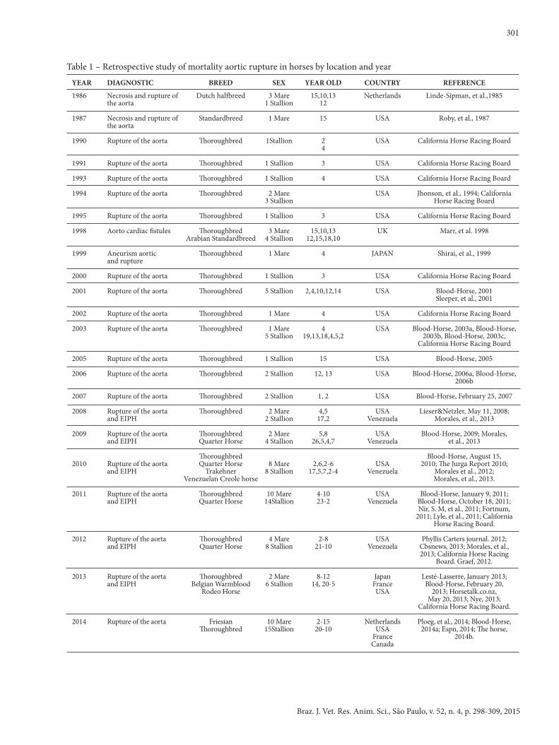
Review of cases of sudden death in horses reported in the literature (period 1986-2014)
Below are the cases of sudden death associated with rupture of the aorta, aneurysm and pulmonary hemorrhage in horses reported in the literature between 1986-2014 are described (Table 1).
Clinical and pathological study The literature has reported a total of 137 cases of
sudden death aortic rupture in horses for 28 years (1986-2014). Approximately five horses died of aortic rupture per year. Clinical history was classified as follows: Horses in athletic activity, showed collapse and sudden death (137 horses), horses with sudden death in the box (04). All horses in athletic activity were asymptomatic before the collapse. In horses with sudden death in the box, two cases presented with severe obesity. The literature report the presenting signs were acute distress and cardiac lesion was detected during a routine examination in one horse. Five horses had monomorphic ventricular tachycardia on admission and another had a history of this arrhythmia. Five horses had a characteristic continuous murmur loudest in the right fourth intercostal space (MARR, 1998). Imaging techniques only in four case reports: Echocardiography revealed an aneurysm of the right
301
Braz. J. Vet. Res. Anim. Sci., São Paulo, v. 52, n. 4, p. 298-309, 2015
Table 1 – Retrospective study of mortality aortic rupture in horses by location and year
YEAR DIAGNOSTIC BREED SEX YEAR OLD COUNTRY REFERENCE
1986 Necrosis and rupture of Dutch halfbreed 3 Mare 15,10,13 Netherlands Linde-Sipman, et al.,1985 the aorta 1 Stallion 12
1987 Necrosis and rupture of Standardbreed 1 Mare 15 USA Roby, et al., 1987 the aorta
1990 Rupture of the aorta Thoroughbred 1Stallion 2 USA California Horse Racing Board 4
1991 Rupture of the aorta Thoroughbred 1 Stallion 3 USA California Horse Racing Board
1993 Rupture of the aorta Thoroughbred 1 Stallion 4 USA California Horse Racing Board
1994 Rupture of the aorta Thoroughbred 2 Mare USA Jhonson, et al., 1994; California 3 Stallion Horse Racing Board 1995 Rupture of the aorta Thoroughbred 1 Stallion 3 USA California Horse Racing Board
1998 Aorto cardiac fistules Thoroughbred 3 Mare 15,10,13 UK Marr, et al. 1998 Arabian Standardbreed 4 Stallion 12,15,18,10
1999 Aneurism aortic Thoroughbred 1 Mare 4 JAPAN Shirai, et al., 1999 and rupture 2000 Rupture of the aorta Thoroughbred 1 Stallion 3 USA California Horse Racing Board
2001 Rupture of the aorta Thoroughbred 5 Stallion 2,4,10,12,14 USA Blood-Horse, 2001 Sleeper, et al., 2001
2002 Rupture of the aorta Thoroughbred 1 Mare 4 USA California Horse Racing Board
2003 Rupture of the aorta Thoroughbred 1 Mare 4 USA Blood-Horse, 2003a, Blood-Horse, 5 Stallion 19,13,18,4,5,2 2003b, Blood-Horse, 2003c, California Horse Racing Board
2005 Rupture of the aorta Thoroughbred 1 Stallion 15 USA Blood-Horse, 2005
2006 Rupture of the aorta Thoroughbred 2 Stallion 12, 13 USA Blood-Horse, 2006a, Blood-Horse, 2006b 2007 Rupture of the aorta Thoroughbred 2 Stallion 1, 2 USA Blood-Horse, February 25, 2007
2008 Rupture of the aorta Thoroughbred 2 Mare 4,5 USA Lieser&Netzler, May 11, 2008; and EIPH 2 Stallion 17,2 Venezuela Morales, et al., 2013
2009 Rupture of the aorta Thoroughbred 2 Mare 5,8 USA Blood-Horse, 2009; Morales, and EIPH Quarter Horse 4 Stallion 26,5,4,7 Venezuela et al., 2013
Thoroughbred Blood-Horse, August 15, 2010 Rupture of the aorta Quarter Horse 8 Mare 2,6,2-6 USA 2010; The Jurga Report 2010; and EIPH Trakehner 8 Stallion 17,5,7,2-4 Venezuela Morales et al., 2012; Venezuelan Creole horse Morales, et al., 2013.
2011 Rupture of the aorta Thoroughbred 10 Mare 4-10 USA Blood-Horse, January 9, 2011; and EIPH Quarter Horse 14Stallion 23-2 Venezuela Blood-Horse, October 18, 2011; Nir, S. M, et al., 2011; Fortnum, 2011; Lyle, et al., 2011; California Horse Racing Board.
2012 Rupture of the aorta Thoroughbred 4 Mare 2-8 USA Phyllis Carters journal. 2012; and EIPH Quarter Horse 8 Stallion 21-10 Venezuela Cbsnews, 2013; Morales, et al., 2013; California Horse Racing Board. Graef, 2012.
2013 Rupture of the aorta Thoroughbred 2 Mare 8-12 Japan Lesté-Lasserre, January 2013; and EIPH Belgian Warmblood 6 Stallion 14, 20-5 France Blood-Horse, February 20, Rodeo Horse USA 2013; Horsetalk.co.nz, May 20, 2013; Nye, 2013; California Horse Racing Board.
2014 Rupture of the aorta Friesian 10 Mare 2-15 Netherlands Ploeg, et al., 2014; Blood-Horse, Thoroughbred 15Stallion 20-10 USA 2014a; Espn, 2014; The horse, France 2014b. Canada

302
Braz. J. Vet. Res. Anim. Sci., São Paulo, v. 52, n. 4, p. 298-309, 2015
aortic sinus. Echocardiography (six horses) and/or post-mortem examination (four horses) revealed the horses had aorto-cardiac fistulas arising from the right aortic sinus in all five horses in which the site was recorded. Two horses had ruptured aneurysm dilatations of the aortic wall at this site. Fistulas extended into the right ventricle in four horses; the right atrium in two horses, the left ventricle in one horse, and five horses had dissecting tracts in the septal myocardium (MARR, 1998). Necropsy: horses were examined by necropsy and samples of tissue were collected (ALUJA; CONSTANTINO, 2002; BANKS, 1996). Histopathology: the tissue samples were fixed in formalin buffered 10% and processed by conventional H&E techniques (BANKS, 1996). The results are presented in table 1 and are described below. All horses in study had sudden death during exercise or during the reproductive activity, except for two horses (2 Creole Venezuelans and 1 Quarter Horse). Clinical evaluation: all horses were completely asymptomatic and showed no apparent clinical signs of cardiovascular failure, except four horses that had a cardiovascular failure at the clinic. Imaging: only four cases revealed an aneurysm of the right aortic sinus by echocardiography. The wall of the aneurysm was thin, and the aneurysm bulged into the right atrium and ventricle at the level of the tricuspid valve (SLEEPER et al., 2001). Color flow Doppler echocardiography did not reveal any blood flow across the aneurysm; however, swirling turbulent flow was evident within the aneurysm (SLEEPER et al., 2001). A small jet of mitral regurgitation was detected with color flow Doppler echocardiography and was deemed to be clinically insignificant at the time of evaluation (SLEEPER et al., 2001). Necropsy: on necropsy, massive hemothorax from severe thoracic aortic aneurysm was observed with ruptured and fibrin in all cases. Observed rupture of the thoracic aorta showed fibrin and necrotic material. In both cases of infestation by Strongylus sp, rupture of the aorta was lumbar level, residues were observed to have larvae
of parasites, with fibrin (quitine). In the Quarter Horses with atherosclerosis, dermatitis alopecic with hyperpigmentation and xantomathosis of subcutaneous tissue was observed. Hemoperitoneum was observed in the abdominal cavity, caused by rupture of the aorta flow. Fat deposits and plates type atheroma (MORALES et al., 2012) were observed in the wall of the aorta. The aorta was assessed in the emergence from the heart, all portions of the aortic arch, thoracic aorta, and abdominal aorta (Figures 1 and 2). Discrete macroscopic degenerative changes in the intima only in the initial portion of aorta were observed (Figures 3, 4 5 and 6). Histopathology: histological examination of the wall of the aorta showed necrotic areas that were scattered throughout the wall in both vessels. In some of these areas, necrotic muscle fibers were still present, whereas in others the muscle fibers had disappeared and the elastic fibers were condensed (Figures 7, 8 and 9). Neutrophils and fibroangioblastic tissue were seen around the areas of necrosis. Areas of calcification were found in horses. All horses had many vasa vasorum in the media and adventitia of the aorta and the pulmonary trunk with intimal thickening, medial fibrosis, or both (LINDE-SIPMAN et al.,1985). The lumen of many vasa vasorum was completely obliterated. Histopathology revealed severed fatty degeneration and hepatic necrosis. The aorta showed lipid-filled foam cell stage (fatty streak) into an advanced, complicated lesion that contains abundant extracellular cholesterol ester in the atheromatous gruel within the arterial intima. On necropsy in two horses the aorta was found, seeding all emerging portions of the aorta in the heart, aortic arch, thoracic aorta, and abdominal aorta. Discrete macroscopic degenerative changes in the intima were observed. The histological evaluation of the emerging portion of the aorta with the heart evidenced degenerative changes with loss of continuity and distribution of elastic fibers. The special reticulin positive stain details the changes of the elastic fiber in electron microscopic study process.

303
Braz. J. Vet. Res. Anim. Sci., São Paulo, v. 52, n. 4, p. 298-309, 2015
Figure 1 – Overview chest cavity (right lung, heart and thoracic aorta)
Source: (BRICEÑO, 2013)
Figure 2 – Thoracic aortaSource: (BRICEÑO, 2013)
Figure 3 – Abdominal aorta in each horseSource: (BRICEÑO, 2013)
Figure 4 – Aorta root originating from the left ventricle of the heart (where rupture commonly occurs)
Source: (BRICEÑO, 2013)
Figure 5 – Aorta root originating from the left ventricle of the heart (where rupture commonly occurs), higher resolution
Source: (BRICEÑO, 2013)
Figure 6 – The histological evaluated in the emerging por-tion of the aorta (H&E 4X)
Source: (BRICEÑO, 2013)

304
Braz. J. Vet. Res. Anim. Sci., São Paulo, v. 52, n. 4, p. 298-309, 2015
Figure 7 – Evaluated in the emerging portion of the aorta with the heart evidencing degenerative changes with loss of continuity and distribution of elastic fibers (H&E 10X)
Source: (BRICEÑO, 2013)
Figure 8 – Evaluated in the emerging portion of the aorta with the heart evidencing degenerative changes with loss of continuity and distribution of elastic fibers (H&E 20X)
Source: (BRICEÑO, 2013)
Figure 9 – Evaluated in the emerging portion of the aorta
with the heart evidencing degenerative changes with loss of continuity and distribution of elastic fibers (H&E 40X)
Source: (BRICEÑO, 2013)
Risk factors The rupture of the aorta is a cause of sudden death
in athletic horses, with no predisposition for race, age, sex, and even equestrian discipline demography. The pathogenesis of the rupture of the aorta is described below as well as its associated factors:
Spontaneous rupture associated with hypertensionSpontaneous rupture of the aorta may occur in
horses. Phenomena associated with hypertension during exercise or post exercise still does not explain the pathogenesis. Apparently, phenomena may be associated with hypertension. This can be submitted during the year when vascular hypertension elastic capacity exceeds wall physiological dynamics of the aorta. This event can occur without presentation of prior clinical signs and revisionary macroscopic and histology aortic endothelial changes are not observed, only the abrupt rupture of the aorta.
Preexisting vascular injury (aneurysm)Preexisting vascular lesions such as aneurysms,
dilations, vascular hypertrophy of the aorta, may predispose equine aortas to rupture. Aneurysms associated with atheromatous plaques, atherosclerosis, and atherosclerosis, with possible perforation, can occur associated with equine metabolic syndrome and obesity. Migration by larvae of Strongyles and verminous arteritis are common in horses with severe intestinal parasites, mostly occurring in the caudal aorta. Alterations of the vasa vasorum of the great vessels may predispose them to rupture from the effect of pressure on the blood vessel wall. Severe lesions of aortic thickening and aortic valve rigidity were observed. Histologically, the tunica media of the aorta, coronary arteries, and pulmonary arteries were expanded by foci of elastin fiber calcification and extracellular matrix with lacunae formation. The vascular lesions are comparative to what has been described as medial arterial calcification, seen in humans suffering from chronic renal failure or diabetes mellitus. No exposure to vitamin D-containing plants or feedstuff could be

305
Braz. J. Vet. Res. Anim. Sci., São Paulo, v. 52, n. 4, p. 298-309, 2015
documented at the time of onset or during the period of clinical signs. The current case describes dramatic lesions of arterial medial calcification of the aorta, coronary, and pulmonary arteries of undetermined cause (FALES-WILLIAMS, et al., 2008). Secondary infections had been reported in foal and development of distal aortic aneurysm (ARCHER et al., 2012).
Dilated / hypertrophic cardiomyopathyPreexisting heart injury can also predispose animals
to risk of rupture of the aorta, such as dilated or hypertrophic cardiomyopathy. Valvular problems such as valvular endocarditis by vegetative or mineralization of the valve can increase the risk of aortic rupture. Dilated hypertrophic concentric and eccentric mural endocarditis is also common in horse necropsies. Thoroughbred athletes tend to show concentric left ventricular hypertrophy.
Copper levels in the endotheliumRupture of the aorta, pulmonary artery, or coronary
artery occurs experimentally, though not as a natural event, in cooper-deficient swine, and has been extensively investigated because of similarities with Marfan syndrome. Marfan syndrome is an inherited disorder in fibrillin metabolism in man and cattle, in which dissecting aortic aneurysm occur (JUBB et al., 2007). Degeneration of elastic appears to be the basis of the vascular lesion of copper deficiency, and is due to a deficiency of a copper-containing enzyme, lysyl oxidase, which is responsible for cross-linking of collagen and elastin (JUBB et al., 2007). Whether hypervitaminosis D plays a role in the condition in racehorses needs to be determined by further study (IMAIZUMI et al., 1989).
Genetic: inbreeding racial predispositionInbreeding in thoroughbred race horses can somehow
play a role in the development of vascular and cardiac malformations that mostly go unreported if they occur in the early stages of life, but are occasionally seen in necropsies. Significant genetic lines prone to certain phenotypic or genotypic traits could possibly be prone
to vascular hypertension. Since aortic and pulmonary trunk ruptures occurred in three Friesian horses which were descendants from the same sire, a genetic cause cannot be excluded (LINDE-SIPMAN et al.,1985).
Toxicity /Foreign substances: Anabolic, stimulants and other drugs
Anabolic steroids also have an immediate effect on heart and blood vessels. In high concentrations they kill heart cells and produce blood clots. These tiny, almost invisible clots can cut tissue from the blood vessel lining. Another negative effect of androgens is that they cause blood vessels to narrow (stenosis). Apparently the effect of corticosteroids in vitro does not injure the endothelium in high doses and stress; however, other studies demonstrate for the first time that glucocorticoids exert direct toxic effect on endothelial cells through caspase-independent cell death mechanisms (VALAMANESH et al., 2009). As we know, racing can somehow generate peripheral vasoconstriction and with a high blood volume and heart physiologically stressed by exercise can lead to saturation and congestion of the aorta with consequent rupture. The anabolic steroid nandrolone can produce direct endothelial damage through the production of small clots predispose to thrombotic MI that can cause hypertension under direct physical damage on the endothelium with rupture (FERENCHICK, 1991). Nandrolone can also produce vascular stenosis with limited aortic arterial vasoconstriction and vasodilatation and numerous toxic side-effects including deleterious cardiovascular changes (FERENCHICK, 1991). Correlation of necropsy and histopathology results with toxicology and pharmacology reports should be further studied for the aorta in horses. Cardio stimulants and anabolic foreign substances like clenbuterol can produce severe hypertensive disorders in cardiac and vascular collapse resulting in injury of the aorta. (SLEEPER et al., 2002). Caffeine and other substances that are cardio-stimulatory could possibly induce super-saturation of the aorta and its subsequent breakup. Although not

306
Braz. J. Vet. Res. Anim. Sci., São Paulo, v. 52, n. 4, p. 298-309, 2015
reported in the literature but observed clinically, it is common in horses medicated with L-carnitine for an antioxidant and development of skeletal muscle mass, one may also observe cardiac muscle hypertrophy. The hardness of drinking water (i.e., the sum of calcium and magnesium concentrations) has been related to cadmium concentration in kidney cortex and to microscopic signs of arteriosclerosis and focal myocardial fibrosis in 50 Swedish horses slaughtered for meat production (ELINDER et al., 1980).
Similarity with pulmonary hemorrhage induced by exercise
Sudden death from ruptured segmental bronchial arteries in horses with exercise induced pulmonary hemorrhage is highly homologous to the rupture of the aorta, which is considered as a differential diagnosis in cases of sudden death. Characteristics associated with equine aortic rupture have been outlined by Linde-Sipman et al. (1985). Aortic ruptures localized just above the aortic valve are a more common cause of acute death in horses; they occur especially in overweight stallions after excitement. Rupture in this location in the aorta may be due to the fact that the aortic wall is thinner in this area than elsewhere. Another explanation may be hemodynamic and mechanical factors, accompanied by degenerative, necrotic, inflammatory or sclerosing alterations of the aortic wall. Histological examination of the wall of the aorta and the pulmonary trunk of the four horses showed almost identical abnormalities. Alterations of the vasa vasorum of great vessels, as found in our horses, is not mentioned in the literature as the cause of ruptures of the aorta and pulmonary trunk. Partial or total obliteration of many vasa vasorum may have caused hypoxia or anoxia of the vessel walls which resulted in local compromised circulation, necrosis, and finally, in a rupture of the wall. The aorta and extrapulmonary artery were examined pathologically in 33 thoroughbred racehorses ranging in age from one to five years. Many of the great vessels of these horses exhibited degenerative or sclerotic changes in the
media with neither lipidosis nor deposits of cholesterol (IMAIZUMI et al., 1989). The severe lesions were predominantly observed at the bifurcation of the pulmonary artery (IMAIZUMI et al., 1989).
Locomotor-associated traumaLocomotion of the horse during exercise (galloping
and jumping) generates multiple loading forces and mass compression effect. The theory is based on the fact that during galloping, the absence of any bone attachment of the forelegs to the spine in the horse causes the shoulder to compress the cranial rib cage (SCHROTER et al., 1998). Thus, the impact surface with the movement generated in cranial viscera during the ventral direction and galloping horses jumping can somehow compress the aorta in its base or in some portion thereof (thoracic, abdominal) and generate a traumatic rupture.
Discussion
Sudden and unexpected death in horses has been reported in the literature since 1982. In recent years, reports have increased globally without a specific geographical area, in prestigious international equestrian events (racing, riding, rodeo, endurance and other competition), major deaths have occurred in horses recognized for their career and industry awards. The rupture of the aorta in horses is a severe pathological condition that has been given less importance but severely affects the equine industry. Aortic lesions appear to be very common in high performance athletic horses worldwide. Multiple factors predispose and determine the rupture of the aorta at a given time, most often occurring during exercise. Only some cases may present first with exercise intolerance but mostly occur suddenly. There is an apparent age-related predisposition in horses over 10 years of age but there is also an increased risk for two-year-old colts, as well as affecting important stallions in reproduction work. In relation to sex, there are as many cases in stallions as in mares. Thoroughbreds reported

307
Braz. J. Vet. Res. Anim. Sci., São Paulo, v. 52, n. 4, p. 298-309, 2015
the highest number of cases, followed by horses and Quarter Horses, Friesians. Other breeds include the Creole Venezuelans, Dutch halfbreeds, Standardbreds, Warmbloods and Trakehners have been reported on a minor scale, it is possible that this is associated with a genetic condition in the horse. Aortocardiac fistulas are usually found in the right aortic sinus in horses, dissecting into the right atrium or ventricle (MARR et al., 1998). The aortocardiac fistula causes biventricular volume overload in the majority of horses. The condition should be considered as a differential diagnosis for horses presenting with acute distress, colic, rapid heart rate with monomorphic ventricular tachycardia, bounding pulses and a continuous right-sided murmur, particularly in middle-aged and older stallions. Echocardiography is the technique of choice for diagnosis of the aortocardiac fistula (MARR et al., 1998). Aorto-pulmonary fistulation in conjunction with aortic rupture is more common in Friesians than previously estimated (PLOET et al., 2013). In some cases, findings demonstrate a progressive pathology rather than acute cardiac failure and sudden death (PLOET et al., 2013). In humans, thoracic aortic rupture (TAR) is recognized as a cause of death in victims of blunt trauma and immediate mortality is 85% (CONOR, 2004). An important point to note is asymptomatic condition of these horses. This point is very important because even though every horse in competition for national and international regulations is so exhaustively examined before the competition by an official veterinarian, it is not possible to diagnose or predict this pathological condition. The toxicological evaluation of these cases was made mostly without detection of banned substances. Recently in California,
USA, have been reports of a significant number of cases of sudden death post-race, which have been described as sudden death syndrome equine. In summary, the rupture of the aorta causes sudden and unexpected death that in most cases does not show obvious clinical signs, only acute collapse and sudden death. The clinical signs are distress, tachycardia, jugular distension, pale mucous membranes and bounding arterial pulse. These signs may be confused with colic. The differential diagnoses are: EIPH (exercise-induced pulmonary hemorrhage), acute heart failure, respiratory failure and the recent equine sudden death syndrome. The diagnosis is made by clinical evaluation, electrocardiogram, echocardiogram and post mortem by necropsy. In most cases the prognosis is reserved.
Conclusion
The rupture of the aorta is an important cause of sudden death in athletic horses. In conclusion, degenerative changes to the discrete elastic fiber of the intima of the aorta in the emergence of the heart seem to predispose the rupture of the wall of the aorta at the time of maximum blood pressure during exercise and the consequent collapse of hypovolemic shock and athletic horse death. Deeper studies are needed to elucidate its etiology and establish some control mechanisms that reduce horse mortality. This severely affects the equine industry for economic losses and the direct impact on the spectators.
Acknowledgments
The authors acknowledge the technical assistance of Mrs. Gema Muñoz and Mr. Antonio Ramirez Career at necropsy and histological processing.
308
Braz. J. Vet. Res. Anim. Sci., São Paulo, v. 52, n. 4, p. 298-309, 2015
ALUJA, A.; CONSTANTINO, C. Technical of necropsy in do-mestic animals. 2nd ed. Mexico: Manual Moderno, 2002. p. 21-49.
ARCHER, R. M.; GORDON, S. J. G.; CARSLAKE, H. B.; COLLET, M. G. Distal aortic aneurysm presumed to be secondary to an infected umbilical artery in a foal. New Zealand Veterinary Journal, v. 60, n. 1, p. 65-68, 2012. Available from: <http://www.tandfonline.com/doi/abs/10.1080/00480169.2011.620546>. Viewed: 22 Oct. 2015. doi: http://dx.doi.org/10.1080/00480169.2011.620546.
BANKS, W. Veterinary applied histology. 2nd ed. Mexico: Manual Moderno, 1996. p. 487-492.
BODEN, L.; CHARLES, J.; SLOCOMBE, R.; SANDY, J.; FINNIN, P.; MORTON, J.; CLARKE, A. Sudden death in racing Thoroughbreds in Victoria, Australia. Equine Veterinary Journal, v. 37, n. 3, p. 269-271, 2005.
CALIFORNIA stallion Lucayan Prince dead. Blood-horse, 2006. Available from: <http://www.bloodhorse.com/horse-racing/articles/33517/california-stallion-lucayan-prince-dead>. Viewed: 22 Oct. 2015.
CALIFORNIA stallion Olympio dies. Blood-horse, 2011. Available from: <http://www.bloodhorse.com/horse-racing/articles/65655/california-stallion-olympio-dies>. Viewed: 22 Oct. 2015.
CBSNEWS. 2012. May, 12. Available from: <http://www.cbsnews.com/8301-400_162-57433263/horse-dies-after-winning-$150000-race/>. Viewed: 22 Oct. 2015.
CONOR, C. Diagnosing traumatic rupture of the thoracic aorta in the emergency department. Emergency Medicine Journal, v. 21, p. 414-141, 2004.
DELESALLE, K. Aortic rupture in Friesian horses. [S.l.]: Welfare Veterinary, 2013.
DONALD, M. Special veterinary pathology. 3rd ed. St. Louis: Mosby, 1999. p. 24-29.
ELINDER, C. G.; JONSSON, L.; STERNSTROM, T.; PISCATOR, M.; LINMAN, L. Water hardness in relation to cadmium accumulation and microscopic signs of cardiovascular disease in horses. Archives of Environmental Health, v. 35, p. 81-84, 1980.
FALLES-WILLIAMS, A.; SPONSELLER, B.; FLAHERTY, H. Idiopatic arterial medial calcification of the arteries in an adult horse. Journal of Veterinary Diagnostic Investigation, v. 20, p. 692-697, 2008.
FERENCHICK, G. S. Anabolic/androgenic steroid abuse and thrombosis: is there a connection? Medical Hypotheses, v. 35, p. 27-31, 1991.
FLORIDA stallion Mister Jolie dead. Blood-horse, 2003. Available from: <http://www.bloodhorse.com/horse-racing/articles/14522/florida-stallion-mister-jolie-dead>. Viewed: 22 Oct. 2015.
FORTNUM, S. A horse suffers aneurysm during Easter jumps carnival. 2011. Available from: <http://www.horseracing.com.au/news/horse-suffers-aneurysm-during-easter-jumps-carnival/>. Viewed: 22 Oct. 2015.
GETTY, R. Domestic animals anatomy. 5. ed. México: SALVAT, 1996. v. 1.
GRAEF, A. More horses die at the Calgary stampede – this savagery must stop. Phyllis Carter Journal. July 16. 2012. Available from: <http://phylliscartersjournal.blogspot.com.es /2012/07/more-horses-die-at-calgary-stampede.html>. Viewed: 22 Oct. 2015.
HALL of famer Manila dead. Blood-horse, 2009. Available from: <https://www.bloodhorse.com/horse-racing/articles/49439/hall-of-famer-manila-dead>. Viewed: 22 Oct. 2015.
ReferencesHORSETALK. Gold medalist King Artus dies after Wiesbaden cross-country. 2013. Available from: <http://horsetalk.co.nz/2013/05/19/gold-medalist-king-artus-dies-wiesbaden-cross>. Viewed: 22 Oct. 2015.
IMAIZUMI, K.; NAKAMURA, T.; KIRYU, K.; KANEMARU, T.; KANEKO, M. Morphological changes of the aorta and pulmonary astery in Thoroughbred racehorses. Journal of Comparative Pathology, v. 101, p. 1-9, 1989.
JOHNSON, B.; STOVER, S.; DAFT, B.; KINDE, H.; READ, D.; BARR, B.; ANDERSON, M.; MOORE, J.; WOODS, L.; STOLTZ, J.; BLANCHARD, P. Causes of death in racehorses over a 2 year period. Equine Veterinary Journal, v. 26, p. 327-330, 1994.
JUBB, K.; PALMER, N. Pathology of domestic animals. 5th ed. Philadelphia: Elsiever, 2007. v. 3, p. 1-107.
JURGA, F. [blog na Internet]. Tragedy in Europe dressage sire Gribaldi dead, sired totilas and painted black. February 14. 2010. Available from: <http://special.equisearch.com/ blog/horsehealth/2010/02/tragedy-european-dressage-sire-gribaldi.html>. Viewed: 22 Oct. 2015.
KENTUCKY stallion Wild Wonder dead. Blood-horse, 2006. Available from: <http://www.bloodhorse.com/horse-racing/articles/31942/kentucky-stallion-wild-wonder-dead>. Viewed: 22 Oct. 2015.
KING, M. Aortic rupture. The Horse, 1999. Available from: <http://www.thehorse.com/articles/10286/aortic-rupture>. Viewed: 22 Oct. 2015.
KRUGEL. L. Chuckwagon horse died of ruptured aortic aneurysm, autopsy shows. 2012. Available from: <http://www.vancouversun.com/sports/animal+welfare+group+wants+chuckwagon+races+stopped+after/6929963/story.html>. Viewed: 22 Oct. 2015.
LESTÉ-LASSERRE, C. Aortic rupture suspected in Olympic show Jumper’s death. 2013. Available from: <http://www.thehorse.com/articles/31223/aortic-rupture-suspected-in-olympic-show-jumpers-death>. Viewed: 22 Oct. 2015.
LIESER, S.; NETZLER, K. Initial necropsy reveals abdominal aortic rupture to blame in Tigger Too’s death. 2008. Available from: <http://www.chronofhorse.com/article/initial-necropsy-reveals-abdominal-aortic-rupture-blame-tigger-toos-death>. Viewed: 22 Oct. 2015.
LINDE-SIPMAN, K. S. van der; KRONEMAN, H.; MEULENA, A. R.; VOS, J. H. Necrosis and rupture of the aorta and pulmonary trunk in four horses. Veterinary Pathology, v. 22, p. 51-53, 1985.
LOST Code succumbs to heart attack. Blood-horse, 2001. Available from: <http://www.bloodhorse.com/horse-racing/articles/2803/lost-code-succumbs-to-heart-attack>. Viewed: 22 Oct. 2015.
LYLE, C.; UZAL, F.; McGORUM, B.; AIDA, H.; BLISSITT, K.; CASE, J.; CHARLES, J.; GARDNER, I.; HORADAGODA, N.; KUSANO, K.; LAM, K.; PACK, J.; PARKIN, T.; SLOCOMBE, R.; STEWART, B.; BODEN, L. Sudden death in racing Thoroughbred horses: an international multicenter study of post mortem findings. Equine Veterinary Journal, v. 43, p. 324-331, 2011.
MARR, C. M.; REEF, V. B.; BRAZIL, T. J.; THOMAS, W. P.; KNOTTENBELT, D. C.; KELLY, D. F.; BAKER, J. R.; REIMER, J. M.; MAXSON, A. D.; CROWHURST, J. S. Aorto-cardiac fistulas in seven horses. Veterinary Radiology & Ultrasound, v. 39, p. 22-31, 1998.
MORALES, A.; VILLORIA, D.; BREWER, K.; TOBIN, T. A toxicology and clinical study of post race epistaxis associated with exercise induced pulmonary hemorrhage in thoroughbred

309
Braz. J. Vet. Res. Anim. Sci., São Paulo, v. 52, n. 4, p. 298-309, 2015
race horses at the racecourse “La Rinconada” Caracas, Venezuela. Open Access Scientific Reports – OASR, v. 1, n. 7, p. 1-2, 2012.
NEW York stallion Claramount dead. Blood-horse, 2003. Available from: <http://www.bloodhorse.com/horse-racing/articles/14191/new-york-stallion-claramount-dead>. Viewed: 22 Oct. 2015.
NIR, S. M. Star show-jumping horse dies during competition. The New York Times, 2011. Available from: <http://www.nytimes.com/2011/11/07/sports/hickstead-star-show-jumping-horse-dies-during-competition.html>. Viewed: 22 Oct. 2015.
NYE, J. The moment horse collapses and dies seconds after bolting as rodeo is accused of using ‘cruel’ cattle prod to buck more wildly. 2012. Available from: <http://www.dailymail.co.uk/news/article-2353510/Tragic-horse-collapses-dies-seconds-bolting-rodeo-accused-using-high-voltage-cattle-prod-stun-animal.html>. Viewed: 22 Oct. 2015.
ORSINI, J.; VIVERS, J. Equine emergencies: treatment and procedure. 3rd ed. San Louis Saunders, 2008. p. 98-99.
PLOEG, M.; SAEY, V.; BRUIJN, C. M.; GRÖNE, A.; CHIERS, K.; LOON, G. van; DUCATELLE, R.; WEEREN, P. R. van; BACK, W.; DELESALLE, C. Aortic rupture and aorto-pulmonary fistulation in the Friesian horse: characterization of the clinical and gross post mortem findings in 24 cases. Equine Veterinary Journal, v. 45, n. 1, p. 101-106, 2013.
PREMIER stallion Dynaformer dies at 27. Blood-horse, 2012. Available from: <http://www.bloodhorse.com/horse-racing/articles/69338/premier-stallion-dynaformer-dies-at-27>. Viewed: 22 Oct. 2015.
REEF, V. B.; ROBY, K. A.; RICHARDSON, D. W.; VAALA, W. E.; JOHNSTON, J. K. Use of ultrasonography for the detection of aortic-iliac thrombosis in horses. Journal of the American Veterinary Medical Association, v. 190, p. 286-288, 1987.
ROBY, K. A.; REEF, V. B.; SHAW, D. P.; SWEENEY, C. R. Rupture of an aortic sinus aneurysm in a 15-year-old broodmare. Journal of the American Veterinary Medical Association, v. 189, p. 305-308, 1986.
ROSENTHAL, M. Equine collapse: once in a lifetime? Blood-horse, 2011. Available from: <http://www.bloodhorse.com/horse-racing/articles/60661/equine-collapse-once-in-a-lifetime>. Viewed: 22 Oct. 2015.
SCHROTER, R. C.; MARLIN, D. J.; DENNY, E. Exercise-induced pulmonary haemorrhage (EIPH) in horses results from locomotory impact-induced trauma – a novel, unifying concept. Equine Veterinary Journal, v. 30, p. 186-192, 1998.
SCOOP the Gold, high yield’s dam, dead. Blood-horse, 2005. Available from: <http://www.bloodhorse.com/horse-racing/articles/27744/scoop-the-gold-high-yields-dam-dead>. Viewed: 22 Oct. 2015.
SHIRAI, W.; MOMOTANI, E.; SATO, T.; KASHIMA, T.; SAITO, T.; ITOI, Y. Dissecting aortic aneurysm in a horse. Journal of Comparative Pathology, v. 120, p. 307-311, 1999.
SLEEPER, M. M.; DURANDO, M.; MILLER, M.; HABECKER, P. L.; REEF, V. B. Aortic root disease in four horses. Journal of the American Veterinary Medical Association, v. 219, p. 491-496, 2001.
SLEEPER, M. M.; KEARNS, C. F.; McKEEVER, K. H. Chronic clenbuterol administration negatively alters cardiac function. Medicine & Science in Sports & Exercise, v. 34, p. 643-650, 2002.
STALLION Captain Steve dies in Japan. Blood-horse, 2013. Available from: <http://www.bloodhorse.com/horse-racing/articles/77723/stallion-captain-steve-dies-in-japan>. Viewed: 22 Oct. 2015.
STORM cat colt died of aneurysm during calder workout. Blood-horse, 2007. Available from: <http://www.bloodhorse.com/horse-racing/articles/37772/storm-cat-colt-died-of-aneurysm-during-calder-workout>. Viewed: 22 Oct. 2015.
TOP race horse, producer Maplejinsky dies. Blood-horse, 2003. Available from: <http://www.bloodhorse.com/horse-racing/articles/19566/top-race-horse-producer-maplejinsky-dies>. Viewed: 22 Oct. 2015.
TUSCAN evening dies following workout. Blood-horse, 2010. Available from: <http://www.bloodhorse.com/horse-racing/articles/58291/tuscan-evening-dies-following-workout>. Viewed: 22 Oct. 2015.
VALAMANESH, F.; BERDUGO, M.; SENNLAUB, F.; SAVOLDELLI, M.; GOUMEAUX, C.; HOUSSIER, M.; JEANNY, J. C.; TORRIGLIA, A.; BEHAR-COHEN, F. Effects of triamcinolone acetonide on vessels of the posterior segment of the eye. Molecular Vision, v. 15, p. 2634-2648, 2009. Available from: <http://www.molvis.org/molvis/v15/a281/>. Viewed: 22 Oct. 2015.