sudden unexpected postnatal collapse at 20 hours from...
TRANSCRIPT

235Folia Neuropathologica 2017; 55/3
Case report
Sudden unexpected postnatal collapse at 20 hours from birth during skin-to-skin care related to brainstem developmental alterations – a case report
Anna M. Lavezzi1, Francesco Piscioli2, Teresa Pusiol2
1“Lino Rossi” Research Center for the study and prevention of unexpected perinatal death and SIDS, Department of Biomedical,
Surgical and Dental Sciences, University of Milan, 2Institute of Pathology, Hospital of Rovereto (Trento), Italy
Folia Neuropathol 2017; 55 (3): 235-241 DOI: https://doi.org/10.5114/fn.2017.70489
A b s t r a c t
This report describes a case of sudden collapse of a 20-hour-old newborn, while he was placed close to their mother according to skin-to-skin care, attributed to developmental alterations of brainstem nuclei involved in regulation of the vital functions. The infant, after a normal pregnancy, appeared well developed at birth, with no evidence of mal-formations or trauma, but showing severe asphyxia. The routine autopsy did not reveal a possible cause of death. Only the in-depth anatomopathological examination of the autonomic nervous system, according to the protocol developed by the “Lino Rossi” Research Center of Milan University, provided an explanation of the pathogenetic mechanism of this early death. The sudden death, a few hours after birth, was the unavoidable outcome of a com-plex of abnormalities of brainstem nuclei, particularly of the Kölliker-Fuse nucleus, an essential structure for eupneic breathing at birth, exacerbated by the prone position implied by the skin-to-skin contact.
Key words: early SIDS, brainstem, neuropathology, collapse, skin-to-skin contact, Kölliker-Fuse nucleus, retrotrapezoid nucleus, raphe obscurus nucleus.
Communicating author
Anna Maria Lavezzi, “Lino Rossi” Research Center for the study and prevention of unexpected perinatal death and SIDS,
Department of Biomedical, Surgical and Dental Sciences, University of Milan, Italy, Via della Commenda 19, 20122 Milano,
phone: +39-02-50320821, fax: +39-02-50320823, e-mail: [email protected]
Introduction
Sudden intrauterine unexplained death syndrome (SIUDS) and sudden infant death syndrome (SIDS) are very frequent events that represent, despite modern advances in maternal-infant care, major unresolved social and health problems of current medicine
[10]. To identify possible pathogenetic mechanisms of these deaths, the Law “Regulations for Diagnos-tic Post Mortem Investigation in Victims of Sudden Infant Death Syndrome (SIDS) and Unexpected Fetal Death” was introduced in 2006 in Italy [1]. This law stipulates that all infants who die suddenly within one year of life without any apparent cause, and
236 Folia Neuropathologica 2017; 55/3
Anna M. Lavezzi, Francesco Piscioli, Teresa Pusiol
fetuses that die after the 25th week of gestation without any apparent cause, must be rapidly sub-mitted to an in-depth diagnostic post mortem inves-tigation, following specific guidelines drawn up by the “Lino Rossi” Research Center of Milan Univer-sity that include, in particular, careful examination of the autonomic nervous system.
The Law has been intensively applied above all in Northeast Italy, allowing one to investigate numer-ous cases of sudden fetal and infant death and to identify, consistently, the presence of developmental alterations of neuronal centers prevalently located in the brainstem [14].
Below, we report one of these cases which deserves to be highlighted. This is a sudden death occurring at 20 hours from birth during skin-to-skin care, which was unexplained after a routine autop-sy. However, the anatomopathological examination of the autonomic nervous system revealed the pres-ence of hypodeve lopment of several brainstem nuclei, above all of the pontine Kölliker-Fuse nucleus, an essential vital nucleus at birth.
Case report
After a 41-week normal pregnancy and an uncom-plicated labor and delivery, a very serious, unexpect-
ed and unexplained asphyxia was detected in a new-born at birth (Apgar score at 5 min: 0). Although heart activity appeared just minutes from birth, following repeated administration of adrenaline, death occurred 20 hours following delivery, while the baby was placed on his mother’s chest. The infant appeared well developed with no evidence of malfor-mations or trauma. His parents stated that they had had no illness of any nature. There was no significant family history relevant to the case. The mother was also asked about drug or alcohol abuse and smoking before and during pregnancy, but she denied these risk factors.
Birth infant weight was 3030 g, length 49 cm and head circumference 32.8 cm. Multiple samples of all organs were collected at routine autopsy, then fixed in 10% formalin buffer, processed, and embedded in paraffin. Five-micrometer (5 µm) sections were then obtained from every sample and stained with hematoxylin/eosin. All the organs appeared unre-markable at gross and microscopic examination. What then could be the cause of the unexpected asphyxia and of the consequent death occurred at birth? The in-depth anatomopathological examina-tion of the brainstem, according to the following protocol, provided a possible explanation.
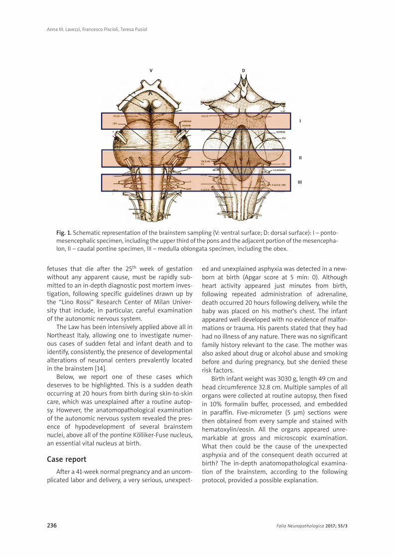
Fig. 1. Schematic representation of the brainstem sampling (V: ventral surface; D: dorsal surface): I – ponto- mesencephalic specimen, including the upper third of the pons and the adjacent portion of the mesencepha-lon, II – caudal pontine specimen, III – medulla oblongata specimen, including the obex.
I
V D
II
III

237Folia Neuropathologica 2017; 55/3
Sudden unexpected postnatal collapse at 20 hours from birth during skin-to-skin care related to brainstem developmental alterations – a case report
Neuropathological examination of the brainstem
Briefly, the applied methodology, in accordance with the aforementioned guidelines provided by Italian law, states that, after fixation in 10% phosphate-buff-ered formalin, the brainstem, where the main struc-tures controlling the vital functions are located, must be processed and embedded in paraffin. Then three specimens are taken (Fig. 1): the first specimen (I) includes the upper third of the pons and the adjacent caudal portion of the midbrain; the second speci-
men (II) is taken from the caudal portion of the pons; and the third specimen (III) is from the medulla oblon-gata around the obex. Transverse serial sections from all the samples are made at intervals of 60 mm. For each level, twelve 4 µm sections are obtained, two of which are routinely stained for histological exam-ination using alternately hemato xylin-eosin and Klüver- Barrera methods. Additional sections are saved and stained as deemed necessary for further investiga-tions. The main nuclei are then analyzed: the para-facial nucleus, the retrotrapezoid nucleus, the locus coeruleus, the Kölliker-Fuse nucleus and the median,
Fig. 2. Image series related to the Kölliker-Fuse nucleus (KFN). Top left: ventral brainstem image with indi-cation of the optimal level for examination of the KFN; right: histological section at the above-mentioned level with bilateral localization of the KFN (encircled area). A) Normal cytoarchitecture of the KFN showing large neurons with a distinct nucleus and evident nucleolus. B) Hypoplasia of the KFN with very few neurons. 4V – fourth ventricle; scp – superior cerebellar peduncle. Klüver-Barrera stain, magnification (A) and (B) 100×, at top 0.5×.
A B

238 Folia Neuropathologica 2017; 55/3
Anna M. Lavezzi, Francesco Piscioli, Teresa Pusiol
Fig. 3. Image series related to the retrotrapezoid nucleus (RTN). Top left: ventral brainstem image with indication of the optimal level for examination of the RTN; right: histological section at the above- mentioned level with bilateral localization of the RTN (encircled area). A) Normal cytoarchitecture of the RTN showing a group of small neurons adjacent to the parafacial nucleus (PFN). B) Hypoplasia of the RTN with very few neurons. Klüver-Barrera stain, magnification (A) and (B) 100×, at top 0.5×.
magnus, dorsal, caudal and linear raphe nuclei in the pons; the hypoglossus, the dorsal motor vagal, the tractus solitarius, the ambiguus, the pre-Bötzing-er, the inferior olivary, the arcuate and the obscurus and pallidus raphe nuclei in the medulla oblongata.
Consent: Written informed consent was obtained from the parents of infant for the autoptic examina-tion and related research. Study approval was grant-ed by the institutional review board of Milan Univer-sity (“Lino Rossi” Research Center).
Results of neuropathological examination
The histological analysis of the brainstem showed a normal cytoarchitecture in relation to age, i.e. com-
pared with the normal structure of age-matched controls, of almost all the above-mentioned nuclei. However, severe hypoplasia of the Kölliker-Fuse nucle-us in the rostral pons (Fig. 2B) and hypodevelopment of the retrotrapezoid nucleus in the caudal pons (Fig. 3B) and of the obscurus raphe nucleus in the medul-la oblongata (Fig. 4B) were diagnosed.
Discussion
The case presented here is of particular interest as the death occurred inexplicably on the first day of life, while the baby was face-down during skin-to-skin care. It is generally understood that SIDS affects infants
A B

239Folia Neuropathologica 2017; 55/3
Sudden unexpected postnatal collapse at 20 hours from birth during skin-to-skin care related to brainstem developmental alterations – a case report
1 month to 1 year old and that the sudden unexpect-ed death of an apparently healthy term infant within the first day of life is a rare event. However, a recent report by Save the Children indicates that over one million newborns around the world die each year on their first day of life [2]. The initial 24 hours are indi-cated as the most critical and dangerous for life, and the present case confirms this assertion.
While the routine autopsy revealed no cause of death, the results obtained from the deep exam-ination of the autonomic nervous system highlighted subtle developmental abnormalities of the brain-stem nuclei controlling the vital functions. The most important alteration, sufficient to explain the patho-
genetic mechanism of death, was the severe hypo-plasia of the Kölliker-Fuse nucleus observed in the rostral pons.
The Kölliker-Fuse nucleus consists of a small group of neurons that have an important function during intrauterine life, inhibiting the response of central and peripheral chemoreceptors (which are already fully formed and potentially functional) and there-fore any respiratory reflex. At birth, the Kölliker-Fuse nucleus abruptly reduces its inhibitory effects and becomes active as the main respiratory center, both stimulating the parafacial and pre-Bötzinger nuclei, assigned to promote the first inspirations, and coor-dinating the pulmonary motor responses to hematic
Fig. 4. Image series related to the raphe obscurus nucleus (RON). Top left: ventral brainstem image with indication of the optimal level for examination of the RON; right: histological section at the above- mentioned level with localization of the RON (encircled area). A) Normal cytoarchitecture of the RON showing a group of neurons distributed along the middle line. B) Hypoplasia of the RON with very few neurons. Klüver-Barrera stain, magnification (A) and (B) 100×, at top 0.5×.
A B

240 Folia Neuropathologica 2017; 55/3
Anna M. Lavezzi, Francesco Piscioli, Teresa Pusiol
oscillations of pO2, pCO2 and pH [5-8]. This activi-ty is also sustained by the chemo receptor nuclei, to which the retrotrapezoid nucleus and the raphe obscurus nucleus belong [3,9].
Then, the severe asphyxia at birth can be explained by the hypoplasia of the Kölliker-Fuse nucleus, able to prevent the start of the eupneic breathing usually initiated by the parafacial and pre-Bötzinger nuclei under its control, despite their normal development. The hypoplasia of the retrotrapezoid and the raphe obscurus nuclei contributed to the lack of ventilatory response to altered levels of the hematic parameters, so further contributing to death. The face pressed against the mother’s chest due to the skin-to-skin position, reducing the amount of oxygen supplied to the brain, has accelerated the fatal outcome.
In our previous studies, we have reported the impli-cation of the Kölliker-Fuse nucleus in the pathogen-esis of SIDS and that its hypoplasia, like other brain-stem alterations, is significantly associated with maternal smoking in pregnancy [5,8]. In this case however the mother admitted no history of ciga-rette smoking. Furthermore, it should be considered that retrospective assessment of maternal smoking, mainly if performed after the fatal event, is some-times unavoidable [18]. Not rarely, smoking mothers are reluctant to honestly report their tobacco use, possibly because of feelings of guilt. In addition to nicotine, other risk factors for delayed development of brain nuclei should be considered. Recently, orig-inal investigations have demonstrated the adverse effects of persistent pollutants such as pesticides and household insecticides (belonging to the cat-egory of endocrine disrupting compounds [EDCs]) on brain development [12,13,15,16]. Traces of highly toxic chemicals, such as organochlorine and organo-phosphate pesticides (α and γ-chlordane, chlorfen-vinfos, chlorpyrifos, p,p-DDT, p,p-DDE, endrin, α- and β-endosulfans), have been directly detected in cere-bral cortex samples of several SIUDS/SIDS cases, frequently from agricultural areas where pesticides are used. These interactions can directly impair autonomic nervous system development, resulting in hypoplasia of the main brainstem centers con-trolling the vital functions, and consequently leading to sudden death.
This case report indicates that also the skin-to-skin contact can be considered as a risk factor for sudden neonatal deaths and that it is imperative to provide to the medical and nursing personnel, as well as mothers,
accurate information regarding this care and the relat-ed risk of collapse. Finally, we underline the need for an in-depth examination of the central nervous sys-tem in victims of sudden perinatal death. The neuro-pathological protocol applied by our research team [17], and briefly described here, should become a specialized component of the fetal and infant autopsy, above all when a clear cause of death is not found. The universal application of this proce-dure is the best way to collect a rich body of infor-mation about all possible neurological alterations in the newborn brain, in order to reduce their incidence and mitigate the related social impact.
Disclosure
Authors report no conflict of interest.
References
1. Constitution of the Italian Republic Law n° 31. Regulations for
diagnostic post-mortem investigation in victims of sudden
infant death syndrome (SIDS) and unexpected fetal death.
Official Gazette of the Italian Republic, General Series 2006;
34: 4. Available at: http://users.unimi.it/centrolinorossi/files/
gazz_ufficiale.pdf.
2. Global Health Workforce Alliance. Available at: http://www.
who.int/workforcealliance/media/news/2014/end_new_born_
death/en/
3. Lavezzi AM, Casale V, Oneda R, Weese-Mayer DE, Matturri L.
Sudden infant death syndrome and sudden intrauterine unex-
plained death: correlation between hypoplasia of raphé nuclei
and serotonin transporter gene promoter polymorphism. Ped
Res 2009; 66: 22-27.
4. Lavezzi AM, Ferrero S, Matturri L, Roncati L, Pusiol T. Develop-
mental neuropathology of brainstem respiratory centers in
unexplained stillbirth: What’s the meaning? Int J Dev Neurosci
2016; 53: 99-106.
5. Lavezzi AM, Ferrero S, Roncati L, Matturri L, Pusiol T. Impaired
orexin receptor expression in the Kölliker-Fuse nucleus in sud-
den infant death syndrome: possible involvement of this nucle-
us in arousal pathophysiology. Neurol Res 2016; 38: 706-716.
6. Lavezzi AM, Ottaviani G, Ballabio G, Rossi L, Matturri L. Prelimi-
nary study on the cytoarchitecture of the human parabrachial/
Kölliker-Fuse complex, with reference to sudden infant death
syndrome and sudden intrauterine unexplained death. Pediatr
Dev Pathol 2004; 7: 171-179.
7. Lavezzi AM, Ottaviani G, Rossi L, Matturri L. Cytoarchitectural
organization of the parabrachial/Kölliker-Fuse complex in man.
Brain Dev 2004; 26: 316-320.
8. Lavezzi AM, Ottaviani G, Rossi L, Matturri L. Hypoplasia of
the Parabrachial/Kölliker-Fuse complex in perinatal death. Biol
Neonate 2004; 86: 92-97.
9. Lavezzi AM, Weese-Mayer DE, Yu MY, Jennings LJ, Corna MF, Casa-
le V, Oneda R, Matturri L. Developmental alterations of the respi-

241Folia Neuropathologica 2017; 55/3
Sudden unexpected postnatal collapse at 20 hours from birth during skin-to-skin care related to brainstem developmental alterations – a case report
ratory human retrotrapezoid nucleus in sudden unexplained fetal and infant death. Auton Neurosci 2012; 170: 12-19.
10. Macdorman MF, Gregory ECW. Fetal and perinatal mortality, United States, 2013. National vital statistics reports. National Center for Health Statistics, Hyattsville 2005; vol. 64, n. 8.
11. Mehboob R, Kabir M, Ahmed N and Ahmad FJ. Towards Better Understanding of the Pathogenesis of Neuronal Respiratory Network in Sudden Perinatal Death. Front Neurol 2017; 8: 320.
12. Roncati L, Piscioli F, Pusiol T. The endocrine disruptors among the environmental risk factors for stillbirth. Sci Total Environ 2016; 563-564: 1086-1087.
13. Roncati L, Piscioli F, Pusiol T. The endocrine disrupting chem-icals as possible stillbirth contributors. Am J Obstet Gynecol 2016; 215: 532-533.
14. Roncati L, Pusiol T, Piscioli F, Barbolini G, Maiorana A, Lavezzi A. The First 5-Year-Long Survey on Intrauterine Unexplained Sud-den Deaths from the Northeast Italy. Fetal Pediatr Pathol 2016; 35: 315-326.
15. Roncati L, Pusiol T, Piscioli F, Lavezzi AM. Neurodevelopmen-tal disorders and pesticide exposure: the northeastern Italian experience. Arch Toxicol 2017; 91: 603-604.
16. Roncati L, Termopoli V, Pusiol T. Negative Role of the Environmen-tal Endocrine Disruptors in the Human Neurodevelopment. Front Neurol 2016; 7: 143.
17. Roncati L, Piscioli F, Pusiol T, Lavezzi AM. Neuropathological pro-tocol for the study of unexplained stillbirth. Folia Neuropathol 2017; 55: 79-85.
18. Shipton D, Tappin DM, Vadiveloo T, Crossley JA, Aitken DA, Chalmers J. Reliability of self reported smoking status by preg-nant women for estimating smoking prevalence: a retrospec-tive, cross sectional study. BMJ 2009; 339: 4347-4354.