sulfonated polyethylenimine for photosensitizer ...jflovell/pubs/chitgupi_bcc_2015.pdf ·...
TRANSCRIPT
Sulfonated Polyethylenimine for Photosensitizer Conjugation andTargetingUpendra Chitgupi,† Yumiao Zhang,†,‡ Chi Y. Lo,‡ Shuai Shao,† Wentao Song,† Jumin Geng,†
Sriram Neelamegham,†,‡ and Jonathan F. Lovell*,†,‡
†Department of Biomedical Engineering and ‡Department of Chemical and Biological Engineering, University at Buffalo, StateUniversity of New York, Buffalo, New York 14260, United States
ABSTRACT: Polysulfonated macromolecules are known to bindselectins, adhesion membrane proteins which are broadly implicated ininflammation. Commercially available branched polyethylenimine(PEI) was reacted with chlorosulfonic acid to generate sulfonatedPEI with varying degrees of sulfonation. Remaining unreacted aminegroups were then used for straightforward conjugation withpyropheophoribide-a, a near-infrared photosensitizer. Photosensitizer-labeled sulfonated PEI conjugates inhibited blood coagulation andwere demonstrated to specifically bind to cells genetically programmedto overexpress L-selectin (CD62L) or P-selectin (CD62P). In vitro,following targeting, selectin-expressing cells could be destroyed viaphotodynamic therapy.
■ INTRODUCTION
Inflammation plays a central role in numerous chronicconditions that adversely affect health including heart disease,1
cancer,2 and metabolic disorders.3 One of the key steps ininflammatory responses involves the migration and extrava-sation of leukocytes from blood vessels to the site of insult. Thisprocess is mediated by selectins, a family of cell-surfaceglycoproteins that include endothelial (E-), platelet (P-), andleukocyte (L-) selectin.4 Selectins contain characteristicextracellular domains that include a (1) calcium-dependent,carbohydrate-binding lectin domain, (2) an epidermal growthfactor domain, and (3) a domain consisting of two to nine shortconsensus repeat units involved in protein binding.5 Followingdamage, tissues release cytokines that induce endothelial cellsto express E- and P-selectin that in turn bind to circulatingleukocytes to induce adhesion to the endothelium. Inflamma-tory activation by molecules like IL-1β and TNFα increases E-selectin expression over a period of hours, whereas othermediators including thrombin, histamine, and peroxides induceP-selectin expression over a period of minutes.6
The binding partners of selectins and their associatedbiological significance are still being elucidated, but arenumerous.7 One selectin-binding surface protein of interestthat is expressed on circulating leukocytes is P-selectinglycoprotein ligand (PSGL-1).8,9 PSGL-1 contains thetetrasaccharide sialyl-Lewis X (sLeX) as well as O-linkedtyrosine-sulfate residues and these two components togetherregulate selectin-binding. The unbranched sulfonated heparinglycosaminoglycan, a broadly used anticoagulant, is known toinhibit acute inflammation by reducing L- and P-selectinbinding function.10 Several other sulfonated macromoleculeshave been reported to interact with selectins to some degree,
including fucoidan, dextran sulfate, chondroitin sulfate, as wellas other sulfonated lipids and sugars.11
Given the importance of selectins in disease, they havebecome a target in molecular imaging research. P-selectinantibodies have been used to functionalize microbubbles forultrasound imaging of renal tissue injury12 and ischemia13 inmice. Other antibody-based approaches have been used totarget E-14 and L-15,16 selectins. Theranostic selectin targetinghas also been described using selectin-binding peptides,17,18
aptamers,19 and sLeX analogues.20 One noteworthy syntheticapproach involves the use of dendritic polyglycerol sulfate(dPGS) as a platform for L- and P-selectin binding.21,22 dPGS,which like heparin is a polysulfonated macromolecule, has alsobeen used as a scaffold for fluorescence imaging ofinflammation using near-infrared dyes23,24 and optoacousticimaging via conjugation to gold nanorods25 and has thecapacity for radiolabeling with synthesis on the kilo scale.26
However, synthesis of dendrimers can be an intensive processand dPGS itself contains only terminal hydroxyl groups whichrequire further functionalization prior to bioconjugation.Therefore, alternate selectin-binding platforms that are morereadily accessible or that are more easily chemically modifiedcould potentially be useful. One candidate includes a derivativeof polyethylenimine (PEI), which is abundantly available,contains a large amount of readily modifiable amine groups,and is easily modified to generate sulfonated PEI (s-PEI). s-PEIhas been explored in diverse applications related to
Received: April 28, 2015Revised: May 31, 2015
Article
pubs.acs.org/bc
© XXXX American Chemical Society A DOI: 10.1021/acs.bioconjchem.5b00241Bioconjugate Chem. XXXX, XXX, XXX−XXX
anticoagulants,27 gene delivery,28 environmental detoxifica-tion,29,30 and membrane processes.31
In this study, we report the synthesis and characterization ofs-PEI and subsequent conjugation to the photosensitizerpyropheophorbide-a (pyro). Photosensitizers are used incombination with light delivery to target tissues in photo-dynamic therapy, which is a clinical procedure used to combatvarious diseases including cancer.32 Molecular targeting ofphotosensitizers aims to increase the amount of photosensitizerin the target tissue, thereby reducing harm to nontargettissues.33−35 Numerous methods have been explored forphotosensitizer targeting including conjugation to antibod-ies,36,37 sugars,38 aptamers,39 and small molecules.40 Photo-sensitizer targeting strategies for cancer typically involve activetargeting to cell surface receptors expressed on cancer cellsthemselves or vascular targeting to tumor blood vessels, eitheractively or passively.41 To the best of our knowledge, thedevelopment of photosensitizers targeted to selectins has notyet been explored. Since E-selectin is overexpressed in cancersincluding breast42 and prostate,43 selectin-targeted photo-sensitizers could possibly offer improved tumor selectivity forPDT treatments. Alternatively, as selectin expression has beenreported to increase shortly following PDT,44,45 it might bepossible to use PDT to strategically induce selectin expression.This would induce a positive feedback effect in attracting moreselectin-targeted photosensitizers to the irradiated tissue. Suchan approach could be effective in lowering the total amount ofinjected photosensitizer, thereby reducing systemic side effectsto the patient such as sunlight skin toxicity.
■ RESULTS AND DISCUSSION
Synthesis and Labeling of Sulfonated Polyethyleni-mine. Commercially available branched PEI was modifiedaccording to published procedures to produce s-PEI with 6%(s6-PEI) and 34% (s34-PEI) sulfonation.27 PEI was stirred inmethanol at 60 °C with varying amounts of chlorosulfonic acidto generate the s-PEI. Figure 1A shows the chemical reaction,with the bulk of the polymer represented by a sphere and an
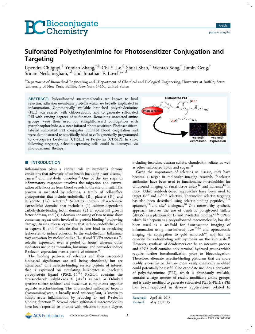
exemplary segment branch shown. Following the reaction, theproduct was dissolved in water, precipitated and washed withmethanol, and then dried under vacuum to obtain s-PEI. Thezeta potential of the s-PEI remained positive, showing thatnumerous free amine groups remained on the polymer,outweighing the sulfate contribution (Figure 1B). The decreasein zeta potential from +19 mV for the unconjugated PEI to +16mV for s6-PEI and +13 mV for s34-PEI was due to the decreasein net positive charge induced by the replacement of cationicamine groups with anionic sulfate residues. A simple andstandard analytical test for the presence of sulfate ions involvesincubation with barium. This results in an insoluble barium-sulfate complex that can be readily detected by an opticalturbidity measurement. We applied this approach to equalconcentrations of PEI or s-PEI (10 mg/mL) to confirm thepresence of sulfate in s-PEI. As shown in Figure 1C, bariumchloride did not induce significant precipitation when added toa solution of standard PEI. However, barium rapidly complexedwith s6-PEI to induce visible aggregation and turbidity in thesolution. s34-PEI generated a greater amount of precipitationrelative to s6-PEI. Fourier transform infrared spectroscopy(FTIR) was used to further validate the sulfate group linkageswith PEI. Absorption bands at 1190 and 990 cm−1 wereobserved in the s-PEI, but absent in the PEI samples (Figure1D). These correspond to SO (asymmetric) and SO(symmetric) bonds, and the observed bands occurred atwavenumbers close to those previously reported for s-PEI byothers.30 The prominent band appearing close to 2800 cm−1 inthe PEI sample is attributed to N−H stretching30 and isweakened in the s-PEI samples. To further confirm the decreasein the number of amine groups due to their conversion tosulfate, we used the ninhydrin assay, which is a common andsimple colorimetric method to determine the presence ofamines. When ninhydrin was added to solutions of PEI and s-PEI, absorption peaks at 570 nm emerged, which are generateddue to the reaction of primary amines with ninhydrin. Thepeaks were integrated and these values are shown in Figure 1E,as a function of the expected sulfonation degree. An inverse
Figure 1. Synthesis and characterization of s-PEI. (A) Reaction of PEI sulfonation. (B) Zeta potential of PEI and s-PEI. (C) Barium chlorideturbidity assay for sulfate detection. (D) FTIR spectra of PEI and s-PEI. (E) Ninhydrin test for free amines.
Bioconjugate Chemistry Article
DOI: 10.1021/acs.bioconjchem.5b00241Bioconjugate Chem. XXXX, XXX, XXX−XXX
B
linear relationship was observed, suggesting that PEI and s-PEIcontained the expected loss of amine groups during theirconversion to sulfates. Although the achieved degree ofsulfonation was assumed to be consistent with publishedpatent literature,27 based on the ninhydrin assay to detect a lossin primary amines, the degree of sulfonation was similar to whatwas expected (7.8% observed vs 6% expected for s6-PEI and38.5% observed vs 34% expected for s34-PEI). Additionalanalysis would be required to more accurately confirm thedegree of sulfonation of the samples. Therefore, based onvarious analytical characterization methods, s-PEI was success-fully synthesized and contained available amine groups forfurther modification.The photosensitizer pyro, which has a single carboxylic acid
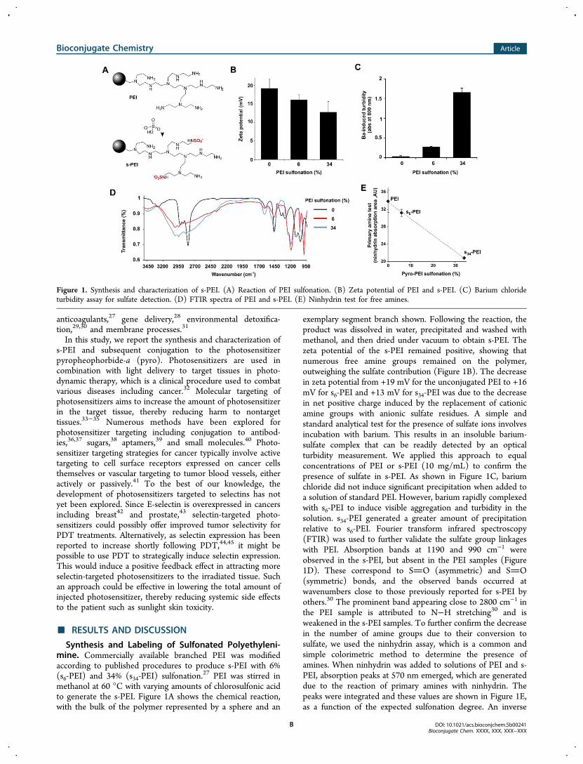
group and has been used previously to make targetedphotosensitizers,38 was next conjugated. Pyro was reactedwith PEI or s-PEI using a condensation reaction in dimethylsulfoxide (DMSO) with HBTU as an acid activator anddiisopropylethylamine (DIPEA) as a base (Figure 2A). As pyrocontains a carboxylic acid group, it could easily react with theamines of PEI and s-PEI to generate pyro-PEI and pyro-s-PEI,respectively. Following the reaction, free pyro was removed byrepeated aqueous extraction and then remaining small moleculereactants were removed with dialysis. The resulting conjugateswere investigated with spectroscopy. As shown in Figure 2B,there was no shift in the peaks of the absorption profile of theconjugated pyro and pyro effectively labeled both the PEI ands-PEI samples, based on the observed absorption intensities.When the absorption of the samples was adjusted to be equal,all the samples exhibited similar fluorescence when measured inmethanol (Figure 2C). However, in water, pyro-PEI exhibitedself-quenching compared to pyro-s6-PEI and pyro-s34-PEI, both
of whose brightness was only slightly attenuated compared tofree pyro in methanol (Figure 2D). It is possible that thesulfonation inhibited self-quenching in the pyro-PEI samples,which may have been caused by structurally induced pyrodimerization. To verify that pyro-s-PEI retained its sulfategroups during the course of pyro conjugation, the bariumturbidity assay was carried out. As expected, with increasingdegree of sulfonation, an increased generation in the turbiditywas observed, confirming the intactness of the sulfate groups(Figure 2E). When freshly drawn mouse blood was mixed withpyro-s-PEI, but not pyro-PEI, coagulation was effectivelyinhibited (Figure 2F). Thus, pyro-s-PEI retained the anti-coagulatory properties of sulfonated macromolecules.
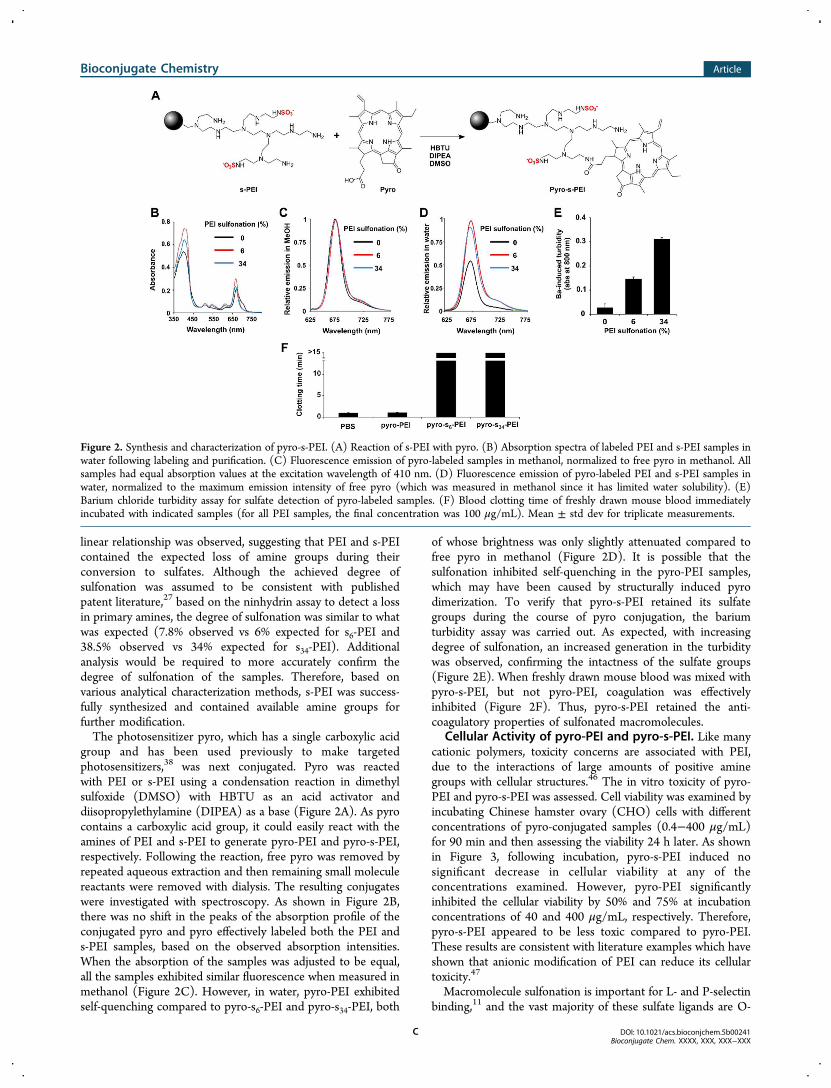
Cellular Activity of pyro-PEI and pyro-s-PEI. Like manycationic polymers, toxicity concerns are associated with PEI,due to the interactions of large amounts of positive aminegroups with cellular structures.46 The in vitro toxicity of pyro-PEI and pyro-s-PEI was assessed. Cell viability was examined byincubating Chinese hamster ovary (CHO) cells with differentconcentrations of pyro-conjugated samples (0.4−400 μg/mL)for 90 min and then assessing the viability 24 h later. As shownin Figure 3, following incubation, pyro-s-PEI induced nosignificant decrease in cellular viability at any of theconcentrations examined. However, pyro-PEI significantlyinhibited the cellular viability by 50% and 75% at incubationconcentrations of 40 and 400 μg/mL, respectively. Therefore,pyro-s-PEI appeared to be less toxic compared to pyro-PEI.These results are consistent with literature examples which haveshown that anionic modification of PEI can reduce its cellulartoxicity.47
Macromolecule sulfonation is important for L- and P-selectinbinding,11 and the vast majority of these sulfate ligands are O-
Figure 2. Synthesis and characterization of pyro-s-PEI. (A) Reaction of s-PEI with pyro. (B) Absorption spectra of labeled PEI and s-PEI samples inwater following labeling and purification. (C) Fluorescence emission of pyro-labeled samples in methanol, normalized to free pyro in methanol. Allsamples had equal absorption values at the excitation wavelength of 410 nm. (D) Fluorescence emission of pyro-labeled PEI and s-PEI samples inwater, normalized to the maximum emission intensity of free pyro (which was measured in methanol since it has limited water solubility). (E)Barium chloride turbidity assay for sulfate detection of pyro-labeled samples. (F) Blood clotting time of freshly drawn mouse blood immediatelyincubated with indicated samples (for all PEI samples, the final concentration was 100 μg/mL). Mean ± std dev for triplicate measurements.
Bioconjugate Chemistry Article
DOI: 10.1021/acs.bioconjchem.5b00241Bioconjugate Chem. XXXX, XXX, XXX−XXX
C
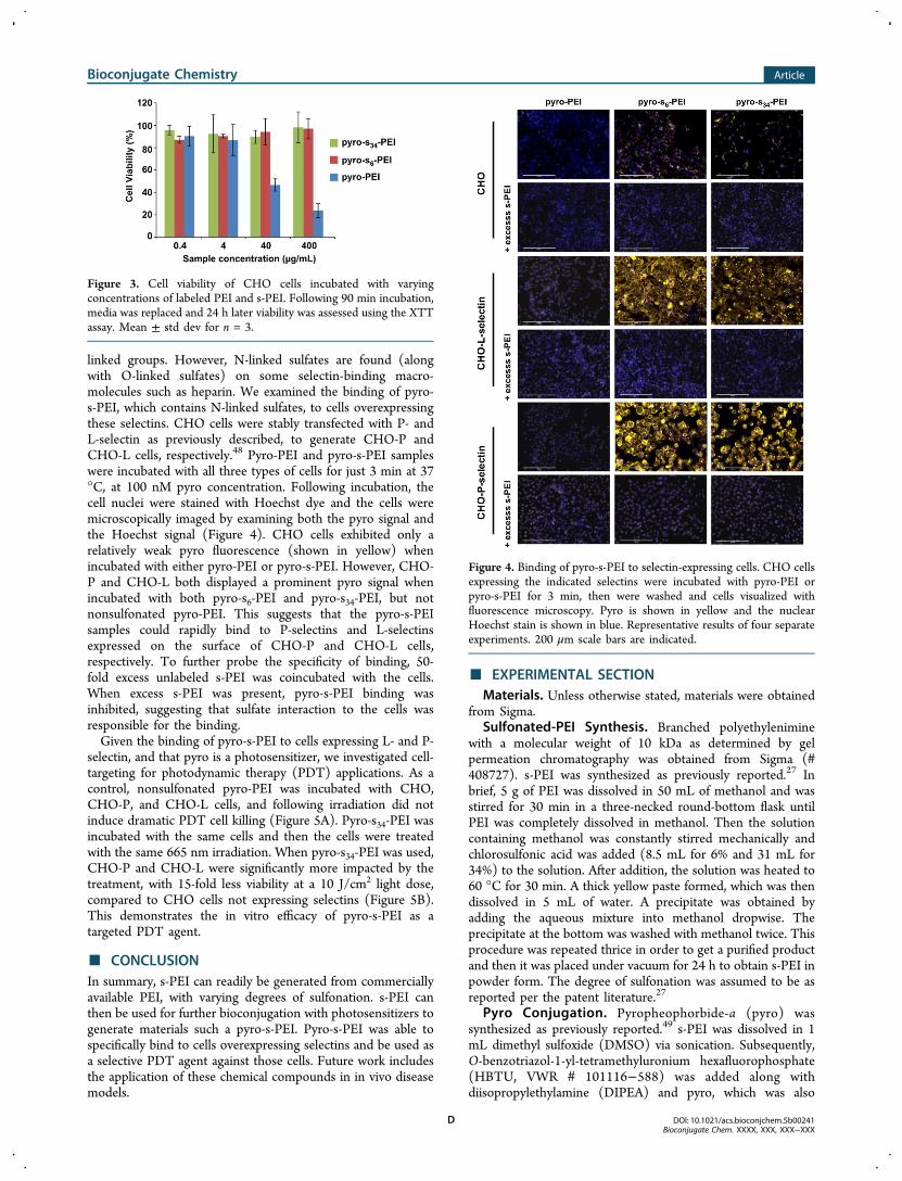
linked groups. However, N-linked sulfates are found (alongwith O-linked sulfates) on some selectin-binding macro-molecules such as heparin. We examined the binding of pyro-s-PEI, which contains N-linked sulfates, to cells overexpressingthese selectins. CHO cells were stably transfected with P- andL-selectin as previously described, to generate CHO-P andCHO-L cells, respectively.48 Pyro-PEI and pyro-s-PEI sampleswere incubated with all three types of cells for just 3 min at 37°C, at 100 nM pyro concentration. Following incubation, thecell nuclei were stained with Hoechst dye and the cells weremicroscopically imaged by examining both the pyro signal andthe Hoechst signal (Figure 4). CHO cells exhibited only arelatively weak pyro fluorescence (shown in yellow) whenincubated with either pyro-PEI or pyro-s-PEI. However, CHO-P and CHO-L both displayed a prominent pyro signal whenincubated with both pyro-s6-PEI and pyro-s34-PEI, but notnonsulfonated pyro-PEI. This suggests that the pyro-s-PEIsamples could rapidly bind to P-selectins and L-selectinsexpressed on the surface of CHO-P and CHO-L cells,respectively. To further probe the specificity of binding, 50-fold excess unlabeled s-PEI was coincubated with the cells.When excess s-PEI was present, pyro-s-PEI binding wasinhibited, suggesting that sulfate interaction to the cells wasresponsible for the binding.Given the binding of pyro-s-PEI to cells expressing L- and P-
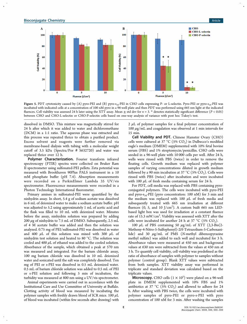
selectin, and that pyro is a photosensitizer, we investigated cell-targeting for photodynamic therapy (PDT) applications. As acontrol, nonsulfonated pyro-PEI was incubated with CHO,CHO-P, and CHO-L cells, and following irradiation did notinduce dramatic PDT cell killing (Figure 5A). Pyro-s34-PEI wasincubated with the same cells and then the cells were treatedwith the same 665 nm irradiation. When pyro-s34-PEI was used,CHO-P and CHO-L were significantly more impacted by thetreatment, with 15-fold less viability at a 10 J/cm2 light dose,compared to CHO cells not expressing selectins (Figure 5B).This demonstrates the in vitro efficacy of pyro-s-PEI as atargeted PDT agent.
■ CONCLUSIONIn summary, s-PEI can readily be generated from commerciallyavailable PEI, with varying degrees of sulfonation. s-PEI canthen be used for further bioconjugation with photosensitizers togenerate materials such a pyro-s-PEI. Pyro-s-PEI was able tospecifically bind to cells overexpressing selectins and be used asa selective PDT agent against those cells. Future work includesthe application of these chemical compounds in in vivo diseasemodels.
■ EXPERIMENTAL SECTIONMaterials. Unless otherwise stated, materials were obtained
from Sigma.Sulfonated-PEI Synthesis. Branched polyethylenimine
with a molecular weight of 10 kDa as determined by gelpermeation chromatography was obtained from Sigma (#408727). s-PEI was synthesized as previously reported.27 Inbrief, 5 g of PEI was dissolved in 50 mL of methanol and wasstirred for 30 min in a three-necked round-bottom flask untilPEI was completely dissolved in methanol. Then the solutioncontaining methanol was constantly stirred mechanically andchlorosulfonic acid was added (8.5 mL for 6% and 31 mL for34%) to the solution. After addition, the solution was heated to60 °C for 30 min. A thick yellow paste formed, which was thendissolved in 5 mL of water. A precipitate was obtained byadding the aqueous mixture into methanol dropwise. Theprecipitate at the bottom was washed with methanol twice. Thisprocedure was repeated thrice in order to get a purified productand then it was placed under vacuum for 24 h to obtain s-PEI inpowder form. The degree of sulfonation was assumed to be asreported per the patent literature.27
Pyro Conjugation. Pyropheophorbide-a (pyro) wassynthesized as previously reported.49 s-PEI was dissolved in 1mL dimethyl sulfoxide (DMSO) via sonication. Subsequently,O-benzotriazol-1-yl-tetramethyluronium hexafluorophosphate(HBTU, VWR # 101116−588) was added along withdiisopropylethylamine (DIPEA) and pyro, which was also
Figure 3. Cell viability of CHO cells incubated with varyingconcentrations of labeled PEI and s-PEI. Following 90 min incubation,media was replaced and 24 h later viability was assessed using the XTTassay. Mean ± std dev for n = 3.
Figure 4. Binding of pyro-s-PEI to selectin-expressing cells. CHO cellsexpressing the indicated selectins were incubated with pyro-PEI orpyro-s-PEI for 3 min, then were washed and cells visualized withfluorescence microscopy. Pyro is shown in yellow and the nuclearHoechst stain is shown in blue. Representative results of four separateexperiments. 200 μm scale bars are indicated.
Bioconjugate Chemistry Article
DOI: 10.1021/acs.bioconjchem.5b00241Bioconjugate Chem. XXXX, XXX, XXX−XXX
D
dissolved in DMSO. This mixture was magnetically stirred for24 h after which it was added to water and dichloromethane(DCM) in a 1:1 ratio. The aqueous phase was extracted andthis process was repeated thrice to obtain a purified product.Excess solvent and reagents were further removed viamembrane-based dialysis with tubing with a molecular weightcutoff of 3.5 kDa (Spectra/Por # S632720) and water wasreplaced thrice over 12 h.Polymer Characterization. Fourier transform infrared
spectroscopy (FTIR) spectra were collected on Bruker RamII spectrometer using sulfonated-PEI pellets. Zeta potential wasmeasured with Brookhaven 90Plus PALS instrument in a 10mM phosphate buffer (pH 7.4). Absorption measurementswere recorded on a PerkinElmer Lambda 35 UV/visspectrometer. Fluorescence measurements were recorded in aPhoton Technology International fluorometer.Primary amines in sulfonated-PEI were quantified by the
ninhydrin assay. In short, 5.4 g of sodium acetate was dissolvedin 6 mL of deionized water to make a sodium acetate buffer. pHwas adjusted to 5.2 using approximately 1 mL of acetic acid andthe flask was filled to 10 mL with deionized water. Minutesbefore the assay, ninhydrin solution was prepared by adding200 μg of ninhydrin to 7.5 mL of DMSO. Subsequently, 2.5 mLof 4 M acetate buffer was added and then the solution wasanalyzed. 0.75 mg of PEI/sulfonated-PEI was dissolved in waterand 400 μL of this solution was mixed with 300 μL ofninhydrin test solution and heated to 80 °C. The solution wascooled and 400 μL of ethanol was added to the cooled solution.Absorbance of the sample, which obtained a peak at 570 nmwas measured and integrated. For the barium chloride assay,100 mg barium chloride was dissolved in 10 mL deionizedwater and sonicated until the salt was completely dissolved. Tenmg of PEI or s-PEI was dissolved in 0.5 mL deionized water.0.5 mL of barium chloride solution was added to 0.5 mL of PEIor s-PEI solution and following 5 min of incubation, theturbidity was measured at 800 nm using a UV/vis spectrometer.Animal experiments were carried out in accordance with the
Institutional Care and Use Committee of University at Buffalo.Clotting activity of blood was measured by incubating thepolymer samples with freshly drawn blood of ICR mice. 100 μLof blood was incubated (within few seconds after drawing) with
2 μL of polymer samples for a final polymer concentration of100 μg/mL and coagulation was observed at 1 min intervals for15 min.
Cell Viability and PDT. Chinese Hamster Ovary (CHO)cells were cultured at 37 °C (5% CO2) in Dulbecco’s modifiedeagle’s medium (DMEM) supplemented with 10% fetal bovineserum (FBS) and 1% streptomycin/penicillin. CHO cells wereseeded in a 96-well plate with 10 000 cells per well. After 24 h,wells were rinsed with PBS (twice) in order to remove thefloating cells. Growth medium was replaced with polymersamples of varying concentrations diluted in growth mediumfollowed by a 90 min incubation at 37 °C (5% CO2). Cells wererinsed with PBS (twice) after incubation and were incubatedwith 100 μL of fresh media containing serum for 24 h.For PDT, cell media was replaced with PBS containing pyro-
conjugated polymers. The cells were incubated with pyro-PEIand pyro-s34-PEI (pyro concentration: 100 nM) for 3 min andthe medium was replaced with 100 μL of fresh media andsubsequently treated with 665 nm irradiation at differentfluences (0, 5, and 10 J/cm2). A custom built 665 nm LED-based light box was used for irradiation at a constant fluencerate of 15.3 mW/cm2. Viability was assessed with XTT after thecells were incubated for another 24 h at 37 °C (5% CO2).100 μL of PBS containing 50 μg/mL of XTT (2,3-Bis(2-
Methoxy-4-Nitro-5-Sulfophenyl)-2H-Tetrazolium-5-Carboxani-lide) and 30 μg/mL of PMS (N-methyl dibenzopyrazinemethyl sulfate) was added to each well and incubated for 3 h.Absorbance values were measured at 450 nm and backgroundvalues at 630 nm were subtracted from the values at 450 nm at3 h. To quantify cell viability, cell viability was predefined as theratio of absorbance of samples with polymer to samples withoutpolymer (control group). Blank XTT values were subtractedfrom both samples. XTT viability assay was performed intriplicate and standard deviation was calculated based on thetriplicate values.
Microscopy. CHO cells (1 × 10⁵) were plated on a 96-wellplate in DMEM supplemented with 10% FBS and 1%antibiotics at 37 °C (5% CO2) and allowed to adhere for 24h. After washing with PBS twice, the cells were incubated withpolymer samples of pyro-PEI or pyro-s-PEI with pyroconcentration of 100 nM for 3 min. After washing the samples
Figure 5. PDT cytotoxicity caused by (A) pyro-PEI and (B) pyro-s34-PEI in CHO cells expressing P- or L-selectin. Pyro-PEI or pyro-s34-PEI wasincubated with indicated cells at a concentration of 100 nM pyro in a 96-well plate and then PDT was performed using 665 nm light at the indicatedfluences. Cell viability was assessed 24 h later using the XTT assay. Mean ± std dev for n = 3. * denotes statistically significant difference (P < 0.05)between CHO and CHO-L-selectin or CHO-P-selectin cells based on one-way analysis of variance with post hoc Tukey’s test.
Bioconjugate Chemistry Article
DOI: 10.1021/acs.bioconjchem.5b00241Bioconjugate Chem. XXXX, XXX, XXX−XXX
E
with PBS twice, nuclei were stained with Hoechst 33342Fluorescent Stain (100 μL of 7.8 μg/mL) and incubated for 15min at 37 °C (5% CO2). The wells were washed with PBSthrice and cells were imaged with an EVOS FL cell imagingsystem.
■ AUTHOR INFORMATIONCorresponding Author*E-mail: [email protected].
NotesThe authors declare no competing financial interest.
■ ACKNOWLEDGMENTSThis work was supported by research funds from the NationalInstitutes of Health (DP5OD017898, HL103411). C.Y.L. wassupported by a T32 NIH Ruth L. Kirschstein PostdoctoralResearch Training Grant.
■ REFERENCES(1) Libby, P., Ridker, P. M., and Maseri, A. (2002) Inflammation andatherosclerosis. Circulation 105, 1135−1143.(2) Coussens, L. M., and Werb, Z. (2002) Inflammation and cancer.Nature 420, 860−867.(3) Hotamisligil, G. S. (2006) Inflammation and metabolic disorders.Nature 444, 860−867.(4) Kansas, G. (1996) Selectins and their ligands: current conceptsand controversies. Blood 88, 3259−3287.(5) Tedder, T. F., Steeber, D. A., Chen, A., and Engel, P. (1995) Theselectins: vascular adhesion molecules. FASEB J. 9, 866−73.(6) Lasky, L. (1992) Selectins: interpreters of cell-specificcarbohydrate information during inflammation. Science 258, 964−969.(7) Varki, A. (1997) Selectin ligands: will the real ones please standup? J. Clin. Invest. 99, 158−162.(8) Simanek, E. E., McGarvey, G. J., Jablonowski, J. A., and Wong, C.-H. (1998) Selectin−carbohydrate interactions: from natural ligands todesigned mimics. Chem. Rev. 98, 833−862.(9) Lo, C. Y., Antonopoulos, A., Gupta, R., Qu, J., Dell, A., Haslam, S.M., and Neelamegham, S. (2013) Competition between core-2GlcNAc-transferase and ST6GalNAc-transferase regulates the syn-thesis of the leukocyte selectin ligand on human P-selectinglycoprotein ligand-1. J. Biol. Chem. 288, 13974−13987.(10) Nelson, R. M., Cecconi, O., Roberts, W. G., Aruffo, A., Linhardt,R. J., and Bevilacqua, M. P. (1993) Heparin oligosaccharides bind L-and P-selectin and inhibit acute inflammation. Blood 82, 3253−3258.(11) Varki, A. (1994) Selectin ligands. Proc. Natl. Acad. Sci. U.S.A. 91,7390−7397.(12) Lindner, J. R., Song, J., Christiansen, J., Klibanov, A. L., Xu, F.,and Ley, K. (2001) Ultrasound assessment of inflammation and renaltissue injury with microbubbles targeted to P-selectin. Circulation 104,2107−2112.(13) Kaufmann, B. A., Lewis, C., Xie, A., Mirza-Mohd, A., andLindner, J. R. (2007) Detection of recent myocardial ischaemia bymolecular imaging of P-selectin with targeted contrast echocardiog-raphy. Eur. Heart J. 28, 2011−2017.(14) Reynolds, P. R., Larkman, D. J., Haskard, D. O., Hajnal, J. V.,Kennea, N. L., George, A. J. T., and Edwards, A. D. (2006) Detectionof vascular expression of E-selectin in vivo with MR imaging. Radiology241, 469−476.(15) Kang, H. W., Josephson, L., Petrovsky, A., Weissleder, R., andBogdanov, A. (2002) Magnetic resonance imaging of inducible E-selectin expression in human endothelial cell culture. BioconjugateChem. 13, 122−127.(16) Hauff, P., Reinhardt, M., Briel, A., Debus, N., and Schirner, M.(2004) Molecular targeting of lymph nodes with L-selectin ligand-specific US contrast agent: a feasibility study in mice and dogs.Radiology 231, 667−673.
(17) Funovics, M., Montet, X., Reynolds, F., Weissleder, R., andJosephson, L. (2005) Nanoparticles for the optical imaging of tumorselectin. Neoplasia 7, 904−911.(18) Jin, A. Y., Tuor, U. I., Rushforth, D., Filfil, R., Kaur, J., Ni, F.,Tomanek, B., and Barber, P. A. (2009) Magnetic resonance molecularimaging of post-stroke neuroinflammation with a P-selectin targetediron oxide nanoparticle. Contrast Media Mol. Imaging 4, 305−311.(19) Mann, A. P., Tanaka, T., Somasunderam, A., Liu, X., Gorenstein,D. G., and Ferrari, M. (2011) E-Selectin-targeted porous siliconparticle for nanoparticle delivery to the bone marrow. Adv. Mater. 23,H278−H282.(20) Jubeli, E., Moine, L., Nicolas, V., and Barratt, G. (2012)Preparation of E-selectin-targeting nanoparticles and preliminary invitro evaluation. Int. J. Pharm. 426, 291−301.(21) Dernedde, J., Rausch, A., Weinhart, M., Enders, S., Tauber, R.,Licha, K., Schirner, M., Zugel, U., von Bonin, A., and Haag, R. (2010)Dendritic polyglycerol sulfates as multivalent inhibitors of inflamma-tion. Proc. Natl. Acad. Sci. U. S. A. 107, 19679−19684.(22) Turk, H., Haag, R., and Alban, S. (2004) Dendritic polyglycerolsulfates as new heparin analogues and potent inhibitors of thecomplement system. Bioconjugate Chem. 15, 162−167.(23) Licha, K., Welker, P., Weinhart, M., Wegner, N., Kern, S.,Reichert, S., Gemeinhardt, I., Weissbach, C., Ebert, B., Haag, R., et al.(2011) Fluorescence imaging with multifunctional polyglycerolsulfates: novel polymeric near-IR probes targeting inflammation.Bioconjugate Chem. 22, 2453−2460.(24) Biffi, S., Dal Monego, S., Dullin, C., Garrovo, C., Bosnjak, B.,Licha, K., Welker, P., Epstein, M. M., and Alves, F. (2013) Dendriticpolyglycerolsulfate near infrared fluorescent (NIRF) dye conjugate fornon-invasively monitoring of inflammation in an allergic asthmamouse model. PLoS One 8, e57150.(25) Vonnemann, J., Beziere, N., Bottcher, C., Riese, S. B., Kuehne,C., Dernedde, J., Licha, K., von Schacky, C., Kosanke, Y., Kimm, et al.(2014) Polyglycerolsulfate functionalized gold nanorods as opto-acoustic signal nanoamplifiers for in vivo bioimaging of rheumatoidarthritis. Theranostics 4, 629−641.(26) Groger, D., Paulus, F., Licha, K., Welker, P., Weinhart, M.,Holzhausen, C., Mundhenk, L., Gruber, A. D., Abram, U., and Haag, R.(2013) Synthesis and biological evaluation of radio and dye labeledamino functionalized dendritic polyglycerol sulfates as multivalent anti-inflammatory compounds. Bioconjugate Chem. 24, 1507−1514.(27) Murashige, Y., Yanagase, A., Kawachi, Y., and Soga, J. (1987)Sulfonated polyethyleneimine useful as blood anticoagulant. U.S.Patent 4,639,339, January 27, 1987.(28) Sun, J., Zeng, F., Jian, H., and Wu, S. (2013) Conjugation withbetaine: a facile and effective approach to significant improvement ofgene delivery properties of PEI. Biomacromolecules 14, 728−736.(29) Leroy, D., Martinot, L., Mignonsin, P., Strivay, D., Weber, G.,Jerome, C., and Jerome, R. (2003) Complexation of uranyl ions bypolypyrrole doped by sulfonated and phosphonated polyethylenei-mine. J. Appl. Polym. Sci. 88, 352−359.(30) Saad, D. M. G., Cukrowska, E. M., and Tutu, H. (2012)Sulfonated cross-linked polyethylenimine for selective removal ofmercury from aqueous solutions. Toxicol. Environ. Chem. 94, 1916−1929.(31) Shen, L.-Q., Xu, Z.-K., Yang, Q., Sun, H.-L., Wang, S.-Y., and Xu,Y.-Y. (2004) Preparation and characterization of sulfonated poly-etherimide/polyetherimide blend membranes. J. Appl. Polym. Sci. 92,1709−1715.(32) Agostinis, P., Berg, K., Cengel, K. A., Foster, T. H., Girotti, A.W., Gollnick, S. O., Hahn, S. M., Hamblin, M. R., Juzeniene, A., Kessel,D., Korbelik, M., Moan, J., Mroz, P., Nowis, D., Piette, J., Wilson, B. C.,and Golab, J. (2011) Photodynamic therapy of cancer: An update. CA:Cancer J. Clin. 61, 250−281.(33) Castano, A. P., Demidova, T. N., and Hamblin, M. R. (2004)Mechanisms in photodynamic therapy: part onephotosensitizers,photochemistry and cellular localization. Photodiagn. Photodyn. Ther. 1,279−293.
Bioconjugate Chemistry Article
DOI: 10.1021/acs.bioconjchem.5b00241Bioconjugate Chem. XXXX, XXX, XXX−XXX
F
(34) Solban, N., Rizvi, I., and Hasan, T. (2006) Targetedphotodynamic therapy. Lasers Surg. Med. 38, 522−531.(35) Verma, S., Watt, G. M., Mai, Z., and Hasan, T. (2007) Strategiesfor enhanced photodynamic therapy effects. Photochem. Photobiol. 83,996−1005.(36) Hudson, R., Carcenac, M., Smith, K., Madden, L., Clarke, O. J.,Pelegrin, A., Greenman, J., and Boyle, R. W. (2005) The developmentand characterisation of porphyrin isothiocyanate-monoclonal antibodyconjugates for photoimmunotherapy. Br. J. Cancer 92, 1442−1449.(37) Del Governatore, M., Hamblin, M. R., Shea, C. R., Rizvi, I.,Molpus, K. G., Tanabe, K. K., and Hasan, T. (2000) Experimentalphotoimmunotherapy of hepatic metastases of colorectal cancer with a17.1A chlorine6 immunoconjugate. Cancer Res. 60, 4200−4205.(38) Zhang, M., Zhang, Z., Blessington, D., Li, H., Busch, T. M.,Madrak, V., Miles, J., Chance, B., Glickson, J. D., and Zheng, G. (2003)Pyropheophorbide 2-deoxyglucosamide: a new photosensitizer target-ing glucose transporters. Bioconjugate Chem. 14, 709−714.(39) Mallikaratchy, P., Tang, Z., and Tan, W. (2008) Cell specificaptamer−photosensitizer conjugates as a molecular tool in photo-dynamic therapy. ChemMedChem 3, 425−428.(40) Schneider, R., Schmitt, F., Frochot, C., Fort, Y., Lourette, N.,Guillemin, F., Muller, J.-F., and Barberi-Heyob, M. (2005) Design,synthesis, and biological evaluation of folic acid targeted tetraphe-nylporphyrin as novel photosensitizers for selective photodynamictherapy. Bioorg. Med. Chem. 13, 2799−2808.(41) Chen, B., Pogue, B. W., Hoopes, P. J., and Hasan, T. (2006)Vascular and cellular targeting for photodynamic therapy. Crit. Rev.Eukaryot. Gene Expr. 16, 279−306.(42) Cazet, A., Julien, S., Bobowski, M., Burchell, J., and Delannoy, P.(2010) Tumour-associated carbohydrate antigens in breast cancer.Breast Cancer Res. 12, 204−204.(43) Cai, Y., Wang, J., Li, R., Ayala, G., Ittmann, M., and Liu, M.(2009) GGAP2/PIKE-A directly activates both the Akt and nuclearfactor-κB pathways and promotes prostate cancer progression. CancerRes. 69, 819−827.(44) Evangelou, G., Farrar, M. D., White, R. D., Sorefan, N. B.,Wright, K. P., McLean, K., Andrew, S., Watson, R. E., and Rhodes, L.E. (2011) Topical aminolaevulinic acid-photodynamic therapyproduces an inflammatory infiltrate but reduces Langerhans cells inhealthy human skin in vivo. Br J. Dermatol. 165, 513−9.(45) Gollnick, S. O., Evans, S. S., Baumann, H., Owczarczak, B.,Maier, P., Vaughan, L., Wang, W. C., Unger, E., and Henderson, B. W.(2003) Role of cytokines in photodynamic therapy-induced local andsystemic inflammation. Br. J. Cancer 88, 1772−9.(46) Lv, H., Zhang, S., Wang, B., Cui, S., and Yan, J. (2006) Toxicityof cationic lipids and cationic polymers in gene delivery. J. ControlledRelease 114, 100−109.(47) Zintchenko, A., Philipp, A., Dehshahri, A., and Wagner, E.(2008) Simple modifications of branched PEI lead to highly efficientsiRNA carriers with low toxicity. Bioconjugate Chem. 19, 1448−1455.(48) Buffone, A., Mondal, N., Gupta, R., McHugh, K. P., Lau, J. T. Y.,and Neelamegham, S. (2013) Silencing α1,3-fucosyltransferases inhuman leukocytes reveals a role for FUT9 enzyme during E-selectin-mediated cell adhesion. J. Biol. Chem. 288, 1620−1633.(49) Pallenberg, A. J., Dobhal, M. P., and Pandey, R. K. (2004)Efficient synthesis of pyropheophorbide-a and its derivatives. Org.Process Res. Dev. 8, 287−290.
Bioconjugate Chemistry Article
DOI: 10.1021/acs.bioconjchem.5b00241Bioconjugate Chem. XXXX, XXX, XXX−XXX
G