sulfur metabolism of three autotrophic iron-oxidizers
TRANSCRIPT

Louisiana State UniversityLSU Digital Commons
LSU Historical Dissertations and Theses Graduate School
1969
Sulfur Metabolism of Three Autotrophic Iron-Oxidizers.Harold Clinton BoundsLouisiana State University and Agricultural & Mechanical College
Follow this and additional works at: https://digitalcommons.lsu.edu/gradschool_disstheses
This Dissertation is brought to you for free and open access by the Graduate School at LSU Digital Commons. It has been accepted for inclusion inLSU Historical Dissertations and Theses by an authorized administrator of LSU Digital Commons. For more information, please [email protected].
Recommended CitationBounds, Harold Clinton, "Sulfur Metabolism of Three Autotrophic Iron-Oxidizers." (1969). LSU Historical Dissertations and Theses.1639.https://digitalcommons.lsu.edu/gradschool_disstheses/1639

This dissertation has been microfilmed exactly as received 70-9039
BOUNDS, Harold Clinton, 1940-SULFUR METABOLISM OF THREE AUTOTROPHIC IRON-OXIDIZERS.
The Louisiana State University and Agricultural and Mechanical College, Ph.D., 1969 Microbiology
University Microfilms, Inc., Ann Arbor, Michigan

SULFUR METABOLISM OF THREE AUTOTROPHIC IRON-OXIDIZERS
A Dissertation
Submitted to the Graduate Faculty of the Louisiana State University and
Agricultural and Mechanical College in partial fulfillment of the requirements for the degree of
Doctor of Philosophy
inThe Department of Microbiology
Harold C.' Bounds B.S., Centenary College, 1963
M.S., Louisiana State University, 1964 August, 1969
ACKNOWLEDGMENT
The author wishes to express his sincere appreciation to Dr. Arthur R. Colmer whose guidance and encouragement made possible this dissertation. He also wishes to thank the other members of the Department of Microbiology for their helpful advice during the experimental phase of this investigation. He would especially like to thank Dr. H. D. Braymer for his cooperation during many phases of this dissertation.
This investigation was supported by public Health Service training grant 5T1-GM00692.
ii

TABLE OF CONTENTS
PageACKNOWLEDGMENT....................................... iiTABLE OF CONTENTS.................................... iiiLIST OF TABLES....................................... viLIST OF FIGURES............................... *......viiABSTRACT................. viiiINTRODUCTION......................................... 1REVIEW OF LI TERATURE................................. 3
Origin of the species............ 3The controversy................................ 4Metabolism of sulfur compounds ............. 8Heterotrophic sulfur oxidizers................ 18Molecular taxonomy............................. 19
MATERIALS AND METHODS r . 25Cultures....................................... 25
*
Medium........................................... 25Isolation of colonies....... 26Cultivation ........... 26Harvesting.......................................27Determination of generation time.............. 28Manometric techniques .................. 28pH measurement ....... 29Protein determinations........... 29
iii

. Thiosulfate determination................... 30Tetrathionate determination.................. 30Disappearance of substrate................... 30Preparation of colloidal sulfur.............. 32DNA extraction.............................. 32Transformation studies......... 33Statistical methods........................... 34
RESULTS AND DISCUSSION ........................ 35Growth Studies................................ 35
Cultural studies ...... 35Generation time.......................... 39
Physiological Studies.................. 44Determination of apparent Michaelis Constant ........................... 44Apparent energy of activation (EaT) 46Effect of pH............ 47
* Oxidation of tetrathionate............ 58Disappearance of substrate.............. 60Sulfur and iron oxidation............... 63
Molecular Studies..................... 65Determination of base ratios............ 65Transformation studies.................. 67
Taxonomic Considerations..................... 69Numerical classification............... 69Proposed nomenclature ............. 76
iv

SUMMARY....LITERATURE CITED VITA............

LIST OF TABLES
Table Page1. Generation times of three iron-
oxidizers in thiosulfate medium....... ........ 40i
2. Cell yield of the iron-sulfur joxidizers................... .....................43
3. Increase in pH during the oxidationof thiosulfate ................. 50
4 . Q02(N) values o f F. ferrooxidans onacid thiosulfate............. 55
5. Decrease in pH during tetrathionateoxidation............. 61
6 . Determination of thiosulfate disappearance.... 627. Determination of tetrathionate
disappearance ............................. 648 . Oxidation of colloidal sulfur and
ferrous iron................................ 669. Base ratios of the iron-sulfur oxidizers 68
10. Preliminary experiments in transformation 7011. Criteria for numerical analysis of three
iron-sulfur oxidizers...................... 7212. Similarity values of the three test
organisms........ 75
vi

LIST OF FIGURES
Figure Pagelo Typical flat, undulate colonies of
T. ferrooxidans on thiosulfate agar............ 362. Typical punctiform colonies of F.
ferrooxidans on thiosulfate agar............ .. 373. F. sulfooxidans on thiosulfate agar
produces two colonial forms ............. 384. A Lineweaver-Burke type plot of the three
iron-oxidizers on thiosulfate. ........ 455. The EaJ values of the three iron organisms
on thiosulfate........... 486 . Effect of initial pH on thiosulfate
oxidation by T. ferrooxidans ..... 517. Effect of initial pH on thiosulfate
oxidation by F. sulfooxidans........ 528 . Response of flask-grown vs . carboy-
grown cells of F. ferrooxidans on thiosulfate oxidation.......................... 54
9. Reversal of pH inhibition of thiosulfateoxidation by the addition of acid...............57
10. Oxidation of various concentrations of tetrathionate by the three iron-sulfur oxidizers ...... 59
11. Colonial appearance of F. sulfooxidans on tetrathionate agar with and without the addition of DNA from T. ferrooxidans.......... 7-1
vii

ABSTRACT
The sulfur metabolism of Thiobacillus ferrooxidans , Ferrobacillus ferrooxidans, and Ferrobacillus sulfooxidans was found to differ in the rate of utilization of thiosulfate, tetrathionate and colloidal sulfur, with tetrathionate oxidation showing the most significant difference between the three organisms. T. ferrooxidans and F. sulfooxidans had maximum Q02(N) values less than 600 when 40-70 pmoles of tetrathionate were used. F. ferrooxidans gave a maximum Q02(N) 1600 with 50 pmolesof tetrathionate.
The apparent Km values of the organisms on thiosulfate~2ranged from 2-5 x 10 M for T. ferrooxidans and F.
sulfooxidans to 5-7 x 10"^ M for F. ferrooxidans. Increasing the temperature caused a greater effect on thiosulfate oxidation by F. ferrooxidans than was the case with the other two as shown by the apparent Ea values (Ea* for F. ferrooxidans, 18,000; F. sulfooxidans, 11,400; T. ferrooxidans, 11,000).
It was found that the initial pH determined the rate of thiosulfate oxidation. When the initial pH was 4.0-4.2, a sharp break in the rate of oxygen uptake was shown by T. ferrooxidans and F. sulfooxidans after 20-30 min of respiration, but not by F. ferrooxidans. If the initial pH was
viii

5.2-5.5, a linear rate of oxidation occurred, but, the rate was much less than that with a more acid initial pH. At pH values of 3.3-3.5, the rate of oxidation was rapid, linear and without a sharp break, except for T. ferrooxidans. All three organisms caused a rise in pH during thiosulfate oxidation, but F. ferrooxidans seldom raised the pH of the contents of Warburg flasks above pH 5.2. Whenever the pH did rise above 5.0-5.2, the rate of oxygen uptake decreased. The addition of acid after the rate change had occurred caused an immediate increase in oxygen uptake which paralleled the rate of oxidation when the pH was initially 3.3-3.5.
Tetrathionate disappearance from the medium was greater with F. ferrooxidans (6.25 mmoles per mg protein) than either T. ferrooxidans (3.7 jumoles per mg protein) or F. sulfooxidans (4.8 jumoles per mg protein).
All three organisms were capable of utilizing thiosulfate, tetrathionate, colloidal sulfur and ferrous iron.
The base ratios of the DNA of the three organisms were determined using the thermal denaturation point method (Tm) , with the moles percent G+C ranging from 52.5-53.6 percent for the three organisms.
Two distinct colony types were produced on thiosulfate agar: T. ferrooxidans produced flat colonies with
ix

undulate margins, F. ferrooxidans showed punctiform colonies, and F. sulfooxidans was polymorphic and produced both colonial forms.
An attempt was made to transform one colony type into another. Preliminary experiments indicate that a change in colony morphology of F. sulfooxidans by DNA from T. ferrooxidans may have occurred.
A numerical analysis, using 20 characteristics obtained from this investigation, was made in an attempt to determine similarities or dissimilarities between the three organisms. It was found that, with the tests used,T. ferrooxidans and F. ferrooxidans were not identical.F. sulfooxidans proved to be intermediate between the two others, but more closely related to T. ferrooxidans.
Because the characteristics used in the numerical analysis measured rates of reactions rather than the presence or absence of enzymes, it is proposed that F. ferrooxidans and F. sulfooxidans be re-named as varieties of T. ferrooxidans and called T. ferrooxidans var. ferrooxidans and T. ferrooxidans var. sulfooxidans, respectively.
x

INTRODUCTION
Thiobacillus ferrooxidans, Ferrobacillus ferrooxidans, and Ferrobacillus sulfooxidans are unique among the chemo- autotrophic bacteria in that they have the ability to utilize either ferrous iron or reduced sulfur compounds as their sole .source of energy. And it is because of the reported differences in the use of sulfur compounds that much controversy has.arisen over the taxonomic relationship of the organisms and their placement into two genera.
All three, organisms were originally isolated from acidic waters of bituminous coal mines of West Virginia and Pennsylvania. The first reports separated the organisms on the basis of whether or not the organisms could oxidize thiosulfate or sulfur in addition to the acknowledged ability to use ferrous iron. However, these criteria no longer are valid as all three have been found by several workers to utilize iron, sulfur or thiosulfate.
While some workers claim that there is no justification for separating the iron-sulfur oxidizers into different genera (Lundgren et al., 1964), the three names still commb|i- ly appear in the literature. The organisms apparently exhibit some differences, such as colonial morphology on thiosulfate agar and their serological reactions, which at present maintain their separation into two genera and
1

2
three species.Recent advances in molecular biology have enabled
the taxonomist to increase the tools for the study of similarities or dissimilarities between organisms. Such techniques as enzyme kinetics, determination of the base ratios of the deoxyribonucleic acid and in vitro hybridization of DNA of different organisms, add greatly to the ability to show overall taxonomic relationships of one organism to another. While these techniques are by no means the ultimate answer, they do provide valuable information when used in conjunction with more conventional methods.
This study, therefore, is concerned with an attempt to gain increased knowledge of the relationship of three iron -sulfur oxidizers by an investigation supported by some newer approaches to bacterial description of which the kinetics of thiosulfate and iron oxidation systems,and base ratio determinations of their DNA are examples.

REVIEW OF LITERATURE
"All of the well established st;tictly autotrophic bacteria are limited for their energy source to the oxidation of a single inorganic material." (Umbreit, 1947)
Origin of the species. Colmer and Hinkle (1947) isolated a bacterium from the acid drainage water of bituminous coal mines which caused the oxidation of ferrous iron to ferric iron. The ferric hydroxide which resulted from this oxidation in the mines and nearby streams precipitated to form yellow deposits called "yellow boy" and were quite distinctive in the water courses.
The organism was unlike any other previously recognized iron bacterium. It was a small Gram-negative rod that grew in a highly acidic environment and appeared to be a true autotrophic organism. Later, Colmer et al. (1950) reported the bacterium could utilize either of two inorganic energy sources, ferrous iron or sodium thiosulfate. Thiosulfate oxidation was demonstrated by a drop in pH and simultaneous disappearance of thiosulfate from the medium. The iron-oxidizer differed from Thiobacillus thiooxidans or T. thioparus, two recognized sulfur bacteria, by. its colonial appearance on thiosulfate agar and by its inability to grow, in their hands at the time of initial studies, on elemental sulfur.
3

4
Temple and Colmer (1951) showed an interdependence of bacterial growth, CO2 fixation and iron oxidation for their bacterium., They concluded the isolate was a true autotrophic iron-oxidizer. However, because of the organism's dissimilarity. to any reported iron-oxidizer and its similarity to To thiooxidans when grown in thiosulfate broth, the bacterium was placed in the genus Thiobacillus, and given the specific epithet, ferrooxidans, to jienote its iron-oxidizing ability.
Thus, Umbreit’s (1947) statement made in an address to the Society of American Bacteriologists was no longer valid.An organism was now known which was not limited to a single inorganic energy source.
The controversy. Leathen and Braley (1954) isolated a bacterium from acid mine water quite similar to T. ferrooxidans but it, in their hands, failed to show any oxidation of sulfur or thiosulfate. They established a new genus, Ferrobacillus, and nanipd their organism;F. ferrooxidans. These two workers felt that the reported thiosulfate oxidation under acid conditions by T. ferrooxidans could t|e attributed to either a purely chemical reaction causing the thiosulfate decomposition, or to the combined activities of T. thiooxidans and an iron-oxidizing bacterium (Leathen et al., 1956).
5
Colmer (1962) answered the question as to whether or not his isolate could utilize thiosulfate by several experiments o In one he inoculated several flasks of thiosulfate broth with cells collected onto a Millipore filter and thus freed of acid. Some of the flasks were steamed at 100 C for 10 min, others were not. Only the test, or unsteamed, flasks became turbid and showed a drop in pH after 48 hr incubation. Colmer also used petri dishes containing iron and thiosulfate agar in the same plate to demonstrate that when cells grown in either iron or thiosulfate broth were streaked across both substrates, typical iron- or sulfur- oxidizer type colonies were produced without regard to the type of inoculum.
Vishniac and Santer (1957) studied the relationship of T. ferrooxidans and F. ferrooxidans and concluded that while the iron-oxidizers may not be members of the genus Thiobacillus they were very closely akin to it. Bryner and Jameson (1958) verified this while studying isolates from the leaching streams of copper mines in Utah and. Sonora, Mexico. They were able to characterize one organism which could oxidize ferrous iron, sulfur or pyrite as well as other sulfide minerals. This organism exhibited nearly the same properties as T. ferrooxidans. It was their conclusion that biological.oxidation of sulfide minerals was not unique to coal mine effluents but occurred wherever conditions

6
were favorable.Normand (1965) reported a similar finding when he
isolated an organism which oxidized iron, sulfur or thiosulfate from the lignitic strata along highway construction sites. The acidity resulting from iron or sulfur oxidation made the soil resistant to sodding and caused serious erosion problems.
Silverman and Lundgren (1959) added more evidence for the similarity of the two organisms when they were able to demonstrate a "slow but significant oxidation of sulfur" by F. ferrooxidans. Their findings showed that F. ferrooxidans could also utilize two energy sources and was, therefore, not too dissimilar to ColmerTs isolate.
Lyalikova (1960) suggested that the nomenclature of the iron-sulfur oxidizers should be simplified by dropping the genus, Ferrobacillus, since F. ferrooxidans and T. ferrooxidans were probably the same organism. Her reasoning was based on experiments by a large number of workers who had shown that a single organism could utilize the reduced compounds of both iron and sulfur.
Unz and Lundgren (1961) agreed with Lyalikovars proposal that the genus Ferrobacillus be abolished after having made a comparative nutritional study of the two organisms.It was found that both F. ferrooxidans and T. ferrooxidans

7
could indeed utilize ferrous iron, elemental sulfur (to some extent) and thiosulfate. The only difference between &hem appeared to be the rate at which thiosulfate could be utilized.
But to complicate matters, Kinsel (1960) reported the finding of a new sulfur-oxidizing iron bacterium, F. sulfooxidans. The bacterium was quite similar to the two previously reported iron-sulfur bacteria, but was differentiated from them by its ability to utilize iron and elemental sulfur, and although Kinsel’s organism did produce colonies on thiosulfate agar, she attributed the growth as occurring at the expense of the sulfur produced from the decomposition of thiosulfate.
New approaches to taxonomy have been proposed over the past several years. One of these is Adansonian, or numerical taxonomy, developed by Sneath (1957). Hutchinson et al.(1966) used a modification of numerical taxonomy, as proposed by Beets and Lockhart (1962), in an attempt to classify the acidophilic thiobacilli. Twenty-two strains were examined using sulfur and thiosulfate as the different substrates. The results indicated two distinct groups corresponding to T. thiooxidans and T. ferrooxidans (this included F. ferrooxidans and F. sulfooxidans in the latter grouping).
The only report that there may be some differences between the three organisms was LaHaye’s (1966) report on

8
serological relationships of the organisms. He found that T. ferrooxidans and F. sulfooxidans were antigenically related, but F. ferrooxidans showed little similarity to the other two. In fact, LaHaye suggested that F. sulfooxidans be considered a strain of T. ferrooxidans.
No report has appeared since that of KinselTs (1960) which indicates that another autotrophic iron oxidizer has been found; thus at this point the literature lists three organisms, T. ferrooxidans, F. ferrooxidans and F. sulfooxidans, all are small Gram-negative rods capable of oxidizing ferrous iron under acid conditions. Seemingly the bone of contention concerning the three bacteria concerns their ability or inability to utilize various forms of sulfur.
Metabolism of sulfur compounds. While the intricacies of sulfur oxidation are still somewhat unclear, two hypotheses have been currently proposed to explain the fate of sulfur; one, the so-called polythionate pathway in which tetra- (S^Op and trithionate (S3O 6) are intermediates of thiosiiifate oxidation,and a second, the sulfur-glutathione reduction pathway, in which sulfite (SO3) is the first intermediate. In either case, the end product is sulfate (SO4) .
Hopkins (1929) seemingly was the first to propose that sulfur reduction to sulfide (S“) was by glutathione or some other sulfhydryl compound. The reaction

9
S° + 2 GSH (reduced glutathione)-£ S + GSSG + 2H+ would yield sulfide, which in the free or perhaps a bound form, could enter the cello
Starkey (1935), however, objected to the idea of sulfur reduction to sulfide and subsequent diffusion or active transport of sulfide because he did not detect a rapid production of I^S by To thiooxidans.
Guittonneau and Keilling (1932) found that sulfur disappeared from soil with the formation of thiosulfate, tetrathionate and other polythionates.. However, the re~ action was carried out by a mixed population. This was later confirmed by several workers (Baalsrud and Baalsrud, 1954; Parker and Prisk, 1953; Vishniac, 1952).
Vishniac and Santer (1957) reported that the for- mation of intermediate oxidation products became apparent from a measurement of growth and thiosulfate utilization; only one-third of the possible growth of the cells was attained when all detectable thiosulfate had disappeared. It was found that thiosulfate was oxidized rapidly, but the rate changed sharply when 0.25 jumoles of 0^ per jumole of thiosulfate had been consumed. Baalsrud and Baalsrud (1954) made a similar observation with T. denitrificans and attributed the break in oxidation rate to injury to the culture suffered from the rise in acidity. Vishniac

10
and Santer (1957) could produce a single linear rate in oxidation of thiosulfate only when they continuously neutralized the medium.
Tetrathionate was believed by Vishniac and Santer to be oxidized to trithionate. Gleen and Quastel (1953) noted that soil once perfused with thiosulfate could later oxidize tetrathionate and trithionate without a lag and at the same rate as thiosulfate. The point at which the change in rate of thiosulfate oxidation occurred was found to correspond to trithionate, i.e., 0.67 yumoles O2 per yumole S2O3 (Vishniac and Santer, 1957) . The oxidation products of trithionate, other than sulfate, were not determined.
Thiosulfate reduction as the first step in its metabolism was implicated when an enzyme isolated from yeast by Kaji and McElroy (1959) catalyzed the reduction of thiosulfate to sulfide and sulfite. Glutathione was employed as the electron donor for the reductive cleavage.The presence of a similar enzyme was suggested by Suzuki and Werkman (1955) after they observed sulfide production from thiosulfate in the presence of glutathione.
Peck and Fisher (1962) confirmed the reductive cleavage of thiosulfate to sulfide and sulfite. Cell extracts of To thioparus were believed to require inorganic phosphate, adenosine monophosphate (AMP) and two enzymes, adenosine-5r-

11
diphosphate sulfurylase. Their data showed that this cell- free system could not produce sulfate nor cause an esterifi- cation of inorganic phosphate unless glutathione was added.The presence of APS reductase in T. thioparus suggested that the enzyme, functioning in reverse to form APS from AMP and sulfite, was an essential component of thiosulfate oxidation. Peck and Fisherrs postulated mechanism of overall thiosulfate metabolism was as followsz
2S203 + 02 + AMP + 2Pi + 4H+ __ ^ 2S° + 2SO4 + ATP + 2H 20 .Thiobacillus thioparus and T. thiooxidans were used by
London and Rittenberg (1964) to demonstrate the presence of intermediates in sulfide and thiosulfate oxidation. In sulfide oxidation by cell-free systems of T. thiooxidans, three intermediates were present at all times tested, thiosulfate being predominate with the polythionates much less abundant. Similarly, the two polythionates were present at all stages of thiosulfate oxidation with tetrathionate in greater concentration than trithionate.
Tetra- and trithionate were also formed in thiosulfate oxidation by T. thioparus extracts. Tetrathionate, however, was not detected late in the oxidation process and the relative concentration of trithionate increased with time.These findings suggested to London and Rittenberg (1964) that the rate limiting step of the reaction was the oxidation of

12
trithionate. They postulated the path of sulfur as4S== ^ 2S2C>6 ^ SO3 + S3O6 ^ 4SO3 ^ 4SO4
in other words, a direct contradiction of Peck and Fisher’s (1962) findings.
Kelly and Syrett (1964) reported that enzymes in cell-free extracts of T. thioparus reduced thiosulfate to sulfide and sulfite, a proposal which agreed with that of Peck and Fisher but opposed the findings of London and Rittenberg. If either sulfide or thiosulfate was added to the dell-free system of Kelly and Syrett, ar\ immediate increase in adenosine triphosphate (ATP) was observed. Incubation of the system with 2 ,4-dinitrophenol nscarcely affected, or enhanced, this increase when thiosulfate was oxidized, but severely depressed ATP formation when sulfide was given.” The authors interpreted this to mean that oxidation of sulfide, but not thiosulfate, was coupled to phosphorylation of the electron transport system and was sensitive to 2 ,4-dinitrophenol0
Trudinger (1964a) studied the oxidation of sulfur compounds by a thiobacillus known simply as Thiobacillus X. Under anaerobic conditions, he found that tetrathionate was oxidized by whole cells to thiosulfate, sulfate and small amounts of trithionate and pentathionate (S5O6). He used.labeled tetrathionate to determine the fate of

13
sulfur atoms, i.e., the "inner” sulfur (i) and the "outer” sulfur (o) of thiosulfate and tetrathionate:
o i = o i i othiosulfate = S--- SO^; tetrathionate = O3S— S-S— SO3
It was found that some oxidation of the inner sulfur of tetrathionate occurred during anaerobic metabolism. The quantitative relationship between the amounts of tetrathionate metabolized and the products produced suggested that a major reaction was a hydrolysis according, to the equation
S40= + 20H~ ___ ^ SO3 + S.(OH)2.
Trithionate oxidation by the same cells (Trudinger, 1964b) was also "sparked” by the addition of thiosulfate and proceeded rapidly only under aerobic conditions and with dense bacterial suspensions. Anaerobically, however, trithionate was oxidized to sulfate and thiosulfate in the following proposed reaction:
S3°6 * H2° — ^ s2°3 + SO; + 2H=
When Thiobacillus X was tested against thiosulfate, Trudinger (1964c) found that cells present in low concentration failed to oxidize thiosulfate completely and as a result tetrathionate accumulated. Yet when-the same organism was grown with a high rate of aeration, the rate of thiosulfate oxidation fell off progressively with time but

14
little or no tetrathionate accumulated. He also observed that sulfite inhibited thiosulfate metabolism and SO3 was formed to a small extent during the reaction. Under all conditions tested by. Tirudinger, approximately two moles of SO4 were formed from the inner sulfur of thiosulfate for every one from the outer sulfur during the early stages of thiosulfate oxidation.
Trudinger (1965a) next was able to demonstrate an effective thiosulfate permeability volume of 2.21 ml/g dry weight of T. neapolitanus. Treatment of the cells with butanol lowered this value to 0.86 ml/g dry weight. He showed that N-ethylmaleimide (NEM) or iodoacetamide, two known thiol-binding agents, would inhibit the reaction, Trudinger concluded that thiosulfate entered the cell by a process other than simple diffusion and suggested the possible role of sulfenyl compounds.
Later, Trudinger (1965b) reported his findings on the effect of thiol-binding reagents on thiosulfate and tetrathionate metabolism of T. neapolitanus. It was found that iodoacetamide, NEM, p-chloromercuribenzoate (CMB), mercuro- chrome and H g C ^ inhibited the oxidation of thiosulfate to sulfate and tetrathionate accumulated. For example, the presence of 1 mM NEM stopped thiosulfate oxidation when about 13 percent of the oxygen was consumed. Tetrathionate

15
oxidation could be blocked by the presence of 0.1 mM NEM. Inhibition of the thiol-binding agents was overcome by washing the cells with Na2S or thioethanol. This supported Trudinger’s proposal in the previous paper that sulfehyl compounds were involved in transport across the cell membrane .
Landesman et al. (1966) used T. ferrooxidans to establish conditions necessary for maximum oxidation rates for various sulfur compounds. Optimum oxidation of elemental sulfur occurred at pH 5.0 and gave a maximum Qo 2(n ) oxidation of thiosulfate gave a Q()2(n ) at ^ 4.0,and tetrathionate and trithionate were oxidized at pH 6.0 with Qo2(n ) va^ues ^03 and 113 respectively. Landesman et al. found that polythionates would accumulate during thiosulfate oxidation but not during the oxidation of elemental sulfur. It was assummed that the initial reaction, i.e. sulfur to.thiosulfate was the slowest step. Another slow step in the reaction sequence was believed to be polythio- nate oxidation as indicated by accumulation of tetra- and trithionate during thiosulfate oxidation. However, Landesman et al. suggested that TTcell permeability to these compounds shoplfl not be overlooked.tf Solutions of thiosulfate at pH 3.0 and 4.0 were centrifuged immediately before their use to remove colloidal sulfur. It was found that "uncentri-

16
fuged” controls gave identical results, thtiis indicating that the colloidal sulfur was not utilized. This apparent contradiction was persumed to opcur because thiosulfate inhibited sulfur oxidation. Razzell and Trussell (1963) had reported a similar occurrence with their isolate of T. ferrooxidans.
More evidence in favor of the polythionate pathway was recently reported by Sinha and Walden (1966). They used T. ferrooxidans as the test organism to demonstrate that the major pathway was
2S203 ^ S4O6 ^ S3O6 + SO4 _ y S2O3 + 2SO4
tlping paper chromatography, they showed that the formation of tetrathionate would precede trithionate during thiosulfate oxidation. When the pH of the medium was controlled at 5.0 by the addition of dilute H2SO4 , t*ie thiosulfate disappeared within 16 hr. Throughout all chromatographic experiments sulfite could not be detected in the spent liquor. Sinha and Walden showed that the pH rose when thiosulfate served as the energy source, but fell when trithionate was used. The accumulation of tetrathionate from thiosulfate under controlled conditions of pH and pH changes with different polythionates indicated to these workers that the reaction of tetrathionate to trithionate was slower than either the reaction of thiosulfate to tetrathio- nate or trithionate to end products.
17
On the other hand, Suzuki (1965) found an enzyme present in cell-free extracts of T. thiooxidans which oxidized sulfur to thiosulfate, but required catalytic amounts of glutathione. The enzyme was partially purified and free of sulfide-oxidizing activity and glutathione reductase. Glutathione could not be replaced by other sulfhydryl compounds, oxidized glutathione or ascorbic acid. Suzuki believed that the polysulfide formed from the glutathione-sulfur complex would be oxidized to thiosulfate, but mentioned that sulfite could also be the initial product.
Suzuki and Silver (1966) did find that sulfite and not thiosulfate was the initial product of the.sulfur- oxidizing enzyme. They showed that the presence of formaldehyde in the reaction mixture trapped the sulfite.If no formaldehyde was present, thiosulfate was formed by a non-enzymatic reaction. Suzuki and Silver suggested that if sulfite, not thiosulfate was the initial product of sulfur oxidation, both atoms of thiosulfate could be oxidized to sulfate by a combined action of the thiosulfate-cleaving system, the sulfur-oxidizing system and the sulfite- oxidizing system, the overall reaction would be as follows:
SSO^ + GS1"* (reduced glutathione) GSS" + S0“GSS"* + 02 + H20 GS' + SO3 + 2H+

18
2SO3 + 2H20 — > 2SO4 + 4e” + 4H+4®” 02- ■** 4H+ — 2H20
SSO= + 202 + H20 — > 2SO4 + 2H+Silver and Lundgren (1968) found a simialr sulfur-
oxidizing enzyme in F. ferrooxidans. The enzyme was Purified 15-fold and required both elemental sulfur and reduced glutathione. The Km value for GSH was reported as 2 x 10”^ M and sulfite was again found to be the end product of the reaction.
Tano and Imai (1968), however, reported their isolation of a sulfur-oxidizing enzyme from T. thiooxidans which produced sulfide and thiosulfate. Thiosulfate was then oxidized to tetrathionate and an unidentified compound (Tano et al., 1968). The activity of the thiosulfate enzyme was found to be associated with the cell membrane.
Heterotrophic sulfur oxidizers. Rolls and Lindstrom (1967) were able to induce a thiosulfate oxidizing enzyme in a photosynthetic organism, Rhodopseudomonas palustris . The crude cell extracts of the organism catalyzed the reaction of thiosulfate to tetrathionate as determined by paper chromatography. The enzyme system was induced when

19
cells were grown in a thiosulfate containing medium and was presumed to be involved in photometabolism of thiosulfate e
Two heterotrophic bacteria were isolated by Trudinger(1967) which oxidized thiosulfate to tetrathionate. Trudinger was able to show that the enzyme system of one of the isolates (C-3) was constitutive while the other isolateTs enzyme system was induced by thiosulfate or tetrathionate.He found that there was no correlation between the formation of the thiosulfate-oxidizing system and the incorporation of thiosulfate sulfur into cellular protein. The addition of thiosulfate to the medium did not effect the growth of the isolate. It is interesting, however, in this regard to note that the heterotrophs gave the same pattern of thiosulfate oxidation as had been previously observed with the thiobacilli. There was a distinct rate change in O2 uptake when the theoretical amount of O2 required for thiosulfate oxidation was consumed, the pH increased and tetrathionate could be reduced to thiosulfate anaerobically if lactate or some other electron donor was present.
Molecular taxonomy. The criteria for bacterial taxonomy are purely arbitrary items, selected by the workers in the field as a means of distinguishing one organism fromIanother. With each group of organisms, emphasis has been

20
placed on whichever test(s) the original investigators thought were highly significant®
Numerical taxonomy is an attempt to reduce an arbitrary emphasis on one test by making all tests used of equal importance® The key to successful use of numerical taxonomy is the use of a large number of tests. Numerical analysis lends itself beautifully to many genera in which a hundred or more physical/biochemical properties can be examined. Since some genera, though, can not be fitted into such a pattern, it follows that another means of showing genetic relatedness would be of value.
Some workers believed that if two organisms were truly different the quantities of the various organic bases which made up the deoxyribonucleic acid (DNA) of the cells should be different. Early work with chemical degradation and subsequent chromatography of the DNA substantiated this view.
Marmur (1961) developed a procedure for the isolation of DNA from microorganisms. Marmur and Doty (1962) found that the "native" or naturally occurring, double-stranded DNA underwent a thermal melting or separation into two single strands as the temperature of the solvent increased.A linear relationship was found to exist between the percentage of guanine and cytosine (moles percent G+C) and...

21
the denaturation temperature, Tm . For a solvent containing 0.2 M Na+ , the Tm is equal to 69.3 + 0.41(G+C) where Tm is degrees centigrade, and the G+C is expressed in moles percent. Marmur and Doty found the Tm values were most simply determined by measuring the absorbance at 260 nm and noting the midpoint of the hyperchromic rise.
Because of its ease of determination and reproducibility of its results, the thermal transition profile (Tm) became a useful taxonomic aid. Hill (1966) made a survey of the literature and compiled a reference list of bacterial species. By this time, data was available on Tm values for over 400 different species.
Garrity et al. (1969) have recently published data which is consistent with the idea that closely related organisms have similar G+C ratios and that mutations within them cause only slight changes in the base composition.They examined 24 strains of Staphylococcus aureus, including eight known mutants, and found a narrow range of G+C content (32.4-35.1 percent). The UV-2 mutant (ATCC-13680), reportedly derived from S. aureus, was shown to be a strain of Corynebacterium by a combination of molecular and standard biochemical tests.
Wayne and Gross (1968) examined the G+C values of 30 cultures of 14 mycobacterial species or varieties and found
22
a bimodal clustering. In general, all species observed seemed to fit one or the other of these percentages (64-66.4 and 66.5-70 percent). The bimodal separation, however, did not correspond to a division of the species into fast versus slow growers; thus no justification was'given for splitting the genus into two genera. Wayne and Gross felt that any further attempts to correlate phenetic classification with mycobacterial DNA would require more specific techniques.
McCarthy and Bolton (1963) had earlier devised a procedure for determining the amount of relatedness between different strands of DNA. Their procedure was based on the assumption that if two organisms had genes in common, the more similar their DNA would be, and, under the proper conditions, complementary strands would.recombine. The McCarthy-Bolton procedure called for denaturing DNA, then trapping the single strands in an agar gel. Then, radioactive denatured DNA from other strains could be allowed to recombine with the trapped DNA. The resulting complex was eluted from the agar and the amount of labeled DNA which had recombined was determined.
Nygaard and Hall (1963) developed a similar system using nitrocellulose filters in place of an agar gel. Both methods gave similar patterns of DNA homology.

23
To demonstrate the effectiveness of the new technique, Kingsbury and Weiss (1968) examined the DNA homology between species of the genus Chlamydia. The base composition of the psittacosis-trachoma group of organisms, C. psittaci and C. trachomatis, showed a significant difference between the two organisms (41-41.5 and 45-45.2 percent G+C). Good homology was obtained between strains of C . psittaci and between three strains of C. trachomatis, but the degree of relatedness between species was only about 10 percent. Reciprocal crosses gave very good agreement in all cases. Thus, organisms with different G+C content showed little DNA homology.
Welker and Campbell (1967) similarly demonstrated the effectiveness of DNA homology as a taxonomic tool when they compared eight strains of Bacillus amyloliquefaciens and Bo subtilis. The base composition of DNA ranged from 41.5-43.5 percent for B. subtilis and 43.5-44.9 percent G+C for B. amyloliquefaciens. However, DNA hybridization, using the agar immobilization technique of McCarthy and Bolton (1963), showed only 14-15 percent DNA hpmology between the two species.
Basden et. al. (1968) showed similar results with six strains of organisms classified as vibrios. The degree of DNA homology varied, yet all strains have very similar base

24
ratios. For example, the bovine R9 strain had a G+C content of 33.4; the avian 1803C strain had a content of 32.4. When RNA-DNA hybridization was carried out using bovine R9 strain as the control, the percent homology was only 2.4 percent.
So it seems that determination of G+C ratios and DNA hybridization are significant taxonomic aids. When the G+C content of two organisms differ, there is no doubt the two are dissimilar. However, if G+C ratios are the same, the organisms may or may not be closely related.Only DNA hybridization can determine this.

MATERIALS AND METHODS
Cultures« The three organisms used in this study,T. ferrooxidans, F. ferrooxidans, and F. sulfooxidans, were initially obtained from the original workers and maintained by Dr. Arthur R. Colmer in his thiosulfate medium (Colmer, 1962) . The stock cultures received from Dr. Colmer were grown in the medium listed below and transferred at weekly intervals.
Medium. The medium used in this investigation was a modification of the basal salts solution of Waksman (1922)of the following composition:
(NH4)2S04 ..... ........ 3.0 gKH2P04 ................. 3.0 gMgS04 '7H20 ............. 0.5 gCaCl2'2H20 ............. 0.25gdistilled w a t e r ..... 1000 mlagar (when desired) ... 15.0 g
It was found that increasing the (NH4)2S04 content to 3.0 g gave much better growth than the original 0.2 g in the Waksman medium. Filter sterilized sodium thiosulfate was added aseptically, after autoclaving of the basal salts, at the rate of 5.0 g per liter. Sodium tetrathionate obtained from K & K Laboratories, Plainview, N. Y., or synthesized
25

26
by the method of Trudinger (1961) was added at the rate of 0.5 g per liter when tetrathionate medium was desired.
Before agar was added to the basal salts, the agar was washed three times in the following manner: 2 g ofagar was placed in a 6 oz prescription bottle and the bottle then filled with distilled water. This was decanted off after the agar had settled and the bottle was refilled with water. After the final washing, the agar was suspended in 50 ml distilled water and autoclaved. Fifty ml of double-strength basal medium plus substrate was added to the melted agar prior to pouring of the plates.
Isolation of colonies. The colonial morphology was determined by viewing the agar plates under the low power objective of a compound microscope. Isolated colonies were picked with a small needle made from the filament of a 60 watt light bulb, and placed in a drop of basal salts medium on the surface of a new plate. After several colonies had been picked, the drops of liquid were spread over the surface of the plate with a sterile bent glass rod.
Cultivation. Stock cultures were maintained in 20 ml of thiosulfate medium in 125 ml Erlenmeyer flasks at 30 C. Transfers were made weekly by adding 0.5 ml of an

27
old culture to fresh medium..Large-scale production of cells was accomplished in
two wayss (a) 10 liters of the thiosulfate medium in a 20 liter carboy was inoculated with 150 ml of a 3-4 day old culture of the desired organism. Forced aeration was obtained using a small, high-speed, piston-type pump capable of delivering 35 psi continuous pressure, A number 12 rubber stopper was fitted with two 5 mm (id) glass tubes for air input and one 7 mm (id) glass tube for exhaust, Air flow was maximum. Incubation was at room temperature for 3,5-4.0 days. In the second method (b),2 liter Erlenmeyer flasks containing 1 liter of thiosulfate medium were inoculated with 20 ml of the desired organism. The flasks were then incubated at room temperature on a rotary shaker at moderate speed (200-220 rpm) for 4 days.
Harvesting. The cells, irrespective of their mode of growth, were collected by centrifugation in a Sharpies continuous flow centrifuge • The input flow rate was 6 i.
liters per hr. The packed cells were resuspended in the mineral salts medium (less thiosulfate) and centrifuged again in a Serval tabletop centrifuge at 4000-5000 rpm.The supernatant fluid. v?as carefully decanted so as not to disturb the colloidal sulfur on the sides of the centri-

28
fuge tubes o To each tube were added 5 ml of the basal salts mediums the cells were suspended in this and then carefully decanted off, leaving the colloidal sulfur adhering to the sides of the tubeso The pooled material was again washed in the basal medium.. Usually two washings were sufficient to remove most of the colloidal sulfur and a relatively "clean11 cell suspension resulted. The cells were suspended in 50-70 Ini basal salts and stored at 4 C. Cells handled in this manner retained acceptable activity when tested for thiosulfate utilization for up to 7 days.
Detemination of generation time.. Generation times were determined by growing the organisms in thiosulfate medium in shaker culture. The growth of the organisms was followed in two wayss (a) increase in numbers by direct cell count, and (b) increase in the hydrogen ion concentration of the medium o
Manometric techniques„ Conventional manometric techniques (Umbreit, 1957) were employed in all experiments. Each flask contained 0.5 ml of cell suspension, 0.2 ml 20 percent KOH in the center well and sufficient basal salts medium (in place of a buffer) plus an amount of the desired substrate to give a final volume of 3.0 ml*
29
The gas phase was air. Temperatures used for individual experiments are given in the Results and Discussion section.
Double-side arm flasks were used for some experiments in which a change in pH was desired after the cells began respiring. Trial and error methods showed the proper ratio of 0.05 N H2SO4 or NaOH to medium to use to obtain the desired pH. Normally, 0.2-0.3 ml of the acid or base was sufficient to change the. pH to the point desired. This value was accounted for by reducing the volume of basal salts medium.
pH. measurements. All pH measurements were made with a Beckman Zeromatic pH meter equipped with a combination electrode.
Protein determinations. Cellular protein was estimated by the procedure of Lowry et al. (1951) after the following trichloroacetic acid (TCA) extractions
1) Use 1 ml each of a ls5 and lslO dilution of the concentrated cell suspension.
2) Extract with 2 ml of cold TCA (10 percent) for 10 min.
3) Centrifuge at 7000 rpm for 10 min; discard supernatant fluid.
4) Suspend the pellet in 2 ml 10 percent TCA and heat at 80 C for 45 min.

30
5) Centrifuge at 7000 rpm for 10 min; discard the supernatant.
6) Resuspend the pellet in 2 ml 0o2 N NaOH and, if necepsary, heat at 100 C until the pellet dissolves (5-10 min). Centrifuge at 7000 rpm for 5 min if the solution is not clear.
7) Proceed with the standard Lowry et al. method. Cellular nitrogen was calulated using the equation
mg proteinmg N = ----------- (Lichstein and Oginsky, 1965) .
6 »25Thiosulfate determination. Thiosulfate was determin-
ed by either of two methodss (a) standard titration with 0.01 N I2 using soluble starch as the indicator, and (b) the colorimetric method of Sorbo (1957) which used a potassium cyanide, cupric chloride and a ferric nitrate reagento A yellow color is formed and the test read at 460 nrrio
Tetrathionate determination. Tetrathionate was converted to thiosulfate by boiling for 5 min in 10 percent KOH (Starkey, 1934), then estimated as thiosulfate by either iodine titration or the Sorbo colorimetric determination.
Disappearance of substrate. Disappearance of thiosulfate and tetrathionate was determined by measuring the1f

31
loss of each compound after 10 min incubation at 30 C.The incubation chamber consisted of a Warburg flask containing 0 o5 ml cell suspension, 200 pmoles thiosulfate or 50 jumoles tetrathionate plus basal salts and K0H in the center well to give a final volume of 3.0 ml. The cells in the sidearm were tipped in after 15 min equilibration and oxygen uptake, pH, and loss of the sulfur compound were determined0 Loss of substrate was expressed as /jrnoles substrate unaccounted for divided by the mg protein used. The following flow sheet shows the procedure used for determining residual thiosulfate and tetrathionate.
Ao Warburg vessel containing cells, substrate (3.0 ml final volume); incubate 10 min at.30 C.
B. Remove 2o5-2°6 ml of cell-substrate mixture; measure pH o
C o Put 2 ml of B into 2 ml K0H
D. Use 0.5 ml and dilute E. Heat remainder in boil-to .Is 100; determine thio- ing water bath for 10 minsulfate dilute to Is 100 and deter
mine thiosulfate

32
When using tetrathionate, heat entire 4 ml sample C and then dilute to ls50. Determine thiosulfate as before.
Preparation of colloidal sulfur. Onp hundred ml of a 10.percent solution of thiosulfate were made acid.to pH 3.0. with 0.1 N and stirred on a magnetic stirrerfor 1 hr. The colloidal sulfur was collected by slow speed centrifugation. The particles of sulfur were washed with the basal salts medium and recentrifuged. The sulfur was finally resuspended in 15-20 ml basal salts medium. The ,mg sulfur per ml was determined by standard gravimetric methods.
DNA extraction. The DNA of freshly harvested, packed cells was extracted by the phenol method of Saito and Miura (1963). Purification of the DNA was accomplished by precipitating the solubilized DNA with isopropanol.The precipitated material was collected by "spooling" on a glass rod and then redissolved in 20 ml 0.1 X SSC (0.015 M NaCl, 0.0015 M sodium citrate) . Sufficient 10 X SSC was added to bring the concentration to 1 X SSC.The purified preparations were then stored at 4 C until needed.
Determination of base ratios. The moles percent G+C was estimated by the melting point method (Tm) of the

33
DNA preparations according to the method of Marmur and Doty (1962). The Tm determinations were made with a Beckman model DB Spectrophotometer equipped with a heating unit which allowed the temperature of the cuvette to rise about one degree centigrade every,3 min. A thermometer which required only 13 mm immersion in fluid was inserted in the 1 X SSC reference cuvette; an insulated chamber cover was fashioned from aluminum foil and styrofoam.
Transformation studies. Genetic transformation of one organism to another was attempted in the following manner.
a) Five ml of one culture in the late logarithmic phase of growth (approximately 2.5-3 days, shaker-grown) was added to a sterile 50 ml EJrlenmeyer flask.
b) DNA preparation (0.5 ml , OD at 260 nm = 1.0-1.5) from another organism- was then added. The mixture was incubated at 30 C for 1 hr.
c) The mixture was plated out at dilutions ofIs 100, Is 10,000, Is 100,000 on thiosulfate agar and Is 10 on tetrathionate agar. All plates were made using the smear plate technique.
d) Controls for transformation consisted of each organism alone on thiosulfate and tetrathionate, and a

34
mixed culture of the two organisms used on thiosulfate and tetrathionate agar, plus the DNA. preparation on thiosulfate agar.
e) All plates were incubated at 30 C and colonies examined at 7 days as previously described.
Statistical methods. Graphs were plotted using the least squares method. The mean values and standard deviations for all determinations listed in the tables were obtained according to Snedecor and Cochran (1967). The generation times of the organisms were tested for significant differences using the analysis of variance (F test) for a randomized block design. Values obtained for the generation times of each organism were compared by orthogonal comparison according to Snedecor and Cochran (1967).

RESULTS AND DISCUSSION
Growth Studies
Cultural studies. LaHaye (1966) found that, while T. ferrooxidans, F. ferrooxidans and F. sulfooxidans produced similar colonies on iron agar, the colonial appearances were quite different on thiosulfate agar. Figure 1 illustrates the colonial appearance of T. ferrooxidans on thiosulfate agar. The colonies are small, flat and exhibit an undulate margin. Figure 2 shows the raised, punctiform colony that is characteristic of F. ferrooxidans. LaHaye reported that at one time both colony types were observed when F. sulfooxidans was streaked on thiosulfate agar, but he could not repeat the occurrence. Figure 3 shows a section of a plate in which F. sulfooxidans pro- duces both colonial forms. To insure purity of the culture, each colony type was carefully picked and restreaked.Both colonial forms resulted from either of the parent types, indicating that this organism is polymorphic.
When the three organisms were streaked on tetrathionate agar, F. ferrooxidans and F. sulfooxidans pro- duced colonies similar to the typical punctiform-type observed on thiosulfate plates. However, T. ferrooxidans produced only relatively few colonies on tetrathionate
35

Figure 1. Typical flat, undulate colonies of T. ferrooxidans on thiosulfate agar. Plates were incubated 7-14 days at 30 C and colonies viewed under 100 X magnification.

36
:|lllfits# mimmi m

Figure 2. Typical punctiform colonies of F. ferrooxidans on thiosulfate agar. Plates were incubated 7-14 days at 30 C and colonies viewed under 100 X magnification.

37
JNriV
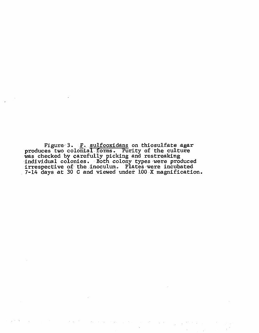
Figure 3. F 0 sulfooxidans on thiosulfate agar produces two colonial forms. Purity of the culture was checked by carefully picking and restreaking individual colonies. Both colony types were produced irrespective of the inoculum 0 Plates were incubated 7-14 days at 30 C and viewed under 100 X magnification.

38
39
and growth was much slower than for the other two organisms o The colony type was the same as T. ferrooxidans produced on thiosulfate agar, i.e., a small, flat colony with undulate margins®
Generation time. The generation times obtained for the three organisms in the thiosulfate medium are shown in Table 1® F. ferrooxidans and F. sulfooxidans have approximately the same rate of growth (7-9 hr) as seen by the mean generation time. The average generation time values are somewhat misleading because of the variation between trials. This is due to technique in culturing the organisms. When T. ferrooxidans gave a long generation time, so did F. ferrooxidans. An analysis of variance shows a very significant difference between the generation times. Orthogonal comparison of the values shows that a very significant difference exists between the generation time for F. sulfooxidans versus the other two. Likewise, a significant difference exists between the average value of T. ferrooxidans and F. ferrooxidans, even though the standard deviation of the values implies an overlapping of the generation times.
A generation time of 13.7 hr for T. ferrooxidans agrees with the time reported by Unz and Lundgren (1961) of 13.7 hr for their culture of T. ferrooxidans. On

40
Table 1. Generation times of the three iron-oxidizers in thiosulfate medium.
T . F . F.Trial ferrooxidans ferrooxidans sulfooxidans
1 18.0 hr 13.3 hr 7.7 hr2 14.0 9.4 7.33 12.1 9.8 7.34 10.7 5.2 7.2
Z 54.8 37.7 29.5X 13.7 + 3.2 9.4 + 3.3 7.4 + 0.2
Analysis of variance: source of error df
Sum of Squares
MeanSquare F value
Total 11 456.69Trials 3 32.19Organisms 2 393.40 196.7 37.97**
3 vs 1, 2 1 78.88 78.88 15.0**1 vs 2 1 36.3 36.3 7.05*
Error 6 31.10 5.18
^significant at the 0.01 level * significant at the .0.05 level

41
the other hand, their reported time of 34 hr for F. ferrooxidans does not agree with the time obtained in this studyo However, Unz and Lundgren stated that their time was "fairly non-representative" of F. ferrooxidansT ability to grow in thiosulfate medium.
The direct microscopic count procedure proved to be very laborious and somewhat inaccurate in the early stages of growth of the cultures. It was observed that, as the organisms first began to increase in number, large amounts of colloidal sulfur formed from the thiosulfate in the medium and interferred with the counting of the cellso The cells would tend to clump around the particles of sulfur and reproducible results were seldom obtained. As the culture entered the later stages of growth, however, the colloidal sulfur was not as great a problem. Presumably, the sulfur was being metabolized by the organisms. This should not be considered as a change in the energy source and, thus, a factor which would affect the generation time. Most workers now agree that S2O3 is not a symmetrical radical. The "outer" sulfur is actually in the form of S“ and oxidation of the outer atom occurs first (Trudinger, 1964a; Suzuki and Silver, 1966). Elemental sulfur is likewise known to form S= non-enzymatically. Therefore, the energy

42
source is the same, regardless of whether the cell starts with thiosulfate or elemental sulfur. The difference in generation times reported for the organisms on thiosulfate or sulfur will be different (Unz and Lundgren, 1961) but this is probably due to the solubility of sulfur and the slow rate at which S“ is formed from S° 0
While determining generation times of the three organisms, it was found that F. ferrooxidans and F. sulfooxidans could be readily cultured in either 2 liter flasks or in 20 liter carboys (see Materials and Methods) but that T. ferrooxidans could not. All attempts to grow T. ferrooxidans in thio- sulfate medium in 20 liter carboys failed. No growth was obtained after 5 days incubation. Aeration did not appear to be the limiting factor as both F. ferrooxidans and F. sulfooxidans grew much better in carboys than in shaker flasks.
Table 2 shows that carboy-grown cultures of F. ferrooxidans and F. sulfooxidans yield two to three times as much protein per liter as shaker-grown cells. Normally, T. ferrooxidans yeilded slightly less cellular material than either of the other two when grown in flasks. No explanation can be given other than the

43
Table 2. Cell yield of the iron-sulfur oxidizers
methodOrganism mg Pr/liter^T . ferrooxidans shaker-flask 4*6 + 1.9(2)F. sulfooxidans shaker-flask 6.4 + 0.6tr tt carboy 13.0 + 2.3F. ferrooxidans shaker-flask 5.0 ± 1.5tt tr carboy 16.1 + 5.4
^ ^mg protein per liter•2)average of three determinations

44
fact that some difference exists between the three organisms in relation to substrate and agitation.
Physiological Studies
Determination of apparent Michaelis Constant. Since it was evident from growth studies on the three organisms that some difference in utilization of thiosulfate did exist, their rates of thiosulfate oxidation were compared. Unz and Lundgren (1961) have previously reported that the only possible dif- ference they could determine between T . ferrooxidans and F. ferrooxidans was in fhe rate of thiosulfate utilization.
Figure 4 illustrates the differences between the three organisms when the apparent Michaelis Constant (Kfm) is determined for each. The K'm for F. ferrooxidans is approximately 10 times greater than that of T. ferrooxidans. F. sulfooxidans ex- hibits a K ’m value intermediate between the other two but is more similar to T. ferrooxidans than F. ferrooxidans.
While it is acknowledged that the Lineweaver- Burke plot is not a "normal" method of expressing data obtained using whole cells, the graph does show

Figure 4. A Lineweaver-Burke type plot of the three iron=oxidizers on thiosulfate.

45
0 -0 T. FERROOXIDANS
d-q F. SULFOOXIDANS
F. FERROOXIDANS
1 0 --
m
KM = 5 X 10
1006020i/tsj

46
that under similar conditions the organisms react differently. The term, K Jm, must be used as the rate of oxidation of thiosulfate by whole cells is not a measure of the kiqetics of a single enzyme but of the slowest reaction of a series of enzymes.Other factors, such as permeability, might be greatly influencing thiosulfate oxidation and/or the intermediates of thiosulfate oxidation. Nevertheless, if the K fm is taken as a rough, inverse measure of affinity of an enzyme for its substrate (Neilands and Stumpf, 1966), then it can be said that F. ferrooxidans has a much greater affinity or rate of utilization of S2O3 than either F. sulfooxidans or T. ferrooxidans.
Apparent energy of activation (EaT) . Enzymatic reactions, like ordinary chemical reactions, are greatly affected by temperature, i.e., as the temperature increases over a certain range the activity or reaction rate increases. Arrhenius (Neilands and Stumpf, 1966) was the first to recognize this phenomenon and derived an equation to express this relationship. The Arrhenius equation, in its most-useful form, using common logarithms and the gas constant, R, in calories, is

47
k2 0.219 Ea (T2 — Tx) log — = — ----------------
kl T1 ~ t2
The value, Ea, is called the energy of activation, or the amount of energy needed for placing molecules in a reactive state.
When the three test organisms were challenged with thiosulfate at various temperatures and the data plotted as the log Q02XN) vs l/absolute T, the E a * could be determined .
Figure 5 illustrates a dramatic difference between T. ferrooxidans (Ea’ = 11,000), F. sulfooxidans (Ea* = 11,400) and F. ferrooxidans (Ea* = 18,700) in regards to the effect of increasing temperatures. Again, it should be noted that this is not the true Ea for thiosulfate but only the Ea’ of prpbably the "pacemaker’* or slowest member of a complicated enzyme sequence. However, the purpose of such an experiment is to elucidate if the three organisms possess similar enzyme capabilities. If they are truly similar, the same Ea* would be expected for each of them.
Effect of pH. During the manometric studies just discussed, it was observed that whenever F. ferrooxidans was used, the jil 02 uptake at 60 min could be very closely

Figure 5. The Ea* values of the three iron organisms on thiosulfate. Each flask contained 200 jumoles thiosulfate per Warburg fkask. The temperatures used were 25, 27, 30, and 35 C; respiration time was 60 min.

LOG
Q0
2(n
)o - o T. FERROOXIDANS
° - 0 F. SULFOOXIDANS
F. FERROOXIDANS4 .2
4 .0Eof * 1 8 ,0 0 0
Ea' - 11,4003.6
3.4
3 .0
3 .353 .25 l/T X 10

49
predicted from the 30 min determination<> The resting cells normally oxidized thiosulfate at a steady rate for at least 60 min. This was not the case with T. ferrooxidans or, F\» sulfObxidans. Particularly with T. ferrooxidans, a sharp break in the rate of oxidation seemed to occur between 20-30 min, and the "expected" pi O2 uptake was never attained.
ThespH of the contents of individual Warburg flasks was measured before and after an experiment to determine if this was a factor. As no buffer was used, only the basal salts medium, any slight changes in H+ concentration would greatly effect the pH of the flask mixture.
Table 3 records the results of several trials in which pH measurements were made. As can be seen, the pH increased sharply for T. ferrooxidans and F. sulfooxidans but not for F. ferrooxidans.
Figures 6 and 7 illustrate the effect of initial pH on thiosulfate oxidation by T. ferrooxidans and F. sulfooxidans, respectively. Also, the relationship between O2 uptake and pH change , over the 60 min interval is demonstrated in these graphs. It appears that whenever the pH rises above 5.0-5.2, the rate of thiosulfate oxidation decreases. Yet, it is interesting to note that the decreased rate of oxidation parallels the rate of

50
Table 3 0 Increase in pH during oxidation of thiosulfate.Time (min)
Organism 0________ 10______ 30 ______ 60____To ferrooxidans 4 d 5o0 5.5 6.15Fo sulfooxidans 4ol 4.6 5o25 5o7F. ferrooxidans 4.1 4.3 4.5 4.8

Figure 6 . Effect of initial pH on thiosulfate oxidation by T. ferrooxidans. Each flask contained 1.01 rag protein and 200 /nmoles thiosulfate. Closed symbols with solid lines represent jul 02 uptake at different initial pH values; dotted lines and open symbols show pH changes. Circles (open and closed)= initial pH 3.5; squares ~ initial pH 4.5; triangles= initial pH 5.5.

oJo
<0
i §

Figure 7o Effect of Initial pH on thiosulfate oxidation by Fo sulfooxidans„ Each flask contained 200 jumoles thiosulfate and 0 .78 mg p r o t e i n S o l i d lines represent jul oxygen uptake while dotted lines show pH changeso Initial pH 5o7 = triangles; initial pH 4 <,3 ,= squares; initial pH 3o3 = circles0 Incubation was at 30 Co

o» (* ' » JP S
PS0/
.0 - -
' ° 0 $
‘°Si

53
thiosulfate oxidation whenever the culture is started at pH levels above 5.5. All three organisms utilized thiosulfate at a very rapid rate in every case in which the initial pH was low (3o3-3„6).
Figure 8 shows that F. ferrooxidans responds differently to thiosulfate depending upon the mode of growing th<£. cells „ Flask-grown cells exhibit some of the change in rate of thiosulfate oxidation which is so characteristic of T. ferrooxidans, but the effect is not so great. Yet, carboy-grown cells respire more rapidly and the pH does not rise as sharply. In fact, the pH of the contents of the Warburg flasks containing carboy- grown cells seldom rises above 5 o0 o This phenomenon is not attributed to cell concentration. Flask-grown cells at different cellular concentrations yield similar curves0 Carboy-grown cells at comparable concentrations do not show the typical change in rate of oxygen uptake o
At pH -values below 3.7-3.8 thiosulfate decomposes to elemental sulfur. A sample of the thiosulfate solution was deliberately made acid and a portion filtered through a Millipore filter to remove the colloidal sulfur. F. ferrooxidans was challenged with both filtered and unfiltered acid thiosulfate. Table 4 shows that the removal of sulfur from the medium did not greatly change

Figure 8 „ Response of flask-grown vs. carboy-? grown cells of F. ferrooxidans on thiosulfate oxidation.. Concentration of thiosulfate was 200 /nmoles. Solid lines represent different concentrations of. flask- grown cells and the dotted line shows carboy-grown cells (0 019 mg protein) o Open circles = 1016 mg protein open squares = 0o58 mg protein; open triangles= 0 o23 mg protein»

2 5 0
200
150
3 1 0 0
10 20 3 0 4 0TIME (MIN)
5 0 6 0 Ln4>-

Table 4 . Q02(N) va ues F0 ferrooxidanson a c id th io s u lfa te .
Substrate *^ _________________ 002 (N)Acid: thiosulfate 3150 550Acid thiosulfate 3438 + 88
(filtered)Colloidal sulfur 320 .+ 10from thiosulfate
f 1)initial pH = 3 . 3

56
the Q02(N) va^ues* In this case the filtered material actually yielded a slightly higher value. This, of course, could be due to experimental error.
To test the hypothesis that an increase in pH would affect oxygen uptake instead of merely the accumulation of end products, the cells of F. ferrooxidans were allowed to respire for a short time in a manometric flask, then base was tipped in to increase the pH. This caused a reduction in the rate of oxidation for the organism which- normally did not increase the pH as rapidly as the other two.
The second approach to the effect of pH usedT. ferrooxidans and F. sulfooxidans. Resting cells of the two were allowed to respire for 60 min, then acid was added to lower the pH. This time, as seen in Figure 9, the rate of oxidation increased and paralleled that of the flasks with an initial pH of 3.3-3.5.
When one looks at the overall picture obtained from this data, it becomes apparent that the pH of the environment greatly, influences the activity of the three iron- sulfur oxidizers. Other workers have related the break in oxygen uptake to the accumulation of tetrathionate (Landesman et al. 1966) or to trithionate (Vishniac and Santer, 1957). It appears, however, from these experi-

Figure 9. Reversal of pH inhibition of thio- sulfate by the addition of acid. Each flask contained 200 /moles thiosulfate and liOl mg protein of T„ ferrooxidans■> Open circles = initial pH 3o5; closed circles = initial pH 4.5» The pH after addition of acid was 3o4o

250 '
200-
Vf-ACID ADDED (PH LOWERED
TO 3.4)
« ■ i ■50 60 TO 8 0
(MIN)

58
ments that it is actually the increase in pH which eauses the organisms to slow down in their oxidation of thiosulfate0 The question now remains as to what causes the rise in pH.
Oxidation of tetrathionate. Sinha and Walden (1966) showed that thiosulfate is oxidized by whole cells of To ferrooxidans to tetrathionate by the following reactions
2S2O3 + ^02 --- ^ + OHIt was believed that the increase in pH in the Warburg flasks might be due to an accumulation of 0H“ from the reaction shown above. The three organisms were placed on various concentrations of tetrathionate to determine if this proposed intermediate could be readily oxidized. Figure 10 shows the results of this experiment.
Fo ferrooxidans a^ain gave the highest Q02(n) values of the three cultures. Concentrations of tetrathionate greater than 50 pamoles inhibited respiration as shown by a drop in the Q02(n)* In the case, of the other two organisms, T. ferrooxidans could withstand slightly higher concentrations of S^Qg than F. ferrooxidans or F. sulfooxidans. However, T. ferrooxidans did not exhibit the affinity for tetrathionate as did

Figure 10o Oxidation of various concentrations of tetrathionate by the three test organisms. The temperature was 30 C and respiration was for 60 min.

59
e - o T FERROOXIDANS
n - O F. SULFOOXIDANS
F. FERROOXIDANS
I
8 0ADDED
602 0 4 0MOLES SA0 i ADDED

60
F. ferrooxidans (as indicated by the lower Qo2(N) va -ues for T. ferrooxidans seen in Figure 10).
It is interesting to note that in all cases where serves as the substrate, the pH drops (see Table 5) .
T. ferrooxidans, F. ferrooxidans and F. sulfooxidans oxidize tetrathionate with a drop in pH that corresponds to the amount of oxidation that occurs. A drop in pH during tetrathionate oxidation does not alter the rate of oxygen uptake. A steady rate of oxidation (unit change/10 min interval) is observed for each organism.This is in contrast to the reaction when thiosulfate serves as the substrate. In this case a rise in pH occurs, followed by a change in rate of oxygen uptake.
Disappearance of substrate. The disappearance of thiosulfate and tetrathionate from the medium is used as an indicator of the affinity of the three test organisms toward the sulfur compounds. For example, Table 6 shows that when the amount of thiosulfate unaccounted for as S4OI; is divided by the mg protein used in the experiment a value of 4.13 can be assigned to T. ferrooxidans. Likewise, F. sulfooxidans yields a value of 5.6, very similar to the first organism. But, F. ferrooxidans, under the same conditions, gives a value of 10.95. This is interpreted to mean that even though thiosulfate disappears

61
Table 5. Decrease in pH during tetrathionate oxidation./angles /nmoles final
Organism S4O6 added O2 used pH
T. ferrooxidans^ 20 1.2 3.6^)30 1.5 3.440 2.6 3.250 2.9 3.1
100 1.6 3.3F . sulfooxidans 20 3.8 3.75
30 5.6 3.740 6.0 3.650 5.7 3.5
100 2.6 3.7F. ferrooxidans 20 1.1 3.2
30 1,2 3.140 1.36 3.050 1.4 3.0
100 0.98 3.25
^ m g protein used varied from 0.412-0.77. (2)v /initial pH in each case was pH 4.0

Table 6. Determination of thiosulfate disappearance.
/imoles/ml S2O3 recovered , . thiosulfate-Organism trial 0 min 10 min as S4Q6___ net loss'* ' disappearance
T. ferrooxidans 1 68.2 64.2 66.5 1.7. . 0 4.40.0 ;« (2)4.13 + 0.36Tt Tt 2 64.5 61.5 62.5 2.0 3.86F. sulfooxidans 1 68 o2 65.2 65.5 •
CM 4.355.6 + 1.77
t t t t 2 64.5 60.0 61.1 3.4 6.85F. ferrooxidans 1 68.2 60.2 63.0 5.2 10.01
10.95 + 1.4t t . , * F . . . .2 . . 64.5. .55.4. . .58,9. . . 5.5 11.9
^^thiosulfate unaccounted for as tetrathionate(2) =expressed as /jmoles S2O3 loss/mg protein
63
rapidly in the presence of each of the organisms, most of the thiosulfate can be recovered as tetrathionate in the case of To ferrooxidans and F. sulfooxidans.On the other hand, F. ferrooxidans utilizes the tetrathionate at a faster rate, i,e0, it is more permeable to the compound. This finding agrees with the Q02(n ) values shown in Figure 10,
Table 7 gives the results obtained for the disappearance of tetrathionate from the medium. Again,F. ferrooxidans shows a greater /imole loss/mg protein used than the other two organisms.
This type of experiment may not measure the true permeability of an organism to a compound. It at least shows that a difference does exist in the absorption/utilization of a compound by various organisms. There seems to be no standard test for permeability in the literature, but at least one other worker has used an approach similar to this (Hart, 1967),
/Sulfur and iron oxidation. Margalith et al. (1966)
reported that cells of F. ferrooxidans, grown repeatedly in thiosulfate for a period of 5 months, readily oxidized iron. They concluded that the iron oxidation system of their organism was constitutive but were not certain about the sulfur-oxidizing system.

Table 7. Determination of tetrathionate disappearance.
, tetrathionateOrganism trials net.loss' ' disappearance
_
T. ferrooxidans 2 1.6 3.7 + 0.8
F. sulfooxidans 2 2.8 4.8 + 1.75
F 0 ferrooxidans 2 3.0 6.25 + 1.2
average value(2) ='expressed as pnoles S4O 6 loss/mg protein

65
A check of the iron-oxidizing ability of the cultures used in this investigation was made since the test organisms had been grown in thiosulfate medium for more than 2 years . The results of this check are shown in Table 8 .
A comparison of the values of the threeorganisms on thiosulfate, sulfur and ferrous iron shows that, while the values vary, all three can utilize each substrateo The findings do not agree exactly with those of these workers (Margalith et al», 1966) in that prolonged growth of F 3 ferrooxidans and F. sulfooxidans on thiosulfate medium causes a loss„in ability to oxidize ferrous iron- However, as can be seen from Table 8 , after £<months of culturing in thiosulfate medium, the organisms utilize iron readily. It is only after 2 years of repeated transfers in thiosulfate broth that the organisms show a decrease in their ability to readily oxidize ferrous iron.
Molecular Studies
Determination of base ratios. The results presented in a recent publication by Garrity et al. (1969) are consistent with the idea that closely related organisms haL\Z& similar base ratios of guanine plus cytosine. The moles

66
Table 8 0 Oxidation of colloidal sulfur and ferrous iron,,
Organism s2°3 S°Q02(n)(1)
Fe^.
To ferrooxidans 1630 988 3400^2) 4150Fo sulfooxidahs 2460 915 5000 10)50F 0 ferrooxidans 8400 1605 2500 1000
^ a t 30 C? |yitial pH with S2O3 = 4„0; initial pH with S® and Fe ~ 3o0o(o)v 'after 6 months repeated transfets in thiosulfate(2 )' -'after 2 years repeated transfers in thiosulfate

67
percent G+C for T. ferrooxidans, F» ferrooxidans and F. sulfooxidans have been determined and are shown in Table 9. The Tm values are very similar for the three organisms. A positive control, Staphylococcus aureus, has a reported G+C range of 32-35 moles percent (Garrity et alOJ 1969) and this is similar to the value obtained in this investigation. Jackson et al. (1968) have reported a G+C ratio for To ferrooxidans of 56-57 percent0
Even though the G+C ratios of the three cultures are similar, this does not mean the organisms are necessarily identical. On the other hand, it does not rule out the posibility that they are the same. As mentioned in the section on Review of Literature, several workers have shown that organisms with similar base ratios may not hybridize to any extent when DNA-DNA hybridization is performed. The use of the DNA-DNA hybridization technique has not been attempted in this study. Thus, all that can be concluded is that the DNA of the organisms may be quite similar.
Transformation studies. An attempt was made to transform cells of T. ferrooxidans, a slow grower on S4O6 agar, with the DNA extracted from F. ferrooxidans and F. sulfooxidans. The reverse was also attempted.

fa! cnj
Table 9» Base ratios of the iron-sulfur oxidizers.
Organism Tm moles percent (G+C)
To ferrooxidans F. sulfooxidans o ferrooxidans o aureus .
90o85 + 0 o2 5 2 o5 ;
91 .30 + 0,.2 53«6 ;
91 .00 + 0o2 5 2 . 9 ;
8 1 . 0 0 . . + .Q.3 . 2 8 o5 /
0 . 5 7
0o 63
0 o 71
0 . 7 1

69
Table 10 shows the results from the preliminary experiments on transformation, from which no conclusive results were obtained0 There is an indication that transformation may have occurred in the case of F. sulfooxidans treated, with the DNA. from T, ferrooxidans.A 10-fold increase in colony count may not really be significanto
But of more interest than mere numbers is the fact that a change in colony morphology seems to have occurred. Figure 11 shows the colonial appearance of F« sulfooxidans on tetrathionate agar both with and without DNA material present from T. ferrooxidans. The presence of the DNA caused a large increase in number of the typical T. ferrooxidans“type colony. It could be that transformation has occurredo Another explanation might be that the presence of the DNA stimulates the polymorphic condition of this organism. More experiments of this type need to be done to verify this observation.
Taxonpmic Considerations
Numerical classification. Numerical analysis was applied to the three organisms used in this study in the manner of Hutchinson et al. (1966). The test used to separate, the organisms are listed in.Table 11. These

70
Table 10e Preliminary experiments in transformation.
Recepient D o n o r ^ Substrate cells/ml
To ferrooxidans - S2°3 n,
CM0
tt it - S4°I n,»d 0F. sulfooxidans - s2°3 8 X
000H
tt tt - s4°6 < 4 X 10 4F. ferrooxidans - S2O3 11 X i o 9
tt tt _ S4OI 8 X 10 6To ferrooxidans II S406 6 X 10⁵tt tt III tt 4 X 10⁸Fo sulfooxidans I tt 1 X 10⁵*Fo ferrooxidans I tt 3 X 10⁶
(1)I = To ferrooxidans; II = F0 ferrooxidans 0 sulfooxidans; III = Fo
(2)not determined
indicates possible transformation has occurred

Figure 11- Colonial appearance of Fo sulfooxidans on tetrathionate agar with (a) and without (b) the addition of DNA from L ferrooxidans o

71
>-■ i«7T ..■*
’*■ l\ - '■' V'

72
Table 11«, Criteria for numerical analysis of three iron-sulfur oxidizers»
Characteristic I^^II III
1) Growth in 20 liter carboys 0<2) + +
2) Growth in shaker-flasks only + 0 03) Punctiform colonies on S2O3 , 0 + +4) Flat colonies oifl S2O3 + + 05) Punctiform colonies on S^Og " 0 + +
6)/ **Average generation time 13 hr + 0 0
7) K*m on S2O3 = 10“^ M + .+ 0
8) K ?m on = 10“^ M 0 0 +9) Q02(^) on s203 y 5000 0 0 +
10)-xQ02(N) on S4O5 >2000 0 0 +U ) Q02(n) °n Cblloidal SfolfUi: >1200 0 0 +12) Q02(n ) on ferrous iron >3000 + + 013) E a ? on S2O3 >15,000 0 0 +14) Sharp break in O2 uptake during
manometric experiments at initial pH of 4o0=4o2
+ + 0
15) Tetrathionate formed from S2O3 + + +16) Average jjmole loss SoCH/mg protein
in 10 min ^4«5 + 0 017) Average famole loss S203/mg protein
in 10 min > 9 0 0 4*18) Average /umole loss S^0g/mg protein
in 10 min > 6 • 2 0 0 +

73
(Table 11 cont.)
Characteristic I II III
19) Moles percent G+C = 52-53 percent + + +■20) DNA causes change in colony
morphology of other culture(s) + - 0 0on tfettathionate agar
(l)l = To ferrooxidans; II = Fo sulfooxidans; III = F. ferrooxidans
(o)v‘c,/+- = positive reaction; 0 = negative reaction

74
are strictly arbitrary criteria derived from all of the preceding experiments. However, using these tests, it is possible to obtain certain similarity (S) values.
According to the S values shown in Table 12, T. ferrooxidans and F. ferrooxidans are not closely related. F. sulfooxidans agaiiv pjroves, to be of intermediate type between the other two. It appears to be more closely related, though, to To ferrooxidans than to F. ferrooxidans.
Hutchinson et al. (1965) reported that the results of a numerical analysis of certain thiobacilli were largely independent of the tests used. A second paper (Hutchinson et alo, 1966) by these workers showed that the acidophilic thiobacilli could be grouped into two distinct groups corresponding to T. thiooxidans and T. ferrooxidans.The latter group included several strains of T. ferrooxidans, F. ferrooxidans and F. sulfooxidans. These three organisms, in Hutchinson et alo hands, had S values of about 9Q-92, regardless of how the three were compared. Yet the order of similarity was always (1)’ T. ferrooxidans, (2) F. sulfooxidans and (3) F. ferrooxidans. It made no difference if the organisms were tested in thiosulfate or sulfur medium.
The findings of these workers agree with the
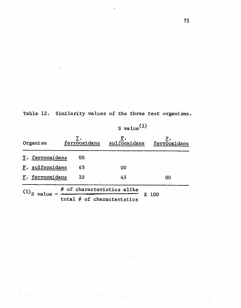
75
Table 12. Similarity values of the three
S value^^To Fc
Organi sm ferrooxidans sulfooxidans
To ferrooxidans 00Fo sulfooxidans 65 00Fo ferrooxidans 10 45/•i\ # of characteristics alikev ' S value = — ' — — X
total # of characteristics
test organisms.
Foferrooxidans
00
100

76
findings presented here, so far as the sequence of similarity is concerned. Because of the more specific tests here, it is possible to distinguish smaller differences between the three organisms than what was reported by the other workerso
Proposed nomenclature. The majority of the tests listed in Table 11 for determining differences in the three organisms are designed to show differences in rate of enzyme activity rather than the presence or absence of certain enzyme systemso
The actual differences between the three organisms, though quite specific for each, may be considered: more as minor differences than major differences. All three organisms carry out essentially the same reactions, i.e., they are capable of oxidizing sulfur, thiosulfate, tetrathionate and ferrous iron. Presumably, each organism carries out these reactions in the same fashion. The major differences appear to be colony morphology on thiosulfate agar, permeability to tetrathionate and antigenic response as demonstrated by LaHaye (1966).
It is possible that all three major differences can be attributed to a single enzyme which is surface bound and. more reactive in one organism (F. ferrooxidans) than in the other two. For example, a change in structure
77
of this enzyme might be enough to account for the antigenic difference noted between T„ ferrooxidans and F. ferrooxidans. It might also be that this difference in activity of the enzyme could be responsible for the two types of colonies produced on §20^ agar. The raised punctiform type could be attributed to a fast-growing culture with the newly formed cells "piling up" and the flat colony type associated with slow-growing cells. The micro-environment in the center of the colony could be inhibitory to continued growth and, thus, the only new growth would be at the periphery of the colony.
The indirect evidence available which does lend support to this hypothesis iss (1) all three organisms are greatly affected by a rise in pH, (2) the rise in pH is probably due to accumulation of S^Og , (3) T. ferrooxidans and F. sulfooxidans are less able to utilize tetrathionate than is F. ferrooxidans, and (4) a change in colony morphology of F„ sulfooxidans occurs when the cells are treated with DNA. from T. ferrooxidans, indicating only a small difference between the two organisms.
Considering all the discussion brought forth in this dissertation, and according to the rules laid down in the seventh edition of Bergey’s Manual of Determinative Bacteriology (1957), it is proposed that the generic names

78
of F. ferrooxidans and F. sulfooxidans be changed to T. ferrooxidans var. ferrooxidans and T. ferrooxidans var. sulfooxidans, respectively.
The description of the three organisms would be as follows ;
Thiobacillus ferrooxidans Temple and Colmer, 1951.Short, Gram-negative rods, 0.5 to 1.0 microns. Motile.Thiosulfate agar colonies; Very small, flat colonies
with undulate margins.Thiosulfate broth; Oxidation characterized by rapid
increase in pH in thiosulfate broth when a heavy inoculum is used and the initial.pH is 4.0-4.2. A well defined break in the rate of oxidation occurs when the pH increases apove 5.2. Generation time in thiosulfate broth approximately 13 hr.
Tetrathionate agar; Very slow growth on 0.05 percent.
Thiobacillus ferrooxidans var. ferrooxidans Leatjien aria Braley, 1954.
Short, Gram-negative rods 0.6 to L 0 microns. Motile.Thiosulfate agar colonies; Punctiform, raised
colonies with entire edges.Thiosulfate broth; Oxidation of thiosulfate in
broth with heavy inoculum characterized by no sharp break

79
In rate of oxidation by resting cells grown in carboys with forced aeration. Cells grown in shaker culture do show a break in rate of oxidation. The pH of the mixture seldom increases above 5.0-5.5. Generation time 7-9 hr in thiosulfate broth.
Tetrathiopate agar: Small colonies appear withinr3-4 days on 0.05 percent tetrathionate.
Thiobacillus ferrooxidans var. sulfooxidans Kinsel,1960.
Short, Gram-negative rods, 0.5 to 1.0 microns. Motile.Thiosulfate agar colonies: Raised, punctiform and
also flat-type with undulate edges appear on the same plate. Each colony type will give rise to both types of colonies.
Thiosulfate broth: Oxidation of thiosulfate brothsimilar to T. ferrooxidans Temple and Colmer, but has consistently higher Q02(n) valueSo
Tetrathionate agar: Small colonies appear within3-4 days. Both colony types present.

SUMMARY
The sulfur metabolism<of three iron-sulfur oxidizers, To ferrooxidans, F. ferrooxidans and F. sulfooxidans, has been examined. Some differences were noted in the rate of utilization of thiosulfate and tetrathionate by the three organisms.
Thiobacillus ferrooxidans, on thiosulfate agar, produced a small, flat colony with undulate margins while F. ferrooxidans produced small punctiform colonies with entire edges. Both colonial forms were exhibited by F. sulfooxidans„ Either colony type of F. sulfooxidans, when isolated colonies were picked and restreaked, repeatedly yielded both morphological forms on thiosulfate agar.
A difference existed in generation time between the three organisms on thiosulfate medium (7-9 hr for F. ferrooxidans and F„ sulfooxidans and 13-14 hr for T. ferrooxidans) <,
The K fm of F. ferrooxidans was found to be approximately 7 x 10"^ M while the K Tm value for the other two
-2was approximately 2-4 x 10 M.The apparent energy of activation (Eaf) on thio
sulfate was 11,000 for T. ferrooxidans, 11,400 for F. sulfooxidans and 18,700 for F. ferrooxidans.
80

81
Thiosulfate oxidation in manometric experiments with To ferrooxidans and F. sulfooxidans was characterized by a sharp break in the rate of oxygen uptake after 20-30 min. The pH of the menstrum increased from an initial pH of 4.0-4.2 to a final pH of 5.8 -6.2 after 60 min. The break in oxygen uptake was associated with a rise in pH above 5.0-5.2. F. ferrooxidans, grown in carboys, did not exhibit this characteristic break in the rate of oxidation. Shaker-grown cells of F. ferrooxidans did show a slight change in the rate of utilization after 30 min.
The addition of acid after 60 min respiration to manometric flasks containing T. ferrooxidans or F. sulfooxidans completely reversed the change in rate of oxidation and paralleled the oxidation rate exhibited by flasks with an initial pH of 3.3-3.5.
Fo ferrooxidans showed a loss of thiosulfate from the medium of 10.95 jamoles/mg protein in 10 min as compared to 4.13 for T. ferrooxidans and 5.6 for F. sulfooxidans. Disappearance of tetrathionate from the F. ferrooxidans medium was 6.25 pmoles/mg protein in 10 min. T. ferrooxidans and F. sulfooxidans were assigned values of 3.7 and 4.8, respectively, for tetrathionate.It is concluded that F. ferrooxidans can more readily

82
utilize tetrathionate than the other two organisms.All three cultures were capable of utilizing thio
sulfate, tetrathionate, colloidal sulfur and ferrous iron, though the Q02(n) values varied with each organism.
The base ratio (moles percent G+C) was determined by the melting point of the DNA (Tm) to be 52.5-53.6 percent for all three organisms.
Attempts at transformation of one organism into another organism as gauged by the colony appearance of the donor of the DNA was attempted. No conclusive results were obtained, but indications exist that a change in colony morphology occurred.
It is proposed that the name T. ferrooxidans be retained and F. ferrooxidans and F. sulfooxidans be changed to T„ ferrooxidans var. ferrooxidans and T. ferrooxidans var. sulfooxidans, respectively.

LITERATURE CITED
Baalsrud, K. and K. S. Baalsrud. 1954 Studies on Thiobacillus denitrificans.Arch. Mikrobiol. 20;34-6Tl
Basden, E. H., M. E. Tourtellotte, W. N. Plastridge, and J. S. Tucker. 1968Genetic relationship among bacteria classified as vibrios. J. Bacteriol. 95:439-443.Beers, R. J. and W. R. Lockhart. 1962 Experimental methods in computer taxonomy.J. Gen. Microbiol. 28:633-640.Breed, R. S., E. G. D. Murray, and N. R. Smith. 1957 Bergey’s manual of determinative bacteriology, 7th ed. Williams and Wilkins Company, Baltimore, Md.Bryner, L. C. and A. K. Jameson. 1958 Microorganisms in leaching sulfide minerals.Appl. Microbiol. 6 i281-287.Colmer, Arthur R. 1962Relation of the iron oxidizer, Thiobacillus ferrooxidans, to thiosulfate.J. Bacteriol. 83:761-765.Colmer, Arthur R. and M. E. Hinkle. 1947 The role of microorganisms in acid mine drainage: a preliminary report.Science, 106:253-256.Colmer, Arthur R., K. L. Temple, and M. E. Hinkle. 1950 An iron-oxidizing bacterium from the acid drainage of some bituminous coal mines.J. Bacteriol. 59:317-328.Garrity, F. L., B. Detrick, and E. R. Kennedy. 1969 Deoxyribonucleic acid base composition in the taxonomy of Staphylococcus.J. Bacteriol. 97:557-560.Gleen, H. and J. H. Quastel. 1953 Sulphur metabolism in soil.Appl. Microbiol. 1:70-77.
83

84
Guittonneau, G. and J. Keilling. 1932L*evolution et la solubilisation du soufre elementairedans la terre arable.Ann. agron. N. S. 600-725.Hart, Lewis T. 1967The response of microorganisms to 2,4-dichlorophenoxy- acetic acid.Ph. D. Dissertation, Louisiana State University, Baton Rouge.Hill, L. R. 1966An index to deoxyribonucleic acid base composition of bacterial species.J. Gen. Microbiol. 44:419-437.Hopkins, F. G. 1929 On glutathione: a reinvestigation.J. Biol Chem. 84:269-320.Hutchinson, M., K. I. Johnstone, and D. White. 1965 The taxonomy of certain thiobacilli.J. Gen Microbiol. 4 H 35 7-366.Hutchinson, M., K. I. Johnstone, and D. White. 1966 Taxonomy of the acidophilic thiobacilli.J. Gen Microbiol. 44:373-381.Jackson, J. F.s D. J. W. Moriarty, and D. J. D Nicholas. 1968Deoxyribonucleic acid base composition and taxonomy of thiobacilli and some nitrifying bacteria.J. Gen. Microbiol. 53:135-138.Kaji, A. and W. D. McElroy. 1959Mechanism of hydrogen sulfide formation from thiosulfate. J. Bacteriol. 77:630-637.Kelly, D. P. and P. J. Syrett. 1964Inhibition of formation of adenosine triphosphate inThiobacillus thioparus by 2 ;4-dinitrophenol.Nature, 202:507-598.Kingsbury, D. T. and E. Weiss. 1968Lack of deoxyribonucleic acid homology between species of the genus Chlamydia.J. Bacteriol. 96:1421-1423.

85
Kinsel, Norma A. 1960New sulfur oxidizing iron bacterium: Ferrobacillusferrooxidans sp.n.J. Bacteriol. 80:628-632.LaHaye, P. A. 1966A study of three iron-oxidizing bacteria.M. S. Thesis, Louisiana State University, Baton Rouge.Landesman, J., D. W. Duncan, and C. C. Walden. 1966 Oxidation of inorganic sulfur compounds by washed cell suspensions of Thiobacillus ferrooxidans.Can. J. Microbiol. 12:957-963"^Leathen, W. W. and S. A. Braley, Srr. ',1954A new iron-oxidizing bacterium: Ferrobacillus ferrooxidans.Bacteriol Proc. 1954:44.Leathen, W. W., N. A. Kinsel, and S. A. Braley, Sr. 1956 Ferrobacillus ferrooxidans: a chemosynthetic autotrophicbacterium.J. Bacteriol. 72:700-704.Lichstein, Herman C. and Evelyn L. Oginsky. 1965 Experimental microbial physiology.W. H. Freeman and Co., San Francisco, p. 2.London, J. and S. C. Rittenberg. 1964Path of sulfur in sulfide and thiosulfate oxidation bythiobacilli.Proc. Nat. Acad. Sci.(U.So), 52:1183-1190.Lowry, Oliver H., Nira J. Rosebrough, A. Lewis Farr, and Rose J. Randall. 1951Protein measurement with the Folin phenol reagent.J. Biol. Chem. 193:265-275.Lundgren, D. G., K. J. Andersen, C. C. Remsen, and R. P. Mahoney. 1964Culture, structure, and physiology of the chemoautotroph Ferrobacillus ferrooxidans.Dev. Ind. Microbiol. j>:250-259.Lyalikova, N. N. 1960The physiology and ecology of Thiobacillus ferrooxidans in relation to its role in the oxidation of sulfide ores (review) .Mikrobiol. 29:773-779.

86
Margalith, P., Marvin Silver, and D, G. Lundgren. 1966 Sulfur oxidation by the iron bacterium Ferrobacillus ferrooxidans.J. Bacteriol. £2:1706-1709.Marmur, J. 1961A procedure for the isolation of DNA from microorganisms,J. Mole Biol. 3:208-218.Marmur, J. and P. Doty. 1962Determination of the base composition of deoxyribonucleic acid from its thermal denaturation temperature.J. Mol. Biol. 5:109-118.McCarthy, B. J. and E. T. Bolton. 1963An approach to the measurement of genetic relatednessamong microorganisms.Pro. Nat. Acad. Sci.(U.S.), 50:156-164.Neilands, J. B. and Paul K. Stumpf. 1966 Outlines of enzyme chemistry.John Wiley & Sons, Inc., N. Y., pp 8 7-134.Normand, R. A. 1965Bacterial iron and sulfur oxidation in some geologic formations.M. S. Thesis, Louisiana State University, Baton Rouge.Nygaard, A. P. and B. D. Hall. 1963 A method for the detection of RNA-DNA complexes.Biochem. Biophys. Res. Commun. 12:98-104.Parker, C. D. and J. Prisk. 1953The oxidation of inorganic compounds of sulphur byvarious bacteria.J. Gen. Microbiol. 8:344-364.Peck, H. D. and E. Fisher. 1962The oxidation of thiosulfate and phosphorylation in extracts of Thiobacillus thioparus.J. Biol. Chem. 237;l$fr::T97.Razzell, W. E. and P. C. Trussell. 1963Isolation and properties of an iron-oxidizing Thiobacillus. J. Bacteriol. 85:595-603.

87
Rolls, J. P. and E. S. Lindstrom. 1967 Induction of a thiosulfate-oxidizing enzyme in Rhodopseudomonas palustris.J. Bacteriol. 9?:784-785.Siato, Hiuga and Kin-Ichiro Miura. 1963 Preparation of transforming deoxyribonucleic acid by phenol treatment.Biochim. Biophys. Acta, 72:619-629.Silver, M. and D. G. Lundgren. 1968Sulfur-oxidizing enzyme of Ferrobacillus ferrooxidans(Thiobacillus ferrooxidans) .Can. J. Biochem. 46:457-461.Silverman, M. P. and D. G. Lundgren. 1959Studies on the chemoautotrophic iron bacterium Ferrobacillus ferrooxidans. II. Manometric studies.J. Bacteriol. 78:326-331.Sinha, D. B. and C. C. Walden. 1966Formation of polythionates and their interrelationships during oxidation of thiosulfate by Thiobacillus ferrooxidans . Can. J. Microbiol. 12:1041-1054.Sneath, P. H. A. 1957The application of computers to taxonomy.J. Gen. Microbiol. 17:201-226.Snedecor, George W. and William G. Cochran. 1967 Statistical methods.Iowa State Uni. Press, Ames, Iowa, p. 32-62, 135-170.Sorbo, B. 1957A colorimetric method for the determination of thiosulfate. Biochim. Biophys. Acta, 23:412-416.Starkey, R. L. 1934The production of polythionates from thiosulfate by microorganisms.J. Bacteriol. 28:387-400.Starkey, R. L. 1935Formation of sulfide by some sulfur bacteria.J. Bacteriol. 33:545-571.

88
Suzuki, I. 1965Oxidation of elemental sulfur by an enzyme system of Thiobacillus thiooxidans.Biochim. Biophys. Acta, 104:359-371.Suzuki, I. and M. Silver. 1966The initial product and properties of the sulfur-oxidizing enzyme of thiobacilli.Biochim. Biophys. Acta, 122:22-33.Suzuki, I. and C. H. Werkman. 1959Glutathione and sulfur oxidation by Thiobacillus thiooxidans. Proc. Natl. Acad. Sci. (U.S.), 45:239-244.Tano, T. and K. Imai. 1968Physiological studies on thiobacilli. Part II. The metabolism of colloidal sulfur by the cell-free enzyme system of Thiobacillus thiooxidans.Agri. Bio. Chem. 32:51-54.Tano, T., H. Asano, and K. Imai. 1968 Physiological studies on thiobacilli. Part III. The oxidation of thiosulfate by particulate fraction of Thiobacillus thiooxidans.Agri. Bio. Chem. 32:140-143.Temple, K. L. and Arthur R. Colmer. 1951The autotrophic oxidation of iron by a new bacterium:Thiobacillus ferrooxidans.J. Bacteriol. 62:605-611.Trudinger, P. A. 1961Thiosulfate oxidation and cytochromes in Thiobacillus X. Biochem. J. 78:680-686.Trudinger, P. A. 1964aProducts of anaerobic metabolism of tetrathionate by Thiobacillus X.Aust. J. Biol. Sci. 17:446-458.Trudinger, P. A. 1964bThe metabolism of trithionate by Thiobacillus X.Aust. J. Biol. Sci. 17:459-468.

89
Trudinger, P. A. 1964cOxidation of thiosiilphate by intact cells of Thiobacillus X: Effects of some experimental conditions.Aust. J. Biol. Sci. JL7:738-751.Trudinger, P. A. 1965aOn the permeability of Thiobacillus neapolitanus to thio- sulphate.Aust. J. Biol. Sci. 18:563-568.Trudinger, P. A. 1965bEffect of thiol-binding reagents on the metabolism of thiosulfate and tetrathionate by Thiobacillus neapolitanus.J. Bacteriol. 89:617-625.Trudinger, P. A. 1967Metabolism of thiosulfate and tetrathionate by hetero- trophic bacteria from soil.J. Bacteriol. 93:550-559.Umbreit, W. W. 1947 Problems of autrophy.Bact. Revs. 11:157-166.Umbreit, W. W., R. H. Burris, and J. F. Stauffer. 1957 Manometric techniques.Burgess Publishing Company, Minneapolis, Minn.Unz, R. F. and D. G. Lundgren. 1961A comparative nutritional study of three chemoautotrophic bacteria: Ferrobacillus ferrooxidans, Thiobacillusferrooxidans and Thiobacillus thiooxidans.Soil Sci. 92:302-3]XVishniac, W. 1952The metabolism of Thiobacillus thioparus. I. The oxidation of thiosulfate.J. Bacteriol. 64:363-373.Vishniac, W. and M. Santer. 1957 The thiobacilli.Bact. Revs. 21:195-213.Waksman, S. A. 1922Microorganisms concerned in the oxidation of sulfur in the soil. III. Media used for the isolation of sulfur bacteria from the soil.Soil Sci. 13:329-336.

90
Wayne, L. G. and W. M. Gross. 1968Base composition of deoxyribonucleic acid isolated from mycobacteria.J. Bacteriol. 96:1915-1919.Welker, N. E. and L. L. Campbell. 1967Unrelatedness of Bacillus amyloliquefaciens and Bacillussubtilis.J. Bacteriol. 94:1124-1130.

VITA
Harold C. Bounds was born August 13, 1940, in Shreveport, Louisiana. He graduated from Fair Park High School in 1958 and entered the United States Coast Guard Reserve for six months active duty.
He received the Bachelor of Science degree from Centenary College in June, 1963 and the Master of Science degree from Louisiana State University in August, 1964.He is presently a candidate for the Doctor of Philosophy degree in Microbiology at Louisiana State University, Baton Rouge.
He is married and has two daughters, Karen Renee •and Leisha Dawn.
He is a member of the American Society for Microbiology, Sigma Xi and Eros Lodge #295, F. & A. M.
91
r

EXAMINATION AND THESIS REPORT
Candidate: Harold C. Bounds
Major Field: Microbiology
Title of Thesis: Sulfur metabolism of three autotrophic iron-oxidizers
Approved:
Major Professor and Chairman
Dean of the Graduate School
EXAMINING COMMITTEE:
C k r $ .
Date of Examination:
July 21. 1969