sumo chain-induced dimerization activates...
TRANSCRIPT

Molecular Cell
Article
SUMO Chain-Induced Dimerization Activates RNF4Alejandro Rojas-Fernandez,1,2 Anna Plechanovova,2 Neil Hattersley,2,3 Ellis Jaffray,2 Michael H. Tatham,2
and Ronald T. Hay1,2,*1Medical Research Council Protein Phosphorylation and Ubiquitylation Unit2Centre for Gene Regulation and Expression, College of Life SciencesUniversity of Dundee, Dundee, Scotland DD1 5EH, UK3Present address: Ludwig Institute for Cancer Research, University of California, San Diego, P.O. Box 12385, La Jolla, CA 92093-2385, USA
*Correspondence: [email protected]
This is an open access article under the CC BY license (http://creativecommons.org/licenses/by/3.0/).http://dx.doi.org/10.1016/j.molcel.2014.02.031
SUMMARY
Dimeric RING E3 ligases interact with protein sub-strates and conformationally restrain the ubiquitin-E2-conjugating enzyme thioester complex suchthat it is primed for catalysis. RNF4 is an E3 ligasecontaining an N-terminal domain that binds its poly-SUMO substrates and a C-terminal RING domainresponsible for dimerization. To investigate howRNF4 activity is controlled, we increased polySUMOsubstrate concentration by ablating expression ofSUMO protease SENP6. Accumulation of SUMOchains in vivo leads to ubiquitin-mediated proteoly-sis of RNF4. In vitro we demonstrate that at concen-trations equivalent to those found in vivo RNF4 ispredominantly monomeric and inactive as an ubiqui-tin E3 ligase. However, in the presence of SUMOchains, RNF4 is activated by dimerization, leadingto both substrate ubiquitylation and autoubiquityla-tion, responsible for degradation of RNF4. Thus theubiquitin E3 ligase activity of RNF4 is directly linkedto the availability of its polySUMO substrates.
INTRODUCTION
Ubiquitin modification is initiated by the ATP-driven formation of
a thioester bond between the C-terminal carboxyl group of
ubiquitin and the catalytic cysteine on one of the two E1-acti-
vating enzymes, UBA1 or UBA6 (Haas et al., 1982). The activated
ubiquitin is then transferred to one of the�40Ubiquitin E2-conju-
gating enzymes, where it again forms a thioester bond. The
E2-Ub complex interacts with ubiquitin E3 ligases that recruit
substrates and confer specificity to ubiquitin modification, lead-
ing to the formation of either an isopeptide or peptide bond
between the ubiquitin C terminus and the ε-amino group of a
lysine or the a-amino group of the protein N terminus, respec-
tively (Deshaies and Joazeiro, 2009; Kravtsova-Ivantsiv and
Ciechanover, 2012; Scheffner et al., 1995; Tatham et al., 2013).
It is thought that there are over 600 E3 ligases encoded in the
human genome, and they fall into two distinct groups based on
their mechanism. When homologous to E6-AP C terminus
880 Molecular Cell 53, 880–892, March 20, 2014 ª2014 The Authors
(HECT) (Scheffner et al., 1995) and RING-between-RING
(RBR) (Wenzel et al., 2011) E3 ligases interact with ubiquitin-
loaded E2, the ubiquitin is first transferred onto an active site
cysteine residue in the E3, and the resulting thioester bond is
attacked by an amino group on the bound substrate to form a
peptide bond between ubiquitin and substrate. In contrast,
Really Interesting New Gene (RING) E3 ligases function by
binding both ubiquitin-loaded E2 and substrate and directly
catalyzing transfer of the ubiquitin to substrate. RING E3s
prime the ubiquitin-loaded E2 for catalysis by folding the
ubiquitin back onto the E2 in a conformation that is optimal for
nucleophilic attack by the amino group of the protein substrate
(Dou et al., 2012, 2013; Plechanovova et al., 2012; Pruneda
et al., 2012).
Small Ubiquitin-like Modifier (SUMO) is encoded by three
functional genes in humans. While the conjugated forms of
SUMO-2 and SUMO-3 are almost identical and functionally
undistinguishable, SUMO-1 is only 45% identical to SUMO-2/
3. Like ubiquitin, SUMO-2/3 can form polymeric chains through
lysine 11, which is located in a SUMO consensus modification
motif (JKXD/E, where J is a large hydrophobic amino acid
and X is any amino acid). Such a consensus modification motif
is lacking in SUMO-1 (Rodriguez et al., 2001; Sampson et al.,
2001; Tatham et al., 2001). In humans the SUMO E1-activating
enzyme is a heterodimer of SAE1 and SAE2 (Desterro et al.,
1999). The SAE2 subunit contains the catalytic cysteine that
forms a thioester with the C terminus of SUMO (Desterro et al.,
1999). Subsequently, SUMO is transferred from SAE2 to the
catalytic cysteine on the unique SUMO E2-conjugating enzyme
(UBC9), forming a new thioester (Desterro et al., 1997; Johnson
and Blobel, 1997). While UBC9 can SUMOylate substrates
directly by recognizing SUMO consensus motifs (Hay, 2005;
Rodriguez et al., 2001), its substrate specificity and catalytic
activity are enhanced by SUMO E3 ligases (Geiss-Friedlander
and Melchior, 2007).
The family of SUMO-Targeted Ubiquitin Ligases (STUbL)
functionally link modifications by SUMO and ubiquitin. STUbLs
bind to SUMO-modified proteins and induce their ubiquitination
(Perry et al., 2008). Recognition of SUMOby STUbLs ismediated
by SUMO-interacting motifs (SIM). SIMs were classically
defined as a consensus of V/L/I, V/L/I, X, V/L/I or V/L/I, X,
V/L/I, and V/L/I, whereas a subgroup of high-affinity SIMs were
described as V/I/L/F/Y,V/I,DLT (Hecker et al., 2006; Song
et al., 2004; Sun and Hunter, 2012).
A
B
DC
Cont
rol
siNT
siSE
NP6
siRN
F4
αSENP6
αRNF4
αTubulin
siNT
siSENP6
#1 #2 #3 #4
αSENP6
αRNF4
αTubulin
U2OS HALO-SENP6 siRNA resistant
siNT siSENP6
Doxycycline
HALO-SENP6Endo SENP6 αSENP6
αRNF4
-250
-130
-95-72
- + - +
αTubulin
siNT
siSE
NP6
siNT
siSE
NP6
250-130-
95-72-55-
36-28-11-
SUM
O c
onju
gate
s
Free SUMO-2
SUM
O c
onju
gate
s
Free SUMO-1
αSUMO-1 αSUMO-2
250-130-
95-72-55-
36-28-11-
αTubulin αTubulin
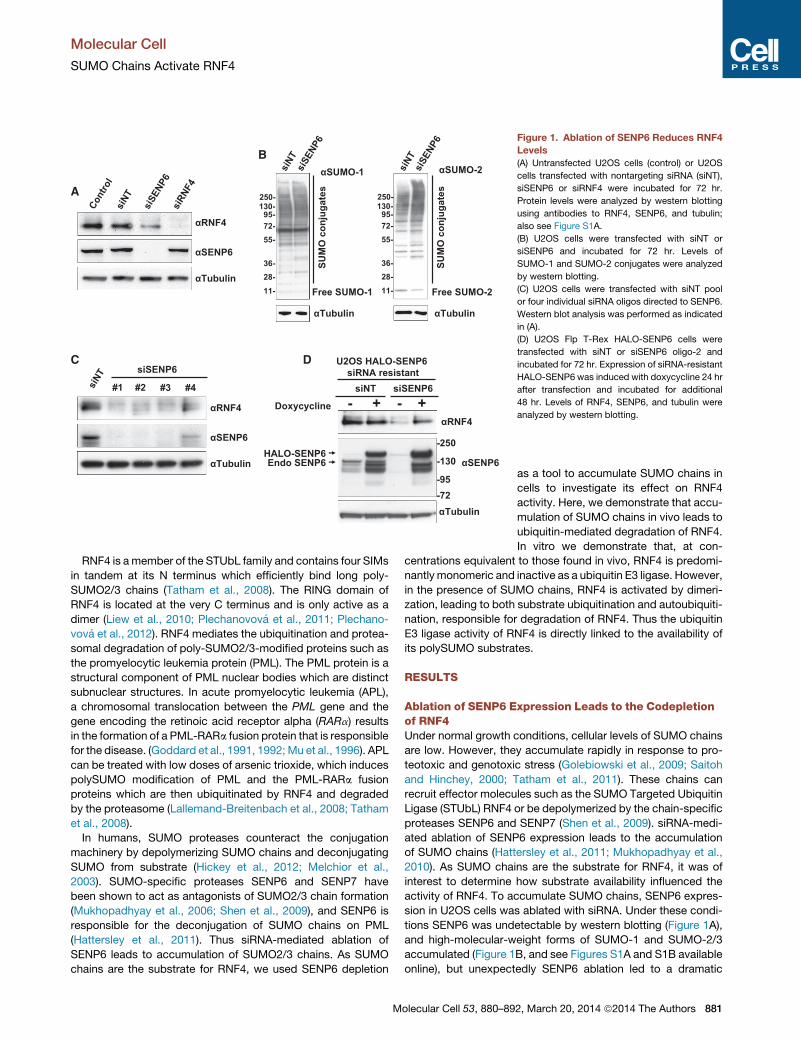
Figure 1. Ablation of SENP6 Reduces RNF4
Levels
(A) Untransfected U2OS cells (control) or U2OS
cells transfected with nontargeting siRNA (siNT),
siSENP6 or siRNF4 were incubated for 72 hr.
Protein levels were analyzed by western blotting
using antibodies to RNF4, SENP6, and tubulin;
also see Figure S1A.
(B) U2OS cells were transfected with siNT or
siSENP6 and incubated for 72 hr. Levels of
SUMO-1 and SUMO-2 conjugates were analyzed
by western blotting.
(C) U2OS cells were transfected with siNT pool
or four individual siRNA oligos directed to SENP6.
Western blot analysis was performed as indicated
in (A).
(D) U2OS Flp T-Rex HALO-SENP6 cells were
transfected with siNT or siSENP6 oligo-2 and
incubated for 72 hr. Expression of siRNA-resistant
HALO-SENP6 was induced with doxycycline 24 hr
after transfection and incubated for additional
48 hr. Levels of RNF4, SENP6, and tubulin were
analyzed by western blotting.
Molecular Cell
SUMO Chains Activate RNF4
RNF4 is amember of the STUbL family and contains four SIMs
in tandem at its N terminus which efficiently bind long poly-
SUMO2/3 chains (Tatham et al., 2008). The RING domain of
RNF4 is located at the very C terminus and is only active as a
dimer (Liew et al., 2010; Plechanovova et al., 2011; Plechano-
vova et al., 2012). RNF4 mediates the ubiquitination and protea-
somal degradation of poly-SUMO2/3-modified proteins such as
the promyelocytic leukemia protein (PML). The PML protein is a
structural component of PML nuclear bodies which are distinct
subnuclear structures. In acute promyelocytic leukemia (APL),
a chromosomal translocation between the PML gene and the
gene encoding the retinoic acid receptor alpha (RARa) results
in the formation of a PML-RARa fusion protein that is responsible
for the disease. (Goddard et al., 1991, 1992; Mu et al., 1996). APL
can be treated with low doses of arsenic trioxide, which induces
polySUMO modification of PML and the PML-RARa fusion
proteins which are then ubiquitinated by RNF4 and degraded
by the proteasome (Lallemand-Breitenbach et al., 2008; Tatham
et al., 2008).
In humans, SUMO proteases counteract the conjugation
machinery by depolymerizing SUMO chains and deconjugating
SUMO from substrate (Hickey et al., 2012; Melchior et al.,
2003). SUMO-specific proteases SENP6 and SENP7 have
been shown to act as antagonists of SUMO2/3 chain formation
(Mukhopadhyay et al., 2006; Shen et al., 2009), and SENP6 is
responsible for the deconjugation of SUMO chains on PML
(Hattersley et al., 2011). Thus siRNA-mediated ablation of
SENP6 leads to accumulation of SUMO2/3 chains. As SUMO
chains are the substrate for RNF4, we used SENP6 depletion
Molecular Cell 53, 880–89
as a tool to accumulate SUMO chains in
cells to investigate its effect on RNF4
activity. Here, we demonstrate that accu-
mulation of SUMO chains in vivo leads to
ubiquitin-mediated degradation of RNF4.
In vitro we demonstrate that, at con-
centrations equivalent to those found in vivo, RNF4 is predomi-
nantly monomeric and inactive as a ubiquitin E3 ligase. However,
in the presence of SUMO chains, RNF4 is activated by dimeri-
zation, leading to both substrate ubiquitination and autoubiquiti-
nation, responsible for degradation of RNF4. Thus the ubiquitin
E3 ligase activity of RNF4 is directly linked to the availability of
its polySUMO substrates.
RESULTS
Ablation of SENP6 Expression Leads to the Codepletionof RNF4Under normal growth conditions, cellular levels of SUMO chains
are low. However, they accumulate rapidly in response to pro-
teotoxic and genotoxic stress (Golebiowski et al., 2009; Saitoh
and Hinchey, 2000; Tatham et al., 2011). These chains can
recruit effector molecules such as the SUMO Targeted Ubiquitin
Ligase (STUbL) RNF4 or be depolymerized by the chain-specific
proteases SENP6 and SENP7 (Shen et al., 2009). siRNA-medi-
ated ablation of SENP6 expression leads to the accumulation
of SUMO chains (Hattersley et al., 2011; Mukhopadhyay et al.,
2010). As SUMO chains are the substrate for RNF4, it was of
interest to determine how substrate availability influenced the
activity of RNF4. To accumulate SUMO chains, SENP6 expres-
sion in U2OS cells was ablated with siRNA. Under these condi-
tions SENP6 was undetectable by western blotting (Figure 1A),
and high-molecular-weight forms of SUMO-1 and SUMO-2/3
accumulated (Figure 1B, and see Figures S1A and S1B available
online), but unexpectedly SENP6 ablation led to a dramatic
2, March 20, 2014 ª2014 The Authors 881

0
2
4
6
8
10
12
siNT siSENP6 siRNF4
Nuc
lear
Bod
ies
RNF4-YFP Nuclear Bodies
05
1015202530354045
siNT siSENP6 siRNF4
Inte
nsity
(A.U
)
RNF4-YFP Nuclear Intensity
A
B
C
ED
GF
H
RNF4-YFP PML MERGE
siN
Tsi
SEN
P6
siNT siSENP6 siRNF4
YFP-
SUM
O-2
siNT siSENP6 siRNF4
YFP-
SUM
O-2
siN
Tsi
SEN
P6
RNF4-YFP SUMO-2 MERGE
SENP6 knockdown (24-60h)
YFP-
SUM
O-2
SENP6 knockdown (48-72)
RN
F4-Y
FPYFP-SUMO-2 Nuclear Bodies
0123456789
10
siNT siSENP6 siRNF4
Nuc
lear
Bod
ies
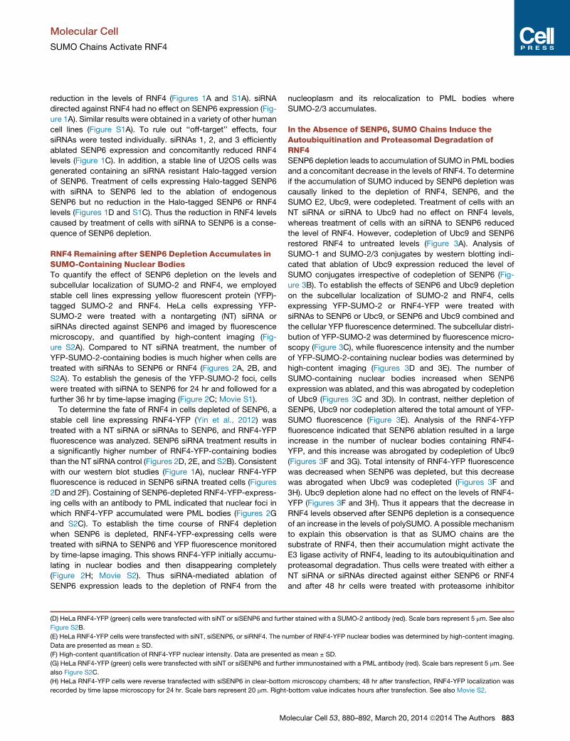
Figure 2. RNF4 Is Relocalized to SUMO-Containing Nuclear Bodies in Response to SENP6 Depletion
(A) HeLa YFP-SUMO-2 cells were treated with siNT, siSENP6, or siRNF4. SUMO localization was determined using structured illumination microscopy. Scale
bars represent 5 mm. Dashed line indicates location of the nucleus visualized by DAPI staining.
(B) HeLa YFP-SUMO-2 cells were transfected in clear-bottom 96-well plates with siNT, siSENP6, or RNF4. Number of YFP-SUMO-2 nuclear bodies was
determined by high-content imaging. Data are presented as mean ± SD.
(C) HeLa YFP-SUMO-2 cells were transfected with siSENP6 pool. Twenty-four hours after transfection, YFP-SUMO-2 localization was recorded by time-lapse
microscopy for 36 hr. Scale bars represent 20 mm. Right-bottom value indicates hours after transfection. See also Movie S1.
(legend continued on next page)
Molecular Cell
SUMO Chains Activate RNF4
882 Molecular Cell 53, 880–892, March 20, 2014 ª2014 The Authors
Molecular Cell
SUMO Chains Activate RNF4
reduction in the levels of RNF4 (Figures 1A and S1A). siRNA
directed against RNF4 had no effect on SENP6 expression (Fig-
ure 1A). Similar results were obtained in a variety of other human
cell lines (Figure S1A). To rule out ‘‘off-target’’ effects, four
siRNAs were tested individually. siRNAs 1, 2, and 3 efficiently
ablated SENP6 expression and concomitantly reduced RNF4
levels (Figure 1C). In addition, a stable line of U2OS cells was
generated containing an siRNA resistant Halo-tagged version
of SENP6. Treatment of cells expressing Halo-tagged SENP6
with siRNA to SENP6 led to the ablation of endogenous
SENP6 but no reduction in the Halo-tagged SENP6 or RNF4
levels (Figures 1D and S1C). Thus the reduction in RNF4 levels
caused by treatment of cells with siRNA to SENP6 is a conse-
quence of SENP6 depletion.
RNF4 Remaining after SENP6 Depletion Accumulates inSUMO-Containing Nuclear BodiesTo quantify the effect of SENP6 depletion on the levels and
subcellular localization of SUMO-2 and RNF4, we employed
stable cell lines expressing yellow fluorescent protein (YFP)-
tagged SUMO-2 and RNF4. HeLa cells expressing YFP-
SUMO-2 were treated with a nontargeting (NT) siRNA or
siRNAs directed against SENP6 and imaged by fluorescence
microscopy, and quantified by high-content imaging (Fig-
ure S2A). Compared to NT siRNA treatment, the number of
YFP-SUMO-2-containing bodies is much higher when cells are
treated with siRNAs to SENP6 or RNF4 (Figures 2A, 2B, and
S2A). To establish the genesis of the YFP-SUMO-2 foci, cells
were treated with siRNA to SENP6 for 24 hr and followed for a
further 36 hr by time-lapse imaging (Figure 2C; Movie S1).
To determine the fate of RNF4 in cells depleted of SENP6, a
stable cell line expressing RNF4-YFP (Yin et al., 2012) was
treated with a NT siRNA or siRNAs to SENP6, and RNF4-YFP
fluorescence was analyzed. SENP6 siRNA treatment results in
a significantly higher number of RNF4-YFP-containing bodies
than the NT siRNA control (Figures 2D, 2E, and S2B). Consistent
with our western blot studies (Figure 1A), nuclear RNF4-YFP
fluorescence is reduced in SENP6 siRNA treated cells (Figures
2D and 2F). Costaining of SENP6-depleted RNF4-YFP-express-
ing cells with an antibody to PML indicated that nuclear foci in
which RNF4-YFP accumulated were PML bodies (Figures 2G
and S2C). To establish the time course of RNF4 depletion
when SENP6 is depleted, RNF4-YFP-expressing cells were
treated with siRNA to SENP6 and YFP fluorescence monitored
by time-lapse imaging. This shows RNF4-YFP initially accumu-
lating in nuclear bodies and then disappearing completely
(Figure 2H; Movie S2). Thus siRNA-mediated ablation of
SENP6 expression leads to the depletion of RNF4 from the
(D) HeLa RNF4-YFP (green) cells were transfected with siNT or siSENP6 and furth
Figure S2B.
(E) HeLa RNF4-YFP cells were transfected with siNT, siSENP6, or siRNF4. The n
Data are presented as mean ± SD.
(F) High-content quantification of RNF4-YFP nuclear intensity. Data are presente
(G) HeLa RNF4-YFP (green) cells were transfected with siNT or siSENP6 and furt
also Figure S2C.
(H) HeLa RNF4-YFP cells were reverse transfected with siSENP6 in clear-bottom
recorded by time lapse microscopy for 24 hr. Scale bars represent 20 mm. Right
M
nucleoplasm and its relocalization to PML bodies where
SUMO-2/3 accumulates.
In the Absence of SENP6, SUMO Chains Induce theAutoubiquitination and Proteasomal Degradation ofRNF4SENP6 depletion leads to accumulation of SUMO in PML bodies
and a concomitant decrease in the levels of RNF4. To determine
if the accumulation of SUMO induced by SENP6 depletion was
causally linked to the depletion of RNF4, SENP6, and the
SUMO E2, Ubc9, were codepleted. Treatment of cells with an
NT siRNA or siRNA to Ubc9 had no effect on RNF4 levels,
whereas treatment of cells with an siRNA to SENP6 reduced
the level of RNF4. However, codepletion of Ubc9 and SENP6
restored RNF4 to untreated levels (Figure 3A). Analysis of
SUMO-1 and SUMO-2/3 conjugates by western blotting indi-
cated that ablation of Ubc9 expression reduced the level of
SUMO conjugates irrespective of codepletion of SENP6 (Fig-
ure 3B). To establish the effects of SENP6 and Ubc9 depletion
on the subcellular localization of SUMO-2 and RNF4, cells
expressing YFP-SUMO-2 or RNF4-YFP were treated with
siRNAs to SENP6 or Ubc9, or SENP6 and Ubc9 combined and
the cellular YFP fluorescence determined. The subcellular distri-
bution of YFP-SUMO-2 was determined by fluorescence micro-
scopy (Figure 3C), while fluorescence intensity and the number
of YFP-SUMO-2-containing nuclear bodies was determined by
high-content imaging (Figures 3D and 3E). The number of
SUMO-containing nuclear bodies increased when SENP6
expression was ablated, and this was abrogated by codepletion
of Ubc9 (Figures 3C and 3D). In contrast, neither depletion of
SENP6, Ubc9 nor codepletion altered the total amount of YFP-
SUMO fluorescence (Figure 3E). Analysis of the RNF4-YFP
fluorescence indicated that SENP6 ablation resulted in a large
increase in the number of nuclear bodies containing RNF4-
YFP, and this increase was abrogated by codepletion of Ubc9
(Figures 3F and 3G). Total intensity of RNF4-YFP fluorescence
was decreased when SENP6 was depleted, but this decrease
was abrogated when Ubc9 was codepleted (Figures 3F and
3H). Ubc9 depletion alone had no effect on the levels of RNF4-
YFP (Figures 3F and 3H). Thus it appears that the decrease in
RNF4 levels observed after SENP6 depletion is a consequence
of an increase in the levels of polySUMO. A possible mechanism
to explain this observation is that as SUMO chains are the
substrate of RNF4, then their accumulation might activate the
E3 ligase activity of RNF4, leading to its autoubiquitination and
proteasomal degradation. Thus cells were treated with either a
NT siRNA or siRNAs directed against either SENP6 or RNF4
and after 48 hr cells were treated with proteasome inhibitor
er stained with a SUMO-2 antibody (red). Scale bars represent 5 mm. See also
umber of RNF4-YFP nuclear bodies was determined by high-content imaging.
d as mean ± SD.
her immunostained with a PML antibody (red). Scale bars represent 5 mm. See
microscopy chambers; 48 hr after transfection, RNF4-YFP localization was
-bottom value indicates hours after transfection. See also Movie S2.
olecular Cell 53, 880–892, March 20, 2014 ª2014 The Authors 883
A B
C
D E
GF H
JI K
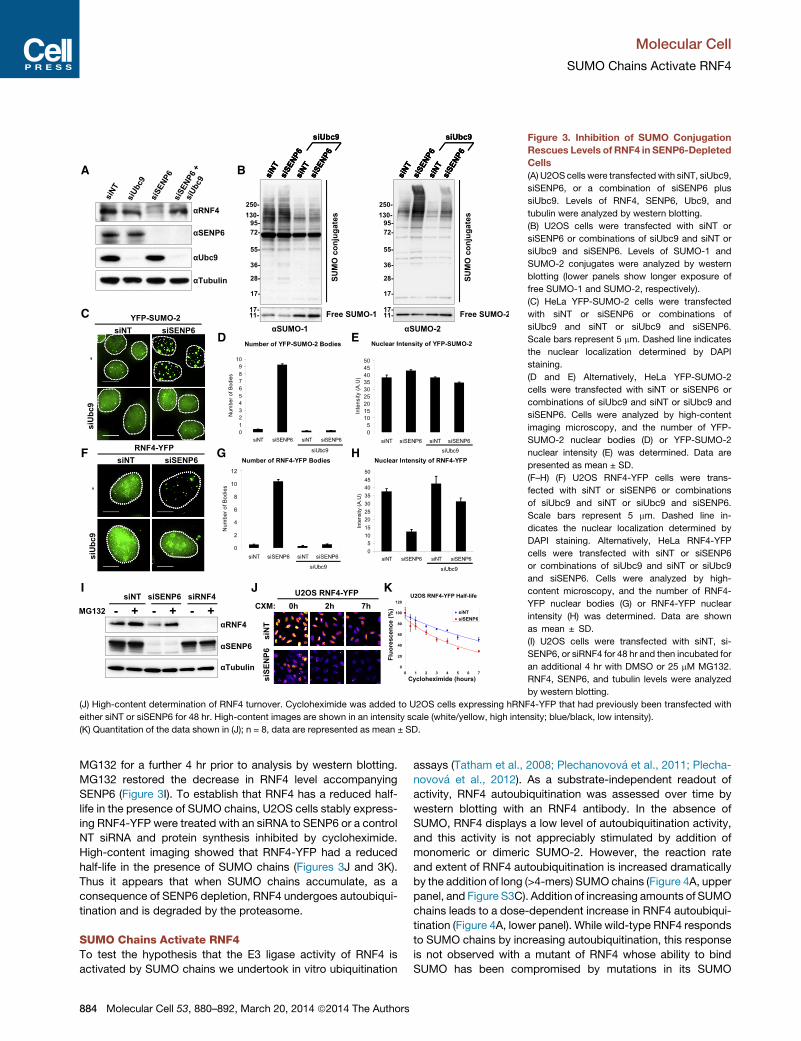
Figure 3. Inhibition of SUMO Conjugation
Rescues Levels of RNF4 in SENP6-Depleted
Cells
(A) U2OS cells were transfectedwith siNT, siUbc9,
siSENP6, or a combination of siSENP6 plus
siUbc9. Levels of RNF4, SENP6, Ubc9, and
tubulin were analyzed by western blotting.
(B) U2OS cells were transfected with siNT or
siSENP6 or combinations of siUbc9 and siNT or
siUbc9 and siSENP6. Levels of SUMO-1 and
SUMO-2 conjugates were analyzed by western
blotting (lower panels show longer exposure of
free SUMO-1 and SUMO-2, respectively).
(C) HeLa YFP-SUMO-2 cells were transfected
with siNT or siSENP6 or combinations of
siUbc9 and siNT or siUbc9 and siSENP6.
Scale bars represent 5 mm. Dashed line indicates
the nuclear localization determined by DAPI
staining.
(D and E) Alternatively, HeLa YFP-SUMO-2
cells were transfected with siNT or siSENP6 or
combinations of siUbc9 and siNT or siUbc9 and
siSENP6. Cells were analyzed by high-content
imaging microscopy, and the number of YFP-
SUMO-2 nuclear bodies (D) or YFP-SUMO-2
nuclear intensity (E) was determined. Data are
presented as mean ± SD.
(F–H) (F) U2OS RNF4-YFP cells were trans-
fected with siNT or siSENP6 or combinations
of siUbc9 and siNT or siUbc9 and siSENP6.
Scale bars represent 5 mm. Dashed line in-
dicates the nuclear localization determined by
DAPI staining. Alternatively, HeLa RNF4-YFP
cells were transfected with siNT or siSENP6
or combinations of siUbc9 and siNT or siUbc9
and siSENP6. Cells were analyzed by high-
content microscopy, and the number of RNF4-
YFP nuclear bodies (G) or RNF4-YFP nuclear
intensity (H) was determined. Data are shown
as mean ± SD.
(I) U2OS cells were transfected with siNT, si-
SENP6, or siRNF4 for 48 hr and then incubated for
an additional 4 hr with DMSO or 25 mM MG132.
RNF4, SENP6, and tubulin levels were analyzed
by western blotting.
(J) High-content determination of RNF4 turnover. Cycloheximide was added to U2OS cells expressing hRNF4-YFP that had previously been transfected with
either siNT or siSENP6 for 48 hr. High-content images are shown in an intensity scale (white/yellow, high intensity; blue/black, low intensity).
(K) Quantitation of the data shown in (J); n = 8, data are represented as mean ± SD.
Molecular Cell
SUMO Chains Activate RNF4
MG132 for a further 4 hr prior to analysis by western blotting.
MG132 restored the decrease in RNF4 level accompanying
SENP6 (Figure 3I). To establish that RNF4 has a reduced half-
life in the presence of SUMO chains, U2OS cells stably express-
ing RNF4-YFP were treated with an siRNA to SENP6 or a control
NT siRNA and protein synthesis inhibited by cycloheximide.
High-content imaging showed that RNF4-YFP had a reduced
half-life in the presence of SUMO chains (Figures 3J and 3K).
Thus it appears that when SUMO chains accumulate, as a
consequence of SENP6 depletion, RNF4 undergoes autoubiqui-
tination and is degraded by the proteasome.
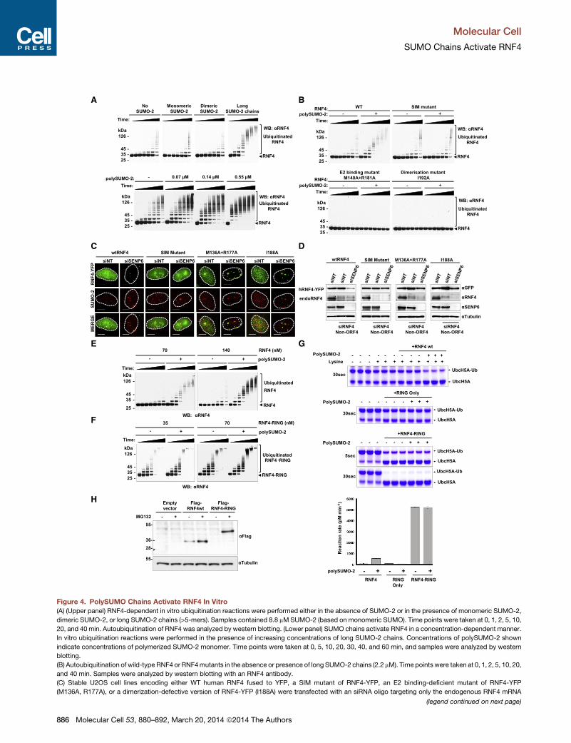
SUMO Chains Activate RNF4To test the hypothesis that the E3 ligase activity of RNF4 is
activated by SUMO chains we undertook in vitro ubiquitination
884 Molecular Cell 53, 880–892, March 20, 2014 ª2014 The Authors
assays (Tatham et al., 2008; Plechanovova et al., 2011; Plecha-
novova et al., 2012). As a substrate-independent readout of
activity, RNF4 autoubiquitination was assessed over time by
western blotting with an RNF4 antibody. In the absence of
SUMO, RNF4 displays a low level of autoubiquitination activity,
and this activity is not appreciably stimulated by addition of
monomeric or dimeric SUMO-2. However, the reaction rate
and extent of RNF4 autoubiquitination is increased dramatically
by the addition of long (>4-mers) SUMO chains (Figure 4A, upper
panel, and Figure S3C). Addition of increasing amounts of SUMO
chains leads to a dose-dependent increase in RNF4 autoubiqui-
tination (Figure 4A, lower panel). While wild-type RNF4 responds
to SUMO chains by increasing autoubiquitination, this response
is not observed with a mutant of RNF4 whose ability to bind
SUMO has been compromised by mutations in its SUMO

Molecular Cell
SUMO Chains Activate RNF4
interaction motifs (SIMs). Rather, the SIM mutant displays the
same low level autoubiquitination in the presence and absence
of SUMO chains as that observed for wild-type RNF4 in the
absence of SUMO chains (Figure 4B, upper panel). Mutants of
RNF4 deficient in E2 binding or dimerization show no autoubiqui-
tination (Figure 4B, lower panel). Thus RNF4 autoubiquitination is
activated by its SIM-directed interaction with SUMO chains and
requires both E2 binding and RING-mediated dimerization.
An alternative RNF4 functional assay is K63-linked ubiquitin
chain synthesis (Tatham et al., 2013). Uev2/Ubc13 generates
unanchored K63 chains in the absence of RNF4, and this activity
is not influenced by the presence of SUMO chains (Figure S3E,
left panel). Addition of RNF4 modestly increased K63 chain
synthesis, but in the presence of SUMO chains long K63 poly-
mers accumulate and free ubiquitin is depleted (Figure S3E, right
panel). Thus SUMO chains stimulate not just RNF4 autoubiquiti-
nation but also the formation of unanchored K63 chains medi-
ated by Uev2/Ubc13.
To establish our findings in vivo, we generated four stable
U2OS cell lines encoding wild-type or various mutants of RNF4
fused to YFP: SIM mutant, an E2 binding-deficient mutant
(M136A,R177A), or a dimerization defective form (I188A) (Figures
4C, S3A, and S3B). To eliminate endogenous RNF4, an siRNA
targeting the non-ORF 50 UTR of RNF4 (siRNF4 non-ORF4) was
used that did not affect expression of RNF4-YFP. Stable cell lines
expressing RNF4-YFP WT and mutants were transfected with
combinations of siRNF4 non-ORF4, NT siRNA, or siRNA directed
against SENP6 and cells stained with a SUMO-2 antibody. While
WT RNF4-YFP was degraded, none of the RNF4 mutants was
degraded in response to SENP6 depletion. The degradation-
resistant E2 binding (M136A, R177A) and dimerization (I188A)
mutants formed very large foci associated with SUMO-2 conju-
gates, while the SIM mutant of RNF4 was not recruited to
SUMO foci (Figures 4C and S3D). western blotting confirmed
that ablation of SENP6 expression resulted in depletion of WT
RNF4-YFP; none of theRNF4mutantswas degraded (Figure 4D).
These data indicate that activation of RNF4 required its SIM
dependent binding to SUMO chains and the ubiquitin ligase
activity of its RING domain in vitro and in cells.
PolySUMO Chains Induce RNF4 DimerizationTo test the hypothesis that SUMOchains induced dimerization of
RNF4, we compared autoubiquitination activity of wild-type
RNF4 (Figure 4E) with a version in which full-length RNF4 was
fused with the RNF4 RING domain in a single polypeptide
(RNF4-RING) (Figure 4F), thus enforcing the dimerization of
the two fused RING domains of RNF4. This form of RNF4 was
previously shown to be fully active in the ubiquitination of
SUMO chains (Plechanovova et al., 2011). At the same concen-
tration of RING domains and in the absence of SUMO chains,
RNF4-RING was substantially more active than wild-type
RNF4 (Figures 4E and 4F). However, in the presence of SUMO
chains the activity of wild-type RNF4 was dramatically stimu-
lated (Figure 4E), whereas little change was observed in the
activity of RNF4-RING (Figure 4F). The RNF4-RING fusion in
the absence of SUMO-2 chains displayed a comparable level
of activity to wild-type RNF4 in the presence of SUMO chains
(Figures 4E and 4F). To quantify the effect of SUMO chains on
M
the E3 ligase activity of RNF4, the RNF4-RING fusion and the
RING alone, we employed a lysine discharge assay which
measures the ability of an E3 ligase to stimulate the transfer of
ubiquitin from a ubiquitin-loaded E2 to free lysine. In the absence
of SUMO chains WT RNF4 and the RNF4 RING domain only had
a similar low level of lysine discharge activity, whereas the RNF4-
RING had a 60-fold higher level of activity. Addition of SUMO
chains to RNF4 increased the initial rate of lysine discharge by
6.5-fold, but SUMO chains did not alter the activity of the RING
alone or the RNF4-RING fusion (Figure 4G). Consistent with
these in vitro observations, ectopic expression of RNF4 could
be detected by western blotting, whereas an RNF4-RING fusion
was almost undetectable under similar conditions. While treat-
ment of cells with proteasome inhibitor MG132 increased
RNF4 levels, it had a dramatic stabilizing effect on the RNF4-
RING fusion (Figure 4H), suggesting that the RNF4-RING fusion
has a faster turnover than RNF4 in cells.
To directly demonstrate that SUMO-2/3 chains induced RNF4
dimerization, we engineered a cysteine residue into the C termi-
nus of RNF4 that, based on structural predictions, could form a
disulphide bond upon RNF4 dimerization. When increasing
concentrations of RNF4 C196 were incubated in the absence
of reducing agent and the products analyzed by western blotting
with an RNF4 antibody, a species accumulated that, based on its
molecular mass and its sensitivity to DTT, was consistent with
the formation of a dimer (Figure 5A). We therefore incubated
RNF4 C196 in the presence of either monomeric SUMO-2 or
polySUMO-2/3 and assessed dimer formation as described
above. At the RNF4 C196 concentration chosen, very little disul-
phide linkedmaterial is detected in the presence of monoSUMO.
However, in the presence of SUMO chains, dimers of RNF4 are
rapidly formed (Figure 5B). These data indicate that SUMO
chains activate the catalytic activity of RNF4 by inducing its
dimerization.
To obtain functional evidence that SUMO chains activate
RNF4 by induced dimerization of the RING domains, we
employed a complementation strategy where two mutants of
RNF4 that are each inactive as homodimers can display activity
only if they can form heterodimers. Y193H RNF4 is inactive as it
fails to interact with the ubiquitin component of the ubiquitin-
loaded E2, whereas M140A+R181A RNF4 is inactive as it cannot
bind the E2. If a heterodimer forms, the E2 component of the
E2�ubiquitin thioester binds to the Y193H subunit, while the
ubiquitin component of the E2�ubiquitin thioester engages
Tyr193 of the M140A+R181A subunit, recreating a functional
E3 ligase (Plechanovova et al., 2011, 2012). We therefore tested
RNF4 autoubiquitination and lysine discharge activities of Y193H
RNF4, M140A+R181A RNF4, or a mix of the two in the presence
and absence of SUMO chains. In the absence of SUMO chains,
neither Y193H, M140A+R181A, nor the mix of the two displays
activity in either of the assays. In the presence of SUMO chains,
M140A+R181A is inactive, Y193H has low activity, but the mix of
the twomutants has robust E3 ligase activity in both assays (Fig-
ures 5C–5E). A similar assay in vivo also indicated that intramo-
lecular complementation between RNF4 mutants was evident
when SUMO chains were increased by ablation of SENP6
expression (Figure 5F). Thus SUMO chains induce the formation
of functional dimers of RNF4.
olecular Cell 53, 880–892, March 20, 2014 ª2014 The Authors 885
A B
C D
E
F
H
G
Figure 4. PolySUMO Chains Activate RNF4 In Vitro
(A) (Upper panel) RNF4-dependent in vitro ubiquitination reactions were performed either in the absence of SUMO-2 or in the presence of monomeric SUMO-2,
dimeric SUMO-2, or long SUMO-2 chains (>5-mers). Samples contained 8.8 mMSUMO-2 (based on monomeric SUMO). Time points were taken at 0, 1, 2, 5, 10,
20, and 40 min. Autoubiquitination of RNF4 was analyzed by western blotting. (Lower panel) SUMO chains activate RNF4 in a concentration-dependent manner.
In vitro ubiquitination reactions were performed in the presence of increasing concentrations of long SUMO-2 chains. Concentrations of polySUMO-2 shown
indicate concentrations of polymerized SUMO-2 monomer. Time points were taken at 0, 5, 10, 20, 30, 40, and 60 min, and samples were analyzed by western
blotting.
(B) Autoubiquitination of wild-type RNF4 or RNF4mutants in the absence or presence of long SUMO-2 chains (2.2 mM). Time points were taken at 0, 1, 2, 5, 10, 20,
and 40 min. Samples were analyzed by western blotting with an RNF4 antibody.
(C) Stable U2OS cell lines encoding either WT human RNF4 fused to YFP, a SIM mutant of RNF4-YFP, an E2 binding-deficient mutant of RNF4-YFP
(M136A, R177A), or a dimerization-defective version of RNF4-YFP (I188A) were transfected with an siRNA oligo targeting only the endogenous RNF4 mRNA
(legend continued on next page)
Molecular Cell
SUMO Chains Activate RNF4
886 Molecular Cell 53, 880–892, March 20, 2014 ª2014 The Authors

A
C
D
E F
B Figure 5. PolySUMO-2 Chains Complement
RNF4 Inactive Mutants by Inducing a Func-
tional RING Dimerization
(A) Mutant RNF4 C55S, C59S was tagged with
glycine-cysteine at its C terminus (RNF4-C196).
Increasing concentrations of the bacterial purified
protein (0.062, 0.12, 0.25, 0.5, and 1 mM) were
incubated at room temperature for 2 hr in the
absence of reducing agents. Nonreducing SDS-
PAGE was used for detection of disulfide dimer
formation (upper panel), and DTT-reduced SDS-
PAGE was used to disrupt the disulfide bonds
(lower panel). RNF4 was detected by western
blotting.
(B) A total of 0.1 mM RNF4-C196 was incubated
in absence of SUMO as control or with increasing
amounts of monomeric SUMO-2 or polySUMO-2
(0.2, 0.4, 1, 2 mM). Disulfide dimer formation
was analyzed by nonreducing (upper panel) or
reducing (lower panel) SDS-PAGE and western
blotting.
(C) Autoubiquitination assay of Y193H RNF4,
M140A+R181A RNF4 and a mix of the two.
Reactions were terminated after 0, 5, 10, 20, 30,
or 40 min and analyzed by western blotting.
(D) Lysine discharge assay of Y193H
RNF4, M140A+R181A RNF4, and a mix of the
two. Reactions (in triplicate) were terminated
after 30 s and analyzed by Coomassie blue
staining.
(E) Quantitation of lysine discharge assays was
as indicated in Figure 4G. Data are presented as
mean ± SD.
(F) U2OS cells transfected with non-ORF siRNF4
plus siNT or siSENP6 siRNA were transiently
transfected with Flag-hRNF4 Y189H (2 mg) and
increasing amounts of hRNF4-YFPM136A, R177A
(0.5, 1, and 2 mg) in 12-well plates. Additionally,
50 ng GFP was cotransfected as control. Protein
levels were detected by western blotting using
Flag, GFP, and SENP6 antibodies.
Molecular Cell
SUMO Chains Activate RNF4
Quantitative Analysis of RNF4 DimerizationTo quantitatively study the effect of SUMO-2 chains on the
dimerization of RNF4, an in vitro Forster resonance energy trans-
fer (FRET) assay was devised. Recombinant forms of RNF4 were
(siRNF4 Non-ORF4). After 24 hr incubation, cells were transfected again either wit
YFP WT and mutants (green) was analyzed together with immunostaining of end
localization of the nucleus determined by DAPI staining. See also Figure S3D. D
(D) Western blot analysis of cells treated as in (C), levels of RNF4-YFP, endogen
(E) In vitro ubiquitination reactions with the indicated concentrations of wild-type
chains (2.2 mM). Time points were taken at 0, 1, 2, 5, 10, 20, and 40 min. Reaction
same concentration of RNF4 and analyzed by western blotting.
(F) In vitro ubiquitination reactions with the indicated concentrations of RNF4-R
SUMO-2 chains (2.2 mM). Time points were taken at 0, 0.5, 1, 2, 5, 10, and 20 m
(G) Lysine discharge assays. UbcH5A loaded with ubiquitin was mixed with SUMO
fusion. Reactions were terminated after 30 s (or 5 s for the RNF4-RING fusion), fra
rates of reactions were determined (shown as mean ± SD of triplicate reactions)
(H) HeLa cells were stable transfected with either pFires-Puro empty vector, Flag-
tagged proteins were detected by western blot using an anti-Flag antibody. Tub
M
expressed with N-terminal fusions of ECFP or YFP. When in
close proximity, emission from ECFP can transfer between the
fluorophores, causing emission from YFP (Martin et al., 2008).
This assay was used to monitor polySUMO-2 effects upon
h siNT or siSENP6 and incubated for an additional 72 hr. Localization of RNF4-
ogenous SUMO-2 (red). Scale bars represent 20 mm. Dashed line shows the
ata are presented as mean ± SD.
ous RNF4, SENP6, and tubulin were determined with specific antibodies.
RNF4 were performed either in the absence or the presence of long SUMO-2
s were stopped by SDS-PAGE sample buffer, and samples were diluted to the
ING linear fusion were performed either in the absence or presence of long
in.
chains, lysine, and either RNF4, the RING domain of RNF4, or an RNF4-RING
ctionated by nonreducing SDS PAGE, and stained with Coomassie blue. Initial
after staining with Sypro Ruby.
hRNF4, or a Flag-RNF4-RING fusion. Cells were treated with MG132, and Flag-
ulin levels were used as a loading control.
olecular Cell 53, 880–892, March 20, 2014 ª2014 The Authors 887
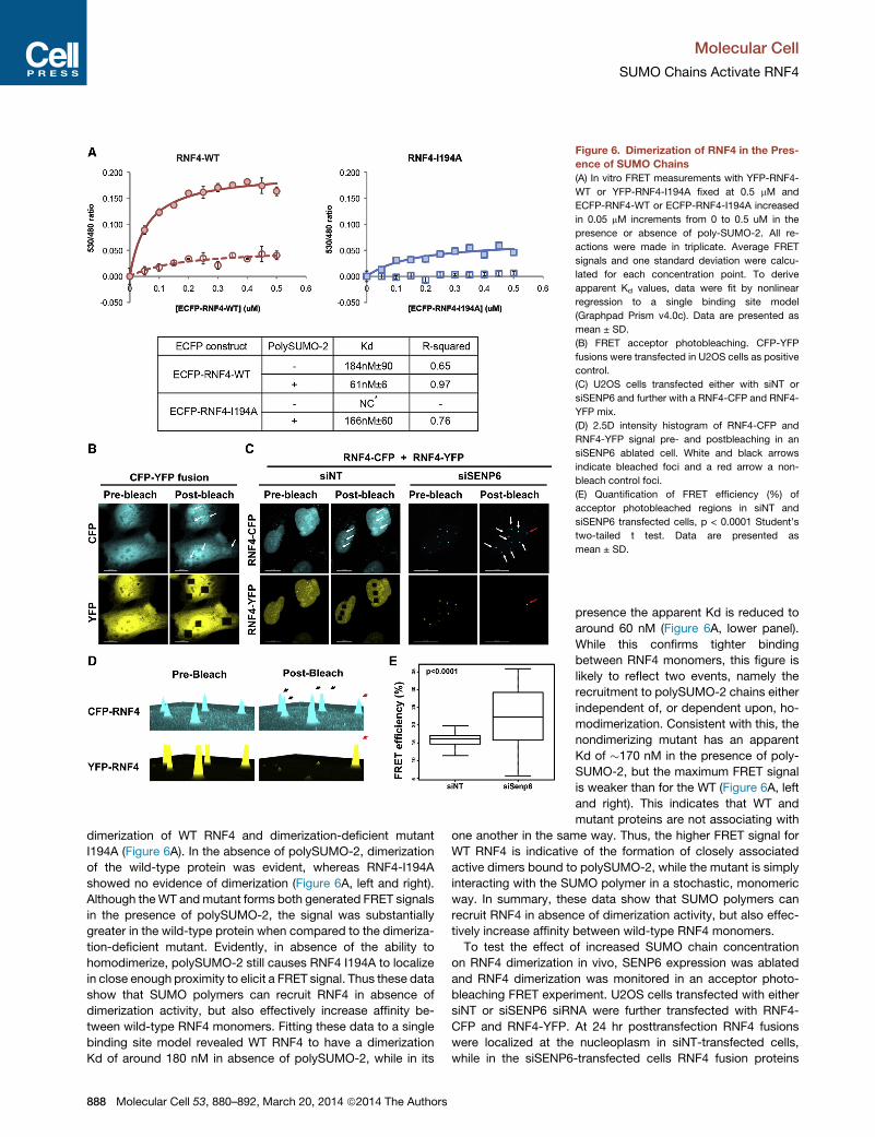
Figure 6. Dimerization of RNF4 in the Pres-
ence of SUMO Chains
(A) In vitro FRET measurements with YFP-RNF4-
WT or YFP-RNF4-I194A fixed at 0.5 mM and
ECFP-RNF4-WT or ECFP-RNF4-I194A increased
in 0.05 mM increments from 0 to 0.5 uM in the
presence or absence of poly-SUMO-2. All re-
actions were made in triplicate. Average FRET
signals and one standard deviation were calcu-
lated for each concentration point. To derive
apparent Kd values, data were fit by nonlinear
regression to a single binding site model
(Graphpad Prism v4.0c). Data are presented as
mean ± SD.
(B) FRET acceptor photobleaching. CFP-YFP
fusions were transfected in U2OS cells as positive
control.
(C) U2OS cells transfected either with siNT or
siSENP6 and further with a RNF4-CFP and RNF4-
YFP mix.
(D) 2.5D intensity histogram of RNF4-CFP and
RNF4-YFP signal pre- and postbleaching in an
siSENP6 ablated cell. White and black arrows
indicate bleached foci and a red arrow a non-
bleach control foci.
(E) Quantification of FRET efficiency (%) of
acceptor photobleached regions in siNT and
siSENP6 transfected cells, p < 0.0001 Student’s
two-tailed t test. Data are presented as
mean ± SD.
Molecular Cell
SUMO Chains Activate RNF4
dimerization of WT RNF4 and dimerization-deficient mutant
I194A (Figure 6A). In the absence of polySUMO-2, dimerization
of the wild-type protein was evident, whereas RNF4-I194A
showed no evidence of dimerization (Figure 6A, left and right).
Although theWT andmutant forms both generated FRET signals
in the presence of polySUMO-2, the signal was substantially
greater in the wild-type protein when compared to the dimeriza-
tion-deficient mutant. Evidently, in absence of the ability to
homodimerize, polySUMO-2 still causes RNF4 I194A to localize
in close enough proximity to elicit a FRET signal. Thus these data
show that SUMO polymers can recruit RNF4 in absence of
dimerization activity, but also effectively increase affinity be-
tween wild-type RNF4 monomers. Fitting these data to a single
binding site model revealed WT RNF4 to have a dimerization
Kd of around 180 nM in absence of polySUMO-2, while in its
888 Molecular Cell 53, 880–892, March 20, 2014 ª2014 The Authors
presence the apparent Kd is reduced to
around 60 nM (Figure 6A, lower panel).
While this confirms tighter binding
between RNF4 monomers, this figure is
likely to reflect two events, namely the
recruitment to polySUMO-2 chains either
independent of, or dependent upon, ho-
modimerization. Consistent with this, the
nondimerizing mutant has an apparent
Kd of �170 nM in the presence of poly-
SUMO-2, but the maximum FRET signal
is weaker than for the WT (Figure 6A, left
and right). This indicates that WT and
mutant proteins are not associating with
one another in the same way. Thus, the higher FRET signal for
WT RNF4 is indicative of the formation of closely associated
active dimers bound to polySUMO-2, while the mutant is simply
interacting with the SUMO polymer in a stochastic, monomeric
way. In summary, these data show that SUMO polymers can
recruit RNF4 in absence of dimerization activity, but also effec-
tively increase affinity between wild-type RNF4 monomers.
To test the effect of increased SUMO chain concentration
on RNF4 dimerization in vivo, SENP6 expression was ablated
and RNF4 dimerization was monitored in an acceptor photo-
bleaching FRET experiment. U2OS cells transfected with either
siNT or siSENP6 siRNA were further transfected with RNF4-
CFP and RNF4-YFP. At 24 hr posttransfection RNF4 fusions
were localized at the nucleoplasm in siNT-transfected cells,
while in the siSENP6-transfected cells RNF4 fusion proteins
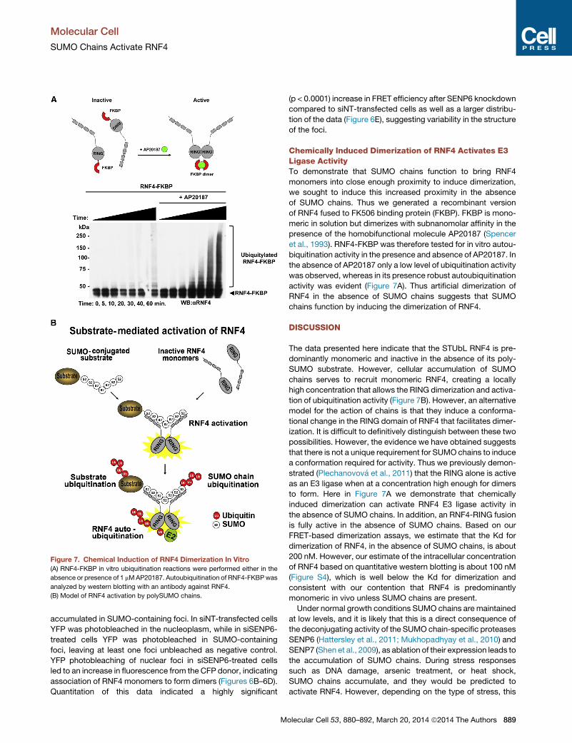
Figure 7. Chemical Induction of RNF4 Dimerization In Vitro
(A) RNF4-FKBP in vitro ubiquitination reactions were performed either in the
absence or presence of 1 mMAP20187. Autoubiquitination of RNF4-FKBPwas
analyzed by western blotting with an antibody against RNF4.
(B) Model of RNF4 activation by polySUMO chains.
Molecular Cell
SUMO Chains Activate RNF4
accumulated in SUMO-containing foci. In siNT-transfected cells
YFP was photobleached in the nucleoplasm, while in siSENP6-
treated cells YFP was photobleached in SUMO-containing
foci, leaving at least one foci unbleached as negative control.
YFP photobleaching of nuclear foci in siSENP6-treated cells
led to an increase in fluorescence from the CFP donor, indicating
association of RNF4 monomers to form dimers (Figures 6B–6D).
Quantitation of this data indicated a highly significant
M
(p < 0.0001) increase in FRET efficiency after SENP6 knockdown
compared to siNT-transfected cells as well as a larger distribu-
tion of the data (Figure 6E), suggesting variability in the structure
of the foci.
Chemically Induced Dimerization of RNF4 Activates E3Ligase ActivityTo demonstrate that SUMO chains function to bring RNF4
monomers into close enough proximity to induce dimerization,
we sought to induce this increased proximity in the absence
of SUMO chains. Thus we generated a recombinant version
of RNF4 fused to FK506 binding protein (FKBP). FKBP is mono-
meric in solution but dimerizes with subnanomolar affinity in the
presence of the homobifunctional molecule AP20187 (Spencer
et al., 1993). RNF4-FKBP was therefore tested for in vitro autou-
biquitination activity in the presence and absence of AP20187. In
the absence of AP20187 only a low level of ubiquitination activity
was observed, whereas in its presence robust autoubiquitination
activity was evident (Figure 7A). Thus artificial dimerization of
RNF4 in the absence of SUMO chains suggests that SUMO
chains function by inducing the dimerization of RNF4.
DISCUSSION
The data presented here indicate that the STUbL RNF4 is pre-
dominantly monomeric and inactive in the absence of its poly-
SUMO substrate. However, cellular accumulation of SUMO
chains serves to recruit monomeric RNF4, creating a locally
high concentration that allows the RING dimerization and activa-
tion of ubiquitination activity (Figure 7B). However, an alternative
model for the action of chains is that they induce a conforma-
tional change in the RING domain of RNF4 that facilitates dimer-
ization. It is difficult to definitively distinguish between these two
possibilities. However, the evidence we have obtained suggests
that there is not a unique requirement for SUMO chains to induce
a conformation required for activity. Thus we previously demon-
strated (Plechanovova et al., 2011) that the RING alone is active
as an E3 ligase when at a concentration high enough for dimers
to form. Here in Figure 7A we demonstrate that chemically
induced dimerization can activate RNF4 E3 ligase activity in
the absence of SUMO chains. In addition, an RNF4-RING fusion
is fully active in the absence of SUMO chains. Based on our
FRET-based dimerization assays, we estimate that the Kd for
dimerization of RNF4, in the absence of SUMO chains, is about
200 nM. However, our estimate of the intracellular concentration
of RNF4 based on quantitative western blotting is about 100 nM
(Figure S4), which is well below the Kd for dimerization and
consistent with our contention that RNF4 is predominantly
monomeric in vivo unless SUMO chains are present.
Under normal growth conditions SUMO chains are maintained
at low levels, and it is likely that this is a direct consequence of
the deconjugating activity of the SUMO chain-specific proteases
SENP6 (Hattersley et al., 2011; Mukhopadhyay et al., 2010) and
SENP7 (Shen et al., 2009), as ablation of their expression leads to
the accumulation of SUMO chains. During stress responses
such as DNA damage, arsenic treatment, or heat shock,
SUMO chains accumulate, and they would be predicted to
activate RNF4. However, depending on the type of stress, this
olecular Cell 53, 880–892, March 20, 2014 ª2014 The Authors 889

Molecular Cell
SUMO Chains Activate RNF4
activation might be highly localized within the cell or on a more
global scale. In the case of arsenic trioxide, SUMO chains form
on PML protein present in nuclear bodies, and RNF4 is rapidly
recruited to these sites in a SIM-dependent, but RING-indepen-
dent, fashion (Geoffroy et al., 2010). Similarly, DNA damage
results in the accumulation of SUMO-2/3 chains at sites of
DNA damage, again resulting in the recruitment of RNF4 to these
localized nuclear subdomains (Galanty et al., 2012; Guzzo et al.,
2012; Vyas et al., 2013; Yin et al., 2012). In contrast, heat stress
or proteasome inhibition results in a global increase of SUMO
chains (Golebiowski et al., 2009; Tatham et al., 2011). After treat-
ment with DNA damaging agents or arsenic trioxide, SUMO
chains accumulate, but there is no evidence for a global degra-
dation of RNF4. This is probably because the generation of
SUMO chains and thus activation of RNF4 is limited by being
transient and confined to distinct subnuclear localizations
(Geoffroy and Hay, 2009; Yin et al., 2012). Although heat shock
leads to a large increase in SUMO chains, this is again transient
and does not appear to induce RNF4 degradation. In contrast,
ablation of SENP6 expression leads to a long-term and global
increase in the level of SUMO chains that ultimately induces
RNF4 degradation.
In addition to RNF4 (Liew et al., 2010; Plechanovova et al.,
2011), many RING type ubiquitin ligases including cIAP (Mace
et al., 2008), TRAF6 (Yin et al., 2009), Mdm2-MdmX (Linke
et al., 2008), BRCA1-BARD1 (Brzovic et al., 2001), and
RING1b-Bmi1 (Buchwald et al., 2006)must formhomo- or hetero-
dimers to display ubiquitin ligase activity, and mutations that
disrupt the dimer interface are defective for ubiquitin ligase activ-
ity (Budhidarmo et al., 2012). Recent studies have revealed that
RING dimerization is required to recruit the ubiquitin-loaded E2
to the E3 and fold the ubiquitin back onto the E2 with one proto-
mer of the E3 binding the E2, while both protomers of the E3
interact with ubiquitin (Plechanovova et al., 2012; Dou et al.,
2012). These multiple interactions of the E3 with E2 and ubiquitin
explain why RING dimerization is required for ubiquitin ligase
activity. Given the critical role of dimerization in E3 ligase activity,
it is not surprising that mechanisms have evolved to regulate this
step. An excellent example of this type of regulation is evident
with the Inhibitor of Apoptosis proteins (IAPs). The ligand for
cIAP1 is the N-terminal tetra peptide (AVPI) of SecondMitochon-
drial Activator of Caspases (SMAC), and in the absence of this
ligand cIAP1 adopts a compact, monomeric conformation in
which the RING domain is occluded and the protein does not
have ubiquitin ligase activity. Engagement of the antagonist
ligand by the Baculoviral IAP Repeat (BIR) domain of cIAP1
induces a conformational change in the cIAP1 protein that
releases the RING domain, facilitating dimerization and allowing
the E3 ligase to assume its dimeric active state (Dueber et al.,
2011; Feltham et al., 2011). SMAC mimetics, which are under-
going clinical trials as anticancer agents, function by inducing
cIAP1 dimerization, autoubiquitination, and proteasomal degra-
dation (Feltham et al., 2011; Dueber et al., 2011) in a manner that
is reminiscent of the substrate-induced degradation of RNF4
reported here. Another potential example of this type of regula-
tion is illustrated by the RNF146/iduna E3 ligase that targets
proteins modified by polyADP ribose (PAR) for ubiquitination
and proteasomal degradation. Although it is not known if PAR
890 Molecular Cell 53, 880–892, March 20, 2014 ª2014 The Authors
induces RING dimerization, it has been reported that addition
of PAR to in vitro reactions containing RNF146/iduna dramati-
cally stimulates both autoubiquitination and ubiquitination of
PAR-modified substrates (Kang et al., 2011; Zhang et al., 2011).
There are many examples where proteins are converted into
substrates for E3 ligases by posttranslational modifications
such as phosphorylation or E3 ligases are activated by phos-
phorylation (de Bie and Ciechanover, 2011) or NEDD8 modifica-
tion (Deshaies and Joazeiro, 2009). E3 ligases can also be
activated by the binding of small molecules, such as auxin plant
hormones (Calderon-Villalobos et al., 2010); however, an inter-
esting aspect of the induction of RNF4 activity by SUMO chains
is that the binding of the chain across the dimer means that the
E3 ligase is not activated until the chain reaches a certain length.
SUMO dimers, for instance, do not induce activation. Thus an
attractive regulatory aspect of RNF4 is that its E3 ligase acti-
vation can be spatially and temporally restricted by the growth
of SUMO chains.
EXPERIMENTAL PROCEDURES
Antibodies
Chicken anti-RNF4 (Yin et al., 2012), sheep anti-SENP6 (Hattersley et al.,
2011), sheep anti-SUMO-1 and sheep anti-SUMO-2 (Tatham et al., 2008),
chicken anti-PML (Geoffroy et al., 2010), and sheep anti-UBC9 were prepared
in house, antigen affinity purified; mouse anti-b-tubulin was from Sigma-
Aldrish T0198, and mouse anti-GFP from Roche.
siRNA
siRNA smart pools (Dhamacom) were reversely transfected using Lipofect-
amine RNAimax according to manufacturers’ instructions at a final concentra-
tion of 20 nM. Oligos were as follows: NT pool siNT (D-001810-10), siRNF4
smart pool (L-006557-00), siSENP6 (L-006044-00), siUBC9 (L-004910-00),
siSENP6 oligo-2 (J-006044-06), siRNF4 non-ORF (50 UTR 256 sense,
GGGCAUGAAAGGUUGAGAAUU).
Immunofluorescence and Microscopy
Cells imaging was performed by using wide-field deconvolutive microscopy,
OMX-structured illumination microscopy (Hattersley et al., 2011; Schermelleh
et al., 2008) and high-content imaging using an InCell2000 instrument as
described in Supplemental Information.
Disulfide Crosslink of RNF4
Based on a structure analysis of RNF4, we engineered a mutant able to form a
disulfide dimer when dimerized. Complete protocol is detailed in Supple-
mental Information.
Further experimental details can be found in Supplemental Information.
SUPPLEMENTAL INFORMATION
Supplemental Information includes four figures, one table, two movies, and
Supplemental Experimental Procedures and can be found with this article at
http://dx.doi.org/10.1016/j.molcel.2014.02.031.
ACKNOWLEDGMENTS
Thanks to Dr. Rakesh Kumar, Oncology R&D, GlaxoSmithKline, for helpful
discussion and support. Thanks to Dr. Katie Hands and Dr. Manu De Rycker
for high-content imaging technology assistance. Thanks to Dr. Markus Posch,
OMX Scientific Officer. The use of the OMX microscope was funded by an
MRC Next Generation Optical Microscopy Award (MR/K015869/1). Thanks
to Dr. Saskia Hutten for help with widefield deconvolution microscopy. Thanks
to Dr. Christophe Lachaud, Dr. Paul Appleton, and Professor Inke Nathke for

Molecular Cell
SUMO Chains Activate RNF4
their assistance with in-cell FRET experiments. Thanks to Dr. Arno Alpi, Dr.
Ivan Munoz, and Dr. Satpal Virdee (MRC-PPU, University of Dundee) for
helpful discussions. Thanks to Dr. Nicola Wood and Melanie Wightman for
generating the vector pcDNA5 FRT/TO-HALO-tev-SENP6 (DSTT, University
of Dundee) and Professor Laura Trinkle-Mulcahy (University of Ottawa) for
the CFP-YFP fusion vector. This work was supported by GlaxoSmithKline,
the Wellcome Trust, Cancer Research UK, and the Medical Research Council.
N.H. was a BBSRC-funded postgraduate student. R.T.H. is a Senior Investi-
gator of the Wellcome Trust.
Received: September 25, 2013
Revised: December 23, 2013
Accepted: February 11, 2014
Published: March 20, 2014
REFERENCES
Brzovic, P.S., Rajagopal, P., Hoyt, D.W., King, M.C., and Klevit, R.E. (2001).
Structure of a BRCA1-BARD1 heterodimeric RING-RING complex. Nat.
Struct. Biol. 8, 833–837.
Buchwald, G., van der Stoop, P., Weichenrieder, O., Perrakis, A., van
Lohuizen, M., and Sixma, T.K. (2006). Structure and E3-ligase activity of the
Ring-Ring complex of polycomb proteins Bmi1 and Ring1b. EMBO J. 25,
2465–2474.
Budhidarmo, R., Nakatani, Y., and Day, C.L. (2012). RINGs hold the key to
ubiquitin transfer. Trends Biochem. Sci. 37, 58–65.
Calderon-Villalobos, L.I., Tan, X., Zheng, N., and Estelle, M. (2010). Auxin
perception—structural insights. Cold Spring Harb. Perspect. Biol. 2, a005546.
de Bie, P., and Ciechanover, A. (2011). Ubiquitination of E3 ligases: self-regu-
lation of the ubiquitin system via proteolytic and non-proteolytic mechanisms.
Cell Death Differ. 18, 1393–1402.
Deshaies, R.J., and Joazeiro, C.A. (2009). RING domain E3 ubiquitin ligases.
Annu. Rev. Biochem. 78, 399–434.
Desterro, J.M., Thomson, J., and Hay, R.T. (1997). Ubch9 conjugates SUMO
but not ubiquitin. FEBS Lett. 417, 297–300.
Desterro, J.M., Rodriguez, M.S., Kemp, G.D., and Hay, R.T. (1999).
Identification of the enzyme required for activation of the small ubiquitin-like
protein SUMO-1. J. Biol. Chem. 274, 10618–10624.
Dou, H., Buetow, L., Sibbet, G.J., Cameron, K., and Huang, D.T. (2012).
BIRC7-E2 ubiquitin conjugate structure reveals the mechanism of ubiquitin
transfer by a RING dimer. Nat. Struct. Mol. Biol. 19, 876–883.
Dou, H., Buetow, L., Sibbet, G.J., Cameron, K., and Huang, D.T. (2013).
Essentiality of a non-RING element in priming donor ubiquitin for catalysis
by a monomeric E3. Nat. Struct. Mol. Biol. 20, 982–986.
Dueber, E.C., Schoeffler, A.J., Lingel, A., Elliott, J.M., Fedorova, A.V.,
Giannetti, A.M., Zobel, K., Maurer, B., Varfolomeev, E., Wu, P., et al. (2011).
Antagonists induce a conformational change in cIAP1 that promotes autoubi-
quitination. Science 334, 376–380.
Feltham, R., Bettjeman, B., Budhidarmo, R., Mace, P.D., Shirley, S., Condon,
S.M., Chunduru, S.K., McKinlay, M.A., Vaux, D.L., Silke, J., and Day, C.L.
(2011). Smac mimetics activate the E3 ligase activity of cIAP1 protein by
promoting RING domain dimerization. J. Biol. Chem. 286, 17015–17028.
Galanty, Y., Belotserkovskaya, R., Coates, J., and Jackson, S.P. (2012). RNF4,
a SUMO-targeted ubiquitin E3 ligase, promotes DNA double-strand break
repair. Genes Dev. 26, 1179–1195.
Geiss-Friedlander, R., and Melchior, F. (2007). Concepts in sumoylation: a
decade on. Nat. Rev. Mol. Cell Biol. 8, 947–956.
Geoffroy, M.C., and Hay, R.T. (2009). An additional role for SUMO in ubiquitin-
mediated proteolysis. Nat. Rev. Mol. Cell Biol. 10, 564–568.
Geoffroy, M.C., Jaffray, E.G., Walker, K.J., and Hay, R.T. (2010). Arsenic-
induced SUMO-dependent recruitment of RNF4 into PML nuclear bodies.
Mol. Biol. Cell 21, 4227–4239.
M
Goddard, A.D., Borrow, J., Freemont, P.S., and Solomon, E. (1991).
Characterization of a zinc finger gene disrupted by the t(15;17) in acute
promyelocytic leukemia. Science 254, 1371–1374.
Goddard, A.D., Borrow, J., and Solomon, E. (1992). A previously uncharacter-
ized gene, PML, is fused to the retinoic acid receptor alpha gene in acute
promyelocytic leukaemia. Leukemia 6 (Suppl 3 ), 117S–119S.
Golebiowski, F., Matic, I., Tatham, M.H., Cole, C., Yin, Y., Nakamura, A., Cox,
J., Barton, G.J., Mann, M., and Hay, R.T. (2009). System-wide changes to
SUMO modifications in response to heat shock. Sci. Signal. 2, ra24.
Guzzo, C.M., Berndsen, C.E., Zhu, J., Gupta, V., Datta, A., Greenberg, R.A.,
Wolberger, C., and Matunis, M.J. (2012). RNF4-dependent hybrid SUMO-
ubiquitin chains are signals for RAP80 and thereby mediate the recruitment
of BRCA1 to sites of DNA damage. Sci. Signal. 5, ra88.
Haas, A.L., Warms, J.V., Hershko, A., and Rose, I.A. (1982). Ubiquitin-acti-
vating enzyme. Mechanism and role in protein-ubiquitin conjugation. J. Biol.
Chem. 257, 2543–2548.
Hattersley, N., Shen, L., Jaffray, E.G., and Hay, R.T. (2011). The SUMO prote-
ase SENP6 is a direct regulator of PML nuclear bodies. Mol. Biol. Cell 22,
78–90.
Hay, R.T. (2005). SUMO: a history of modification. Mol. Cell 18, 1–12.
Hecker, C.M., Rabiller, M., Haglund, K., Bayer, P., and Dikic, I. (2006).
Specification of SUMO1- and SUMO2-interacting motifs. J. Biol. Chem. 281,
16117–16127.
Hickey, C.M., Wilson, N.R., and Hochstrasser, M. (2012). Function and regula-
tion of SUMO proteases. Nat. Rev. Mol. Cell Biol. 13, 755–766.
Johnson, E.S., and Blobel, G. (1997). Ubc9p is the conjugating enzyme for the
ubiquitin-like protein Smt3p. J. Biol. Chem. 272, 26799–26802.
Kang, H.C., Lee, Y.I., Shin, J.H., Andrabi, S.A., Chi, Z., Gagne, J.P., Lee, Y., Ko,
H.S., Lee, B.D., Poirier, G.G., et al. (2011). Iduna is a poly(ADP-ribose) (PAR)-
dependent E3 ubiquitin ligase that regulates DNA damage. Proc. Natl. Acad.
Sci. USA 108, 14103–14108.
Kravtsova-Ivantsiv, Y., and Ciechanover, A. (2012). Non-canonical ubiquitin-
based signals for proteasomal degradation. J. Cell Sci. 125, 539–548.
Lallemand-Breitenbach, V., Jeanne,M., Benhenda, S., Nasr, R., Lei, M., Peres,
L., Zhou, J., Zhu, J., Raught, B., and de The, H. (2008). Arsenic degrades PML
or PML-RARalpha through a SUMO-triggered RNF4/ubiquitin-mediated
pathway. Nat. Cell Biol. 10, 547–555.
Liew, C.W., Sun, H., Hunter, T., and Day, C.L. (2010). RING domain dimeriza-
tion is essential for RNF4 function. Biochem. J. 431, 23–29.
Linke, K., Mace, P.D., Smith, C.A., Vaux, D.L., Silke, J., and Day, C.L. (2008).
Structure of theMDM2/MDMX RING domain heterodimer reveals dimerization
is required for their ubiquitylation in trans. Cell Death Differ. 15, 841–848.
Mace, P.D., Linke, K., Feltham, R., Schumacher, F.R., Smith, C.A., Vaux, D.L.,
Silke, J., and Day, C.L. (2008). Structures of the cIAP2 RING domain reveal
conformational changes associated with ubiquitin-conjugating enzyme (E2)
recruitment. J. Biol. Chem. 283, 31633–31640.
Martin, S.F., Tatham, M.H., Hay, R.T., and Samuel, I.D. (2008). Quantitative
analysis of multi-protein interactions using FRET: application to the SUMO
pathway. Protein Sci. 17, 777–784.
Melchior, F., Schergaut, M., and Pichler, A. (2003). SUMO: ligases, isopepti-
dases and nuclear pores. Trends Biochem. Sci. 28, 612–618.
Mu, Z.M., Le, X.F., Glassman, A.B., and Chang, K.S. (1996). The biologic func-
tion of PML and its role in acute promyelocytic leukemia. Leuk. Lymphoma 23,
277–285.
Mukhopadhyay, D., Ayaydin, F., Kolli, N., Tan, S.H., Anan, T., Kametaka, A.,
Azuma, Y., Wilkinson, K.D., and Dasso, M. (2006). SUSP1 antagonizes forma-
tion of highly SUMO2/3-conjugated species. J. Cell Biol. 174, 939–949.
Mukhopadhyay, D., Arnaoutov, A., and Dasso, M. (2010). The SUMO protease
SENP6 is essential for inner kinetochore assembly. J. Cell Biol. 188, 681–692.
Perry, J.J., Tainer, J.A., and Boddy, M.N. (2008). A SIM-ultaneous role for
SUMO and ubiquitin. Trends Biochem. Sci. 33, 201–208.
olecular Cell 53, 880–892, March 20, 2014 ª2014 The Authors 891
Molecular Cell
SUMO Chains Activate RNF4
Plechanovova, A., Jaffray, E.G., McMahon, S.A., Johnson, K.A., Navratilova, I.,
Naismith, J.H., and Hay, R.T. (2011). Mechanism of ubiquitylation by dimeric
RING ligase RNF4. Nat. Struct. Mol. Biol. 18, 1052–1059.
Plechanovova, A., Jaffray, E.G., Tatham, M.H., Naismith, J.H., and Hay, R.T.
(2012). Structure of a RING E3 ligase and ubiquitin-loaded E2 primed for catal-
ysis. Nature 489, 115–120.
Pruneda, J.N., Littlefield, P.J., Soss, S.E., Nordquist, K.A., Chazin, W.J.,
Brzovic, P.S., and Klevit, R.E. (2012). Structure of an E3:E2�Ub complex
reveals an allosteric mechanism shared among RING/U-box ligases. Mol.
Cell 47, 933–942.
Rodriguez, M.S., Dargemont, C., and Hay, R.T. (2001). SUMO-1 conjugation
in vivo requires both a consensus modification motif and nuclear targeting.
J. Biol. Chem. 276, 12654–12659.
Saitoh, H., and Hinchey, J. (2000). Functional heterogeneity of small ubiquitin-
related protein modifiers SUMO-1 versus SUMO-2/3. J. Biol. Chem. 275,
6252–6258.
Sampson, D.A., Wang, M., and Matunis, M.J. (2001). The small ubiquitin-like
modifier-1 (SUMO-1) consensus sequence mediates Ubc9 binding and is
essential for SUMO-1 modification. J. Biol. Chem. 276, 21664–21669.
Scheffner, M., Nuber, U., and Huibregtse, J.M. (1995). Protein ubiquitination
involving an E1-E2-E3 enzyme ubiquitin thioester cascade. Nature 373, 81–83.
Schermelleh, L., Carlton, P.M., Haase, S., Shao, L., Winoto, L., Kner, P., Burke,
B., Cardoso, M.C., Agard, D.A., Gustafsson, M.G., et al. (2008). Subdiffraction
multicolor imaging of the nuclear periphery with 3D structured illumination
microscopy. Science 320, 1332–1336.
Shen, L.N., Geoffroy, M.C., Jaffray, E.G., and Hay, R.T. (2009).
Characterization of SENP7, a SUMO-2/3-specific isopeptidase. Biochem. J.
421, 223–230.
Song, J., Durrin, L.K., Wilkinson, T.A., Krontiris, T.G., and Chen, Y. (2004).
Identification of a SUMO-binding motif that recognizes SUMO-modified
proteins. Proc. Natl. Acad. Sci. USA 101, 14373–14378.
Spencer, D.M., Wandless, T.J., Schreiber, S.L., and Crabtree, G.R. (1993).
Controlling signal transduction with synthetic ligands. Science 262, 1019–
1024.
892 Molecular Cell 53, 880–892, March 20, 2014 ª2014 The Authors
Sun, H., and Hunter, T. (2012). Poly-small ubiquitin-like modifier (PolySUMO)-
binding proteins identified through a string search. J. Biol. Chem. 287, 42071–
42083.
Tatham, M.H., Jaffray, E., Vaughan, O.A., Desterro, J.M., Botting, C.H.,
Naismith, J.H., and Hay, R.T. (2001). Polymeric chains of SUMO-2 and
SUMO-3 are conjugated to protein substrates by SAE1/SAE2 and Ubc9.
J. Biol. Chem. 276, 35368–35374.
Tatham, M.H., Geoffroy, M.C., Shen, L., Plechanovova, A., Hattersley, N.,
Jaffray, E.G., Palvimo, J.J., and Hay, R.T. (2008). RNF4 is a poly-SUMO-
specific E3 ubiquitin ligase required for arsenic-induced PML degradation.
Nat. Cell Biol. 10, 538–546.
Tatham, M.H., Matic, I., Mann, M., and Hay, R.T. (2011). Comparative proteo-
mic analysis identifies a role for SUMO in protein quality control. Sci. Signal. 4,
rs4.
Tatham, M.H., Plechanovova, A., Jaffray, E.G., Salmen, H., and Hay, R.T.
(2013). Ube2W conjugates ubiquitin to a-amino groups of protein N-termini.
Biochem. J. 453, 137–145.
Vyas, R., Kumar, R., Clermont, F., Helfricht, A., Kalev, P., Sotiropoulou, P.,
Hendriks, I.A., Radaelli, E., Hochepied, T., Blanpain, C., et al. (2013). RNF4
is required for DNA double-strand break repair in vivo. Cell Death Differ. 20,
490–502.
Wenzel, D.M., Lissounov, A., Brzovic, P.S., and Klevit, R.E. (2011). UBCH7
reactivity profile reveals parkin and HHARI to be RING/HECT hybrids. Nature
474, 105–108.
Yin, Q., Lin, S.C., Lamothe, B., Lu, M., Lo, Y.C., Hura, G., Zheng, L., Rich, R.L.,
Campos, A.D., Myszka, D.G., et al. (2009). E2 interaction and dimerization in
the crystal structure of TRAF6. Nat. Struct. Mol. Biol. 16, 658–666.
Yin, Y., Seifert, A., Chua, J.S., Maure, J.F., Golebiowski, F., and Hay, R.T.
(2012). SUMO-targeted ubiquitin E3 ligase RNF4 is required for the response
of human cells to DNA damage. Genes Dev. 26, 1196–1208.
Zhang, Y., Liu, S., Mickanin, C., Feng, Y., Charlat, O., Michaud, G.A., Schirle,
M., Shi, X., Hild, M., Bauer, A., et al. (2011). RNF146 is a poly(ADP-ribose)-
directed E3 ligase that regulates axin degradation and Wnt signalling. Nat.
Cell Biol. 13, 623–629.