sundt iii and james d. thomas published online march 3,...
TRANSCRIPT

Sundt III and James D. ThomasRobert A. Guyton, Patrick T. O'Gara, Carlos E. Ruiz, Nikolaos J. Skubas, Paul Sorajja, Thoralf M. Rick A. Nishimura, Catherine M. Otto, Robert O. Bonow, Blase A. Carabello, John P. Erwin III,
Association Task Force on Practice GuidelinesExecutive Summary: A Report of the American College of Cardiology/American Heart
2014 AHA/ACC Guideline for the Management of Patients With Valvular Heart Disease:
Print ISSN: 0009-7322. Online ISSN: 1524-4539 Copyright © 2014 American Heart Association, Inc. All rights reserved.
is published by the American Heart Association, 7272 Greenville Avenue, Dallas, TX 75231Circulation published online March 3, 2014;Circulation.
http://circ.ahajournals.org/content/early/2014/02/27/CIR.0000000000000029.citationWorld Wide Web at:
The online version of this article, along with updated information and services, is located on the
http://circ.ahajournals.org/content/suppl/2014/03/03/CIR.0000000000000029.DC2.html http://circ.ahajournals.org/content/suppl/2014/03/03/CIR.0000000000000029.DC1.html
Data Supplement (unedited) at:
http://circ.ahajournals.org//subscriptions/
is online at: Circulation Information about subscribing to Subscriptions:
http://www.lww.com/reprints Information about reprints can be found online at: Reprints:
document. Permissions and Rights Question and Answer available in the
Permissions in the middle column of the Web page under Services. Further information about this process isOnce the online version of the published article for which permission is being requested is located, click Request
can be obtained via RightsLink, a service of the Copyright Clearance Center, not the Editorial Office.Circulation Requests for permissions to reproduce figures, tables, or portions of articles originally published inPermissions:
by guest on October 16, 2014http://circ.ahajournals.org/Downloaded from by guest on October 16, 2014http://circ.ahajournals.org/Downloaded from by guest on October 16, 2014http://circ.ahajournals.org/Downloaded from by guest on October 16, 2014http://circ.ahajournals.org/Downloaded from by guest on October 16, 2014http://circ.ahajournals.org/Downloaded from by guest on October 16, 2014http://circ.ahajournals.org/Downloaded from by guest on October 16, 2014http://circ.ahajournals.org/Downloaded from by guest on October 16, 2014http://circ.ahajournals.org/Downloaded from by guest on October 16, 2014http://circ.ahajournals.org/Downloaded from by guest on October 16, 2014http://circ.ahajournals.org/Downloaded from by guest on October 16, 2014http://circ.ahajournals.org/Downloaded from by guest on October 16, 2014http://circ.ahajournals.org/Downloaded from by guest on October 16, 2014http://circ.ahajournals.org/Downloaded from by guest on October 16, 2014http://circ.ahajournals.org/Downloaded from by guest on October 16, 2014http://circ.ahajournals.org/Downloaded from by guest on October 16, 2014http://circ.ahajournals.org/Downloaded from by guest on October 16, 2014http://circ.ahajournals.org/Downloaded from by guest on October 16, 2014http://circ.ahajournals.org/Downloaded from by guest on October 16, 2014http://circ.ahajournals.org/Downloaded from by guest on October 16, 2014http://circ.ahajournals.org/Downloaded from by guest on October 16, 2014http://circ.ahajournals.org/Downloaded from by guest on October 16, 2014http://circ.ahajournals.org/Downloaded from by guest on October 16, 2014http://circ.ahajournals.org/Downloaded from by guest on October 16, 2014http://circ.ahajournals.org/Downloaded from by guest on October 16, 2014http://circ.ahajournals.org/Downloaded from by guest on October 16, 2014http://circ.ahajournals.org/Downloaded from by guest on October 16, 2014http://circ.ahajournals.org/Downloaded from by guest on October 16, 2014http://circ.ahajournals.org/Downloaded from by guest on October 16, 2014http://circ.ahajournals.org/Downloaded from by guest on October 16, 2014http://circ.ahajournals.org/Downloaded from by guest on October 16, 2014http://circ.ahajournals.org/Downloaded from by guest on October 16, 2014http://circ.ahajournals.org/Downloaded from by guest on October 16, 2014http://circ.ahajournals.org/Downloaded from by guest on October 16, 2014http://circ.ahajournals.org/Downloaded from by guest on October 16, 2014http://circ.ahajournals.org/Downloaded from by guest on October 16, 2014http://circ.ahajournals.org/Downloaded from by guest on October 16, 2014http://circ.ahajournals.org/Downloaded from by guest on October 16, 2014http://circ.ahajournals.org/Downloaded from by guest on October 16, 2014http://circ.ahajournals.org/Downloaded from by guest on October 16, 2014http://circ.ahajournals.org/Downloaded from by guest on October 16, 2014http://circ.ahajournals.org/Downloaded from by guest on October 16, 2014http://circ.ahajournals.org/Downloaded from by guest on October 16, 2014http://circ.ahajournals.org/Downloaded from by guest on October 16, 2014http://circ.ahajournals.org/Downloaded from by guest on October 16, 2014http://circ.ahajournals.org/Downloaded from by guest on October 16, 2014http://circ.ahajournals.org/Downloaded from by guest on October 16, 2014http://circ.ahajournals.org/Downloaded from by guest on October 16, 2014http://circ.ahajournals.org/Downloaded from by guest on October 16, 2014http://circ.ahajournals.org/Downloaded from by guest on October 16, 2014http://circ.ahajournals.org/Downloaded from by guest on October 16, 2014http://circ.ahajournals.org/Downloaded from by guest on October 16, 2014http://circ.ahajournals.org/Downloaded from by guest on October 16, 2014http://circ.ahajournals.org/Downloaded from by guest on October 16, 2014http://circ.ahajournals.org/Downloaded from by guest on October 16, 2014http://circ.ahajournals.org/Downloaded from by guest on October 16, 2014http://circ.ahajournals.org/Downloaded from by guest on October 16, 2014http://circ.ahajournals.org/Downloaded from by guest on October 16, 2014http://circ.ahajournals.org/Downloaded from by guest on October 16, 2014http://circ.ahajournals.org/Downloaded from by guest on October 16, 2014http://circ.ahajournals.org/Downloaded from by guest on October 16, 2014http://circ.ahajournals.org/Downloaded from by guest on October 16, 2014http://circ.ahajournals.org/Downloaded from by guest on October 16, 2014http://circ.ahajournals.org/Downloaded from by guest on October 16, 2014http://circ.ahajournals.org/Downloaded from by guest on October 16, 2014http://circ.ahajournals.org/Downloaded from by guest on October 16, 2014http://circ.ahajournals.org/Downloaded from by guest on October 16, 2014http://circ.ahajournals.org/Downloaded from by guest on October 16, 2014http://circ.ahajournals.org/Downloaded from by guest on October 16, 2014http://circ.ahajournals.org/Downloaded from by guest on October 16, 2014http://circ.ahajournals.org/Downloaded from by guest on October 16, 2014http://circ.ahajournals.org/Downloaded from by guest on October 16, 2014http://circ.ahajournals.org/Downloaded from by guest on October 16, 2014http://circ.ahajournals.org/Downloaded from by guest on October 16, 2014http://circ.ahajournals.org/Downloaded from

Nishimura, RA et al. 2014 AHA/ACC Valvular Heart Disease Guideline
Page 1 of 96
2014 AHA/ACC Guideline for the Management of Patients With Valvular Heart Disease: Executive Summary
A Report of the American College of Cardiology/American Heart Association Task Force on Practice Guidelines
Developed in Collaboration With the American Association for Thoracic Surgery, American Society of
Echocardiography, Society for Cardiovascular Angiography and Interventions, Society of Cardiovascular Anesthesiologists, and Society of Thoracic Surgeons
WRITING COMMITTEE MEMBERS*
Rick A. Nishimura, MD, MACC, FAHA, Co-Chair† Catherine M. Otto, MD, FACC, FAHA, Co-Chair†
Robert O. Bonow, MD, MACC, FAHA† Carlos E. Ruiz, MD, PhD, FACC† Blase A. Carabello, MD, FACC*† Nikolaos J. Skubas, MD, FASE¶ John P. Erwin III, MD, FACC, FAHA‡ Paul Sorajja, MD, FACC, FAHA# Robert A. Guyton, MD, FACC*§ Thoralf M. Sundt III, MD* **†† Patrick T. O’Gara, MD, FACC, FAHA† James D. Thomas, MD, FASE, FACC, FAHA‡‡
ACC/AHA TASK FORCE MEMBERS
Jeffrey L. Anderson, MD, FACC, FAHA, Chair
Jonathan L. Halperin, MD, FACC, FAHA, Chair-Elect Nancy M. Albert, PhD, CCNS, CCRN, FAHA Judith S. Hochman, MD, FACC, FAHA Biykem Bozkurt, MD, PhD, FACC, FAHA Richard J. Kovacs, MD, FACC, FAHA Ralph G. Brindis, MD, MPH, MACC E. Magnus Ohman, MD, FACC Mark A. Creager, MD, FACC, FAHA§§ Susan J. Pressler, PhD, RN, FAHA Lesley H. Curtis, PhD, FAHA Frank W. Sellke, MD, FACC, FAHA David DeMets, PhD Win-Kuang Shen, MD, FACC, FAHA Robert A. Guyton, MD, FACC§§ William G. Stevenson, MD, FACC, FAHA§§
Clyde W. Yancy, MD, FACC, FAHA§§ *Writing committee members are required to recuse themselves from voting on sections to which their specific relationships with industry and other entities may apply; see Appendix 1 for recusal information. †ACC/AHA representative. ‡ACC/AHA Task Force on Performance Measures liaison. §ACC/AHA Task Force on Practice Guidelines liaison. ¶Society of Cardiovascular Anesthesiologists representative. #Society for Cardiovascular Angiography and Interventions representative. **American Association for Thoracic Surgery representative. ††Society of Thoracic Surgeons representative. ‡‡American Society of Echocardiography representative. §§Former Task Force member during the writing effort.
by guest on October 16, 2014http://circ.ahajournals.org/Downloaded from
Nishimura, RA et al. 2014 AHA/ACC Valvular Heart Disease Guideline
Page 2 of 96
Full-text guideline available at: Circulation. 2014;129:xxx-xxx. This document was approved by the American College of Cardiology Board of Trustees and the American Heart Association Science Advisory and Coordinating Committee in January 2014. The online-only Data Supplement is available with this article at
http://circ.ahajournals.org/lookup/suppl/doi:10.1161/CIR.0000000000000029/-/DC1.
The online-only Comprehensive Relationships With Industry table is available with this article at
http://circ.ahajournals.org/lookup/suppl/doi:10.1161/CIR.0000000000000029/-/DC2.
The American Heart Association requests that this document be cited as follows: Nishimura RA, Otto CM, Bonow RO, Carabello BA, Erwin JP III, Guyton RA, O’Gara PT, Ruiz CE, Skubas NJ, Sorajja P, Sundt TM III, Thomas JD. 2014 AHA/ACC guideline for the management of patients with valvular heart disease: executive summary: a report of the American College of Cardiology/American Heart Association Task Force on Practice Guidelines. Circulation. 2014; 129:–. This article has been copublished in the Journal of the American College of Cardiology. Copies: This document is available on the World Wide Web sites of the American College of Cardiology (www.cardiosource.org) and the American Heart Association (my.americanheart.org). A copy of the document is available at http://my.americanheart.org/statements by selecting either the “By Topic” link or the “By Publication Date” link. To purchase additional reprints, call 843-216-2533 or e-mail [email protected]. Expert peer review of AHA Scientific Statements is conducted by the AHA Office of Science Operations. For more on AHA statements and guidelines development, visit http://my.americanheart.org/statements and select the “Policies and Development” link. Permissions: Multiple copies, modification, alteration, enhancement, and/or distribution of this document are not permitted without the express permission of the American Heart Association. Instructions for obtaining permission are located at http://www.heart.org/HEARTORG/General/Copyright-Permission-Guidelines_UCM_300404_Article.jsp. A link to the “Copyright Permissions Request Form” appears on the right side of the page. (Circulation. 2014;129:000–000.) © 2014 by the American Heart Association, Inc., and the American College of Cardiology Foundation.
by guest on October 16, 2014http://circ.ahajournals.org/Downloaded from
Nishimura, RA et al. 2014 AHA/ACC Valvular Heart Disease Guideline
Page 3 of 96
Table of Contents Preamble ................................................................................................................................................................................... 5 1. Introduction .......................................................................................................................................................................... 9
1.1. Methodology and Evidence Review ............................................................................................................................. 9 1.2. Organization of the Writing Committee ....................................................................................................................... 9 1.3. Document Review and Approval .................................................................................................................................. 9 1.4. Scope of the Guideline ............................................................................................................................................... 10
2. General Principles .............................................................................................................................................................. 11 2.1. Evaluation of the Patient With Suspected VHD ......................................................................................................... 11 2.2. Definitions of Severity of Valve Disease ................................................................................................................... 11 2.3. Diagnostic TestingDiagnosis and Follow-Up: Recommendations ......................................................................... 12 2.4. Basic Principles of Medical Therapy: Recommendations .......................................................................................... 13 2.5. Evaluation of Surgical and Interventional Risk .......................................................................................................... 14 2.6. The Heart Valve Team and Heart Valve Centers of Excellence: Recommendations ................................................. 14
3. Aortic Stenosis: Recommendations .................................................................................................................................... 16 3.1. Stages of Valvular AS ................................................................................................................................................ 16 3.2. Diagnosis and Follow-Up ........................................................................................................................................... 19 3.3. Medical Therapy ......................................................................................................................................................... 19 3.4. Timing of Intervention ................................................................................................................................................ 19 3.5. Choice of Intervention ................................................................................................................................................ 22
4. Aortic Regurgitation: Recommendations ........................................................................................................................... 23 4.1. Stages of Chronic Aortic Regurgitation ...................................................................................................................... 23 4.2. Diagnosis and Follow-Up ........................................................................................................................................... 27 4.3. Medical Therapy ......................................................................................................................................................... 27 4.4. Timing of Intervention ................................................................................................................................................ 27
5. Bicuspid Aortic Valve and Aortopathy: Recommendations .............................................................................................. 29 5.1. Diagnosis and Follow-Up ........................................................................................................................................... 29 5.2. Intervention ................................................................................................................................................................. 29
6. Mitral Stenosis: Recommendations .................................................................................................................................... 29 6.1. Stages of MS ............................................................................................................................................................... 29 6.2. Diagnosis and Follow-Up ........................................................................................................................................... 32 6.3. Medical Therapy ......................................................................................................................................................... 32 6.4. Intervention ................................................................................................................................................................. 32
7. Mitral Regurgitation: Recommendations ........................................................................................................................... 34 7.1. Stages of Chronic MR ................................................................................................................................................ 34 7.2. Chronic Primary MR .................................................................................................................................................. 38
7.2.1. Diagnosis and Follow-Up ................................................................................................................................... 38 7.2.2. Medical Therapy ................................................................................................................................................. 38 7.2.3. Intervention ......................................................................................................................................................... 38
7.3. Chronic Secondary MR .............................................................................................................................................. 40 7.3.1. Diagnosis and Follow-Up ................................................................................................................................... 40 7.3.2. Medical Therapy ................................................................................................................................................. 40 7.3.3. Intervention ......................................................................................................................................................... 41
8. Tricuspid Valve Disease: Recommendations ..................................................................................................................... 42 8.1. Stages of TR ............................................................................................................................................................... 42 8.2. Tricuspid Regurgitation .............................................................................................................................................. 46
8.2.1. Diagnosis and Follow-Up ................................................................................................................................... 46 8.2.2. Medical Therapy ................................................................................................................................................. 46 8.2.3. Intervention ......................................................................................................................................................... 46
8.3. Stages of Tricuspid Stenosis ....................................................................................................................................... 47 8.4. Tricuspid Stenosis....................................................................................................................................................... 48
8.4.1. Diagnosis and Follow-Up ................................................................................................................................... 48 8.4.2. Intervention ......................................................................................................................................................... 48
9. Stages of Pulmonic Valve Disease ..................................................................................................................................... 48 10. Prosthetic Valves: Recommendations .............................................................................................................................. 49
10.1. Evaluation and Selection of Prosthetic Valves ......................................................................................................... 49 10.1.1. Diagnosis and Follow-Up ................................................................................................................................. 49
by guest on October 16, 2014http://circ.ahajournals.org/Downloaded from

Nishimura, RA et al. 2014 AHA/ACC Valvular Heart Disease Guideline
Page 4 of 96
10.1.2. Intervention ....................................................................................................................................................... 49 10.2. Antithrombotic Therapy for Prosthetic Valves ......................................................................................................... 50 10.3. Bridging Therapy for Prosthetic Valves ................................................................................................................... 51 10.4. Excessive Anticoagulation and Serious Bleeding With Prosthetic Valves ............................................................... 51 10.5. Prosthetic Valve Thrombosis .................................................................................................................................... 52
10.5.1. Diagnosis and Follow-Up ................................................................................................................................. 52 10.5.2. Medical Therapy ............................................................................................................................................... 52 10.5.3. Intervention ....................................................................................................................................................... 53
10.6. Prosthetic Valve Stenosis ......................................................................................................................................... 54 10.7. Prosthetic Valve Regurgitation ................................................................................................................................. 54
11. Infective Endocarditis: Recommendations ....................................................................................................................... 54 11.1. Diagnosis and Follow-Up ......................................................................................................................................... 54 11.2. Medical Therapy ....................................................................................................................................................... 55 11.3. Intervention ............................................................................................................................................................... 56
12. Pregnancy and VHD: Recommendations ......................................................................................................................... 57 12.1. Native Valve Stenosis ............................................................................................................................................... 57
12.1.1. Diagnosis and Follow-Up ................................................................................................................................. 58 12.1.2. Medical Therapy ............................................................................................................................................... 58 12.1.3. Intervention ....................................................................................................................................................... 58
12.2. Native Valve Regurgitation ...................................................................................................................................... 59 12.2.1. Diagnosis and Follow-Up ................................................................................................................................. 59 12.2.2. Medical Therapy ............................................................................................................................................... 59 12.2.3. Intervention ....................................................................................................................................................... 59
12.3. Prosthetic Valves in Pregnancy ................................................................................................................................ 60 12.3.1. Diagnosis and Follow-Up ................................................................................................................................. 60 12.3.2. Medical Therapy ............................................................................................................................................... 60
13. Surgical Considerations: Recommendations .................................................................................................................... 63 13.1. Evaluation of Coronary Anatomy ............................................................................................................................. 63 13.2. Concomitant Procedures ........................................................................................................................................... 63
13.2.1. Intervention for CAD ........................................................................................................................................ 63 13.2.2. Intervention for AF ........................................................................................................................................... 64
14. Noncardiac Surgery in Patients With VHD: Recommendations ...................................................................................... 65 Appendix 1. Author Relationships With Industry and Other Entities (Relevant) .................................................................. 66 Appendix 2. Reviewer Relationships With Industry and Other Entities (Relevant) .............................................................. 69 References .............................................................................................................................................................................. 80
by guest on October 16, 2014http://circ.ahajournals.org/Downloaded from

Nishimura, RA et al. 2014 AHA/ACC Valvular Heart Disease Guideline
Page 5 of 96
Preamble The medical profession should play a central role in evaluating evidence related to drugs, devices, and
procedures for detection, management, and prevention of disease. When properly applied, expert analysis of
available data on the benefits and risks of these therapies and procedures can improve the quality of care,
optimize patient outcomes, and favorably affect costs by focusing resources on the most effective strategies. An
organized and directed approach to a thorough review of evidence has resulted in the production of clinical
practice guidelines that assist clinicians in selecting the best management strategy for an individual patient.
Moreover, clinical practice guidelines can provide a foundation for other applications, such as performance
measures, appropriate use criteria, and both quality improvement and clinical decision support tools.
The American College of Cardiology (ACC) and the American Heart Association (AHA) have jointly
engaged in the production of guidelines in the area of cardiovascular disease since 1980. The ACC/AHA Task
Force on Practice Guidelines (Task Force) directs this effort by developing, updating, and revising practice
guidelines for cardiovascular diseases and procedures
Experts in the subject under consideration are selected from both ACC and AHA to examine subject-
specific data and write guidelines. Writing committees are specifically charged with performing a literature
review, weighing the strength of evidence for or against particular tests, treatments, or procedures, and including
estimates of expected health outcomes where such data exist. Patient-specific modifiers, comorbidities, and
issues of patient preference that may influence the choice of tests or therapies are considered, as well as
frequency of follow-up and cost effectiveness. When available, information from studies on cost is considered;
however, review of data on efficacy and outcomes constitutes the primary basis for preparing recommendations
in this guideline.
In analyzing the data and developing recommendations and supporting text, the writing committee uses
evidence-based methodologies developed by the Task Force (1). The Class of Recommendation (COR) is an
estimate of the size of the treatment effect, with consideration given to risks versus benefits, as well as evidence
and/or agreement that a given treatment or procedure is or is not useful/effective or in some situations may cause
harm. The Level of Evidence (LOE) is an estimate of the certainty or precision of the treatment effect. The
writing committee reviews and ranks evidence supporting each recommendation, with the weight of evidence
ranked as LOE A, B, or C, according to specific definitions. The schema for the COR and LOE is summarized
in Table 1, which also provides suggested phrases for writing recommendations within each COR. Studies are
identified as observational, retrospective, prospective, or randomized, as appropriate. For certain conditions for
which inadequate data are available, recommendations are based on expert consensus and clinical experience
and are ranked as LOE C. When recommendations at LOE C are supported by historical clinical data,
appropriate references (including clinical reviews) are cited if available. For issues with sparse available data, a
survey of current practice among the clinician members of the writing committee is the basis for LOE C
recommendations and no references are cited.
by guest on October 16, 2014http://circ.ahajournals.org/Downloaded from

Nishimura, RA et al. 2014 AHA/ACC Valvular Heart Disease Guideline
Page 6 of 96
A new addition to this methodology is separation of the Class III recommendations to delineate whether
the recommendation is determined to be of “no benefit” or is associated with “harm” to the patient. In addition,
in view of the increasing number of comparative effectiveness studies, comparator verbs and suggested phrases
for writing recommendations for the comparative effectiveness of one treatment or strategy versus another are
included for COR I and IIa, LOE A or B only.
In view of the advances in medical therapy across the spectrum of cardiovascular diseases, the Task
Force has designated the term guideline-directed medical therapy (GDMT) to represent optimal medical therapy
as defined by ACC/AHA guideline (primarily Class I)-recommended therapies. This new term, GDMT, is used
herein and throughout subsequent guidelines.
Because the ACC/AHA practice guidelines address patient populations (and clinicians) residing in
North America, drugs that are not currently available in North America are discussed in the text without a
specific COR. For studies performed in large numbers of subjects outside North America, each writing
committee reviews the potential impact of different practice patterns and patient populations on the treatment
effect and relevance to the ACC/AHA target population to determine whether the findings should inform a
specific recommendation.
The ACC/AHA practice guidelines are intended to assist clinicians in clinical decision making by
describing a range of generally acceptable approaches to the diagnosis, management, and prevention of specific
diseases or conditions. The guidelines attempt to define practices that meet the needs of most patients in most
circumstances. The ultimate judgment about care of a particular patient must be made by the clinician and
patient in light of all the circumstances presented by that patient. As a result, situations may arise in which
deviations from these guidelines may be appropriate. Clinical decision making should involve consideration of
the quality and availability of expertise in the area where care is provided. When these guidelines are used as the
basis for regulatory or payer decisions, the goal should be improvement in quality of care. The Task Force
recognizes that situations arise in which additional data are needed to inform patient care more effectively; these
areas are identified within each respective guideline when appropriate.
Prescribed courses of treatment in accordance with these recommendations are effective only if
followed. Because lack of patient understanding and adherence may adversely affect outcomes, clinicians
should make every effort to engage the patient’s active participation in prescribed medical regimens and
lifestyles. In addition, patients should be informed of the risks, benefits, and alternatives to a particular treatment
and should be involved in shared decision making whenever feasible, particularly for COR IIa and IIb, for
which the benefit-to-risk ratio may be lower.
The Task Force makes every effort to avoid actual, potential, or perceived conflicts of interest that may
arise as a result of relationships with industry and other entities (RWI) among the members of the writing
committee. All writing committee members and peer reviewers of the guideline are required to disclose all
by guest on October 16, 2014http://circ.ahajournals.org/Downloaded from

Nishimura, RA et al. 2014 AHA/ACC Valvular Heart Disease Guideline
Page 7 of 96
current healthcare-related relationships, including those existing 12 months before initiation of the writing
effort.
In December 2009, the ACC and AHA implemented a new RWI policy that requires the writing
committee chair plus a minimum of 50% of the writing committee to have no relevant RWI (Appendix 1
includes the ACC/AHA definition of relevance). The Task Force and all writing committee members review
their respective RWI disclosures during each conference call and/or meeting of the writing committee, and
members provide updates to their RWI as changes occur. All guideline recommendations require a confidential
vote by the writing committee and require approval by a consensus of the voting members. Authors’ and peer
reviewers’ RWI pertinent to this guideline are disclosed in Appendixes 1 and 2. Members may not draft or vote
on any recommendations pertaining to their RWI. Members who recused themselves from voting are indicated
in the list of writing committee members with specific section recusals noted in Appendix 1. In addition, to
ensure complete transparency, writing committee members’ comprehensive disclosure informationincluding
RWI not pertinent to this documentis available as an online supplement at
http://circ.ahajournals.org/lookup/suppl/doi:10.1161/CIR.0000000000000029/-/DC2.
Comprehensive disclosure information for the Task Force is also available online at
http://www.cardiosource.org/en/ACC/About-ACC/Who-We-Are/Leadership/Guidelines-and-Documents-Task-
Forces.aspx. The ACC and AHA exclusively sponsor the work of the writing committee without commercial
support. Writing committee members volunteered their time for this activity. Guidelines are official policy of
both the ACC and AHA.
In an effort to maintain relevance at the point of care for clinicians, the Task Force continues to oversee
an ongoing process improvement initiative. As a result, several changes to these guidelines will be apparent,
including limited narrative text, a focus on summary and evidence tables (with references linked to abstracts in
PubMed), and more liberal use of summary recommendation tables (with references that support LOE) to serve
as a quick reference.
In April 2011, the Institute of Medicine released 2 reports: Finding What Works in Health Care:
Standards for Systematic Reviews and Clinical Practice Guidelines We Can Trust (2, 3). It is noteworthy that
the Institute of Medicine cited ACC/AHA practice guidelines as being compliant with many of the proposed
standards. A thorough review of these reports and of our current methodology is under way, with further
enhancements anticipated.
The recommendations in this guideline are considered current until they are superseded by a focused
update, the full-text guideline is revised, or until a published addendum declares it out of date and no longer
official ACC/AHA policy. The reader is encouraged to consult the full-text guideline (4) for additional guidance
and details about valvular heart disease (VHD), since the executive summary contains only the
recommendations.
by guest on October 16, 2014http://circ.ahajournals.org/Downloaded from

Nishimura, RA et al. 2014 AHA/ACC Valvular Heart Disease Guideline
Page 8 of 96
Jeffrey L. Anderson, MD, FACC, FAHA Chair, ACC/AHA Task Force on Practice Guidelines Table 1. Applying Classification of Recommendations and Level of Evidence
A recommendation with Level of Evidence B or C does not imply that the recommendation is weak. Many important clinical questions addressed in the guidelines do not lend themselves to clinical trials. Although randomized trials are unavailable, there may be a very clear clinical consensus that a particular test or therapy is useful or effective. *Data available from clinical trials or registries about the usefulness/efficacy in different subpopulations, such as sex, age, history of diabetes mellitus, history of prior myocardial infarction, history of heart failure, and prior aspirin use. †For comparative-effectiveness recommendations (Class I and IIa; Level of Evidence A and B only), studies that support the use of comparator verbs should involve direct comparisons of the treatments or strategies being evaluated.
by guest on October 16, 2014http://circ.ahajournals.org/Downloaded from
Nishimura, RA et al. 2014 AHA/ACC Valvular Heart Disease Guideline
Page 9 of 96
1. Introduction
1.1. Methodology and Evidence Review The recommendations listed in this document are, whenever possible, evidence based. An extensive review was
conducted on literature published through November 2012, and other selected references through October 2013
were reviewed by the guideline writing committee. The relevant data are included in evidence tables in the Data
Supplement available online at (http://circ.ahajournals.org/lookup/suppl/doi:10.1161/CIR.0000000000000029/-
/DC1). Searches were extended to studies, reviews, and other evidence conducted on human subjects and that
were published in English from PubMed, EMBASE, Cochrane, Agency for Healthcare Research and Quality
Reports, and other selected databases relevant to this guideline. Key search words included but were not limited
to the following: valvular heart disease, aortic stenosis, aortic regurgitation, bicuspid aortic valve, mitral
stenosis, mitral regurgitation, tricuspid stenosis, tricuspid regurgitation, pulmonic stenosis, pulmonic
regurgitation, prosthetic valves, anticoagulation therapy, infective endocarditis, cardiac surgery, and
transcatheter aortic valve replacement. Additionally, the committee reviewed documents related to the subject
matter previously published by the ACC and AHA. The references selected and published in this document are
representative and not all-inclusive.
1.2. Organization of the Writing Committee The committee was composed of clinicians, which included cardiologists, interventionalists, surgeons, and
anesthesiologists. The committee included representatives from the American Association for Thoracic Surgery,
American Society of Echocardiography (ASE), Society for Cardiovascular Angiography and Interventions,
Society of Cardiovascular Anesthesiologists, and Society of Thoracic Surgeons (STS).
1.3. Document Review and Approval This document was reviewed by 2 official reviewers each nominated by both the ACC and the AHA, as well as
1 reviewer each from the American Association for Thoracic Surgery, ASE, Society for Cardiovascular
Angiography and Interventions, Society of Cardiovascular Anesthesiologists, and STS and 39 individual content
reviewers (which included representatives from the following ACC committees and councils: Adult Congenital
and Pediatric Cardiology Section, Association of International Governors, Council on Clinical Practice,
Cardiovascular Section Leadership Council, Geriatric Cardiology Section Leadership Council, Heart Failure and
Transplant Council, Interventional Council, Lifelong Learning Oversight Committee, Prevention of
Cardiovascular Disease Committee, and Surgeon Council). Reviewers’ RWI information was distributed to the
writing committee and is published in this document (Appendix 2).
This document was approved for publication by the governing bodies of the ACC and the AHA and
endorsed by the American Association for Thoracic Surgery, ASE, Society for Cardiovascular Angiography and
Interventions, Society of Cardiovascular Anesthesiologists, and STS.
by guest on October 16, 2014http://circ.ahajournals.org/Downloaded from

Nishimura, RA et al. 2014 AHA/ACC Valvular Heart Disease Guideline
Page 10 of 96
1.4. Scope of the Guideline The focus of this guideline is the diagnosis and management of adult patients with valvular heart disease (VHD).
A full revision of the original 1998 VHD guideline was made in 2006, and an update was made in 2008 (5).
Some recommendations from the earlier VHD guidelines have been updated as warranted by new evidence or a
better understanding of earlier evidence, whereas others that were inaccurate, irrelevant, or overlapping were
deleted or modified. Throughout, our goal was to provide the clinician with concise, evidence-based,
contemporary recommendations and the supporting documentation to encourage their use.
The full-text version of this guideline (4) was created in a different format from prior VHD guidelines to
facilitate the access of concise, relevant bytes of information at the point of care when clinical knowledge is
needed the most. Thus, each COR is followed by a brief paragraph of supporting text and references. Where
applicable, sections were divided into subsections of 1) diagnosis and follow-up, 2) medical therapy, and 3)
intervention. The purpose of these subsections was to categorize the COR according to the clinical decision-
making pathways that caregivers use in the management of patients with VHD. New recommendations for
assessment of the severity of valve lesions have been proposed, based on current natural history studies of
patients with VHD. The relevant data are included in evidence tables in the Data Supplement of the full-text
guideline (4).
The present document applies to adult patients with VHD. Management of patients with congenital
heart disease (CHD) and infants and children with valve disease are not addressed here. The document
recommends a combination of lifestyle modifications and medications that constitute GDMT. Both for GDMT
and other recommended drug treatment regimens, the reader is advised to confirm dosages with product insert
material and to carefully evaluate for contraindications and drug–drug interactions. Table 2 is a list of associated
guidelines that may be of interest to the reader. The table is intended for use as a resource and obviates the need
to repeat already extant guideline recommendations.
Table 2. Associated Guidelines and Statements
Title Organization Publication Year/Reference Recommendations for Evaluation of the Severity of Native Valvular Regurgitation With Two-Dimensional and Doppler Echocardiography
ASE 2003 (6)
Guidelines for the Management of Patients With Atrial Fibrillation
ACC/AHA/ESC 2006 (7)*
Guidelines for the Management of Adults With Congenital Heart Disease
ACC/AHA 2008 (8)
Echocardiographic Assessment of Valve Stenosis: EAE/ASE Recommendations for Clinical Practice
EAE/ASE 2009 (9)
Recommendations for Evaluation of Prosthetic Valves With Echocardiography and Doppler Ultrasound
ASE 2009 (10)
Guideline for the Diagnosis and Treatment of Hypertrophic Cardiomyopathy
ACCF/AHA 2011 (11)
Guidelines on the Management of Cardiovascular Diseases During Pregnancy
ESC 2011 (12)
Antithrombotic and Thrombolytic Therapy for Valvular ACCP 2012 (13)
by guest on October 16, 2014http://circ.ahajournals.org/Downloaded from

Nishimura, RA et al. 2014 AHA/ACC Valvular Heart Disease Guideline
Page 11 of 96
Disease: Antithrombotic Therapy and Prevention of Thrombosis Guidelines on the Management of Valvular Heart Disease ESC/EACTS 2012 (14) Guideline for the Management of Heart Failure ACCF/AHA 2013 (15) *The “ACC/AHA/ESC 2006 Guidelines for the Management of Patients With Atrial Fibrillation” and the 2 subsequent focused updates from 2011 (7, 16, 17) are considered policy at the time of publication of the VHD guideline. However, a fully revised AF guideline is in development and will include updated recommendations on AF; it is expected that the revised AF guideline will be published in 2014. ACC indicates American College of Cardiology; ACCF, American College of Cardiology Foundation; ACCP, American College of Chest Physicians; AF, atrial fibrillation; AHA, American Heart Association; ASE, American Society of Echocardiography; EACTS, European Association of Cardio Thoracic Surgery; EAE, European Association of Echocardiography; ESC, European Society of Cardiology; and VHD, valvular heart disease.
2. General Principles
2.1. Evaluation of the Patient With Suspected VHD Patients with VHD may present with a heart murmur, symptoms, or incidental findings of valvular abnormalities
on chest imaging or noninvasive testing. Irrespective of the presentation, all patients with known or suspected
VHD should undergo an initial meticulous history and physical examination, as well as a chest x-ray and
electrocardiogram. A comprehensive transthoracic echocardiogram (TTE) with 2-dimensional imaging and
Doppler interrogation should then be performed to correlate findings with initial impressions based on the initial
clinical evaluation. The TTE will also be able to provide additional information, such as the effect of the valve
lesion on the cardiac chambers and great vessels, and to assess for other concomitant valve lesions. Other
ancillary testing such as transesophageal echocardiography (TEE), computed tomography (CT) or cardiac
magnetic resonance (CMR) imaging, stress testing, and diagnostic hemodynamic cardiac catheterization may be
required to determine the optimal treatment for a patient with VHD. An evaluation of the possible surgical risk
for each individual patient should be performed if intervention is contemplated, as well as other contributing
factors such as the presence and extent of comorbidities and frailty. Follow-up of these patients is important and
should consist of an annual history and physical examination in most stable patients. An evaluation of the
patient may be necessary sooner than annually if there is a change in the patient’s symptoms. In some valve
lesions there may be unpredictable adverse consequences on the left ventricle in the absence of symptoms
necessitating more frequent follow-up. The frequency of repeat testing, such as echocardiography, will be
dependent on the severity of the valve lesion and its effect on the left or right ventricle, coupled with the known
natural history of the valve lesion.
2.2. Definitions of Severity of Valve Disease Classification of the severity of valve lesions should be based on multiple criteria, including the initial findings
on the physical examination, which should then be correlated with data from a comprehensive TTE. Intervention
should primarily be performed on patients with severe VHD in addition to other criteria outlined in this
document.
by guest on October 16, 2014http://circ.ahajournals.org/Downloaded from

Nishimura, RA et al. 2014 AHA/ACC Valvular Heart Disease Guideline
Page 12 of 96
This document provides a classification of the progression of VHD with 4 stages (A to D) similar to that
proposed by the “2013 ACCF/AHA Guideline for the Management of Heart Failure” (18). Indication for
intervention in patients with VHD is dependent on 1) the presence or absence of symptoms; 2) the severity of
VHD; 3) the response of the left and/or right ventricle to the volume or pressure overload caused by VHD; 4)
the effect on the pulmonary or systemic circulation; and 5) a change in heart rhythm. The stages take into
consideration all of these important factors (Table 3). The criteria for the stages of each individual valve lesion
are listed in Section 3.1 (Table 6), Section 4.1 (Table 9), Section 6.1 (Table 11), Section 7.1 (Tables 13 and 14),
Section 8.1 (Table 17), Section 8.3 (Table 18), and Section 9 (Tables 19 and 20).
Table 3. Stages of Progression of VHD Stage Definition Description
A At risk Patients with risk factors for development of VHD B Progressive Patients with progressive VHD (mild-to-moderate severity and asymptomatic) C Asymptomatic severe Asymptomatic patients who have the criteria for severe VHD:
C1: Asymptomatic patients with severe VHD in whom the left or right ventricle remains compensated
C2: Asymptomatic patients with severe VHD, with decompensation of the left or right ventricle
D Symptomatic severe Patients who have developed symptoms as a result of VHD VHD indicates valvular heart disease.
The purpose of valvular intervention is to improve symptoms and/or prolong survival, as well as to
minimize the risk of VHD-related complications such as asymptomatic irreversible ventricular dysfunction,
pulmonary hypertension, stroke, and atrial fibrillation (AF). Thus, the criteria for “severe” VHD are based on
studies describing the natural history of patients with unoperated VHD, as well as observational studies relating
the onset of symptoms to measurements of severity. In patients with stenotic lesions, there is an additional
category of “very severe” stenosis based on studies of the natural history showing that prognosis becomes
poorer as the severity of stenosis increases.
2.3. Diagnostic TestingDiagnosis and Follow-Up: Recommendations See Table 4 for the frequency of echocardiograms in asymptomatic patients with VHD and normal left ventricular function. Class I
1. TTE is recommended in the initial evaluation of patients with known or suspected VHD to confirm the diagnosis, establish etiology, determine severity, assess hemodynamic consequences, determine prognosis, and evaluate for timing of intervention (19-34). (Level of Evidence: B)
2. TTE is recommended in patients with known VHD with any change in symptoms or physical examination findings. (Level of Evidence: C)
3. Periodic monitoring with TTE is recommended in asymptomatic patients with known VHD at intervals depending on valve lesion, severity, ventricular size, and ventricular function. (Level of Evidence: C)
4. Cardiac catheterization for hemodynamic assessment is recommended in symptomatic patients when noninvasive tests are inconclusive or when there is a discrepancy between the findings on
by guest on October 16, 2014http://circ.ahajournals.org/Downloaded from

Nishimura, RA et al. 2014 AHA/ACC Valvular Heart Disease Guideline
Page 13 of 96
noninvasive testing and physical examination regarding severity of the valve lesion. (Level of Evidence: C)
Class IIa
1. Exercise testing is reasonable in selected patients with asymptomatic severe VHD to 1) confirm the absence of symptoms, or 2) assess the hemodynamic response to exercise, or 3) determine prognosis (35-39). (Level of Evidence: B)
Table 4. Frequency of Echocardiograms in Asymptomatic Patients with VHD and Normal Left Ventricular Function
Stage Valve Lesion Stage Aortic Stenosis* Aortic Regurgitation Mitral Stenosis Mitral Regurgitation
Progressive (stage B)
Every 3–5 y (mild severity Vmax 2.0–2.9 m/s)
Every 3–5 y (mild severity) Every 1–2 y (moderate severity)
Every 3–5 y (MVA >1.5 cm2)
Every 3–5 y (mild severity) Every 1–2 y (moderate severity)
every 1–2 y (moderate severity Vmax 3.0–3.9 m/s)
Severe (stage C)
Every 6-12 mo (Vmax ≥4 m/s)
Every 6–12 mo Dilating LV: more frequently
Every 1–2 y (MVA 1.0–1.5 cm2) Once every year (MVA <1.0 cm2)
Every 6–12 mo Dilating LV: more frequently
Patients with mixed valve disease may require serial evaluations at intervals earlier than recommended for single valve lesions. *With normal stroke volume. LV indicates left ventricle; MVA, mitral valve area; VHD, valvular heart disease; and Vmax, maximum velocity.
2.4. Basic Principles of Medical Therapy: Recommendations Class I
1. Secondary prevention of rheumatic fever is indicated in patients with rheumatic heart disease, specifically mitral stenosis (MS) (40). (Level of Evidence: C)
Class IIa
1. Prophylaxis against infective endocarditis (IE) is reasonable for the following patients at highest risk for adverse outcomes from IE before dental procedures that involve manipulation of gingival tissue, manipulation of the periapical region of teeth, or perforation of the oral mucosa (41-43), (Level of Evidence: B):
Patients with prosthetic cardiac valves; Patients with previous IE; Cardiac transplant recipients with valve regurgitation due to a structurally abnormal
valve; or Patients with CHD with:
o Unrepaired cyanotic CHD, including palliative shunts and conduits; o Completely repaired congenital heart defect repaired with prosthetic material or
device, whether placed by surgery or catheter intervention, during the first 6 months after the procedure; or
o Repaired CHD with residual defects at the site or adjacent to the site of a prosthetic patch or prosthetic device.
Class III: No Benefit
by guest on October 16, 2014http://circ.ahajournals.org/Downloaded from

Nishimura, RA et al. 2014 AHA/ACC Valvular Heart Disease Guideline
Page 14 of 96
1. Prophylaxis against IE is not recommended in patients with VHD who are at risk of IE for nondental procedures (e.g., TEE, esophagogastroduodenoscopy, colonoscopy, or cystoscopy) in the absence of active infection (44). (Level of Evidence: B)
2.5. Evaluation of Surgical and Interventional Risk See Table 5 for risk assessment combining STS risk estimate, frailty, major organ system dysfunction, and procedure-specific impediments. Table 5. Risk Assessment Combining STS Risk Estimate, Frailty, Major Organ System Dysfunction, and Procedure-Specific Impediments
Low Risk (Must Meet ALL Criteria in This Column )
Intermediate Risk (Any 1 Criterion in This Column)
High Risk (Any 1 Criterion in This Column)
Prohibitive Risk (Any 1 Criterion in This Column)
STS PROM* <4% AND
4% to 8% OR
>8% OR
Predicted risk with surgery of death or major morbidity (all-cause) >50% at 1 y OR
Frailty† None AND
1 Index (mild) OR
≥2 Indices (moderate to severe) OR
Major organ system compromise not to be improved postoperatively‡
None AND
1 Organ system OR
No more than 2 organ systems OR
≥3 Organ systems OR
Procedure-specific impediment§
None Possible procedure-specific impediment
Possible procedure-specific impediment
Severe procedure-specific impediment
*Use of the STS PROM to predict risk in a given institution with reasonable reliability is appropriate only if institutional outcomes are within 1 standard deviation of STS average observed/expected ratio for the procedure in question. †Seven frailty indices: Katz Activities of Daily Living (independence in feeding, bathing, dressing, transferring, toileting, and urinary continence) and independence in ambulation (no walking aid or assist required or 5-meter walk in <6 s). Other scoring systems can be applied to calculate no, mild-, or moderate-to-severe frailty. ‡Examples of major organ system compromise: Cardiac—severe LV systolic or diastolic dysfunction or RV dysfunction, fixed pulmonary hypertension; CKD stage 3 or worse; pulmonary dysfunction with FEV1 <50% or DLCO2 <50% of predicted; CNS dysfunction (dementia, Alzheimer’s disease, Parkinson’s disease, CVA with persistent physical limitation); GI dysfunction—Crohn’s disease, ulcerative colitis, nutritional impairment, or serum albumin <3.0; cancer—active malignancy; and liver—any history of cirrhosis, variceal bleeding, or elevated INR in the absence of VKA therapy. §Examples: tracheostomy present, heavily calcified ascending aorta, chest malformation, arterial coronary graft adherent to posterior chest wall, or radiation damage. CKD indicates chronic kidney disease; CNS, central nervous system; CVA, stroke; DLCO2, diffusion capacity for carbon dioxide; FEV1, forced expiratory volume in 1 s; GI, gastrointestinal; INR, international normalized ratio; LV, left ventricular; PROM, predicted risk of mortality; RV, right ventricular; STS, Society of Thoracic Surgeons; and VKA, vitamin K antagonist.
2.6. The Heart Valve Team and Heart Valve Centers of Excellence: Recommendations Class I
1. Patients with severe VHD should be evaluated by a multidisciplinary Heart Valve Team when intervention is considered. (Level of Evidence: C)
Class IIa
by guest on October 16, 2014http://circ.ahajournals.org/Downloaded from

Nishimura, RA et al. 2014 AHA/ACC Valvular Heart Disease Guideline
Page 15 of 96
1. Consultation with or referral to a Heart Valve Center of Excellence is reasonable when discussing treatment options for 1) asymptomatic patients with severe VHD, 2) patients who may benefit from valve repair versus valve replacement, or 3) patients with multiple comorbidities for whom valve intervention is considered. (Level of Evidence: C)
A competent, practicing cardiologist should have the ability to diagnose and direct the treatment of most patients
with VHD. For instance, otherwise healthy patients with severe VHD who become symptomatic should nearly
always be considered for intervention. However, more complex decision-making processes may be required in
select patient populations, such as those who have asymptomatic severe VHD, those who are at high risk for
intervention, or those who could benefit from specialized therapies such as valve repair or transcatheter valve
intervention.
The management of patients with complex severe VHD is best achieved by a Heart Valve Team
composed primarily of a cardiologist and surgeon (including a structural valve interventionist if a catheter-based
therapy is being considered). In selected cases, there may be a multidisciplinary, collaborative group of
caregivers, including cardiologists, structural valve interventionalists, cardiovascular imaging specialists,
cardiovascular surgeons, anesthesiologists, and nurses, all of whom have expertise in the management and
outcomes of patients with complex VHD. The Heart Valve Team should optimize patient selection for available
procedures through a comprehensive understanding of the risk–benefit ratio of different treatment strategies.
This is particularly beneficial in patients in whom there are several options for treatment, such as the elderly
high-risk patient with severe symptomatic aortic stenosis (AS) being considered for transcatheter aortic valve
replacement (TAVR) or surgical aortic valve replacement (AVR). The patient and family should be sufficiently
educated by the Heart Valve Team about all alternatives for treatment so that their expectations can be met as
fully as possible using a shared decision-making approach.
The optimal care of the patient with complex heart disease is best performed in centers that can provide
all available options for diagnosis and management, including the expertise for complex aortic or mitral valve
repair, aortic surgery, and transcatheter therapies. This has led to the development of Heart Valve Centers of
Excellence. Heart Valve Centers of Excellence 1) are composed of experienced healthcare providers with
expertise from multiple disciplines; 2) offer all available options for diagnosis and management, including
complex valve repair, aortic surgery, and transcatheter therapies; 3) participate in regional or national outcome
registries; 4) demonstrate adherence to national guidelines; 5) participate in continued evaluation and quality
improvement processes to enhance patient outcomes; and 6) publicly report their available mortality and success
rates. Decisions about intervention at the Heart Valve Centers of Excellence should be dependent on the centers’
publicly available mortality rates and operative outcomes. It is recognized that some Heart Valve Centers of
Excellence may have expertise in select valve problems.
by guest on October 16, 2014http://circ.ahajournals.org/Downloaded from

Nishimura, RA et al. 2014 AHA/ACC Valvular Heart Disease Guideline
Page 16 of 96
3. Aortic Stenosis: Recommendations See Table 6 for the stages of valvular AS; Tables 7 and 8 for a summary of recommendations for choice and timing of intervention; and Figure 1 for indications for AVR in patients with AS.
3.1. Stages of Valvular AS Medical and interventional approaches to the management of patients with valvular AS depend on accurate
diagnosis of the cause and stage of the disease process. Table 6 shows the stages of AS ranging from patients at
risk of AS (stage A) or with progressive hemodynamic obstruction (stage B) to severe asymptomatic (stage C)
and symptomatic AS (stage D). Each of these stages is defined by valve anatomy, valve hemodynamics, the
consequences of valve obstruction on the left ventricle and vasculature, as well as by patient symptoms.
Hemodynamic severity is best characterized by the transaortic maximum velocity (or mean pressure gradient)
when the transaortic volume flow rate is normal. However, some patients with AS have a low transaortic
volume flow rate due to either left ventricular (LV) systolic dysfunction with a low left ventricular ejection
fraction (LVEF) or due to a small hypertrophied left ventricle with a low stroke volume. These categories of
severe AS pose a diagnostic and management challenge distinctly different from the majority of patients with
AS who have a high gradient and velocity when AS is severe. These special subgroups with low-flow AS are
designated D2 (with a low LVEF) and D3 (with a normal LVEF).
The definition of severe AS is based on natural history studies of patients with unoperated AS, which
show that the prognosis is poor once there is a peak aortic valve velocity of >4.0 m per second, corresponding to
a mean aortic valve gradient >40 mm Hg. In patients with low forward flow, severe AS can be present with
lower aortic valve velocities and lower aortic valve gradients. Thus, an aortic valve area should be calculated in
these patients. The prognosis of patients with AS is poorer when the aortic valve area is <1.0 cm2. At normal
flow rates, an aortic valve area of <0.8 cm2 correlates with a mean aortic valve gradient >40 mm Hg. However,
symptomatic patients with a calcified aortic valve with reduced opening and an aortic valve area between 0.8
cm2 and 1.0 cm2 should be closely evaluated to determine whether they would benefit from valve intervention.
Meticulous attention to detail is required when assessing aortic valve hemodynamics, either with Doppler
echocardiography or cardiac catheterization, and the inherent variability of the measurements and calculations
should always be considered in clinical-decision making.
by guest on October 16, 2014http://circ.ahajournals.org/Downloaded from

Nishimura, RA et al. 2014 AHA/ACC Valvular Heart Disease Guideline
Page 17 of 96
Table 6. Stages of Valvular AS Stage Definition Valve Anatomy Valve Hemodynamics Hemodynamic
Consequences Symptoms
A At risk of AS Bicuspid aortic valve (or other congenital valve anomaly)
Aortic valve sclerosis
Aortic Vmax <2 m/s None None
B Progressive AS Mild-to-moderate leaflet calcification of a bicuspid or trileaflet valve with some reduction in systolic motion or
Rheumatic valve changes with commissural fusion
Mild AS: Aortic Vmax 2.0–2.9 m/s or mean P <20 mm Hg
Moderate AS: Aortic Vmax 3.0–3.9 m/s or mean P 20–39 mm Hg
Early LV diastolic dysfunction may be present
Normal LVEF
None
C: Asymptomatic severe AS C1 Asymptomatic severe AS Severe leaflet calcification
or congenital stenosis with severely reduced leaflet opening
Aortic Vmax 4 m/s or mean P ≥40 mm Hg
AVA typically is ≤1.0 cm2 (or AVAi 0.6 cm2/m2)
Very severe AS is an aortic Vmax ≥5 m/s or
mean P ≥60 mm Hg
LV diastolic dysfunction
Mild LV hypertrophy
Normal LVEF
None: Exercise testing is reasonable to confirm symptom status
C2 Asymptomatic severe AS with LV dysfunction
Severe leaflet calcification or congenital stenosis with severely reduced leaflet opening
Aortic Vmax ≥4 m/s or mean P ≥40 mm Hg
AVA typically ≤1.0 cm2 (or AVAi 0.6 cm2/m2)
LVEF <50% None
D: Symptomatic severe AS D1 Symptomatic severe high-gradient
AS Severe leaflet calcification
or congenital stenosis with severely reduced leaflet opening
Aortic Vmax ≥4 m/s or mean P ≥40 mm Hg
AVA typically 1.0 cm2 (or AVAi 0.6 cm2/m2) but may be larger with mixed AS/AR
LV diastolic dysfunction
LV hypertrophy Pulmonary
hypertension may be present
Exertional dyspnea or decreased exercise tolerance
Exertional angina Exertional syncope
or presyncope D2 Symptomatic severe low-flow/low-
gradient AS with reduced LVEF Severe leaflet calcification
with severely reduced leaflet motion
AVA 1.0 cm2 with resting aortic Vmax <4 m/s or mean P <40 mm Hg
Dobutamine stress echocardiography shows
AVA 1.0 cm2 with Vmax 4 m/s at any flow rate
LV diastolic dysfunction
LV hypertrophy LVEF <50%
HF Angina Syncope or
presyncope
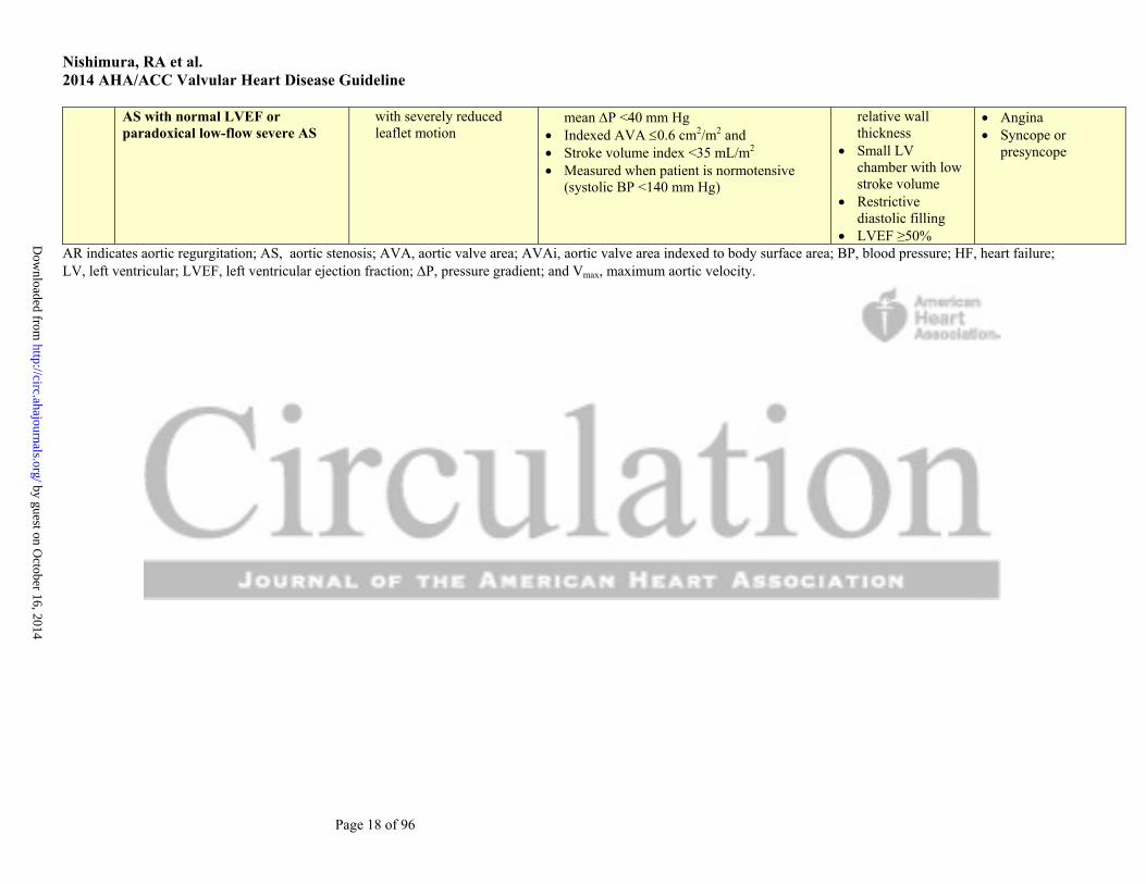
D3 Symptomatic severe low-gradient Severe leaflet calcification AVA 1.0 cm2 with aortic Vmax <4 m/s or Increased LV HF
by guest on October 16, 2014
http://circ.ahajournals.org/D
ownloaded from
Nishimura, RA et al. 2014 AHA/ACC Valvular Heart Disease Guideline
Page 18 of 96
AS with normal LVEF or paradoxical low-flow severe AS
with severely reduced leaflet motion
mean P <40 mm Hg Indexed AVA 0.6 cm2/m2 and Stroke volume index <35 mL/m2 Measured when patient is normotensive
(systolic BP <140 mm Hg)
relative wall thickness
Small LV chamber with low stroke volume
Restrictive diastolic filling
LVEF ≥50%
Angina Syncope or
presyncope
AR indicates aortic regurgitation; AS, aortic stenosis; AVA, aortic valve area; AVAi, aortic valve area indexed to body surface area; BP, blood pressure; HF, heart failure; LV, left ventricular; LVEF, left ventricular ejection fraction; P, pressure gradient; and Vmax, maximum aortic velocity.
by guest on October 16, 2014
http://circ.ahajournals.org/D
ownloaded from

Nishimura, RA et al. 2014 AHA/ACC Valvular Heart Disease Guideline
Page 19 of 96
3.2. Diagnosis and Follow-Up The overall approach to the initial diagnosis of VHD is discussed in Section 2.3, and additional considerations
specific to patients with AS are addressed here.
Class I
1. TTE is indicated in patients with signs or symptoms of AS or a bicuspid aortic valve for accurate diagnosis of the cause of AS, hemodynamic severity, LV size and systolic function, and for determining prognosis and timing of valve intervention (26, 27, 45). (Level of Evidence: B)
Class IIa
1. Low-dose dobutamine stress testing using echocardiographic or invasive hemodynamic measurements is reasonable in patients with stage D2 AS with all of the following (46-48), (Level of Evidence: B):
a. Calcified aortic valve with reduced systolic opening; b. LVEF less than 50%; c. Calculated valve area 1.0 cm2 or less; and d. Aortic velocity less than 4.0 m per second or mean pressure gradient less than 40 mm Hg.
2. Exercise testing is reasonable to assess physiological changes with exercise and to confirm the absence of symptoms in asymptomatic patients with a calcified aortic valve and an aortic velocity 4.0 m per second or greater or mean pressure gradient 40 mm Hg or higher (stage C) (27, 37, 38, 49). (Level of Evidence: B)
Class III: Harm
1. Exercise testing should not be performed in symptomatic patients with AS when the aortic velocity is 4.0 m per second or greater or mean pressure gradient is 40 mm Hg or higher (stage D) (50). (Level of Evidence: B)
3.3. Medical Therapy Class I
1. Hypertension in patients at risk for developing AS (stage A) and in patients with asymptomatic AS (stages B and C) should be treated according to standard GDMT, started at a low dose, and gradually titrated upward as needed with frequent clinical monitoring (51-53). (Level of Evidence: B)
Class IIb
1. Vasodilator therapy may be reasonable if used with invasive hemodynamic monitoring in the acute management of patients with severe decompensated AS (stage D) with New York Heart Association (NYHA) class IV heart failure (HF) symptoms. (Level of Evidence: C)
Class III: No Benefit
1. Statin therapy is not indicated for prevention of hemodynamic progression of AS in patients with mild-to-moderate calcific valve disease (stages B to D) (54-56). (Level of Evidence: A)
3.4. Timing of Intervention See Table 7 for a summary of recommendations from this section. Class I
by guest on October 16, 2014http://circ.ahajournals.org/Downloaded from

Nishimura, RA et al. 2014 AHA/ACC Valvular Heart Disease Guideline
Page 20 of 96
1. AVR is recommended in symptomatic patients with severe AS (stage D1) with (57-60), (Level of Evidence: B):
a. Decreased systolic opening of a calcified or congenitally stenotic aortic valve; and b. An aortic velocity 4.0 m per second or greater or mean pressure gradient 40 mm Hg or
higher; and c. Symptoms of HF, syncope, exertional dyspnea, angina, or presyncope by history or on
exercise testing. 2. AVR is recommended for asymptomatic patients with severe AS (stage C2) and an LVEF less
than 50% with decreased systolic opening of a calcified aortic valve with an aortic velocity 4.0 m per second or greater or mean pressure gradient 40 mm Hg or higher (61, 62). (Level of Evidence: B)
3. AVR is indicated for patients with severe AS (stage C or D) when undergoing cardiac surgery for other indications when there is decreased systolic opening of a calcified aortic valve and an aortic velocity 4.0 m per second or greater or mean pressure gradient 40 mm Hg or higher (63, 64). (Level of Evidence: B)
Class IIa
1. AVR is reasonable for asymptomatic patients with very severe AS (stage C1) with (65, 66), (Level of Evidence: B):
a. Decreased systolic opening of a calcified valve; b. An aortic velocity 5.0 m per second or greater or mean pressure gradient 60 mm Hg or
higher; and c. A low surgical risk.
2. AVR is reasonable in apparently asymptomatic patients with severe AS (stage C1) with (27, 38), (Level of Evidence: B):
a. A calcified aortic valve; b. An aortic velocity of 4.0 m per second to 4.9 m per second or mean pressure gradient of 40
mm Hg to 59 mm Hg; and c. An exercise test demonstrating decreased exercise tolerance or a fall in systolic blood
pressure (BP). 3. AVR is reasonable in symptomatic patients with low-flow/low-gradient severe AS with reduced
LVEF (stage D2) with a (67-69), (Level of Evidence: B): a. Calcified aortic valve with reduced systolic opening; b. Resting valve area 1.0 cm2 or less; c. Aortic velocity less than 4.0 m per second or mean pressure gradient less than 40 mm Hg; d. LVEF less than 50%; and e. A low-dose dobutamine stress study that shows an aortic velocity 4.0 m per second or
greater or mean pressure gradient 40 mm Hg or higher with a valve area 1.0 cm2 or less at any dobutamine dose.
4. AVR is reasonable in symptomatic patients with low-flow/low-gradient severe AS (stage D3) with an LVEF 50% or greater, a calcified aortic valve with significantly reduced leaflet motion, and a valve area 1.0 cm2 or less only if clinical, hemodynamic, and anatomic data support valve obstruction as the most likely cause of symptoms and data recorded when the patient is normotensive (systolic BP <140 mm Hg) indicate (Level of Evidence: C):
a. An aortic velocity less than 4.0 m per second or mean pressure gradient less than 40 mm Hg; and
b. A stroke volume index less than 35 mL/m2; and c. An indexed valve area 0.6 cm2/m2 or less.
5. AVR is reasonable for patients with moderate AS (stage B) with an aortic velocity between 3.0 m per second and 3.9 m per second or mean pressure gradient between 20 mm Hg and 39 mm Hg who are undergoing cardiac surgery for other indications. (Level of Evidence: C)
by guest on October 16, 2014http://circ.ahajournals.org/Downloaded from

Nishimura, RA et al. 2014 AHA/ACC Valvular Heart Disease Guideline
Page 21 of 96
Class IIb 1. AVR may be considered for asymptomatic patients with severe AS (stage C1) with an aortic
velocity 4.0 m per second or greater or mean pressure gradient 40 mm Hg or higher if the patient is at low surgical risk and serial testing shows an increase in aortic velocity 0.3 m/s or greater per year. (Level of Evidence: C)
Table 7. Summary of Recommendations for AS: Timing of Intervention
Recommendations COR LOE References AVR is recommended with severe high-gradient AS who have symptoms by history or on exercise testing (stage D1)
I B (10, 57-59)
AVR is recommended for asymptomatic patients with severe AS (stage C2) and LVEF <50%
I B (61, 62)
AVR is indicated for patients with severe AS (stage C or D) when undergoing other cardiac surgery
I B (63, 64)
AVR is reasonable for asymptomatic patients with very severe AS (stage C1, aortic velocity ≥5.0 m/s) and low surgical risk
IIa B (65, 66)
AVR is reasonable in asymptomatic patients (stage C1) with severe AS and decreased exercise tolerance or an exercise fall in BP
IIa B (27, 38)
AVR is reasonable in symptomatic patients with low-flow/low-gradient severe AS with reduced LVEF (stage D2) with a low-dose dobutamine stress study that shows an aortic velocity 4.0 m/s (or mean pressure gradient 40 mm Hg) with a valve area 1.0 cm2 at any dobutamine dose
IIa B (67-69)
AVR is reasonable in symptomatic patients who have low-flow/low-gradient severe AS (stage D3) who are normotensive and have an LVEF ≥50% if clinical, hemodynamic, and anatomic data support valve obstruction as the most likely cause of symptoms
IIa C N/A
AVR is reasonable for patients with moderate AS (stage B) (aortic velocity 3.0–3.9 m/s) who are undergoing other cardiac surgery
IIa C N/A
AVR may be considered for asymptomatic patients with severe AS (stage C1) and rapid disease progression and low surgical risk
IIb C N/A
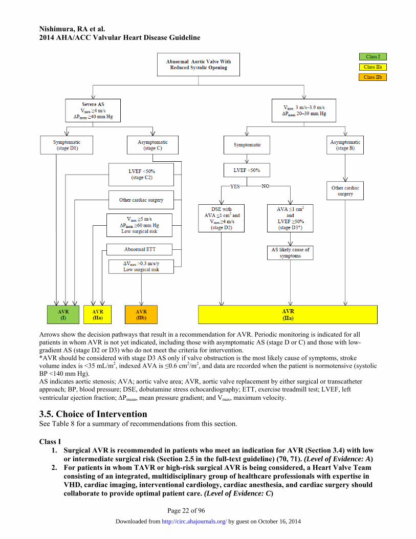
AS indicates aortic stenosis; AVR, aortic valve replacement by either surgical or transcatheter approach; BP, blood pressure; COR, Class of Recommendation; LOE, Level of Evidence; LVEF, left ventricular ejection fraction; and N/A, not applicable. Figure 1. Indications for AVR in Patients With AS
by guest on October 16, 2014http://circ.ahajournals.org/Downloaded from
Nishimura, RA et al. 2014 AHA/ACC Valvular Heart Disease Guideline
Page 22 of 96
Arrows show the decision pathways that result in a recommendation for AVR. Periodic monitoring is indicated for all patients in whom AVR is not yet indicated, including those with asymptomatic AS (stage D or C) and those with low-gradient AS (stage D2 or D3) who do not meet the criteria for intervention. *AVR should be considered with stage D3 AS only if valve obstruction is the most likely cause of symptoms, stroke volume index is <35 mL/m2, indexed AVA is ≤0.6 cm2/m2, and data are recorded when the patient is normotensive (systolic BP <140 mm Hg). AS indicates aortic stenosis; AVA; aortic valve area; AVR, aortic valve replacement by either surgical or transcatheter approach; BP, blood pressure; DSE, dobutamine stress echocardiography; ETT, exercise treadmill test; LVEF, left ventricular ejection fraction; Pmean, mean pressure gradient; and Vmax, maximum velocity.
3.5. Choice of Intervention See Table 8 for a summary of recommendations from this section. Class I
1. Surgical AVR is recommended in patients who meet an indication for AVR (Section 3.4) with low or intermediate surgical risk (Section 2.5 in the full-text guideline) (70, 71). (Level of Evidence: A)
2. For patients in whom TAVR or high-risk surgical AVR is being considered, a Heart Valve Team consisting of an integrated, multidisciplinary group of healthcare professionals with expertise in VHD, cardiac imaging, interventional cardiology, cardiac anesthesia, and cardiac surgery should collaborate to provide optimal patient care. (Level of Evidence: C)
by guest on October 16, 2014http://circ.ahajournals.org/Downloaded from

Nishimura, RA et al. 2014 AHA/ACC Valvular Heart Disease Guideline
Page 23 of 96
3. TAVR is recommended in patients who meet an indication for AVR (Section 3.4) who have a prohibitive risk for surgical AVR (Section 2.5 in the full-text guideline) and a predicted post-TAVR survival greater than 12 months (72, 73). (Level of Evidence: B)
Class IIa
1. TAVR is a reasonable alternative to surgical AVR in patients who meet an indication for AVR (Section 3.4) and who have high surgical risk for surgical AVR (Section 2.5 in the full-text guideline) (74, 75). (Level of Evidence: B)
Class IIb
1. Percutaneous aortic balloon dilation may be considered as a bridge to surgical AVR or TAVR in patients with severe symptomatic AS. (Level of Evidence: C)
Class III: No Benefit 1. TAVR is not recommended in patients in whom existing comorbidities would preclude the
expected benefit from correction of AS (72). (Level of Evidence: B)
Table 8. Summary of Recommendations for AS: Choice of Surgical or Transcatheter Intervention
Recommendations COR LOE References
Surgical AVR is recommended in patients who meet an indication for AVR (Section 3.4) with low or intermediate surgical risk (Section 2.5 in the full-text guideline)
I A (70, 71)
For patients in whom TAVR or high-risk surgical AVR is being considered, members of a Heart Valve Team should collaborate to provide optimal patient care
I C N/A
TAVR is recommended in patients who meet an indication for AVR for AS who have a prohibitive surgical risk and a predicted post-TAVR survival >12 mo
I B (72, 73)
TAVR is a reasonable alternative to surgical AVR in patients who meet an indication for AVR (Section 3.4) and who have high surgical risk (Section 2.5 in the full-text guideline)
IIa B (74, 75)
Percutaneous aortic balloon dilation may be considered as a bridge to surgical or transcatheter AVR in severely symptomatic patients with severe AS
IIb C N/A
TAVR is not recommended in patients in whom existing comorbidities would preclude the expected benefit from correction of AS
III: No Benefit
B (72)
AS indicates aortic stenosis; AVR, aortic valve replacement; COR, Class of Recommendation; LOE, Level of Evidence; N/A, not applicable; and TAVR, transcatheter aortic valve replacement. 4. Aortic Regurgitation: Recommendations
4.1. Stages of Chronic Aortic Regurgitation The most common causes of chronic aortic regurgitation (AR) in the United States and other developed
countries are bicuspid aortic valve and calcific valve disease. In addition, AR frequently arises from primary
diseases causing dilation of the ascending aorta or the sinuses of Valsalva. Another cause of AR is rheumatic
heart disease (the leading cause in many developing countries). In the majority of patients with AR, the disease
course is chronic and slowly progressive with increasing LV volume overload and LV adaptation via chamber
dilation and hypertrophy. Management of patients with AR depends on accurate diagnosis of the cause and stage
by guest on October 16, 2014http://circ.ahajournals.org/Downloaded from
Nishimura, RA et al. 2014 AHA/ACC Valvular Heart Disease Guideline
Page 24 of 96
of the disease process. Table 9 shows the stages of AR ranging from patients at risk of AR (stage A) or with
progressive mild-to-moderate AR (stage B) to severe asymptomatic (stage C) and symptomatic AR (stage D).
Each of these stages is defined by valve anatomy, valve hemodynamics, severity of LV dilation, and LV systolic
function, as well as by patient symptoms.
by guest on October 16, 2014http://circ.ahajournals.org/Downloaded from
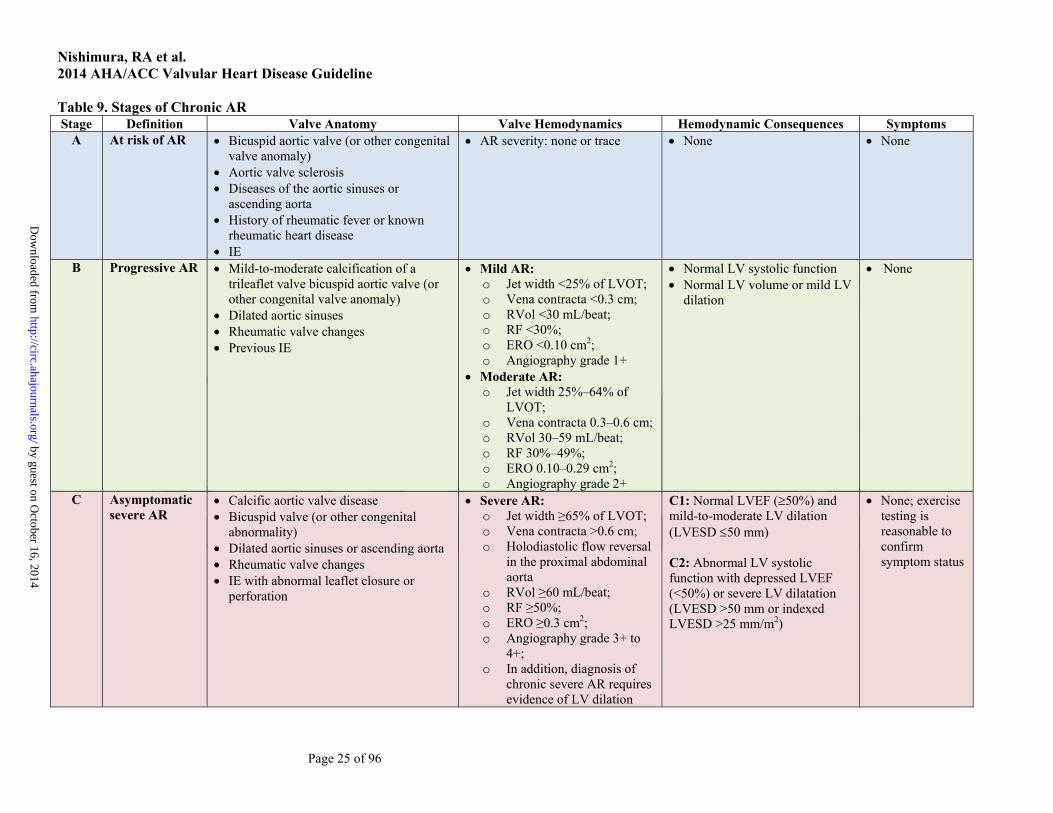
Nishimura, RA et al. 2014 AHA/ACC Valvular Heart Disease Guideline
Page 25 of 96
Table 9. Stages of Chronic AR Stage Definition Valve Anatomy Valve Hemodynamics Hemodynamic Consequences Symptoms
A At risk of AR Bicuspid aortic valve (or other congenital valve anomaly)
Aortic valve sclerosis Diseases of the aortic sinuses or
ascending aorta History of rheumatic fever or known
rheumatic heart disease IE
AR severity: none or trace None None
B Progressive AR Mild-to-moderate calcification of a trileaflet valve bicuspid aortic valve (or other congenital valve anomaly)
Dilated aortic sinuses Rheumatic valve changes Previous IE
Mild AR: o Jet width <25% of LVOT; o Vena contracta <0.3 cm; o RVol <30 mL/beat; o RF <30%; o ERO <0.10 cm2; o Angiography grade 1+
Moderate AR: o Jet width 25%–64% of
LVOT; o Vena contracta 0.3–0.6 cm; o RVol 30–59 mL/beat; o RF 30%–49%; o ERO 0.10–0.29 cm2; o Angiography grade 2+
Normal LV systolic function Normal LV volume or mild LV
dilation
None
C Asymptomatic severe AR
Calcific aortic valve disease Bicuspid valve (or other congenital
abnormality) Dilated aortic sinuses or ascending aorta Rheumatic valve changes IE with abnormal leaflet closure or
perforation
Severe AR: o Jet width ≥65% of LVOT; o Vena contracta >0.6 cm; o Holodiastolic flow reversal
in the proximal abdominal aorta
o RVol ≥60 mL/beat; o RF ≥50%; o ERO ≥0.3 cm2; o Angiography grade 3+ to
4+; o In addition, diagnosis of
chronic severe AR requires evidence of LV dilation
C1: Normal LVEF (50%) and mild-to-moderate LV dilation (LVESD 50 mm) C2: Abnormal LV systolic function with depressed LVEF (<50%) or severe LV dilatation (LVESD >50 mm or indexed LVESD >25 mm/m2)
None; exercise testing is reasonable to confirm symptom status
by guest on October 16, 2014
http://circ.ahajournals.org/D
ownloaded from
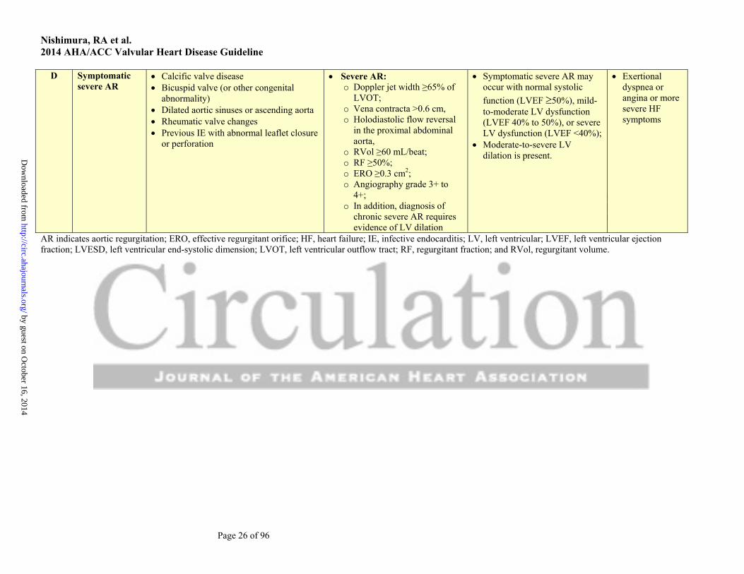
Nishimura, RA et al. 2014 AHA/ACC Valvular Heart Disease Guideline
Page 26 of 96
D Symptomatic severe AR
Calcific valve disease Bicuspid valve (or other congenital
abnormality) Dilated aortic sinuses or ascending aorta Rheumatic valve changes Previous IE with abnormal leaflet closure
or perforation
Severe AR: o Doppler jet width ≥65% of
LVOT; o Vena contracta >0.6 cm, o Holodiastolic flow reversal
in the proximal abdominal aorta,
o RVol ≥60 mL/beat; o RF ≥50%; o ERO ≥0.3 cm2; o Angiography grade 3+ to
4+; o In addition, diagnosis of
chronic severe AR requires evidence of LV dilation
Symptomatic severe AR may occur with normal systolic
function (LVEF 50%), mild-to-moderate LV dysfunction (LVEF 40% to 50%), or severe LV dysfunction (LVEF <40%);
Moderate-to-severe LV dilation is present.
Exertional dyspnea or angina or more severe HF symptoms
AR indicates aortic regurgitation; ERO, effective regurgitant orifice; HF, heart failure; IE, infective endocarditis; LV, left ventricular; LVEF, left ventricular ejection fraction; LVESD, left ventricular end-systolic dimension; LVOT, left ventricular outflow tract; RF, regurgitant fraction; and RVol, regurgitant volume.
by guest on October 16, 2014
http://circ.ahajournals.org/D
ownloaded from

Nishimura, RA et al. 2014 AHA/ACC Valvular Heart Disease Guideline
Page 27 of 96
See Figure 2 for indications for AVR for chronic AR
4.2. Diagnosis and Follow-Up Class I
1. TTE is indicated in patients with signs or symptoms of AR (stages A to D) for accurate diagnosis of the cause of regurgitation, regurgitant severity, and LV size and systolic function, and for determining clinical outcome and timing of valve intervention (34, 76-85). (Level of Evidence: B)
2. TTE is indicated in patients with dilated aortic sinuses or ascending aorta or with a bicuspid aortic valve (stages A and B) to evaluate the presence and severity of AR (86). (Level of Evidence: B)
3. CMR is indicated in patients with moderate or severe AR (stages B, C, and D) and suboptimal echocardiographic images for the assessment of LV systolic function, systolic and diastolic volumes, and measurement of AR severity (87, 88). (Level of Evidence: B)
4.3. Medical Therapy Class I
1. Treatment of hypertension (systolic BP >140 mm Hg) is recommended in patients with chronic AR (stages B and C), preferably with dihydropyridine calcium channel blockers or angiotensin-converting enzyme (ACE) inhibitors/angiotensin-receptor blockers (ARBs) (84, 89). (Level of Evidence: B)
Class IIa
1. Medical therapy with ACE inhibitors/ARBs and beta blockers is reasonable in patients with severe AR who have symptoms and/or LV dysfunction (stages C2 and D) when surgery is not performed because of comorbidities (90, 91). (Level of Evidence: B)
4.4. Timing of Intervention See Table 10 for a summary of recommendations from this section. Class I
1. AVR is indicated for symptomatic patients with severe AR regardless of LV systolic function (stage D) (33, 92, 93). (Level of Evidence: B)
2. AVR is indicated for asymptomatic patients with chronic severe AR and LV systolic dysfunction (LVEF <50%) at rest (stage C2) if no other cause for systolic dysfunction is identified (92, 94-96). (Level of Evidence: B)
3. AVR is indicated for patients with severe AR (stage C or D) while undergoing cardiac surgery for other indications. (Level of Evidence: C)
Class IIa
1. AVR is reasonable for asymptomatic patients with severe AR with normal LV systolic function (LVEF 50%) but with severe LV dilation (LV end-systolic dimension [LVESD] >50 mm or indexed LVESD >25 mm/m2) (stage C2) (97-99). (Level of Evidence: B)
2. AVR is reasonable in patients with moderate AR (stage B) while undergoing surgery on the ascending aorta, coronary artery bypass graft (CABG), or mitral valve surgery. (Level of Evidence: C)
Class IIb
1. AVR may be considered for asymptomatic patients with severe AR and normal LV systolic function at rest (LVEF 50%, stage C1) but with progressive severe LV dilatation (LV end-diastolic dimension >65 mm) if surgical risk is low. (Level of Evidence: C)
by guest on October 16, 2014http://circ.ahajournals.org/Downloaded from

Nishimura, RA et al. 2014 AHA/ACC Valvular Heart Disease Guideline
Page 28 of 96
Table 10. Summary of Recommendations for AR Intervention Recommendations COR LOE References
AVR is indicated for symptomatic patients with severe AR regardless of LV systolic function (stage D)
I B (33, 92, 93)
AVR is indicated for asymptomatic patients with chronic severe AR and LV systolic dysfunction (LVEF <50%) (stage C2)
I B (92, 94-96)
AVR is indicated for patients with severe AR (stage C or D) while undergoing cardiac surgery for other indications
I C N/A
AVR is reasonable for asymptomatic patients with severe AR with normal LV systolic function (LVEF 50%) but with severe LV dilation (LVESD >50 mm, stage C2)
IIa B (97-99)
AVR is reasonable in patients with moderate AR (stage B) who are undergoing other cardiac surgery
IIa C N/A
AVR may be considered for asymptomatic patients with severe AR and normal LV systolic function (LVEF ≥50%, stage C1) but with progressive severe LV dilation (LVEDD >65 mm) if surgical risk is low*
IIb C N/A
*Particularly in the setting of progressive LV enlargement. AR indicates aortic regurgitation; AVR, aortic valve replacement; COR, Class of Recommendation; LOE, Level of Evidence; LV, left ventricular; LVEDD, left ventricular end-diastolic dimension; LVEF, left ventricular ejection fraction; LVESD, left ventricular end-systolic dimension; and N/A, not applicable. Figure 2. Indications for AVR for Chronic AR
AR indicates aortic regurgitation; AVR, aortic valve replacement (valve repair may be appropriate in selected patients); ERO, effective regurgitant orifice; LV, left ventricular; LVEDD, left ventricular end-diastolic dimension; LVEF, left
by guest on October 16, 2014http://circ.ahajournals.org/Downloaded from

Nishimura, RA et al. 2014 AHA/ACC Valvular Heart Disease Guideline
Page 29 of 96
ventricular ejection fraction; LVESD, left ventricular end-systolic dimension; RF, regurgitant fraction; and RVol, regurgitant volume.
5. Bicuspid Aortic Valve and Aortopathy: Recommendations
5.1. Diagnosis and Follow-Up Class I
1. An initial TTE is indicated in patients with a known bicuspid aortic valve to evaluate valve morphology, to measure the severity of AS and AR, and to assess the shape and diameter of the aortic sinuses and ascending aorta for prediction of clinical outcome and to determine timing of intervention (100-105). (Level of Evidence: B)
2. Aortic magnetic resonance angiography or CT angiography is indicated in patients with a bicuspid aortic valve when morphology of the aortic sinuses, sinotubular junction, or ascending aorta cannot be assessed accurately or fully by echocardiography. (Level of Evidence: C)
3. Serial evaluation of the size and morphology of the aortic sinuses and ascending aorta by echocardiography, CMR, or CT angiography is recommended in patients with a bicuspid aortic valve and an aortic diameter greater than 4.0 cm, with the examination interval determined by the degree and rate of progression of aortic dilation and by family history. In patients with an aortic diameter greater than 4.5 cm, this evaluation should be performed annually. (Level of Evidence: C)
5.2. Intervention Class I
1. Operative intervention to repair the aortic sinuses or replace the ascending aorta is indicated in patients with a bicuspid aortic valve if the diameter of the aortic sinuses or ascending aorta is greater than 5.5 cm (106-108). (Level of Evidence: B)
Class IIa 1. Operative intervention to repair the aortic sinuses or replace the ascending aorta is reasonable in
patients with bicuspid aortic valves if the diameter of the aortic sinuses or ascending aorta is greater than 5.0 cm and a risk factor for dissection is present (family history of aortic dissection or if the rate of increase in diameter is ≥0.5 cm per year). (Level of Evidence: C)
2. Replacement of the ascending aorta is reasonable in patients with a bicuspid aortic valve who are undergoing aortic valve surgery because of severe AS or AR (Sections 3.4 and 4.4) if the diameter of the ascending aorta is greater than 4.5 cm. (Level of Evidence: C)
6. Mitral Stenosis: Recommendations
6.1. Stages of MS Medical and interventional approaches to the management of patients with valvular MS depend on accurate
diagnosis of the cause and stage of the disease process. Table 11 shows the stages of mitral valve disease
ranging from patients at risk of MS (stage A) or with progressive hemodynamic obstruction (stage B) to severe
asymptomatic (stage C) and symptomatic MS (stage D). Each of these stages is defined by valve anatomy, valve
hemodynamics, the consequences of valve obstruction on the left atrium (LA) and pulmonary circulation, and
patient symptoms. The anatomic features of the stages of MS are based on a rheumatic etiology for the disease.
There are patients who have a nonrheumatic etiology of MS due to senile calcific disease (Section 6.3 in the full
by guest on October 16, 2014http://circ.ahajournals.org/Downloaded from
Nishimura, RA et al. 2014 AHA/ACC Valvular Heart Disease Guideline
Page 30 of 96
text) in whom there is a heavily calcified mitral annulus with extension of the calcium into the leaflets.
Hemodynamic severity is best characterized by the planimetered mitral valve area and the calculated mitral
valve area from the diastolic pressure half-time. The definition of “severe” MS is based on the severity at which
symptoms occur as well as the severity at which intervention will improve symptoms. Thus, a mitral valve area
≤1.5 cm2 is considered severe. This usually corresponds to a transmitral mean gradient of >5 mm Hg to 10 mm
Hg at a normal heart rate. However, the mean pressure gradient is highly dependent on the transvalvular flow
and diastolic filling period and will vary greatly with changes in heart rate. The diastolic pressure half-time is
dependent not only on the degree of mitral obstruction but also the compliance of the left ventricle and LA and
other measures of mitral valve area, such as the continuity equation or the proximal isovelocity surface area,
may be used if discrepancies exist (109-115).
by guest on October 16, 2014http://circ.ahajournals.org/Downloaded from

Nishimura, RA et al. 2014 AHA/ACC Valvular Heart Disease Guideline
Page 31 of 96
Table 11. Stages of MS Stage Definition Valve Anatomy Valve Hemodynamics Hemodynamic Consequences Symptoms
A At risk of MS Mild valve doming during diastole
Normal transmitral flow velocity None None
B Progressive MS Rheumatic valve changes with commissural fusion and diastolic doming of the mitral valve leaflets
Planimetered MVA >1.5 cm2
Increased transmitral flow velocities
MVA >1.5 cm2 Diastolic pressure half-time
<150 ms
Mild-to-moderate LA enlargement
Normal pulmonary pressure at rest
None
C Asymptomatic severe MS
Rheumatic valve changes with commissural fusion and diastolic doming of the mitral valve leaflets
Planimetered MVA ≤1.5 cm2 (MVA ≤1.0 cm2 with very
severe MS)
MVA ≤1.5 cm2 (MVA ≤1.0 cm2 with very
severe MS) Diastolic pressure half-time
≥150 ms (Diastolic pressure half-time
≥220 ms with very severe MS)
Severe LA enlargement Elevated PASP >30 mm Hg
None
D Symptomatic severe MS
Rheumatic valve changes with commissural fusion and diastolic doming of the mitral valve leaflets
Planimetered MVA ≤1.5 cm2
MVA ≤1.5 cm2 (MVA ≤1.0 cm2 with very
severe MS) Diastolic pressure half-time
≥150 ms (Diastolic pressure half-time
≥220 ms with very severe MS)
Severe LA enlargement Elevated PASP >30 mm Hg
Decreased exercise tolerance
Exertional dyspnea
The transmitral mean pressure gradient should be obtained to further determine the hemodynamic effect of the MS and is usually >5 mm Hg to 10 mm Hg in severe MS; however, due to the variability of the mean pressure gradient with heart rate and forward flow, it has not been included in the criteria for severity. LA indicates left atrial; LV, left ventricular; MS, mitral stenosis; MVA, mitral valve area; and PASP, pulmonary artery systolic pressure.
by guest on October 16, 2014
http://circ.ahajournals.org/D
ownloaded from
Nishimura, RA et al. 2014 AHA/ACC Valvular Heart Disease Guideline
Page 32 of 96
See Figure 3 for indications for intervention for rheumatic MS.
6.2. Diagnosis and Follow-Up Class I
1. TTE is indicated in patients with signs or symptoms of MS to establish the diagnosis, quantify hemodynamic severity (mean pressure gradient, mitral valve area, and pulmonary artery pressure), assess concomitant valvular lesions, and demonstrate valve morphology (to determine suitability for mitral commissurotomy) (9, 60, 116-123). (Level of Evidence: B)
2. TEE should be performed in patients considered for percutaneous mitral balloon commissurotomy to assess the presence or absence of left atrial thrombus and to further evaluate the severity of mitral regurgitation (MR) (117, 124-126). (Level of Evidence: B)
3. Exercise testing with Doppler or invasive hemodynamic assessment is recommended to evaluate the response of the mean mitral gradient and pulmonary artery pressure in patients with MS when there is a discrepancy between resting Doppler echocardiographic findings and clinical symptoms or signs. (Level of Evidence: C)
6.3. Medical Therapy Class I
1. Anticoagulation (vitamin K antagonist [VKA] or heparin) is indicated in patients with 1) MS and AF (paroxysmal, persistent, or permanent), or 2) MS and a prior embolic event, or 3) MS and a left atrial thrombus (127-133). (Level of Evidence: B)
Class IIa
1. Heart rate control can be beneficial in patients with MS and AF and fast ventricular response. (Level of Evidence: C)
Class IIb
1. Heart rate control may be considered for patients with MS in normal sinus rhythm and symptoms associated with exercise (134, 135). (Level of Evidence: B)
6.4. Intervention See Table 12 for a summary of recommendations from this section. Class I
1. Percutaneous mitral balloon commissurotomy is recommended for symptomatic patients with severe MS (mitral valve area ≤1.5 cm2, stage D) and favorable valve morphology in the absence of left atrial thrombus or moderate-to-severe MR (109-113, 115, 136). (Level of Evidence: A)
2. Mitral valve surgery (repair, commissurotomy, or valve replacement) is indicated in severely symptomatic patients (NYHA class III to IV) with severe MS (mitral valve area ≤1.5 cm2, stage D) who are not high risk for surgery and who are not candidates for or who have failed previous percutaneous mitral balloon commissurotomy (137-142). (Level of Evidence: B)
3. Concomitant mitral valve surgery is indicated for patients with severe MS (mitral valve area ≤1.5 cm2, stage C or D) undergoing cardiac surgery for other indications. (Level of Evidence: C)
Class IIa
1. Percutaneous mitral balloon commissurotomy is reasonable for asymptomatic patients with very severe MS (mitral valve area ≤1.0 cm2, stage C) and favorable valve morphology in the absence of left atrial thrombus or moderate-to-severe MR (121, 143-145). (Level of Evidence: C)
by guest on October 16, 2014http://circ.ahajournals.org/Downloaded from

Nishimura, RA et al. 2014 AHA/ACC Valvular Heart Disease Guideline
Page 33 of 96
2. Mitral valve surgery is reasonable for severely symptomatic patients (NYHA class III to IV) with severe MS (mitral valve area ≤1.5 cm2, stage D), provided there are other operative indications (e.g., aortic valve disease, coronary artery disease (CAD), tricuspid regurgitation (TR), aortic aneurysm). (Level of Evidence: C)
Class IIb 1. Percutaneous mitral balloon commissurotomy may be considered for asymptomatic patients with
severe MS (mitral valve area ≤1.5 cm2, stage C) and valve morphology favorable for percutaneous mitral balloon commissurotomy in the absence of left atrial thrombus or moderate-to-severe MR who have new onset of AF. (Level of Evidence: C)
2. Percutaneous mitral balloon commissurotomy may be considered for symptomatic patients with mitral valve area greater than 1.5 cm2 if there is evidence of hemodynamically significant MS based on pulmonary artery wedge pressure greater than 25 mm Hg or mean mitral valve gradient greater than 15 mm Hg during exercise. (Level of Evidence: C)
3. Percutaneous mitral balloon commissurotomy may be considered for severely symptomatic patients (NYHA class III to IV) with severe MS (mitral valve area ≤1.5 cm2, stage D) who have a suboptimal valve anatomy and who are not candidates for surgery or at high risk for surgery. (Level of Evidence: C)
4. Concomitant mitral valve surgery may be considered for patients with moderate MS (mitral valve area 1.6 cm2 to 2.0 cm2) undergoing cardiac surgery for other indications. (Level of Evidence: C)
5. Mitral valve surgery and excision of the left atrial appendage may be considered for patients with severe MS (mitral valve area ≤1.5 cm2, stages C and D) who have had recurrent embolic events while receiving adequate anticoagulation. (Level of Evidence: C)
Table 12. Summary of Recommendations for MS Intervention Recommendations COR LOE References
PMBC is recommended for symptomatic patients with severe MS (MVA <1.5 cm2, stage D) and favorable valve morphology in the absence of contraindications
I A (109-113, 115)
Mitral valve surgery is indicated in severely symptomatic patients (NYHA class III/IV) with severe MS (MVA <1.5 cm2, stage D) who are not high risk for surgery and who are not candidates for or failed previous PMBC
I B (137-142)
Concomitant mitral valve surgery is indicated for patients with severe MS (MVA ≤1.5 cm2, stage C or D) undergoing other cardiac surgery
I C N/A
PMBC is reasonable for asymptomatic patients with very severe MS (MVA ≤1.0 cm2, stage C) and favorable valve morphology in the absence of contraindications
IIa C (121, 143-145)
Mitral valve surgery is reasonable for severely symptomatic patients (NYHA class III/IV) with severe MS (MVA ≤1.5 cm2, stage D), provided there are other operative indications
IIa C N/A
PMBC may be considered for asymptomatic patients with severe MS (MVA ≤1.5 cm2, stage C) and favorable valve morphology who have new onset of AF in the absence of contraindications
IIb C N/A
PMBC may be considered for symptomatic patients with MVA >1.5 cm2 if there is evidence of hemodynamically significant MS during exercise
IIb C N/A
PMBC may be considered for severely symptomatic patients (NYHA class III/IV) with severe MS (MVA ≤1.5 cm2, stage D) who have suboptimal valve anatomy and are not candidates for surgery or at high risk for surgery
IIb C N/A
Concomitant mitral valve surgery may be considered for patients with moderate MS (MVA 1.6–2.0 cm2) undergoing other cardiac surgery
IIb C N/A
Mitral valve surgery and excision of the left atrial appendage may be IIb C N/A
by guest on October 16, 2014http://circ.ahajournals.org/Downloaded from
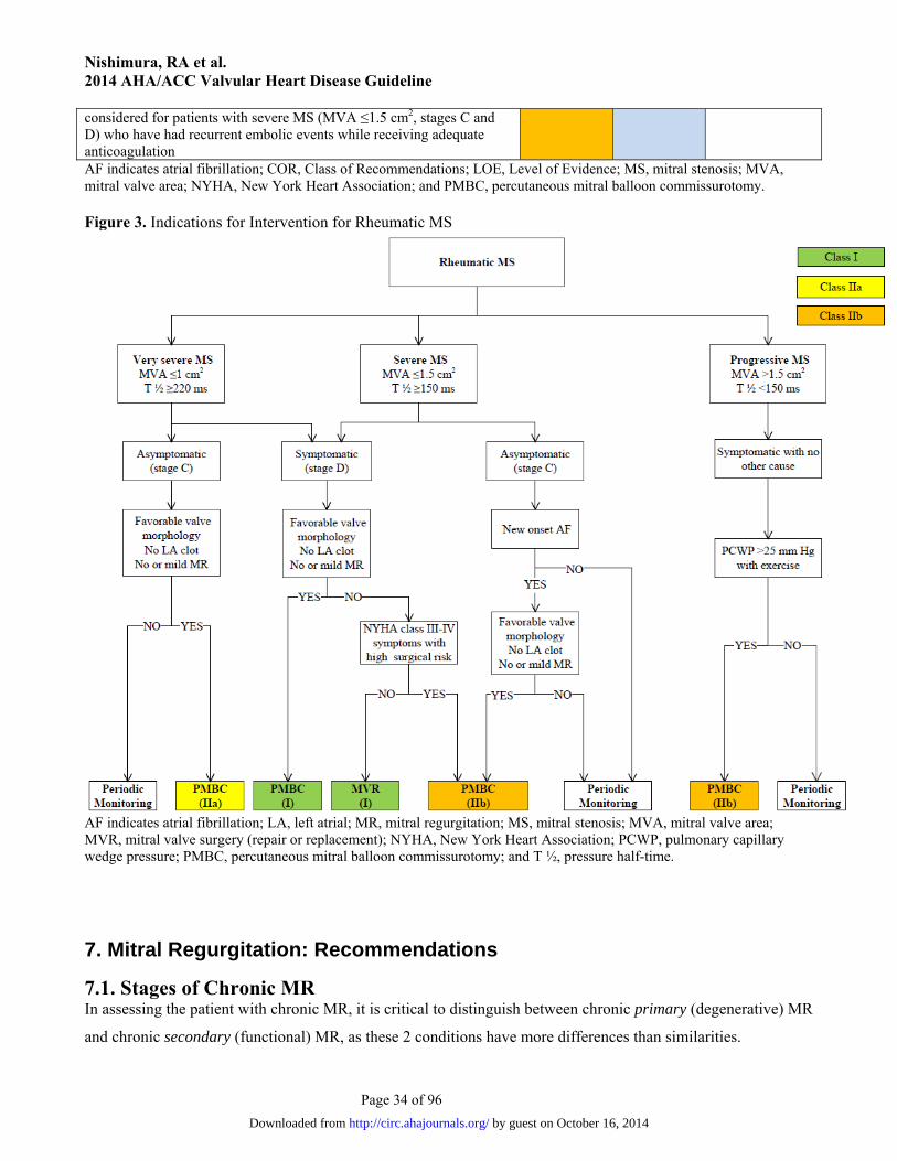
Nishimura, RA et al. 2014 AHA/ACC Valvular Heart Disease Guideline
Page 34 of 96
considered for patients with severe MS (MVA ≤1.5 cm2, stages C and D) who have had recurrent embolic events while receiving adequate anticoagulation AF indicates atrial fibrillation; COR, Class of Recommendations; LOE, Level of Evidence; MS, mitral stenosis; MVA, mitral valve area; NYHA, New York Heart Association; and PMBC, percutaneous mitral balloon commissurotomy. Figure 3. Indications for Intervention for Rheumatic MS
AF indicates atrial fibrillation; LA, left atrial; MR, mitral regurgitation; MS, mitral stenosis; MVA, mitral valve area; MVR, mitral valve surgery (repair or replacement); NYHA, New York Heart Association; PCWP, pulmonary capillary wedge pressure; PMBC, percutaneous mitral balloon commissurotomy; and T ½, pressure half-time.
7. Mitral Regurgitation: Recommendations
7.1. Stages of Chronic MR In assessing the patient with chronic MR, it is critical to distinguish between chronic primary (degenerative) MR
and chronic secondary (functional) MR, as these 2 conditions have more differences than similarities.
by guest on October 16, 2014http://circ.ahajournals.org/Downloaded from
Nishimura, RA et al. 2014 AHA/ACC Valvular Heart Disease Guideline
Page 35 of 96
In chronic primary MR, the pathology of ≥1 of the components of the valve (leaflets, chordae tendineae,
papillary muscles, annulus) causes valve incompetence with systolic regurgitation of blood from the left
ventricle to the LA (Table 13). The most common cause of chronic primary MR in developed countries is mitral
valve prolapse, which has a wide spectrum of etiology and presentation. Younger populations present with
severe myxomatous degeneration with gross redundancy of both anterior and posterior leaflets and the chordal
apparatus (Barlow’s valve). Alternatively, older populations present with fibroelastic deficiency disease, in
which lack of connective tissue leads to chordal rupture. The differentiation between these 2 etiologies has
important implications for operative intervention. Other less common causes of chronic primary MR include IE,
connective tissue disorders, rheumatic heart disease, cleft mitral valve, and radiation heart disease. If the
subsequent volume overload of chronic primary MR is prolonged and severe, it causes myocardial damage, HF,
and eventual death. Correction of the MR is curative. Thus, MR is “the disease.”
In chronic secondary MR, the mitral valve is usually normal (Table 14). Instead, severe LV dysfunction
is caused either by CAD, related myocardial infarction (ischemic chronic secondary MR), or idiopathic
myocardial disease (nonischemic chronic secondary MR). The abnormal and dilated left ventricle causes
papillary muscle displacement, which in turn results in leaflet tethering with associated annular dilation that
prevents coaptation. Because MR is only 1 component of the disease (severe LV dysfunction, coronary disease,
or idiopathic myocardial disease are the others), restoration of mitral valve competence is not by itself curative;
thus, the best therapy for chronic secondary MR is much less clear than it is for chronic primary MR. The data
are limited, and there is greater difficulty in defining the severity of MR in patients with secondary MR than in
those with primary MR. In patients with secondary MR, adverse outcomes are associated with a smaller
calculated effective regurgitant orifice compared to primary MR due to multiple reasons. The MR will likely
progress due to the associated progressive LV systolic dysfunction and adverse remodeling. In addition, there is
an underestimation of effective regurgitant orifice area by the 2-dimensional echocardiographyderived flow
convergence method due to the crescenticshape of the regurgitant orifice. There are the additional clinical
effects of a smaller amount of regurgitation in the presence of compromised LV systolic function and baseline
elevated filling pressures.
by guest on October 16, 2014http://circ.ahajournals.org/Downloaded from

Nishimura, RA et al. 2014 AHA/ACC Valvular Heart Disease Guideline
Page 36 of 96
Table 13. Stages of Primary MR Grade Definition Valve Anatomy Valve Hemodynamics* Hemodynamic
Consequences Symptoms
A At risk of MR Mild mitral valve prolapse with normal coaptation
Mild valve thickening and leaflet restriction
No MR jet or small central jet area <20% LA on Doppler
Small vena contracta <0.3 cm
None None
B Progressive MR Severe mitral valve prolapse with normal coaptation
Rheumatic valve changes with leaflet restriction and loss of central coaptation
Prior IE
Central jet MR 20%–40% LA or late systolic eccentric jet MR
Vena contracta <0.7 cm Regurgitant volume <60 mL Regurgitant fraction <50% ERO <0.40 cm2 Angiographic grade 1–2+
Mild LA enlargement No LV enlargement Normal pulmonary
pressure
None
C Asymptomatic severe MR
Severe mitral valve prolapse with loss of coaptation or flail leaflet
Rheumatic valve changes with leaflet restriction and loss of central coaptation
Prior IE Thickening of leaflets with
radiation heart disease
Central jet MR >40% LA or holosystolic eccentric jet MR
Vena contracta ≥0.7 cm Regurgitant volume ≥60 mL Regurgitant fraction ≥50% ERO ≥0.40 cm2 Angiographic grade 3–4+
Moderate or severe LA enlargement
LV enlargement Pulmonary hypertension
may be present at rest or with exercise
C1: LVEF >60% and LVESD <40 mm
C2: LVEF ≤60% and LVESD ≥40 mm
None
D Symptomatic severe MR
Severe mitral valve prolapse with loss of coaptation or flail leaflet
Rheumatic valve changes with leaflet restriction and loss of central coaptation
Prior IE Thickening of leaflets with
radiation heart disease
Central jet MR >40% LA or holosystolic eccentric jet MR
Vena contracta ≥0.7 cm Regurgitant volume ≥60 mL Regurgitant fraction ≥50% ERO ≥0.40 cm2 Angiographic grade 3–4+
Moderate or severe LA enlargement
LV enlargement Pulmonary hypertension
present
Decreased exercise tolerance
Exertional dyspnea
*Several valve hemodynamic criteria are provided for assessment of MR severity, but not all criteria for each category will be present in each patient. Categorization of MR severity as mild, moderate, or severe depends on data quality and integration of these parameters in conjunction with other clinical evidence. ERO indicates effective regurgitant orifice; IE, infective endocarditis; LA, left atrium/atrial; LV, left ventricular; LVEF, left ventricular ejection fraction; LVESD; left ventricular end-systolic dimension; and MR, mitral regurgitation
by guest on October 16, 2014
http://circ.ahajournals.org/D
ownloaded from
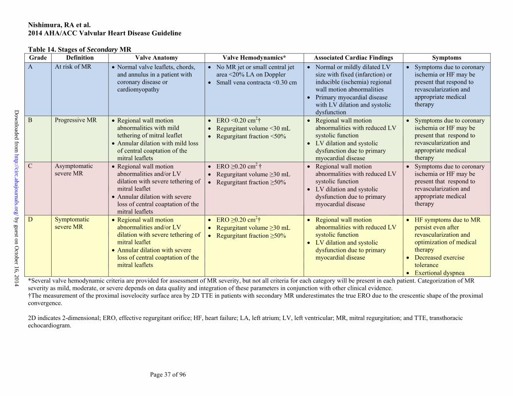
Nishimura, RA et al. 2014 AHA/ACC Valvular Heart Disease Guideline
Page 37 of 96
Table 14. Stages of Secondary MR Grade Definition Valve Anatomy Valve Hemodynamics* Associated Cardiac Findings Symptoms A At risk of MR Normal valve leaflets, chords,
and annulus in a patient with coronary disease or cardiomyopathy
No MR jet or small central jet area <20% LA on Doppler
Small vena contracta <0.30 cm
Normal or mildly dilated LV size with fixed (infarction) or inducible (ischemia) regional wall motion abnormalities
Primary myocardial disease with LV dilation and systolic dysfunction
Symptoms due to coronary ischemia or HF may be present that respond to revascularization and appropriate medical therapy
B Progressive MR Regional wall motion abnormalities with mild tethering of mitral leaflet
Annular dilation with mild loss of central coaptation of the mitral leaflets
ERO <0.20 cm2† Regurgitant volume <30 mL Regurgitant fraction <50%
Regional wall motion abnormalities with reduced LV systolic function
LV dilation and systolic dysfunction due to primary myocardial disease
Symptoms due to coronary ischemia or HF may be present that respond to revascularization and appropriate medical therapy
C Asymptomatic severe MR
Regional wall motion abnormalities and/or LV dilation with severe tethering of mitral leaflet
Annular dilation with severe loss of central coaptation of the mitral leaflets
ERO ≥0.20 cm2 † Regurgitant volume ≥30 mL Regurgitant fraction ≥50%
Regional wall motion abnormalities with reduced LV systolic function
LV dilation and systolic dysfunction due to primary myocardial disease
Symptoms due to coronary ischemia or HF may be present that respond to revascularization and appropriate medical therapy
D Symptomatic severe MR
Regional wall motion abnormalities and/or LV dilation with severe tethering of mitral leaflet
Annular dilation with severe loss of central coaptation of the mitral leaflets
ERO ≥0.20 cm2† Regurgitant volume ≥30 mL Regurgitant fraction ≥50%
Regional wall motion abnormalities with reduced LV systolic function
LV dilation and systolic dysfunction due to primary myocardial disease
HF symptoms due to MR persist even after revascularization and optimization of medical therapy
Decreased exercise tolerance
Exertional dyspnea *Several valve hemodynamic criteria are provided for assessment of MR severity, but not all criteria for each category will be present in each patient. Categorization of MR severity as mild, moderate, or severe depends on data quality and integration of these parameters in conjunction with other clinical evidence. †The measurement of the proximal isovelocity surface area by 2D TTE in patients with secondary MR underestimates the true ERO due to the crescentic shape of the proximal convergence. 2D indicates 2-dimensional; ERO, effective regurgitant orifice; HF, heart failure; LA, left atrium; LV, left ventricular; MR, mitral regurgitation; and TTE, transthoracic echocardiogram.
by guest on October 16, 2014
http://circ.ahajournals.org/D
ownloaded from

Nishimura, RA et al. 2014 AHA/ACC Valvular Heart Disease Guideline
Page 38 of 96
7.2. Chronic Primary MR
7.2.1. Diagnosis and Follow-Up Class I
1. TTE is indicated for baseline evaluation of LV size and function, right ventricular (RV) function and left atrial size, pulmonary artery pressure, and mechanism and severity of primary MR (stages A to D) in any patient suspected of having chronic primary MR (6, 23, 146-162). (Level of Evidence: B)
2. CMR is indicated in patients with chronic primary MR to assess LV and RV volumes, function, or MR severity and when these issues are not satisfactorily addressed by TTE (157, 163, 164). (Level of Evidence: B)
3. Intraoperative TEE is indicated to establish the anatomic basis for chronic primary MR (stages C and D) and to guide repair (165, 166). (Level of Evidence: B)
4. TEE is indicated for evaluation of patients with chronic primary MR (stages B to D) in whom noninvasive imaging provides nondiagnostic information about severity of MR, mechanism of MR, and/or status of LV function. (Level of Evidence: C)
Class IIa
1. Exercise hemodynamics with either Doppler echocardiography or cardiac catheterization is reasonable in symptomatic patients with chronic primary MR where there is a discrepancy between symptoms and the severity of MR at rest (stages B and C) (167, 168). (Level of Evidence: B)
2. Exercise treadmill testing can be useful in patients with chronic primary MR to establish symptom status and exercise tolerance (stages B and C). (Level of Evidence: C)
7.2.2. Medical Therapy Class IIa
1. Medical therapy for systolic dysfunction is reasonable in symptomatic patients with chronic primary MR (stage D) and LVEF less than 60% in whom surgery is not contemplated (169-173). (Level of Evidence: B)
Class III: No Benefit
1. Vasodilator therapy is not indicated for normotensive asymptomatic patients with chronic primary MR (stages B and C1) and normal systolic LV function (173-178). (Level of Evidence: B)
7.2.3. Intervention See Table 15 for a summary of recommendations from this section. Class I
1. Mitral valve surgery is recommended for symptomatic patients with chronic severe primary MR (stage D) and LVEF greater than 30% (156, 179). (Level of Evidence: B)
2. Mitral valve surgery is recommended for asymptomatic patients with chronic severe primary MR and LV dysfunction (LVEF 30% to 60% and/or LVESD ≥40 mm, stage C2) (150-153, 180-182). (Level of Evidence: B)
3. Mitral valve repair is recommended in preference to mitral valve replacement (MVR) when surgical treatment is indicated for patients with chronic severe primary MR limited to the posterior leaflet (155, 183-198). (Level of Evidence: B)
4. Mitral valve repair is recommended in preference to MVR when surgical treatment is indicated for patients with chronic severe primary MR involving the anterior leaflet or both leaflets when a successful and durable repair can be accomplished (195-197, 199-203). (Level of Evidence: B)
by guest on October 16, 2014http://circ.ahajournals.org/Downloaded from

Nishimura, RA et al. 2014 AHA/ACC Valvular Heart Disease Guideline
Page 39 of 96
5. Concomitant mitral valve repair or MVR is indicated in patients with chronic severe primary MR undergoing cardiac surgery for other indications (204). (Level of Evidence: B)
Class IIa
1. Mitral valve repair is reasonable in asymptomatic patients with chronic severe primary MR (stage C1) with preserved LV function (LVEF >60% and LVESD <40 mm) in whom the likelihood of a successful and durable repair without residual MR is greater than 95% with an expected mortality rate of less than 1% when performed at a Heart Valve Center of Excellence (149, 203, 205-209). (Level of Evidence: B)
2. Mitral valve repair is reasonable for asymptomatic patients with chronic severe nonrheumatic primary MR (stage C1) and preserved LV function (LVEF >60% and LVESD <40 mm) in whom there is a high likelihood of a successful and durable repair with 1) new onset of AF or 2) resting pulmonary hypertension (pulmonary artery systolic arterial pressure >50 mm Hg) (154, 205, 210-215). (Level of Evidence: B)
3. Concomitant mitral valve repair is reasonable in patients with chronic moderate primary MR (stage B) when undergoing cardiac surgery for other indications. (Level of Evidence: C)
Class IIb
1. Mitral valve surgery may be considered in symptomatic patients with chronic severe primary MR and LVEF less than or equal to 30% (stage D). (Level of Evidence: C)
2. Mitral valve repair may be considered in patients with rheumatic mitral valve disease when surgical treatment is indicated if a durable and successful repair is likely or when the reliability of long-term anticoagulation management is questionable (194, 202, 203). (Level of Evidence: B)
3. Transcatheter mitral valve repair may be considered for severely symptomatic patients (NYHA class III to IV) with chronic severe primary MR (stage D) who have favorable anatomy for the repair procedure and a reasonable life expectancy but who have a prohibitive surgical risk because of severe comorbidities and remain severely symptomatic despite optimal GDMT for HF (216). (Level of Evidence: B)
Class III: Harm 1. MVR should not be performed for the treatment of isolated severe primary MR limited to less
than one half of the posterior leaflet unless mitral valve repair has been attempted and was unsuccessful (195-198). (Level of Evidence: B)
Table 15. Summary of Recommendations for Chronic Primary MR
Recommendations COR LOE References
MV surgery is recommended for symptomatic patients with chronic severe primary MR (stage D) and LVEF >30%
I B (156, 179)
MV surgery is recommended for asymptomatic patients with chronic severe primary MR and LV dysfunction (LVEF 30%–60% and/or LVESD ≥40 mm, stage C2)
I B (150-153, 180-
182)
MV repair is recommended in preference to MVR when surgical treatment is indicated for patients with chronic severe primary MR limited to the posterior leaflet
I B (155, 183-198)
MV repair is recommended in preference to MVR when surgical treatment is indicated for patients with chronic severe primary MR involving the anterior leaflet or both leaflets when a successful and durable repair can be accomplished
I B (195-197, 199-
203)
Concomitant MV repair or replacement is indicated in patients with chronic severe primary MR undergoing cardiac surgery for other indications
I B (204)
by guest on October 16, 2014http://circ.ahajournals.org/Downloaded from

Nishimura, RA et al. 2014 AHA/ACC Valvular Heart Disease Guideline
Page 40 of 96
MV repair is reasonable in asymptomatic patients with chronic severe primary MR (stage C1) with preserved LV function (LVEF >60% and LVESD <40 mm) in whom the likelihood of a successful and durable repair without residual MR is >95% with an expected mortality rate of <1% when performed at a Heart Valve Center of Excellence
IIa B (149, 203, 205-
209)
MV repair is reasonable for asymptomatic patients with chronic severe nonrheumatic primary MR (stage C1) and preserved LV function in whom there is a high likelihood of a successful and durable repair with 1) new onset of AF or 2) resting pulmonary hypertension (PA systolic arterial pressure >50 mm Hg)
IIa B (154, 205, 210-
215)
Concomitant MV repair is reasonable in patients with chronic moderate primary MR (stage B) undergoing cardiac surgery for other indications
IIa C N/A
MV surgery may be considered in symptomatic patients with chronic severe primary MR and LVEF 30% (stage D)
IIb C N/A
MV repair may be considered in patients with rheumatic mitral valve disease when surgical treatment is indicated if a durable and successful repair is likely or if the reliability of long-term anticoagulation management is questionable
IIb B (194, 202, 203)
Transcatheter MV repair may be considered for severely symptomatic patients (NYHA class III/IV) with chronic severe primary MR (stage D) who have a reasonable life expectancy but a prohibitive surgical risk because of severe comorbidities
IIb B (216)
MVR should not be performed for treatment of isolated severe primary MR limited to less than one half of the posterior leaflet unless MV repair has been attempted and was unsuccessful
III: Harm B (195-198)
AF indicates atrial fibrillation; COR, Class of Recommendation; LOE, Level of Evidence; LV, left ventricular; LVEF, left ventricular ejection fraction; LVESD, left ventricular end-systolic dimension; MR, mitral regurgitation; MV, mitral valve; MVR, mitral valve replacement; N/A, not applicable; NYHA, New York Heart Association; and PA, pulmonary artery.
7.3. Chronic Secondary MR
7.3.1. Diagnosis and Follow-Up Class I
1. TTE is useful to establish the etiology of chronic secondary MR (stages B to D) and the extent and location of wall motion abnormalities and to assess global LV function, severity of MR, and magnitude of pulmonary hypertension. (Level of Evidence: C)
2. Noninvasive imaging (stress nuclear/positron emission tomography, CMR, or stress echocardiography), cardiac CT angiography, or cardiac catheterization, including coronary arteriography, is useful to establish etiology of chronic secondary MR (stages B to D) and/or to assess myocardial viability, which in turn may influence management of functional MR. (Level of Evidence: C)
7.3.2. Medical Therapy Class I
1. Patients with chronic secondary MR (stages B to D) and HF with reduced LVEF should receive standard GDMT therapy for HF, including ACE inhibitors, ARBs, beta blockers, and/or aldosterone antagonists as indicated (128, 217-221). (Level of Evidence: A)
2. Cardiac resynchronization therapy with biventricular pacing is recommended for symptomatic patients with chronic severe secondary MR (stages B to D) who meet the indications for device therapy (222, 223). (Level of Evidence: A)
by guest on October 16, 2014http://circ.ahajournals.org/Downloaded from

Nishimura, RA et al. 2014 AHA/ACC Valvular Heart Disease Guideline
Page 41 of 96
7.3.3. Intervention See Table 16 for a summary of recommendations for this section and Figure 4 for indications for surgery for MR. Class IIa
1. Mitral valve surgery is reasonable for patients with chronic severe secondary MR (stages C and D) who are undergoing CABG or AVR. (Level of Evidence: C)
Class IIb
1. Mitral valve repair or replacement may be considered for severely symptomatic patients (NYHA class III to IV) with chronic severe secondary MR (stage D) who have persistent symptoms despite optimal GDMT for HF (224-235). (Level of Evidence: B)
2. Mitral valve repair may be considered for patients with chronic moderate secondary MR (stage B) who are undergoing other cardiac surgery. (Level of Evidence: C)
Table 16. Summary of Recommendations for Chronic Severe Secondary MR Recommendations COR LOE References
MV surgery is reasonable for patients with chronic severe secondary MR (stages C and D) who are undergoing CABG or AVR
IIa C N/A
MV surgery may be considered for severely symptomatic patients (NYHA class III/IV) with chronic severe secondary MR (stage D)
IIb B (224-235)
MV repair may be considered for patients with chronic moderate secondary MR (stage B) who are undergoing other cardiac surgery
IIb C N/A
AVR indicates aortic valve replacement; CABG, coronary artery bypass graft; COR, Class of Recommendation; LOE, Level of Evidence; MR, mitral regurgitation; MV, mitral valve; N/A, not applicable; and NYHA, New York Heart Association.
Figure 4. Indications for Surgery for MR
by guest on October 16, 2014http://circ.ahajournals.org/Downloaded from

Nishimura, RA et al. 2014 AHA/ACC Valvular Heart Disease Guideline
Page 42 of 96
*Mitral valve repair is preferred over MVR when possible. AF indicates atrial fibrillation; CAD, coronary artery disease; CRT, cardiac resynchronization therapy; ERO, effective regurgitant orifice; HF, heart failure; LV, left ventricular; LVEF, left ventricular ejection fraction; LVESD, left ventricular end-systolic dimension; MR, mitral regurgitation, MV, mitral valve; MVR, mitral valve replacement; NYHA, New York Heart Association; PASP, pulmonary artery systolic pressure; RF, regurgitant fraction; RVol, regurgitant volume; and Rx, therapy.
8. Tricuspid Valve Disease: Recommendations
8.1. Stages of TR Trace-to-mild degrees of TR of no physiological consequence are commonly detected on TTE in subjects with
anatomically normal valves. Primary disorders of the tricuspid apparatus that can lead to more significant
degrees of TR include rheumatic disease, prolapse, congenital disease (Ebstein’s), IE, radiation, carcinoid, blunt
chest wall trauma, RV endomyocardial biopsy–related trauma, and intra-annular RV pacemaker or implantable
cardioverter-defibrillator leads. Approximately 80% of cases of significant TR are functional in nature and
related to tricuspid annular dilation and leaflet tethering in the setting of RV remodeling due to pressure and/or
volume overload. The tricuspid annulus is a saddle-shaped ellipsoid that becomes planar and circular as it dilates
in an anterior-posterior direction and will often not return to its normal size and configuration after relief of RV
by guest on October 16, 2014http://circ.ahajournals.org/Downloaded from
Nishimura, RA et al. 2014 AHA/ACC Valvular Heart Disease Guideline
Page 43 of 96
overload. Table 17 shows the stages (A through D) of primary and functional TR as defined for other valve
lesions. Severe TR (stages C and D) is associated with poor prognosis independent of age, LV and RV function,
and RV size. Patients with signs or symptoms of right HF would fit into the stage D category even if they do not
meet other hemodynamic or morphological criteria.
Supporting Reference: (236)
by guest on October 16, 2014http://circ.ahajournals.org/Downloaded from

Nishimura, RA et al. 2014 AHA/ACC Valvular Heart Disease Guideline
Page 44 of 96
Table 17. Stages of TR Stage Definition Valve Anatomy Valve Hemodynamics* Hemodynamic
Consequences Symptoms
A At risk of TR Primary Mild rheumatic change Mild prolapse Other (e.g., IE with
vegetation, early carcinoid deposition, radiation)
Intra-annular RV pacemaker or ICD lead
Postcardiac transplant (biopsy related)
Functional Normal Early annular dilation
No or trace TR None None or in relation to other left heart or pulmonary/pulmonary vascular disease
B Progressive TR Primary Progressive leaflet
deterioration/destruction Moderate-to-severe
prolapse, limited chordal rupture
Functional Early annular dilation Moderate leaflet tethering
Mild TR Central jet area <5.0 cm2 Vena contracta width not
defined CW jet density and contour:
soft and parabolic Hepatic vein flow: systolic
dominance Moderate TR Central jet area 5–10 cm2 Vena contracta width not
defined but <0.70 cm CW jet density and contour:
dense, variable contour Hepatic vein flow: systolic
blunting
Mild TR RV/RA/IVC size normal Moderate TR No RV enlargement No or mild RA
enlargement No or mild IVC
enlargement with normal respirophasic variation
Normal RA pressure
None or in relation to other left heart or pulmonary/pulmonary vascular disease
C Asymptomatic, severe TR
Primary Flail or grossly distorted
leaflets Functional Severe annular dilation
Central jet area >10.0 cm2 Vena contracta width >0.7 cm CW jet density and contour:
dense, triangular with early peak
Hepatic vein flow: systolic
RV/RA/IVC dilated with decreased IVC respirophasic variation
Elevated RA pressure with “c-V” wave
Diastolic interventricular
None, or in relation to other left heart or pulmonary/pulmonary vascular disease
by guest on October 16, 2014
http://circ.ahajournals.org/D
ownloaded from
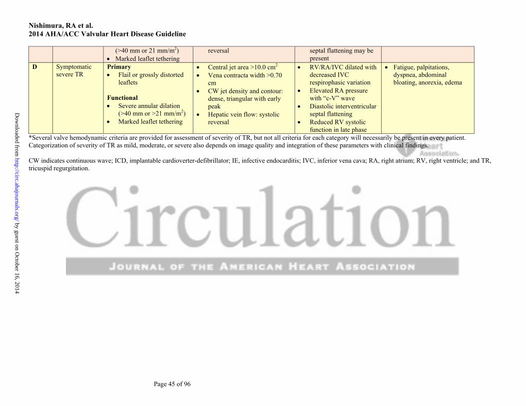
Nishimura, RA et al. 2014 AHA/ACC Valvular Heart Disease Guideline
Page 45 of 96
(>40 mm or 21 mm/m2) Marked leaflet tethering
reversal septal flattening may be present
D Symptomatic severe TR
Primary Flail or grossly distorted
leaflets
Functional Severe annular dilation
(>40 mm or >21 mm/m2) Marked leaflet tethering
Central jet area >10.0 cm2 Vena contracta width >0.70
cm CW jet density and contour:
dense, triangular with early peak
Hepatic vein flow: systolic reversal
RV/RA/IVC dilated with decreased IVC respirophasic variation
Elevated RA pressure with “c-V” wave
Diastolic interventricular septal flattening
Reduced RV systolic function in late phase
Fatigue, palpitations, dyspnea, abdominal bloating, anorexia, edema
*Several valve hemodynamic criteria are provided for assessment of severity of TR, but not all criteria for each category will necessarily be present in every patient. Categorization of severity of TR as mild, moderate, or severe also depends on image quality and integration of these parameters with clinical findings. CW indicates continuous wave; ICD, implantable cardioverter-defibrillator; IE, infective endocarditis; IVC, inferior vena cava; RA, right atrium; RV, right ventricle; and TR, tricuspid regurgitation.
by guest on October 16, 2014
http://circ.ahajournals.org/D
ownloaded from

Nishimura, RA et al. 2014 AHA/ACC Valvular Heart Disease Guideline
Page 46 of 96
8.2. Tricuspid Regurgitation See Figure 5 (Section 8.2.3) for indications for surgery.
8.2.1. Diagnosis and Follow-Up Class I
1. TTE is indicated to evaluate severity of TR, determine etiology, measure sizes of right-sided chambers and inferior vena cava, assess RV systolic function, estimate pulmonary artery systolic pressure, and characterize any associated left-sided heart disease. (Level of Evidence: C)
Class IIa
1. Invasive measurement of pulmonary artery pressures and pulmonary vascular resistance can be useful in patients with TR when clinical and noninvasive data regarding their values are discordant. (Level of Evidence: C)
Class IIb
1. CMR or real-time 3-dimensional echocardiography may be considered for assessment of RV systolic function and systolic and diastolic volumes in patients with severe TR (stages C and D) and suboptimal 2-dimensional echocardiograms. (Level of Evidence: C)
2. Exercise testing may be considered for the assessment of exercise capacity in patients with severe TR with no or minimal symptoms (stage C). (Level of Evidence: C)
8.2.2. Medical Therapy Class IIa
1. Diuretics can be useful for patients with severe TR and signs of right-sided HF (stage D). (Level of Evidence: C)
Class IIb
1. Medical therapies to reduce elevated pulmonary artery pressures and/or pulmonary vascular resistance might be considered in patients with severe functional TR (stages C and D). (Level of Evidence: C)
8.2.3. Intervention Class I
1. Tricuspid valve surgery is recommended for patients with severe TR (stages C and D) undergoing left-sided valve surgery. (Level of Evidence: C)
Class IIa
1. Tricuspid valve repair can be beneficial for patients with mild, moderate, or greater functional TR (stage B) at the time of left-sided valve surgery with either 1) tricuspid annular dilation or 2) prior evidence of right HF (237-246). (Level of Evidence: B)
2. Tricuspid valve surgery can be beneficial for patients with symptoms due to severe primary TR that are unresponsive to medical therapy (stage D). (Level of Evidence: C)
Class IIb
1. Tricuspid valve repair may be considered for patients with moderate functional TR (stage B) and pulmonary artery hypertension at the time of left-sided valve surgery. (Level of Evidence: C)
by guest on October 16, 2014http://circ.ahajournals.org/Downloaded from

Nishimura, RA et al. 2014 AHA/ACC Valvular Heart Disease Guideline
Page 47 of 96
2. Tricuspid valve surgery may be considered for asymptomatic or minimally symptomatic patients with severe primary TR (stage C) and progressive degrees of moderate or greater RV dilation and/or systolic dysfunction. (Level of Evidence: C)
3. Reoperation for isolated tricuspid valve repair or replacement may be considered for persistent symptoms due to severe TR (stage D) in patients who have undergone previous left-sided valve surgery and who do not have severe pulmonary hypertension or significant RV systolic dysfunction. (Level of Evidence: C)
Figure 5. Indications for Surgery
*See Table 17 for definition of stages. TA dilation is defined by >40 mm on TTE (>21 mm/m2) or >70 mm on direct intraoperative measurement. LV indicates left ventricular; PHTN, pulmonary hypertension; RV, right ventricular; TA, tricuspid annular; TR, tricuspid regurgitation; TTE, transthoracic echocardiogram; TV, tricuspid valve; and TVR, tricuspid valve replacement.
8.3. Stages of Tricuspid Stenosis See Table 18 for the stages of severe tricuspid stenosis (TS). Table 18. Stages of Severe TS
Stage Definition Valve Anatomy Valve Hemodynamics Hemodynamic Consequences
Symptoms
C, D Severe TS Thickened, distorted, calcified leaflets
T ½ ≥190 ms Valve area ≤1.0 cm2
RA/IVC enlargement
None or variable and dependent on severity of associated valve disease and degree of
by guest on October 16, 2014http://circ.ahajournals.org/Downloaded from

Nishimura, RA et al. 2014 AHA/ACC Valvular Heart Disease Guideline
Page 48 of 96
Stage Definition Valve Anatomy Valve Hemodynamics Hemodynamic Consequences
Symptoms
obstruction The transtricuspid diastolic gradient is highly variable and is affected by heart rate, forward flow, and phases of the respiratory cycle. However, severe TS usually has mean pressure gradients >5 to 10 mm Hg at heart rate 70. IVC indicates inferior vena cava; RA, right atrium; T ½, pressure half-time; and TS, tricuspid stenosis. (9)
8.4. Tricuspid Stenosis
8.4.1. Diagnosis and Follow-Up Class I
1. TTE is indicated in patients with TS to assess the anatomy of the valve complex, evaluate severity of stenosis, and characterize any associated regurgitation and/or left-sided valve disease. (Level of Evidence: C)
Class IIb
1. Invasive hemodynamic assessment of severity of TS may be considered in symptomatic patients when clinical and noninvasive data are discordant. (Level of Evidence: C)
8.4.2. Intervention Class I
1. Tricuspid valve surgery is recommended for patients with severe TS at the time of operation for left-sided valve disease. (Level of Evidence: C)
2. Tricuspid valve surgery is recommended for patients with isolated, symptomatic severe TS. (Level of Evidence: C)
Class IIb
1. Percutaneous balloon tricuspid commissurotomy might be considered in patients with isolated, symptomatic severe TS without accompanying TR. (Level of Evidence: C)
9. Stages of Pulmonic Valve Disease See Table 19 for the stages of severe pulmonic regurgitation and Table 20 for the stages of severe pulmonic stenosis. Table 19. Stages of Severe Pulmonic Regurgitation
Stage Definition Valve Anatomy Valve Hemodynamics Hemodynamic Consequences
Symptoms
C, D Severe PR Distorted or absent leaflets, annular dilation
Color jet fills RVOT CW jet density and
contour: dense laminar flow with steep deceleration slope; may terminate abruptly
Paradoxical septal motion (volume overload pattern)
RV enlargement
None or variable and dependent on cause of PR and RV function
CW indicates continuous wave; PR, pulmonic regurgitation; RV, right ventricular; and RVOT, right ventricular outflow tract. (247) Table 20. Stages of Severe Pulmonic Stenosis
by guest on October 16, 2014http://circ.ahajournals.org/Downloaded from
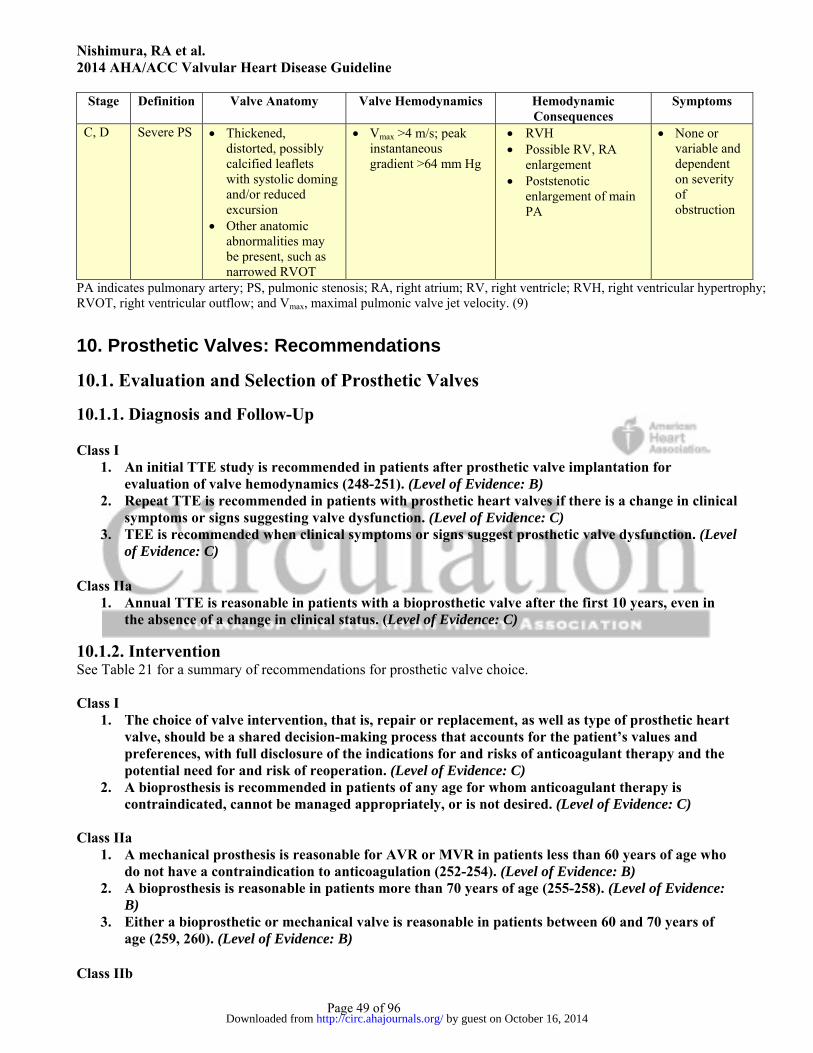
Nishimura, RA et al. 2014 AHA/ACC Valvular Heart Disease Guideline
Page 49 of 96
Stage Definition Valve Anatomy Valve Hemodynamics Hemodynamic Consequences
Symptoms
C, D Severe PS Thickened, distorted, possibly calcified leaflets with systolic doming and/or reduced excursion
Other anatomic abnormalities may be present, such as narrowed RVOT
Vmax >4 m/s; peak instantaneous gradient >64 mm Hg
RVH Possible RV, RA
enlargement Poststenotic
enlargement of main PA
None or variable and dependent on severity of obstruction
PA indicates pulmonary artery; PS, pulmonic stenosis; RA, right atrium; RV, right ventricle; RVH, right ventricular hypertrophy; RVOT, right ventricular outflow; and Vmax, maximal pulmonic valve jet velocity. (9)
10. Prosthetic Valves: Recommendations
10.1. Evaluation and Selection of Prosthetic Valves
10.1.1. Diagnosis and Follow-Up Class I
1. An initial TTE study is recommended in patients after prosthetic valve implantation for evaluation of valve hemodynamics (248-251). (Level of Evidence: B)
2. Repeat TTE is recommended in patients with prosthetic heart valves if there is a change in clinical symptoms or signs suggesting valve dysfunction. (Level of Evidence: C)
3. TEE is recommended when clinical symptoms or signs suggest prosthetic valve dysfunction. (Level of Evidence: C)
Class IIa
1. Annual TTE is reasonable in patients with a bioprosthetic valve after the first 10 years, even in the absence of a change in clinical status. (Level of Evidence: C)
10.1.2. Intervention See Table 21 for a summary of recommendations for prosthetic valve choice. Class I
1. The choice of valve intervention, that is, repair or replacement, as well as type of prosthetic heart valve, should be a shared decision-making process that accounts for the patient’s values and preferences, with full disclosure of the indications for and risks of anticoagulant therapy and the potential need for and risk of reoperation. (Level of Evidence: C)
2. A bioprosthesis is recommended in patients of any age for whom anticoagulant therapy is contraindicated, cannot be managed appropriately, or is not desired. (Level of Evidence: C)
Class IIa
1. A mechanical prosthesis is reasonable for AVR or MVR in patients less than 60 years of age who do not have a contraindication to anticoagulation (252-254). (Level of Evidence: B)
2. A bioprosthesis is reasonable in patients more than 70 years of age (255-258). (Level of Evidence: B)
3. Either a bioprosthetic or mechanical valve is reasonable in patients between 60 and 70 years of age (259, 260). (Level of Evidence: B)
Class IIb
by guest on October 16, 2014http://circ.ahajournals.org/Downloaded from

Nishimura, RA et al. 2014 AHA/ACC Valvular Heart Disease Guideline
Page 50 of 96
1. Replacement of the aortic valve by a pulmonary autograft (the Ross procedure), when performed by an experienced surgeon, may be considered in young patients when VKA anticoagulation is contraindicated or undesirable. (Level of Evidence: C)
Table 21. Summary of Recommendations for Prosthetic Valve Choice
Recommendations COR LOE References Choice of valve intervention and prosthetic valve type should be a shared decision process
I C N/A
A bioprosthesis is recommended in patients of any age for whom anticoagulant therapy is contraindicated, cannot be managed appropriately, or is not desired
I C N/A
A mechanical prosthesis is reasonable for AVR or MVR in patients <60 y of age who do not have a contraindication to anticoagulation
IIa B (252-254)
A bioprosthesis is reasonable in patients >70 y of age IIa B (255-258)
Either a bioprosthetic or mechanical valve is reasonable in patients between 60 y and 70 y of age
IIa B (259, 260)
Replacement of the aortic valve by a pulmonary autograft (the Ross procedure), when performed by an experienced surgeon, may be considered in young patients when VKA anticoagulation is contraindicated or undesirable
IIb C N/A
AVR indicates aortic valve replacement; COR, Class of Recommendation; LOE, Level of Evidence; MVR, mitral valve replacement; N/A, not applicable; and VKA, vitamin K antagonist.
10.2. Antithrombotic Therapy for Prosthetic Valves Class I
1. Anticoagulation with a VKA and international normalized ratio (INR) monitoring is recommended in patients with a mechanical prosthetic valve (261-263). (Level of Evidence: A)
2. Anticoagulation with a VKA to achieve an INR of 2.5 is recommended in patients with a mechanical AVR (bileaflet or current-generation single tilting disc) and no risk factors for thromboembolism (264-266). (Level of Evidence: B)
3. Anticoagulation with a VKA is indicated to achieve an INR of 3.0 in patients with a mechanical AVR and additional risk factors for thromboembolic events (AF, previous thromboembolism, LV dysfunction, or hypercoagulable conditions) or an older-generation mechanical AVR (such as ball-in-cage) (267). (Level of Evidence: B)
4. Anticoagulation with a VKA is indicated to achieve an INR of 3.0 in patients with a mechanical MVR (267, 268). (Level of Evidence: B)
5. Aspirin 75 mg to 100 mg daily is recommended in addition to anticoagulation with a VKA in patients with a mechanical valve prosthesis (269, 270). (Level of Evidence: A)
Class IIa
1. Aspirin 75 mg to 100 mg per day is reasonable in all patients with a bioprosthetic aortic or mitral valve (271-274). (Level of Evidence: B)
2. Anticoagulation with a VKA is reasonable for the first 3 months after bioprosthetic MVR or repair to achieve an INR of 2.5 (275). (Level of Evidence: C)
Class IIb 1. Anticoagulation, with a VKA, to achieve an INR of 2.5 may be reasonable for the first 3 months
after bioprosthetic AVR (276). (Level of Evidence: B) 2. Clopidogrel 75 mg daily may be reasonable for the first 6 months after TAVR in addition to life-
long aspirin 75 mg to 100 mg daily. (Level of Evidence: C)
by guest on October 16, 2014http://circ.ahajournals.org/Downloaded from
Nishimura, RA et al. 2014 AHA/ACC Valvular Heart Disease Guideline
Page 51 of 96
Class III: Harm 1. Anticoagulant therapy with oral direct thrombin inhibitors or anti-Xa agents should not be used
in patients with mechanical valve prostheses (277-279). (Level of Evidence: B)
10.3. Bridging Therapy for Prosthetic Valves Class I
1. Continuation of VKA anticoagulation with a therapeutic INR is recommended in patients with mechanical heart valves undergoing minor procedures (such as dental extractions or cataract removal) where bleeding is easily controlled. (Level of Evidence: C)
2. Temporary interruption of VKA anticoagulation, without bridging agents while the INR is subtherapeutic, is recommended in patients with a bileaflet mechanical AVR and no other risk factors for thrombosis who are undergoing invasive or surgical procedures. (Level of Evidence: C)
3. Bridging anticoagulation with either intravenous unfractionated heparin (UFH) or subcutaneous low-molecular-weight heparin (LMWH) is recommended during the time interval when the INR is subtherapeutic preoperatively in patients who are undergoing invasive or surgical procedures with a 1) mechanical AVR and any thromboembolic risk factor, 2) older-generation mechanical AVR, or 3) mechanical MVR. (Level of Evidence: C)
Class IIa
1. Administration of fresh frozen plasma or prothrombin complex concentrate is reasonable in patients with mechanical valves receiving VKA therapy who require emergency noncardiac surgery or invasive procedures. (Level of Evidence: C)
10.4. Excessive Anticoagulation and Serious Bleeding With Prosthetic Valves See Figure 6 for anticoagulation for prosthetic valves. Class IIa
1. Administration of fresh frozen plasma or prothrombin complex concentrate is reasonable in patients with mechanical valves and uncontrollable bleeding who require reversal of anticoagulation (280, 281). (Level of Evidence: B)
Figure 6. Anticoagulation for Prosthetic Valves
by guest on October 16, 2014http://circ.ahajournals.org/Downloaded from

Nishimura, RA et al. 2014 AHA/ACC Valvular Heart Disease Guideline
Page 52 of 96
Risk factors include AF, previous thromboembolism, LV dysfunction, hypercoagulable condition, and older-generation mechanical AVR. AF indicates atrial fibrillation; ASA, aspirin; AVR, aortic valve replacement; INR, international normalized ratio; LMWH, low-molecular-weight heparin; MVR, mitral valve replacement; PO, by mouth; QD, every day; SC, subcutaneous; TAVR, transcatheter aortic valve replacement; UFH, unfractionated heparin; and VKA, vitamin K antagonist.
10.5. Prosthetic Valve Thrombosis See Figure 7 for evaluation and management of suspected valve thrombosis.
10.5.1. Diagnosis and Follow-Up Class I
1. TTE is indicated in patients with suspected prosthetic valve thrombosis to assess hemodynamic severity and follow resolution of valve dysfunction (282, 283). (Level of Evidence: B)
2. TEE is indicated in patients with suspected prosthetic valve thrombosis to assess thrombus size and valve motion (283-285). (Level of Evidence: B)
Class IIa
1. Fluoroscopy or CT is reasonable in patients with suspected valve thrombosis to assess valve motion. (Level of Evidence: C)
10.5.2. Medical Therapy
by guest on October 16, 2014http://circ.ahajournals.org/Downloaded from

Nishimura, RA et al. 2014 AHA/ACC Valvular Heart Disease Guideline
Page 53 of 96
Class IIa 1. Fibrinolytic therapy is reasonable for patients with a thrombosed left-sided prosthetic heart valve,
recent onset (<14 days) of NYHA class I to II symptoms, and a small thrombus (<0.8 cm2) (283, 286). (Level of Evidence: B)
2. Fibrinolytic therapy is reasonable for thrombosed right-sided prosthetic heart valves (287, 288). (Level of Evidence: B)
10.5.3. Intervention Class I
1. Emergency surgery is recommended for patients with a thrombosed left-sided prosthetic heart valve with NYHA class III to IV symptoms (287, 289, 290). (Level of Evidence: B)
Class IIa
1. Emergency surgery is reasonable for patients with a thrombosed left-sided prosthetic heart valve with a mobile or large thrombus (>0.8 cm2) (283, 285, 290). (Level of Evidence: C)
Figure 7. Evaluation and Management of Suspected Prosthetic Valve Thrombosis
*See full-text guideline for dosage recommendations.
by guest on October 16, 2014http://circ.ahajournals.org/Downloaded from

Nishimura, RA et al. 2014 AHA/ACC Valvular Heart Disease Guideline
Page 54 of 96
CT indicates computed tomography; IV, intravenous; NYHA, New York Heart Association; Rx, therapy; TEE, transesophageal echocardiography; and TTE, transthoracic echocardiography.
10.6. Prosthetic Valve Stenosis Class I
1. Repeat valve replacement is indicated for severe symptomatic prosthetic valve stenosis. (Level of Evidence: C)
10.7. Prosthetic Valve Regurgitation Class I
1. Surgery is recommended for operable patients with mechanical heart valves with intractable hemolysis or HF due to severe prosthetic or paraprosthetic regurgitation (291, 292). (Level of Evidence: B)
Class IIa
1. Surgery is reasonable for operable patients with severe symptomatic or asymptomatic bioprosthetic regurgitation. (Level of Evidence C)
2. Percutaneous repair of paravalvular regurgitation is reasonable in patients with prosthetic heart valves and intractable hemolysis or NYHA class III/IV HF who are at high risk for surgery and have anatomic features suitable for catheter-based therapy when performed in centers with expertise in the procedure (293-295). (Level of Evidence B)
11. Infective Endocarditis: Recommendations
11.1. Diagnosis and Follow-Up See Figure 8 for recommendations for imaging studies in native valve endocarditis and prosthetic valve endocarditis. Class I
1. At least 2 sets of blood cultures should be obtained in patients at risk for IE (e.g., those with congenital or acquired VHD, previous IE, prosthetic heart valves, certain congenital or heritable heart malformations, immunodeficiency states, or injection drug users) who have unexplained fever for more than 48 hours (296) (Level of Evidence: B) or patients with newly diagnosed left-sided valve regurgitation. (Level of Evidence: C)
2. The Modified Duke Criteria should be used in evaluating a patient with suspected IE (Tables 24 and 25 in the full-text guideline) (297-300). (Level of Evidence: B)
3. Patients with IE should be evaluated and managed with consultation of a multispecialty Heart Valve Team including an infectious disease specialist, cardiologist, and cardiac surgeon. In surgically managed patients, this team should also include a cardiac anesthesiologist (301). (Level of Evidence: B)
4. TTE is recommended in patients with suspected IE to identify vegetations, characterize the hemodynamic severity of valvular lesions, assess ventricular function and pulmonary pressures, and detect complications (302-306). (Level of Evidence: B)
5. TEE is recommended in all patients with known or suspected IE when TTE is nondiagnostic, when complications have developed or are clinically suspected, or when intracardiac device leads are present (307-315). (Level of Evidence: B)
6. TTE and/or TEE are recommended for reevaluation of patients with IE who have a change in clinical signs or symptoms (e.g., new murmur, embolism, persistent fever, HF, abscess, or atrioventricular heart block) and in patients at high risk of complications (e.g., extensive infected
by guest on October 16, 2014http://circ.ahajournals.org/Downloaded from
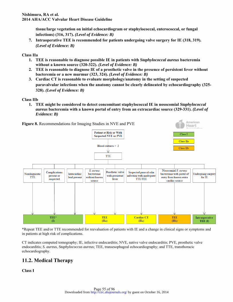
Nishimura, RA et al. 2014 AHA/ACC Valvular Heart Disease Guideline
Page 55 of 96
tissue/large vegetation on initial echocardiogram or staphylococcal, enterococcal, or fungal infections) (316, 317). (Level of Evidence: B)
7. Intraoperative TEE is recommended for patients undergoing valve surgery for IE (318, 319). (Level of Evidence: B)
Class IIa 1. TEE is reasonable to diagnose possible IE in patients with Staphylococcal aureus bacteremia
without a known source (320-322). (Level of Evidence: B) 2. TEE is reasonable to diagnose IE of a prosthetic valve in the presence of persistent fever without
bacteremia or a new murmur (323, 324). (Level of Evidence: B) 3. Cardiac CT is reasonable to evaluate morphology/anatomy in the setting of suspected
paravalvular infections when the anatomy cannot be clearly delineated by echocardiography (325-328). (Level of Evidence: B)
Class IIb 1. TEE might be considered to detect concomitant staphylococcal IE in nosocomial Staphylococcal
aureus bacteremia with a known portal of entry from an extracardiac source (329-331). (Level of Evidence: B)
Figure 8. Recommendations for Imaging Studies in NVE and PVE
*Repeat TEE and/or TTE recommended for reevaluation of patients with IE and a change in clinical signs or symptoms and in patients at high risk of complications. CT indicates computed tomography; IE, infective endocarditis; NVE, native valve endocarditis; PVE, prosthetic valve endocarditis; S. aureus, Staphylococcus aureus; TEE, transesophageal echocardiography; and TTE, transthoracic echocardiography.
11.2. Medical Therapy Class I
by guest on October 16, 2014http://circ.ahajournals.org/Downloaded from

Nishimura, RA et al. 2014 AHA/ACC Valvular Heart Disease Guideline
Page 56 of 96
1. Appropriate antibiotic therapy should be initiated and continued after blood cultures are obtained with guidance from antibiotic sensitivity data and infectious disease consultants (296). (Level of Evidence: B)
Class IIa 1. It is reasonable to temporarily discontinue anticoagulation in patients with IE who develop
central nervous system symptoms compatible with embolism or stroke regardless of the other indications for anticoagulation (332-337). (Level of Evidence: B)
Class IIb 1. Temporary discontinuation of VKA anticoagulation might be considered in patients receiving
VKA anticoagulation at the time of IE diagnosis (333, 338-341). (Level of Evidence: B) Class III: Harm
1. Patients with known VHD should not receive antibiotics before blood cultures are obtained for unexplained fever. (Level of Evidence: C)
11.3. Intervention See Figure 9 for diagnosis and treatment of IE. Class I
1. Decisions about timing of surgical intervention should be made by a multispecialty Heart Valve Team of cardiology, cardiothoracic surgery, and infectious disease specialists (301). (Level of Evidence: B)
2. Early surgery (during initial hospitalization before completion of a full therapeutic course of antibiotics) is indicated in patients with IE who present with valve dysfunction resulting in symptoms of HF (342-347). (Level of Evidence: B)
3. Early surgery (during initial hospitalization before completion of a full therapeutic course of antibiotics) is indicated in patients with left-sided IE caused by Staphylococcal aureus, fungal, or other highly resistant organisms (347-354). (Level of Evidence: B)
4. Early surgery (during initial hospitalization before completion of a full therapeutic course of antibiotics) is indicated in patients with IE complicated by heart block, annular or aortic abscess, or destructive penetrating lesions (347, 355-359). (Level of Evidence: B)
5. Early surgery (during initial hospitalization before completion of a full therapeutic course of antibiotics) for IE is indicated in patients with evidence of persistent infection as manifested by persistent bacteremia or fevers lasting longer than 5 to 7 days after onset of appropriate antimicrobial therapy (347, 352, 353, 360-362). (Level of Evidence: B)
6. Surgery is recommended for patients with prosthetic valve endocarditis and relapsing infection (defined as recurrence of bacteremia after a complete course of appropriate antibiotics and subsequently negative blood cultures) without other identifiable source for portal of infection. (Level of Evidence: C)
7. Complete removal of pacemaker or defibrillator systems, including all leads and the generator, is indicated as part of the early management plan in patients with IE with documented infection of the device or leads (363-366). (Level of Evidence: B)
Class IIa 1. Complete removal of pacemaker or defibrillator systems, including all leads and the generator, is
reasonable in patients with valvular IE caused by Staphylococcal aureus or fungi, even without evidence of device or lead infection (363-366). (Level of Evidence: B)
2. Complete removal of pacemaker or defibrillator systems, including all leads and the generator, is reasonable in patients undergoing valve surgery for valvular IE. (Level of Evidence: C)
3. Early surgery (during initial hospitalization before completion of a full therapeutic course of antibiotics) is reasonable in patients with IE who present with recurrent emboli and persistent vegetations despite appropriate antibiotic therapy (302, 367, 368). (Level of Evidence: B)
by guest on October 16, 2014http://circ.ahajournals.org/Downloaded from
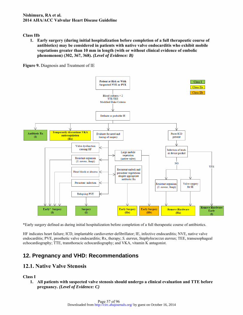
Nishimura, RA et al. 2014 AHA/ACC Valvular Heart Disease Guideline
Page 57 of 96
Class IIb
1. Early surgery (during initial hospitalization before completion of a full therapeutic course of antibiotics) may be considered in patients with native valve endocarditis who exhibit mobile vegetations greater than 10 mm in length (with or without clinical evidence of embolic phenomenon) (302, 367, 368). (Level of Evidence: B)
Figure 9. Diagnosis and Treatment of IE
*Early surgery defined as during initial hospitalization before completion of a full therapeutic course of antibiotics. HF indicates heart failure; ICD, implantable cardioverter-defibrillator; IE, infective endocarditis; NVE, native valve endocarditis; PVE, prosthetic valve endocarditis; Rx, therapy; S. aureus, Staphylococcus aureus; TEE, transesophageal echocardiography; TTE, transthoracic echocardiography; and VKA, vitamin K antagonist.
12. Pregnancy and VHD: Recommendations
12.1. Native Valve Stenosis Class I
1. All patients with suspected valve stenosis should undergo a clinical evaluation and TTE before pregnancy. (Level of Evidence: C)
by guest on October 16, 2014http://circ.ahajournals.org/Downloaded from

Nishimura, RA et al. 2014 AHA/ACC Valvular Heart Disease Guideline
Page 58 of 96
2. All patients with severe valve stenosis (stages C and D) should undergo prepregnancy counseling by a cardiologist with expertise in managing patients with VHD during pregnancy. (Level of Evidence: C)
3. All patients referred for a valve operation before pregnancy should receive prepregnancy counseling by a cardiologist with expertise in managing patients with VHD during pregnancy about the risks and benefits of all options for operative interventions, including mechanical prosthesis, bioprosthesis, and valve repair. (Level of Evidence: C)
4. Pregnant patients with severe valve stenosis (stages C and D) should be monitored in a tertiary care center with a dedicated Heart Valve Team of cardiologists, surgeons, anesthesiologists, and obstetricians with expertise in the management of high-risk cardiac patients during pregnancy. (Level of Evidence: C)
12.1.1. Diagnosis and Follow-Up Class IIa
1. Exercise testing is reasonable in asymptomatic patients with severe AS (aortic velocity ≥4 m per second or mean pressure gradient ≥40 mm Hg, stage C) before pregnancy. (Level of Evidence: C)
12.1.2. Medical Therapy Class I
1. Anticoagulation should be given to pregnant patients with MS and AF unless contraindicated. (Level of Evidence: C)
Class IIa
1. Use of beta blockers as required for rate control is reasonable for pregnant patients with MS in the absence of contraindication if tolerated. (Level of Evidence: C)
Class IIb
1. Use of diuretics may be reasonable for pregnant patients with MS and HF symptoms (stage D). (Level of Evidence: C)
Class III: Harm
1. ACE inhibitors and ARBs should not be given to pregnant patients with valve stenosis (369-371). (Level of Evidence: B)
12.1.3. Intervention Class I
1. Valve intervention is recommended before pregnancy for symptomatic patients with severe AS (aortic velocity ≥4.0 m per second or mean pressure gradient ≥40 mm Hg, stage D). (Level of Evidence: C)
2. Valve intervention is recommended before pregnancy for symptomatic patients with severe MS (mitral valve area ≤1.5 cm2, stage D). (Level of Evidence: C)
3. Percutaneous mitral balloon commissurotomy is recommended before pregnancy for asymptomatic patients with severe MS (mitral valve area ≤1.5 cm2, stage C) who have valve morphology favorable for percutaneous mitral balloon commissurotomy. (Level of Evidence: C)
Class IIa
1. Valve intervention is reasonable before pregnancy for asymptomatic patients with severe AS (aortic velocity ≥4.0 m per second or mean pressure gradient ≥40 mm Hg, stage C). (Level of Evidence: C)
by guest on October 16, 2014http://circ.ahajournals.org/Downloaded from

Nishimura, RA et al. 2014 AHA/ACC Valvular Heart Disease Guideline
Page 59 of 96
2. Percutaneous mitral balloon commissurotomy is reasonable for pregnant patients with severe MS (mitral valve area ≤1.5 cm2, stage D) with valve morphology favorable for percutaneous mitral balloon commissurotomy who remain symptomatic with NYHA class III to IV HF symptoms despite medical therapy (372-376). (Level of Evidence: B)
3. Valve intervention is reasonable for pregnant patients with severe MS (mitral valve area ≤1.5 cm2, stage D) and valve morphology not favorable for percutaneous mitral balloon commissurotomy only if there are refractory NYHA class IV HF symptoms. (Level of Evidence: C)
4. Valve intervention is reasonable for pregnant patients with severe AS (mean pressure gradient ≥40 mm Hg, stage D) only if there is hemodynamic deterioration or NYHA class III to IV HF symptoms (377-383). (Level of Evidence: B)
Class III: Harm
1. Valve operation should not be performed in pregnant patients with valve stenosis in the absence of severe HF symptoms. (Level of Evidence: C)
12.2. Native Valve Regurgitation
12.2.1. Diagnosis and Follow-Up Class I
1. All patients with suspected valve regurgitation should undergo a clinical evaluation and TTE before pregnancy. (Level of Evidence: C)
2. All patients with severe valve regurgitation (stages C and D) should undergo prepregnancy counseling by a cardiologist with expertise in managing patients with VHD during pregnancy. (Level of Evidence: C)
3. All patients referred for a valve operation before pregnancy should receive prepregnancy counseling by a cardiologist with expertise in managing patients with VHD during pregnancy regarding the risks and benefits of all options for operative interventions, including mechanical prosthesis, bioprosthesis, and valve repair. (Level of Evidence: C)
4. Pregnant patients with severe regurgitation (stages C and D) should be monitored in a tertiary care center with a dedicated Heart Valve Team of cardiologists, surgeons, anesthesiologists, and obstetricians with expertise in managing high-risk cardiac patients. (Level of Evidence: C)
Class IIa
1. Exercise testing is reasonable in asymptomatic patients with severe valve regurgitation (stage C) before pregnancy. (Level of Evidence: C)
12.2.2. Medical Therapy Class III: Harm
1. ACE inhibitors and ARBs should not be given to pregnant patients with valve regurgitation (369-371). (Level of Evidence: B)
12.2.3. Intervention Class I
1. Valve repair or replacement is recommended before pregnancy for symptomatic women with severe valve regurgitation (stage D). (Level of Evidence: C)
Class IIa
1. Valve operation for pregnant patients with severe valve regurgitation is reasonable only if there are refractory NYHA class IV HF symptoms (stage D). (Level of Evidence: C)
by guest on October 16, 2014http://circ.ahajournals.org/Downloaded from

Nishimura, RA et al. 2014 AHA/ACC Valvular Heart Disease Guideline
Page 60 of 96
Class IIb 1. Valve repair before pregnancy may be considered in the asymptomatic patient with severe MR
(stage C) and a valve suitable for valve repair, but only after detailed discussion with the patient about the risks and benefits of the operation and its outcome on future pregnancies. (Level of Evidence: C)
Class III: Harm
1. Valve operations should not be performed in pregnant patients with valve regurgitation in the absence of severe intractable HF symptoms. (Level of Evidence: C)
12.3. Prosthetic Valves in Pregnancy
12.3.1. Diagnosis and Follow-Up Class I
1. All patients with a prosthetic valve should undergo a clinical evaluation and baseline TTE before pregnancy. (Level of Evidence: C)
2. All patients with a prosthetic valve should undergo prepregnancy counseling by a cardiologist with expertise in managing patients with VHD during pregnancy. (Level of Evidence: C)
3. TTE should be performed in all pregnant patients with a prosthetic valve if not done before pregnancy. (Level of Evidence: C)
4. Repeat TTE should be performed in all pregnant patients with a prosthetic valve who develop symptoms. (Level of Evidence: C)
5. TEE should be performed in all pregnant patients with a mechanical prosthetic valve who have prosthetic valve obstruction or experience an embolic event. (Level of Evidence: C)
6. Pregnant patients with a mechanical prosthesis should be monitored in a tertiary care center with a dedicated Heart Valve Team of cardiologists, surgeons, anesthesiologists, and obstetricians with expertise in the management of high-risk cardiac patients. (Level of Evidence: C)
12.3.2. Medical Therapy See Figure 10 for anticoagulation of pregnant patients with mechanical valves. Class I
1. Therapeutic anticoagulation with frequent monitoring is recommended for all pregnant patients with a mechanical prosthesis (384, 385). (Level of Evidence: B)
2. Warfarin is recommended in pregnant patients with a mechanical prosthesis to achieve a therapeutic INR in the second and third trimesters (386-391). (Level of Evidence: B)
3. Discontinuation of warfarin with initiation of intravenous UFH (with an activated partial thromboplastin time [aPTT] >2 times control) is recommended before planned vaginal delivery in pregnant patients with a mechanical prosthesis. (Level of Evidence: C)
4. Low-dose aspirin (75 mg to 100 mg) once per day is recommended for pregnant patients in the second and third trimesters with either a mechanical prosthesis or bioprosthesis. (Level of Evidence: C)
Class IIa
1. Continuation of warfarin during the first trimester is reasonable for pregnant patients with a mechanical prosthesis if the dose of warfarin to achieve a therapeutic INR is 5 mg per day or less after full discussion with the patient about risks and benefits (384, 385, 390-393). (Level of Evidence: B)
2. Dose-adjusted LMWH at least 2 times per day (with a target anti-Xa level of 0.8 U/mL to 1.2 U/mL, 4 to 6 hours postdose) during the first trimester is reasonable for pregnant patients with a
by guest on October 16, 2014http://circ.ahajournals.org/Downloaded from

Nishimura, RA et al. 2014 AHA/ACC Valvular Heart Disease Guideline
Page 61 of 96
mechanical prosthesis if the dose of warfarin is greater than 5 mg per day to achieve a therapeutic INR (386-389, 394, 395). (Level of Evidence: B)
3. Dose-adjusted continuous intravenous UFH (with an aPTT at least 2 times control) during the first trimester is reasonable for pregnant patients with a mechanical prosthesis if the dose of warfarin is greater than 5 mg per day to achieve a therapeutic INR (384, 385, 392). (Level of Evidence: B)
Class IIb
1. Dose-adjusted LMWH at least 2 times per day (with a target anti-Xa level of 0.8 U/mL to 1.2 U/mL, 4 to 6 hours postdose) during the first trimester may be reasonable for pregnant patients with a mechanical prosthesis if the dose of warfarin is 5 mg per day or less to achieve a therapeutic INR (386-389, 394-396). (Level of Evidence: B)
2. Dose-adjusted continuous infusion of UFH (with aPTT at least 2 times control) during the first trimester may be reasonable for pregnant patients with a mechanical prosthesis if the dose of warfarin is 5 mg per day or less to achieve a therapeutic INR (384, 385, 392). (Level of Evidence: B)
Class III: Harm
1. LMWH should not be administered to pregnant patients with mechanical prostheses unless anti-Xa levels are monitored 4 to 6 hours after administration (387, 388, 394, 395, 397). (Level of Evidence: B)
Figure 10. Anticoagulation of Pregnant Patients With Mechanical Valves
by guest on October 16, 2014http://circ.ahajournals.org/Downloaded from

Nishimura, RA et al. 2014 AHA/ACC Valvular Heart Disease Guideline
Page 62 of 96
aPTT indicates activated partial thromboplastin time; ASA, aspirin; INR, international normalized ratio; LMWH, low-molecular-weight heparin; QD, once daily; and UFH, unfractionated heparin.
by guest on October 16, 2014http://circ.ahajournals.org/Downloaded from

Nishimura, RA et al. 2014 AHA/ACC Valvular Heart Disease Guideline
Page 63 of 96
13. Surgical Considerations: Recommendations
13.1. Evaluation of Coronary Anatomy See Figure 11 for evaluation and management of CAD in patients undergoing valve surgery.
Class I 1. Coronary angiography is indicated before valve intervention in patients with symptoms of angina,
objective evidence of ischemia, decreased LV systolic function, history of CAD, or coronary risk factors (including men age >40 years and postmenopausal women). (Level of Evidence: C)
2. Coronary angiography should be performed as part of the evaluation of patients with chronic severe secondary MR. (Level of Evidence: C)
Class IIa 1. Surgery without coronary angiography is reasonable for patients having emergency valve surgery
for acute valve regurgitation, disease of the aortic sinuses or ascending aorta, or IE. (Level of Evidence: C)
2. CT coronary angiography is reasonable to exclude the presence of significant obstructive CAD in selected patients with a low/intermediate pretest probability of CAD. A positive coronary CT angiogram (the presence of any epicardial CAD) can be confirmed with invasive coronary angiography (398-404). (Level of Evidence: B)
13.2. Concomitant Procedures
13.2.1. Intervention for CAD Class IIa
1. CABG or percutaneous coronary intervention is reasonable in patients undergoing valve repair or replacement with significant CAD (≥70% reduction in luminal diameter in major coronary arteries or ≥50% reduction in luminal diameter in the left main coronary artery). (Level of Evidence: C)
Figure 11. Evaluation and Management of CAD in Patients Undergoing Valve Surgery
by guest on October 16, 2014http://circ.ahajournals.org/Downloaded from
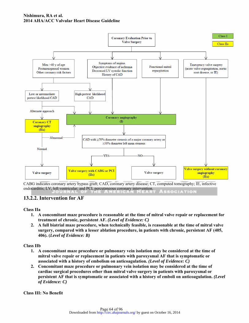
Nishimura, RA et al. 2014 AHA/ACC Valvular Heart Disease Guideline
Page 64 of 96
CABG indicates coronary artery bypass graft; CAD, coronary artery disease; CT, computed tomography; IE, infective endocarditis; LV, left ventricular; and PCI, percutaneous coronary intervention.
13.2.2. Intervention for AF Class IIa
1. A concomitant maze procedure is reasonable at the time of mitral valve repair or replacement for treatment of chronic, persistent AF. (Level of Evidence: C)
2. A full biatrial maze procedure, when technically feasible, is reasonable at the time of mitral valve surgery, compared with a lesser ablation procedure, in patients with chronic, persistent AF (405, 406). (Level of Evidence: B)
Class IIb
1. A concomitant maze procedure or pulmonary vein isolation may be considered at the time of mitral valve repair or replacement in patients with paroxysmal AF that is symptomatic or associated with a history of embolism on anticoagulation. (Level of Evidence: C)
2. Concomitant maze procedure or pulmonary vein isolation may be considered at the time of cardiac surgical procedures other than mitral valve surgery in patients with paroxysmal or persistent AF that is symptomatic or associated with a history of emboli on anticoagulation. (Level of Evidence: C)
Class III: No Benefit
by guest on October 16, 2014http://circ.ahajournals.org/Downloaded from

Nishimura, RA et al. 2014 AHA/ACC Valvular Heart Disease Guideline
Page 65 of 96
1. Catheter ablation for AF should not be performed in patients with severe MR when mitral repair or replacement is anticipated, with preference for the combined maze procedure plus mitral valve repair (407). (Level of Evidence: B)
14. Noncardiac Surgery in Patients With VHD: Recommendations Class IIa
1. Moderate-risk elective noncardiac surgery with appropriate intraoperative and postoperative hemodynamic monitoring is reasonable to perform in patients with asymptomatic severe AS (408-411). (Level of Evidence: B)
2. Moderate-risk elective noncardiac surgery with appropriate intraoperative and postoperative hemodynamic monitoring is reasonable to perform in patients with asymptomatic severe MR. (Level of Evidence: C)
3. Moderate-risk elective noncardiac surgery with appropriate intraoperative and postoperative hemodynamic monitoring is reasonable to perform in patients with asymptomatic severe AR and a normal LVEF. (Level of Evidence: C)
Class IIb
1. Moderate-risk elective noncardiac surgery in patients with appropriate intraoperative and postoperative hemodynamic monitoring may be reasonable to perform in asymptomatic patients with severe MS if valve morphology is not favorable for percutaneous balloon mitral commissurotomy. (Level of Evidence: C)
Presidents and Staff American College of Cardiology John Gordon Harold, MD, MACC, President Shalom Jacobovitz, Chief Executive Officer William J. Oetgen, MD, MBA, FACC, Executive Vice President, Science, Education, and Quality Charlene L. May, Senior Director, Science and Clinical Policy American College of Cardiology/American Heart Association Lisa Bradfield, CAE, Director, Science and Clinical Policy Emily Cottrell, MA, Specialist, Science and Clinical Policy American Heart Association Mariell Jessup, MD, FACC, FAHA, President Nancy Brown, Chief Executive Officer Rose Marie Robertson, MD, FAHA, Chief Science Officer Gayle R. Whitman, PhD, RN, FAHA, FAAN, Senior Vice President, Office of Science Operations Marco Di Buono, PhD, Vice President of Science and Research Jody Hundley, Production Manager, Scientific Publications, Office of Science Operations Key Words: ACC/AHA Practice Guidelines ■ anticoagulation therapy ■ aortic stenosis ■ aortic regurgitation■ bicuspid aortic valve ■ cardiac surgery ■ heart valves ■ infective endocarditis ■ mitral stenosis ■ mitral regurgitation ■ prosthetic valves ■ pulmonic regurgitation ■ pulmonic stenosis ■ transcatheter aortic valve replacement ■ tricuspid stenosis ■ tricuspid regurgitation ■ valvular heart disease.
by guest on October 16, 2014http://circ.ahajournals.org/Downloaded from

Nishimura, RA et al. 2014 AHA/ACC Valvular Heart Disease Guideline
Page 66 of 96
Appendix 1. Author Relationships With Industry and Other Entities (Relevant)–AHA/ACC Guideline for the Management of Patients With Valvular Heart Disease
Committee Member
Employment Consultant Speaker’s Bureau
Ownership/ Partnership/
Principal
Personal Research
Institutional, Organizational,
or Other Financial Benefit
Expert Witness
Voting Recusals by Section*
Rick A. Nishimura, Co-Chair
Mayo Clinic, Division of Cardiovascular Disease—Judd and Mary Morris Leighton Professor of Medicine
None None None None None None None
Catherine M. Otto, Co-Chair
University of Washington Division of Cardiology—Professor of Medicine
None None None None None None None
Robert O. Bonow
Northwestern University Medical School—Goldberg Distinguished Professor
None None None None None None None
Blasé A. Carabello
VA Medical Center—Professor of Medicine, Baylor College of Medicine
None None None None Edwards Lifesciences (DSMB)†
Medtronic†
None 2.4.2, 3.2.3, 3.2.4, 4.3.3, 5.1.3, 6.2.3, 7.3.1.1, 7.3.3, 7.4.3, 8.2.3, 11.1.1, 11.1.2, 11.2.2, 11.3.2, 11.4, 11.6.1, 11.6.2, 11.6.3, 11.7.3, 11.8.3, 12.2.1, 12.2.3, 13.1, 13.1.2, 13.1.3, 13.2.1, 13.2.3, 13.3.1, 13.3.2, 14.1, and 14.2.2.
John P. Erwin, III
Scott and White Hospital and Clinic—Senior Staff Cardiologist, Associate Professor of Medicine
None None None None None None None
Robert A. Guyton
Emory Clinic, Inc.—Professor and Chief, Division of
Medtronic
None None None None Defendant, Cardiac Surgery,
2.4.2, 3.2.3, 3.2.4, 4.3.3, 5.1.3, 6.2.3, 7.3.1.1, 7.3.3, 7.4.3,
by guest on October 16, 2014
http://circ.ahajournals.org/D
ownloaded from

Nishimura, RA et al. 2014 AHA/ACC Valvular Heart Disease Guideline
Page 67 of 96
Cardiothoracic Surgery 2013 8.2.3, 11.1.1, 11.1.2, 11.2.2, 11.3.2, 11.4, 11.6.1, 11.6.2, 11.6.3, 11.7.3, 11.8.3, 12.2.1, 12.2.3, 13.1, 13.1.2, 13.1.3, 13.2.1, 13.2.3, 13.3.1, 13.3.2, 14.1, and 14.2.2.
Patrick T. O’Gara
Brigham and Women’s Hospital—Professor of Medicine; Harvard Medical School—Director of Clinical Cardiology
None None None None None None None
Carlos E. Ruiz Lenox Hill Heart and Vascular Institute of New York—Professor and Chief, Division of Pediatric Cardiology
None None None None None None None
Nikolaos J. Skubas
Weill Cornell Medical College—Associate Professor of Anesthesiology and Director of Cardiac Anesthesia
None None None None None None None
Paul Sorajja Mayo Clinic—Associate Professor of Medicine
None None Intellectual property in patent on percutaneous closure of paravalvular prosthetic regurgitation
None None None None
Thoralf M. Sundt, III
Massachusetts General Hospital—Chief, Division Cardiac Surgery
St. Jude Medical
None None None None AATS, Secretary- Elect
2.4.2, 3.2.3, 3.2.4, 4.3.3, 5.1.3, 6.2.3, 7.3.1.1, 7.3.3, 7.4.3, 8.2.3, 11.1.1, 11.1.2, 11.2.2, 11.3.2, 11.4, 11.6.1, 11.6.2, 11.6.3, 11.7.3, 11.8.3, 12.2.1, 12.2.3, 13.1, 13.1.2,
by guest on October 16, 2014
http://circ.ahajournals.org/D
ownloaded from

Nishimura, RA et al. 2014 AHA/ACC Valvular Heart Disease Guideline
Page 68 of 96
13.1.3, 13.2.1, 13.2.3, 13.3.1, 13.3.2, 14.1, and 14.2.2.
James D. Thomas
Cleveland Clinic—Professor of Medicine and Biomedical Engineering
None None None None None None None
This table represents the relationships of committee members with industry and other entities that were determined to be relevant to this document. These relationships were reviewed and updated in conjunction with all meetings and/or conference calls of the writing committee during the document development process. The table does not necessarily reflect relationships with industry at the time of publication. A person is deemed to have a significant interest in a business if the interest represents ownership of ≥5% of the voting stock or share of the business entity, or ownership of ≥$10,000 of the fair market value of the business entity; or if funds received by the person from the business entity exceed 5% of the person’s gross income for the previous year. Relationships that exist with no financial benefit are also included for the purpose of transparency. Relationships in this table are modest unless otherwise noted. According to the ACC/AHA, a person has a relevant relationship IF: a) The relationship or interest relates to the same or similar subject matter, intellectual property or asset, topic, or issue addressed in the document; or b) The company/entity (with whom the relationship exists) makes a drug, drug class, or device addressed in the document, or makes a competing drug or device addressed in the document; or c) The person or a member of the person’s household, has a reasonable potential for financial, professional or other personal gain or loss as a result of the issues/content addressed in the document. *Writing committee members are required to recuse themselves from voting on sections to which their specific relationships with industry and other entities may apply. Section numbers pertain to those in the full-text guideline. †No financial benefit. AATS indicates American Association of Thoracic Surgery; DSMB, data safety monitoring board; and VA, Veterans Affair.
by guest on October 16, 2014
http://circ.ahajournals.org/D
ownloaded from

Nishimura, RA et al. 2014 AHA/ACC Valvular Heart Disease Guideline
Page 69 of 96
Appendix 2. Reviewer Relationships With Industry and Other Entities (Relevant)–AHA/ACC Guideline for the Management of Patients With Valvular Heart Disease
Reviewer Representation Employment Consultant Speaker’s Bureau
Ownership/ Partnership/
Principal
Personal Research
Institutional, Organizational,
or Other Financial Benefit
Expert Witness
Blair D. Erb Official Reviewer—ACC Board of Trustees
Bozeman Deaconess Hospital, Cardiology Consultants—Physician
None None None None Medtronic None
Mario J. Garcia
Official Reviewer—AHA
Montefiore Medical Center-Albert Einstein College of Medicine —Chief, Division of Cardiology
None None None None Medtronic† Pfizer
None
Smadar Kort Official Reviewer—ACC Board of Governors
Stony Brook University Medical Center—Clinical Professor of Medicine; Director, Echocardiography Laboratory; Director, Cardiovascular Imaging
None None None None Pfizer None
Richard J. Kovacs
Official Reviewer—ACC/AHA Task Force on Practice Guidelines
Indiana University—Clinical Director and Professor of Clinical Medicine,
Biomedical Systems
Insight Pharmaceuticals
None None None Cook Incorporated-Med Institute*
Eli Lilly* (DSMB)
None
by guest on October 16, 2014
http://circ.ahajournals.org/D
ownloaded from

Nishimura, RA et al. 2014 AHA/ACC Valvular Heart Disease Guideline
Page 70 of 96
Krannert Institute of Cardiology; Associate Dean for Clinical Research
Theravance*
David H. Adams
Organizational Reviewer—AATS
The Mount Sinai Medical Center—Marie-Josée and Henry R. Kravis Professor; Chairman, Department of Cardiothoracic Surgery
None None None None Edward Lifesciences*
Medtronic
None
Howard Herrmann
Organizational Reviewer—SCAI
University of Pennsylvania Perelman School of Medicine—Professor of Medicine; Director, Interventional Cardiology Program
Paieon Siemens
Medical St. Jude
Medical
None Micro-Interventional Devices*
Abbott Vascular*
Edward Lifesciences*
Medtronic† Siemens
Medical* St. Jude
Medical WL Gore and
Associates
None None
Sunil V. Mankad
Organizational Reviewer—ASE
Mayo Clinic—Associate Professor of Medicine
None None None None None None
Patrick McCarthy
Organizational Reviewer—STS
Northwestern University, Feinberg School of Medicine—Surgical Director, Bluhm Cardiovascular Institute
Abbott Vascular*
Baxter Edward
Lifesciences*
None Cardious Edward
Lifesciences*
MiCardia
None Direct Flow None
Stanton K. Shernan
Organizational Reviewer—SCA
Brigham and Women’s Hospital
None Philips Healthcare
None None National Board of
Defendant, Echocardiog
by guest on October 16, 2014
http://circ.ahajournals.org/D
ownloaded from

Nishimura, RA et al. 2014 AHA/ACC Valvular Heart Disease Guideline
Page 71 of 96
Echocardiography Officer†
raphy, 2012
Mouaz H. Al-Mallah
Content Reviewer—Prevention of Cardiovascular Disease Committee
King Abdul-Aziz Cardiac Center—Associate Professor of Medicine
Bracco None None None None None
Nancy M. Albert
Content Reviewer— ACC/AHA Task Force on Practice Guidelines
Cleveland Clinic Foundation—Senior Director of Nursing Research and Clinical Nursing Specialists, Kaufman Center for Heart Failure
Medtronic None None None None None
Jeffrey L. Anderson
Content Reviewer— ACC/AHA Task Force on Practice Guidelines
Intermountain Medical Center—Associate Chief of Cardiology
Sanofi-aventis
None None GlaxoSmithKline
None None
Robert H. Beekman
Content Reviewer—Adult Congenital and Pediatric Cardiology Section
Cincinnati Children's Hospital Medical Center— Division of Cardiology
St. Jude Medical
None None None None None
Vera A. Bittner
Content Reviewer— Prevention of Cardiovascular Disease Committee
University of Alabama at Birmingham—Professor of Medicine; Director, Cardiac Rehabilitation
Novartis None None Amgen AstraZeneca† Eli Lilly† GlaxoSmithK
line* NIH/Joint
Abbott* Sanofi-
aventis† Schering
Plough†
Pfizer None
Biykem Content Michael E. None None None Forest Amgen None
by guest on October 16, 2014
http://circ.ahajournals.org/D
ownloaded from
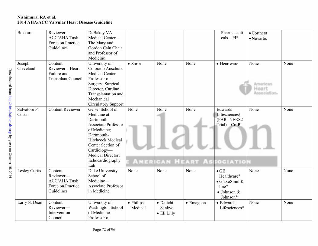
Nishimura, RA et al. 2014 AHA/ACC Valvular Heart Disease Guideline
Page 72 of 96
Bozkurt Reviewer— ACC/AHA Task Force on Practice Guidelines
DeBakey VA Medical Center—The Mary and Gordon Cain Chair and Professor of Medicine
Pharmaceuticals—PI*
Corthera Novartis
Joseph Cleveland
Content Reviewer—Heart Failure and Transplant Council
University of Colorado Anschutz Medical Center—Professor of Surgery; Surgical Director, Cardiac Transplantation and Mechanical Circulatory Support
Sorin None None Heartware None None
Salvatore P. Costa
Content Reviewer Geisel School of Medicine at Dartmouth—Associate Professor of Medicine; Dartmouth-Hitchcock Medical Center Section of Cardiology—Medical Director, Echocardiography Lab
None None None Edwards Lifesciences† (PARTNERS2 Trial)—Co-PI
None None
Lesley Curtis Content Reviewer— ACC/AHA Task Force on Practice Guidelines
Duke University School of Medicine—Associate Professor in Medicine
None None None GE Healthcare*
GlaxoSmithKline* Johnson &
Johnson*
None None
Larry S. Dean Content Reviewer—Intervention Council
University of Washington School of Medicine—Professor of
Philips Medical
Daiichi-Sankyo
Eli Lilly
Emageon Edwards Lifesciences*
None None
by guest on October 16, 2014
http://circ.ahajournals.org/D
ownloaded from
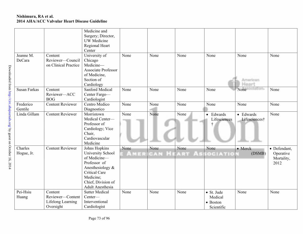
Nishimura, RA et al. 2014 AHA/ACC Valvular Heart Disease Guideline
Page 73 of 96
Medicine and Surgery; Director, UW Medicine Regional Heart Center
Jeanne M. DeCara
Content Reviewer—Council on Clinical Practice
University of Chicago Medicine—Associate Professor of Medicine, Section of Cardiology
None None None None None None
Susan Farkas Content Reviewer—ACC BOG
Sanford Medical Center Fargo—Cardiologist
None None None None None None
Frederico Gentile
Content Reviewer Centro Medico Diagnostico
None None None None None None
Linda Gillam Content Reviewer Morristown Medical Center—Professor of Cardiology; Vice Chair, Cardiovascular Medicine
None None None Edwards Lifesciences†
Edwards Lifesciences†
None
Charles Hogue, Jr.
Content Reviewer Johns Hopkins University School of Medicine—Professor of Anesthesiology & Critical Care Medicine; Chief, Division of Adult Anesthesia
None None None None Merck (DSMB)
Defendant, Operative Mortality, 2012
Pei-Hsiu Huang
Content Reviewer—Content Lifelong Learning Oversight
Sutter Medical Center—Interventional Cardiologist
None None None St. Jude Medical
Boston Scientific
None None
by guest on October 16, 2014
http://circ.ahajournals.org/D
ownloaded from

Nishimura, RA et al. 2014 AHA/ACC Valvular Heart Disease Guideline
Page 74 of 96
Committee (Educational)
Abbott Vascular
Judy W. Hung Content Reviewer Harvard Medical School—Associate Professor of Medicine; Massachusetts General Hospital—Associate Director, Echocardiography
None None None None None None
Bernard Iung Content Reviewer Bichat Hospital—Professor of Cardiology
Abbott Boehringer
Ingelheim Edwards
Lifesciences
Valtech
None None None Archives of Cardiovascular Disease† (Associate Editor)
Boehringer Ingelheim†
None
Amar Krishnaswamy
Content Reviewer—Content Lifelong Learning Oversight Committee (Educational)
Cleveland Clinic—Associate Staff, Robert and Suzanne Tomsich Department of Cardiovascular Medicine
None None None None None None
David Lanfear Content Reviewer—Heart Failure and Transplant Council
Henry Ford Hospital— Assistant Professor, Heart and Vascular Institute in the Center for Health Services Research
Thoratec None None Amgen* Biocontrol* CardioRentis
None None
Richard Lange Content Reviewer University of Texas Health Science Center at San Antonio—Professor of
None None None None None None
by guest on October 16, 2014
http://circ.ahajournals.org/D
ownloaded from
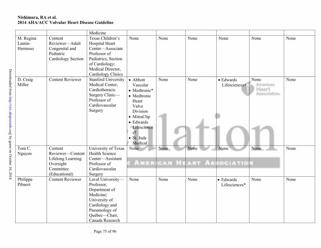
Nishimura, RA et al. 2014 AHA/ACC Valvular Heart Disease Guideline
Page 75 of 96
Medicine M. Regina Lantin-Hermoso
Content Reviewer—Adult Congenital and Pediatric Cardiology Section
Texas Children’s Hospital Heart Center—Associate Professor of Pediatrics, Section of Cardiology; Medical Director, Cardiology Clinics
None None None None None None
D. Craig Miller
Content Reviewer Stanford University Medical Center, Cardiothoracic Surgery Clinic—Professor of Cardiovascular Surgery
Abbott Vascular
Medtronic* Medtronic
Heart Valve Division
MitraClip Edwards
Lifesciences†
St. Jude Medical
None None Edwards Lifesciences†
None None
Tom C. Nguyen
Content Reviewer—Content Lifelong Learning Oversight Committee (Educational)
University of Texas Health Science Center—Assistant Professor of Cardiovascular Surgery
None None None None None None
Philippe Pibarot
Content Reviewer
Laval University—Professor, Department of Medicine; University of Cardiology and Pneumology of Québec—Chair, Canada Research
None None None Edwards Lifesciences*
None None
by guest on October 16, 2014
http://circ.ahajournals.org/D
ownloaded from

Nishimura, RA et al. 2014 AHA/ACC Valvular Heart Disease Guideline
Page 76 of 96
Chair in Valvular Heart Diseases
Geetha Raghuveer
Content Reviewer—ACC BOG
University of Missouri-Kansas City School of Medicine— Associate Professor of Pediatrics; Pediatric Cardiologist
None None None None None None
Michael J. Reardon
Content Reviewer Methodist DeBakey Heart & Vascular Center—Professor of Cardiothoracic Surgery
Medtronic None None Medtronic None None
Raphael Rosenhek
Content Reviewer Medical University of Vienna—Associate Professor, Department of Cardiology
None Abbott Edwards
Lifesciences
None None None None
Hartzell V. Schaff
Content Reviewer Mayo Clinic—Stuart W. Harrington Professor of Surgery
None None None None None None
Allan Schwartz
Content Reviewer Columbia University—Chief, Division of Cardiology
None None None None None None
Frank W. Sellke
Content Reviewer—ACC/AHA Task Force on Practice Guidelines
Rhode Island Hospital—Chief, Division of Cardiothoracic Surgery and Lifespan Heart
None None None CLS Behring The
Medicines Company
CLS Behring The Medicines
Company
None
by guest on October 16, 2014
http://circ.ahajournals.org/D
ownloaded from
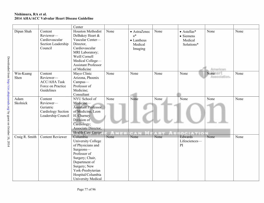
Nishimura, RA et al. 2014 AHA/ACC Valvular Heart Disease Guideline
Page 77 of 96
Center Dipan Shah Content
Reviewer— Cardiovascular Section Leadership Council
Houston Methodist DeBakey Heart & Vascular Center—Director, Cardiovascular MRI Laboratory; Weill Cornell Medical College—Assistant Professor of Medicine
None AstraZeneca*
Lantheus Medical Imaging
None Astellas* Siemens
Medical Solutions*
None None
Win-Kuang Shen
Content Reviewer— ACC/AHA Task Force on Practice Guidelines
Mayo Clinic Arizona, Phoenix Campus—Professor of Medicine; Consultant
None None None None None None
Adam Skolnick
Content Reviewer—Geriatric Cardiology Section Leadership Council
NYU School of Medicine— Assistant Professor of Medicine, Leon H. Charney Division of Cardiology; Associate Director, Health Care Center
None None None None None None
Craig R. Smith Content Reviewer Columbia University College of Physicians and Surgeons—Professor of Surgery; Chair, Department of Surgery; New York-Presbyterian Hospital/Columbia University Medical
None None None Edwards Lifesciences—PI
None None
by guest on October 16, 2014
http://circ.ahajournals.org/D
ownloaded from

Nishimura, RA et al. 2014 AHA/ACC Valvular Heart Disease Guideline
Page 78 of 96
Center—Surgeon-in-Chief
Ruth H. Strasser
Content Reviewer—AIG
Heart Centre, University Hospital, University of Technology, Dresden—Professor, Director, and Chair, Internal Medicine and Cardiology Clinic; Medical Director, Heart Centre
None None None None Abbott† AstraZeneca† Bayer† Biosensors† Pfizer†
None
Rakesh Suri Content Reviewer Mayo Clinic—Associate Professor of Surgery
None None None None Edwards Lifesciences†
Sorin† St. Jude
Medical†
None
Vinod Thourani
Content Reviewer—Surgeon Council
Emory University Edward Lifesciences
Sorin St. Jude
Medical
None Apica Cardiovascular†
Maquet None None
Alec Vahanian Content Reviewer Hospital Bichat—Department de Cardiologie
Abbott Vascular
Edwards Lifesciences
Medtronic St. Jude
Medical Valtech
None None None None None
Andrew Wang Content Reviewer Duke University Medical Center—Professor of
None None None Abbott Vascular*
None Defendant, Sudden death,
by guest on October 16, 2014
http://circ.ahajournals.org/D
ownloaded from

Nishimura, RA et al. 2014 AHA/ACC Valvular Heart Disease Guideline
Page 79 of 96
Medicine Edwards Lifesciences*
2012
This table represents the relationships of reviewers with industry and other entities that were disclosed at the time of peer review and determined to be relevant to this document. It does not necessarily reflect relationships with industry at the time of publication. A person is deemed to have a significant interest in a business if the interest represents ownership of ≥5% of the voting stock or share of the business entity, or ownership of ≥$10 000 of the fair market value of the business entity; or if funds received by the person from the business entity exceed 5% of the person’s gross income for the previous year. A relationship is considered to be modest if it is less than significant under the preceding definition. Relationships that exist with no financial benefit are also included for the purpose of transparency. Relationships in this table are modest unless otherwise noted. Names are listed in alphabetical order within each category of review.
According to the ACC/AHA, a person has a relevant relationship IF: a) The relationship or interest relates to the same or similar subject matter, intellectual property or asset, topic, or issue addressed in the document; or b) The company/entity (with whom the relationship exists) makes a drug, drug class, or device addressed in the document, or makes a competing drug or device addressed in the document; or c) The person or a member of the person’s household, has a reasonable potential for financial, professional or other personal gain or loss as a result of the issues/content addressed in the document. *No financial benefit †Significant relationship AATS indicates American Association for Thoracic Surgery; ACC, American College of Cardiology; AHA, American Heart Association; AIG, Association of International Governors; ASE, American Society of Echocardiography; BOG, Board of Governors; DSMB, Data and Safety Monitoring Board; NIH, National Institutes of Health; PARTNERS, Placement Of Aortic Transcatheter Valves; PI, Principal Investigator; SCA, Society of Cardiovascular Anesthesiologists; SCAI, Society for Cardiovascular Angiography and Interventions; and STS, Society of Thoracic Surgeons. by guest on O
ctober 16, 2014http://circ.ahajournals.org/
Dow
nloaded from

Nishimura, RA et al. 2014 AHA/ACC Valvular Heart Disease Guideline
Page 80 of 96
References
1. ACCF.AHA Task Force on Practice Guidelines. Methodology Manual and Policies From the ACCF/AHA Task Force on Practice Guidelines. American College of Cardiology Foundation and American Heart Association, Inc. cardiosource.org. 2010. Available at: http://assets.cardiosource.com/Methodology_Manual_for_ACC_AHA_Writing_Committees.pdf and http://my.americanheart.org/idc/groups/ahamah-public/@wcm/@sop/documents/downloadable/ucm_319826.pdf. Accessed February 19, 2014.
2. Committee on Standards for Developing Trustworthy Clinical Practice Guidelines; Institute of Medicine. Clinical Practice Guidelines We Can Trust. Washington, D.C.: The National Academies Press, 2013.
3. Committee on Standards for Systematic Reviews of Comparative Effectiveness Research, Institute of Medicine. Finding What Works in Health Care: Standards for Systematic Reviews. Washington, DC: The National Academies Press, 2011.
4. Nishimura R, Otto CM, Bonow RO, et al. 2014 AHA/ACC guideline for the management of patients with valvular heart disease: a report of the American College of Cardiology/American Heart Association Task Force on Practice Guidelines. In press. Journal of the American College of Cardiology. 2014;
5. Bonow RO, Carabello BA, Chatterjee K, et al. 2008 focused update incorporated into the ACC/AHA 2006 guidelines for the management of patients with valvular heart disease: a report of the American College of Cardiology/American Heart Association Task Force on Practice Guidelines (Writing Committee to revise the 1998 guidelines for the management of patients with valvular heart disease). Endorsed by the Society of Cardiovascular Anesthesiologists, Society for Cardiovascular Angiography and Interventions, and Society of Thoracic Surgeons. J Am Coll Cardiol. 2008;52:e1-142.
6. Zoghbi WA, Enriquez-Sarano M, Foster E, et al. Recommendations for evaluation of the severity of native valvular regurgitation with two-dimensional and Doppler echocardiography. J Am Soc Echocardiogr. 2003;16:777-802.
7. Fuster V, Ryden LE, Cannom DS, et al. ACC/AHA/ESC 2006 guidelines for the management of patients with atrial fibrillation--executive summary: a report of the American College of Cardiology/American Heart Association Task Force on Practice Guidelines and the European Society of Cardiology Committee for Practice Guidelines (Writing Committee to Revise the 2001 Guidelines for the Management of Patients With Atrial Fibrillation). J Am Coll Cardiol. 2006;48:854-906.
8. Warnes CA, Williams RG, Bashore TM, et al. ACC/AHA 2008 guidelines for the management of adults with congenital heart disease: a report of the American College of Cardiology/American Heart Association Task Force on Practice Guidelines (Writing Committee to Develop Guidelines on the Management of Adults With Congenital Heart Disease). Developed in Collaboration With the American Society of Echocardiography, Heart Rhythm Society, International Society for Adult Congenital Heart Disease, Society for Cardiovascular Angiography and Interventions, and Society of Thoracic Surgeons. J Am Coll Cardiol. 2008;52:e143-e263.
9. Baumgartner H, Hung J, Bermejo J, et al. Echocardiographic assessment of valve stenosis: EAE/ASE recommendations for clinical practice. Eur J Echocardiogr. 2009;10:1-25.
10. Zoghbi WA, Chambers JB, Dumesnil JG, et al. Recommendations for evaluation of prosthetic valves with echocardiography and Doppler ultrasound: a report from the American Society of Echocardiography's Guidelines and Standards Committee and the Task Force on Prosthetic Valves, developed in conjunction with the American College of Cardiology Cardiovascular Imaging Committee, Cardiac Imaging Committee of the American Heart Association, the European Association of Echocardiography, a registered branch of the European Society of Cardiology, the Japanese Society of Echocardiography and the Canadian Society of Echocardiography. J Am Soc Echocardiogr. 2009;22:975-1014.
11. Gersh BJ, Maron BJ, Bonow RO, et al. 2011 ACCF/AHA guideline for the diagnosis and treatment of hypertrophic cardiomyopathy: a report of the American College of Cardiology Foundation/American Heart Association Task Force on Practice Guidelines. Developed in collaboration with the American Association for Thoracic Surgery, American Society of Echocardiography, American Society of Nuclear Cardiology, Heart Failure Society of America, Heart Rhythm Society, Society for Cardiovascular Angiography and Interventions, and Society of Thoracic Surgeons. J Am Coll Cardiol. 2011;58:e212-e260.
12. Regitz-Zagrosek V, Blomstrom LC, Borghi C, et al. ESC guidelines on the management of cardiovascular diseases during pregnancy: the Task Force on the Management of Cardiovascular Diseases during Pregnancy of the European Society of Cardiology (ESC). Eur Heart J. 2011;32:3147-97.
by guest on October 16, 2014http://circ.ahajournals.org/Downloaded from

Nishimura, RA et al. 2014 AHA/ACC Valvular Heart Disease Guideline
Page 81 of 96
13. Whitlock RP, Sun JC, Fremes SE, et al. Antithrombotic and thrombolytic therapy for valvular disease: antithrombotic therapy and prevention of thrombosis, 9th ed: American College of Chest Physicians Evidence-Based Clinical Practice Guidelines. Chest. 2012;141:e576S-e600S.
14. Vahanian A, Alfieri O, Andreotti F, et al. Guidelines on the management of valvular heart disease (version 2012). Eur Heart J. 2012;33:2451-96.
15. Yancy CW, Jessup M, Bozkurt B, et al. 2013 ACCF/AHA guideline for the management of heart failure: a report of the American College of Cardiology Foundation/American Heart Association Task Force on Practice Guidelines. J Am Coll Cardiol. 2013;62:e147-e239.
16. Wann LS, Curtis AB, January CT, et al. 2011 ACCF/AHA/HRS focused update on the management of patients with atrial fibrillation (updating the 2006 guideline): a report of the American College of Cardiology Foundation/American Heart Association Task Force on Practice Guidelines. J Am Coll Cardiol. 2011;57:223-42.
17. Wann LS, Curtis AB, Ellenbogen KA, et al. 2011 ACCF/AHA/HRS focused update on the management of patients with atrial fibrillation (update on dabigatran): a report of the American College of Cardiology Foundation/American Heart Association Task Force on practice guidelines. J Am Coll Cardiol. 2011;57:1330-7.
18. Yancy CW, Jessup M, Bozkurt B, et al. 2013 ACCF/AHA Guideline for the Management of Heart Failure: A Report of the American College of Cardiology Foundation/American Heart Association Task Force on Practice Guidelines. J Am Coll Cardiol. 2013;62:e147-e239.
19. Carabello BA, Williams H, Gash AK, et al. Hemodynamic predictors of outcome in patients undergoing valve replacement. Circulation. 1986;74:1309-16.
20. Currie PJ, Seward JB, Chan KL, et al. Continuous wave Doppler determination of right ventricular pressure: a simultaneous Doppler-catheterization study in 127 patients. J Am Coll Cardiol. 1985;6:750-6.
21. Currie PJ, Seward JB, Reeder GS, et al. Continuous-wave Doppler echocardiographic assessment of severity of calcific aortic stenosis: a simultaneous Doppler-catheter correlative study in 100 adult patients. Circulation. 1985;71:1162-9.
22. Dujardin KS, Seward JB, Orszulak TA, et al. Outcome after surgery for mitral regurgitation. Determinants of postoperative morbidity and mortality. J Heart Valve Dis. 1997;6:17-21.
23. Enriquez-Sarano M, Avierinos JF, Messika-Zeitoun D, et al. Quantitative determinants of the outcome of asymptomatic mitral regurgitation. N Engl J Med. 2005;352:875-83.
24. Enriquez-Sarano M, Tajik AJ, Schaff HV, et al. Echocardiographic prediction of left ventricular function after correction of mitral regurgitation: results and clinical implications. J Am Coll Cardiol. 1994;24:1536-43.
25. Nishimura RA, Rihal CS, Tajik AJ, et al. Accurate measurement of the transmitral gradient in patients with mitral stenosis: a simultaneous catheterization and Doppler echocardiographic study. J Am Coll Cardiol. 1994;24:152-8.
26. Oh JK, Taliercio CP, Holmes DRJ, et al. Prediction of the severity of aortic stenosis by Doppler aortic valve area determination: prospective Doppler-catheterization correlation in 100 patients. J Am Coll Cardiol. 1988;11:1227-34.
27. Otto CM, Burwash IG, Legget ME, et al. Prospective study of asymptomatic valvular aortic stenosis. Clinical, echocardiographic, and exercise predictors of outcome. Circulation. 1997;95:2262-70.
28. Otto CM, Nishimura RA, Davis KB, et al. Doppler echocardiographic findings in adults with severe symptomatic valvular aortic stenosis. Balloon Valvuloplasty Registry Echocardiographers. Am J Cardiol. 1991;68:1477-84.
29. Otto CM, Pearlman AS, Comess KA, et al. Determination of the stenotic aortic valve area in adults using Doppler echocardiography. J Am Coll Cardiol. 1986;7:509-17.
30. Otto CM, Pearlman AS, Gardner CL. Hemodynamic progression of aortic stenosis in adults assessed by Doppler echocardiography. J Am Coll Cardiol. 1989;13:545-50.
31. Pellikka PA, Sarano ME, Nishimura RA, et al. Outcome of 622 adults with asymptomatic, hemodynamically significant aortic stenosis during prolonged follow-up. Circulation. 2005;111:3290-5.
32. Zile MR, Gaasch WH, Carroll JD, et al. Chronic mitral regurgitation: predictive value of preoperative echocardiographic indexes of left ventricular function and wall stress. J Am Coll Cardiol. 1984;3:235-42.
33. Dujardin KS, Enriquez-Sarano M, Schaff HV, et al. Mortality and morbidity of aortic regurgitation in clinical practice. A long-term follow-up study. Circulation. 1999;99:1851-7.
34. Bonow RO, Lakatos E, Maron BJ, et al. Serial long-term assessment of the natural history of asymptomatic patients with chronic aortic regurgitation and normal left ventricular systolic function. Circulation. 1991;84:1625-35.
35. Aviles RJ, Nishimura RA, Pellikka PA, et al. Utility of stress Doppler echocardiography in patients undergoing percutaneous mitral balloon valvotomy. J Am Soc Echocardiogr. 2001;14:676-81.
36. Otto CM, Pearlman AS, Kraft CD, et al. Physiologic changes with maximal exercise in asymptomatic valvular aortic stenosis assessed by Doppler echocardiography. J Am Coll Cardiol. 1992;20:1160-7.
37. Lancellotti P, Lebois F, Simon M, et al. Prognostic importance of quantitative exercise Doppler echocardiography in asymptomatic valvular aortic stenosis. Circulation. 2005;112:I377-I382.
by guest on October 16, 2014http://circ.ahajournals.org/Downloaded from
Nishimura, RA et al. 2014 AHA/ACC Valvular Heart Disease Guideline
Page 82 of 96
38. Marechaux S, Hachicha Z, Bellouin A, et al. Usefulness of exercise-stress echocardiography for risk stratification of true asymptomatic patients with aortic valve stenosis. Eur Heart J. 2010;31:1390-7.
39. Messika-Zeitoun D, Johnson BD, Nkomo V, et al. Cardiopulmonary exercise testing determination of functional capacity in mitral regurgitation: physiologic and outcome implications. J Am Coll Cardiol. 2006;47:2521-7.
40. Gerber MA, Baltimore RS, Eaton CB, et al. Prevention of rheumatic fever and diagnosis and treatment of acute Streptococcal pharyngitis: a scientific statement from the American Heart Association Rheumatic Fever, Endocarditis, and Kawasaki Disease Committee of the Council on Cardiovascular Disease in the Young, the Interdisciplinary Council on Functional Genomics and Translational Biology, and the Interdisciplinary Council on Quality of Care and Outcomes Research: endorsed by the American Academy of Pediatrics. Circulation. 2009;119:1541-51.
41. Horstkotte D. Contribution for choosing the optimal prophylaxis of bacterial endocarditis. Eur Heart J. 1987;379-81.
42. Strom BL, Abrutyn E, Berlin JA, et al. Dental and cardiac risk factors for infective endocarditis. A population-based, case-control study. Ann Intern Med. 1998;129:761-9.
43. Duval X, Alla F, Hoen B, et al. Estimated risk of endocarditis in adults with predisposing cardiac conditions undergoing dental procedures with or without antibiotic prophylaxis. Clin Infect Dis. 2006;42:e102-e107.
44. Guarner-Argente C, Shah P, Buchner A, et al. Use of antimicrobials for EUS-guided FNA of pancreatic cysts: a retrospective, comparative analysis. Gastrointest Endosc. 2011;74:81-6.
45. Galan A, Zoghbi WA, Quinones MA. Determination of severity of valvular aortic stenosis by Doppler echocardiography and relation of findings to clinical outcome and agreement with hemodynamic measurements determined at cardiac catheterization. Am J Cardiol. 1991;67:1007-12.
46. Lin SS, Roger VL, Pascoe R, et al. Dobutamine stress Doppler hemodynamics in patients with aortic stenosis: feasibility, safety, and surgical correlations. Am Heart J. 1998;136:1010-6.
47. Monin JL, Monchi M, Gest V, et al. Aortic stenosis with severe left ventricular dysfunction and low transvalvular pressure gradients: risk stratification by low-dose dobutamine echocardiography. J Am Coll Cardiol. 2001;37:2101-7.
48. Clavel MA, Fuchs C, Burwash IG, et al. Predictors of outcomes in low-flow, low-gradient aortic stenosis: results of the multicenter TOPAS Study. Circulation. 2008;118:S234-S242.
49. Das P, Rimington H, Chambers J. Exercise testing to stratify risk in aortic stenosis. Eur Heart J. 2005;26:1309-13. 50. Atterhog JH, Jonsson B, Samuelsson R. Exercise testing: a prospective study of complication rates. Am Heart J.
1979;98:572-9. 51. O'Brien KD, Zhao XQ, Shavelle DM, et al. Hemodynamic effects of the angiotensin-converting enzyme inhibitor,
ramipril, in patients with mild to moderate aortic stenosis and preserved left ventricular function. J Investig Med. 2004;52:185-91.
52. Chockalingam A, Venkatesan S, Subramaniam T, et al. Safety and efficacy of angiotensin-converting enzyme inhibitors in symptomatic severe aortic stenosis: Symptomatic Cardiac Obstruction-Pilot Study of Enalapril in Aortic Stenosis (SCOPE-AS). Am Heart J. 2004;147:E19.
53. Nadir MA, Wei L, Elder DH, et al. Impact of renin-angiotensin system blockade therapy on outcome in aortic stenosis. J Am Coll Cardiol. 2011;58:570-6.
54. Rossebo AB, Pedersen TR, Boman K, et al. Intensive lipid lowering with simvastatin and ezetimibe in aortic stenosis. N Engl J Med. 2008;359:1343-56.
55. Cowell SJ, Newby DE, Prescott RJ, et al. A randomized trial of intensive lipid-lowering therapy in calcific aortic stenosis. N Engl J Med. 2005;352:2389-97.
56. Chan KL, Teo K, Dumesnil JG, et al. Effect of Lipid lowering with rosuvastatin on progression of aortic stenosis: results of the aortic stenosis progression observation: measuring effects of rosuvastatin (ASTRONOMER) trial. Circulation. 2010;121:306-14.
57. Otto CM, Pearlman AS. Doppler echocardiography in adults with symptomatic aortic stenosis. Diagnostic utility and cost-effectiveness. Arch Intern Med. 1988;148:2553-60.
58. Turina J, Hess O, Sepulcri F, et al. Spontaneous course of aortic valve disease. Eur Heart J. 1987;8:471-83. 59. Kelly TA, Rothbart RM, Cooper CM, et al. Comparison of outcome of asymptomatic to symptomatic patients
older than 20 years of age with valvular aortic stenosis. Am J Cardiol. 1988;61:123-30. 60. Zoghbi WA, Chambers JB, Dumesnil JG, et al. Recommendations for evaluation of prosthetic valves with
echocardiography and doppler ultrasound: a report From the American Society of Echocardiography's Guidelines and Standards Committee and the Task Force on Prosthetic Valves, developed in conjunction with the American College of Cardiology Cardiovascular Imaging Committee, Cardiac Imaging Committee of the American Heart Association, the European Association of Echocardiography, a registered branch of the European Society of Cardiology, the Japanese Society of Echocardiography and the Canadian Society of Echocardiography, endorsed by the American College of Cardiology Foundation, American Heart Association, European Association of
by guest on October 16, 2014http://circ.ahajournals.org/Downloaded from
Nishimura, RA et al. 2014 AHA/ACC Valvular Heart Disease Guideline
Page 83 of 96
Echocardiography, a registered branch of the European Society of Cardiology, the Japanese Society of Echocardiography, and Canadian Society of Echocardiography. J Am Soc Echocardiogr. 2009;22:975-1014.
61. Connolly HM, Oh JK, Orszulak TA, et al. Aortic valve replacement for aortic stenosis with severe left ventricular dysfunction. Prognostic indicators. Circulation. 1997;95:2395-400.
62. Tribouilloy C, Levy F, Rusinaru D, et al. Outcome after aortic valve replacement for low-flow/low-gradient aortic stenosis without contractile reserve on dobutamine stress echocardiography. J Am Coll Cardiol. 2009;53:1865-73.
63. Rosenhek R, Binder T, Porenta G, et al. Predictors of outcome in severe, asymptomatic aortic stenosis. N Engl J Med. 2000;343:611-7.
64. Smith WT, Ferguson TB, Jr., Ryan T, et al. Should coronary artery bypass graft surgery patients with mild or moderate aortic stenosis undergo concomitant aortic valve replacement? A decision analysis approach to the surgical dilemma. J Am Coll Cardiol. 2004;44:1241-7.
65. Lancellotti P, Donal E, Magne J, et al. Risk stratification in asymptomatic moderate to severe aortic stenosis: the importance of the valvular, arterial and ventricular interplay. Heart. 2010;96:1364-71.
66. Rosenhek R, Zilberszac R, Schemper M, et al. Natural history of very severe aortic stenosis. Circulation. 2010;121:151-6.
67. Nishimura RA, Grantham JA, Connolly HM, et al. Low-output, low-gradient aortic stenosis in patients with depressed left ventricular systolic function: the clinical utility of the dobutamine challenge in the catheterization laboratory. Circulation. 2002;106:809-13.
68. Monin JL, Quere JP, Monchi M, et al. Low-gradient aortic stenosis: operative risk stratification and predictors for long-term outcome: a multicenter study using dobutamine stress hemodynamics. Circulation. 2003;108:319-24.
69. Fougeres E, Tribouilloy C, Monchi M, et al. Outcomes of pseudo-severe aortic stenosis under conservative treatment. Eur Heart J. 2012;33:2426-33.
70. Horstkotte D, Loogen F. The natural history of aortic valve stenosis. Eur Heart J. 1988;9 Suppl E:57-64. 71. O'Brien SM, Shahian DM, Filardo G, et al. The Society of Thoracic Surgeons 2008 cardiac surgery risk models:
part 2--isolated valve surgery. Ann Thorac Surg. 2009;88:S23-S42. 72. Kodali SK, Williams MR, Smith CR, et al. Two-year outcomes after transcatheter or surgical aortic-valve
replacement. N Engl J Med. 2012;366:1686-95. 73. Leon MB, Smith CR, Mack M, et al. Transcatheter aortic-valve implantation for aortic stenosis in patients who
cannot undergo surgery. N Engl J Med. 2010;363:1597-607. 74. Makkar RR, Fontana GP, Jilaihawi H, et al. Transcatheter aortic-valve replacement for inoperable severe aortic
stenosis. N Engl J Med. 2012;366:1696-704. 75. Smith CR, Leon MB, Mack MJ, et al. Transcatheter versus surgical aortic-valve replacement in high-risk patients.
N Engl J Med. 2011;364:2187-98. 76. Detaint D, Messika-Zeitoun D, Maalouf J, et al. Quantitative echocardiographic determinants of clinical outcome
in asymptomatic patients with aortic regurgitation: a prospective study. JACC Cardiovasc Imaging. 2008;1:1-11. 77. Pizarro R, Bazzino OO, Oberti PF, et al. Prospective validation of the prognostic usefulness of B-type natriuretic
peptide in asymptomatic patients with chronic severe aortic regurgitation. J Am Coll Cardiol. 2011;58:1705-14. 78. Teague SM, Heinsimer JA, Anderson JL, et al. Quantification of aortic regurgitation utilizing continuous wave
Doppler ultrasound. J Am Coll Cardiol. 1986;8:592-9. 79. Bonow RO, Rosing DR, McIntosh CL, et al. The natural history of asymptomatic patients with aortic regurgitation
and normal left ventricular function. Circulation. 1983;68:509-17. 80. Scognamiglio R, Fasoli G, Dalla VS. Progression of myocardial dysfunction in asymptomatic patients with severe
aortic insufficiency. Clin Cardiol. 1986;9:151-6. 81. Siemienczuk D, Greenberg B, Morris C, et al. Chronic aortic insufficiency: factors associated with progression to
aortic valve replacement. Ann Intern Med. 1989;110:587-92. 82. Tornos MP, Olona M, Permanyer-Miralda G, et al. Clinical outcome of severe asymptomatic chronic aortic
regurgitation: a long-term prospective follow-up study. Am Heart J. 1995;130:333-9. 83. Ishii K, Hirota Y, Suwa M, et al. Natural history and left ventricular response in chronic aortic regurgitation. Am J
Cardiol. 1996;78:357-61. 84. Scognamiglio R, Rahimtoola SH, Fasoli G, et al. Nifedipine in asymptomatic patients with severe aortic
regurgitation and normal left ventricular function. N Engl J Med. 1994;331:689-94. 85. Borer JS, Hochreiter C, Herrold EM, et al. Prediction of indications for valve replacement among asymptomatic or
minimally symptomatic patients with chronic aortic regurgitation and normal left ventricular performance. Circulation. 1998;97:525-34.
86. Attenhofer JCH, Turina J, Mayer K, et al. Echocardiography in the evaluation of systolic murmurs of unknown cause. Am J Med. 2000;614-20.
by guest on October 16, 2014http://circ.ahajournals.org/Downloaded from

Nishimura, RA et al. 2014 AHA/ACC Valvular Heart Disease Guideline
Page 84 of 96
87. Gelfand EV, Hughes S, Hauser TH, et al. Severity of mitral and aortic regurgitation as assessed by cardiovascular magnetic resonance: optimizing correlation with Doppler echocardiography. J Cardiovasc Magn Reson. 2006;8:503-7.
88. Cawley PJ, Hamilton-Craig C, Owens DS, et al. Prospective comparison of valve regurgitation quantitation by cardiac magnetic resonance imaging and transthoracic echocardiography. Circ Cardiovasc Imaging. 2013;6:48-57.
89. Evangelista A, Tornos P, Sambola A, et al. Long-term vasodilator therapy in patients with severe aortic regurgitation. N Engl J Med. 2005;353:1342-9.
90. Sondergaard L, Aldershvile J, Hildebrandt P, et al. Vasodilatation with felodipine in chronic asymptomatic aortic regurgitation. Am Heart J. 2000;139:667-74.
91. Elder DH, Wei L, Szwejkowski BR, et al. The impact of renin-angiotensin-aldosterone system blockade on heart failure outcomes and mortality in patients identified to have aortic regurgitation: a large population cohort study. J Am Coll Cardiol. 2011;58:2084-91.
92. Greves J, Rahimtoola SH, McAnulty JH, et al. Preoperative criteria predictive of late survival following valve replacement for severe aortic regurgitation. Am Heart J. 1981;101:300-8.
93. Klodas E, Enriquez-Sarano M, Tajik AJ, et al. Optimizing timing of surgical correction in patients with severe aortic regurgitation: role of symptoms. J Am Coll Cardiol. 1997;30:746-52.
94. Forman R, Firth BG, Barnard MS. Prognostic significance of preoperative left ventricular ejection fraction and valve lesion in patients with aortic valve replacement. Am J Cardiol. 1980;45:1120-5.
95. Chaliki HP, Mohty D, Avierinos JF, et al. Outcomes after aortic valve replacement in patients with severe aortic regurgitation and markedly reduced left ventricular function. Circulation. 2002;106:2687-93.
96. Bhudia SK, McCarthy PM, Kumpati GS, et al. Improved outcomes after aortic valve surgery for chronic aortic regurgitation with severe left ventricular dysfunction. J Am Coll Cardiol. 2007;49:1465-71.
97. Bonow RO, Dodd JT, Maron BJ, et al. Long-term serial changes in left ventricular function and reversal of ventricular dilatation after valve replacement for chronic aortic regurgitation. Circulation. 1988;78:1108-20.
98. Van Rossum AC, Visser FC, Sprenger M, et al. Evaluation of magnetic resonance imaging for determination of left ventricular ejection fraction and comparison with angiography. Am J Cardiol. 1988;62:628-33.
99. Gaasch WH, Carroll JD, Levine HJ, et al. Chronic aortic regurgitation: prognostic value of left ventricular end-systolic dimension and end-diastolic radius/thickness ratio. J Am Coll Cardiol. 1983;1:775-82.
100. Pachulski RT, Weinberg AL, Chan KL. Aortic aneurysm in patients with functionally normal or minimally stenotic bicuspid aortic valve. Am J Cardiol. 1991;67:781-2.
101. Hahn RT, Roman MJ, Mogtader AH, et al. Association of aortic dilation with regurgitant, stenotic and functionally normal bicuspid aortic valves. J Am Coll Cardiol. 1992;19:283-8.
102. Nistri S, Sorbo MD, Marin M, et al. Aortic root dilatation in young men with normally functioning bicuspid aortic valves. Heart. 1999;82:19-22.
103. Keane MG, Wiegers SE, Plappert T, et al. Bicuspid aortic valves are associated with aortic dilatation out of proportion to coexistent valvular lesions. Circulation. 2000;102:III35-III39.
104. Novaro GM, Tiong IY, Pearce GL, et al. Features and predictors of ascending aortic dilatation in association with a congenital bicuspid aortic valve. Am J Cardiol. 2003;92:99-101.
105. Schaefer BM, Lewin MB, Stout KK, et al. The bicuspid aortic valve: an integrated phenotypic classification of leaflet morphology and aortic root shape. Heart. 2008;94:1634-8.
106. Tzemos N, Therrien J, Yip J, et al. Outcomes in adults with bicuspid aortic valves. JAMA. 2008;300:1317-25. 107. Michelena HI, Khanna AD, Mahoney D, et al. Incidence of aortic complications in patients with bicuspid aortic
valves. JAMA. 2011;306:1104-12. 108. Davies RR, Goldstein LJ, Coady MA, et al. Yearly rupture or dissection rates for thoracic aortic aneurysms:
simple prediction based on size. Ann Thorac Surg. 2002;73:17-27. 109. Arora R, Nair M, Kalra GS, et al. Immediate and long-term results of balloon and surgical closed mitral
valvotomy: a randomized comparative study. Am Heart J. 1993;125:1091-4. 110. Turi ZG, Reyes VP, Raju BS, et al. Percutaneous balloon versus surgical closed commissurotomy for mitral
stenosis. A prospective, randomized trial. Circulation. 1991;83:1179-85. 111. Patel JJ, Shama D, Mitha AS, et al. Balloon valvuloplasty versus closed commissurotomy for pliable mitral
stenosis: a prospective hemodynamic study. J Am Coll Cardiol. 1991;18:1318-22. 112. Ben FM, Ayari M, Maatouk F, et al. Percutaneous balloon versus surgical closed and open mitral
commissurotomy: seven-year follow-up results of a randomized trial. Circulation. 1998;97:245-50. 113. Cotrufo M, Renzulli A, Ismeno G, et al. Percutaneous mitral commissurotomy versus open mitral
commissurotomy: a comparative study. Eur J Cardiothorac Surg. 1999;15:646-51. 114. Hugenholtz PG, Ryan TJ, Stein SW, et al. The spectrum of pure mitral stenosis. Hemodynamic studies in relation
to clinical disability. Am J Cardiol. 1962;10:773-84.
by guest on October 16, 2014http://circ.ahajournals.org/Downloaded from
Nishimura, RA et al. 2014 AHA/ACC Valvular Heart Disease Guideline
Page 85 of 96
115. Reyes VP, Raju BS, Wynne J, et al. Percutaneous balloon valvuloplasty compared with open surgical commissurotomy for mitral stenosis. N Engl J Med. 1994;331:961-7.
116. Sugeng L, Weinert L, Lammertin G, et al. Accuracy of mitral valve area measurements using transthoracic rapid freehand 3-dimensional scanning: comparison with noninvasive and invasive methods. J Am Soc Echocardiogr. 2003;16:1292-300.
117. Schlosshan D, Aggarwal G, Mathur G, et al. Real-time 3D transesophageal echocardiography for the evaluation of rheumatic mitral stenosis. JACC Cardiovasc Imaging. 2011;4:580-8.
118. Leavitt JI, Coats MH, Falk RH. Effects of exercise on transmitral gradient and pulmonary artery pressure in patients with mitral stenosis or a prosthetic mitral valve: a Doppler echocardiographic study. J Am Coll Cardiol. 1991;17:1520-6.
119. Chung CS, Karamanoglu M, Kovacs SJ. Duration of diastole and its phases as a function of heart rate during supine bicycle exercise. Am J Physiol Heart Circ Physiol. 2004;287:H2003-H2008.
120. Wilkins GT, Weyman AE, Abascal VM, et al. Percutaneous balloon dilatation of the mitral valve: an analysis of echocardiographic variables related to outcome and the mechanism of dilatation. Br Heart J. 1988;60:299-308.
121. Abascal VM, Wilkins GT, O'Shea JP, et al. Prediction of successful outcome in 130 patients undergoing percutaneous balloon mitral valvotomy. Circulation. 1990;82:448-56.
122. Cannan CR, Nishimura RA, Reeder GS, et al. Echocardiographic assessment of commissural calcium: a simple predictor of outcome after percutaneous mitral balloon valvotomy. J Am Coll Cardiol. 1997;29:175-80.
123. Thomas JD, Wilkins GT, Choong CY, et al. Inaccuracy of mitral pressure half-time immediately after percutaneous mitral valvotomy. Dependence on transmitral gradient and left atrial and ventricular compliance. Circulation. 1988;78:980-93.
124. Ellis K, Ziada KM, Vivekananthan D, et al. Transthoracic echocardiographic predictors of left atrial appendage thrombus. Am J Cardiol. 2006;97:421-5.
125. Kronzon I, Tunick PA, Glassman E, et al. Transesophageal echocardiography to detect atrial clots in candidates for percutaneous transseptal mitral balloon valvuloplasty. J Am Coll Cardiol. 1990;16:1320-2.
126. Tessier P, Mercier LA, Burelle D, et al. Results of percutaneous mitral commissurotomy in patients with a left atrial appendage thrombus detected by transesophageal echocardiography. J Am Soc Echocardiogr. 1994;7:394-9.
127. Wilson JK, Greenwood WF. The natural history of mitral stenosis. Can Med Assoc J. 1954;71:323-31. 128. Rowe JC, Bland EF, Sprague HB, et al. The course of mitral stenosis without surgery: ten- and twenty-year
perspectives. Ann Intern Med. 1960;52:741-9. 129. Olesen KH. The natural history of 271 patients with mitral stenosis under medical treatment. Br Heart J.
1962;24:349-57. 130. Szekely P. Systemic embolism and anticoagulant prophylaxis in rheumatic heart disease. Br Med J. 1964;1:1209-
12. 131. Perez-Gomez F, Alegria E, Berjon J, et al. Comparative effects of antiplatelet, anticoagulant, or combined therapy
in patients with valvular and nonvalvular atrial fibrillation: a randomized multicenter study. J Am Coll Cardiol. 2004;44:1557-66.
132. Omran H, Rang B, Schmidt H, et al. Incidence of left atrial thrombi in patients in sinus rhythm and with a recent neurologic deficit. Am Heart J. 2000;140:658-62.
133. Singer DE, Albers GW, Dalen JE, et al. Antithrombotic therapy in atrial fibrillation: American College of Chest Physicians Evidence-Based Clinical Practice Guidelines (8th Edition). Chest. 2008;133:546S-92S.
134. Stoll BC, Ashcom TL, Johns JP, et al. Effects of atenolol on rest and exercise hemodynamics in patients with mitral stenosis. Am J Cardiol. 1995;75:482-4.
135. Monmeneu Menadas JV, Marin OF, Reyes GF, et al. Beta-blockade and exercise capacity in patients with mitral stenosis in sinus rhythm. J Heart Valve Dis. 2002;11:199-203.
136. Bouleti C, Iung B, Laouenan C, et al. Late results of percutaneous mitral commissurotomy up to 20 years: development and validation of a risk score predicting late functional results from a series of 912 patients. Circulation. 2012;125:2119-27.
137. Ellis LB, Singh JB, Morales DD, et al. Fifteen-to twenty-year study of one thousand patients undergoing closed mitral valvuloplasty. Circulation. 1973;48:357-64.
138. John S, Bashi VV, Jairaj PS, et al. Closed mitral valvotomy: early results and long-term follow-up of 3724 consecutive patients. Circulation. 1983;68:891-6.
139. Finnegan JO, Gray DC, MacVaugh H, III, et al. The open approach to mitral commissurotomy. J Thorac Cardiovasc Surg. 1974;67:75-82.
140. Mullin MJ, Engelman RM, Isom OW, et al. Experience with open mitral commissurotomy in 100 consecutive patients. Surgery. 1974;76:974-82.
141. Halseth WL, Elliott DP, Walker EL, et al. Open mitral commissurotomy. A modern re-evaluation. J Thorac Cardiovasc Surg. 1980;80:842-8.
by guest on October 16, 2014http://circ.ahajournals.org/Downloaded from

Nishimura, RA et al. 2014 AHA/ACC Valvular Heart Disease Guideline
Page 86 of 96
142. Gross RI, Cunningham JN, Jr., Snively SL, et al. Long-term results of open radical mitral commissurotomy: ten year follow-up study of 202 patients. Am J Cardiol. 1981;47:821-5.
143. Iung B, Cormier B, Ducimetiere P, et al. Functional results 5 years after successful percutaneous mitral commissurotomy in a series of 528 patients and analysis of predictive factors. J Am Coll Cardiol. 1996;27:407-14.
144. Arat N, Altay H, Korkmaz S, et al. The effect of baseline pulmonary artery pressure on right ventricular functions after mitral balloon valvuloplasty for rheumatic mitral stenosis: a tissue Doppler imaging study. Turk Kardiyol Dern Ars. 2008;36:223-30.
145. Vincens JJ, Temizer D, Post JR, et al. Long-term outcome of cardiac surgery in patients with mitral stenosis and severe pulmonary hypertension. Circulation. 1995;92:II137-II142.
146. Recusani F, Bargiggia GS, Yoganathan AP, et al. A new method for quantification of regurgitant flow rate using color Doppler flow imaging of the flow convergence region proximal to a discrete orifice. An in vitro study. Circulation. 1991;83:594-604.
147. Bargiggia GS, Tronconi L, Sahn DJ, et al. A new method for quantitation of mitral regurgitation based on color flow Doppler imaging of flow convergence proximal to regurgitant orifice. Circulation. 1991;84:1481-9.
148. Rivera JM, Vandervoort PM, Thoreau DH, et al. Quantification of mitral regurgitation with the proximal flow convergence method: a clinical study. Am Heart J. 1992;124:1289-96.
149. Rosenhek R, Rader F, Klaar U, et al. Outcome of watchful waiting in asymptomatic severe mitral regurgitation. Circulation. 2006;113:2238-44.
150. Crawford MH, Souchek J, Oprian CA, et al. Determinants of survival and left ventricular performance after mitral valve replacement. Department of Veterans Affairs Cooperative Study on Valvular Heart Disease. Circulation. 1990;81:1173-81.
151. Enriquez-Sarano M, Tajik AJ, Schaff HV, et al. Echocardiographic prediction of survival after surgical correction of organic mitral regurgitation. Circulation. 1994;90:830-7.
152. Tribouilloy C, Grigioni F, Avierinos JF, et al. Survival implication of left ventricular end-systolic diameter in mitral regurgitation due to flail leaflets a long-term follow-up multicenter study. J Am Coll Cardiol. 2009;54:1961-8.
153. Grigioni F, Tribouilloy C, Avierinos JF, et al. Outcomes in mitral regurgitation due to flail leaflets a multicenter European study. JACC Cardiovasc Imaging. 2008;1:133-41.
154. Ghoreishi M, Evans CF, deFilippi CR, et al. Pulmonary hypertension adversely affects short- and long-term survival after mitral valve operation for mitral regurgitation: implications for timing of surgery. J Thorac Cardiovasc Surg. 2011;142:1439-52.
155. Rozich JD, Carabello BA, Usher BW, et al. Mitral valve replacement with and without chordal preservation in patients with chronic mitral regurgitation. Mechanisms for differences in postoperative ejection performance. Circulation. 1992;86:1718-26.
156. Tribouilloy CM, Enriquez-Sarano M, Schaff HV, et al. Impact of preoperative symptoms on survival after surgical correction of organic mitral regurgitation: rationale for optimizing surgical indications. Circulation. 1999;99:400-5.
157. Pflugfelder PW, Sechtem UP, White RD, et al. Noninvasive evaluation of mitral regurgitation by analysis of left atrial signal loss in cine magnetic resonance. Am Heart J. 1989;117:1113-9.
158. Pu M, Prior DL, Fan X, et al. Calculation of mitral regurgitant orifice area with use of a simplified proximal convergence method: initial clinical application. J Am Soc Echocardiogr. 2001;14:180-5.
159. Pu M, Vandervoort PM, Greenberg NL, et al. Impact of wall constraint on velocity distribution in proximal flow convergence zone. Implications for color Doppler quantification of mitral regurgitation. J Am Coll Cardiol. 1996;27:706-13.
160. Lang RM, Badano LP, Tsang W, et al. EAE/ASE recommendations for image acquisition and display using three-dimensional echocardiography. J Am Soc Echocardiogr. 2012;25:3-46.
161. Witkowski TG, Thomas JD, Debonnaire PJ, et al. Global longitudinal strain predicts left ventricular dysfunction after mitral valve repair. Eur Heart J Cardiovasc Imaging. 2013;14:69-76.
162. Magne J, Mahjoub H, Pierard LA, et al. Prognostic importance of brain natriuretic peptide and left ventricular longitudinal function in asymptomatic degenerative mitral regurgitation. Heart. 2012;98:584-91.
163. Ozdogan O, Yuksel A, Gurgun C, et al. Evaluation of the severity of mitral regurgitation by the use of signal void in magnetic resonance imaging. Echocardiography. 2009;26:1127-35.
164. Myerson SG, Francis JM, Neubauer S. Direct and indirect quantification of mitral regurgitation with cardiovascular magnetic resonance, and the effect of heart rate variability. MAGMA. 2010;23:243-9.
165. Dahm M, Iversen S, Schmid FX, et al. Intraoperative evaluation of reconstruction of the atrioventricular valves by transesophageal echocardiography. Thorac Cardiovasc Surg. 1987;35 Spec No 2:140-2.
166. Saiki Y, Kasegawa H, Kawase M, et al. Intraoperative TEE during mitral valve repair: does it predict early and late postoperative mitral valve dysfunction? Ann Thorac Surg. 1998;66:1277-81.
by guest on October 16, 2014http://circ.ahajournals.org/Downloaded from

Nishimura, RA et al. 2014 AHA/ACC Valvular Heart Disease Guideline
Page 87 of 96
167. Tischler MD, Cooper KA, Rowen M, et al. Mitral valve replacement versus mitral valve repair. A Doppler and quantitative stress echocardiographic study. Circulation. 1994;89:132-7.
168. Magne J, Lancellotti P, Pierard LA. Exercise-induced changes in degenerative mitral regurgitation. J Am Coll Cardiol. 2010;56:300-9.
169. Tsutsui H, Spinale FG, Nagatsu M, et al. Effects of chronic beta-adrenergic blockade on the left ventricular and cardiocyte abnormalities of chronic canine mitral regurgitation. J Clin Invest. 1994;93:2639-48.
170. Varadarajan P, Joshi N, Appel D, et al. Effect of Beta-blocker therapy on survival in patients with severe mitral regurgitation and normal left ventricular ejection fraction. Am J Cardiol. 2008;102:611-5.
171. Ahmed MI, Aban I, Lloyd SG, et al. A randomized controlled phase IIb trial of beta(1)-receptor blockade for chronic degenerative mitral regurgitation. J Am Coll Cardiol. 2012;60:833-8.
172. Nemoto S, Hamawaki M, De Freitas G, et al. Differential effects of the angiotensin-converting enzyme inhibitor lisinopril versus the beta-adrenergic receptor blocker atenolol on hemodynamics and left ventricular contractile function in experimental mitral regurgitation. J Am Coll Cardiol. 2002;40:149-54.
173. Schon HR. Hemodynamic and morphologic changes after long-term angiotensin converting enzyme inhibition in patients with chronic valvular regurgitation. J Hypertens Suppl. 1994;12:S95-S104.
174. Tischler MD, Rowan M, LeWinter MM. Effect of enalapril therapy on left ventricular mass and volumes in asymptomatic chronic, severe mitral regurgitation secondary to mitral valve prolapse. Am J Cardiol. 1998;82:242-5.
175. Wisenbaugh T, Sinovich V, Dullabh A, et al. Six month pilot study of captopril for mildly symptomatic, severe isolated mitral and isolated aortic regurgitation. J Heart Valve Dis. 1994;3:197-204.
176. Dujardin KS, Enriquez-Sarano M, Bailey KR, et al. Effect of losartan on degree of mitral regurgitation quantified by echocardiography. Am J Cardiol. 2001;87:570-6.
177. Harris KM, Aeppli DM, Carey CF. Effects of angiotensin-converting enzyme inhibition on mitral regurgitation severity, left ventricular size, and functional capacity. Am Heart J. 2005;150:1106.
178. Kizilbash AM, Willett DL, Brickner ME, et al. Effects of afterload reduction on vena contracta width in mitral regurgitation. J Am Coll Cardiol. 1998;32:427-31.
179. Gillinov AM, Mihaljevic T, Blackstone EH, et al. Should patients with severe degenerative mitral regurgitation delay surgery until symptoms develop? Ann Thorac Surg. 2010;90:481-8.
180. Grigioni F, Enriquez-Sarano M, Ling LH, et al. Sudden death in mitral regurgitation due to flail leaflet. J Am Coll Cardiol. 1999;34:2078-85.
181. Schuler G, Peterson KL, Johnson A, et al. Temporal response of left ventricular performance to mitral valve surgery. Circulation. 1979;59:1218-31.
182. Starling MR. Effects of valve surgery on left ventricular contractile function in patients with long-term mitral regurgitation. Circulation. 1995;92:811-8.
183. Rushmer RF. Initial phase of ventricular systole: asynchronous contraction. Am J Physiol. 1956;184:188-94. 184. Hansen DE, Sarris GE, Niczyporuk MA, et al. Physiologic role of the mitral apparatus in left ventricular regional
mechanics, contraction synergy, and global systolic performance. J Thorac Cardiovasc Surg. 1989;97:521-33. 185. Sarris GE, Cahill PD, Hansen DE, et al. Restoration of left ventricular systolic performance after reattachment of
the mitral chordae tendineae. The importance of valvular-ventricular interaction. J Thorac Cardiovasc Surg. 1988;95:969-79.
186. Goldman ME, Mora F, Guarino T, et al. Mitral valvuloplasty is superior to valve replacement for preservation of left ventricular function: an intraoperative two-dimensional echocardiographic study. J Am Coll Cardiol. 1987;10:568-75.
187. David TE, Burns RJ, Bacchus CM, et al. Mitral valve replacement for mitral regurgitation with and without preservation of chordae tendineae. J Thorac Cardiovasc Surg. 1984;88:718-25.
188. Hennein HA, Swain JA, McIntosh CL, et al. Comparative assessment of chordal preservation versus chordal resection during mitral valve replacement. J Thorac Cardiovasc Surg. 1990;99:828-36.
189. Cohn LH. Surgery for mitral regurgitation. JAMA. 1988;260:2883-7. 190. Cosgrove DM, Chavez AM, Lytle BW, et al. Results of mitral valve reconstruction. Circulation. 1986;74:I82-I87. 191. STS online risk calculator. Available at: http://riskcalc.sts.org/STSWebRiskCalc273/de.aspx.2013.Accessed on
February 20, 2014. 192. David TE, Uden DE, Strauss HD. The importance of the mitral apparatus in left ventricular function after
correction of mitral regurgitation. Circulation. 1983;68:II76-II82. 193. Horskotte D, Schulte HD, Bircks W, et al. The effect of chordal preservation on late outcome after mitral valve
replacement: a randomized study. J Heart Valve Dis. 1993;2:150-8. 194. Vassileva CM, Mishkel G, McNeely C, et al. Long-term survival of patients undergoing mitral valve repair and
replacement: a longitudinal analysis of Medicare fee-for-service beneficiaries. Circulation. 2013;127:1870-6.
by guest on October 16, 2014http://circ.ahajournals.org/Downloaded from
Nishimura, RA et al. 2014 AHA/ACC Valvular Heart Disease Guideline
Page 88 of 96
195. Braunberger E, Deloche A, Berrebi A, et al. Very long-term results (more than 20 years) of valve repair with carpentier's techniques in nonrheumatic mitral valve insufficiency. Circulation. 2001;104:I8-11.
196. David TE, Ivanov J, Armstrong S, et al. A comparison of outcomes of mitral valve repair for degenerative disease with posterior, anterior, and bileaflet prolapse. J Thorac Cardiovasc Surg. 2005;130:1242-9.
197. McClure RS, Athanasopoulos LV, McGurk S, et al. One thousand minimally invasive mitral valve operations: early outcomes, late outcomes, and echocardiographic follow-up. J Thorac Cardiovasc Surg. 2013;145:1199-206.
198. Gammie JS, Sheng S, Griffith BP, et al. Trends in mitral valve surgery in the United States: results from the Society of Thoracic Surgeons Adult Cardiac Surgery Database. Ann Thorac Surg. 2009;87:1431-7.
199. Chikwe J, Goldstone AB, Passage J, et al. A propensity score-adjusted retrospective comparison of early and mid-term results of mitral valve repair versus replacement in octogenarians. Eur Heart J. 2011;32:618-26.
200. Badhwar V, Peterson ED, Jacobs JP, et al. Longitudinal outcome of isolated mitral repair in older patients: results from 14,604 procedures performed from 1991 to 2007. Ann Thorac Surg. 2012;94:1870-7.
201. Grossi EA, Galloway AC, Miller JS, et al. Valve repair versus replacement for mitral insufficiency: when is a mechanical valve still indicated? J Thorac Cardiovasc Surg. 1998;115:389-94.
202. Chauvaud S, Fuzellier JF, Berrebi A, et al. Long-term (29 years) results of reconstructive surgery in rheumatic mitral valve insufficiency. Circulation. 2001;104:I12-I15.
203. Bolling SF, Li S, O'Brien SM, et al. Predictors of mitral valve repair: clinical and surgeon factors. Ann Thorac Surg. 2010;90:1904-11.
204. Gillinov AM, Blackstone EH, Cosgrove DM, III, et al. Mitral valve repair with aortic valve replacement is superior to double valve replacement. J Thorac Cardiovasc Surg. 2003;125:1372-87.
205. Kang DH, Kim JH, Rim JH, et al. Comparison of early surgery versus conventional treatment in asymptomatic severe mitral regurgitation. Circulation. 2009;119:797-804.
206. Gillinov AM, Blackstone EH, Nowicki ER, et al. Valve repair versus valve replacement for degenerative mitral valve disease. J Thorac Cardiovasc Surg. 2008;135:885-93, 893.
207. Duran CM, Gometza B, Saad E. Valve repair in rheumatic mitral disease: an unsolved problem. J Card Surg. 1994;9:282-5.
208. Suri RM, Vanoverschelde JL, Grigioni F, et al. Association between early surgical intervention vs watchful waiting and outcomes for mitral regurgitation due to flail mitral valve leaflets. JAMA. 2013;310:609-16.
209. Suri RM, Schaff HV, Dearani JA, et al. Recovery of left ventricular function after surgical correction of mitral regurgitation caused by leaflet prolapse. J Thorac Cardiovasc Surg. 2009;137:1071-6.
210. Ngaage DL, Schaff HV, Mullany CJ, et al. Influence of preoperative atrial fibrillation on late results of mitral repair: is concomitant ablation justified? Ann Thorac Surg. 2007;84:434-42.
211. Raine D, Dark J, Bourke JP. Effect of mitral valve repair/replacement surgery on atrial arrhythmia behavior. J Heart Valve Dis. 2004;13:615-21.
212. Cox JL. The surgical treatment of atrial fibrillation. IV. Surgical technique. J Thorac Cardiovasc Surg. 1991;101:584-92.
213. Kobayashi J, Kosakai Y, Isobe F, et al. Rationale of the Cox maze procedure for atrial fibrillation during redo mitral valve operations. J Thorac Cardiovasc Surg. 1996;112:1216-21.
214. Kawaguchi AT, Kosakai Y, Sasako Y, et al. Risks and benefits of combined maze procedure for atrial fibrillation associated with organic heart disease. J Am Coll Cardiol. 1996;28:985-90.
215. Olasinska-Wisniewska A, Mularek-Kubzdela T, Grajek S, et al. Impact of atrial remodeling on heart rhythm after radiofrequency ablation and mitral valve operations. Ann Thorac Surg. 2012;93:1449-55.
216. Feldman T, Foster E, Glower DD, et al. Percutaneous repair or surgery for mitral regurgitation. N Engl J Med. 2011;364:1395-406.
217. Effect of enalapril on mortality and the development of heart failure in asymptomatic patients with reduced left ventricular ejection fractions. The SOLVD Investigattors. N Engl J Med. 1992;327:685-91.
218. Granger CB, McMurray JJ, Yusuf S, et al. Effects of candesartan in patients with chronic heart failure and reduced left-ventricular systolic function intolerant to angiotensin-converting-enzyme inhibitors: the CHARM-Alternative trial. Lancet. 2003;362:772-6.
219. Eriksson SV, Eneroth P, Kjekshus J, et al. Neuroendocrine activation in relation to left ventricular function in chronic severe congestive heart failure: a subgroup analysis from the Cooperative North Scandinavian Enalapril Survival Study (CONSENSUS). Clin Cardiol. 1994;17:603-6.
220. Pitt B, Zannad F, Remme WJ, et al. The effect of spironolactone on morbidity and mortality in patients with severe heart failure. Randomized Aldactone Evaluation Study Investigators. N Engl J Med. 1999;341:709-17.
221. Krum H, Roecker EB, Mohacsi P, et al. Effects of initiating carvedilol in patients with severe chronic heart failure: results from the COPERNICUS Study. JAMA. 2003;289:712-8.
222. St John Sutton MG, Plappert T, Abraham WT, et al. Effect of cardiac resynchronization therapy on left ventricular size and function in chronic heart failure. Circulation. 2003;107:1985-90.
by guest on October 16, 2014http://circ.ahajournals.org/Downloaded from

Nishimura, RA et al. 2014 AHA/ACC Valvular Heart Disease Guideline
Page 89 of 96
223. van Bommel RJ, Marsan NA, Delgado V, et al. Cardiac resynchronization therapy as a therapeutic option in patients with moderate-severe functional mitral regurgitation and high operative risk. Circulation. 2011;124:912-9.
224. Grigioni F, Enriquez-Sarano M, Zehr KJ, et al. Ischemic mitral regurgitation: long-term outcome and prognostic implications with quantitative Doppler assessment. Circulation. 2001;103:1759-64.
225. Lancellotti P, Gerard PL, Pierard LA. Long-term outcome of patients with heart failure and dynamic functional mitral regurgitation. Eur Heart J. 2005;26:1528-32.
226. Trichon BH, Felker GM, Shaw LK, et al. Relation of frequency and severity of mitral regurgitation to survival among patients with left ventricular systolic dysfunction and heart failure. Am J Cardiol. 2003;91:538-43.
227. Rossi A, Dini FL, Faggiano P, et al. Independent prognostic value of functional mitral regurgitation in patients with heart failure. A quantitative analysis of 1256 patients with ischaemic and non-ischaemic dilated cardiomyopathy. Heart. 2011;97:1675-80.
228. Fattouch K, Guccione F, Sampognaro R, et al. POINT: Efficacy of adding mitral valve restrictive annuloplasty to coronary artery bypass grafting in patients with moderate ischemic mitral valve regurgitation: a randomized trial. J Thorac Cardiovasc Surg. 2009;138:278-85.
229. Mihaljevic T, Lam BK, Rajeswaran J, et al. Impact of mitral valve annuloplasty combined with revascularization in patients with functional ischemic mitral regurgitation. J Am Coll Cardiol. 2007;49:2191-201.
230. Wu AH, Aaronson KD, Bolling SF, et al. Impact of mitral valve annuloplasty on mortality risk in patients with mitral regurgitation and left ventricular systolic dysfunction. J Am Coll Cardiol. 2005;45:381-7.
231. Harris KM, Sundt TM, III, Aeppli D, et al. Can late survival of patients with moderate ischemic mitral regurgitation be impacted by intervention on the valve? Ann Thorac Surg. 2002;74:1468-75.
232. Benedetto U, Melina G, Roscitano A, et al. Does combined mitral valve surgery improve survival when compared to revascularization alone in patients with ischemic mitral regurgitation? A meta-analysis on 2479 patients. J Cardiovasc Med (Hagerstown). 2009;10:109-14.
233. Deja MA, Grayburn PA, Sun B, et al. Influence of mitral regurgitation repair on survival in the surgical treatment for ischemic heart failure trial. Circulation. 2012;125:2639-48.
234. Cohn LH, Rizzo RJ, Adams DH, et al. The effect of pathophysiology on the surgical treatment of ischemic mitral regurgitation: operative and late risks of repair versus replacement. Eur J Cardiothorac Surg. 1995;9:568-74.
235. Chan KM, Punjabi PP, Flather M, et al. Coronary artery bypass surgery with or without mitral valve annuloplasty in moderate functional ischemic mitral regurgitation: final results of the Randomized Ischemic Mitral Evaluation (RIME) trial. Circulation. 2012;126:2502-10.
236. Nath J, Foster E, Heidenreich PA. Impact of tricuspid regurgitation on long-term survival. J Am Coll Cardiol. 2004;43:405-9.
237. Dreyfus GD, Corbi PJ, Chan KM, et al. Secondary tricuspid regurgitation or dilatation: which should be the criteria for surgical repair? Ann Thorac Surg. 2005;79:127-32.
238. Chan V, Burwash IG, Lam BK, et al. Clinical and echocardiographic impact of functional tricuspid regurgitation repair at the time of mitral valve replacement. Ann Thorac Surg. 2009;88:1209-15.
239. Calafiore AM, Gallina S, Iaco AL, et al. Mitral valve surgery for functional mitral regurgitation: should moderate-or-more tricuspid regurgitation be treated? a propensity score analysis. Ann Thorac Surg. 2009;87:698-703.
240. Di MM, Bivona A, Iaco AL, et al. Mitral valve surgery for functional mitral regurgitation: prognostic role of tricuspid regurgitation. Eur J Cardiothorac Surg. 2009;35:635-9.
241. Van de Veire NR, Braun J, Delgado V, et al. Tricuspid annuloplasty prevents right ventricular dilatation and progression of tricuspid regurgitation in patients with tricuspid annular dilatation undergoing mitral valve repair. J Thorac Cardiovasc Surg. 2011;141:1431-9.
242. Yilmaz O, Suri RM, Dearani JA, et al. Functional tricuspid regurgitation at the time of mitral valve repair for degenerative leaflet prolapse: the case for a selective approach. J Thorac Cardiovasc Surg. 2011;142:608-13.
243. Calafiore AM, Iaco AL, Romeo A, et al. Echocardiographic-based treatment of functional tricuspid regurgitation. J Thorac Cardiovasc Surg. 2011;142:308-13.
244. Navia JL, Brozzi NA, Klein AL, et al. Moderate tricuspid regurgitation with left-sided degenerative heart valve disease: to repair or not to repair? Ann Thorac Surg. 2012;93:59-67.
245. Kim JB, Yoo DG, Kim GS, et al. Mild-to-moderate functional tricuspid regurgitation in patients undergoing valve replacement for rheumatic mitral disease: the influence of tricuspid valve repair on clinical and echocardiographic outcomes. Heart. 2012;98:24-30.
246. Benedetto U, Melina G, Angeloni E, et al. Prophylactic tricuspid annuloplasty in patients with dilated tricuspid annulus undergoing mitral valve surgery. J Thorac Cardiovasc Surg. 2012;143:632-8.
247. Lancellotti P, Tribouilloy C, Hagendorff A, et al. European Association of Echocardiography recommendations for the assessment of valvular regurgitation. Part 1: aortic and pulmonary regurgitation (native valve disease). Eur J Echocardiogr. 2010;11:223-44.
by guest on October 16, 2014http://circ.ahajournals.org/Downloaded from

Nishimura, RA et al. 2014 AHA/ACC Valvular Heart Disease Guideline
Page 90 of 96
248. Burstow DJ, Nishimura RA, Bailey KR, et al. Continuous wave Doppler echocardiographic measurement of prosthetic valve gradients. A simultaneous Doppler-catheter correlative study. Circulation. 1989;80:504-14.
249. Baumgartner H, Khan S, DeRobertis M, et al. Effect of prosthetic aortic valve design on the Doppler-catheter gradient correlation: an in vitro study of normal St. Jude, Medtronic-Hall, Starr-Edwards and Hancock valves. J Am Coll Cardiol. 1992;19:324-32.
250. Vandervoort PM, Greenberg NL, Powell KA, et al. Pressure recovery in bileaflet heart valve prostheses. Localized high velocities and gradients in central and side orifices with implications for Doppler-catheter gradient relation in aortic and mitral position. Circulation. 1995;92:3464-72.
251. Dumesnil JG, Honos GN, Lemieux M, et al. Validation and applications of indexed aortic prosthetic valve areas calculated by Doppler echocardiography. J Am Coll Cardiol. 1990;16:637-43.
252. Hammermeister K, Sethi GK, Henderson WG, et al. Outcomes 15 years after valve replacement with a mechanical versus a bioprosthetic valve: final report of the Veterans Affairs randomized trial. J Am Coll Cardiol. 2000;36:1152-8.
253. Badhwar V, Ofenloch JC, Rovin JD, et al. Noninferiority of closely monitored mechanical valves to bioprostheses overshadowed by early mortality benefit in younger patients. Ann Thorac Surg. 2012;93:748-53.
254. Weber A, Noureddine H, Englberger L, et al. Ten-year comparison of pericardial tissue valves versus mechanical prostheses for aortic valve replacement in patients younger than 60 years of age. J Thorac Cardiovasc Surg. 2012;144:1075-83.
255. Banbury MK, Cosgrove DM, III, Thomas JD, et al. Hemodynamic stability during 17 years of the Carpentier-Edwards aortic pericardial bioprosthesis. Ann Thorac Surg. 2002;73:1460-5.
256. Dellgren G, David TE, Raanani E, et al. Late hemodynamic and clinical outcomes of aortic valve replacement with the Carpentier-Edwards Perimount pericardial bioprosthesis. J Thorac Cardiovasc Surg. 2002;124:146-54.
257. Borger MA, Ivanov J, Armstrong S, et al. Twenty-year results of the Hancock II bioprosthesis. J Heart Valve Dis. 2006;15:49-55.
258. Myken PS, Bech-Hansen O. A 20-year experience of 1712 patients with the Biocor porcine bioprosthesis. J Thorac Cardiovasc Surg. 2009;137:76-81.
259. Oxenham H, Bloomfield P, Wheatley DJ, et al. Twenty year comparison of a Bjork-Shiley mechanical heart valve with porcine bioprostheses. Heart. 2003;89:715-21.
260. Stassano P, Di Tommaso L., Monaco M, et al. Aortic valve replacement: a prospective randomized evaluation of mechanical versus biological valves in patients ages 55 to 70 years. J Am Coll Cardiol. 2009;54:1862-8.
261. Cannegieter SC, Rosendaal FR, Briet E. Thromboembolic and bleeding complications in patients with mechanical heart valve prostheses. Circulation. 1994;89:635-41.
262. Stein PD, Alpert JS, Bussey HI, et al. Antithrombotic therapy in patients with mechanical and biological prosthetic heart valves. Chest. 2001;119:220S-7S.
263. Schlitt A, von Bardeleben RS, Ehrlich A, et al. Clopidogrel and aspirin in the prevention of thromboembolic complications after mechanical aortic valve replacement (CAPTA). Thromb Res. 2003;109:131-5.
264. Torella M, Torella D, Chiodini P, et al. LOWERing the INtensity of oral anticoaGulant Therapy in patients with bileaflet mechanical aortic valve replacement: results from the "LOWERING-IT" Trial. Am Heart J. 2010;160:171-8.
265. Hering D, Piper C, Bergemann R, et al. Thromboembolic and bleeding complications following St. Jude Medical valve replacement: results of the German Experience With Low-Intensity Anticoagulation Study. Chest. 2005;127:53-9.
266. Acar J, Iung B, Boissel JP, et al. AREVA: multicenter randomized comparison of low-dose versus standard-dose anticoagulation in patients with mechanical prosthetic heart valves. Circulation. 1996;94:2107-12.
267. Horstkotte D, Scharf RE, Schultheiss HP. Intracardiac thrombosis: patient-related and device-related factors. J Heart Valve Dis. 1995;4:114-20.
268. Pruefer D, Dahm M, Dohmen G. Intensity of oral anticoagulation after implantation of St. Jude Medical mitral or multiple valve replacement: lessons learned from GELIA (GELIA 5). Eur Heart J. 2001;3:Q43.
269. Meschengieser SS, Fondevila CG, Frontroth J, et al. Low-intensity oral anticoagulation plus low-dose aspirin versus high-intensity oral anticoagulation alone: a randomized trial in patients with mechanical prosthetic heart valves. J Thorac Cardiovasc Surg. 1997;113:910-6.
270. Turpie AG, Gent M, Laupacis A, et al. A comparison of aspirin with placebo in patients treated with warfarin after heart-valve replacement. N Engl J Med. 1993;329:524-9.
271. Heras M, Chesebro JH, Fuster V, et al. High risk of thromboemboli early after bioprosthetic cardiac valve replacement. J Am Coll Cardiol. 1995;25:1111-9.
272. Colli A, Mestres CA, Castella M, et al. Comparing warfarin to aspirin (WoA) after aortic valve replacement with the St. Jude Medical Epic heart valve bioprosthesis: results of the WoA Epic pilot trial. J Heart Valve Dis. 2007;16:667-71.
by guest on October 16, 2014http://circ.ahajournals.org/Downloaded from

Nishimura, RA et al. 2014 AHA/ACC Valvular Heart Disease Guideline
Page 91 of 96
273. Aramendi JI, Mestres CA, Martinez-Leon J, et al. Triflusal versus oral anticoagulation for primary prevention of thromboembolism after bioprosthetic valve replacement (trac): prospective, randomized, co-operative trial. Eur J Cardiothorac Surg. 2005;27:854-60.
274. Nunez L, Gil AM, Larrea JL, et al. Prevention of thromboembolism using aspirin after mitral valve replacement with porcine bioprosthesis. Ann Thorac Surg. 1984;37:84-7.
275. Russo A, Grigioni F, Avierinos JF, et al. Thromboembolic complications after surgical correction of mitral regurgitation incidence, predictors, and clinical implications. J Am Coll Cardiol. 2008;51:1203-11.
276. Merie C, Kober L, Skov OP, et al. Association of warfarin therapy duration after bioprosthetic aortic valve replacement with risk of mortality, thromboembolic complications, and bleeding. JAMA. 2012;308:2118-25.
277. FDA Drug Safety Communication: Pradaxa (dabigatran etexilate mesylate) should not be used in patients with mechanical prosthetic heart valves. FDA. 2012. Available at: http://www.fda.gov/Drugs/DrugSafety/ucm332912.htm. Accessed February 20, 2014.
278. Van de Werf F, Brueckmann M, Connolly SJ, et al. A comparison of dabigatran etexilate with warfarin in patients with mechanical heart valves: THE Randomized, phase II study to evaluate the safety and pharmacokinetics of oral dabigatran etexilate in patients after heart valve replacement (RE-ALIGN). Am Heart J. 2012;163:931-7.
279. Eikelboom JW, Connolly SJ, Brueckmann M, et al. Dabigatran versus warfarin in patients with mechanical heart valves. N Engl J Med. 2013;369:1206-14.
280. Weibert RT, Le DT, Kayser SR, et al. Correction of excessive anticoagulation with low-dose oral vitamin K1. Ann Intern Med. 1997;126:959-62.
281. Yiu KH, Siu CW, Jim MH, et al. Comparison of the efficacy and safety profiles of intravenous vitamin K and fresh frozen plasma as treatment of warfarin-related over-anticoagulation in patients with mechanical heart valves. Am J Cardiol. 2006;97:409-11.
282. Barbetseas J, Nagueh SF, Pitsavos C, et al. Differentiating thrombus from pannus formation in obstructed mechanical prosthetic valves: an evaluation of clinical, transthoracic and transesophageal echocardiographic parameters. J Am Coll Cardiol. 1998;32:1410-7.
283. Tong AT, Roudaut R, Ozkan M, et al. Transesophageal echocardiography improves risk assessment of thrombolysis of prosthetic valve thrombosis: results of the international PRO-TEE registry. J Am Coll Cardiol. 2004;43:77-84.
284. Roudaut R, Serri K, Lafitte S. Thrombosis of prosthetic heart valves: diagnosis and therapeutic considerations. Heart. 2007;93:137-42.
285. Deviri E, Sareli P, Wisenbaugh T, et al. Obstruction of mechanical heart valve prostheses: clinical aspects and surgical management. J Am Coll Cardiol. 1991;17:646-50.
286. Roudaut R, Lafitte S, Roudaut MF, et al. Fibrinolysis of mechanical prosthetic valve thrombosis: a single-center study of 127 cases. J Am Coll Cardiol. 2003;41:653-8.
287. Keuleers S, Herijgers P, Herregods MC, et al. Comparison of thrombolysis versus surgery as a first line therapy for prosthetic heart valve thrombosis. Am J Cardiol. 2011;107:275-9.
288. Caceres-Loriga FM, Perez-Lopez H, Morlans-Hernandez K, et al. Thrombolysis as first choice therapy in prosthetic heart valve thrombosis. A study of 68 patients. J Thromb Thrombolysis. 2006;21:185-90.
289. Karthikeyan G, Senguttuvan NB, Joseph J, et al. Urgent surgery compared with fibrinolytic therapy for the treatment of left-sided prosthetic heart valve thrombosis: a systematic review and meta-analysis of observational studies. Eur Heart J. 2013;34:1557-66.
290. Roudaut R, Lafitte S, Roudaut MF, et al. Management of prosthetic heart valve obstruction: fibrinolysis versus surgery. Early results and long-term follow-up in a single-centre study of 263 cases. Arch Cardiovasc Dis. 2009;102:269-77.
291. Miller DL, Morris JJ, Schaff HV, et al. Reoperation for aortic valve periprosthetic leakage: identification of patients at risk and results of operation. J Heart Valve Dis. 1995;4:160-5.
292. Akins CW, Bitondo JM, Hilgenberg AD, et al. Early and late results of the surgical correction of cardiac prosthetic paravalvular leaks. J Heart Valve Dis. 2005;14:792-9.
293. Sorajja P, Cabalka AK, Hagler DJ, et al. Percutaneous repair of paravalvular prosthetic regurgitation: acute and 30-day outcomes in 115 patients. Circ Cardiovasc Interv. 2011;4:314-21.
294. Ruiz CE, Jelnin V, Kronzon I, et al. Clinical outcomes in patients undergoing percutaneous closure of periprosthetic paravalvular leaks. J Am Coll Cardiol. 2011;58:2210-7.
295. Sorajja P, Cabalka AK, Hagler DJ, et al. Long-term follow-up of percutaneous repair of paravalvular prosthetic regurgitation. J Am Coll Cardiol. 2011;58:2218-24.
296. Lopez J, Sevilla T, Vilacosta I, et al. Prognostic role of persistent positive blood cultures after initiation of antibiotic therapy in left-sided infective endocarditis. Eur Heart J. 2012.
297. Durack DT, Lukes AS, Bright DK. New criteria for diagnosis of infective endocarditis: utilization of specific echocardiographic findings. Duke Endocarditis Service. Am J Med. 1994;96:200-9.
by guest on October 16, 2014http://circ.ahajournals.org/Downloaded from

Nishimura, RA et al. 2014 AHA/ACC Valvular Heart Disease Guideline
Page 92 of 96
298. Kupferwasser LI, Darius H, Muller AM, et al. Diagnosis of culture-negative endocarditis: the role of the Duke criteria and the impact of transesophageal echocardiography. Am Heart J. 2001;142:146-52.
299. Li JS, Sexton DJ, Mick N, et al. Proposed modifications to the Duke criteria for the diagnosis of infective endocarditis. Clin Infect Dis. 2000;30:633-8.
300. Perez-Vazquez A, Farinas MC, Garcia-Palomo JD, et al. Evaluation of the Duke criteria in 93 episodes of prosthetic valve endocarditis: could sensitivity be improved? Arch Intern Med. 2000;160:1185-91.
301. Botelho-Nevers E, Thuny F, Casalta JP, et al. Dramatic reduction in infective endocarditis-related mortality with a management-based approach. Arch Intern Med. 2009;169:1290-8.
302. Mugge A, Daniel WG, Frank G, et al. Echocardiography in infective endocarditis: reassessment of prognostic implications of vegetation size determined by the transthoracic and the transesophageal approach. J Am Coll Cardiol. 1989;14:631-8.
303. Burger AJ, Peart B, Jabi H, et al. The role of two-dimensional echocardiology in the diagnosis of infective endocarditis [corrected]. Angiology. 1991;42:552-60.
304. Irani WN, Grayburn PA, Afridi I. A negative transthoracic echocardiogram obviates the need for transesophageal echocardiography in patients with suspected native valve active infective endocarditis. Am J Cardiol. 1996;78:101-3.
305. Liu YW, Tsai WC, Hsu CH, et al. Judicious use of transthoracic echocardiography in infective endocarditis screening. Can J Cardiol. 2009;25:703-5.
306. Kemp WE, Jr., Citrin B, Byrd BF, III. Echocardiography in infective endocarditis. South Med J. 1999;92:744-54. 307. Erbel R, Rohmann S, Drexler M, et al. Improved diagnostic value of echocardiography in patients with infective
endocarditis by transoesophageal approach. A prospective study. Eur Heart J. 1988;9:43-53. 308. Daniel WG, Mugge A, Martin RP, et al. Improvement in the diagnosis of abscesses associated with endocarditis
by transesophageal echocardiography. N Engl J Med. 1991;324:795-800. 309. Sochowski RA, Chan KL. Implication of negative results on a monoplane transesophageal echocardiographic
study in patients with suspected infective endocarditis. J Am Coll Cardiol. 1993;21:216-21. 310. Shively BK, Gurule FT, Roldan CA, et al. Diagnostic value of transesophageal compared with transthoracic
echocardiography in infective endocarditis. J Am Coll Cardiol. 1991;18:391-7. 311. Pedersen WR, Walker M, Olson JD, et al. Value of transesophageal echocardiography as an adjunct to
transthoracic echocardiography in evaluation of native and prosthetic valve endocarditis. Chest. 1991;100:351-6. 312. Ronderos RE, Portis M, Stoermann W, et al. Are all echocardiographic findings equally predictive for diagnosis in
prosthetic endocarditis? J Am Soc Echocardiogr. 2004;17:664-9. 313. Roe MT, Abramson MA, Li J, et al. Clinical information determines the impact of transesophageal
echocardiography on the diagnosis of infective endocarditis by the duke criteria. Am Heart J. 2000;139:945-51. 314. Karalis DG, Bansal RC, Hauck AJ, et al. Transesophageal echocardiographic recognition of subaortic
complications in aortic valve endocarditis. Clinical and surgical implications. Circulation. 1992;86:353-62. 315. El-Ahdab F, Benjamin DK, Jr., Wang A, et al. Risk of endocarditis among patients with prosthetic valves and
Staphylococcus aureus bacteremia. Am J Med. 2005;118:225-9. 316. Mylonakis E, Calderwood SB. Infective endocarditis in adults. N Engl J Med. 2001;345:1318-30. 317. Rohmann S, Erbel R, Darius H, et al. Prediction of rapid versus prolonged healing of infective endocarditis by
monitoring vegetation size. J Am Soc Echocardiogr. 1991;4:465-74. 318. Shapira Y, Weisenberg DE, Vaturi M, et al. The impact of intraoperative transesophageal echocardiography in
infective endocarditis. Isr Med Assoc J. 2007;9:299-302. 319. Yao F, Han L, Xu ZY, et al. Surgical treatment of multivalvular endocarditis: twenty-one-year single center
experience. J Thorac Cardiovasc Surg. 2009;137:1475-80. 320. Watanakunakorn C. Staphylococcus aureus endocarditis at a community teaching hospital, 1980 to 1991. An
analysis of 106 cases. Arch Intern Med. 1994;154:2330-5. 321. Abraham J, Mansour C, Veledar E, et al. Staphylococcus aureus bacteremia and endocarditis: the Grady Memorial
Hospital experience with methicillin-sensitive S aureus and methicillin-resistant S aureus bacteremia. Am Heart J. 2004;147:536-9.
322. Kaasch AJ, Fowler VG, Jr., Rieg S, et al. Use of a simple criteria set for guiding echocardiography in nosocomial Staphylococcus aureus bacteremia. Clin Infect Dis. 2011;53:1-9.
323. San Martin J, Sarria C, de las Cuevas C, et al. Relevance of clinical presentation and period of diagnosis in prosthetic valve endocarditis. J Heart Valve Dis. 2010;19:131-8.
324. Knudsen JB, Fuursted K, Petersen E, et al. Failure of clinical features of low probability endocarditis. The early echo remains essential. Scand Cardiovasc J. 2011;45:133-8.
325. Feuchtner GM, Stolzmann P, Dichtl W, et al. Multislice computed tomography in infective endocarditis: comparison with transesophageal echocardiography and intraoperative findings. J Am Coll Cardiol. 2009;53:436-44.
by guest on October 16, 2014http://circ.ahajournals.org/Downloaded from

Nishimura, RA et al. 2014 AHA/ACC Valvular Heart Disease Guideline
Page 93 of 96
326. Gahide G, Bommart S, Demaria R, et al. Preoperative evaluation in aortic endocarditis: findings on cardiac CT. AJR Am J Roentgenol. 2010;194:574-8.
327. Lentini S, Monaco F, Tancredi F, et al. Aortic valve infective endocarditis: could multi-detector CT scan be proposed for routine screening of concomitant coronary artery disease before surgery? Ann Thorac Surg. 2009;87:1585-7.
328. Fagman E, Perrotta S, Bech-Hanssen O, et al. ECG-gated computed tomography: a new role for patients with suspected aortic prosthetic valve endocarditis. Eur Radiol. 2012;22:2407-14.
329. Fowler VG, Jr., Li J, Corey GR, et al. Role of echocardiography in evaluation of patients with Staphylococcus aureus bacteremia: experience in 103 patients. J Am Coll Cardiol. 1997;30:1072-8.
330. Rasmussen RV, Host U, Arpi M, et al. Prevalence of infective endocarditis in patients with Staphylococcus aureus bacteraemia: the value of screening with echocardiography. Eur J Echocardiogr. 2011;12:414-20.
331. Sullenberger AL, Avedissian LS, Kent SM. Importance of transesophageal echocardiography in the evaluation of Staphylococcus aureus bacteremia. J Heart Valve Dis. 2005;14:23-8.
332. Masuda J, Yutani C, Waki R, et al. Histopathological analysis of the mechanisms of intracranial hemorrhage complicating infective endocarditis. Stroke. 1992;23:843-50.
333. Tornos P, Almirante B, Mirabet S, et al. Infective endocarditis due to Staphylococcus aureus: deleterious effect of anticoagulant therapy. Arch Intern Med. 1999;159:473-5.
334. Carpenter JL, McAllister CK. Anticoagulation in prosthetic valve endocarditis. South Med J. 1983;76:1372-5. 335. Lieberman A, Hass WK, Pinto R, et al. Intracranial hemorrhage and infarction in anticoagulated patients with
prosthetic heart valves. Stroke. 1978;9:18-24. 336. Wilson WR, Geraci JE, Danielson GK, et al. Anticoagulant therapy and central nervous system complications in
patients with prosthetic valve endocarditis. Circulation. 1978;57:1004-7. 337. Ananthasubramaniam K, Beattie JN, Rosman HS, et al. How safely and for how long can warfarin therapy be
withheld in prosthetic heart valve patients hospitalized with a major hemorrhage? Chest. 2001;119:478-84. 338. Pruitt AA, Rubin RH, Karchmer AW, et al. Neurologic complications of bacterial endocarditis. Medicine
(Baltimore). 1978;57:329-43. 339. Chan KL, Tam J, Dumesnil JG, et al. Effect of long-term aspirin use on embolic events in infective endocarditis.
Clin Infect Dis. 2008;46:37-41. 340. Fang MC, Go AS, Chang Y, et al. Death and disability from warfarin-associated intracranial and extracranial
hemorrhages. Am J Med. 2007;120:700-5. 341. Rasmussen RV, Snygg-Martin U, Olaison L, et al. Major cerebral events in Staphylococcus aureus infective
endocarditis: is anticoagulant therapy safe? Cardiology. 2009;114:284-91. 342. Jault F, Gandjbakhch I, Rama A, et al. Active native valve endocarditis: determinants of operative death and late
mortality. Ann Thorac Surg. 1997;63:1737-41. 343. Hasbun R, Vikram HR, Barakat LA, et al. Complicated left-sided native valve endocarditis in adults: risk
classification for mortality. JAMA. 2003;289:1933-40. 344. Kiefer T, Park L, Tribouilloy C, et al. Association between valvular surgery and mortality among patients with
infective endocarditis complicated by heart failure. JAMA. 2011;306:2239-47. 345. Tornos P, Sanz E, Permanyer-Miralda G, et al. Late prosthetic valve endocarditis. Immediate and long-term
prognosis. Chest. 1992;101:37-41. 346. Gordon SM, Serkey JM, Longworth DL, et al. Early onset prosthetic valve endocarditis: the Cleveland Clinic
experience 1992-1997. Ann Thorac Surg. 2000;69:1388-92. 347. Wang A, Athan E, Pappas PA, et al. Contemporary clinical profile and outcome of prosthetic valve endocarditis.
JAMA. 2007;297:1354-61. 348. Remadi JP, Habib G, Nadji G, et al. Predictors of death and impact of surgery in Staphylococcus aureus infective
endocarditis. Ann Thorac Surg. 2007;83:1295-302. 349. Hill EE, Herijgers P, Claus P, et al. Infective endocarditis: changing epidemiology and predictors of 6-month
mortality: a prospective cohort study. Eur Heart J. 2007;28:196-203. 350. Aksoy O, Sexton DJ, Wang A, et al. Early surgery in patients with infective endocarditis: a propensity score
analysis. Clin Infect Dis. 2007;44:364-72. 351. Ellis ME, Al-Abdely H, Sandridge A, et al. Fungal endocarditis: evidence in the world literature, 1965-1995. Clin
Infect Dis. 2001;32:50-62. 352. Wolff M, Witchitz S, Chastang C, et al. Prosthetic valve endocarditis in the ICU. Prognostic factors of overall
survival in a series of 122 cases and consequences for treatment decision. Chest. 1995;108:688-94. 353. Chirouze C, Cabell CH, Fowler VG, Jr., et al. Prognostic factors in 61 cases of Staphylococcus aureus prosthetic
valve infective endocarditis from the International Collaboration on Endocarditis merged database. Clin Infect Dis. 2004;38:1323-7.
by guest on October 16, 2014http://circ.ahajournals.org/Downloaded from
Nishimura, RA et al. 2014 AHA/ACC Valvular Heart Disease Guideline
Page 94 of 96
354. Melgar GR, Nasser RM, Gordon SM, et al. Fungal prosthetic valve endocarditis in 16 patients. An 11-year experience in a tertiary care hospital. Medicine (Baltimore). 1997;76:94-103.
355. Wang K, Gobel F, Gleason DF, et al. Complete heart block complicating bacterial endocarditis. Circulation. 1972;46:939-47.
356. Middlemost S, Wisenbaugh T, Meyerowitz C, et al. A case for early surgery in native left-sided endocarditis complicated by heart failure: results in 203 patients. J Am Coll Cardiol. 1991;18:663-7.
357. Chan KL. Early clinical course and long-term outcome of patients with infective endocarditis complicated by perivalvular abscess. CMAJ. 2002;167:19-24.
358. Jault F, Gandjbakhch I, Chastre JC, et al. Prosthetic valve endocarditis with ring abscesses. Surgical management and long-term results. J Thorac Cardiovasc Surg. 1993;105:1106-13.
359. Anguera I, Miro JM, Vilacosta I, et al. Aorto-cavitary fistulous tract formation in infective endocarditis: clinical and echocardiographic features of 76 cases and risk factors for mortality. Eur Heart J. 2005;26:288-97.
360. Klieverik LM, Yacoub MH, Edwards S, et al. Surgical treatment of active native aortic valve endocarditis with allografts and mechanical prostheses. Ann Thorac Surg. 2009;88:1814-21.
361. Hill EE, Herijgers P, Claus P, et al. Abscess in infective endocarditis: the value of transesophageal echocardiography and outcome: a 5-year study. Am Heart J. 2007;154:923-8.
362. Manne MB, Shrestha NK, Lytle BW, et al. Outcomes after surgical treatment of native and prosthetic valve infective endocarditis. Ann Thorac Surg. 2012;93:489-93.
363. Sohail MR, Uslan DZ, Khan AH, et al. Infective endocarditis complicating permanent pacemaker and implantable cardioverter-defibrillator infection. Mayo Clin Proc. 2008;83:46-53.
364. Athan E, Chu VH, Tattevin P, et al. Clinical characteristics and outcome of infective endocarditis involving implantable cardiac devices. JAMA. 2012;307:1727-35.
365. Rundstrom H, Kennergren C, Andersson R, et al. Pacemaker endocarditis during 18 years in Goteborg. Scand J Infect Dis. 2004;36:674-9.
366. Ho HH, Siu CW, Yiu KH, et al. Prosthetic valve endocarditis in a multicenter registry of Chinese patients. Asian Cardiovasc Thorac Ann. 2010;18:430-4.
367. Thuny F, Di Salvo G., Belliard O, et al. Risk of embolism and death in infective endocarditis: prognostic value of echocardiography: a prospective multicenter study. Circulation. 2005;112:69-75.
368. Kang DH, Kim YJ, Kim SH, et al. Early surgery versus conventional treatment for infective endocarditis. N Engl J Med. 2012;366:2466-73.
369. Schaefer C. Angiotensin II-receptor-antagonists: further evidence of fetotoxicity but not teratogenicity. Birth Defects Res A Clin Mol Teratol. 2003;67:591-4.
370. Cooper WO, Hernandez-Diaz S, Arbogast PG, et al. Major congenital malformations after first-trimester exposure to ACE inhibitors. N Engl J Med. 2006;354:2443-51.
371. Shotan A, Widerhorn J, Hurst A, et al. Risks of angiotensin-converting enzyme inhibition during pregnancy: experimental and clinical evidence, potential mechanisms, and recommendations for use. Am J Med. 1994;96:451-6.
372. Abouzied AM, Al Abbady M, Al Gendy MF, et al. Percutaneous balloon mitral commissurotomy during pregnancy. Angiology. 2001;52:205-9.
373. Ben FM, Gamra H, Betbout F, et al. Percutaneous balloon mitral commissurotomy during pregnancy. Heart. 1997;77:564-7.
374. de Souza JA, Martinez EE, Jr., Ambrose JA, et al. Percutaneous balloon mitral valvuloplasty in comparison with open mitral valve commissurotomy for mitral stenosis during pregnancy. J Am Coll Cardiol. 2001;37:900-3.
375. Glantz JC, Pomerantz RM, Cunningham MJ, et al. Percutaneous balloon valvuloplasty for severe mitral stenosis during pregnancy: a review of therapeutic options. Obstet Gynecol Surv. 1993;48:503-8.
376. Iung B, Cormier B, Elias J, et al. Usefulness of percutaneous balloon commissurotomy for mitral stenosis during pregnancy. Am J Cardiol. 1994;73:398-400.
377. Tzemos N, Silversides CK, Colman JM, et al. Late cardiac outcomes after pregnancy in women with congenital aortic stenosis. Am Heart J. 2009;157:474-80.
378. Banning AP, Pearson JF, Hall RJ. Role of balloon dilatation of the aortic valve in pregnant patients with severe aortic stenosis. Br Heart J. 1993;70:544-5.
379. Easterling TR, Chadwick HS, Otto CM, et al. Aortic stenosis in pregnancy. Obstet Gynecol. 1988;72:113-8. 380. Lao TT, Adelman AG, Sermer M, et al. Balloon valvuloplasty for congenital aortic stenosis in pregnancy. Br J
Obstet Gynaecol. 1993;100:1141-2. 381. McIvor RA. Percutaneous balloon aortic valvuloplasty during pregnancy. Int J Cardiol. 1991;32:1-3. 382. Myerson SG, Mitchell AR, Ormerod OJ, et al. What is the role of balloon dilatation for severe aortic stenosis
during pregnancy? J Heart Valve Dis. 2005;14:147-50.
by guest on October 16, 2014http://circ.ahajournals.org/Downloaded from
Nishimura, RA et al. 2014 AHA/ACC Valvular Heart Disease Guideline
Page 95 of 96
383. Tumelero RT, Duda NT, Tognon AP, et al. Percutaneous balloon aortic valvuloplasty in a pregnant adolescent. Arq Bras Cardiol. 2004;82:98-7.
384. Chan WS, Anand S, Ginsberg JS. Anticoagulation of pregnant women with mechanical heart valves: a systematic review of the literature. Arch Intern Med. 2000;160:191-6.
385. Meschengieser SS, Fondevila CG, Santarelli MT, et al. Anticoagulation in pregnant women with mechanical heart valve prostheses. Heart. 1999;82:23-6.
386. Abildgaard U, Sandset PM, Hammerstrom J, et al. Management of pregnant women with mechanical heart valve prosthesis: thromboprophylaxis with low molecular weight heparin. Thromb Res. 2009;124:262-7.
387. McLintock C, McCowan LM, North RA. Maternal complications and pregnancy outcome in women with mechanical prosthetic heart valves treated with enoxaparin. BJOG. 2009;116:1585-92.
388. Oran B, Lee-Parritz A, Ansell J. Low molecular weight heparin for the prophylaxis of thromboembolism in women with prosthetic mechanical heart valves during pregnancy. Thromb Haemost. 2004;92:747-51.
389. Quinn J, Von Klemperer K., Brooks R, et al. Use of high intensity adjusted dose low molecular weight heparin in women with mechanical heart valves during pregnancy: a single-center experience. Haematologica. 2009;94:1608-12.
390. Sillesen M, Hjortdal V, Vejlstrup N, et al. Pregnancy with prosthetic heart valves - 30 years' nationwide experience in Denmark. Eur J Cardiothorac Surg. 2011;40:448-54.
391. De Santo LS, Romano G, Della Corte A., et al. Mechanical aortic valve replacement in young women planning on pregnancy: maternal and fetal outcomes under low oral anticoagulation, a pilot observational study on a comprehensive pre-operative counseling protocol. J Am Coll Cardiol. 2012;59:1110-5.
392. Salazar E, Izaguirre R, Verdejo J, et al. Failure of adjusted doses of subcutaneous heparin to prevent thromboembolic phenomena in pregnant patients with mechanical cardiac valve prostheses. J Am Coll Cardiol. 1996;27:1698-703.
393. Vitale N, De Feo M., Cotrufo M. Anticoagulation for prosthetic heart valves during pregnancy: the importance of warfarin daily dose. Eur J Cardiothorac Surg. 2002;22:656.
394. Rowan JA, McCowan LM, Raudkivi PJ, et al. Enoxaparin treatment in women with mechanical heart valves during pregnancy. Am J Obstet Gynecol. 2001;185:633-7.
395. James AH, Brancazio LR, Gehrig TR, et al. Low-molecular-weight heparin for thromboprophylaxis in pregnant women with mechanical heart valves. J Matern Fetal Neonatal Med. 2006;19:543-9.
396. Yinon Y, Siu SC, Warshafsky C, et al. Use of low molecular weight heparin in pregnant women with mechanical heart valves. Am J Cardiol. 2009;104:1259-63.
397. Ginsberg JS, Chan WS, Bates SM, et al. Anticoagulation of pregnant women with mechanical heart valves. Arch Intern Med. 2003;163:694-8.
398. Mark DB, Berman DS, Budoff MJ, et al. ACCF/ACR/AHA/NASCI/SAIP/SCAI/SCCT 2010 expert consensus document on coronary computed tomographic angiography: a report of the American College of Cardiology Foundation Task Force on Expert Consensus Documents. J Am Coll Cardiol. 2010;55:2663-99.
399. Gilard M, Cornily JC, Pennec PY, et al. Accuracy of multislice computed tomography in the preoperative assessment of coronary disease in patients with aortic valve stenosis. J Am Coll Cardiol. 2006;47:2020-4.
400. Manghat NE, Morgan-Hughes GJ, Broadley AJ, et al. 16-detector row computed tomographic coronary angiography in patients undergoing evaluation for aortic valve replacement: comparison with catheter angiography. Clin Radiol. 2006;61:749-57.
401. Meijboom WB, Mollet NR, Van Mieghem CA, et al. Pre-operative computed tomography coronary angiography to detect significant coronary artery disease in patients referred for cardiac valve surgery. J Am Coll Cardiol. 2006;48:1658-65.
402. Reant P, Brunot S, Lafitte S, et al. Predictive value of noninvasive coronary angiography with multidetector computed tomography to detect significant coronary stenosis before valve surgery. Am J Cardiol. 2006;97:1506-10.
403. Scheffel H, Leschka S, Plass A, et al. Accuracy of 64-slice computed tomography for the preoperative detection of coronary artery disease in patients with chronic aortic regurgitation. Am J Cardiol. 2007;100:701-6.
404. Galas A, Hryniewiecki T, Kepka C, et al. May dual-source computed tomography angiography replace invasive coronary angiography in the evaluation of patients referred for valvular disease surgery? Kardiol Pol. 2012;70:877-82.
405. Doukas G, Samani NJ, Alexiou C, et al. Left atrial radiofrequency ablation during mitral valve surgery for continuous atrial fibrillation: a randomized controlled trial. JAMA. 2005;294:2323-9.
406. Blomstrom-Lundqvist C, Johansson B, Berglin E, et al. A randomized double-blind study of epicardial left atrial cryoablation for permanent atrial fibrillation in patients undergoing mitral valve surgery: the SWEDish Multicentre Atrial Fibrillation study (SWEDMAF). Eur Heart J. 2007;28:2902-8.
by guest on October 16, 2014http://circ.ahajournals.org/Downloaded from

Nishimura, RA et al. 2014 AHA/ACC Valvular Heart Disease Guideline
Page 96 of 96
407. Liu X, Tan HW, Wang XH, et al. Efficacy of catheter ablation and surgical CryoMaze procedure in patients with long-lasting persistent atrial fibrillation and rheumatic heart disease: a randomized trial. Eur Heart J. 2010;31:2633-41.
408. Calleja AM, Dommaraju S, Gaddam R, et al. Cardiac risk in patients aged >75 years with asymptomatic, severe aortic stenosis undergoing noncardiac surgery. Am J Cardiol. 2010;105:1159-63.
409. Zahid M, Sonel AF, Saba S, et al. Perioperative risk of noncardiac surgery associated with aortic stenosis. Am J Cardiol. 2005;96:436-8.
410. Torsher LC, Shub C, Rettke SR, et al. Risk of patients with severe aortic stenosis undergoing noncardiac surgery. Am J Cardiol. 1998;81:448-52.
411. Agarwal S, Rajamanickam A, Bajaj NS, et al. Impact of aortic stenosis on postoperative outcomes after noncardiac surgeries. Circ Cardiovasc Qual Outcomes. 2013;6:193-200.
by guest on October 16, 2014http://circ.ahajournals.org/Downloaded from

2014 Valvular Heart Disease Guideline Data Supplements
(Section numbers correspond to the full-text guideline.)
Table of Contents Data Supplement 1. Outcomes in Adults With Low-Flow/Low-Gradient Aortic Stenosis With Reduced Left Ventricular Ejection Fraction (stage S1) (Sections 3.2.1.1 and 3.2.3) ............... 2 Data Supplement 2. Hemodynamic Progression of Aortic Stenosis in Adult Patients (stages B and C) (Section 3.2.1.3) ............................................................................................................ 5 Data Supplement 3. Exercise Stress Testing in Asymptomatic Adults With Aortic Stenosis (stages B and C) (Sections 3.2.1.5 and 3.2.3) ................................................................................ 6 Data Supplement 4. Clinical Trials of Lipid Lowering Therapy in Adults With Asymptomatic Mild to Moderate Aortic Stenosis (stage B (Section 3.2.2) ...................................................... 8 Data Supplement 5. Clinical Outcomes in Asymptomatic Adults With Aortic Stenosis (stages B and C) of Known Hemodynamic Severity (Section 3.2.3) .................................................... 9 Data Supplement 6. Incidence of Sudden Death in Asymptomatic Adults With Aortic Stenosis (stages B and C) (Section 3.2.3) ............................................................................................ 11 Data Supplement 7. Clinical Outcomes in Symptomatic Adults With Aortic Stenosis of Known Hemodynamic Severity (Section 3.2.3) ............................................................................... 12 Data Supplement 8. Outcomes in Adults With Low-Flow/Low-Gradient Aortic Stenosis With Preserved Left Ventricular Ejection Fraction (stage S2) (Section 3.2.3) ............................... 14 Data Supplement 9. Choice of Intervention in Symptomatic Adults With Severe Aortic Stenosis (stage D): Surgical Versus Transcatheter Aortic Valve Replacement (Section 3.2.4) ........ 16 Data Supplement 10. Clinical Outcomes of Asymptomatic Patients With Chronic Aortic Regurgitation (Sections 4.3.1.1 and 4.3.3) ...................................................................................... 17 Data Supplement 11. Vasodilator Therapy in Asymptomatic Patients With Chronic Aortic Regurgitation (Section 4.3.2) ....................................................................................................... 20 Data Supplement 12. Determinants of Outcome After Surgery for Chronic Aortic Regurgitation (Section 4.3.3) ..................................................................................................................... 21 Data Supplement 13. Hemodynamic Effects Percutaneous Mitral Balloon Commissurotomy (PMBC) Compared to Surgical Closed Commissurotomy (CC) or Open Commissurotomy (OC) (Section 6.2.3) ...................................................................................................................................................................................................................................................................... 27 Data Supplement 14. Echocardiographic Prediction of Outcome of Percutaneous Balloon Mitral Commissurotomy (Section 6.2.3) ....................................................................................... 28 Data Supplement 15. Randomized Trials of Percutaneous Mitral Balloon Commissurotomy Versus Surgery for Mitral Stenosis (Section 6.2.3) .................................................................... 29 Data Supplement 16. Preoperative Predictors of Surgical Outcome in Mitral Regurgitation (Section 7.3.3) .............................................................................................................................. 31 Data Supplement 17. Primary Mitral Regurgitation—Evidence for Intervention (Section 7.3.3) ................................................................................................................................................ 32 Data Supplement 18. Secondary Mitral Regurgitation—Evidence for Intervention (7.4.3) ......................................................................................................................................................... 34 Data Supplement 19. Functional Tricuspid Regurgitation: Outcomes Following Tricuspid Valve Surgery (Sections 8.2.3 and 8.4.3) ...................................................................................... 35 Data Supplement 20. Clinical Outcomes With Bioprosthetic and Mechanical Valves (Section 11.1.2) ...................................................................................................................................... 37 Data Supplement 21. Bridging Anticoagulation Therapy for Mechanical Heart Valves (Section 11.3.2) ................................................................................................................................... 40 Data Supplement 22. Fibrinolytic Therapy for Prosthetic Valve Thrombosis (Section 11.6.2) ................................................................................................................................................... 42 Data Supplement 23. Paravalvular Regurgitation (Section 11.8.3) .............................................................................................................................................................................................. 44 Data Supplement 24. Surgical Outcome in Infective Endocarditis (Section 12) .......................................................................................................................................................................... 46 Data Supplement 25.Outcomes in Pregnant Women With a Mechanical Prosthetic Valve Treated with Warfarin or Unfractionated Heparin (UFH) (Section 13.3.2) ................................... 51 Data Supplement 26. Outcomes in Pregnant Women With a Mechanical Prosthetic Valve Treated With Low Molecular Weight Heparin (LMWH) (Section 13.3.2) ................................... 54 Data Supplement 27. Outcomes With the Maze Procedure for Atrial Fibrillation in Patients With Valvular Heart Disease (Section 14.2.2) ............................................................................ 56 Data Supplement 28. Noncardiac Surgery in Patients With Valvular Heart Disease (Section 15.3) ........................................................................................................................................... 59 References ..................................................................................................................................................................................................................................................................................... 61

2014 Valvular Heart Disease Guideline Data Supplements
2
Data Supplement 1. Outcomes in Adults With Low-Flow/Low-Gradient Aortic Stenosis With Reduced Left Ventricular Ejection Fraction (stage S1) (Sections 3.2.1.1 and 3.2.3)
Study Aim of Study Study Type Study Size Definition of LFLG Severe AS With rLVEF
Exclusion Criteria
Stress Findings/Clinical Outcomes
Comments
DeFillippi, 1995 (1) 7810504
To determine if DSE can distinguish severe fixed AS from flow-dependent AS
Prospective 24 AVAi ≤0.5 cm2/m2 ∆Pmean ≤30 mm Hg LVEF ≤45% All symptomatic
Too ill AF
IA. (n=7, 39%) No change in AVA with ≥20% improvement in LVEF (contractile reserve). IB. (n=5, 28%) ↑AVA ≥0.3 cm2 and contractile reserve. II. (n=6, 33%) No contractile reserve.
IA. 4 underwent AVR with improved symptoms (1 perioperative death). IB. 4 medical Rx and alive at 1 y. 1 CAD death. II. 3 deaths and 3 persistent CHF.
Connolly, 1997 (2) 9170402
Determine outcome after AVR for severe AS with LG and low LVEF
Retrospective surgical database
154 LVEF ≤35% Undergoing AVR
Other valve disease
Baseline mean AVA 0.6±0.2 cm2, Mean cardiac output 4.1±1.5 L/min, Perioperative (30 d) mortality 9%, Postoperative LVEF improved in 76% of pts.
Study group had low LVEF, but not all had LG or LF.
Pereira, 2002 (3) 11955855
Evaluate outcome with AVR vs. medical Rx in LFLG severe AS
Retrospective, propensity score matched
68 AVA ≤0.75 cm2 ∆Pmean ≤30 mm Hg LVEF ≤35%
Other valve disease.
In propensity matched pts, survival at 4 y was 78% with AVR vs.15% with medical Rx (p<0.0001).
Multivariate predictors of survival were AVR, age, and renal function.
Nishimura, 2002 (4) 12176952
Diagnostic value of invasive hemodynamics with dobutamine stress
Prospective, comparison with surgical findings
32 AVA <1.0 cm2 ∆Pmean <40 mm Hg LVEF <40%
N/A With dobutamine, final AVA ≤1.2 cm2 with a ∆Pmean >30 mm Hg in 21 pts; severe AS confirmed at surgery. In 15 pts with CR, mortality was 7% (1 death) with medical therapy.
CR defined as ↑SV ≥20% with dobutamine.
Monin, 2003 (5) 12835219
Assess prognostic value of DSE in LFLG AS
Prospective, multicenter
136 AVA ≤1.0 cm2 Cardiac index ≤3 L/min/m2 ∆Pmean ≤40 mm Hg
Other valve disease, severe comorbidities
Operative mortality 5% with CR vs. 32% without CR (p=0.0002). Predictors of long-term survival were AVR and CR.
CR defined as ↑SV ≥20% on DSE.
Quere, 2006 (6) 16585393
Determine relationship between CR on DSE and postoperative LVEF
Prospective, multicenter
66 AVA ≤1.0 cm2 ∆Pmean ≤40 mm Hg LVEF ≤40% All symptomatic
Excluded operative deaths
I. CR in 70%; post-AVR LVEF improved ≥10 LVEF units in 83%. II. No contractile reserve in 30%; post-AVR LVEF improved ≥10 LVEF units in 65%.
Symptoms improved by ≥2 classes after AVR in 58%. Mean LVEF increased from 29±6% to 47±11% after AVR.
Blais, 2006 (7) 16461844
Improve differentiation of true from pseudo severe AS on DSE
In vitro model and prospective pt group
23 AVAi ≤0.6 cm2/m2 LVEF ≤40% ∆Pmean ≤40 mm Hg All symptomatic undergoing AVR
Other valve disease AF or paced rhythm
Projected effective orifice area at a normal transvalvular flow rate was accurate for identifying true vs. pseudo severe AS in comparison to surgical findings.
No outcome data.
Bergler-Klein, 2007 (8) 15117847
Relationship between BNP and outcome in LFLG AS
Prospective, multicenter
69 AVAi <0.6 cm2/m2 ∆Pmean ≤40 mm Hg LVEF ≤40%
Other valve disease, AF, or paced rhythm
BNP was higher with true-severe AS compared to pseudo-severe AS (p=0.12). 1-y survival 47±9% with BNP ≥550 pg/mL vs. 97±3% with BNP <550 pg/mL (p=0.0001).
Classified as severe AS if DSE showed AVA ≤1.0 cm2 at projected flow rate of 250 mL/s; pseudo-severe if AVA >1.0 cm2 projected at 250 mL/s.
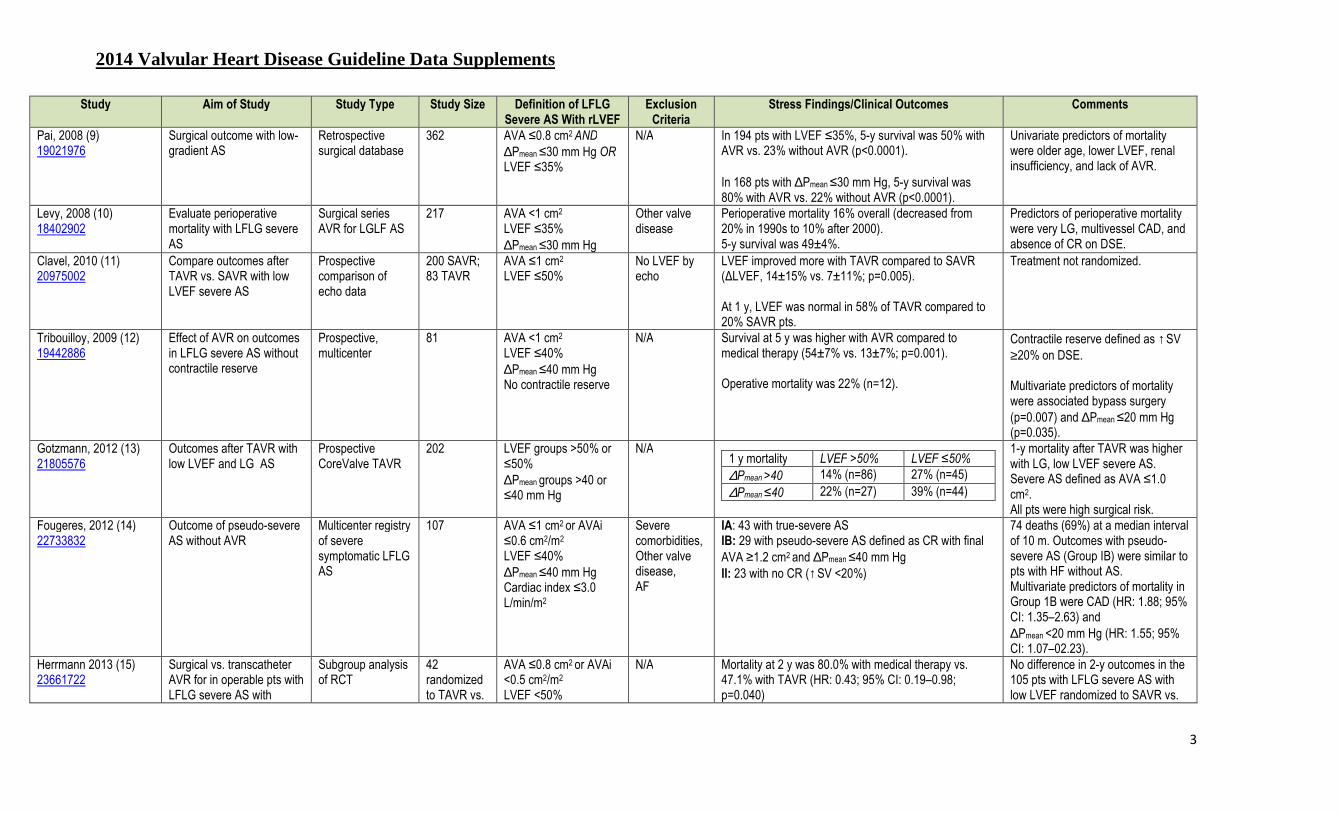
2014 Valvular Heart Disease Guideline Data Supplements
3
Study Aim of Study Study Type Study Size Definition of LFLG Severe AS With rLVEF
Exclusion Criteria
Stress Findings/Clinical Outcomes
Comments
Pai, 2008 (9) 19021976
Surgical outcome with low-gradient AS
Retrospective surgical database
362 AVA ≤0.8 cm2 AND ∆Pmean ≤30 mm Hg OR LVEF ≤35%
N/A In 194 pts with LVEF ≤35%, 5-y survival was 50% with AVR vs. 23% without AVR (p<0.0001). In 168 pts with ∆Pmean ≤30 mm Hg, 5-y survival was 80% with AVR vs. 22% without AVR (p<0.0001).
Univariate predictors of mortality were older age, lower LVEF, renal insufficiency, and lack of AVR.
Levy, 2008 (10) 18402902
Evaluate perioperative mortality with LFLG severe AS
Surgical series AVR for LGLF AS
217 AVA <1 cm2 LVEF ≤35% ∆Pmean ≤30 mm Hg
Other valve disease
Perioperative mortality 16% overall (decreased from 20% in 1990s to 10% after 2000). 5-y survival was 49±4%.
Predictors of perioperative mortality were very LG, multivessel CAD, and absence of CR on DSE.
Clavel, 2010 (11) 20975002
Compare outcomes after TAVR vs. SAVR with low LVEF severe AS
Prospective comparison of echo data
200 SAVR; 83 TAVR
AVA ≤1 cm2 LVEF ≤50%
No LVEF by echo
LVEF improved more with TAVR compared to SAVR (∆LVEF, 14±15% vs. 7±11%; p=0.005). At 1 y, LVEF was normal in 58% of TAVR compared to 20% SAVR pts.
Treatment not randomized.
Tribouilloy, 2009 (12) 19442886
Effect of AVR on outcomes in LFLG severe AS without contractile reserve
Prospective, multicenter
81 AVA <1 cm2 LVEF ≤40% ∆Pmean ≤40 mm Hg No contractile reserve
N/A Survival at 5 y was higher with AVR compared to medical therapy (54±7% vs. 13±7%; p=0.001). Operative mortality was 22% (n=12).
Contractile reserve defined as ↑SV ≥20% on DSE. Multivariate predictors of mortality were associated bypass surgery (p=0.007) and ∆Pmean ≤20 mm Hg (p=0.035).
Gotzmann, 2012 (13) 21805576
Outcomes after TAVR with low LVEF and LG AS
Prospective CoreValve TAVR
202 LVEF groups >50% or ≤50% ∆Pmean groups >40 or ≤40 mm Hg
N/A
1 y mortality LVEF >50% LVEF ≤50%
∆Pmean >40 14% (n=86) 27% (n=45) ∆Pmean ≤40 22% (n=27) 39% (n=44)
1-y mortality after TAVR was higher with LG, low LVEF severe AS. Severe AS defined as AVA ≤1.0 cm2. All pts were high surgical risk.
Fougeres, 2012 (14) 22733832
Outcome of pseudo-severe AS without AVR
Multicenter registry of severe symptomatic LFLG AS
107 AVA ≤1 cm2 or AVAi ≤0.6 cm2/m2 LVEF ≤40% ∆Pmean ≤40 mm Hg Cardiac index ≤3.0 L/min/m2
Severe comorbidities, Other valve disease, AF
IA: 43 with true-severe AS IB: 29 with pseudo-severe AS defined as CR with final AVA ≥1.2 cm2 and ∆Pmean ≤40 mm Hg II: 23 with no CR (↑SV <20%)
74 deaths (69%) at a median interval of 10 m. Outcomes with pseudo-severe AS (Group IB) were similar to pts with HF without AS. Multivariate predictors of mortality in Group 1B were CAD (HR: 1.88; 95% CI: 1.35–2.63) and ∆Pmean <20 mm Hg (HR: 1.55; 95% CI: 1.07–02.23).
Herrmann 2013 (15) 23661722
Surgical vs. transcatheter AVR for in operable pts with LFLG severe AS with
Subgroup analysis of RCT
42 randomized to TAVR vs.
AVA ≤0.8 cm2 or AVAi <0.5 cm2/m2 LVEF <50%
N/A Mortality at 2 y was 80.0% with medical therapy vs. 47.1% with TAVR (HR: 0.43; 95% CI: 0.19–0.98; p=0.040)
No difference in 2-y outcomes in the 105 pts with LFLG severe AS with low LVEF randomized to SAVR vs.

2014 Valvular Heart Disease Guideline Data Supplements
4
Study Aim of Study Study Type Study Size Definition of LFLG Severe AS With rLVEF
Exclusion Criteria
Stress Findings/Clinical Outcomes
Comments
reduced LVEF medical Rx ∆Pmean ≤40 mm Hg SVi < 35 mL/m2
TAVR (42.9% vs. 37.1%; HR: 1.25, 95% CI: 0.66–2.36; p=0.50).
AF indicates atrial fibrillation; AS, aortic stenosis; AVA, aortic valve area; AVAi, aortic valve area indexed to body surface area; AVR, aortic valve replacement; BNP, brain natriuretic peptide; CAD, coronary artery disease; CHF, congestive heart failure; CR, contractile reserve; DSE, dobutamine stress echocardiography; HF, heart failure; LFLG, low-flow/low-gradient; LF, low flow; LG, low gradient; N/A, nonapplicable; ∆Pmean, mean transaortic systolic pressure gradient; pts, adult patients; Rx, prescription; rLVEF, left ventricular reduced ejection fraction; ∆Pmean , mean transaortic pressure gradient; SAVR, surgical aortic valve replacement; SV, stroke volume; SVi, stroke volume indexed to body surface area; and TAVR, transcatheter aortic valve replacement.

2014 Valvular Heart Disease Guideline Data Supplements
5
Data Supplement 2. Hemodynamic Progression of Aortic Stenosis in Adult Patients (stages B and C) (Section 3.2.1.3) First Author, Year N Type of Study Entry Criteria Mean Follow-up (y) Increase in ∆∆∆∆Pmean
(mmHg/y) (mean± SD)
Increase in Vmax (m/s/y) (mean± SD)
Decrease in AVA (cm2/y) (mean± SD)
Otto, 1989 (16) 2918158
42 Prospective Asymptomatic; Vmax >2.5 m/s 1.7 8 (-7–23) 0.36±0.31 0.1
Roger, 1990 (17) 2301222
112 Retrospective AS on echo 2.1 N/A 0.23±0.37 N/A
Faggiano, 1992 (18) 1626512
45 Prospective AS on echo 1.5 N/A 0.4±0.3 0.1±0.13
Peter, 1993 (19) 8404089
49 Retrospective AS on echo 2.7 7.2 N/A N/A
Brener, 1995 (20) 7829781
394 Retrospective AS on echo 6.3 N/A N/A 0.14
Otto, 1997 (21) 9142003
123 Prospective Asymptomatic, Vmax >2.5 m/s 2.5 7±7 0.32±0.34 0.12±0.19
Bahler, 1999 (22) 10569661
91 Retrospective AS on echo 1.8 2.8 0.2 0.04
Palta, 2000 (23) 10831524
170 Retrospective AS on echo 1.9 N/A N/A 0.10±0.27
Rosenhek, 2000 (24) 10965007
128 Prospective Vmax >4.0m/s 1.8 Slow 0.14±0.18 N/A Rapid 0.45±0.38 N/A
Rosenhek, 2004 (25) 14972419
176 Retrospective Vmax 2.5–3.9 m/s 3.8 N/A 0.24±0.30 N/A
Rossebo, 2008 (26) 18765433
1,875 Prospective Vmax 2.5–4 m/s 4.3 Statin Rx 0.15±0.01 0.03±0.1 Placebo 0.16±0.01 0.03±0.1
AS indicates aortic stenosis; AVA, aortic valve area; echo, echocardiography; N/A, not applicable; ∆Pmean , mean transaortic pressure gradient; Vmax, maximum velocity.

2014 Valvular Heart Disease Guideline Data Supplements
6
Data Supplement 3. Exercise Stress Testing in Asymptomatic Adults With Aortic Stenosis (stages B and C) (Sections 3.2.1.5 and 3.2.3) Study Aim of Study Study Type Study Size Inclusion Criteria Exclusion
Criteria Exercise Findings/Clinical Outcomes
Comments
Nylander, 1986 (27) 3707789
Describe hemodynamics, clinical features, noninvasive findings in elderly pts with suspected severe symptomatic AS
Observational, exercise test
76 (37 in NHYA class III/IV)
Suspected symptomatic severe AS, Mean age 65 y
N/A Inadequate BP increase with exercise in 82%. ETT was at variance with reported NYHA class in 25%. Exercise tolerance was <80% expected for age.
ETT stopped for low BP in 36% and chest pain in 29%. No clinical outcome data. Most pts were symptomatic at baseline.
Clyne, 1991 (28) 1746429
Evaluate exercise response ETT, Thallium perfusion imaging, MUGA
14 Asymptomatic AS
N/A AS pts had decreased exercise tolerance and VO2max vs. controls
ST depression >1 mm flat or downsloping in 71%. Reversible perfusion defect in 21%. ↓BP >10 mm Hg in 7%. No clinical outcome data.
Otto, 1992 (29) 1401617
Measure physiologic response to exercise
Prospective, Bruce protocol ETT, Doppler echo
28 Asymptomatic AS
N/A Exercise duration 6.7±4.3 min Vmax ↑3.99±0.93 to 4.61±1.12 m/s (p<0.0001) ∆Pmean ↑39±20 to 52±26 mm Hg (p<0.0001) Stroke volume ↓98±29 to 89±32 mL (p=0.01) Qmax ↑422±117 to 523±209 mL/s (p<0.0001) SEP ↓0.33±0.04 to 0.24±0.002 (p<0.0001) Cardiac output ↑6.5±1.7 to 10.2 4.4 L/min (p<0.0001) AVA 1.17±0.45 to 1.28±0.65 (p=NS)
↓BP >10 mm Hg in 11%. ST depression >1 mm flat or downsloping in 75%. Occasional PVCS in 39%. Asymptomatic 3-beat VT in 4% (1 pt.). No clinical outcome data.
Otto, 1997 (21) 9142003
Identify predictors of clinical outcome
Prospective, clinical, echo, and ETT data
104 pts 274 exercise tests
Asymptomatic AS (Vmax >2.5 m/s)
Unable to walk on treadmill
Univariate predictors of clinical outcome (AVR or death) included a smaller exercise ↑AVA, BP, and cardiac output and ↓stroke volume with exercise. Multivariate predictors of outcome were resting Vmax, the rate of change in Vmax (m/s/y), and functional status score; exercise variables did provide additive prognostic information.
No complication in 85%. ↓BP >10 mm Hg in 9%. ST depression >1 mm flat or downsloping in 69%. ST depression >2 mm flat or downsloping persisting >5 m in recovery in 2%.
Amato, 2001(30) 11559673
To determine prognostic value of exercise testing
Prospective 66 Mean age 49.5 y, 67% men
Severe AS (AVA ≤1.0 cm2)
CAD, arrhythmias, abnormal baseline ECG, comorbid disease
Main outcome measure of sudden death (6%) or symptom onset (52%). Positive ETT in 67%: symptoms in 35%, BP rise <20 mm Hg in 20%, ST changes alone in 12%, ventricular arrhythmia in 7%. Event free survival at 2 y was 19% with a positive ETT and 85% with a negative ETT.
Dizziness during ETT in 12%, no other complications of ETT. The 66 pts were derived from a cohort of 853 consecutive pts. These data may not apply to all AS pts.
Alborino, 2002 (31)
Risk stratification of asymptomatic pts with
Prospective 30 Mean age
Asymptomatic AS N/A Abnormal ETT in 18 (60%) with: Fall in BP (3), angina (1), ECG ST changes (3), dyspnea
At 1 y: All 12 pts with a normal ETT

2014 Valvular Heart Disease Guideline Data Supplements
7
Study Aim of Study Study Type Study Size Inclusion Criteria Exclusion Criteria
Exercise Findings/Clinical Outcomes
Comments
12000161 moderate-severe AS 62±14 y (11) remained symptom free. 10/18 with abnormal ETT required AVR
Das, 2005 (32) 15820999
Accuracy of stress testing to predict symptom onset at 12 mo
Prospective 125 Asymptomatic AS AVA <1.4 (mean 0.9±0.2) cm2/m2 Normal LVEF
Other valve disease. Regional wall motion.
At 1-y follow-up, 36 (29%) developed symptoms. ETT provoked symptoms in 26 (72%) of these pts. Abnormal BP response or ST changes did not improve accuracy of ETT for predicting symptom onset.
Symptoms provoked by ETT had a PPV of 57% and NPV of 87% for onset of symptoms within 1 y. Accuracy was higher in pts under 70 y of age.
Lancellotti, 2005 (33) 16159850
Role of quantitative exercise Doppler
Prospective 69 Asymptomatic AS AVA <1.0 cm2
Other valve disease, AF, AVR within 2 mo
Abnormal exercise response in 26 (38%) including symptoms, ST depression, failure of BP rise.
Cardiac events (n=18) at 15±7 mo follow-up were predicted by an exercise ↑∆Pmean ≥18 mm Hg, an abnormal exercise test or an AVA <0.75 cm2.
Marechaux, 2010 (34) 20308041
Assess if exercise hemodynamics provide incremental prognostic value to standard ETT data
Prospective, multicenter
186 Moderate-severe AS Normal LV (LVEF ≥50%)
Symptoms Other valve disease CAD AF/flutter
In the 73% with a normal ETT, 67 had an event (AVR or CV death) at 20±14 mo follow-up. The 27% with an abnormal ETT (symptoms limiting exercise, fall in BP below baseline or complex ventricular arrhythmias) were excluded from analysis.
Adverse events associated with age 65 y, diabetes mellitus, LVH, resting ∆Pmean 35 mm Hg, exercise ↑∆Pmean >20 mm Hg.
Rajani, 2010 (35) 11479246
Test if exercise symptoms are due to changes in LV function
Prospective 38 Asymptomatic AVA <1.5 cm2
N/A ETT revealed symptom in 10 (26%) which was associated with a lower cardiac index, stroke index, and VO2max
compared to those without symptoms.
The only independent predictor of peak cardiac index was the log BNP level (p<0.001; r=0.71)
AF indicates atrial fibrillation, AS, aortic stenosis; AVA, aortic valve area; AVR, aortic valve replacement; BNP, brain natriuretic peptide; BP, blood pressure; CAD, coronary artery disease; CV, cardiovascular, echo; echocardiography; ECG, electrocardiogram; ETT, exercise treadmill test; LV, left ventricular, LVEF, left ventricular ejection fraction; LVH; left ventricular hypertrophy; MUGA; multi gated acquisition scan; N/A, nonapplicable; NS, nonsignificant; NPV, negative predictive value; NYHA, New York Heart Association; ∆Pmean, mean transaortic systolic pressure gradient ; PPV, positive predictive value; pt(s), patients; PVCs, premature ventricular contractions; Qmax, maximum flow rate; SEP, systolic ejection period; VO2max, maximal oxygen consumption.

2014 Valvular Heart Disease Guideline Data Supplements
8
Data Supplement 4. Clinical Trials of Lipid Lowering Therapy in Adults With Asymptomatic Mild to Moderate Aortic Stenosis (stage B (Section 3.2.2) Study Name, First Author,
Year
Type of Study, Mean Follow-
Up (y)
N Entry Criteria Exclusion Criteria Treatment Group
Serum LDL on Rx (% change from baseline)
Increase in Vmax (m/s/y)
or ∆∆∆∆Pmean
(mm Hg/y)
Decrease in AVA (cm2/y)
Other Endpoints Clinical Endpoints
Study Limitations and Adverse Events
SALTIRE Cowell, 2005 (36) 15944423
Randomized, double-blind, Placebo controlled 2.1 y
134 Vmax >2.5 m/s Aortic valve Ca++ Age >8 y Asymptomatic
Severe MS, AR, or MR LVEF <35% Statin Rx or indication Cholesterol <150 mg/dL Pacer or ICD Child bearing potential Liver disease Alcohol or drug abuse history
Atorvastatin 80 mg/d (n=77)
63±23 mg/dL (↓53%)
Vmax 0.2±0.21 0.08±0.11 CT valve Ca++ ↑22.3±21.0 %/y
Primary endpoints were hemodynamics and valve Ca++
Study drug discontinued in 5% of placebo and 9% of treatment groups. Study not powered for clinical outcomes.
Placebo (n=78)
130±30 mg/dL (0%)
0.2±0.21 (p=NS)
0.08±0.11 (p=NS)
CT valve Ca++ ↑21.7±19.8 %/y
RAAVE Moura, 2007 (37) 17276178
Open-label, prospective. 1.4 y
121 AVA 1.0–1.5 cm2
Asymptomatic CAD, rheumatic mitral valve disease, BAV, liver disease, elevated creatinine, comorbidities
Rosuvastatin 20 mg/d (n=61)
93±21 mg/dL (↓42%)
Vmax 0.04±0.38
0.05±0.12 Inflammatory markers showed ↓CRP in statin group; ↓IL-6 and ↓sCD4OL in both groups
Endpoints were cholesterol levels and AS severity
Pts with LDL >130 mg/dL at baseline were treated, those with LDL <130 received placebo
No statin (n=60)
118±29 mg/dL (0%)
0.24±0.30 (p=0.007)
0.1±0.09 (p=0.041)
ASTRONOMER Chan, 2010 (38) 20048204
Randomized, double-blind, Placebo controlled 3.5 y
269 Vmax 2.5–4.0 m/s Age 18–82 y Asymptomatic Trileaflet or bicuspid (49%) valve
Clinical indication for statin including CAD, CVD, PVD
Rosuvastatin 40 mg/d (n=134)
1.45 mmol/L (↓54%)
∆Pmean 3.8±4.4
0.08±0.21 7 cardiac deaths 55 AVR
No difference in survival or AVR between groups.
Primary endpoint was AS progression. Composite clinical outcome was secondary outcome.
Placebo (n=135)
1.61 mmol/L (↑1.8%)
3.9 ±4.9 (p=NS)
0.07±0.15 (p=NS)
SEAS Rossebø, 2008 (26) 18765433
Randomized, double-blind, Placebo controlled 5.4 y
1,873 Vmax 2.5–4.0 m/s Age 45–85 y Asymptomatic
CAD, PVD, CVD, DM Clinical indication for statin
Simvastatin 40 mg plus ezetimibe 10 mg/d (n=944)
53±23 mg/dl (↓61%)
Vmax
0.15±0.01 0.03±0.01 333 composite
outcome of CV death, AVR, CHF, and CAD events
No difference for aortic valve related events HR: 1.00; 95% CI: 0.84–1.18
Noncardiac deaths occurred in 5.9% of treatment group and 4.75 of placebo group (p=0.26) Placebo
(n=929) 139±35 mg/dL (0%)
0.16±0.01 (p=NS)
0.03±0.01 (p=NS)
335 composite outcome (p=NS)
AR indicates aortic regurgitation; AS, aortic stenosis; AVA, aortic valve area; AVR, aortic valve replacement; BAV, bicuspid aortic valve; CA++, calcium; CAD, coronary artery disease; CHF, congestive heart failure; CI, 95% confidence interval; CRP,C-reactive protein; CVD, cardiovascular disease; DM, diabetes mellitus; HR, hazard ratio; ICD, implantable cardioverter defibrillator; IL-6; interleukin-6; LDL, low density lipoprotein; LVEF, left ventricular ejection fraction; MR, mitral regurgitation; MS, mitral stenosis; NS, non-significant; ∆Pmean, mean transaortic systolic pressure gradient; PVD, peripheral vascular disease; pt(s), patient(s); Rx, prescription; sCD4OL soluble CD40 ligand; Vmax, maximum transvalvular aortic velocity.

2014 Valvular Heart Disease Guideline Data Supplements
9
Data Supplement 5. Clinical Outcomes in Asymptomatic Adults With Aortic Stenosis (stages B and C) of Known Hemodynamic Severity (Section 3.2.3) Author, Year Study
Size (N) Patient Population Inclusion Criteria
Exclusion Criteria Pt. Age (y)
% Male
Follow-Up (mo)
AS Severity at Entry
Event-Free Survival Cardiac Events Multivariate Predictors of Clinical Outcome
Kelly, 1988 (39) 3337000
51 Vmax ≥3.5 m/s Asymptomatic
Other valve disease 63±19 75% 17±0 ∆P 68±19 mm Hg 60% at 2 y 21 AS symptom onset 8 deaths (2 cardiac)
N/A
Pellikka, 1990 (40) 2312954
113 Vmax ≥4.0 m/s Age≥40 y Asymptomatic
Other valve disease CAD Prior valve procedure Early aortic intervention
70 (40–94)
67% 20 Vmax 4.3 (4–6) m/s 62% at 2 y 37 AS symptoms (20 with AVR) 14 deaths (6 cardiac)
Vmax ≥4.5 m/s; RR: 4.9 (1.64–14.6) LVEF <50%; RR: 2.93 (0.84–10.2)
Kennedy, 1991 (41) 1991886
66 AVA 0.7–1.2 cm2 at cath
Other valve disease Previous valve surgery
67±10 77% 35 AVA 0.92±0.13cm2 59% at 4 y 21 AVR (13 for symptoms) 14 deaths due to AS
LVEF <50%; RR: 1.94 (0.86–4.41). LV-end diastolic pressure >18 mm Hg RR: 2.71 (1.23–5.97). AVA index <0.5 cm2 RR: 1.93 (0.89–4.23).
Otto, 1997 (21) 9142003
123 Vmax >2.6 m/s Asymptomatic
Severe comorbid disease
63±16 70% 30 Vmax <3 m/s 84% at 2 y 48 AVR for symptoms 8 deaths
Vmax Functional status score Rate of change in Vmax
Vmax 3–4 m/s 66% at 2 y Vmax >4 m/s 21% at 2 y
Rosenhek, 2000 (24) 10965007
128 Vmax ≥4.0 m/s Asymptomatic
Other valve disease 60±18 54% 22±18 Vmax 5.0±0.7 m/s 67% at 1 y 59 AVR for symptoms 8 deaths
Extent of valve calcification RR: 4.6 (1.6–14.0). 56% at 2 y
33% at 4 y Rosenhek, 2004 (25) 14972419
176 Vmax 2.5–3.9 m/s LVEF >50% No AS symptoms
Other valve disease 58±19 59% 48±19 Vmax 3.1±0.4 m/s 95% at 1 y 33 AVR for symptoms 34 deaths
Severe valve calcification RR: 2.0 (1.3–3.3). Vmax ≥3 m/s RR: 1.6 (1.04–2.8). CAD RR: 1.7 (1.2–2.7).
75% at 2 y 60% at 5 y
Pellikka, 2005 (42) 15956131
622 Vmax ≥4.0 m/s No AS symptoms
Other valve disease CAD
72±11 62% 65±48 Vmax 4.4 ±0.4 m/s 82% at 1 y 297 AS symptoms (AVR in 207 of these) 103 deaths without AVR or AS symptoms
AVA HR: 0.33 for a 1 cm2 increase (95%CI: 0.15–0.71). LVH by ECG HR: 1.39 (95% CI: 1.02–1.89).
67% at 2 y 33% at 5 y
Rossebo, 2008 (26) 18765433
1,873 Vmax 2.5 m/s to 4.0 m/s
CAD, CHF, diabetes mellitus, CVA, PVD, and other valve disease
68±9 59% 52 (median)
Vmax 3.1±0.55 65% at 5 y 668 (36%) Major CV events (death, AVR, CHF, coronary events, and ischemic stroke)
No effect of statin therapy on major CV events.
Lancellotti, 2010 (43) 20483891
163 AVAi ≤0.6 cm2/m2
No AS symptoms LVEF ≥55%
Nonsinus rhythm Other valve disease
70±10 65% 20±19 ≤0.6 cm2/m2 50% at 2 y 11 symptoms, but no AVR 57 AVR 6 deaths
Vmax ≥4.4 m/s, LV longitudinal deformation ≤15.9%, valvulo-arterial impedance ≥4.9 mm Hg/m2, LA area
44% at 4 y
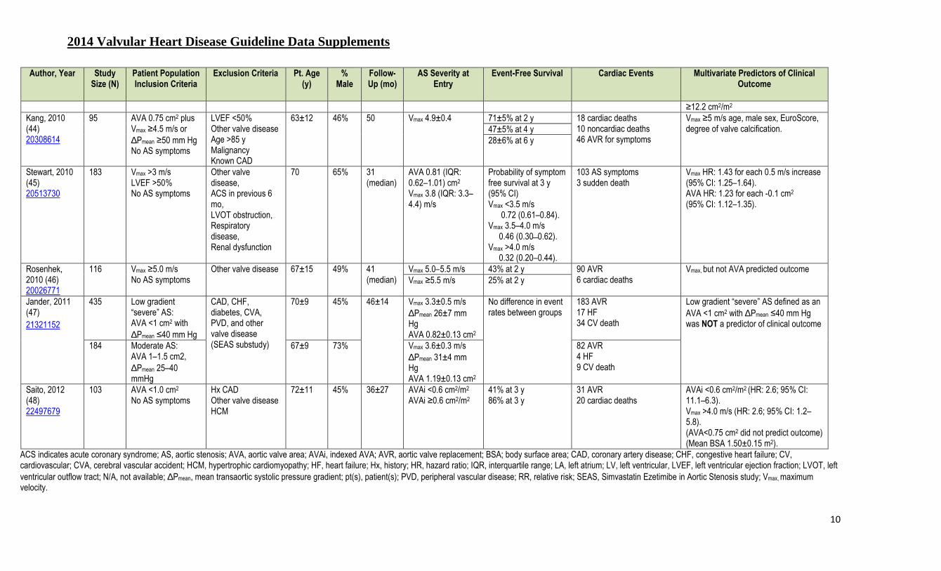
2014 Valvular Heart Disease Guideline Data Supplements
10
Author, Year Study Size (N)
Patient Population Inclusion Criteria
Exclusion Criteria Pt. Age (y)
% Male
Follow-Up (mo)
AS Severity at Entry
Event-Free Survival Cardiac Events Multivariate Predictors of Clinical Outcome
≥12.2 cm2/m2 Kang, 2010 (44) 20308614
95 AVA 0.75 cm2 plus Vmax ≥4.5 m/s or ∆Pmean ≥50 mm Hg No AS symptoms
LVEF <50% Other valve disease Age >85 y Malignancy Known CAD
63±12 46% 50 Vmax 4.9±0.4 71±5% at 2 y 18 cardiac deaths 10 noncardiac deaths 46 AVR for symptoms
Vmax ≥5 m/s age, male sex, EuroScore, degree of valve calcification. 47±5% at 4 y
28±6% at 6 y
Stewart, 2010 (45) 20513730
183 Vmax >3 m/s LVEF >50% No AS symptoms
Other valve disease, ACS in previous 6 mo, LVOT obstruction, Respiratory disease, Renal dysfunction
70 65% 31 (median)
AVA 0.81 (IQR: 0.62–1.01) cm2
Vmax 3.8 (IQR: 3.3–4.4) m/s
Probability of symptom free survival at 3 y (95% CI) Vmax <3.5 m/s 0.72 (0.61–0.84). Vmax 3.5–4.0 m/s 0.46 (0.30 ̶ 0.62). Vmax >4.0 m/s 0.32 (0.20–0.44).
103 AS symptoms 3 sudden death
Vmax HR: 1.43 for each 0.5 m/s increase (95% CI: 1.25–1.64). AVA HR: 1.23 for each -0.1 cm2 (95% CI: 1.12–1.35).
Rosenhek, 2010 (46) 20026771
116 Vmax ≥5.0 m/s No AS symptoms
Other valve disease 67±15 49% 41 (median)
Vmax 5.0–5.5 m/s 43% at 2 y 90 AVR 6 cardiac deaths
Vmax, but not AVA predicted outcome Vmax ≥5.5 m/s 25% at 2 y
Jander, 2011 (47) 21321152
435 Low gradient “severe” AS: AVA <1 cm2 with ∆Pmean ≤40 mm Hg
CAD, CHF, diabetes, CVA, PVD, and other valve disease (SEAS substudy)
70±9 45% 46±14 Vmax 3.3±0.5 m/s ∆Pmean 26±7 mm Hg AVA 0.82±0.13 cm2
No difference in event rates between groups
183 AVR 17 HF 34 CV death
Low gradient “severe” AS defined as an AVA <1 cm2 with ∆Pmean ≤40 mm Hg was NOT a predictor of clinical outcome
184 Moderate AS: AVA 1–1.5 cm2, ∆Pmean 25–40 mmHg
67±9 73% Vmax 3.6±0.3 m/s ∆Pmean 31±4 mm Hg AVA 1.19±0.13 cm2
82 AVR 4 HF 9 CV death
Saito, 2012 (48) 22497679
103 AVA <1.0 cm2
No AS symptoms Hx CAD Other valve disease HCM
72±11 45% 36±27 AVAi <0.6 cm2/m2 AVAi ≥0.6 cm2/m2
41% at 3 y 86% at 3 y
31 AVR 20 cardiac deaths
AVAi <0.6 cm2/m2 (HR: 2.6; 95% CI: 11.1–6.3). Vmax >4.0 m/s (HR: 2.6; 95% CI: 1.2–5.8). (AVA<0.75 cm2 did not predict outcome) (Mean BSA 1.50±0.15 m2).
ACS indicates acute coronary syndrome; AS, aortic stenosis; AVA, aortic valve area; AVAi, indexed AVA; AVR, aortic valve replacement; BSA; body surface area; CAD, coronary artery disease; CHF, congestive heart failure; CV, cardiovascular; CVA, cerebral vascular accident; HCM, hypertrophic cardiomyopathy; HF, heart failure; Hx, history; HR, hazard ratio; IQR, interquartile range; LA, left atrium; LV, left ventricular, LVEF, left ventricular ejection fraction; LVOT, left ventricular outflow tract; N/A, not available; ∆Pmean, mean transaortic systolic pressure gradient; pt(s), patient(s); PVD, peripheral vascular disease; RR, relative risk; SEAS, Simvastatin Ezetimibe in Aortic Stenosis study; Vmax, maximum velocity.
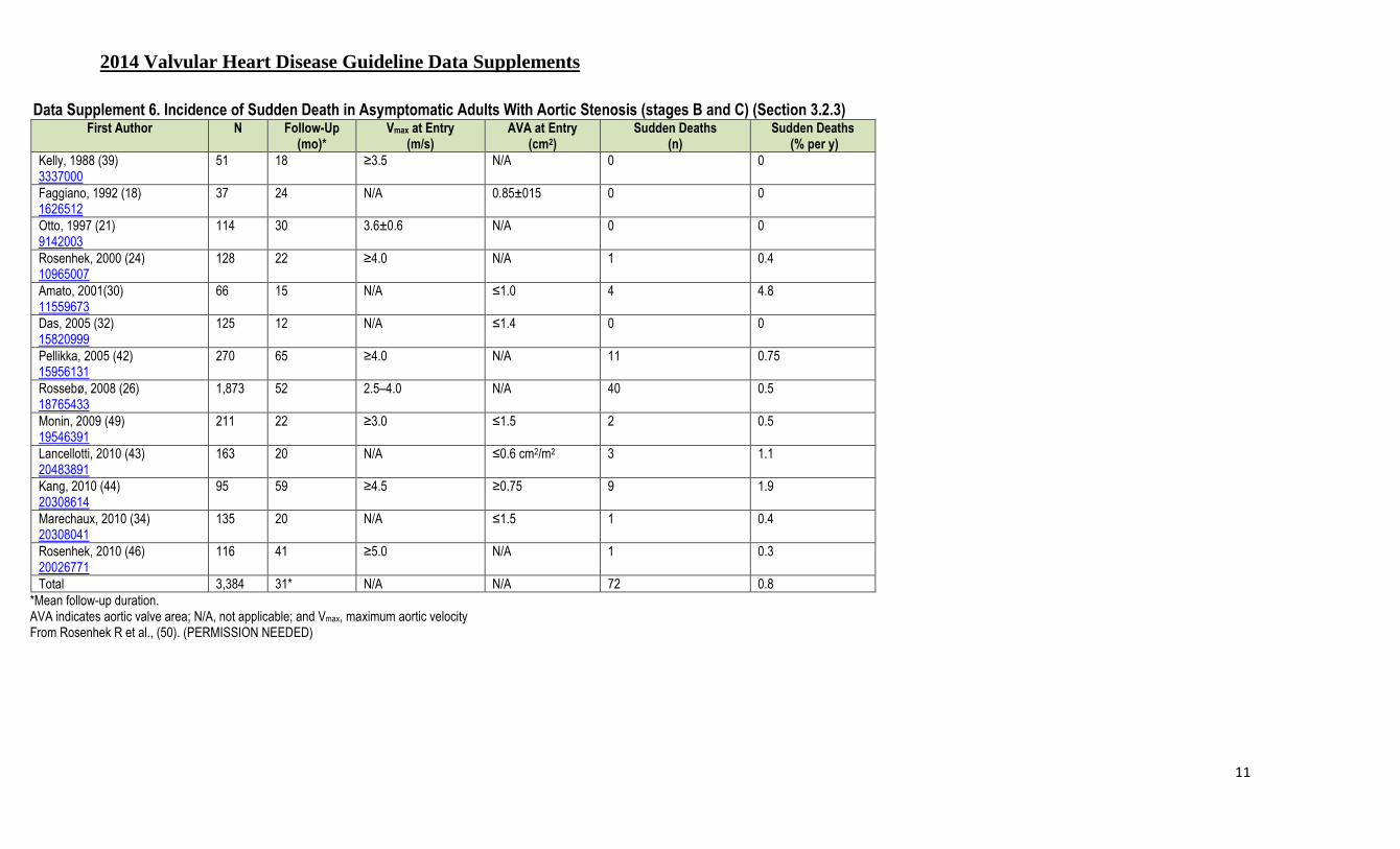
2014 Valvular Heart Disease Guideline Data Supplements
11
Data Supplement 6. Incidence of Sudden Death in Asymptomatic Adults With Aortic Stenosis (stages B and C) (Section 3.2.3) First Author N Follow-Up
(mo)* Vmax at Entry
(m/s) AVA at Entry
(cm2) Sudden Deaths
(n) Sudden Deaths
(% per y)
Kelly, 1988 (39) 3337000
51 18 ≥3.5 N/A 0 0
Faggiano, 1992 (18) 1626512
37 24 N/A 0.85±015 0 0
Otto, 1997 (21) 9142003
114 30 3.6±0.6 N/A 0 0
Rosenhek, 2000 (24) 10965007
128 22 ≥4.0 N/A 1 0.4
Amato, 2001(30) 11559673
66 15 N/A ≤1.0 4 4.8
Das, 2005 (32) 15820999
125 12 N/A ≤1.4 0 0
Pellikka, 2005 (42) 15956131
270 65 ≥4.0 N/A 11 0.75
Rossebø, 2008 (26) 18765433
1,873 52 2.5–4.0 N/A 40 0.5
Monin, 2009 (49) 19546391
211 22 ≥3.0 ≤1.5 2 0.5
Lancellotti, 2010 (43) 20483891
163 20 N/A ≤0.6 cm2/m2 3 1.1
Kang, 2010 (44) 20308614
95 59 ≥4.5 ≥0.75 9 1.9
Marechaux, 2010 (34) 20308041
135 20 N/A ≤1.5 1 0.4
Rosenhek, 2010 (46) 20026771
116 41 ≥5.0 N/A 1 0.3
Total 3,384 31* N/A N/A 72 0.8 *Mean follow-up duration. AVA indicates aortic valve area; N/A, not applicable; and Vmax, maximum aortic velocity From Rosenhek R et al., (50). (PERMISSION NEEDED)
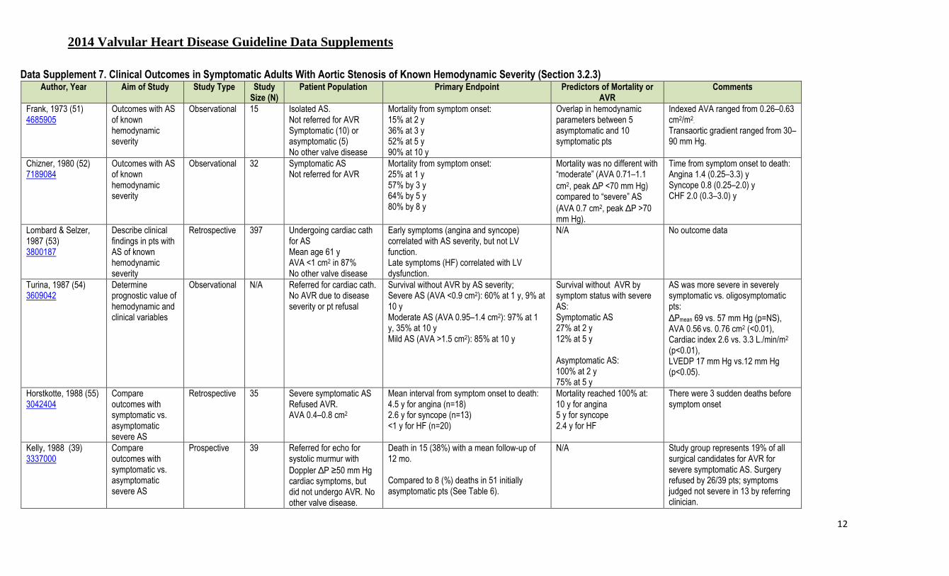
2014 Valvular Heart Disease Guideline Data Supplements
12
Data Supplement 7. Clinical Outcomes in Symptomatic Adults With Aortic Stenosis of Known Hemodynamic Severity (Section 3.2.3) Author, Year Aim of Study Study Type Study
Size (N) Patient Population Primary Endpoint Predictors of Mortality or
AVR Comments
Frank, 1973 (51) 4685905
Outcomes with AS of known hemodynamic severity
Observational 15 Isolated AS. Not referred for AVR Symptomatic (10) or asymptomatic (5) No other valve disease
Mortality from symptom onset: 15% at 2 y 36% at 3 y 52% at 5 y 90% at 10 y
Overlap in hemodynamic parameters between 5 asymptomatic and 10 symptomatic pts
Indexed AVA ranged from 0.26–0.63 cm2/m2.
Transaortic gradient ranged from 30–90 mm Hg.
Chizner, 1980 (52) 7189084
Outcomes with AS of known hemodynamic severity
Observational 32 Symptomatic AS Not referred for AVR
Mortality from symptom onset: 25% at 1 y 57% by 3 y 64% by 5 y 80% by 8 y
Mortality was no different with “moderate” (AVA 0.71–1.1 cm2, peak ∆P <70 mm Hg) compared to “severe” AS (AVA 0.7 cm2, peak ∆P >70 mm Hg).
Time from symptom onset to death: Angina 1.4 (0.25–3.3) y Syncope 0.8 (0.25–2.0) y CHF 2.0 (0.3–3.0) y
Lombard & Selzer, 1987 (53) 3800187
Describe clinical findings in pts with AS of known hemodynamic severity
Retrospective 397 Undergoing cardiac cath for AS Mean age 61 y AVA <1 cm2 in 87% No other valve disease
Early symptoms (angina and syncope) correlated with AS severity, but not LV function. Late symptoms (HF) correlated with LV dysfunction.
N/A No outcome data
Turina, 1987 (54) 3609042
Determine prognostic value of hemodynamic and clinical variables
Observational N/A Referred for cardiac cath. No AVR due to disease severity or pt refusal
Survival without AVR by AS severity; Severe AS (AVA <0.9 cm2): 60% at 1 y, 9% at 10 y Moderate AS (AVA 0.95–1.4 cm2): 97% at 1 y, 35% at 10 y Mild AS (AVA >1.5 cm2): 85% at 10 y
Survival without AVR by symptom status with severe AS: Symptomatic AS 27% at 2 y 12% at 5 y Asymptomatic AS: 100% at 2 y 75% at 5 y
AS was more severe in severely symptomatic vs. oligosymptomatic pts: ∆Pmean 69 vs. 57 mm Hg (p=NS), AVA 0.56 vs. 0.76 cm2 (<0.01), Cardiac index 2.6 vs. 3.3 L./min/m2 (p<0.01), LVEDP 17 mm Hg vs.12 mm Hg (p<0.05).
Horstkotte, 1988 (55) 3042404
Compare outcomes with symptomatic vs. asymptomatic severe AS
Retrospective 35 Severe symptomatic AS Refused AVR. AVA 0.4–0.8 cm2
Mean interval from symptom onset to death: 4.5 y for angina (n=18) 2.6 y for syncope (n=13) <1 y for HF (n=20)
Mortality reached 100% at: 10 y for angina 5 y for syncope 2.4 y for HF
There were 3 sudden deaths before symptom onset
Kelly, 1988 (39) 3337000
Compare outcomes with symptomatic vs. asymptomatic severe AS
Prospective 39 Referred for echo for systolic murmur with Doppler ∆P ≥50 mm Hg cardiac symptoms, but did not undergo AVR. No other valve disease.
Death in 15 (38%) with a mean follow-up of 12 mo. Compared to 8 (%) deaths in 51 initially asymptomatic pts (See Table 6).
N/A Study group represents 19% of all surgical candidates for AVR for severe symptomatic AS. Surgery refused by 26/39 pts; symptoms judged not severe in 13 by referring clinician.
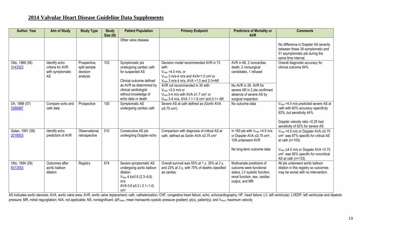
2014 Valvular Heart Disease Guideline Data Supplements
13
Author, Year Aim of Study Study Type Study Size (N)
Patient Population Primary Endpoint Predictors of Mortality or AVR
Comments
Other valve disease. No difference in Doppler AS severity between these 39 symptomatic and 51 asymptomatic pts during the same time interval.
Otto, 1988 (56) 3143323
Identify echo criteria for AVR with symptomatic AS
Prospective, split sample decision analysis
103 Symptomatic pts undergoing cardiac cath for suspected AS Clinical outcome defined as AVR as determined by clinical cardiologist without knowledge of echo data or death
Decision model recommended AVR in 73 with: Vmax >4.0 m/s, or Vmax 3 m/s-4 m/s and AVA<1.0 cm2 or Vmax 3 m/s-4 m/s, AVA >1.0 and 2-3+AR
AVR in 68, 2 noncardiac death, 2 nonsurgical candidates, 1 refused
Overall diagnostic accuracy for clinical outcome 94%
AVR not recommended in 30 with: Vmax <3.0 m/s or Vmax 3-4 m/s with AVA ≥1.7 cm2 or Vmax 3-4 m/s, AVA 1.1-1.6 cm2 and 0-1+ AR
No AVR in 28. AVR for severe AR in 2 pts confirmed absence of severe AS by surgical inspection
Oh, 1988 (57) 3366997
Compare echo and cath data
Prospective 100 Symptomatic AS undergoing cardiac cath
Severe AS at cath defined as (Gorlin AVA ≤0.75 cm2)
No outcome data Vmax >4.5 m/s predicted severe AS at cath with 60% accuracy–specificity 93%, but sensitivity 44% Doppler velocity ratio <0.25 had sensitivity of 92% for severe AS
Galan, 1991 (58) 2018003
Identify echo predictors of AVR
Observational, retrospective
510 Consecutive AS pts undergoing Doppler echo
Comparison with diagnosis of critical AS at cath, defined as Gorlin AVA ≤0.75 cm2
In 160 pts with Vmax >4.5 m/s or Doppler AVA ≤0.75 cm2, 109 underwent AVR No long-term outcome data
Vmax >4.5 m/s or Doppler AVA ≤0.75 cm2 was 97% specific for critical AS at cath (n=105) Vmax ≤4.5 m/s or Doppler AVA >0.75 cm2 was 95% specific for noncritical AS at cath (n=133)
Otto, 1994 (59) 8313553
Outcomes after aortic balloon dilation
Registry 674 Severe symptomatic AS undergoing aortic balloon dilation Vmax 4.4±0.8 (2.3–6.6) m/s AVA 0.6 ±0.2 ( 0.1–1.4) cm2
Overall survival was 55% at 1 y, 35% at 2 y, and 23% at 3 y, with 70% of deaths classified as cardiac
Multivariate predictors of outcome were functional status, LV systolic function, renal function, sex, cardiac output, and MR
All pts underwent aortic balloon dilation in this registry so outcomes may be worse with no intervention.
AS indicates aortic stenosis; AVA, aortic valve area; AVR, aortic valve replacement; cath, catheterization; CHF, congestive heart failure, echo, echocardiography; HF, heart failure; LV, left ventricular; LVEDP, left ventricular end diastolic pressure; MR, mitral regurgitation; N/A, not applicable; NS, nonsignificant; ∆Pmean, mean transaortic systolic pressure gradient; pt(s), patient(s); and Vmax, maximum velocity.
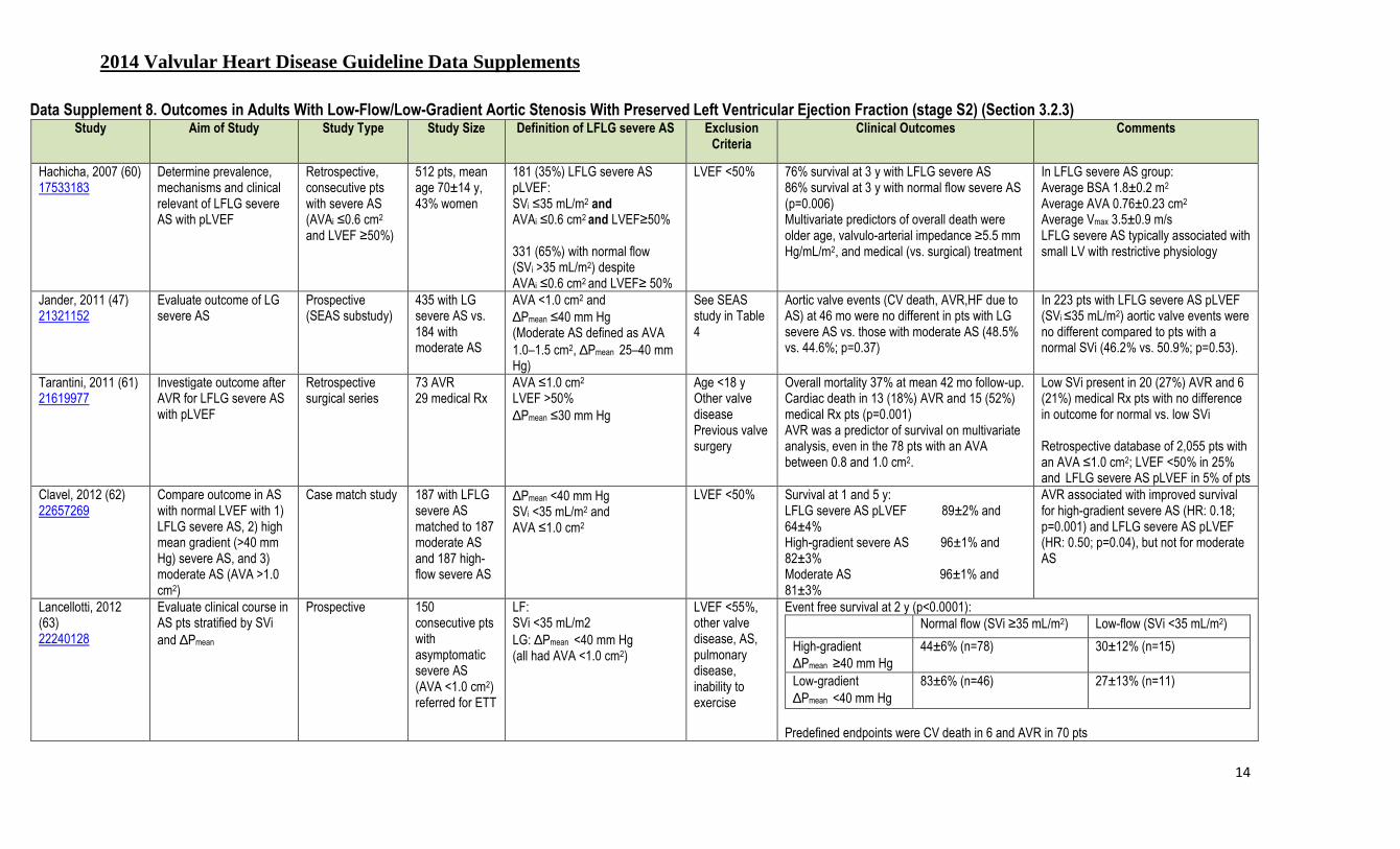
2014 Valvular Heart Disease Guideline Data Supplements
14
Data Supplement 8. Outcomes in Adults With Low-Flow/Low-Gradient Aortic Stenosis With Preserved Left Ventricular Ejection Fraction (stage S2) (Section 3.2.3) Study Aim of Study Study Type Study Size Definition of LFLG severe AS Exclusion
Criteria Clinical Outcomes Comments
Hachicha, 2007 (60) 17533183
Determine prevalence, mechanisms and clinical relevant of LFLG severe AS with pLVEF
Retrospective, consecutive pts with severe AS (AVAi ≤0.6 cm2
and LVEF ≥50%)
512 pts, mean age 70±14 y, 43% women
181 (35%) LFLG severe AS pLVEF: SVi ≤35 mL/m2 and AVAi ≤0.6 cm2 and LVEF≥50% 331 (65%) with normal flow (SVi >35 mL/m2) despite AVAi ≤0.6 cm2 and LVEF≥ 50%
LVEF <50% 76% survival at 3 y with LFLG severe AS 86% survival at 3 y with normal flow severe AS (p=0.006) Multivariate predictors of overall death were older age, valvulo-arterial impedance ≥5.5 mm Hg/mL/m2, and medical (vs. surgical) treatment
In LFLG severe AS group: Average BSA 1.8±0.2 m2
Average AVA 0.76±0.23 cm2
Average Vmax 3.5±0.9 m/s LFLG severe AS typically associated with small LV with restrictive physiology
Jander, 2011 (47) 21321152
Evaluate outcome of LG severe AS
Prospective (SEAS substudy)
435 with LG severe AS vs. 184 with moderate AS
AVA <1.0 cm2 and ∆Pmean ≤40 mm Hg (Moderate AS defined as AVA 1.0–1.5 cm2, ∆Pmean 25–40 mm Hg)
See SEAS study in Table 4
Aortic valve events (CV death, AVR,HF due to AS) at 46 mo were no different in pts with LG severe AS vs. those with moderate AS (48.5% vs. 44.6%; p=0.37)
In 223 pts with LFLG severe AS pLVEF (SVi ≤35 mL/m2) aortic valve events were no different compared to pts with a normal SVi (46.2% vs. 50.9%; p=0.53).
Tarantini, 2011 (61) 21619977
Investigate outcome after AVR for LFLG severe AS with pLVEF
Retrospective surgical series
73 AVR 29 medical Rx
AVA ≤1.0 cm2 LVEF >50% ∆Pmean ≤30 mm Hg
Age <18 y Other valve disease Previous valve surgery
Overall mortality 37% at mean 42 mo follow-up. Cardiac death in 13 (18%) AVR and 15 (52%) medical Rx pts (p=0.001) AVR was a predictor of survival on multivariate analysis, even in the 78 pts with an AVA between 0.8 and 1.0 cm2.
Low SVi present in 20 (27%) AVR and 6 (21%) medical Rx pts with no difference in outcome for normal vs. low SVi Retrospective database of 2,055 pts with an AVA ≤1.0 cm2; LVEF <50% in 25% and LFLG severe AS pLVEF in 5% of pts
Clavel, 2012 (62) 22657269
Compare outcome in AS with normal LVEF with 1) LFLG severe AS, 2) high mean gradient (>40 mm Hg) severe AS, and 3) moderate AS (AVA >1.0 cm2)
Case match study 187 with LFLG severe AS matched to 187 moderate AS and 187 high-flow severe AS
∆Pmean <40 mm Hg SVi <35 mL/m2 and AVA ≤1.0 cm2
LVEF <50% Survival at 1 and 5 y: LFLG severe AS pLVEF 89±2% and 64±4% High-gradient severe AS 96±1% and 82±3% Moderate AS 96±1% and 81±3%
AVR associated with improved survival for high-gradient severe AS (HR: 0.18; p=0.001) and LFLG severe AS pLVEF (HR: 0.50; p=0.04), but not for moderate AS
Lancellotti, 2012 (63) 22240128
Evaluate clinical course in AS pts stratified by SVi and ∆Pmean
Prospective 150 consecutive pts with asymptomatic severe AS (AVA <1.0 cm2) referred for ETT
LF: SVi <35 mL/m2 LG: ∆Pmean <40 mm Hg (all had AVA <1.0 cm2)
LVEF <55%, other valve disease, AS, pulmonary disease, inability to exercise
Event free survival at 2 y (p<0.0001): Normal flow (SVi ≥35 mL/m2) Low-flow (SVi <35 mL/m2)
High-gradient ∆Pmean ≥40 mm Hg
44±6% (n=78) 30±12% (n=15)
Low-gradient ∆Pmean <40 mm Hg
83±6% (n=46) 27±13% (n=11)
Predefined endpoints were CV death in 6 and AVR in 70 pts

2014 Valvular Heart Disease Guideline Data Supplements
15
Study Aim of Study Study Type Study Size Definition of LFLG severe AS Exclusion Criteria
Clinical Outcomes Comments
Herrmann 2013 (15) 23661722
Evaluate outcomes with TAVR compared to medical therapy with LG severe AS
Subgroup analysis of RCT
52 inoperable symptomatic pts with LFLG severe AS with normal LVEF
∆Pmean <40 mm Hg SVi <35 mL/m2 and AVA <0.8 cm2 or AVAi <0.5 cm2/m2
LVEF <50%
In 52 inoperable pts with LFLG severe AS with preserved LVEF, 1-y mortality was 66% with TAVR compared to 35% with medical therapy (HR: 0.38; p=0.02). In 87 pts at high risk for surgery, there was no difference between TAVR and SAVR (39.0% vs. 38.3%; HR: 0.91; 95% CI: 0.57–1.45; p=0.69.
Le Ven 2013 (64) 23770162
Evaluate effect of LV EF and gradient on outcomes after TAVR
Retrospective analysis of registry data
639 severe AS undergoing TAVR
Low flow (SVi <35 mL/m2) with a normal EF (>50%) was present in 86 (13%) of pts
--- Low flow (but not low EF) was an independent predictor of 30-day mortality (odds ratio: 1.94, p=0.026), cumulative all-cause mortality (hazard ratio: 1.27 per 10 mL/m² SVi decrease, p=0.016), and cumulative cardiovascular mortality (hazard ratio: 1.29 per 10 mL/m² decrease, p=0.04).
Mehrotra 2013 (65) 23533186
Compare clinical characteristics and outcomes in AS subgroups
Retrospective echocardiographic database
LFLG severe AS in 38 pts, compared to 75 normal flow low gradient and 70 moderate AS pts.
AVA ≤1.0 cm2 with LVEF≥ 55%, mead gradient <40 mm Hg and SVi <35 mL/m2.
Mitral valve disease, aortic regurgitation, poor quality study. Severe AS with mean gradient >40 mm Hg.
Survival at 3 years was significantly lower in LF LG compared with NF LG (p=0.006) and moderate AS (p=0.002), but not different between NF LG and moderate AS (p=0.49).
Ozkan 2013 (66) 23812184
Compare outcomes of LG severe AS with AVR or medical therapy
Prospective follow-up of symptomatic severe LG AS
260 pts with symptomatic severe AS (AVAi ≤0.6 cm2/m2 ) and mean gradient <40 mm Hg
Normal flow present in 125; low flow (SVi ≤35 mL/m2) in 135.
Mitral disease, aortic regurgitation
At 28 ±24 mos follow-up, 105 pts died (40%): 32 (30%) in the AVR group and 73 (70%) in the medical treatment group. AVR (hazard ratio, 0.54; 95% confidence interval, 0.32–0.94; p<0.001) was independently associated with outcome and remained a strong predictor of survival after adjustment for propensity score. The protective effect of AVR was similar in 125 pts with normal flow (stroke volume index >35 mL/m2; p=0.22).
Eleid 2013 (67) 24048203
Evaluate impact of stroke volume with normal EF on outcomes with severe AS
Echocardiographic database.
1,704 consecutive pts with severe AS (AVA <1.0 cm2) and LVEF≥50%
Low flow = SVi ≤35 mL/m2
Low gradient <40 mm Hg. LFLG present in 53 pts (3%) compared to normal flow LG (n=352, 21%) and to high gradient severe AS.
Prosthetic valve, congenital or other native valve disease
AVR was associated with a 69% mortality reduction (HR 0.31 (0.25, 0.39) p<0.0001) in LF/LG and NF/HG, with no survival benefit associated with AVR in NF/LG and LF/HG.
AS indicates aortic stenosis; AVAi, aortic valve area indexed to body surface area; AVR, aortic valve replacement; BSA, body surface area; CV, cardiovascular; ETT, exercise treadmill testing; HG, high gradient; HF, heart failure; LFLG, low-flow low-gradient; LF, low-flow; LG, low-gradient; LV, left ventricular; NF, normal flow; pLVEF, preserved left ventricular ejection fraction; ∆Pmean, mean transaortic systolic pressure gradient; RCT, randomized controlled clinical trial; Rx, prescription; SEAS, Simvastatin Ezetimibe in Aortic Stenosis study; SVi, stroke volume index; TAVR, transcatheter aortic valve replacement; and Vmax, maximum velocity.

2014 Valvular Heart Disease Guideline Data Supplements
16
Data Supplement 9. Choice of Intervention in Symptomatic Adults With Severe Aortic Stenosis (stage D): Surgical Versus Transcatheter Aortic Valve Replacement (Section 3.2.4) Study Aim of Study Study
Type Study Groups (N) Patient Population Major Endpoints Other Results
PARTNER COHORT A (high-surgical risk) (68) 21639811 (69) 22443479
To show that TAVR is not inferior to SAVR
RCT TAVR 348 vs. SAVR 351 TAVR was transfemoral in 244 and transapical in 104
Severe symptomatic calcific AS defined as AVA <0.8 cm2 plus a mean ∆P ≥40 mm Hg or Vmax ≥4.0 m/s with NYHA class II-IV symptoms. High surgical risk defined as ≥15% risk of death by 30 d after the procedure. An STS score ≥10% was used for guidance with an actual mean STS score of 11.8±3.3% Exclusions were bicuspid aortic valve, AMI, significant CAD, LVEF<20%, aortic annulus <18 or >25 mm, severe AR or MR, TIA within 6 mo, or severe renal insufficiency
All cause death (intention to treat analysis):
TAVR SAVR p-value
30 d 3.4% 6.5% 0.07 1 y* 24.2% 26.8% 0.44% 2 y 33.9% 35.0% 0.78
*(p=0.001 for noninferiority) Composite endpoint at 2 y –all-cause death or stroke: TAVR 37.1% vs. SAVR 36.4% (p=0.85) HR: 0.93; 95% CI: 0.73–1.18; p=0.55
Stroke or TIA at 2 y: TAVR 11.2 % vs. SAVR 6.5 % (p=0.05) Major vascular complications at 30 d: TAVR 11.0% vs. SAVR 3.2% (p<0.001) Major bleeding at 30 d: TAVR 9.3% vs. SAVR 19.5% (p<0.001) New-onset AF at 30 d: TAVR 8.6% vs. SAVR 16.0% (p=0.006).
PARTNER COHORT B (inoperable) (70) 22443478 (71) 20961243
Compare TAVR to medical Rx in inoperable pts with severe symptomatic AS
RCT TAVR in 179 vs. standard medical therapy in 179 (including BAV in 150 (84%)
Severe symptomatic calcific AS defined as AVA <0.8 cm2 plus a mean ∆P ≥40 mm Hg or Vmax ≥4.0 m/s with NYHA class II-IV symptoms. Inoperable due to coexisting conditions with predicted ≥50% risk of death within 30 d of intervention or a serious irreversible condition. Exclusions were bicuspid aortic valve, AMI, significant CAD, LVEF<20%, aortic annulus <18 or >25 mm, severe AR or MR, TIA within 6 mo, or severe renal insufficiency
All-cause death at 2 y (Kaplan–Meier): TAVR 43.3% vs. standard therapy 68% HR: with TAVR, 0.58 (95% CI: 0.36–0.92; p=0.02). Repeat hospitalization: TAVR 55% vs. 72.5% standard therapy (p<0.001). Survival benefit of TAVR stratified by STS score: STS score <5% HR: 0.37 (95% CI: 0.13–1.01 ); p=0.04 STS score 5%–14.9% HR: 0.58 (95% CI: 0.41–0.81); p=0.002 STS score ≥15% HR: 0.77 (95% CI: 0.46–1.28); p=0.31
Cardiac symptoms (NYHA class III or IV) were present in 25.2% of survivors at 1 y after TAVR vs. 58% with standard therapy (p<0.001). Major stroke rate at 30 d, was 5.0% with TAVR vs. 1.1% with standard therapy (p=0.06) and remained high at 2 y 13.8% with TAVR vs. 5.5% (p=0.01) Major vascular complications occurred in 16.2% with TAVR vs. 1.1% with standard therapy (p<0.001).
AF indicates atrial fibrillation; AMI, acute myocardial infarction; AS, aortic stenosis; AR, aortic regurgitation; AVA, aortic valve area; CAD, coronary artery disease; LVEF, left ventricular ejection fraction; MI, myocardial infarction; MR, mitral regurgitation; NYHA, New York Heart Association; ∆P, mean transaortic pressure gradient; pt(s), patient(s); RCT, randomized controlled trial; Rx, prescription; SAVR, surgical aortic valve replacement; STS, Society of Thoracic Surgeons; TAVR, transcatheter aortic valve replacement; TIA, transient ischemic attack; and Vmax, aortic valve maximum velocity.

2014 Valvular Heart Disease Guideline Data Supplements
17
Data Supplement 10. Clinical Outcomes of Asymptomatic Patients With Chronic Aortic Regurgitation (Sections 4.3.1.1 and 4.3.3) Study, Year Aim of Study Study Type Study
Size (n) Mean
Follow-Up (y)
Inclusion Criteria, Details
Outcomes Comments, Limitations
Bonow, 1983 (72) 6872164
Determine clinical outcome of asymptomatic pts with chronic AR and normal LV systolic function
Prospective, observational series; consecutive pts enrolled 1973-1982; single institution
77 4.1 Initially asymptomatic pts with chronic AR and normal LV systolic function Mean age 37 y (range 17–67) Serial echo and radionuclide angiographic studies 63 pts had 3+–4+ AR on aortic root angiography, and the other 14 pts had pulse pressures >70 mm Hg Endpoints: death, symptoms, LV systolic dysfunction
No pt died 12 pts underwent AVR because of symptoms (n=11) or asymptomatic LV dysfunction (n=1) Progression to symptoms or LV dysfunction: less than 4%/y No perioperative deaths in pts who underwent AVR
Percent of pts who did not need surgery was 90±3% (±SE) at 3 y, 81+6% at 5 y, and 75±7% at 7 y. Outcome associated with LVESD, LVEDD, FS, change in LVEF with exercise
Scognomiglio, 1986 (73) 3720042
Determine factors predictive of progression to LV systolic dysfunction
Observational series; single institution
30 4.7 38 initially asymptomatic pts with chronic AR, 30 of whom had normal LV fractional shortening Mean age 26±10 y Serial echo studies Endpoints: death, symptoms, subnormal LV fractional shortening
No pt died Progression to symptoms or LV dysfunction: 2.1%/y Progression to asymptomatic LV dysfunction: 2.1%/y
3 pts developing asymptomatic LV dysfunction had lower initial PAP/ESV ratios and trend toward higher LVESD and LVEDD and lower fractional shortening
Siemienczuk, 1989 (74) 2930091
Determine clinical outcome of asymptomatic pts with chronic AR and normal LV function.
Observational series derived from screening for randomized clinical trial; single institution
50 3.7 Pts included those receiving placebo and medical dropouts in a randomized drug trial of hydralazine therapy; included some pts with NYHA II symptoms. Mean age 48±16 y Serial echo and radionuclide LV angiographic studies
No pt died Progression to symptoms or LV dysfunction: 4.0%/y Progression to asymptomatic LV dysfunction: 0.5%/y
Outcome associated with LVESV, EDV, change in LVEF with exercise, and end-systolic wall stress
Bonow, 1991 (75) 1914102
Determine outcomes of asymptomatic pts with chronic AR; extension of Bonow, 1983
Prospective, observational series; consecutive pts enrolled 1973-1988; single institution
104 8.0 Initially asymptomatic pts with chronic AR and normal LV systolic function Mean age 37 y (range 17–67) Serial echo (average 7.5 per pt) and radionuclide LV angiographic (average 5.0 per pt) studies Endpoints: death, symptoms, LV systolic dysfunction
2 pts died suddenly Progression to symptoms or LV dysfunction: 2.1%/y Progression to asymptomatic LV dysfunction: 2.1%/y
Outcome associated with age, LVESD, LVEDD, change in LVEF with exercise, and rate of change in LVESD and LVEF at rest with time Initial LVESD >50 mm was associated with risk of death, symptoms, and/or LV dysfunction of 19% per y
Scognomiglio, 1994 (76) 8058074
Effect of nifedipine on outcomes of pts with severe AR and normal LV function
Randomized clinical drug trial (see Data Supplement 11); single institution
74 6.0 Initially asymptomatic pts with chronic AR and normal LV systolic function Mean age 36±12 y Serial echo studies Endpoints: death, symptoms, LV systolic dysfunction
No pt died Progression to death, symptoms or LV dysfunction: 5.7%/y Progression to asymptomatic LV dysfunction: 3.4%/y
This table include only the pts who received digoxin as part of a randomized trial See Data Supplement 11 for outcomes in those receiving active drug (nifedipine, n=69)

2014 Valvular Heart Disease Guideline Data Supplements
18
Study, Year Aim of Study Study Type Study Size (n)
Mean Follow-Up (y)
Inclusion Criteria, Details
Outcomes Comments, Limitations
Tornos, 1995 (77) 7631617
Determine clinical outcome of asymptomatic pts with chronic AR and normal LV systolic function
Prospective, observational series; consecutive pts beginning in 1982; single institution
101 4.6 Initially asymptomatic pts with chronic AR and normal LV systolic function Mean age 41±14 y Serial echo and radionuclide LV angiographic studies Endpoints: death, symptoms, LV systolic dysfunction
No pt died Progression to symptoms or LV dysfunction: 3.0%/y Progression to asymptomatic LV dysfunction: 1.3%/y
Outcome associated with pulse pressure, LVESD, LVEDD, and LVEF at rest Initial LVESD >50 mm was associated with risk of death, symptoms, and/or LV dysfunction of 7% per y
Ishii, 1996 (78) 8759822
Clinical outcome and LV response to chronic AR
Prospective, observational series; consecutive pts 1970-1990; single institution
27 14.2 94 consecutive pts followed for ≥6 mo; the 27 asymptomatic pts with normal LV function are included here Mean age 42±12 y LV function assessed by echo
No pt died Progression to symptoms or LV dysfunction: 3.6%/y
Development of symptoms associated with systolic BP, LVESD, LVEDD, mass index, and wall thickness. LV function not reported in all pts
Borer, 1998 (79) 9494022
Determine clinical outcome of asymptomatic pts with chronic AR and normal LV systolic function
Prospective, observational series; consecutive pts beginning in 1979; single institution
104 7.3 Initially asymptomatic pts with chronic AR and normal LV systolic function Mean age 46±15 y 20% of pts in NYHA II initially Serial echo and radionuclide LV angiographic studies Endpoints: death, symptoms, LV systolic dysfunction
4 pts died suddenly Progression to symptoms or LV dysfunction: 6.2%/y Progression to asymptomatic LV dysfunction: 0.9%/y
Change in LVEF from rest to exercise, normalized for change in end-systolic stress from rest to exercise was strongest predictor of any endpoint or of sudden cardiac death alone Outcome also associated with initial NYHA II symptoms, change in LVEF with exercise, LVESD, and LVFS
Tarasoutchi, 2003 (80) 12706927
Clinical outcome of asymptomatic pts with chronic AR and normal LV systolic function
Prospective, observational series; consecutive pts beginning in 1979; single institution
72 10 Initially asymptomatic pts with chronic AR and normal LV systolic function Mean age 28±9 y Serial echo and radionuclide LV angiographic studies Endpoints: death, symptoms, LV systolic dysfunction
No pt died Progression to symptoms or LV dysfunction: 4.7%/y Progression to asymptomatic LV dysfunction: 0.1%/y
AR of predominant rheumatic etiology LV function not reported in all pts Development of symptoms associated with LVESD and LVEDD Initial LVESD >50 mm was associated with risk of symptoms and/or LV dysfunction of 7.6%/y
Evangelista, 2005 (81) 16192479
Effect of nifedipine versus enalapril on outcomes of pts with severe AR and normal LV function
Randomized clinical drug trial (see Data Supplement 11); single institution
31 7 Initially asymptomatic pts with chronic AR and normal LV systolic function Mean age 42±15 y Serial echo studies Endpoints: death, symptoms, LV systolic dysfunction
1 pt died from HF Progression to death, symptoms or LV dysfunction: 3.6%/y
Pts reported here were in the control (placebo) group of this clinical trial See Data Supplement 11 for pts receiving active drugs nifedipine (n=32) and enalapril (n=31)

2014 Valvular Heart Disease Guideline Data Supplements
19
Study, Year Aim of Study Study Type Study Size (n)
Mean Follow-Up (y)
Inclusion Criteria, Details
Outcomes Comments, Limitations
Detaint, 2008 (82) 19356398
Predictive value of quantitative measures of AR severity and LV volumes in asymptomatic pts with chronic AR and normal LV systolic function
Prospective, observational series; consecutive pts enrolled from 1991–2003; single institution.
251 8 Initially asymptomatic pts with chronic AR and normal LV systolic function Mean age 60±17 y Serial echo studies to assess severity of AR (ROA and RV) as well as LV dimensions and volumes Endpoints: death, HF, AF, surgery
33 pts died Progression to death or surgery: 5.0%/y Survival at 10 y: Mild AR: 92±4% Moderate AR: 75±6% Severe AR: 69±9% Survival free from AVR at 10 y: Mild AR: 92±4% Moderate AR: 57±6% Severe AR: 20±5%
Surgical indications included symptoms (n=38), LV dysfunction or enlargement (n=17), aortic aneurysm (n=11), IE (n=3, and clinician and/or pt preference [n=11]) Cardiac events (defined as cardiac death, HR, or new onset of AF) associated with RV and ROA as well as ESV index, which superseded M-mode LV dimensions Mortality rate in this series is highest of all series Pts in this series older than all others; only 1 death in pts <50 y in this series
Pizzaro, 2011 (83) 21982316
Predictive value of BNP and quantitative measures of AR severity and LV volumes in asymptomatic pts with chronic AR and normal LV systolic function
Prospective, observational series; consecutive pts enrolled from 1991–2003; single institution
294 3.5 Initially asymptomatic pts with chronic AR and normal LV systolic function The first 160 consecutive pts were analyzed as the derivation set of data (mean age 51±9 y) The next 134 consecutive pts were analyzed as the validation set (mean age 53±10 y) BNP and serial echo studies to assess severity of AR (ROA and RV) as well as LV dimensions and volumes
5 pts died Progression to symptoms or LV dysfunction: 10%/y Progression to asymptomatic LV dysfunction: 2.8%/y
Outcome associated with BNP >130 pg/mL Outcome also associated with RV, ROA, LVESD index, LVEDD index, ESV index, and EDV index
Olsen, 2011 (84) 21414568
Predictive value of speckle-tracking echo in asymptomatic pts with chronic AR and normal LV systolic function
N/A 35 1.6 35 initially asymptomatic pts with chronic AR and normal LV systolic function were followed sequentially Mean age 56±14 y Serial echo studies Endpoints: symptoms, increase in LVEDV >15%, or decrease in LVEF >10% 29 additional pts who underwent AVR at the outset are not reported here
No pts died Progression to death, symptoms, increase in LVEDV or decrease in LVEF: 14.3%/y
Disease progression defined as symptoms, increase in LVEDV >15%, or decrease in LVEF >10% Disease progression associated with reduced myocardial systolic strain, systolic strain rate, and early diastolic strain rate
AF indicates atrial fibrillation; AR, aortic regurgitation; AVR, aortic valve replacement; BNP; brain natriuretic peptide; BP, blood pressure; EDV, end-diastolic volume; ESV, end-systolic volume; HF, heart failure; Hx, history; LV, left ventricular; LVEDD, end-diastolic dimension; LVEDV, left ventricular end-diastolic volume; LVEF, left ventricular ejection fraction; LVESD, left ventricular end-systolic volume; IE, infective endocarditis; N/A, not applicable; NYHA, New York Heart Association; PAP, pulmonary artery pressure; pt(s), patient(s); ROA, regurgitant orifice area; RV, regurgitant volume; and SE, standard error.

2014 Valvular Heart Disease Guideline Data Supplements
20
Data Supplement 11. Vasodilator Therapy in Asymptomatic Patients With Chronic Aortic Regurgitation (Section 4.3.2) Study Name, Author, Year
Study Aim Study Type/ Size (N)
Intervention vs. Comparator (n)
Patient Population Study Intervention Study Comparator
Endpoints Results
Inclusion Criteria Exclusion Criteria
Primary Endpoint &
Results
Evangelista, 2005 (81) 16192479
Effects of vasodilator therapy on LV function and time to AVR
RCT/95 Intervention: open-label nifedipine-32 pts (20 mg every 12 h) or open label enalapril-32 pts (20 mg every 12 h) vs. Comparator: no treatment-31 pts
Asymptomatic, chronic, severe AR and normal LV function
LVEF <50%., other valve disease. Hypertension, AF, CAD, aortic aneurysm
Open-label nifedipine (20 mg every 12 h) or open-label enalapril (20 mg/d)
No treatment LVEF Time to AVR
Rate of AVR was similar among the groups: Control group 39% Enalapril group 50% Nifedipine group 41%; p=0.62) No significant group differences in AR severity, LV size or LVEF. Follow-up mean 7 y
Scognomiglio, 1994 (76) 8058074
Assess whether vasodilator therapy reduces or delays the need for AVR
RCT/143 Intervention: Nifedipine (20 mg twice daily)-69 pts vs. Comparator: Digoxin (0.25 mg twice daily)-74 pts
Asymptomatic chronic severe AR with normal LV function
LVEF <50%, recent or worsening AR, hypertension, CAD, AS, other valve disease.
Nifedipine Digoxin Time to AVR AVR in 34%+6% of pts on digoxin versus 15%+3% of pts on nifedipine pts (p<0.001) at 6 y follow-up
AF indicates atrial fibrillation; AR, aortic regurgitation; AS, aortic stenosis; AVR, aortic valve replacement; CAD, coronary artery disease; LV, left ventricular; LVEF, left ventricular ejection fraction; pts, patients; and, RCT, randomized controlled trial.
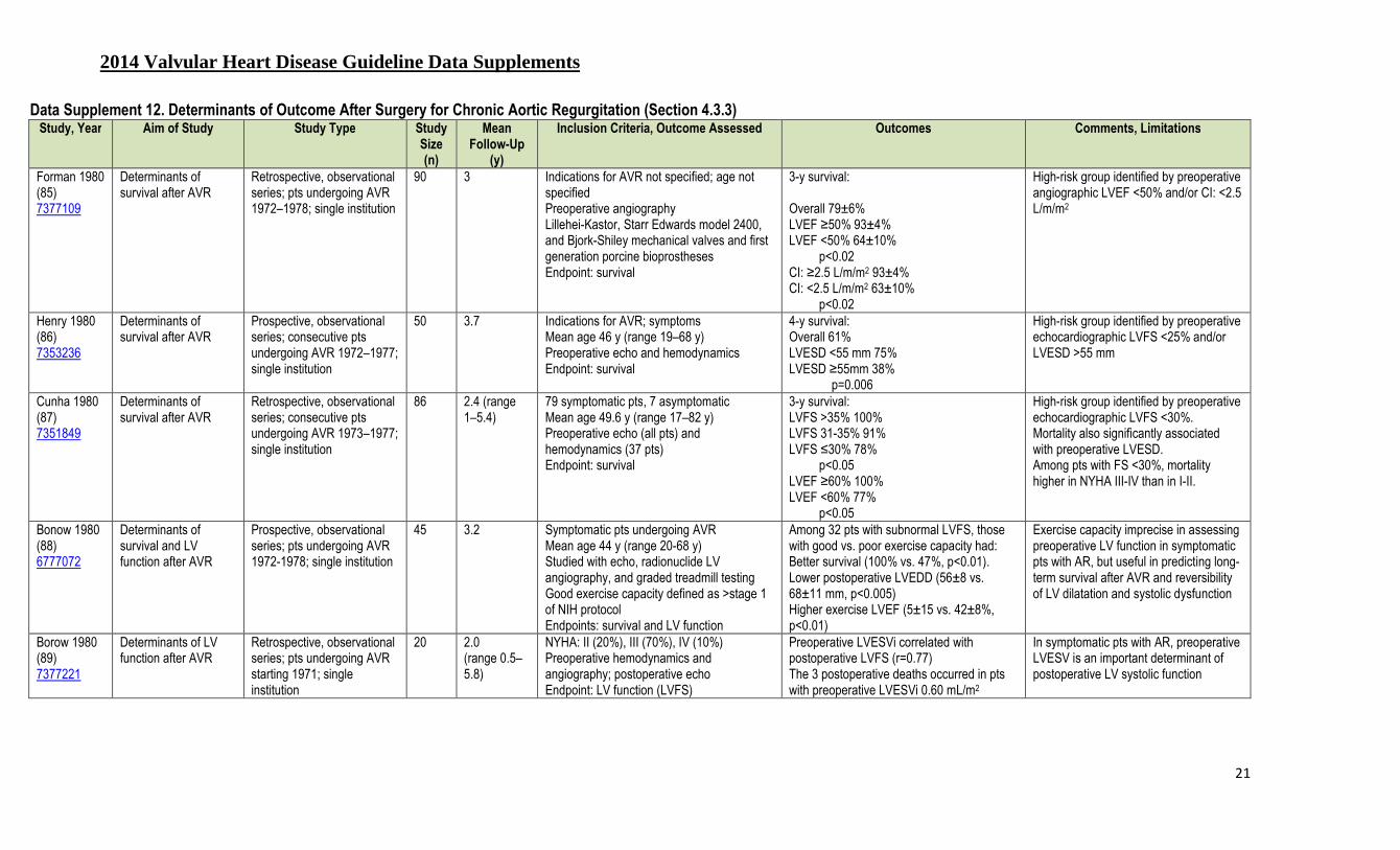
2014 Valvular Heart Disease Guideline Data Supplements
21
Data Supplement 12. Determinants of Outcome After Surgery for Chronic Aortic Regurgitation (Section 4.3.3) Study, Year Aim of Study Study Type Study
Size (n)
Mean Follow-Up
(y)
Inclusion Criteria, Outcome Assessed Outcomes Comments, Limitations
Forman 1980 (85) 7377109
Determinants of survival after AVR
Retrospective, observational series; pts undergoing AVR 1972–1978; single institution
90 3 Indications for AVR not specified; age not specified Preoperative angiography Lillehei-Kastor, Starr Edwards model 2400, and Bjork-Shiley mechanical valves and first generation porcine bioprostheses Endpoint: survival
3-y survival: Overall 79±6% LVEF ≥50% 93±4% LVEF <50% 64±10%
p<0.02 CI: ≥2.5 L/m/m2 93±4% CI: <2.5 L/m/m2 63±10%
p<0.02
High-risk group identified by preoperative angiographic LVEF <50% and/or CI: <2.5 L/m/m2
Henry 1980 (86) 7353236
Determinants of survival after AVR
Prospective, observational series; consecutive pts undergoing AVR 1972–1977; single institution
50 3.7 Indications for AVR; symptoms Mean age 46 y (range 19–68 y) Preoperative echo and hemodynamics Endpoint: survival
4-y survival: Overall 61% LVESD <55 mm 75% LVESD ≥55mm 38%
p=0.006
High-risk group identified by preoperative echocardiographic LVFS <25% and/or LVESD >55 mm
Cunha 1980 (87) 7351849
Determinants of survival after AVR
Retrospective, observational series; consecutive pts undergoing AVR 1973–1977; single institution
86 2.4 (range 1–5.4)
79 symptomatic pts, 7 asymptomatic Mean age 49.6 y (range 17–82 y) Preoperative echo (all pts) and hemodynamics (37 pts) Endpoint: survival
3-y survival: LVFS >35% 100% LVFS 31-35% 91% LVFS ≤30% 78%
p<0.05 LVEF ≥60% 100% LVEF <60% 77%
p<0.05
High-risk group identified by preoperative echocardiographic LVFS <30%. Mortality also significantly associated with preoperative LVESD. Among pts with FS <30%, mortality higher in NYHA III-IV than in I-II.
Bonow 1980 (88) 6777072
Determinants of survival and LV function after AVR
Prospective, observational series; pts undergoing AVR 1972-1978; single institution
45 3.2 Symptomatic pts undergoing AVR Mean age 44 y (range 20-68 y) Studied with echo, radionuclide LV angiography, and graded treadmill testing Good exercise capacity defined as >stage 1 of NIH protocol Endpoints: survival and LV function
Among 32 pts with subnormal LVFS, those with good vs. poor exercise capacity had: Better survival (100% vs. 47%, p<0.01). Lower postoperative LVEDD (56±8 vs. 68±11 mm, p<0.005) Higher exercise LVEF (5±15 vs. 42±8%, p<0.01)
Exercise capacity imprecise in assessing preoperative LV function in symptomatic pts with AR, but useful in predicting long-term survival after AVR and reversibility of LV dilatation and systolic dysfunction
Borow 1980 (89) 7377221
Determinants of LV function after AVR
Retrospective, observational series; pts undergoing AVR starting 1971; single institution
20 2.0 (range 0.5–5.8)
NYHA: II (20%), III (70%), IV (10%) Preoperative hemodynamics and angiography; postoperative echo Endpoint: LV function (LVFS)
Preoperative LVESVi correlated with postoperative LVFS (r=0.77) The 3 postoperative deaths occurred in pts with preoperative LVESVi 0.60 mL/m2
In symptomatic pts with AR, preoperative LVESV is an important determinant of postoperative LV systolic function

2014 Valvular Heart Disease Guideline Data Supplements
22
Study, Year Aim of Study Study Type Study Size (n)
Mean Follow-Up
(y)
Inclusion Criteria, Outcome Assessed Outcomes Comments, Limitations
Greves 1981 (90) 6451163
Determinants of survival after AVR
Retrospective, observational series; pts undergoing AVR 1973–1979; single institution
42 3.7 (range 0.2–6.6)
38 symptomatic pts, 4 asymptomatic Mean age 45 (range 14–74) Preoperative hemodynamics and angiography Endpoint: survival
5-y survival: Overall 65.3±7.8% (SE) LVEF ≥45% 86.6±6.2% LVEF <45% 53.6±20.1%
p=0.04 Cardiac index: ≥2.5L/m/m2 92±6% Cardiac index: <2.5L/m/m2 66±16.1%
p<0.02
High-risk group identified by preoperative angiographic LVEF <45% and/or cardiac index: <2.5 L/m/m2 Among pts with LVEF <45%, mortality higher in NYHA III-IV than in I-II.
Kumpuris 1982 (91) 6461239
Determinants of survival, LV function, symptoms after AVR
Prospective, observational series; consecutive pts undergoing AVR 1973–1979; single institution
43 0.67 43 pts with chronic AR and 14 pts with acute AR; only the pts with chronic AR reported here Mean age 46 y (range 18–72 y) Pre- and postoperative echos Endpoint: survival, HF, LV function
Prediction of persistent LV dilatation after AVR (LVEDD >58 mm): Index Accuracy LVEDD 72 mm 77% LVESD 50 mm 86% FS 28% 70% Mean R/Th 2.5 93% MWS 300 mm Hg 88% ESS 235 mm Hg 91%
Persistent LV dilatation after AVR predicted by preoperative LVESD, R/Th ratio, mean and end-systolic wall stress; greater precision than LVFS or LVEDD. All deaths occurred in pts with persistent LV dilatation.
Gaasch 1983 (92) 6219153
Determinants of LV function, symptoms after AVR
Prospective, observational series; pts undergoing AVR 1975–1980; single institution
32 Range 1–6 Group A: 25 pts with normal LVEDD after AVR (mean age 45 y, range 18–63 y) Group B: 7 pts with LVEDD >33 mm/m2 after AVR (mean age 58 y, range 23–74 y) 24 symptomatic pts, 9 asymptomatic (8 in Group A) Pre- and serial postoperative echos Endpoint: symptoms, LV function
Preoperative data, Group A vs. Group B (p<0.001): —LVEDD 69±6 mm vs. 79±6 mm —LVESD 46±7 mm vs. 58±7 mm —LVFS 34±6% vs. 27±6% —R/Th 3.4±0.4 vs. 4.1±0.3 More postoperative symptoms in Group B
Persistent LV dilatation after AVR predicted by echocardiographic LVESD >2.6 cm/m2 and R/Th ratio >3.8. Trend toward worse survival in Group B (but only 2 deaths in each group at 4 y). Note: Group B was also 12 y older than Group A and more symptomatic.
Fioretti 1983 (93) 6847800
Determinants of LV function after AVR
Retrospective, observational series; consecutive pts undergoing AVR 1972–1980; single institution
47 3.4 (range 0.5–6.3)
All pts symptomatic Group A: 27 pts with LVESD <55 mm (45 y of age, range 22-75 y) Group B: 20 pts with LVESD ≥55 mm (49 y of age, range 22-65 y) NYHA III-IV: Group A 26%, Group B 65% Preoperative echo and angiographic data; postoperative echo at 3 mo and 36 mo Endpoint: LV function
Preoperative data, Group A vs. Group B (p<0.001): —LVEDD 67±7 vs. 82±6 mm —LVFS 33±6 vs. 24±6% —LVEDV 147±43 vs. 247±42 mL/m2 —LVEF 54±7 vs. 42±9% Postoperative data, Group A vs. Group B: —LVEDD 53±8 vs. 63±7 mm (p<0.001)
Persistent LV dysfunction predicted by preoperative LVEDD ≥75 mm and/or LVESD ≥55 mm. Note greater preoperative symptoms in Group B than Group A

2014 Valvular Heart Disease Guideline Data Supplements
23
Study, Year Aim of Study Study Type Study Size (n)
Mean Follow-Up
(y)
Inclusion Criteria, Outcome Assessed Outcomes Comments, Limitations
Stone 1984 (94) 6707364
Determinants of LV function after AVR
Prospective, observational series; consecutive pts undergoing AVR 1962–1977; single institution.
113 4.6±3.3 108 pts symptomatic Mean age 51 y (range 25–77 y) Hemodynamics and angiography in all pts; echo in 44 pts 20 pts with pre- and postoperative echos Endpoint: survival (all pts) and LV function (20 pts)
43 pts died after AVR (8 from HF), no predictors of death Predictors of postoperative LVEDD ≤57 mm: LVESD, LVFS, R/Th ratio Predictors of postoperative LVESD ≤40 mm: LVESD, LVEDD, LV mass
No preoperative variable predicted postoperative LV function. Normal LV size after AVR most likely in pts with preoperative LVFS >26%, LVESD <55 mm, and LVEDD <80 mm
Bonow 1985 (95) 4064269
Determinants of survival and LV function after AVR
Prospective, observational series; consecutive pts undergoing AVR 1976–1983; single institution.
80 3.75 (range 0.5–7.5)
96 consecutive pts; 16 with CAD excluded Group A: 30 pts with normal LVEF Group B: 50 pts with subnormal LVEF Mean age 44 y (range 15–74 y) Preoperative and postoperative echo and radionuclide angiography; preoperative exercise testing Endpoint: Survival, LV function
5 y survival was 83±5%, significantly better than pts undergoing AVR from 1972–1976 (62±9%) Preoperative determinants of postoperative survival: LVEF and FS (both p<0.001) and LVESD (p<0.01) 5 y survival: 96±3% in Group A, 63±12% in Group B (p<0.001)
High-risk group identified by subnormal LVEF at rest. Pts in Group B with poor exercise tolerance and prolonged duration of LV dysfunction were the highest-risk group (5 y survival 52±11) and had greater LVEDD and lower LVEF (both p<0.001) than the others.
Daniel 1985 (96) 3156010
Determinants of survival, symptoms and LV function after AVR
Retrospective, observational series; pts undergoing AVR 1975–1983; single institution.
84 2.5 Consecutive series of pts with high-quality echos Preoperative symptoms not specified Age 46±11 y (range 18–71) Pts with CAD excluded Endpoint: Survival, symptoms, LV function
Survival at 2.5 y: 90.5% in pts with LVFS >25% and LVESD ≤55 mm, but only 70% with LVESD >55 mm and LVFS ≤25%. Survival at 2.5 y: 79% in pts with LVESD >55 mm or LVFS ≤25%.
Outcome after AVR predicted by preoperative LVFS and LVESD. Pts with preoperative LVFS ≤25% had greater postoperative LVEDD compared to those with LVFS >25%: 62±10 vs. 54±7 mm (p<0.05)
Cormier 1986 (97) 3727677
Determinants of survival after AVR
Prospective, observational series; consecutive pts undergoing AVR 1968–1983; single institution.
73 4.9±0.8 (range 0.3–14)
All pts in NYHA FC I-II (26 FC I, 47 FC II) Age 46±11 y (range 14–76 y) Echo in 58 pts (LVEDD 70±12 mm; hemodynamics and angiography in 62 pts) (LVEDV 222±55 mL/m2) Pts with CAD excluded Endpoint: Survival
84% survival at 8 y There were only 2 determinants of survival after AVR: LVEF (p<0.05) and LVESD (p<0.05)
Overall survival good in asymptomatic/mildly symptomatic pts High-risk group identified by preoperative LVEF <40% and LVESD ≥55 mm.
Sheiban 1986 (98) 3727678
Determinants of survival after AVR
Retrospective, observational series; consecutive pts undergoing AVR 1973–1982; single institution.
84 6.5 (range 3–10)
NYHA: I (12%), II (33%), III (45%), IV (10%) Mean age 42 y (range 20–68) Echo, hemodynamics, and angiography Endpoint: Survival
10-y survival (p<0.01): NYHA I 100%, II 86%, III 70%, IV 0% 5-y survival (p<0.01): —82% in LVESD ≤55 mm; —37% in LVESD <55 mm —81% in LVEF ≥50% ; 62% in LVEF <50%
High-risk group identified by preoperative LVEF <50% and LVESD >55 mm. Severity of preoperative symptoms associated with late survival
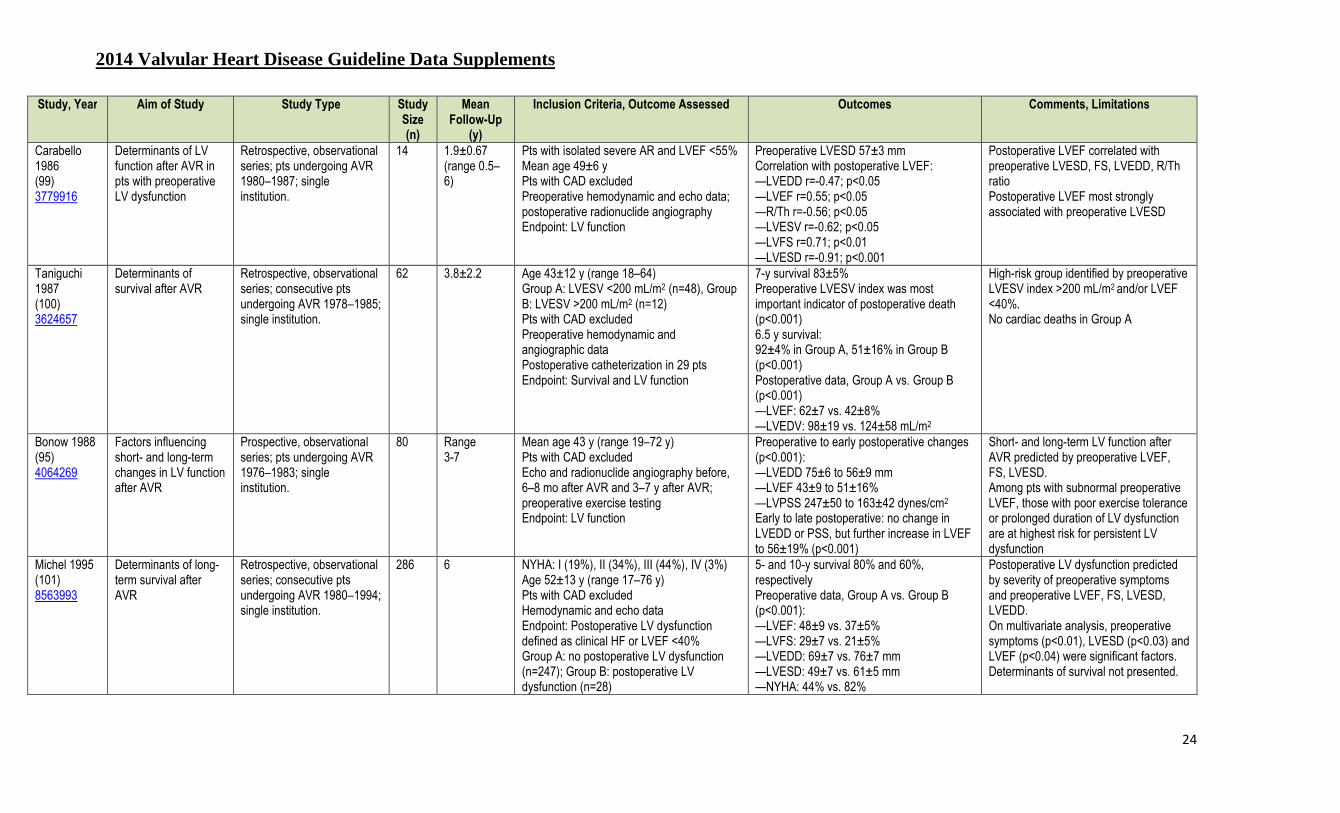
2014 Valvular Heart Disease Guideline Data Supplements
24
Study, Year Aim of Study Study Type Study Size (n)
Mean Follow-Up
(y)
Inclusion Criteria, Outcome Assessed Outcomes Comments, Limitations
Carabello 1986 (99) 3779916
Determinants of LV function after AVR in pts with preoperative LV dysfunction
Retrospective, observational series; pts undergoing AVR 1980–1987; single institution.
14 1.9±0.67 (range 0.5–6)
Pts with isolated severe AR and LVEF <55% Mean age 49±6 y Pts with CAD excluded Preoperative hemodynamic and echo data; postoperative radionuclide angiography Endpoint: LV function
Preoperative LVESD 57±3 mm Correlation with postoperative LVEF: —LVEDD r=-0.47; p<0.05 —LVEF r=0.55; p<0.05 —R/Th r=-0.56; p<0.05 —LVESV r=-0.62; p<0.05 —LVFS r=0.71; p<0.01 —LVESD r=-0.91; p<0.001
Postoperative LVEF correlated with preoperative LVESD, FS, LVEDD, R/Th ratio Postoperative LVEF most strongly associated with preoperative LVESD
Taniguchi 1987 (100) 3624657
Determinants of survival after AVR
Retrospective, observational series; consecutive pts undergoing AVR 1978–1985; single institution.
62 3.8±2.2 Age 43±12 y (range 18–64) Group A: LVESV <200 mL/m2 (n=48), Group B: LVESV >200 mL/m2 (n=12) Pts with CAD excluded Preoperative hemodynamic and angiographic data Postoperative catheterization in 29 pts Endpoint: Survival and LV function
7-y survival 83±5% Preoperative LVESV index was most important indicator of postoperative death (p<0.001) 6.5 y survival: 92±4% in Group A, 51±16% in Group B (p<0.001) Postoperative data, Group A vs. Group B (p<0.001) —LVEF: 62±7 vs. 42±8% —LVEDV: 98±19 vs. 124±58 mL/m2
High-risk group identified by preoperative LVESV index >200 mL/m2 and/or LVEF <40%. No cardiac deaths in Group A
Bonow 1988 (95) 4064269
Factors influencing short- and long-term changes in LV function after AVR
Prospective, observational series; pts undergoing AVR 1976–1983; single institution.
80 Range 3-7
Mean age 43 y (range 19–72 y) Pts with CAD excluded Echo and radionuclide angiography before, 6–8 mo after AVR and 3–7 y after AVR; preoperative exercise testing Endpoint: LV function
Preoperative to early postoperative changes (p<0.001): —LVEDD 75±6 to 56±9 mm —LVEF 43±9 to 51±16% —LVPSS 247±50 to 163±42 dynes/cm2 Early to late postoperative: no change in LVEDD or PSS, but further increase in LVEF to 56±19% (p<0.001)
Short- and long-term LV function after AVR predicted by preoperative LVEF, FS, LVESD. Among pts with subnormal preoperative LVEF, those with poor exercise tolerance or prolonged duration of LV dysfunction are at highest risk for persistent LV dysfunction
Michel 1995 (101) 8563993
Determinants of long-term survival after AVR
Retrospective, observational series; consecutive pts undergoing AVR 1980–1994; single institution.
286 6 NYHA: I (19%), II (34%), III (44%), IV (3%) Age 52±13 y (range 17–76 y) Pts with CAD excluded Hemodynamic and echo data Endpoint: Postoperative LV dysfunction defined as clinical HF or LVEF <40% Group A: no postoperative LV dysfunction (n=247); Group B: postoperative LV dysfunction (n=28)
5- and 10-y survival 80% and 60%, respectively Preoperative data, Group A vs. Group B (p<0.001): —LVEF: 48±9 vs. 37±5% —LVFS: 29±7 vs. 21±5% —LVEDD: 69±7 vs. 76±7 mm —LVESD: 49±7 vs. 61±5 mm —NYHA: 44% vs. 82%
Postoperative LV dysfunction predicted by severity of preoperative symptoms and preoperative LVEF, FS, LVESD, LVEDD. On multivariate analysis, preoperative symptoms (p<0.01), LVESD (p<0.03) and LVEF (p<0.04) were significant factors. Determinants of survival not presented.

2014 Valvular Heart Disease Guideline Data Supplements
25
Study, Year Aim of Study Study Type Study Size (n)
Mean Follow-Up
(y)
Inclusion Criteria, Outcome Assessed Outcomes Comments, Limitations
Klodas 1996 (102) 8606280
Impact of LV function on survival after AVR
Retrospective, observational series; consecutive pts undergoing AVR 1980–1989; single institution
219 5-y and 10-y survival data reported
Group A: preoperative LVEDD <80 mm (n=188, age 55±16 y) Group B: preoperative LVDD ≥80 mm (n=31, age 50±15 y) NYHA III-IV symptoms: Group A 37%, Group B 29% Includes pts with CAD: Group A 37%, Group B 29% Endpoint: Survival
Preoperative data, Group A vs. Group B (p<0.001): —LVEF: 53±11 vs. 43±12% —LVEDD: 67±8 vs. 84±4 mm —LVESD: 45±9 vs. 63±8 mm —LVESS: 96±39 vs. 147±39 dynes x 105/s Postoperative survival, Group A vs. Group B (p=NS): —5 y: 89±3% vs. 87±6% —10 y: 73±5% vs. 71±9% Postoperative survival, LVEF ≥50% vs. <50% (p<0.01): —10 y: 80±5% vs. 63±7%
Extreme LV dilatation associated with LV systolic dysfunction Preoperative LVEF, not degree of LV dilatation, associated with survival
Klodas 1997 (103) 9283535
Impact of symptom severity on survival after AVR
Retrospective, observational series; consecutive pts undergoing AVR 1980–1989; single institution
289 5-y and 10-y survival data reported
Group A: NYHA I-II (n=161, age 50±16 y, 86% men) Group B: NYHA III-IV (n=128, age 61±14 y, 70% men) Includes pts with CAD: Group A 11%, Group B 35%; including AVR plus CABG: Group A 8%, Group B 32% (both p<0.0001) Echo data in 249 pts Endpoint: survival
Preoperative data, Group A vs. Group B (p<0.05): —LVEF: 5 3±11 vs. 49±14% 10-y survival, Group A vs. Group B (p<0.001) —Total: 78±7% vs.45±4% —LVEF ≥50%: 82% vs. 40% —LVEF <50%: 73% vs. 40% —Men: 80% vs. 55% —Women: 73% vs. 21% —CAD: 76% vs. 39% —No CAD: 79% vs. 48%
High-risk group identified by symptom severity and preoperative LVEF <50% Survival in Group A equivalent to normal age/sex matched population Note higher frequency of CAD and CABG surgery (and other comorbidities) in the more symptomatic Group B
Turina 1998 (104) 9852889
Determinants of survival after AVR
Retrospective, observational series; consecutive pts undergoing AVR 1970–1983; single institution
192 18.7 (range 13–26)
Mean age 44 y Endpoint: Survival
Survival rates 76% at 10 y, 55% at 20 y. 83% of long-term survivors in NYHA I-II. Multivariate predictors of late survival: age, LVESV, NYHA, previous IE. LVEF significant in univariate analysis.
High-risk group identified by symptom severity, low LVEF, and elevated ESV.
Chaliki 2002 (105) 12438294
Survival after AVR in pts with normal versus reduced LV function
Retrospective, observational series; consecutive pts undergoing AVR 1980–1995; single institution
450 8.1 (median)
Group A (273 pts, age 56±16) with LVEF ≥50% Group B (134 pts, age 58±15) with LVEF 35%–50% Group C (43 pts, age 58±14) with LVEF <35% LVEF measured by left ventriculography
Operative mortality, Group A vs. B vs. C: 3.7%, 6.7%, 14% (p=0.02) 10-y mortality, Group A vs. B vs. C: 30%, 44%, 59% (p<0.001) 10-y HF rate, Group A vs. B vs. C: 9%, 17%, 25% (p<0.003) Postoperative change in LVEF, Group A vs.
Pts with markedly low LVEF incur have high rates of short- and long-term mortality and HF after AVR. However, postoperative LVEF improves significantly, and most pts survive without recurrence of HF. Thus they should not be denied benefits

2014 Valvular Heart Disease Guideline Data Supplements
26
Study, Year Aim of Study Study Type Study Size (n)
Mean Follow-Up
(y)
Inclusion Criteria, Outcome Assessed Outcomes Comments, Limitations
and/or echo Endpoint: Survival
B vs. C: -2.3%, 4%, 4.9% (p<0.01)
of AVR.
Tornos 2006 (106) 16516086
Determinants of survival after AVR
Prospective, observational series; consecutive pts undergoing AVR 1982–2002; single institution
170 10±6 (range 1–22)
Group A (60 pts age 47±15) mild symptoms (NYHA II), mild LV dysfunction (LVEF 45–50%) or LVESD 50–55 mm Group B (110 pts age 53±14) with NYHA III-IV symptoms or more severe LV dysfunction (LVEF <45% or LVESD >55 mm) Echo data Endpoint: Survival
Cardiac deaths: 5 (9%) in Group A, 28 (28%) in Group B (p=0.002). Survival Group A vs. Group B (p=0.009):
90% vs. 75% at 5 y, 86% vs. 64% at 10 y, 78% vs. 53% at 15 y
Early AVR as defined in the 2006 ACCF/AHA guidelines improves long-term survival in pts with chronic AR. Delaying AVR until more severe symptoms or more severe LV dysfunction decreases postoperative survival.
Bhudia 2007 (107) 17397676
Survival after AVR in pts with marked LV dysfunction compared to normal LV function or mild LV dysfunction
Prospective, observational series; consecutive pts undergoing AVR 1972–1999; single institution
724 8.3±6.5 Group A (88 pts, age 56±12) with severe LV dysfunction (LVEF <30%) Group B (636 pts, age 50±15) with either less severe LV dysfunction or normal LV function Endpoint: Survival
Survival diminished in Group A (severe LV dysfunction) compared to Group B (p=0.04):
81% vs. 92% at 1 y, 68% vs. 81% at 5 y, 46% vs. 62% at 10 y, 26% vs. 41% at 15 y, 12% vs. 24% at 20 y
In propensity matched pts since 1985, these survival trends persisted, but were not significant between pts in Groups A and B (p=0.9):
92% vs. 96% at 1 y, 79% vs. 83% at 5 y, 51% vs. 55% at 10 y
AR indicates aortic regurgitation; AVR, aortic valve replacement; CABG, coronary artery bypass graft surgery; CAD, coronary artery disease; echo, echocardiography; ESS, end-systolic stress; ESV, end-systolic volume; FS, fractional shortening; HF, heart failure; IE, infective endocarditis; LV, left ventricular; LVEDD, left ejection end-diastolic dimension; LVEF, left ventricular ejection fraction; LVESD, left ventricular end-systolic dimension; LVESV (i), left ejection end-systolic volume (indexed to body surface area); LVFS, left ventricular fractional shortening; LVPSS, left ventricular peak systolic wall stress; MWS, mean wall stress; NIH, National Institute of Health; NYHA , New York Heart Association; PSS, peak systolic wall stress; pts, patients; and, R/Th, radius to wall thickness ratio.

2014 Valvular Heart Disease Guideline Data Supplements
27
Data Supplement 13. Hemodynamic Effects Percutaneous Mitral Balloon Commissurotomy (PMBC) Compared to Surgical Closed Commissurotomy (CC) or Open Commissurotomy (OC) (Section 6.2.3)
Author, Year Mean Follow-Up
Procedure No. of Patients
Age, y Average Morphology
Score*
Mitral Gradient (mm Hg) Mitral Valve Area (cm2) Restenosis (%) Freedom From Reintervention (%)
NYHA I (%)
Pre Post Pre Post
Patel 1991 (108) 1918709
Immediate PMBC
23 30±11 6.0 12±4 4±3 0.8±0.3 2.1±0.7† N/A N/A 91
CC 22 26±26 6.0 12±5 6±3 0.7±0.2 1.3±0.3 N/A N/A N/A Turi 1991 (109) 2013139
7 mo PMBC 20 27±8 7.2 18±4 10±2 0.8±2 1.6±0.2 N/A N/A N/A
CC 20 28±1 8.4 20±6 12±2 0.9±0.4 1.7±0.2 N/A N/A N/A Arora 1993 (110) 8465732
22 mo PMBC 100 19±5 N/A N/A N/A 0.8±0.3 2.3±0.1 5 N/A N/A
CC 100 20±6 N/A N/A N/A 0.8±0.2 2.1±0.4 4 N/A N/A Reyes 1994 (111) 8084354
3 y PMBC 30 30±9 6.7 N/A N/A 0.9±0.3 2.4±0.4† 10 N/A 72
CC 30 31±9 7.0 N/A N/A 0.9±0.3 1.8±0.4 13 N/A 57 Ben Farhat 1998 (112) 9462525
7 y PMBC 30 29±12 6.0 N/A N/A 0.9±0.2 1.8±0.4 N/A 90 87
OC 30 27±9 6.0 N/A N/A 0.9±0.2 1.8±0.3 N/A 93 90 CC 30 28±10 6.0 N/A N/A 0.9±0.2 1.3±0.3 N/A 50 33 Cotrufo 1999 (113) 10386411
38 mo PMBC 111 47±14 7.6 N/A N/A 1.0±0.2 1.8±0.3 28 88 67
50 mo OC 82 49±10 8.2 N/A N/A 1.0±0.2 2.3±0.3 18 96 84 *Wilkins echocardiographic mitral valve morphology score, the sum of a 0 to 4 score for each of 4 characteristics: eaflet mobility, thickness, calcification and chordal involvement . †Significant difference (p<0.05) in increased mitral valve area by PMBC compared with surgical commissurotomy. CC indicates closed commissurotomy; N/A, not available; NYHA, New York Heart Association; OC, open commissurotomy; Post, postprocedure; PMBC, percutaneous mitral balloon commissurotomy; and, Pre, preprocedure. Adapted from Bonow et al. (114). Bhudia SK, McCarthy PM, Kumpati GS, et al. Improved outcomes after aortic valve surgery for chronic aortic regurgitation with severe left ventricular dysfunction. J Am Coll Cardiol. 2007;49;1465-71.

2014 Valvular Heart Disease Guideline Data Supplements
28
Data Supplement 14. Echocardiographic Prediction of Outcome of Percutaneous Balloon Mitral Commissurotomy (Section 6.2.3) Author, Year Mean Follow-
Up, mo Echo Criteria Number of
Patients Age
(y±SD) Survival (%) Survival Free of Events (%) Events
Cohen et al., 1992 (115) 1406834
36±20 Score ≤8 Score >8
84 52
N/A N/A 68% at 5 y 28% at 5 y
Death, MVR, repeat PMBC
Palacios et al., 1995 (116) 7828292
20±12 Score ≤8 Score >8
211 116
48±14 64±11
98% at 4 y 39% at 4 y
98% at 4 y 39% at 4 y
Death, MVR, NYHA III-IV symptoms
Dean et al., 1996 (117) 8917257
38±16 Score ≤8 Score 8–12 Score >12
272 306 24
49±13 58±15 58±15
95% at 4 y 83% at 4 y 24% at 4 y
N/A Death
Iung et al., 1996 (118) 8557913
32±18 Group 1 Group 2 Group 3
87 311 130
46±13
N/A 89% at 3 y 78% at 3 y 65% at 3 y
Death, MVR, repeat PMBC, FC III-IV symptoms
Cannan et al., 1997 (119) 8996311
22±10 Com Ca- Com Ca+
120 29
N/A N/A 86% at 3 y 40% at 3 y
Death, MVR, repeat PMBC
Palacios et al., 2002 (120) 11914256
50±44 Score >8 Score <8
278 601
63+14 51+14
82% at 12 y 57% at 12 y
38% at 12 y 22% at 12 y
Death, MVR, repeat PMBC
Echo score based on scoring system of Wilkins et al. (121) mitral valve morphology score, the sum of a 0 to 4 score for each of 4 characteristics: leaflet mobility, thickness, calcification and chordal involvement. Echo groups defined as 1, 2, or 3 based on valve flexibility, chordal fusion and valve calcification (Iung, et al. (112)). Com Ca indicates commissural calcification; echo, echocardiographic; MVR, mitral valve replacement; N/A, not available; NYHA, New York Heart Association; and, PMBC, percutaneous mitral balloon commissurotomy.

2014 Valvular Heart Disease Guideline Data Supplements
29
Data Supplement 15. Randomized Trials of Percutaneous Mitral Balloon Commissurotomy Versus Surgery for Mitral Stenosis (Section 6.2.3) Study Name, Author, Year
Study Aim Study Type/
Size (N)
Intervention vs. Comparator (n)
Patient Population Study Intervention
Study Comparator
Results
Inclusion Criteria Exclusion Criteria
Patel 1991 (108) 1918709
Compare PMBC by single catheter technique versus CC
RCT/45
Intervention: 23 PMBC vs. comparator: 22 CC
Symptomatic NYHA II or III, tight MS
Mitral valve calcification or left atrial thrombus on 2D echo, more than mild MR or AR, history of systemic embolism within 3 mo of presentation
PBMC Closed surgical valvotomy
PBMC: MVA increased from 0.8+0.3 cm2 to 2.1±0.7 cm2
(p<0.001) CC: MVA increased from 0.7±0.2 cm2 to 1.3±0.3 cm2 (p<0.001)
Ben 1998 (112) 9462525
Compare the early invasive and long-term (7 y) clinical and echo follow-up results of PBMC with those of OC and CC for the treatment of tight pliable rheumatic MS
RCT/90 Intervention: PBMC vs. comparator: CC; OC
Rheumatic tight rheumatic mitral valve stenosis (MVA <1.3 cm2),
Other valve disease, previous thromboembolism, mitral valve calcification, and left atrium thrombus, AF, severe pulmonary hypertension or mild-to-moderate TR
PBMC CC or OC Increase in Gorlin MVA : PBMC (from 0.9±0.16 to 2.2±0.4 cm2), OC (from 0.9+0.2 to 2.2±0.4 cm2), CC (from 0.9±0.2 to 1.6±0.4 cm2). Residual MS (MVA <1.5 cm2): 0% after PBMC or OC and 27% after CC. No early or late mortality or thromboembolism among the 3 groups. At 7-y follow-up, echo MVA was similar and greater after PBMC and OC (1.8±0.4 cm2) than after CC (1.3±0.3 cm2; p<0.001). Restenosis (MVA <1.5 cm2) rate was 6.6% after PBMC or OC vs. 37% after CC. Residual ASD in 2 pts and 3+ MR in 1 pt in the PBMC group. NYHA class I in 87% of pts after PBMC and 90% of pts after OC vs. CC 33% (p<0.0001) Freedom from reintervention 90% after PBMC, 93% after OC, and 50% after CC.
Turi 1991 (109) 2013139
Compare PBMC with surgical CC
RCT/40 Intervention: 20 PBMC vs. Comparator: 20 CC
Pts deemed acceptable as candidates for both procedures
N/A PBMC Surgical CC No differences between groups in pulmonary artery wedge pressures, mitral valve gradients, and MVA at 1 wk and at 8 mo. (all p>0.4).
Arora 1993 (110) 8465732
Compare the immediate and long-term results of PBMC vs. CMC
RCT/200 Intervention: 100 vs. Comparator: 100
Symptomatic pts with moderate-to-severe MS
Pts with more than minimal mitral valve calcification AF, or >2+ MR
PBMC CC Both procedures resulted in significant and similar increases in MVA (PBMV: 0.85+0.28 to 2.39±0.94 cm2; CC: 0.79±0.21 to 2.2±0.85 cm2; p=NS). MR developed in 14 pts after PBMC and in 12 pts after CC. Restenosis (defined as a loss of >50% of the achieved

2014 Valvular Heart Disease Guideline Data Supplements
30
Study Name, Author, Year
Study Aim Study Type/
Size (N)
Intervention vs. Comparator (n)
Patient Population Study Intervention
Study Comparator
Results
Inclusion Criteria Exclusion Criteria
increase in MVA) was seen in 4 (5%) pts after PBMV and in 3 (4%) after CC.
Reyes 1994 (111) 8084354
Compare PBMC to OC for treatment of rheumatic MS
RCT/60 Intervention: 30 vs. Comparator: 30
Severe rheumatic MS, in sinus rhythm, no severe subvalvular disease/ calcification or more than mild MR
Coexisting other cardiac or valve disease, stroke, severe pulmonary hypertension, low body weight, Lutembacjer's syndrome, and pt decision not to be randomized
PBMC Open surgical commissurotomy
MVA at 3 years was larger after PBMC (2.4±0.6 cm2 ) vs. OC (1.8±0.4 cm2).
NYHA class I at 3 years in 72% or PBMC pts and 57% of OC pts
Cotrufo 1999 (113) 10386411
Compare PPMC vs. OC
RCT/193 Intervention: PBMC 111 vs. Comparator: OC 82
N/A N/A PBMC OC Survival, event free analysis, recurrent restenosis No hospital mortality in both groups (p=0.3) Hospital complications: 4/111 PBMC vs. 1/82 OC (p=0.3)
2D indicates 2-dimensional; AF, atrial fibrillation; AR, aortic regurgitation; ASD, atrial septal defect; CC, closed commissurotomy; echo, echocardiography; MR, mitral regurgitation, MS, mitral stenosis; MVA, mitral valve area; N/A, not applicable; NS, nonsignificant; NYHA, New York Heart Association; OC, open commissurotomy; PMBC; percutaneous mitral balloon commissurotomy; pts, patients; RCT, randomized controlled trial; and, TR, tricuspid regurgitation.
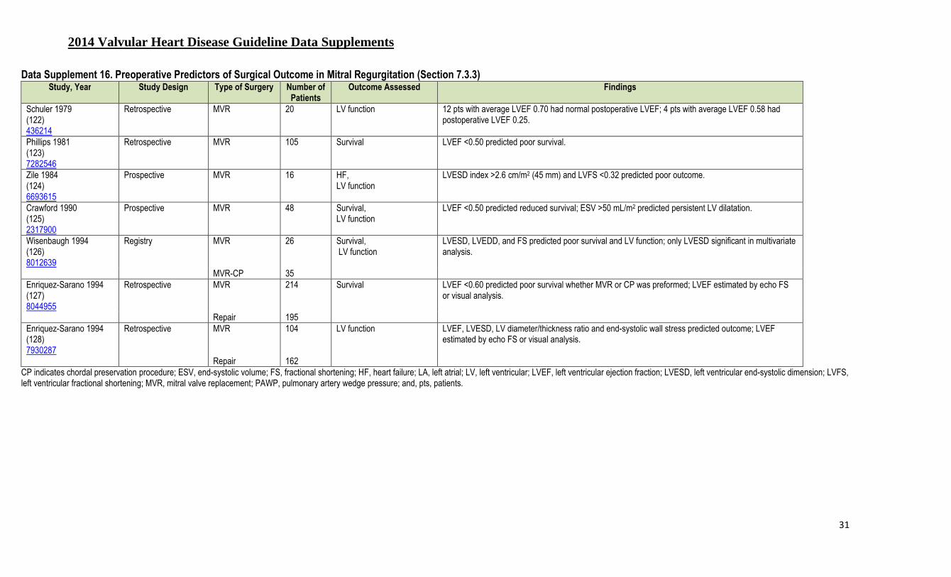
2014 Valvular Heart Disease Guideline Data Supplements
31
Data Supplement 16. Preoperative Predictors of Surgical Outcome in Mitral Regurgitation (Section 7.3.3) Study, Year Study Design Type of Surgery Number of
Patients Outcome Assessed Findings
Schuler 1979 (122) 436214
Retrospective MVR 20 LV function 12 pts with average LVEF 0.70 had normal postoperative LVEF; 4 pts with average LVEF 0.58 had postoperative LVEF 0.25.
Phillips 1981 (123) 7282546
Retrospective MVR 105 Survival LVEF <0.50 predicted poor survival.
Zile 1984 (124) 6693615
Prospective MVR 16 HF, LV function
LVESD index >2.6 cm/m2 (45 mm) and LVFS <0.32 predicted poor outcome.
Crawford 1990 (125) 2317900
Prospective MVR 48 Survival, LV function
LVEF <0.50 predicted reduced survival; ESV >50 mL/m2 predicted persistent LV dilatation.
Wisenbaugh 1994 (126) 8012639
Registry MVR 26 Survival, LV function
LVESD, LVEDD, and FS predicted poor survival and LV function; only LVESD significant in multivariate analysis.
MVR-CP 35 Enriquez-Sarano 1994 (127) 8044955
Retrospective MVR 214 Survival LVEF <0.60 predicted poor survival whether MVR or CP was preformed; LVEF estimated by echo FS or visual analysis.
Repair 195 Enriquez-Sarano 1994 (128) 7930287
Retrospective MVR 104 LV function LVEF, LVESD, LV diameter/thickness ratio and end-systolic wall stress predicted outcome; LVEF estimated by echo FS or visual analysis.
Repair 162 CP indicates chordal preservation procedure; ESV, end-systolic volume; FS, fractional shortening; HF, heart failure; LA, left atrial; LV, left ventricular; LVEF, left ventricular ejection fraction; LVESD, left ventricular end-systolic dimension; LVFS, left ventricular fractional shortening; MVR, mitral valve replacement; PAWP, pulmonary artery wedge pressure; and, pts, patients.

2014 Valvular Heart Disease Guideline Data Supplements
32
Data Supplement 17. Primary Mitral Regurgitation—Evidence for Intervention (Section 7.3.3) Study Name, Author,
Year Aim of Study Study Type Study Size (N) Study Intervention Group
(n) Study Comparator Group (n) Outcome
Tribouilloy 1999 (129) 9918527
Assess impact of symptom status on outcome
Retrospective 478 Mitral surgery NYHA class I,II, III, IV Advanced preoperative symptoms increased operative mortality by 10 fold. Long-term survival also reduced.
Gillinov 2010 (130) 20667334
Assess impact of symptoms on outcomes
Retrospective propensity-matched
4,253 MVR NYHA all class Even NYHA class II preoperative symptoms impaired late survival.
Rosenhek 2006 (131) 16651470
Assess outcome with watchful waiting
Prospective 132 Watchful waiting for severe MR
N/A Survival for watchful waiting identical to age normal population, but triggers for surgery occurred early after enrollment in 50%.
Kang 2009 (132) 19188506
Assess outcome with watchful waiting
Prospective 447 Mitral surgery Early surgery vs. watchful waiting Early surgery appeared superior, but several unoperated pts refused surgery despite presence of triggers.
Enriquez-Sarano 1994 (127) 8044955
Assess predictors of outcome Retrospective 409 Mitral surgery LVEF >60, 50-60, <50 Survival at 10 y, 72% for LVEF >60, 53%, 50–60, 32%, <50.
Tribouilloy 2009 (133) 19909877
Assess impact of LVESD on outcome
Retrospective 739 Mitral surgery LVESD <40 vs. ≥40 LVESD >40 mm nearly doubled late mortality risk.
Enriquez-Sarano 2005 (134) 15745978
Assess impact of MR severity Prospective 450 N/A ERO of different sizes ERO >0.4 cm2 nearly tripled mortality, but mortality was reduced by surgery.
Ghoreshi 2011 (135) 21962906
Assess impact of pulmonary HTN on outcome
Retrospective 873 Mitral surgery Preoperative-pulmonary HTN of various degrees
5 y survival 88% for PAP <40 vs. 52% PAP >60.
Goldman 1987 (136) 3624663
Compare LV function after replace vs. repair
Prospective 18 Mitral surgery Repair vs. replacement LVEF fell following replacement, but not repair.
David 1984 (137) 6492840
Compare outcome with and without chordal presentation
Prospective 27 Mitral surgery MV surgery with and without chordal preservation
LVEF decreased without preservation, but was maintained with preservation.
Rozich 1992 (138) 1451243
Examined LVEF Retrospective 15 Mitral surgery Chordal preservation vs. destruction
Afterload increased following chordal destruction, but decreases following preservation.
Grigioni 2008 (139) 19356418
Outcome of repair vs. replacement
Prospective 394 Mitral surgery Repair vs. replacement vs. nonsurgery
92% 54 y survival for repair 80% for replacement.

2014 Valvular Heart Disease Guideline Data Supplements
33
Study Name, Author, Year
Aim of Study Study Type Study Size (N) Study Intervention Group (n)
Study Comparator Group (n) Outcome
Gillinov 2008 (140) 18721551
Outcome of repair vs. replacement
Retrospective 328 N/A Repair vs. replacement propensity
5, 10, 15 y survival 95, 87, 68 repair vs. -80, 60, 44 replacement.
ERO indicates effective regurgitant orifice; HTN, hypertension; LV, left ventricular; LVEF, left ventricular ejection fraction; LVESD, left ventricular end-systolic dimension; MR, mitral regurgitation; MV, mitral valve; MVR, mitral valve repair; N/A not applicable; NYHA, New York Heart Association; PAP, pulmonary artery pressure; and, pts, patients.
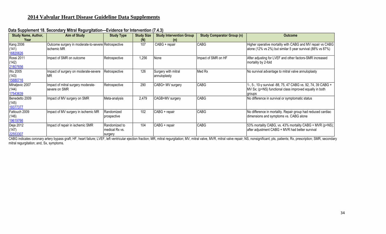
2014 Valvular Heart Disease Guideline Data Supplements
34
Data Supplement 18. Secondary Mitral Regurgitation—Evidence for Intervention (7.4.3) Study Name, Author,
Year Aim of Study Study Type Study Size
(N) Study Intervention Group
(n) Study Comparator Group (n) Outcome
Kang 2006 (141) 16820626
Outcome surgery in moderate-to-severe ischemic MR
Retrospective 107 CABG + repair CABG Higher operative mortality with CABG and MV repair vs CABG alone (12% vs 2%) but similar 5 year survival (88% vs 87%)
Rossi 2011 (142) 21807656
Impact of SMR on outcome Retrospective 1,256 None Impact of SMR on HF After adjusting for LVEF and other factors-SMR increased mortality by 2-fold
Wu 2005 (143) 15680716
Impact of surgery on moderate-severe MR
Retrospective 126 Surgery with mitral annuloplasty
Med Rx No survival advantage to mitral valve annuloplasty
Mihaljevic 2007 (144) 17543639
Impact of mitral surgery moderate-severe on SMR
Retrospective 290 CABG+ MV surgery CABG 1-, 5-, 10-y survival -88, 75, 47 CABG vs. 92, 74, 39 CABG + MV Sx; (p=NS) functional class improved equally in both groups
Benedetto 2009 (145) 19377377
Impact of MV surgery on SMR Meta-analysis 2,479 CAGB+MV surgery CABG No difference in survival or symptomatic status
Fattouch 2009 (146) 19619766
Impact of MV surgery in ischemic MR Randomized prospective
102 CABG + repair CABG No difference in mortality. Repair group had reduced cardiac dimensions and symptoms vs. CABG alone
Deja 2012 (147) 22553307
Impact of repair in ischemic SMR Randomized to medical Rx vs. surgery
104 CABG + repair CABG 53% mortality CABG, vs. 43% mortality CABG + MVR (p=NS); after adjustment CABG + MVR had better survival
CABG indicates coronary artery bypass graft; HF, heart failure; LVEF, left ventricular ejection fraction; MR, mitral regurgitation; MV, mitral valve, MVR, mitral valve repair; NS, nonsignificant; pts, patients; Rx, prescription; SMR, secondary mitral regurgitation; and, Sx, symptoms.
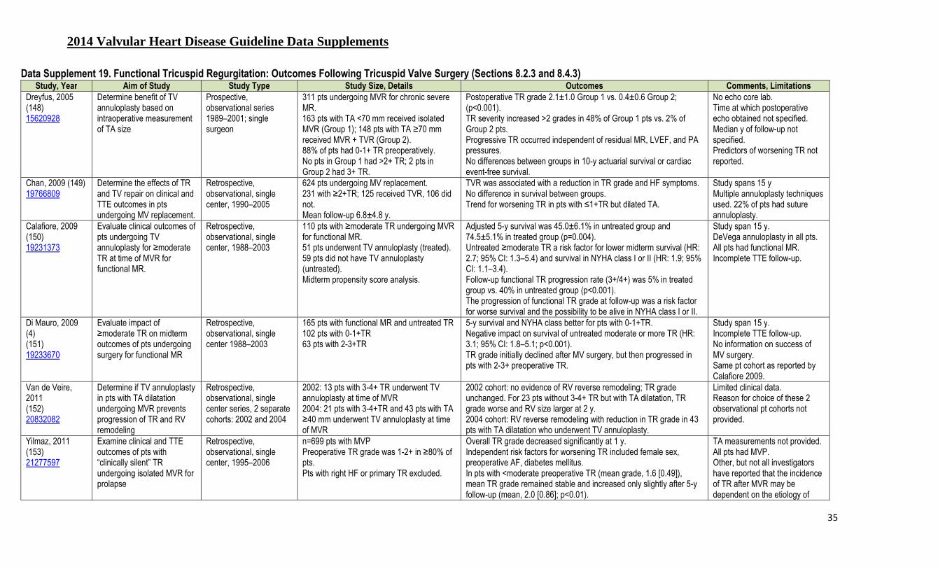
2014 Valvular Heart Disease Guideline Data Supplements
35
Data Supplement 19. Functional Tricuspid Regurgitation: Outcomes Following Tricuspid Valve Surgery (Sections 8.2.3 and 8.4.3) Study, Year Aim of Study Study Type Study Size, Details Outcomes Comments, Limitations
Dreyfus, 2005 (148) 15620928
Determine benefit of TV annuloplasty based on intraoperative measurement of TA size
Prospective, observational series 1989–2001; single surgeon
311 pts undergoing MVR for chronic severe MR. 163 pts with TA <70 mm received isolated MVR (Group 1); 148 pts with TA ≥70 mm received MVR + TVR (Group 2). 88% of pts had 0-1+ TR preoperatively. No pts in Group 1 had >2+ TR; 2 pts in Group 2 had 3+ TR.
Postoperative TR grade 2.1±1.0 Group 1 vs. 0.4±0.6 Group 2; (p<0.001). TR severity increased >2 grades in 48% of Group 1 pts vs. 2% of Group 2 pts. Progressive TR occurred independent of residual MR, LVEF, and PA pressures. No differences between groups in 10-y actuarial survival or cardiac event-free survival.
No echo core lab. Time at which postoperative echo obtained not specified. Median y of follow-up not specified. Predictors of worsening TR not reported.
Chan, 2009 (149) 19766809
Determine the effects of TR and TV repair on clinical and TTE outcomes in pts undergoing MV replacement.
Retrospective, observational, single center, 1990–2005
624 pts undergoing MV replacement. 231 with ≥2+TR; 125 received TVR, 106 did not. Mean follow-up 6.8±4.8 y.
TVR was associated with a reduction in TR grade and HF symptoms. No difference in survival between groups. Trend for worsening TR in pts with ≤1+TR but dilated TA.
Study spans 15 y Multiple annuloplasty techniques used. 22% of pts had suture annuloplasty.
Calafiore, 2009 (150) 19231373
Evaluate clinical outcomes of pts undergoing TV annuloplasty for ≥moderate TR at time of MVR for functional MR.
Retrospective, observational, single center, 1988–2003
110 pts with ≥moderate TR undergoing MVR for functional MR. 51 pts underwent TV annuloplasty (treated). 59 pts did not have TV annuloplasty (untreated). Midterm propensity score analysis.
Adjusted 5-y survival was 45.0±6.1% in untreated group and 74.5±5.1% in treated group (p=0.004). Untreated ≥moderate TR a risk factor for lower midterm survival (HR: 2.7; 95% CI: 1.3–5.4) and survival in NYHA class I or II (HR: 1.9; 95% CI: 1.1–3.4). Follow-up functional TR progression rate (3+/4+) was 5% in treated group vs. 40% in untreated group (p<0.001). The progression of functional TR grade at follow-up was a risk factor for worse survival and the possibility to be alive in NYHA class I or II.
Study span 15 y. DeVega annuloplasty in all pts. All pts had functional MR. Incomplete TTE follow-up.
Di Mauro, 2009 (4) (151) 19233670
Evaluate impact of ≥moderate TR on midterm outcomes of pts undergoing surgery for functional MR
Retrospective, observational, single center 1988–2003
165 pts with functional MR and untreated TR 102 pts with 0-1+TR 63 pts with 2-3+TR
5-y survival and NYHA class better for pts with 0-1+TR. Negative impact on survival of untreated moderate or more TR (HR: 3.1; 95% CI: 1.8–5.1; p<0.001). TR grade initially declined after MV surgery, but then progressed in pts with 2-3+ preoperative TR.
Study span 15 y. Incomplete TTE follow-up. No information on success of MV surgery. Same pt cohort as reported by Calafiore 2009.
Van de Veire, 2011 (152) 20832082
Determine if TV annuloplasty in pts with TA dilatation undergoing MVR prevents progression of TR and RV remodeling
Retrospective, observational, single center series, 2 separate cohorts: 2002 and 2004
2002: 13 pts with 3-4+ TR underwent TV annuloplasty at time of MVR 2004: 21 pts with 3-4+TR and 43 pts with TA ≥40 mm underwent TV annuloplasty at time of MVR
2002 cohort: no evidence of RV reverse remodeling; TR grade unchanged. For 23 pts without 3-4+ TR but with TA dilatation, TR grade worse and RV size larger at 2 y. 2004 cohort: RV reverse remodeling with reduction in TR grade in 43 pts with TA dilatation who underwent TV annuloplasty.
Limited clinical data. Reason for choice of these 2 observational pt cohorts not provided.
Yilmaz, 2011 (153) 21277597
Examine clinical and TTE outcomes of pts with “clinically silent” TR undergoing isolated MVR for prolapse
Retrospective, observational, single center, 1995–2006
n=699 pts with MVP Preoperative TR grade was 1-2+ in ≥80% of pts. Pts with right HF or primary TR excluded.
Overall TR grade decreased significantly at 1 y. Independent risk factors for worsening TR included female sex, preoperative AF, diabetes mellitus. In pts with <moderate preoperative TR (mean grade, 1.6 [0.49]), mean TR grade remained stable and increased only slightly after 5-y follow-up (mean, 2.0 [0.86]; p<0.01).
TA measurements not provided. All pts had MVP. Other, but not all investigators have reported that the incidence of TR after MVR may be dependent on the etiology of

2014 Valvular Heart Disease Guideline Data Supplements
36
Study, Year Aim of Study Study Type Study Size, Details Outcomes Comments, Limitations
In pts with at least moderate preoperative TR, mean TR grade decreased significantly from preoperative values after MVR (p<0.001 at hospital discharge, <1 y, and 1–3 y). Mean TR grade trended down at 3 and 5 y after surgery (p=0.18 after 3 y; p=0.33 after 5 y). Degree of preoperative TR was not associated with early or late mortality.
MR. Effect of recurrent TR on survival not reported.
Calafiore, 2011 (154) 21163499
Determine benefit of TV annuloplasty for TR based on TA diameter
Retrospective, observational, single center 2006–2008
298 pts with ≥1+ TR undergoing MV surgery. 167 underwent TVR, 108 with ≥moderate TR and 59 with TA >24 mm. 137 did not have TVR, 16 with ≥moderate TR and 81 with TA >24 mm.
In pts who did not undergo TVR, TA >24 mm was a risk factor for increasing TR grade during follow-up (HR: 2.4; 95% CI: 1.4–5.1; p=0.020).
DeVega annuloplasty used in all pts with TA <28 mm. Small cohort sizes.
Navia, 2012 (155) 22093694
Identify factors associated with TVR; assess safety and efficacy of TVR
Retrospective, observational, single center series 1997–2008
91(5%) of 1,724 pts with 2+ TR undergoing left-sided heart valve surgery. Propensity analysis performed for 91 matched pairs. Pts nonrandomly selected for TVR had more severe indices of RV remodeling with TV tethering.
In propensity-matched groups, prevalence of early postoperative TR grades 0 and 1 was 83% after TVR vs. 46% in the no-repair group 11% of the repair group had persistent grade 2+ TR after TVR, compared with 39% of the no-repair group. Worse TR on was present in 7% of the TVR group, vs. 15% of the no-repair group (p<0.0001). Differences in TR grade for matched pts were sustained at over 3 y. TVR did not add significant in-hospital morbidity or mortality. Long-term survival of propensity matched pts did not differ.
Multiple TVR techniques used Limited long-term outcome and TTE data. Matched pairs differed significantly.
Kim, 2012 (156) 21930721
Assess clinical and TTE outcomes of TVR in pts with mild-to-moderate TR at time of MV replacement
Retrospective, observational, single center, 1997-2008
236 pts with mild-moderate TR undergoing mechanical MV replacement for rheumatic disease. 123 pts underwent TVR. 113 pts did not undergo TVR.
Freedom from moderate-severe TR at 5 y 92.9±2.9% in repair group vs. 60.8+/16.9% in nonrepair group (p<0.001). Approximately 10% of pts with mild TR who did not have repair progressed to ≥moderate TR over 10 y. No differences between groups in mortality, need for TV reoperation, or HF. Postoperative moderate-severe TR an independent predictor of poorer event-free survival (HR: 2.90; p=0.038).
All pts had rheumatic MV disease. Groups significantly unbalanced at baseline. Limited TTE follow-up information, especially regarding MV prosthesis function, PA pressures, etc.
Benedetto 2012 (157) 22244561
Determine if TV annuloplasty in pts with TA dilatation and ≤moderate TR prevents TR progression after MV surgery
Randomized, prospective, single institution, 2008-2009
44 pts undergoing MV surgery with ≤2+ TR and TA ≥40 mm on preoperative TTE. Randomized 1:1 to TV annuloplasty with a flexible ring or no TV annuloplasty. Primary endpoint: ≥3+ TR at 1 y.
≥3+ TR at 1 y 0% in TV annuloplasty group vs. 28% in no annuloplasty group (p=0.02). Compared with no annuloplasty, TV annuloplasty resulted in significant RV reverse remodeling. Distance during 6-min walk test greater in the TV annuloplasty group (p=0.008).
Small sample size. Nonblinded endpoint assessment.
AF indicates atrial fibrillation; echo, echocardiography; HF, heart failure; LVEF, left ventricular ejection fraction; MR, mitral regurgitation; MV, mitral valve; MVP, mitral valve prolapse; MVR, mitral valve repair; NYHA, New York Heart Association; PA, pulmonary artery; pt(s), patients; RV, right ventricle; TA, tricuspid annulus; TR, tricuspid regurgitation; TTE, transthoracic echocardiography TV, tricuspid valve; and, TVR, tricuspid valve repair.
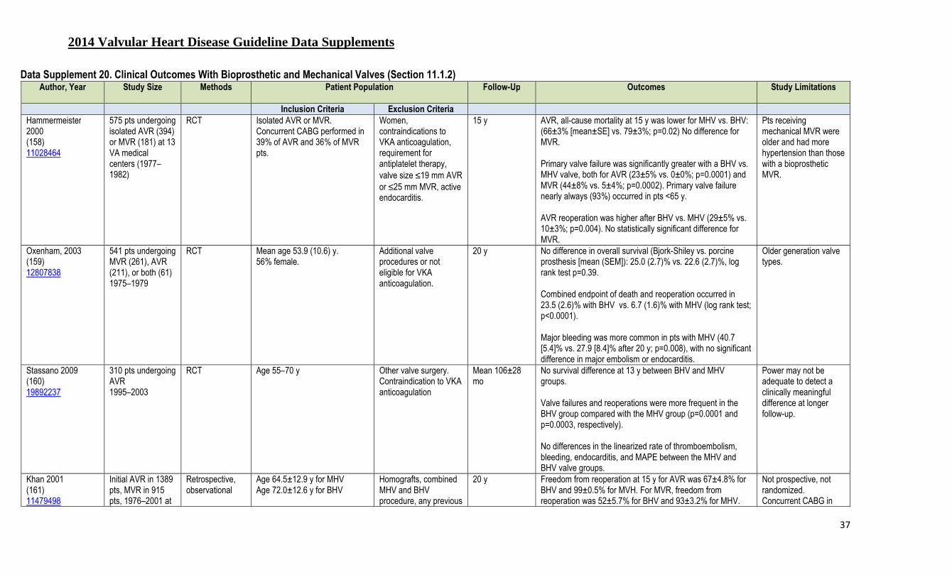
2014 Valvular Heart Disease Guideline Data Supplements
37
Data Supplement 20. Clinical Outcomes With Bioprosthetic and Mechanical Valves (Section 11.1.2) Author, Year Study Size Methods Patient Population
Follow-Up Outcomes Study Limitations
Inclusion Criteria Exclusion Criteria
Hammermeister 2000 (158) 11028464
575 pts undergoing isolated AVR (394) or MVR (181) at 13 VA medical centers (1977–1982)
RCT Isolated AVR or MVR. Concurrent CABG performed in 39% of AVR and 36% of MVR pts.
Women, contraindications to VKA anticoagulation, requirement for antiplatelet therapy, valve size ≤19 mm AVR or ≤25 mm MVR, active endocarditis.
15 y AVR, all-cause mortality at 15 y was lower for MHV vs. BHV: (66±3% [mean±SE] vs. 79±3%; p=0.02) No difference for MVR. Primary valve failure was significantly greater with a BHV vs. MHV valve, both for AVR (23±5% vs. 0±0%; p=0.0001) and MVR (44±8% vs. 5±4%; p=0.0002). Primary valve failure nearly always (93%) occurred in pts <65 y. AVR reoperation was higher after BHV vs. MHV (29±5% vs. 10±3%; p=0.004). No statistically significant difference for MVR.
Pts receiving mechanical MVR were older and had more hypertension than those with a bioprosthetic MVR.
Oxenham, 2003 (159) 12807838
541 pts undergoing MVR (261), AVR (211), or both (61) 1975–1979
RCT Mean age 53.9 (10.6) y. 56% female.
Additional valve procedures or not eligible for VKA anticoagulation.
20 y No difference in overall survival (Bjork-Shiley vs. porcine prosthesis [mean (SEM]): 25.0 (2.7)% vs. 22.6 (2.7)%, log rank test p=0.39. Combined endpoint of death and reoperation occurred in 23.5 (2.6)% with BHV vs. 6.7 (1.6)% with MHV (log rank test; p<0.0001). Major bleeding was more common in pts with MHV (40.7 [5.4]% vs. 27.9 [8.4]% after 20 y; p=0.008), with no significant difference in major embolism or endocarditis.
Older generation valve types.
Stassano 2009 (160) 19892237
310 pts undergoing AVR 1995–2003
RCT Age 55–70 y Other valve surgery. Contraindication to VKA anticoagulation
Mean 106±28 mo
No survival difference at 13 y between BHV and MHV groups. Valve failures and reoperations were more frequent in the BHV group compared with the MHV group (p=0.0001 and p=0.0003, respectively). No differences in the linearized rate of thromboembolism, bleeding, endocarditis, and MAPE between the MHV and BHV valve groups.
Power may not be adequate to detect a clinically meaningful difference at longer follow-up.
Khan 2001 (161) 11479498
Initial AVR in 1389 pts, MVR in 915 pts, 1976–2001 at
Retrospective, observational
Age 64.5±12.9 y for MHV Age 72.0±12.6 y for BHV
Homografts, combined MHV and BHV procedure, any previous
20 y Freedom from reoperation at 15 y for AVR was 67±4.8% for BHV and 99±0.5% for MVH. For MVR, freedom from reoperation was 52±5.7% for BHV and 93±3.2% for MHV.
Not prospective, not randomized. Concurrent CABG in

2014 Valvular Heart Disease Guideline Data Supplements
38
Author, Year Study Size Methods Patient Population
Follow-Up Outcomes Study Limitations
Inclusion Criteria Exclusion Criteria
a single medical center.
valve surgery Survival at 15 y (BHV vs. MHV, p=NS for all): AVR in pts <65 y (55±5.9 vs. 61±5.3%), AVR in pts >65 y (17±3.4 vs. 17±3.8%). MVR in pts <65 y (32±5.5 vs. 51±5.4%), MVR in pts >65 y (12±3.5 vs. 18±3.8%)
50%.
Chan 2006 (162) 16733156
3,063 pts undergoing AVR 1982–1998
Retrospective, observational
2,195 BHV and 980 MHV. Previous cardiac surgery
Average follow-ups in y for the BHV and MHV groups were 7.5±4.7% and 5.9±3.3% (p<0.001), respectively
Valve-related mortality (per pt-y): BHV 1.0% vs. MHV 0.7% Valve-related reoperation (per pt-y): BHV 1.3% vs. MHV 0.3% (p<0.001) Valve-related morbidity: BHV 0.4% vs. MHV 2.1% (p<0.001) Actual freedom from valve-related reoperation favored MHV for pts <60 y. Actual freedom from valve-related morbidity favored BHV for pts >40 y. Actual freedom from valve-related mortality was similar for BHV vs. MHV >50 y.
Not randomized. AVR only. Concomitant CABG in 43.5% of BHV pts and 26.0% of MHV pts.
Kulik 2006 (163) 16857373
659 pts age 50–65 y with initial AVR or MVR
Prospective, observational
AVR in 388 (MHV 306, BHV 48). MVR in 236 (MHV 188, BHV 48).
Enrolled only if survived perioperative period. Valve repair excluded.
Mean 5.1±4.1 y; maximum 18.3 y
Freedom from primary endpoint MAPE at 10 y (reoperation, endocarditis, major bleeding, or thromboembolism): AVR MHV 70±4.1% vs. BHV 41.0±30.3% (p=0.55) MVR MHV 53.3±8.8% vs. BHV 61.2±9.2% (p=0.34) Multivariate analysis did not identify valve type as an independent risk factor for MAPE
Not randomized. Surgeon choice of valve type. Concurrent CABG in 29%.
Ruel 2007 (164) 17846320
567 pts undergoing AVR or MVR
Retrospective, observational
Age <60 y. First heart valve operation.
N/A Mean survivor follow-up, 24.0±3.1 y
Survival in AVR: no difference between BHV vs. MHV (HR:0.95, 95% CI: 0.7–1.3); Survival in MVR: no difference between BHV or MHV (HR: 0.9, 95% CI: 0.5–1.4); Long-term survival worse in MVR than AVR (HR: 1.4, 95% CI: 1.1–1.8); Reoperation in 89% of BHV AVR and 84% of BHV MVR (older generation devices) with reoperative mortality 4.3%.
Not randomized or prospective, follow-up available in only 23% of original cohort.
van Geldorp 2009 (165) 19327512
Bioprosthetic AVR=2,860 (73%) vs. mechanical AVR=1,074 (27%)
Retrospective cohort (1982–2003) Microsimulation used to calculate age-specific pt
Bioprosthetic AVR: mean age=70 y, mean follow-up=6.1 y, CABG=47% vs. Mechanical AVR: mean age=58 y, mean follow-up=8.5 y, CABG=28%
N/A Bioprosthetic AVR: mean follow-up=6.1 y. Mechanical AVR: mean follow-up=8.5 y.
Simulated events for a 60-y man undergoing AVR, favors a BP vs. MP:
• life-expectancy: 11.9 vs. 12.2 y, • event-free survival: 9.8 vs. 9.3 y, • reoperation-free: 10.5 vs. 11.9 y, • reoperation risk: 25% vs. 3%,
Methodology of microsimulation is dependent on quality of dataset, wide chronological age of prostheses.

2014 Valvular Heart Disease Guideline Data Supplements
39
Author, Year Study Size Methods Patient Population
Follow-Up Outcomes Study Limitations
Inclusion Criteria Exclusion Criteria
outcome after AVR
• risk of bleeding: 12% vs. 41%
Badhwar 2012 (166) 22364968
172 pts undergoing isolated AVR or MVR (2003–2007)
Prospective, nonrandomized, matched pairs for BP vs. MP
Mean age 56.2±9.6 y (range, 24–72 y).
Limited 5 y survival based on comorbidity
Median follow-up 4.0 y
At a median 4-y follow-up, thromboembolism was 0.77% for MP and 0.78% for BP (p=NS) There was a survival benefit of mechanical prostheses at 7.5 y. Noninferiority to bioprosthetic AVR for bleeding and thromboembolic complications.
Prosthesis choice by surgeon, not randomized. Low INR targets (AVR: 2.0, MVR: 2.5) with home monitoring point-of-care system
Weber 2012 (167) 22341653
206 pts undergoing AVR (2000–2009)
Retrospective, with propensity matching of 103 BP to 103 MP AVR
Age <60 y. AVR with or without concurrent CABG, aortic root surgery, mitral or tricuspid valve repair.
Additional valve replacement.
Median follow-up 33±24 mo (2–120 mo)
Overall survival was worse with BP (90.3% vs. MP=98%, p=0.038; HR:0.243, 0.054–0.923 Freedom from valve related complication complications was similar: BP=54.5% vs. MP=51.6%, p=NS
Concurrent CABG in 49.9%, 14% were reoperations
AVR indicates aortic valve replacement; BHV, bioprosthetic heart valve; CABG, coronary artery bypass graft; HTN, hypertension; INR, international normalized ratio; MAPE, major adverse prosthesis-related events; MHV, mechanical heart valve; MVR, mitral valve replacement; N/A, not applicable; NS, nonsignificant; RCT, randomized controlled trial; pts, patients; VA, Veterans Affairs; and, VKA, vitamin K antagonist.

2014 Valvular Heart Disease Guideline Data Supplements
40
Data Supplement 21. Bridging Anticoagulation Therapy for Mechanical Heart Valves (Section 11.3.2) Author, Year Study Type Patient Population
Study Size and Comparator (N) Outcomes Study Limitations
Inclusion Criteria Exclusion Criteria
Hammerstingl 2007 (168) 17578050
Prospective, observational
Pts with MHV undergoing major surgery (n=25) or minor surgery (n=36), pacer implantation (n=21), or cardiac cath (n=34)
N/A 116 pts: MVR 31) , AVR (76) or DVR (9) Bridging with enoxaparin in all (renal function dose adjusted)
No thromboembolic (95% CI: 0–3.1%) complications. 1 major bleeding complication (0.86%; 95% CI: 0.02–4.7%). Minor bleeding in 10 pts (8.6%; 95% CI: 4.2–15.3%) at a mean of 5.4±1.4 d LMWH therapy.
Not randomized, no comparison group, relatively small study group.
Spyropoulos 2008 (169) 18805116
Observational, prospective, multicenter registry in USA, Canada
Adults undergoing elective surgery or invasive procedure with a mechanical valve on long-term VKA
Enrolled in another bridging study within 30 d.
73 with IV UFH (1,535±532 U/h) vs. 172 with SQ LMWH (76% enoxaparin 1 mg/kg bid, 13% dalteparin 100 U/kg bid, 4% tinzaparin 175 U/kg/d)
Major adverse event rates (5.5% vs. 10.3%; p=0.23) and major bleeds (4.2% vs. 8.8%; p=0.17) were similar in the LMWH and UFH groups, respectively; 1 arterial thromboembolic event occurred in each group. More LMWH-bridged pts were treated as outpts or discharged from the hospital in <24 hours (68.6% vs. 6.8%; p <0.0001). Multivariate logistic analysis found no significant differences in major bleeds and major composite adverse events when adjusting for cardiothoracic or major surgery between groups.
Not randomized, bridging therapy chosen by clinician. The LMWH group was less likely to undergo major surgery (33.7% vs. 58.9%; p=0.0002) and cardiothoracic surgery (7.6% vs. 19.2%; p=0.008), and to receive intraprocedural anticoagulants or thrombolytics (4.1% vs. 13.7%; p=0.007)
Pengo 2009 (170) 19470892
Prospective inception cohort at 22 Italian centers, 2005–2007
Adults undergoing surgical or invasive procedures that required interruption of long-term VKA therapy
Body weight <40 kg. Creatinine >2.0 mg/dL, contraindication to LMWH, need for dual antiplatelet Rx
N=189 MHV valve pts (15% of total study size of 1,262). Bridging with 70 anti-Xa U/kg/bid for high-risk pts.
Intention-to-treat analysis for the entire study population: Thromboembolic events in 5 pts (0.4%; 95% CI: 0.1–0.9), all in high-thromboembolic-risk pts Major bleeding in 15 (1.2%; 95% CI: 0.7–2.0) and minor bleeding in 53 pts (4.2%; 95% CI: 3.2–5.5). Major bleeding was associated with twice-daily LMWH (high-risk pts), but not with the bleeding risk of the procedure.
Only 15% had mechanical valves, no comparison group. Safety in pts with MHV valves has not been conclusively established

2014 Valvular Heart Disease Guideline Data Supplements
41
Author, Year Study Type Patient Population
Study Size and Comparator (N) Outcomes Study Limitations
Inclusion Criteria Exclusion Criteria
Daniels 2009 (171) 19232682
Retrospective cohort, 1997–2003
MHV on chronic VKA therapy undergoing invasive procedures or surgery
N/A A total of 580 procedures: 372 AVR, 136 MVR and 48 multivalvular. UFH or LMWH bridging used in high-risk pts (older AVR, any MVR, additional risk factors for TE). No bridging in isolated AVR pts.
Events at 3 mo N (%)
No Heparin
LMWH Only Any UFH
N=213 N=243 N=99
Thromboembolism 1 (0.5) 2 (0.8) 2 (3.1) Major Bleeding 5 (2.4) 9 (3.7) 6 (6.1) Minor Bleeding 13 (6.1) 13 (5.4) 8 (8.1)
Overall cumulative incidence of TE at 3 mo was 0.9%; all events occurred within 1 wk of the procedure. No TE events in 93 pts with isolated AVR with no bridging.
Not randomized, choice of therapy individualized based on estimated TE and bleeding risk. Most frequent procedures were GI endoscopy (19.1%), urologic procedures (14.0%), angiography or transcatheter interventions (10.5%), and orthopedic surgery (10.3%).
Bui HT 2009 (172) 19892063
Retrospective cohort study
173 pts on VKA anticoagulation for MHV (n=90) or for nonvalvular AF undergoing invasive or surgical procedures
Age <18 y, Pregnancy, Hypercoagulable condition, bioprosthetic valve
130 bridging episodes with LMWH were used to compare outcomes in MHV vs. pts with AF.
No deaths or thromboembolic events at 2 mo. Major and minor bleeding rates were similar between the MHV and AF groups (3.2% and 2.9%, 14.5% and 13.2% respectively, p=NS).
Isolated AVR in 43 (48%) of mechanical valve pts. Not randomized. Comparator group of AF may not require bridging. No sample size calculation for power of study.
Biteker 2012 (173) 22591673
Prospective cohort, single center
Consecutive pts undergoing noncardiac surgery
Bioprosthetic valves, severe liver or renal disease, contraindication to heparin
140 pts with MHV (77 AVR, 46 MVR, and 17 DVR) receiving enoxaparin 1 mg/kg bid compared to 1,200 pts with native valves (control group) receiving no anticoagulation.
Events (3 mo) N (%)
MHV with LMWH
Native valves
N=140 N=1200 p-value
Bleeding 18.6% 14.2% NS Thromboembolism 3.6% 2% NS Mortality 1.4% 1.3% NS Cardiovascular events 10.8% 10.7% NS
Not randomized. Comparison group did not have valve disease. No power calculation with small number of MHV pts.
Weiss 2013 (174) 23648452
Retrospective, single-center cohort study
Consecutive pts requiring postoperative bridging therapy after cardiac surgery during a 19 mo period
N/A N=402 receiving LMWH (enoxaparin): comparison of full-dose (FD=1 mg/kg bodyweight bid) to half-dose (HD=0.5 mg/kg bid) with renal function dose adjustment.
Events (by hospital discharge) N (%)
Full dose LMWH
Low dose LVWH
N=210 N=210 p-value
Mortality 0.5% 5.5% 0.003 Thromboembolism 5% 9% 0.277 Bleeding 11% 5% 0.126 Hospital stay (d) 15.1±9.3 12.5±8.1 0.003
Not randomized, but well matched (first half of cohort received FD, second half HD) Included only 100 (25.9% of total) pts with MHV, also included AF in 83.6%.
AF indicates atrial fibrillation; AVR, aortic valve replacement; DVR, double-valve replacement; FD, dull dose; GI, gastrointestinal; HD, half dose; LMWH, low molecular weight heparin; MHV, mechanical heart valve; MVR, mitral valve replacement; N/A, not available; NS, nonsignificant; pt(s), patient(s); TE, thromboembolism; UFH, unfractionated heparin; USA, United States of America; and, VKA, vitamin K antagonist.
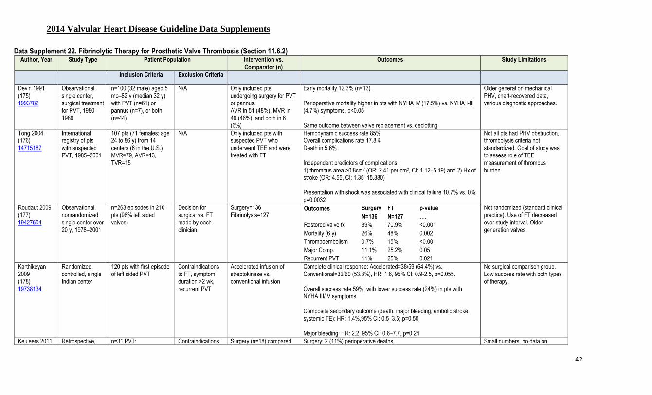
2014 Valvular Heart Disease Guideline Data Supplements
42
Data Supplement 22. Fibrinolytic Therapy for Prosthetic Valve Thrombosis (Section 11.6.2) Author, Year Study Type Patient Population Intervention vs.
Comparator (n) Outcomes Study Limitations
Inclusion Criteria Exclusion Criteria
Deviri 1991 (175) 1993782
Observational, single center, surgical treatment for PVT, 1980–1989
n=100 (32 male) aged 5 mo–82 y (median 32 y) with PVT (n=61) or pannus (n=7), or both (n=44)
N/A Only included pts undergoing surgery for PVT or pannus. AVR in 51 (48%), MVR in 49 (46%), and both in 6 (6%)
Early mortality 12.3% (n=13) Perioperative mortality higher in pts with NYHA IV (17.5%) vs. NYHA I-III (4.7%) symptoms, p<0.05 Same outcome between valve replacement vs. declotting
Older generation mechanical PHV, chart-recovered data, various diagnostic approaches.
Tong 2004 (176) 14715187
International registry of pts with suspected PVT, 1985–2001
107 pts (71 females; age 24 to 86 y) from 14 centers (6 in the U.S.) MVR=79, AVR=13, TVR=15
N/A Only included pts with suspected PVT who underwent TEE and were treated with FT
Hemodynamic success rate 85% Overall complications rate 17.8% Death in 5.6% Independent predictors of complications: 1) thrombus area >0.8cm2 (OR: 2.41 per cm2, CI: 1.12–5.19) and 2) Hx of stroke (OR: 4.55, CI: 1.35–15.380) Presentation with shock was associated with clinical failure 10.7% vs. 0%; p=0.0032
Not all pts had PHV obstruction, thrombolysis criteria not standardized. Goal of study was to assess role of TEE measurement of thrombus burden.
Roudaut 2009 (177) 19427604
Observational, nonrandomized single center over 20 y, 1978–2001
n=263 episodes in 210 pts (98% left sided valves)
Decision for surgical vs. FT made by each clinician.
Surgery=136 Fibrinolysis=127
Outcomes
Surgery FT p-value
N=136 N=127 ….
Restored valve fx 89% 70.9% <0.001 Mortality (6 y) 26% 48% 0.002 Thromboembolism 0.7% 15% <0.001 Major Comp. 11.1% 25.2% 0.05 Recurrent PVT 11% 25% 0.021
Not randomized (standard clinical practice). Use of FT decreased over study interval. Older generation valves.
Karthikeyan 2009 (178) 19738134
Randomized, controlled, single Indian center
120 pts with first episode of left sided PVT
Contraindications to FT, symptom duration >2 wk, recurrent PVT
Accelerated infusion of streptokinase vs. conventional infusion
Complete clinical response: Accelerated=38/59 (64.4%) vs. Conventional=32/60 (53.3%), HR: 1.6, 95% CI: 0.9-2.5, p=0.055. Overall success rate 59%, with lower success rate (24%) in pts with NYHA III/IV symptoms. Composite secondary outcome (death, major bleeding, embolic stroke, systemic TE): HR: 1.4%,95% CI: 0.5–3.5; p=0.50 Major bleeding: HR: 2.2, 95% CI: 0.6–7.7, p=0.24
No surgical comparison group. Low success rate with both types of therapy.
Keuleers 2011 Retrospective, n=31 PVT: Contraindications Surgery (n=18) compared Surgery: 2 (11%) perioperative deaths, Small numbers, no data on

2014 Valvular Heart Disease Guideline Data Supplements
43
Author, Year Study Type Patient Population Intervention vs. Comparator (n)
Outcomes Study Limitations
Inclusion Criteria Exclusion Criteria
(179) 21211605
nonrandomized, single center, 1988–2008
MVR=17 (55%), AVR=8 (26%), TVR=6 (19%).
to FT to FT (n=13) 2 (11%) recurrent PVT (follow-up 76 mo) FT: 8 (61%) with restoration of normal valve function. 4 (31%) recurrent PVT (follow-up 18 mo)
4 (31%) major complications (death, stroke, TIA, or bleeding requiring surgery)
thrombus size
Özkan 2013 (66) 23489534
Observational , single center clinical experience, 1993–2009
TEE-guided FT in 182 consecutive pts with 220 episodes of PVT in 220 different episodes (156 women; mean age, 43.2±13.06 y).
Contraindications to FT, asymptomatic PVT with normal valve hemodynamics and no TE or with, thrombus size <10 mm.
FT regimen adjusted over study duration with Groups: I–Slow streptokinase II–Rapid streptokinase III–tPA 100 mg (bolus) IV–tPA 50 mg 6 h infusion V–tPA 25 mg 6 h infusion
Outcomes N
I II III IV V p-value
16 41 12 27 124 Overall success 68.8% 85.4% 75% 81.5% 85.5% 0.46 Major nonfatal comp. 12.5% 12.2% 8.3% 11.1% 4.8% NS Death 12.5% 2.4% 16.7% 3.7% 0% 0.01 Multivariate predictors of mortality plus major nonfatal complications: Any thrombolytic therapy regimen other than Group V and a history of stroke/TIA.
Karthikeyan 2013 (180) 23329151
Meta-analysis Published articles on left-sided PVT with at least 5 pts each treated with surgery and FT
Lack of data on primary outcome (restoration of normal valve function)
7 studies with 690 episodes of left sided PVT, 446 treated with surgery, and 244 with FT.
Outcomes
Surgery FT OR p-value
N=446 N=244
Restored valve Fx 86.5% 69.7% 2.53, 95% CI: 0.94–6.78 0.066 Death 13.5% 9% 1.95, 95% CI: 0.63–5.98 0.244 Thromboembolism 1.6% 16% 0.10, 95% CI: 0.04–0.24 <0.001 Major Bleeding 1.4% 5% 0.27, 95% CI: 0.08–0.98 0.046 Recurrent PVT 7.1% 25.4% 0.25, 95% CI: 0.08–0.74 0.013
AVR indicates aortic valve replacement; FT, fibrinolytic therapy; fx, function; Hx, history; MVR, mitral valve replacement, N/A, not available; NS, nonsignificant; NYHA, New York Heart Association; PHV, prosthetic heart valves; pts, patients; PVT, prosthetic valve thrombosis; TE, thromboembolism, TEE, transesophageal echocardiography; TIA, transient ischemic attack (stroke); TVR, tricuspid valve replacement; and, U.S., United States.

2014 Valvular Heart Disease Guideline Data Supplements
44
Data Supplement 23. Paravalvular Regurgitation (Section 11.8.3) Study Name,
Author, Year
Study Aim Study Type/Size
(N)
Intervention vs.
Comparator (n)
Patient Population Endpoints Adverse Events
Inclusion/Exclusion Criteria
Primary Endpoint & Results Secondary Endpoint & Results
Orszulak 1983 (181) 6860002
To report outcome with surgical reoperation for PVR
Retrospective N=105
Surgical reoperative repair of prosthetic PVR
Aortic PVR (n=75) and mitral PVR (n=29)
Early mortality for entire cohort: 5.7%. 5-y survival was 94% for aortic PVR pts and 75% for mitral PVR pts.
21 pts required multiple operations for persistent PVR. 85% of survivors at follow-up up to 14 y were NYHA I or II. Murmur of residual or recurrent PVR evident in 21% of pts.
N/A
Miller 1995 (182) 8556176
To identify clinical features that predict occurrence of PVR. Outcome after surgical repair also reported
Retrospective N=30
Surgical reoperative repair of aortic prosthetic PVR
Aortic prosthetic PVR 30-d survival=90%; 5-d survival=73%
Prosthesis replacement in 26, suture repair in 4. Trivial or no residual regurgitation in 16 of 20 with echocardiography in follow-up.
N/A
Akins 2005 (183) 16359061
To examine acute and long-term outcome of surgery for PVR
Retrospective N=136
Surgical reoperative repair of aortic or mitral prosthetic PVR
Mitral PVR in 68% Aortic PVR in 32%
Operative mortality, 6.6% Perioperative stroke, 5.1% 10-y survival, 30%
Primary repair in 48%, prosthesis replacement in 52%
N/A
Pate 2006 (184) 16969856
To describe outcome in series of pts undergoing percutaneous repair of PVR
Retrospective N=10 (10 defects)
Percutaneous repair of PVR
Mitral PVR (n=9) and aortic PVR (n=1); 9 were not surgical candidates
7 with successful procedure 3 pts died at 1 y
4 of 10 required second procedure 6 with sustained improvement in symptoms
1 retroperitoneal bleed 1 device dislodgement
Shapira 2007 (185) 11479246
To examine the feasibility and early outcome of percutaneous repair of PVR
Retrospective N=11 (13 defects)
Percutaneous repair of PVR
Mitral PVR (n=8), aortic PVR (n=1), and both aortic and mitral PVR (n=2) Estimated surgical mortality, 17.8%
10 with device deployment 6 with reduction in regurgitation 5 with NYHA improvement by 1 class
Hemolysis improved in 4, worsened in 4, and was unchanged in 2 in early follow-up 3 deaths in follow-up
N/A
Cortes 2008 (186) 18237605
To examine utility of TEE in percutaneous repair of PVR
Retrospective N=27 (27 defects)
TEE before and procedure (n=27) and at follow-up ≥1 mo (n=17)
Mechanical mitral PVR in pts at high risk for surgery
62% with procedure success TEE helped guide procedure and identified variety of complications
N/A 2 stroke 1 cardiac perforation 6 needing blood transfusion for postprocedural anemia
Ruiz 2011 (187) 22078427
To examine feasibility and efficacy of the percutaneous repair of PVR
Retrospective/ N=43 (57 defects)
Percutaneous repair of PVR
Mitral PVR (n=36), aortic PVR (n=9), and both aortic and mitral PVR (n=2)
Device deployment success in 86% of pts and 86% of leaks Survival: 92% at 6 m, 86% at 18 m
12 pts required multiple procedures Reduction in need for transfusions or EPO from 56–5% NYHA class improved by ≥1 in 28/35 pts
2 device embolizations 1 emergency surgery 1 vascular complication 1 procedural death
Sorajja 2011 To examine the feasibility and Retrospective Percutaneous repair of 78% mitral PVR, 22% aortic Device deployment in 89% Leaflet impingement in 4.3% 30-d events

2014 Valvular Heart Disease Guideline Data Supplements
45
Study Name,
Author, Year
Study Aim Study Type/Size
(N)
Intervention vs.
Comparator (n)
Patient Population Endpoints Adverse Events
Inclusion/Exclusion Criteria
Primary Endpoint & Results Secondary Endpoint & Results
(188) 21791673
early outcome of percutaneous repair of PVR
N=115 pts (141 defects)
PVR PVR Average STS risk score=6.9%
Mild or no residual regurgitation in 77% No procedural death
Procedure time average 147 min and decreased with case experience
Death, 1.7% Stroke, 2.6% Emergency surgery, 0.9%, Bleeding=5.2%
Sorajja 2011 (189) 22078428
To determine the long-term clinical efficacy of percutaneous repair of PVR
Retrospective N=126 (154 defects)
Percutaneous repair of PVR
79% mitral PVR, 21% aortic PVR Average STS risk score=6.7%
3-y survival, 64% HF accounted to 37% of deaths; noncardiac cause in 30%
Symptom improvement occurred only in pts with mild or no residual regurgitation Hemolytic anemia persisted in 14 of 29 pts
Survival free of death or need for cardiac surgery was 54% at 3 y Need for cardiac surgery related to degree of residual regurgitation
EPO indicates erythropoietin; HF, heart failure; N/A, not applicable; NYHA, New York Heart Association; pts, patients; PVR, paravalvular regurgitation; STS, Society of Thoracic Surgeons; and, TEE, transesophageal echocardiography.

2014 Valvular Heart Disease Guideline Data Supplements
46
Data Supplement 24. Surgical Outcome in Infective Endocarditis (Section 12) Author/
Year Aim of Study Study Type Study
Size (N)
Patient Population
Study Intervention Primary Endpoint Predictors of Outcome
Jault, 1997 (190) 9205176
Identify significant predictors of operative mortality, reoperation, and recurrent IEs
Retrospective single-center surgical cohort study
247 NVE alone; surgery 100%
Registration of epidemiological and microbiological features, echocardiography data, treatment strategy
Operative mortality was 7.6% (n=19). Overall survival
rate (operative mortality excluded) was 71.3% at 9 y. The probability of freedom from reoperation (operative mortality included) was 73.3±4.2% at 8 y. The rate of IE of the implanted prosthetic valve was 7%.
Increased age, cardiogenic shock at the time of operation, insidious illness, and greater thoracic ratio (>0.5) were the predominant risk factors for operative mortality; the length of antibiotic therapy appeared to have no influence. Increased age, preoperative neurologic complications,
cardiogenic shock at the time of operation, shorter duration of the illness, insidious illness before the operation, and mitral valve endocarditis were the predominant risk factors for late mortality. Risk factors for reoperation were younger age and aortic valve endocarditis.
Castillo, 2000 (191) 10768901
To determine the clinical features and long-term prognosis of IE in pts who were not drug users.
Prospective single-center case series
138 NVE 69%, PVE 31%; surgery 51%
Registration of epidemiological and microbiological features, echocardiography data, treatment strategy
Severe complications (HF, embolic phenomenon, severe valve dysfunction, abscesses, renal failure, and immunologic phenomenon) occurred in 83% of pts. 51% of pts underwent surgery during the active phase (22% was emergency surgery) Inpt mortality was 21%. Overall 10 y survival was 71%
There were no significant differences in survival depending on the type of treatment received during the hospital stay (medical vs. combined medical-surgical) in this observational study.
Alexiou, 2000 (192) 10881821
Single center experience in the surgical treatment of active culture-positive
IE and identify determinants of early and late outcome
Retrospective single-center surgical cohort study
118 NVE 70%, PVE 30%; 100% of pts underwent surgery
Registration of epidemiological and microbiological features, echocardiography data, treatment strategy
Operative mortality was 7.6% (9 pts). Endocarditis recurred in 8 (6.7%). A reoperation was required in 12 (10.2%). There were 24 late deaths, 17 of them cardiac. Actuarial freedom from recurrent endocarditis, reoperation, late cardiac death, and long-term survival at 10 y were 85.9%, 87.2%, 85.2%, and 73.1%, respectively.
Predictors of operative mortality: HF, impaired LV function. Predictors of recurrence: PVE. Predictors of late mortality: myocardial invasion, reoperation. Predictors of poor long-term survival: coagulase-negative staphylococcus, annular abscess, long ICU stay.
Wallace, 2002 (193) 12067945
To identify clinical markers available within the first 48 h of admission that are associated with poor outcome in IE
Retrospective single-center cohort study
208 NVE 68%, PVE 32%; surgery 52%
Registration of epidemiological, clinical, microbiological and other laboratory features, echocardiography data, and treatment strategy
Mortality at discharge was 18% and at 6 mo 27%. Surgery was performed in 107 (51%) pts. In-hospital mortality was not influenced by surgery (23% vs. 15% in the nonsurgical group); p=0.3 At 6 mo there was a trend towards increased mortality in the surgical group (33% vs. 20%)
Duration of illness, age, gender, site of infection, organism, and LV function did not predict outcome. Abnormal white cell count, raised creatinine, ≥2 major Duke criteria, or visible vegetation conferred poor prognosis.
Hasbun, 2003 (194)
To derive and externally validate a prognostic
Retrospective multicenter cohort study
513 Pts with left-sided NVE with current
Registration of clinical information, sociodemographic data, comorbid conditions, previous heart disease,
In the derivation and validation cohorts, the 6-mo mortality rates were 25% and 26%, respectively. In the derivation cohort, pts were classified into 4
5 baseline features were independently associated with 6 mo mortality (comorbidity [p=0.03], abnormal mental status [p=0.02], moderate-to-severe congestive HF
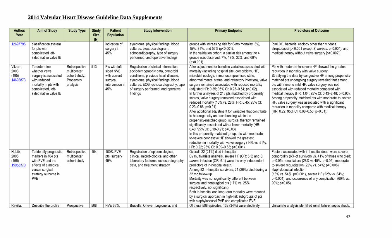
2014 Valvular Heart Disease Guideline Data Supplements
47
Author/ Year
Aim of Study Study Type Study Size (N)
Patient Population
Study Intervention Primary Endpoint Predictors of Outcome
12697795 classification system for pts with complicated left-sided native valve IE
indication of surgery in 45%`
symptoms, physical findings, blood cultures, electrocardiogram, echocardiography, type of surgery performed, and operative findings
groups with increasing risk for 6-mo mortality: 5%, 15%, 31%, and 59% (p<0.001). In the validation cohort, a similar risk among the 4 groups was observed: 7%, 19%, 32%, and 69% (p<0.001).
[p=0.01], bacterial etiology other than viridans streptococci [p<0.001 except S. aureus, p=0.004], and medical therapy without valve surgery [p=0.002])
Vikram, 2003 (195) 14693873
To determine whether valve surgery is associated with reduced mortality in pts with complicated, left-sided native valve IE
Retrospective multicenter cohort study; Propensity analysis
513 Pts with left sided NVE with current surgical intervention in 45%
Registration of clinical information, sociodemographic data, comorbid conditions, previous heart disease, symptoms, physical findings, blood cultures, ECG, echocardiography, type of surgery performed, and operative findings
After adjustment for baseline variables associated with mortality (including hospital site, comorbidity, HF, microbial etiology, immunocompromised state, abnormal mental status, and refractory infection), valve surgery remained associated with reduced mortality (adjusted HR: 0.35; 95% CI: 0.23–0.54; p<0.02). In further analyses of 218 pts matched by propensity scores, valve surgery remained associated with reduced mortality (15% vs. 28%; HR: 0.45; 95% CI: 0.23–0.86; p=0.01). After additional adjustment for variables that contribute to heterogeneity and confounding within the propensity-matched group, surgical therapy remained significantly associated with a lower mortality (HR: 0.40; 95% CI: 0.18-0.91; p=0.03). In this propensity-matched group, pts with moderate-to-severe congestive HF showed the greatest reduction in mortality with valve surgery (14% vs. 51%; HR: 0.22; 95% CI: 0.09–0.53; p=0.001).
Pts with moderate-to-severe HF showed the greatest reduction in mortality with valve surgery. Stratifying the data by congestive HF among propensity-matched pts undergoing surgery revealed that among pts with none to mild HF, valve surgery was not associated with reduced mortality compared with medical therapy (HR: 1.04; 95% CI: 0.43–2.48; p=0.93). Among propensity-matched pts with moderate-to-severe HF, valve surgery was associated with a significant reduction in mortality compared with medical therapy (HR: 0.22; 95% CI: 0.08–0.53; p=0.01).
Habib, 2005 (196) 15958370
To identify prognostic markers in 104 pts with PVE and the effects of a medical versus surgical strategy outcome in PVE
Retrospective multicenter cohort study
104 100% PVE pts; surgery 49%
Registration of epidemiological, clinical, microbiological and other laboratory features, echocardiography data, and treatment strategy
Overall, 22 (21%) died in hospital. By multivariate analysis, severe HF (OR: 5.5) and S. aureus infection (OR: 6.1) were the only independent predictors of in-hospital death. Among 82 in-hospital survivors, 21 (26%) died during a 32 mo follow-up. Mortality was not significantly different between surgical and nonsurgical pts (17% vs. 25%, respectively, not significant). Both in-hospital and long-term mortality were reduced by a surgical approach in high-risk subgroups of pts with staphylococcal PVE and complicated PVE.
Factors associated with in-hospital death were severe comorbidity (6% of survivors vs. 41% of those who died; p=0.05), renal failure (28% vs.45%, p=0.05), moderate-to-severe regurgitation (22% vs. 54%; p=0.006), staphylococcal infection (16% vs. 54%; p=0.001), severe HF (22% vs. 64%; p=0.001), and occurrence of any complication (60% vs. 90%; p=0.05).
Revilla, Describe the profile Prospective 508 NVE 66%, Brucella, Q fever, Legionella, and Of these 508 episodes, 132 (34%) were electively Univariate analysis identified renal failure, septic shock,
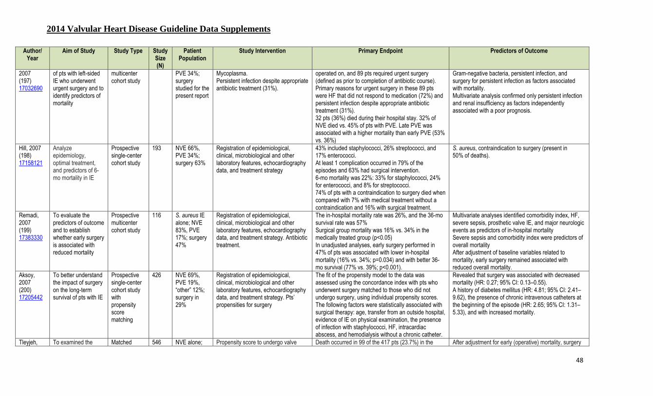
2014 Valvular Heart Disease Guideline Data Supplements
48
Author/ Year
Aim of Study Study Type Study Size (N)
Patient Population
Study Intervention Primary Endpoint Predictors of Outcome
2007 (197) 17032690
of pts with left-sided IE who underwent urgent surgery and to identify predictors of mortality
multicenter cohort study
PVE 34%; surgery studied for the present report
Mycoplasma. Persistent infection despite appropriate antibiotic treatment (31%).
operated on, and 89 pts required urgent surgery (defined as prior to completion of antibiotic course). Primary reasons for urgent surgery in these 89 pts were HF that did not respond to medication (72%) and persistent infection despite appropriate antibiotic treatment (31%). 32 pts (36%) died during their hospital stay. 32% of NVE died vs. 45% of pts with PVE. Late PVE was associated with a higher mortality than early PVE (53% vs. 36%)
Gram-negative bacteria, persistent infection, and surgery for persistent infection as factors associated with mortality. Multivariate analysis confirmed only persistent infection and renal insufficiency as factors independently associated with a poor prognosis.
Hill, 2007 (198) 17158121
Analyze epidemiology, optimal treatment, and predictors of 6-mo mortality in IE
Prospective single-center cohort study
193 NVE 66%, PVE 34%; surgery 63%
Registration of epidemiological, clinical, microbiological and other laboratory features, echocardiography data, and treatment strategy
43% included staphylococci, 26% streptococci, and 17% enterococci. At least 1 complication occurred in 79% of the episodes and 63% had surgical intervention. 6-mo mortality was 22%: 33% for staphylococci, 24% for enterococci, and 8% for streptococci. 74% of pts with a contraindication to surgery died when compared with 7% with medical treatment without a contraindication and 16% with surgical treatment.
S. aureus, contraindication to surgery (present in 50% of deaths).
Remadi, 2007 (199) 17383330
To evaluate the predictors of outcome and to establish whether early surgery is associated with reduced mortality
Prospective multicenter cohort study
116 S. aureus IE alone; NVE 83%, PVE 17%; surgery 47%
Registration of epidemiological, clinical, microbiological and other laboratory features, echocardiography data, and treatment strategy. Antibiotic treatment.
The in-hospital mortality rate was 26%, and the 36-mo survival rate was 57% Surgical group mortality was 16% vs. 34% in the medically treated group (p<0.05) In unadjusted analyses, early surgery performed in 47% of pts was associated with lower in-hospital mortality (16% vs. 34%; p=0.034) and with better 36-mo survival (77% vs. 39%; p<0.001).
Multivariate analyses identified comorbidity index, HF, severe sepsis, prosthetic valve IE, and major neurologic events as predictors of in-hospital mortality Severe sepsis and comorbidity index were predictors of overall mortality After adjustment of baseline variables related to mortality, early surgery remained associated with reduced overall mortality.
Aksoy, 2007 (200) 17205442
To better understand the impact of surgery on the long-term survival of pts with IE
Prospective single-center cohort study with propensity score matching
426 NVE 69%, PVE 19%, “other” 12%; surgery in 29%
Registration of epidemiological, clinical, microbiological and other laboratory features, echocardiography data, and treatment strategy. Pts’ propensities for surgery
The fit of the propensity model to the data was assessed using the concordance index with pts who underwent surgery matched to those who did not undergo surgery, using individual propensity scores. The following factors were statistically associated with surgical therapy: age, transfer from an outside hospital, evidence of IE on physical examination, the presence of infection with staphylococci, HF, intracardiac abscess, and hemodialysis without a chronic catheter.
Revealed that surgery was associated with decreased mortality (HR: 0.27; 95% CI: 0.13–0.55). A history of diabetes mellitus (HR: 4.81; 95% CI: 2.41–9.62), the presence of chronic intravenous catheters at the beginning of the episode (HR: 2.65; 95% CI: 1.31–5.33), and with increased mortality.
Tleyjeh, To examined the Matched 546 NVE alone; Propensity score to undergo valve Death occurred in 99 of the 417 pts (23.7%) in the After adjustment for early (operative) mortality, surgery

2014 Valvular Heart Disease Guideline Data Supplements
49
Author/ Year
Aim of Study Study Type Study Size (N)
Patient Population
Study Intervention Primary Endpoint Predictors of Outcome
2007 (201) 17372170
association between valve surgery and all-cause 6 mo mortality among pts with left-sided IE
propensity analysis
surgery 24% surgery was used to match pts in the surgical and nonsurgical groups. To adjust for survivor bias, the follow-up time was matched so that each pt in the nonsurgical group survived at least as long as the time to surgery in the respective surgically-treated pt. Valve surgery was used as a time-dependent covariate in different Cox models.
nonsurgical group vs. 35 deaths among the 129 pts (27.1%) in the surgical group. 18 of 35 (51%) pts in the surgical group died within 7 d of valve surgery.
was not associated with a survival benefit (adjusted HR: 0.92; 95% CI: 0.48–1.76).
Tleyjeh, 2008 (202) 18308866
To examine the association between the timing of valve surgery after IE diagnosis and 6-mo mortality among pts with left-sided IE
Retrospective single-center cohort propensity analysis
546 NVE alone; surgery 24%
The association between time from IE diagnosis to surgery and all-cause 6 mo mortality was assessed using Cox proportional hazards modeling after adjusting for the propensity score (to undergo surgery 0–11 d vs. 11 d, median time, after IE diagnosis).
The median time between IE diagnosis and surgery was 11 d (range 1–30). Using Cox proportional hazards modeling, propensity score and longer time to surgery (in d) were associated with unadjusted HRs of (1.15, 95% CI: 1.04–1.28, per 0.10 unit change; p=0.009) and (0.93; 95% CI: 0.88–0.99, per d; p=0.03), respectively. In multivariate analysis, a longer time to surgery was associated with an adjusted HR (0.97; 95% CI: 0.90–1.03). The propensity score and time from diagnosis to surgery had a correlation coefficient of r=20.63, making multicollinearity an issue in the multivariable model.
On univariate analysis, a longer time to surgery showed a significant protective effect for the outcome of mortality. After adjusting for the propensity to undergo surgery early versus late, a longer time to surgery was no longer significant, but remained in the protective direction.
Thuny, 2009 (203) 19329497
To determine whether the timing of surgery could influence mortality and morbidity in pts with complicated IE
Retrospective single-center cohort propensity analysis
291 NVE 82%, PVE 18%; surgery 100%
The time between the beginning of the appropriate antimicrobial therapy and surgery was used as a continuous variable and as a categorical variable with a cut-off of 7 d to assess the impact of timing of surgery. 2 groups of pts were formed according to the timing of surgery: the “<1st wk surgery group” and the “>1st wk surgery group”. The impact of the timing of surgery on 6 mo mortality, relapses, and PVD was analyzed using PS analyses.
1st wk surgery was associated with a trend of decrease in 6-mo mortality in the quintile of pts with the most likelihood of undergoing this early surgical management (quintile 5: 11% vs. 33%, OR: 0.18, 95% CI: 0.04 –0.83; p=0.03). Pts of this subgroup were younger, were more likely to have S. aureus infections, congestive HF, and larger vegetations. <1st wk surgery was associated with an increased number of relapses or PVD (16% vs. 4%, adjusted OR: 2.9, 95% CI: 0.99–8.40; p=0.05).
Very early surgery (<7 d) associated with improved survival (especially in highest risk pts), but greater likelihood of relapse or post-operative valve dysfunction.
Manne, 2012 (204)
Describe the morbidity and mortality associated
Retrospective single-center surgical
428 NVE 58%, PVE 42%; surgery 100%
Registration of epidemiological, clinical, microbiological and other laboratory features, echocardiography
Overall 90% of pts survived to hospital discharge. When compared with pts with NVE, pts with PVE had significantly higher 30-d mortality (13% vs. 5.6%;
Pts with IE caused by S. aureus had significantly higher hospital mortality (15% vs. 8.4%; p<0.05), 6 mo mortality (23% vs. 15%; p=0.05), and 1 y mortality (28%

2014 Valvular Heart Disease Guideline Data Supplements
50
Author/ Year
Aim of Study Study Type Study Size (N)
Patient Population
Study Intervention Primary Endpoint Predictors of Outcome
22206953 with surgery for IE and compare differences in characteristics, pathogens, and outcomes for pts with NVE and PVE from a large surgery-minded tertiary referral center
cohort study data, and treatment strategy p<0.01), but long-term survival was not significantly different (35% vs. 29%; p=0.19).
vs. 18%; p=0.02) compared with non–S. aureus IE.
Kang, 2012 (205) 22738096
To compare clinical outcomes of early surgery and conventional treatment in pts with IE
Prospective randomized trial at 2 centers with intention to treat analysis
76 Left-side NVE and high risk of embolism to early surgery (49%) vs. conventional treatment (51%)
Pts were randomly assigned in a 1:1 ratio to the early-surgery group or the conventional-treatment group with the use of a Web-based interactive response system. The protocol specified that pts who were assigned to the early-surgery group should undergo surgery within 48 h after randomization. Pts assigned to the conventional-treatment group were treated according to the AHA guidelines, and surgery was performed only if complications requiring urgent surgery developed during medical treatment or if symptoms persisted after the completion of antibiotic therapy.
The primary endpoint (composite of in-hospital death and embolic events that occurred within 6 wk after randomization) occurred in 1 pt (3%) in the early surgery group as compared with 9 (23%) in the conventional-treatment group (HR: 0.10; 95% CI: 0.01 –0.82; p=0.03). There was no significant difference in all-cause mortality at 6 mo in the early-surgery and conventional-treatment groups (3% and 5%, respectively; HR: 0.51; 95% CI: 0.05–5.66; p=0.59). The rate of the composite en point of death from any cause, embolic events, or recurrence of IE at 6 mo was 3% in the early-surgery group and 28% in the conventional-treatment group (HR: 0.08; 95% CI: 0.01–0.65; p=0.02).
As compared with conventional treatment, early surgery in pts with IE and large vegetations significantly reduced the composite endpoint of death from any cause and embolic events by effectively decreasing the risk of systemic embolism.
AHA indicates American Heart Association; HF, heart failure; ICU, intensive care unit, IE, infective endocarditis; NVE, native valve endocarditis; pts, patients; PVE, prosthetic valve; and S. aureus, Staphylococcus aureus. Table modified from Prendergast BD and Tornos P. Surgery for infective endocarditis: who and when? Circulation 2010, 121:1141-1152.

2014 Valvular Heart Disease Guideline Data Supplements
51
Data Supplement 25.Outcomes in Pregnant Women With a Mechanical Prosthetic Valve Treated with Warfarin or Unfractionated Heparin (UFH) (Section 13.3.2) Author, Year Study Aim Study Size
(N) Patient Population Study Type Type of
Anticoagulation Endpoints Summary Study
Limitations
Maternal Fetal
Chan, 2000 (206) 10647757
Systematic review anticoagulation mechanical valves
1,234 pregnancies in 976 women
All pts with mechanical prosthesis–40 articles–treated with differing anticoagulation regimens 1966–1997
Systematic review of literature
1. Warfarin throughout 2. UFH 1st trimester, then warfarin 3. UFH throughout pregnancy 4. No AC
Maternal Death 1. 1.8% 2. 4.2% 3. 15% 4. 4.7% Thromboembolic 1. 3.9% 2. 9.2% 3. 33% 4. 24%
Fetal anomalies 1. 6.4% 2. 3.4% 3. 0% 4. 3.3% Fetal wastage 1. 33% 2. 26% 3. 43% 4. 20%
Reduction of thromboembolic events for mother greatest with warfarin throughout pregnancy, worse maternal outcome with heparin throughout pregnancy. Heparin in 1st trimester reduces risk of fetopathic effects, but with increased risk of thromboembolic embolic events.
Retrospective systematic review–prior to LMWH use
Meschengieser, 1999 (207) 10377303
Single center experience anticoagulation mechanical valves
92 pregnancies in 59 women
Consecutive unselected pregnancies between 1986–1997
Observational 1. Warfarin throughout pregnancy 2. UFH 1st trimester, then warfarin 3. UFH throughout pregnancy 4. No A/C
Thromboembolic 1. 0.3 episodes/100 pt mo 2. 4.9 episodes/100 pt mo
Fetal wastage 1. 25% 2. 19%
Reduction of thromboembolic events for mother greatest with warfarin throughout pregnancy. No maternal deaths or valve thrombosis occurred in this study.
Retrospective review of small number pts–prior to LMWH use
Vitale, 1999 (208) 10334435
Single center experience anticoagulation mechanical valves
58 pregnancies in 43 pts
Consecutive unselected pregnancies between 1987–1997
Observational Warfarin throughout pregnancy: A. Dose <5 mg vs. B. Dose >5 mg
Maternal Death None Valve thrombosis 2 pts
Fetal complications A. 4 SA and 1 GR (28/32 healthy babies) vs. B: 2 WE, 18 SA, 1 SB, 1 VSD (3/25 healthy babies)
First to show that fetal complications are dose-dependent, relatively safe if dose ≤5 mg
Retrospective review–only warfarin throughout was used
Salazar, 1996 (209) 8636556
Single center experience anticoagulation mechanical valves
40 pregnancies in 37 pts
Single center experience of a prospective protocol using UFH SQ during the 1st trimester
Prospective cohort trial
All pts had SQ UFH from 6–12 wk and then during the last 2 wks of gestation
2 cases of massive thrombosis of a MVR tilting disk. 1 death from GI bleeding during warfarin.
37% spontaneous abortion 2.5% neonatal death No embryopathy
UFH is a poor anticoagulant and does not prevent massive thrombosis
Trial stopped after 2 events occurred
Sbarouni, 1994 (210) 8130033
Questionnaire to all cardiac centers in Europe
214 pregnancies in 182 pts (133 with
Questionnaire sent 1994 to all cardiac centers in Europe
Questionnaire data
N/A 6 maternal deaths (4 valve thrombosis, 1 cerebral embolism, 1 pulmonary edema)
No embryopathies in 36 women on warfarin
Heparin is neither effective or safe for both fetus and mother with increased risk thromboembolism and bleeding
No detailed information on level of anticoagulation

2014 Valvular Heart Disease Guideline Data Supplements
52
Author, Year Study Aim Study Size (N)
Patient Population Study Type Type of Anticoagulation
Endpoints Summary Study Limitations
Maternal Fetal
mechanical prosthesis)
13 valve thrombosis–10/13 on heparin, 12/13 MVR 8 embolic events–5/8 heparin
Fetal outcome similar for warfarin vs. heparin–22% abortion and 10% stillbirths
dose. Selection bias of those who responded to the questionnaire
Al-Lawati 2002 (211) 12142189
Single center experience anticoagulation mechanical valves from country of Oman
63 pregnancies in 21 pts
Consecutive unselected pregnancies between 1983–1997
Observational 1. Warfarin throughout 2. UFH 1st trimester, then Warfarin
Thrombosis of valves 1. None 2. 2 pts
Fetal complications 1. 74% live
babies 2. 71% live
babies Spontaneous abortion 1. 26% 2. 14% No embryopathy (2 pts with 6 mg, rest with ≤5 mg)
Role of warfarin embryopathy overstated. Warfarin recommended, especially with low dose of warfarin. Valve thrombosis occurred only in pts with UFH during 1st trimester–none with warfarin.
Retrospective review—only warfarin throughout was used
Sadler 2000 (212) 10688509
Historical cohort of women with mechanical, bioprosthetic and homograft valves from New Zealand
147 pregnancies in 79 pts
All women in New Zealand who had valve replacement 1972–1992 and had subsequent pregnancy
Observational 1. Warfarin throughout pregnancy 2. Warfarin for 6 wk then subq UFH 3. Warfarin for 28 wk then subq UFH
Valve thrombosis 1. 0% 2. 20% 3. 0% Embolic events 1. 0% 2. 20% 3. 25% Hemorrhage 1. 3% 2. 30% 3. 25%
Pregnancy loss 1. 70% 2. 22% 3. 33%
Warfarin had high rate of fetal loss High rate of thromboemboli on heparin (29%) Bioprosthesis or homografts were associated with successful pregnancies
Retrospective review of small number pts—prior to LMWH use
De Santo 2005 (213) 15999035
Single center experience of all pts who had mechanical prosthesis and became pregnant
48 pregnancies in 37 pts
All women from a single center who had MVR 1975 to 2002 and had subsequent pregnancy
Observational 1. Warfarin throughout A. Dose <5 mg B. Dose >5 mg 2. 2 pts with UFH
2/2 pts with UFH had valve thrombosis No pt with warfarin had adverse cardiac or valve related event
1A. 2/23 (8.6%) adverse fetal event 1B. 17/21 (81%) adverse fetal event
If continue warfarin throughout pregnancy, there are no maternal events Adverse fetal events mainly if dose >5 mg
Retrospective review of small number pts—prior to LMWH use

2014 Valvular Heart Disease Guideline Data Supplements
53
AC indicates anticoagulation; GI, gastrointestinal; GR, growth retardation; LMWH, low molecular weight heparin; MVR, mitral valve replacement; N/A, not available; pts, patients; SA, spontaneous abortion; SB, still birth; SQ subcutaneous; UFH, unfractionated heparin; VSD, ventricular septal defect; and, WE, warfarin embryopathy.

2014 Valvular Heart Disease Guideline Data Supplements
54
Data Supplement 26. Outcomes in Pregnant Women With a Mechanical Prosthetic Valve Treated With Low Molecular Weight Heparin (LMWH) (Section 13.3.2) Author, Year Study Aim Study Size (N) Type of
Anticoagulant Patient Population Study Type Endpoints Summary Study Limitations
Maternal Fetal
Rowan 2001 (214) 11568791
Examine pregnancy outcomes in women with mechanical prosthesis treated with LMWH throughout pregnancy
14 pregnancies in 11 women
LMWH throughout pregnancy
All pts with mechanical prosthesis treated with LMWH single center—1997–1999—fixed dose LMWH
Observational One valve thrombosis 14.3% hemorrhage
9 live births 3 miscarriages 2 terminations
Can achieve successful pregnancy using LMWH throughout pregnancy, but risk of valve thrombosis
Use fixed dose LMWH with mean anti-Xa level 0.46 pre- and 0.89 post dose. Retrospective review of small number pts.
James, 2006 (215) 16966122
Examine pregnancy outcomes in women with mechanical prosthesis treated with LMWH throughout pregnancy
76 pregnancies LMWH throughout pregnancy
Medline search of 73 cases 1966–2006 and 3 of single center using LMWH throughout pregnancy
Meta-analysis 22% thrombotic events 4% maternal mortality
No congenital anomalies 8 spontaneous abortions
Use of LMWH during pregnancy associated with high risk of life threatening thrombosis
No anti X-a levels performed. Meta-analysis only
Abildgaard, 2009 (216) 19162303
Examine pregnancy outcomes in women with mechanical prosthesis treated with LMWH throughout pregnancy
12 pregnancies in 12 women
LMWH throughout pregnancy
All pts with mechanical prosthesis treated with LMWH throughout pregnancy in country Norway—1997–2008—use anti-Xa levels
Observational 1 systemic embolism and 1 valve thrombosis (both subtherapeutic doses) Pooled risk of thromboembolism 7.1% vs. prior data 25% with UFH
13 healthy babies If use anti-Xa levels, successful in 10/12 pregnancies, risk lower than UFH by retrospective comparison
Retrospective review of small number pts
Oran, 2004 (217) 15467905
Meta-analysis of pregnancy outcomes in women with mechanical prosthesis treated with differing anticoagulation regimens, including LMWH
10 reports (2 prospective) 81 pregnancies in 75 women
LMWH 1st trimester, then warfarin vs. LMWH throughout pregnancy
Medline search of studies in pts with prostheses receiving LMWH from 1989–2004
Meta-analysis 12% had thromboemboli–all MVR–all with LMWH throughout–9/10 did not have anti-Xa monitoring. Valve thrombosis 8.6%
Spontaneous abortion in 7.4% Stillbirth in 1.2% 87% live births
All thromboemboli occurred in pts with mitral prosthesis who had LMWH throughout pregnancy. Anti Xa levels were not monitored in 90% of thromboembolic events.
Meta-analysis only
McLintock, 2009 (218) 19681850
Examine pregnancy outcomes in women with mechanical prosthesis treated with differing anticoagulation regimens including LMWH
47 pregnancies in 31 women
Warfarin throughout pregnancy vs. LMWH 1st trimester, then warfarin vs. LMWH throughout pregnancy
All pts with mechanical prosthesis treated with differing anticoagulation regimens including LMWH—2 centers—1997–2008—use anti-Xa levels
Observational Thromboembolism 7 total–5 (10.6%) LMWH Antepartum bleeding 10.6% LMWH Postpartum bleeding 12.7% LMWH
96% live births with LMWH vs. 75% live births with warfarin
Poor compliance or subtherapeutic anti-Xa levels were present in all valve thrombosis on LMWH
Retrospective review of small number pts

2014 Valvular Heart Disease Guideline Data Supplements
55
Author, Year Study Aim Study Size (N) Type of Anticoagulant
Patient Population Study Type Endpoints Summary Study Limitations
Maternal Fetal
Yinon, 2009 (219) 19840573
Examine pregnancy outcomes in women with mechanical prosthesis treated with LMWH throughout pregnancy
23 pregnancies in 17 women
LMWH throughout pregnancy
All pts with mechanical prosthesis treated with LMWH—single center 1998–2008—use anti-Xa levels
Observational 1 (4%) maternal thrombosis died 5 (22%) pulmonary edema, arrhythmias, and endocarditis 13% postpartum hemorrhage
19 live births 2 first trimester miscarriages 2 intrauterine deaths
Even with careful monitoring of anti X-a levels thrombosis may occur, even with low risk AVR
Retrospective review of small number pts
Quinn, 2009 (220) 19880782
Examine pregnancy outcomes in women with mechanical prosthesis treated with LMWH throughout pregnancy
12 pregnancies in 11 women
LMWH throughout pregnancy
All pts with mechanical prosthesis treated with LMWH—single center—2001–2007—use anti-Xa levels
Observational 3 major bleeds 3 minor bleeds BS MVR thrombosis 1 pt (Xa level not done and later subtherapeutic)
11/12 live births Increasing dose LMWH during pregnancy necessary Only valve thrombosis occurred in pt with subtherapeutic level Xa
Retrospective review of small number pts
AVR indicates aortic valve replacement; BS, Bjork-Shiley; GI, gastrointestinal; LMWH, low molecular weight heparin; MVR, mitral valve replacement; N/A, not available; pts, patients; UFH, unfractionated heparin; and, SES, socioeconomic status.

2014 Valvular Heart Disease Guideline Data Supplements
56
Data Supplement 27. Outcomes With the Maze Procedure for Atrial Fibrillation in Patients With Valvular Heart Disease (Section 14.2.2) Author, Year Aim of Study Study Type Study Size (N) Study “Intervention”
Group (n) Study Comparator Group
(n) Outcomes
Prognostic Significant of AF at Time of Surgery
Eguchi et al 2005 (221) 15845559
Examine impact of preoperative AF on outcome of MV repair for 1° MR
Retrospective observational
283 pts with moderate-to-severe MR who underwent MV repair between 1991 and 2002
129 in AF Age 59±13 y 60% male
154 in NSR Age 52±14 y 67% male
5 y outcomes were better in pts in NSR vs. AF for: survival (96±2.1 vs. 87±3.2%; p=0.002) and freedom from cardiac events (96±2.0 vs. 75±4.4%; p<0.001)
Alexiou 2007 (222) 17280837
Impact of preoperative AF on early and late outcome after MV repair
Retrospective observational
349 pts undergoing MV repair for primary MR
152 (44%) in AF 197 (56%) in NSR Kaplan-Meier survival at 7 y was 75±6% for AF pts vs. 90±3% (p=0.005) for SR pts.
Ngaage 2006 (223) 17643612
Prognostic significance of preoperative AF at the time of AVR
Retrospective observational, cohort comparison
381 AVR 1993 and 2002 matched for age, gender, and LVEF
Preoperative AF (n=129) Preoperative NSR (n=252) Pts with preoperative AF had had worse late survival (RR for death=1.5; p=0.03) with 1-, 5-, and 7-y survival rates of 94%, 87%, and 50%, respectively, for those in AF vs. 98%, 90%, and 61% for pts in SR preoperatively. Pts with AF more frequently developed HF (25% vs. 10%; p=0.005) and stroke (16% vs. 5%; p=0.005). By multivariable analysis, preoperative AF was an independent predictor of late adverse cardiac and cerebrovascular events, but not late death.
Predictors of Return of Sinus Rhythm After Valve Surgery
Chua 1994 (224) 8302059
Determine frequency of reversion to NSR after MV repair among pts with preoperative AF
Retrospective, observational
323 consecutive pts who underwent surgical MV valvuloplasty for MR from 1980–1991
97 in AF before surgery 216 in NSR before surgery At late follow-up (mean 2.6 y, range 3 mo–10 y), AF was present in 5% pts with preoperative NSR, 80% pts with preoperative chronic AF, and 0% pts with preoperative recent onset AF (p<0.01)
Obadia 1997 (225) 9270633
Determine predictors for return to NSR after MVR
Retrospective, observational
191 pts undergoing surgery for MVR
Preoperative AF in 96 (50%)
Preoperative NSR in 95 (40%) The probability of return to stable NSR was 93.7% when NSR was already present before the operation and 80% when AF was intermittent or of less than 1 y duration; probability of postop NSR declined abruptly for preoperative duration of AF >1 y
Jessurun 2000 (226) 10814915
Outcome analysis of arrhythmias after MV surgery
Retrospective, observational
162 consecutive pts undergoing MV surgery between 1990 and 1993
Preoperative chronic AF in 74 (46%) and paroxysmal AF in 29 (18%)
Preoperative NSR in 59 (36%) NSR present postop in 40 of 57 (70%) pts with preop NSR. AF present postop in 58 of 68 (85%) of pts with preop chronic AF (>1 y). NSR present postop in 10 of 29 (34%) pts with preoperative paroxysmal AF.
Outcomes With Surgical Maze for AF
Deneke 2002 (227) 11922646
Efficacy of a modified maze procedure in pts with chronic AF undergoing MVR
Prospective randomized
30 consecutive pts undergoing MVR
Modified maze at time of MVR
MVR alone After 12 mo, NSR was present significantly more often in pts undergoing modified maze (cumulative rate NSR=0.800) compared to pts with MV replacement alone (0.267) (p<0.01)
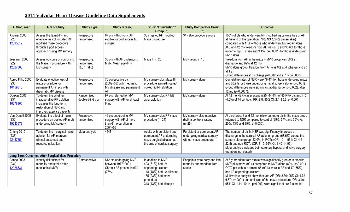
2014 Valvular Heart Disease Guideline Data Supplements
57
Author, Year Aim of Study Study Type Study Size (N) Study “Intervention” Group (n)
Study Comparator Group (n)
Outcomes
Akpinar 2003 (228) 12895612
Assess the feasibility and effectiveness of irrigated RF modified maze procedure through a port access approach during MV surgery
Prospective randomized
67 pts with chronic AF eligible for port access MV surgery
33 irrigated RF modified Maze procedure
34 valve procedure alone 100% of pts who underwent RF modified maze were free of AF at the end of the operation (76% NSR, 24% pacemaker) compared with 41% of those who underwent MV repair alone. At 6 and 12 mo freedom from AF was 87.2 and 93.6% for those undergoing RF maze and 9.4% (p=0.0001) for those undergoing MVR alone
Jessarun 2003 (229) 12627066
Assess outcome of combining the Maze III procedure with MV surgery
Prospective. randomized (2.5:1 ratio)
35 pts with AF undergoing MVR. Mean age 64 y
Maze III in 25 MVR along in 10 Freedom from AF in the maze + MVR group was 56% at discharge and 92% at 12 mo. MVR alone group, freedom from AF was 0% at discharge and 20 at 1 y. Group differences at discharge p=0.002 and at 1 y p=0.0007.
Abreu Filho 2005 (230) 16159816
Evaluate effectiveness of maze procedure for permanent AF in pts with rheumatic MV disease
Prospective randomized
70 consecutive pts (2002−03) with rheumatic MV disease and permanent AF
MV surgery plus Maze III procedure saline-Irrigated cooled-tip RF ablation
MV surgery alone Cumulative rates of NSR were 79.4% for those undergoing maze and 26.9% for those undergoing mitral surgery alone (p=0.001). Group differences were significant at discharge (p=0.002), after 12 mo (p=0.0007).
Doukas 2005 (231) 16278360
To determine whether intraoperative RF ablation increases the long-term restoration of NSR and improves exercise capacity
Randomized, double-blind trial
97 pts referred for MV surgery with AF for at least 6 mo
MV surgery plus RF left atrial ablation
MV surgery alone At 12 mo NSR was present in 20 (44.4%) of 45 RFA pts and in 2 (4.5%) of 44 controls, RR: 9.8; 95% CI: 2.4–86.3; p<0.001
Von Oppell 2009 (232) 19233678
Evaluate the effect of maze procedure on postop AF in pts undergoing MV surgery
Prospective randomized
49 pts undergoing MV surgery with AF of more than 6 mo duration in 2004−06
MV surgery plus RF maze procedure (n=24)
MV surgery plus intensive rhythm control strategy (n=25).
At discharge, 3 and 12 mo follow-up, more pts in the maze group returned to NSR compared to control (29%, 57% and 75% vs. 20%, 43% and 39%; p=0.030).
Cheng 2010 (233) 22437354
To determine if surgical maze ablation for AF improves clinical outcomes and resource utilization
Meta-analysis 4647 Adults with persistent and permanent AF undergoing maze surgical ablation at the time of cardiac surgery
Persistent or permanent AF undergoing cardiac surgery without maze procedure
The number of pts in NSR was significantly improved at discharge in the surgical AF ablation group (68.6%) versus the surgery alone group (23.0%) in RCTs (OR: 10.1, 95% CI: 4.5-22.5) and non-RCTs (OR: 7.15, 95% CI: 3.42-14.95). Meta-analysis includes both coronary bypass and valve surgery (numbers not stated).
Long-Term Outcomes After Surgical Maze Procedure
Bando 2003 (234) 12928631
Identify risk factors for mortality and stroke after mechanical MVR
Retrospective 812 pts undergoing MVR between 1977−2001. Chronic AF present in 630 (78%)
In addition to MVR: 493 (61%) had LV appendage closure 148 (18%) had LA plication 185 (23%) had maze procedure 348 (43%) had tricuspid
Endpoints were early and late mortality and freedom from stroke
At 8 y, freedom from stroke was significantly greater in pts with MVR plus maze (99%) compared to MVR alone (89%, p<0.001) Of 72 pts with late stroke, 65 (90%) were in AF and 47 (65%) had LA appendage closure. Multivariate analysis show that late AF (OR: 3.39; 95% CI: 1.72–6.67; p=.0001) and omission of the maze procedure (OR: 3.40; 95% CI: 1.14–10.14; p=0.003) were significant risk factors for

2014 Valvular Heart Disease Guideline Data Supplements
58
Author, Year Aim of Study Study Type Study Size (N) Study “Intervention” Group (n)
Study Comparator Group (n)
Outcomes
annuloplasty. late stroke. Bum Kim 2012 (235) 22456472
Evaluate long-term benefits of the maze procedure in pts with chronic AF undergoing mechanical MVR
Retrospective, observational
569 pts undergoing mechanical MVR between 1997−2010
317 with MVR plus a concomitant maze procedure
252 with MVR alone Pts who had undergone the maze procedure were at similar risks of death (HR: 1.15; 95% CI: 0.65–2.03; p=0.63) and the composite outcomes (HR: 0.82; 95% CI: 0.50–1.34; p=0.42), but a significantly lower risk of thromboembolic events (HR: 0.29; 95% CI: 0.12–0.73; p=0.008) compared with those who underwent valve replacement alone
Malaisrie 2012 (236) 22808837
Determine the impact of concomitant AF ablation in pts undergoing AVR
Retrospective, observational
124 pts (mean age 74±12 y) with pre-existing AF undergoing AVR
80 (65%) had concomitant surgical AF ablation
44 had AVR alone Postop freedom from AF when not receiving anti-arrhythmic drugs occurred in 58 pts (82%) in the ablation group, compared to 8 (36%) in the nonablation group (p<0.001)
Liu 2010 (237) 20573636
Compare pulmonary vein isolation versus maze procedure for treatment of permanent AF
Prospective randomized
99 with rheumatic heart disease and permanent AF
49 with valve surgery plus circumferential pulmonary vein isolation
50 with valve surgery plus maze procedure for AF
After one procedure, pts undergoing the maze procedure had a significantly higher freedom from atrial arrhythmias (82% vs. 55.2%, p<0.001). At 15–20 mo follow-up, cumulative rates of sinus rhythm were 71% vs. 88% (p<0.001).
1° indicates primary; AF, atrial fibrillation; AVR, aortic valve replacement; CABG, coronary artery bypass grafting; LA, left atrial; LV, left ventricle; LVEF, left ventricular ejection fraction; MR, mitral regurgitation; MV, mitral valve; MVR, mitral valve replacement; NSR, normal sinus rhythm; preop, preoperative; postop, postoperative; pts, patients; RCT, randomized clinical trial; RF, radiofrequency; RFA, radiofrequency ablation; and, SR, sinus rhythm.
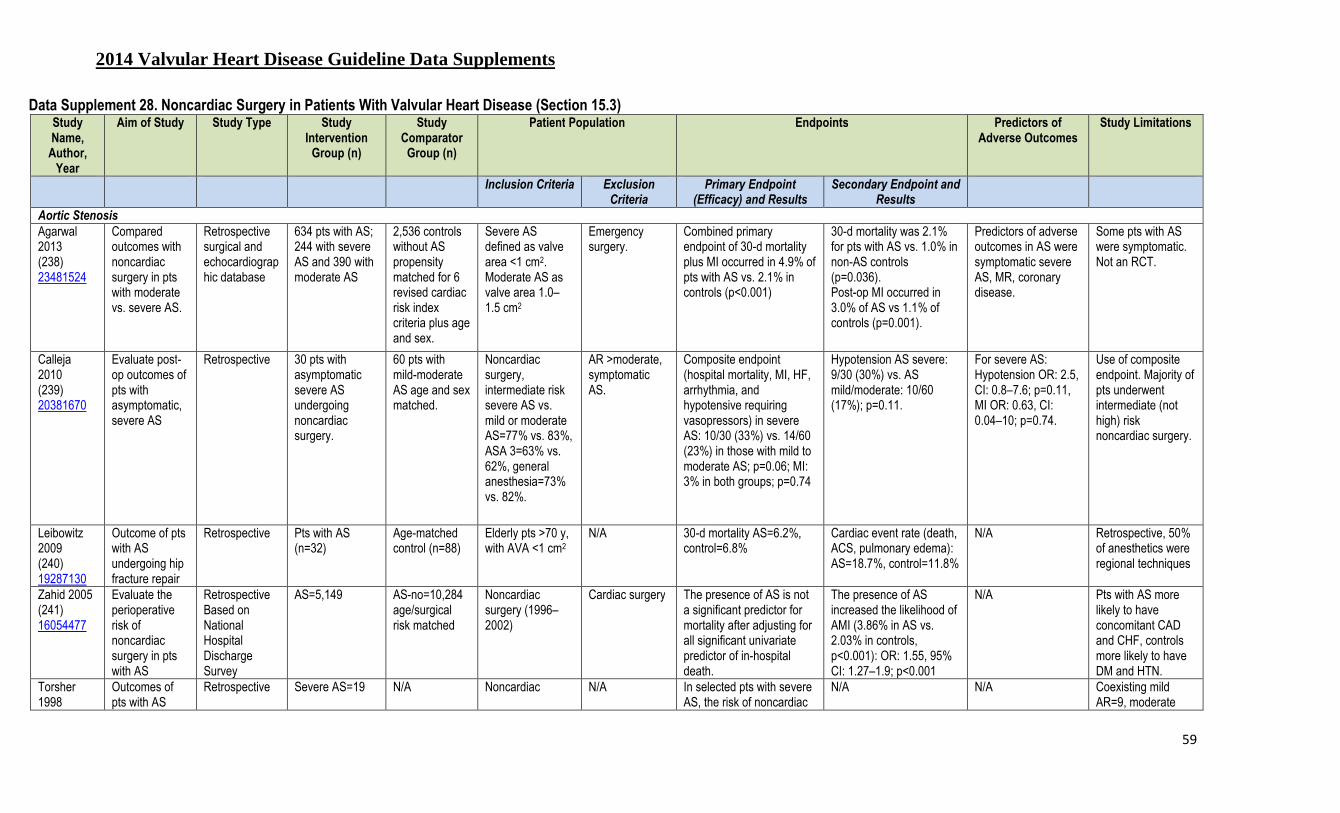
2014 Valvular Heart Disease Guideline Data Supplements
59
Data Supplement 28. Noncardiac Surgery in Patients With Valvular Heart Disease (Section 15.3) Study Name,
Author, Year
Aim of Study Study Type Study Intervention
Group (n)
Study Comparator
Group (n)
Patient Population Endpoints Predictors of Adverse Outcomes
Study Limitations
Inclusion Criteria Exclusion Criteria
Primary Endpoint (Efficacy) and Results
Secondary Endpoint and Results
Aortic Stenosis
Agarwal 2013 (238) 23481524
Compared outcomes with noncardiac surgery in pts with moderate vs. severe AS.
Retrospective surgical and echocardiographic database
634 pts with AS; 244 with severe AS and 390 with moderate AS
2,536 controls without AS propensity matched for 6 revised cardiac risk index criteria plus age and sex.
Severe AS defined as valve area <1 cm2. Moderate AS as valve area 1.0–1.5 cm2
Emergency surgery.
Combined primary endpoint of 30-d mortality plus MI occurred in 4.9% of pts with AS vs. 2.1% in controls (p<0.001)
30-d mortality was 2.1% for pts with AS vs. 1.0% in non-AS controls (p=0.036). Post-op MI occurred in 3.0% of AS vs 1.1% of controls (p=0.001).
Predictors of adverse outcomes in AS were symptomatic severe AS, MR, coronary disease.
Some pts with AS were symptomatic. Not an RCT.
Calleja 2010 (239) 20381670
Evaluate post-op outcomes of pts with asymptomatic, severe AS
Retrospective 30 pts with asymptomatic severe AS undergoing noncardiac surgery.
60 pts with mild-moderate AS age and sex matched.
Noncardiac surgery, intermediate risk severe AS vs. mild or moderate AS=77% vs. 83%, ASA 3=63% vs. 62%, general anesthesia=73% vs. 82%.
AR >moderate, symptomatic AS.
Composite endpoint (hospital mortality, MI, HF, arrhythmia, and hypotensive requiring vasopressors) in severe AS: 10/30 (33%) vs. 14/60 (23%) in those with mild to moderate AS; p=0.06; MI: 3% in both groups; p=0.74
Hypotension AS severe: 9/30 (30%) vs. AS mild/moderate: 10/60 (17%); p=0.11.
For severe AS: Hypotension OR: 2.5, CI: 0.8–7.6; p=0.11, MI OR: 0.63, CI: 0.04–10; p=0.74.
Use of composite endpoint. Majority of pts underwent intermediate (not high) risk noncardiac surgery.
Leibowitz 2009 (240) 19287130
Outcome of pts with AS undergoing hip fracture repair
Retrospective Pts with AS (n=32)
Age-matched control (n=88)
Elderly pts >70 y, with AVA <1 cm2
N/A 30-d mortality AS=6.2%, control=6.8%
Cardiac event rate (death, ACS, pulmonary edema): AS=18.7%, control=11.8%
N/A Retrospective, 50% of anesthetics were regional techniques
Zahid 2005 (241) 16054477
Evaluate the perioperative risk of noncardiac surgery in pts with AS
Retrospective Based on National Hospital Discharge Survey
AS=5,149 AS-no=10,284 age/surgical risk matched
Noncardiac surgery (1996–2002)
Cardiac surgery The presence of AS is not a significant predictor for mortality after adjusting for all significant univariate predictor of in-hospital death.
The presence of AS increased the likelihood of AMI (3.86% in AS vs. 2.03% in controls, p<0.001): OR: 1.55, 95% CI: 1.27–1.9; p<0.001
N/A Pts with AS more likely to have concomitant CAD and CHF, controls more likely to have DM and HTN.
Torsher 1998
Outcomes of pts with AS
Retrospective Severe AS=19 N/A Noncardiac N/A In selected pts with severe AS, the risk of noncardiac
N/A
N/A Coexisting mild AR=9, moderate
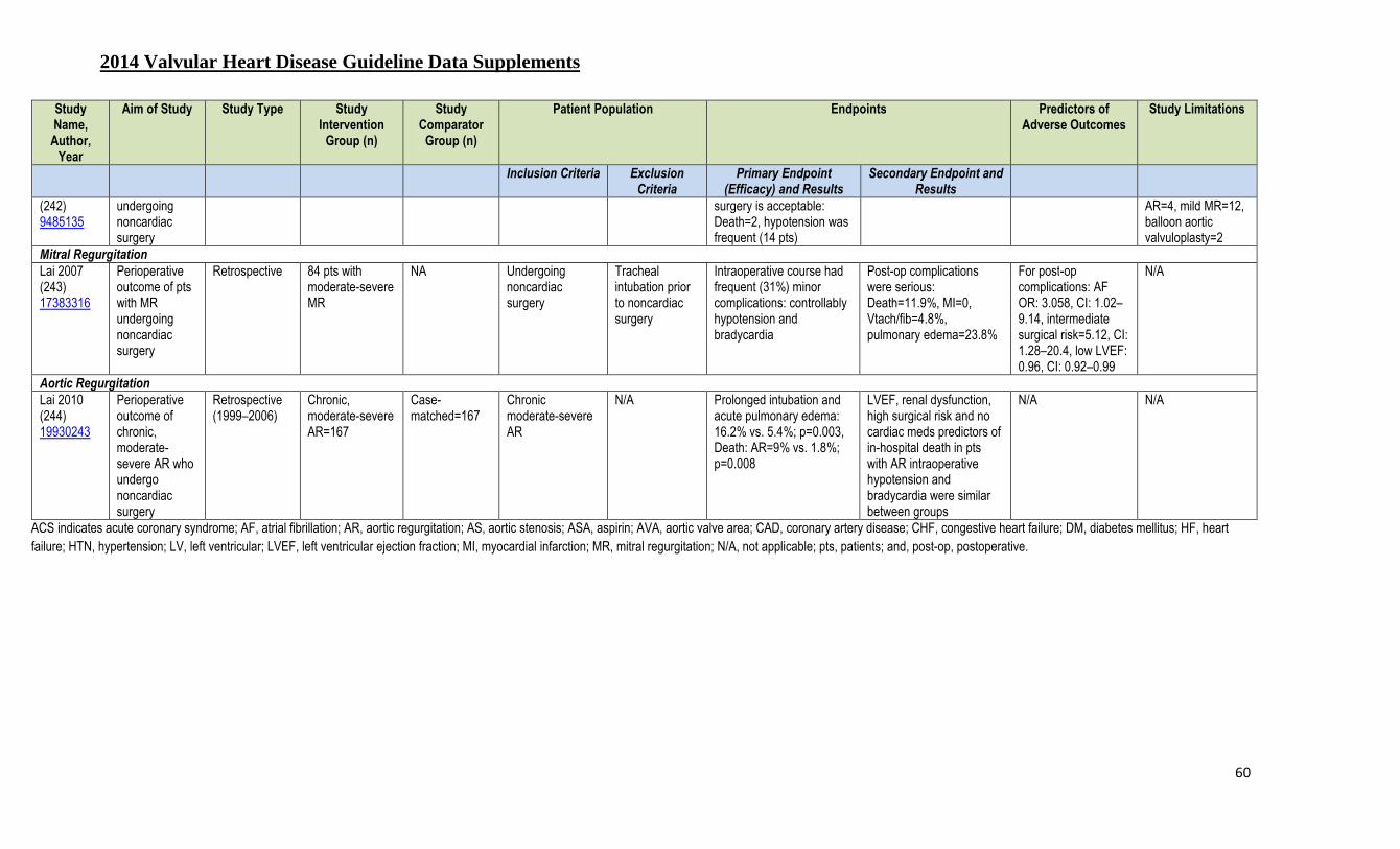
2014 Valvular Heart Disease Guideline Data Supplements
60
Study Name,
Author, Year
Aim of Study Study Type Study Intervention
Group (n)
Study Comparator
Group (n)
Patient Population Endpoints Predictors of Adverse Outcomes
Study Limitations
Inclusion Criteria Exclusion Criteria
Primary Endpoint (Efficacy) and Results
Secondary Endpoint and Results
(242) 9485135
undergoing noncardiac surgery
surgery is acceptable: Death=2, hypotension was frequent (14 pts)
AR=4, mild MR=12, balloon aortic valvuloplasty=2
Mitral Regurgitation Lai 2007 (243) 17383316
Perioperative outcome of pts with MR undergoing noncardiac surgery
Retrospective 84 pts with moderate-severe MR
NA Undergoing noncardiac surgery
Tracheal intubation prior to noncardiac surgery
Intraoperative course had frequent (31%) minor complications: controllably hypotension and bradycardia
Post-op complications were serious: Death=11.9%, MI=0, Vtach/fib=4.8%, pulmonary edema=23.8%
For post-op complications: AF OR: 3.058, CI: 1.02–9.14, intermediate surgical risk=5.12, CI: 1.28–20.4, low LVEF: 0.96, CI: 0.92–0.99
N/A
Aortic Regurgitation Lai 2010 (244) 19930243
Perioperative outcome of chronic, moderate-severe AR who undergo noncardiac surgery
Retrospective (1999–2006)
Chronic, moderate-severe AR=167
Case-matched=167
Chronic moderate-severe AR
N/A Prolonged intubation and acute pulmonary edema: 16.2% vs. 5.4%; p=0.003, Death: AR=9% vs. 1.8%; p=0.008
LVEF, renal dysfunction, high surgical risk and no cardiac meds predictors of in-hospital death in pts with AR intraoperative hypotension and bradycardia were similar between groups
N/A N/A
ACS indicates acute coronary syndrome; AF, atrial fibrillation; AR, aortic regurgitation; AS, aortic stenosis; ASA, aspirin; AVA, aortic valve area; CAD, coronary artery disease; CHF, congestive heart failure; DM, diabetes mellitus; HF, heart failure; HTN, hypertension; LV, left ventricular; LVEF, left ventricular ejection fraction; MI, myocardial infarction; MR, mitral regurgitation; N/A, not applicable; pts, patients; and, post-op, postoperative.

2014 Valvular Heart Disease Guideline Data Supplements
61
References
1. deFilippi CR, Willett DL, Brickner ME, et al. Usefulness of dobutamine echocardiography in distinguishing severe from nonsevere valvular aortic stenosis in patients with depressed left ventricular function and low transvalvular gradients. Am J Cardiol. 1995;75:191-4.
2. Connolly HM, Oh JK, Orszulak TA, et al. Aortic valve replacement for aortic stenosis with severe left ventricular dysfunction. Prognostic indicators. Circulation. 1997;95:2395-400. 3. Pereira JJ, Lauer MS, Bashir M, et al. Survival after aortic valve replacement for severe aortic stenosis with low transvalvular gradients and severe left ventricular dysfunction. J Am Coll Cardiol.
2002;39:1356-63. 4. Nishimura RA, Grantham JA, Connolly HM, et al. Low-output, low-gradient aortic stenosis in patients with depressed left ventricular systolic function: the clinical utility of the dobutamine
challenge in the catheterization laboratory. Circulation. 2002;106:809-13. 5. Monin JL, Quere JP, Monchi M, et al. Low-gradient aortic stenosis: operative risk stratification and predictors for long-term outcome: a multicenter study using dobutamine stress hemodynamics.
Circulation. 2003;108:319-24. 6. Quere JP, Monin JL, Levy F, et al. Influence of preoperative left ventricular contractile reserve on postoperative ejection fraction in low-gradient aortic stenosis. Circulation. 2006;113:1738-44. 7. Blais C, Burwash IG, Mundigler G, et al. Projected valve area at normal flow rate improves the assessment of stenosis severity in patients with low-flow, low-gradient aortic stenosis: the
multicenter TOPAS (Truly or Pseudo-Severe Aortic Stenosis) study. Circulation. 2006;113:711-21. 8. Bergler-Klein J, Klaar U, Heger M, et al. Natriuretic peptides predict symptom-free survival and postoperative outcome in severe aortic stenosis. Circulation. 2004;109:2302-8. 9. Pai RG, Varadarajan P, Razzouk A. Survival benefit of aortic valve replacement in patients with severe aortic stenosis with low ejection fraction and low gradient with normal ejection fraction. Ann
Thorac Surg. 2008;86:1781-9. 10. Levy F, Laurent M, Monin JL, et al. Aortic valve replacement for low-flow/low-gradient aortic stenosis operative risk stratification and long-term outcome: a European multicenter study. J Am Coll
Cardiol. 2008;51:1466-72. 11. Clavel MA, Webb JG, Rodes-Cabau J, et al. Comparison between transcatheter and surgical prosthetic valve implantation in patients with severe aortic stenosis and reduced left ventricular ejection
fraction. Circulation. 2010;122:1928-36. 12. Tribouilloy C, Levy F, Rusinaru D, et al. Outcome after aortic valve replacement for low-flow/low-gradient aortic stenosis without contractile reserve on dobutamine stress echocardiography. J Am
Coll Cardiol. 2009;53:1865-73. 13. Gotzmann M, Lindstaedt M, Bojara W, et al. Clinical outcome of transcatheter aortic valve implantation in patients with low-flow, low gradient aortic stenosis. Catheter Cardiovasc Interv.
2012;79:693-701. 14. Fougeres E, Tribouilloy C, Monchi M, et al. Outcomes of pseudo-severe aortic stenosis under conservative treatment. Eur Heart J. 2012;33:2426-33. 15. Herrmann HC, Pibarot P, Hueter I, et al. Predictors of Mortality and Outcomes of Therapy in Low-Flow Severe Aortic Stenosis: A Placement of Aortic Transcatheter Valves (PARTNER) Trial
Analysis. Circulation. 2013;127:2316-26. 16. Otto CM, Pearlman AS, Gardner CL. Hemodynamic progression of aortic stenosis in adults assessed by Doppler echocardiography. J Am Coll Cardiol. 1989;13:545-50. 17. Roger VL, Tajik AJ, Bailey KR, et al. Progression of aortic stenosis in adults: new appraisal using Doppler echocardiography. Am Heart J. 1990;119:331-8. 18. Faggiano P, Ghizzoni G, Sorgato A, et al. Rate of progression of valvular aortic stenosis in adults. Am J Cardiol. 1992;70:229-33. 19. Peter M, Hoffmann A, Parker C, et al. Progression of aortic stenosis. Role of age and concomitant coronary artery disease. Chest. 1993;103:1715-9. 20. Brener SJ, Duffy CI, Thomas JD, et al. Progression of aortic stenosis in 394 patients: relation to changes in myocardial and mitral valve dysfunction. J Am Coll Cardiol. 1995;25:305-10. 21. Otto CM, Burwash IG, Legget ME, et al. Prospective study of asymptomatic valvular aortic stenosis. Clinical, echocardiographic, and exercise predictors of outcome. Circulation. 1997;95:2262-70. 22. Bahler RC, Desser DR, Finkelhor RS, et al. Factors leading to progression of valvular aortic stenosis. Am J Cardiol. 1999;84:1044-8. 23. Palta S, Pai AM, Gill KS, et al. New insights into the progression of aortic stenosis: implications for secondary prevention. Circulation. 2000;101:2497-502. 24. Rosenhek R, Binder T, Porenta G, et al. Predictors of outcome in severe, asymptomatic aortic stenosis. N Engl J Med. 2000;343:611-7. 25. Rosenhek R, Klaar U, Schemper M, et al. Mild and moderate aortic stenosis. Natural history and risk stratification by echocardiography. Eur Heart J. 2004;25:199-205. 26. Rossebo AB, Pedersen TR, Boman K, et al. Intensive lipid lowering with simvastatin and ezetimibe in aortic stenosis. N Engl J Med. 2008;359:1343-56.

2014 Valvular Heart Disease Guideline Data Supplements
62
27. Nylander E, Ekman I, Marklund T, et al. Severe aortic stenosis in elderly patients. Br Heart J. 1986;55:480-7. 28. Clyne CA, Arrighi JA, Maron BJ, et al. Systemic and left ventricular responses to exercise stress in asymptomatic patients with valvular aortic stenosis. Am J Cardiol. 1991;68:1469-76. 29. Otto CM, Pearlman AS, Kraft CD, et al. Physiologic changes with maximal exercise in asymptomatic valvular aortic stenosis assessed by Doppler echocardiography. J Am Coll Cardiol.
1992;20:1160-7. 30. Amato MC, Moffa PJ, Werner KE, et al. Treatment decision in asymptomatic aortic valve stenosis: role of exercise testing. Heart. 2001;86:381-6. 31. Alborino D, Hoffmann JL, Fournet PC, et al. Value of exercise testing to evaluate the indication for surgery in asymptomatic patients with valvular aortic stenosis. J Heart Valve Dis. 2002;11:204-9. 32. Das P, Rimington H, Chambers J. Exercise testing to stratify risk in aortic stenosis. Eur Heart J. 2005;26:1309-13. 33. Lancellotti P, Lebois F, Simon M, et al. Prognostic importance of quantitative exercise Doppler echocardiography in asymptomatic valvular aortic stenosis. Circulation. 2005;112:I377-I382. 34. Marechaux S, Hachicha Z, Bellouin A, et al. Usefulness of exercise-stress echocardiography for risk stratification of true asymptomatic patients with aortic valve stenosis. Eur Heart J.
2010;31:1390-7. 35. Rajani R, Rimington H, Chambers JB. Treadmill exercise in apparently asymptomatic patients with moderate or severe aortic stenosis: relationship between cardiac index and revealed symptoms.
Heart. 2010;96:689-95. 36. Cowell SJ, Newby DE, Prescott RJ, et al. A randomized trial of intensive lipid-lowering therapy in calcific aortic stenosis. N Engl J Med. 2005;352:2389-97. 37. Moura LM, Ramos SF, Zamorano JL, et al. Rosuvastatin affecting aortic valve endothelium to slow the progression of aortic stenosis. J Am Coll Cardiol. 2007;49:554-61. 38. Chan KL, Teo K, Dumesnil JG, et al. Effect of Lipid lowering with rosuvastatin on progression of aortic stenosis: results of the aortic stenosis progression observation: measuring effects of
rosuvastatin (ASTRONOMER) trial. Circulation. 2010;121:306-14. 39. Kelly TA, Rothbart RM, Cooper CM, et al. Comparison of outcome of asymptomatic to symptomatic patients older than 20 years of age with valvular aortic stenosis. Am J Cardiol. 1988;61:123-30. 40. Pellikka PA, Nishimura RA, Bailey KR, et al. The natural history of adults with asymptomatic, hemodynamically significant aortic stenosis. J Am Coll Cardiol. 1990;15:1012-7. 41. Kennedy KD, Nishimura RA, Holmes DR, Jr., et al. Natural history of moderate aortic stenosis. J Am Coll Cardiol. 1991;17:313-9. 42. Pellikka PA, Sarano ME, Nishimura RA, et al. Outcome of 622 adults with asymptomatic, hemodynamically significant aortic stenosis during prolonged follow-up. Circulation. 2005;111:3290-5. 43. Lancellotti P, Donal E, Magne J, et al. Risk stratification in asymptomatic moderate to severe aortic stenosis: the importance of the valvular, arterial and ventricular interplay. Heart. 2010;96:1364-
71. 44. Kang DH, Park SJ, Rim JH, et al. Early surgery versus conventional treatment in asymptomatic very severe aortic stenosis. Circulation. 2010;121:1502-9. 45. Stewart RA, Kerr AJ, Whalley GA, et al. Left ventricular systolic and diastolic function assessed by tissue Doppler imaging and outcome in asymptomatic aortic stenosis. Eur Heart J.
2010;31:2216-22. 46. Rosenhek R, Zilberszac R, Schemper M, et al. Natural history of very severe aortic stenosis. Circulation. 2010;121:151-6. 47. Jander N, Minners J, Holme I, et al. Outcome of patients with low-gradient "severe" aortic stenosis and preserved ejection fraction. Circulation. 2011;123:887-95. 48. Saito T, Muro T, Takeda H, et al. Prognostic value of aortic valve area index in asymptomatic patients with severe aortic stenosis. Am J Cardiol. 2012;110:93-7. 49. Monin JL, Lancellotti P, Monchi M, et al. Risk score for predicting outcome in patients with asymptomatic aortic stenosis. Circulation. 2009;120:69-75. 50. Rosenhek R, Baumgartner H. Aortic Stenosis. In: The Practice of Clinical Echocardiography. Elsevier/Saunders ; 2012. 51. Frank S, Johnson A, Ross J, Jr. Natural history of valvular aortic stenosis. Br Heart J. 1973;35:41-6. 52. Chizner MA, Pearle DL, deLeon AC, Jr. The natural history of aortic stenosis in adults. Am Heart J. 1980;99:419-24. 53. Lombard JT, Selzer A. Valvular aortic stenosis. A clinical and hemodynamic profile of patients. Ann Intern Med. 1987;106:292-8. 54. Turina J, Hess O, Sepulcri F, et al. Spontaneous course of aortic valve disease. Eur Heart J. 1987;8:471-83. 55. Horstkotte D, Loogen F. The natural history of aortic valve stenosis. Eur Heart J. 1988;9 Suppl E:57-64. 56. Otto CM, Pearlman AS. Doppler echocardiography in adults with symptomatic aortic stenosis. Diagnostic utility and cost-effectiveness. Arch Intern Med. 1988;148:2553-60. 57. Oh JK, Taliercio CP, Holmes DR, Jr., et al. Prediction of the severity of aortic stenosis by Doppler aortic valve area determination: prospective Doppler-catheterization correlation in 100 patients. J
Am Coll Cardiol. 1988;11:1227-34. 58. Galan A, Zoghbi WA, Quinones MA. Determination of severity of valvular aortic stenosis by Doppler echocardiography and relation of findings to clinical outcome and agreement with
hemodynamic measurements determined at cardiac catheterization. Am J Cardiol. 1991;67:1007-12.

2014 Valvular Heart Disease Guideline Data Supplements
63
59. Otto CM, Mickel MC, Kennedy JW, et al. Three-year outcome after balloon aortic valvuloplasty. Insights into prognosis of valvular aortic stenosis. Circulation. 1994;89:642-50. 60. Hachicha Z, Dumesnil JG, Bogaty P, et al. Paradoxical low-flow, low-gradient severe aortic stenosis despite preserved ejection fraction is associated with higher afterload and reduced survival.
Circulation. 2007;115:2856-64. 61. Tarantini G, Covolo E, Razzolini R, et al. Valve replacement for severe aortic stenosis with low transvalvular gradient and left ventricular ejection fraction exceeding 0.50. Ann Thorac Surg.
2011;91:1808-15. 62. Clavel MA, Dumesnil JG, Capoulade R, et al. Outcome of patients with aortic stenosis, small valve area, and low-flow, low-gradient despite preserved left ventricular ejection fraction. J Am Coll
Cardiol. 2012;60:1259-67. 63. Lancellotti P, Magne J, Donal E, et al. Clinical outcome in asymptomatic severe aortic stenosis: insights from the new proposed aortic stenosis grading classification. J Am Coll Cardiol.
2012;59:235-43. 64. Le VF, Freeman M, Webb J, et al. Impact of low flow on the outcome of high-risk patients undergoing transcatheter aortic valve replacement. J Am Coll Cardiol. 2013;62:782-8. 65. Mehrotra P, Jansen K, Flynn AW, et al. Differential left ventricular remodelling and longitudinal function distinguishes low flow from normal-flow preserved ejection fraction low-gradient severe
aortic stenosis. Eur Heart J. 2013;34:1906-14. 66. Ozkan M, Gunduz S, Biteker M, et al. Comparison of different TEE-guided thrombolytic regimens for prosthetic valve thrombosis: the TROIA trial. JACC Cardiovasc Imaging. 2013;6:206-16. 67. Eleid MF, Sorajja P, Michelena HI, et al. Flow-gradient patterns in severe aortic stenosis with preserved ejection fraction: clinical characteristics and predictors of survival. Circulation.
2013;128:1781-9. 68. Smith CR, Leon MB, Mack MJ, et al. Transcatheter versus surgical aortic-valve replacement in high-risk patients. N Engl J Med. 2011;364:2187-98. 69. Kodali SK, Williams MR, Smith CR, et al. Two-year outcomes after transcatheter or surgical aortic-valve replacement. N Engl J Med. 2012;366:1686-95. 70. Makkar RR, Fontana GP, Jilaihawi H, et al. Transcatheter aortic-valve replacement for inoperable severe aortic stenosis. N Engl J Med. 2012;366:1696-704. 71. Leon MB, Smith CR, Mack M, et al. Transcatheter aortic-valve implantation for aortic stenosis in patients who cannot undergo surgery. N Engl J Med. 2010;363:1597-607. 72. Bonow RO, Rosing DR, McIntosh CL, et al. The natural history of asymptomatic patients with aortic regurgitation and normal left ventricular function. Circulation. 1983;68:509-17. 73. Scognamiglio R, Fasoli G, Dalla VS. Progression of myocardial dysfunction in asymptomatic patients with severe aortic insufficiency. Clin Cardiol. 1986;9:151-6. 74. Siemienczuk D, Greenberg B, Morris C, et al. Chronic aortic insufficiency: factors associated with progression to aortic valve replacement. Ann Intern Med. 1989;110:587-92. 75. Bonow RO, Lakatos E, Maron BJ, et al. Serial long-term assessment of the natural history of asymptomatic patients with chronic aortic regurgitation and normal left ventricular systolic function.
Circulation. 1991;84:1625-35. 76. Scognamiglio R, Rahimtoola SH, Fasoli G, et al. Nifedipine in asymptomatic patients with severe aortic regurgitation and normal left ventricular function. N Engl J Med. 1994;331:689-94. 77. Tornos MP, Olona M, Permanyer-Miralda G, et al. Clinical outcome of severe asymptomatic chronic aortic regurgitation: a long-term prospective follow-up study. Am Heart J. 1995;130:333-9. 78. Ishii K, Hirota Y, Suwa M, et al. Natural history and left ventricular response in chronic aortic regurgitation. Am J Cardiol. 1996;78:357-61. 79. Borer JS, Hochreiter C, Herrold EM, et al. Prediction of indications for valve replacement among asymptomatic or minimally symptomatic patients with chronic aortic regurgitation and normal left
ventricular performance. Circulation. 1998;97:525-34. 80. Tarasoutchi F, Grinberg M, Spina GS, et al. Ten-year clinical laboratory follow-up after application of a symptom-based therapeutic strategy to patients with severe chronic aortic regurgitation of
predominant rheumatic etiology. J Am Coll Cardiol. 2003;41:1316-24. 81. Evangelista A, Tornos P, Sambola A, et al. Long-term vasodilator therapy in patients with severe aortic regurgitation. N Engl J Med. 2005;353:1342-9. 82. Detaint D, Messika-Zeitoun D, Maalouf J, et al. Quantitative echocardiographic determinants of clinical outcome in asymptomatic patients with aortic regurgitation: a prospective study. JACC
Cardiovasc Imaging. 2008;1:1-11. 83. Pizarro R, Bazzino OO, Oberti PF, et al. Prospective validation of the prognostic usefulness of B-type natriuretic peptide in asymptomatic patients with chronic severe aortic regurgitation. J Am
Coll Cardiol. 2011;58:1705-14. 84. Olsen NT, Sogaard P, Larsson HB, et al. Speckle-tracking echocardiography for predicting outcome in chronic aortic regurgitation during conservative management and after surgery. JACC
Cardiovasc Imaging. 2011;4:223-30. 85. Forman R, Firth BG, Barnard MS. Prognostic significance of preoperative left ventricular ejection fraction and valve lesion in patients with aortic valve replacement. Am J Cardiol. 1980;45:1120-5.

2014 Valvular Heart Disease Guideline Data Supplements
64
86. Henry WL, Bonow RO, Borer JS, et al. Observations on the optimum time for operative intervention for aortic regurgitation. I. Evaluation of the results of aortic valve replacement in symptomatic patients. Circulation. 1980;61:471-83.
87. Cunha CL, Giuliani ER, Fuster V, et al. Preoperative M-mode echocardiography as a predictor of surgical results in chronic aortic insufficiency. J Thorac Cardiovasc Surg. 1980;79:256-65. 88. Bonow RO, Borer JS, Rosing DR, et al. Preoperative exercise capacity in symptomatic patients with aortic regurgitation as a predictor of postoperative left ventricular function and long-term
prognosis. Circulation. 1980;62:1280-90. 89. Borow KM, Green LH, Mann T, et al. End-systolic volume as a predictor of postoperative left ventricular performance in volume overload from valvular regurgitation. Am J Med. 1980;68:655-63. 90. Greves J, Rahimtoola SH, McAnulty JH, et al. Preoperative criteria predictive of late survival following valve replacement for severe aortic regurgitation. Am Heart J. 1981;101:300-8. 91. Kumpuris AG, Quinones MA, Waggoner AD, et al. Importance of preoperative hypertrophy, wall stress and end-systolic dimension as echocardiographic predictors of normalization of left
ventricular dilatation after valve replacement in chronic aortic insufficiency. Am J Cardiol. 1982;49:1091-100. 92. Gaasch WH, Carroll JD, Levine HJ, et al. Chronic aortic regurgitation: prognostic value of left ventricular end-systolic dimension and end-diastolic radius/thickness ratio. J Am Coll Cardiol.
1983;1:775-82. 93. Fioretti P, Roelandt J, Bos RJ, et al. Echocardiography in chronic aortic insufficiency. Is valve replacement too late when left ventricular end-systolic dimension reaches 55 mm? Circulation.
1983;67:216-21. 94. Stone PH, Clark RD, Goldschlager N, et al. Determinants of prognosis of patients with aortic regurgitation who undergo aortic valve replacement. J Am Coll Cardiol. 1984;3:1118-26. 95. Bonow RO, Picone AL, McIntosh CL, et al. Survival and functional results after valve replacement for aortic regurgitation from 1976 to 1983: impact of preoperative left ventricular function.
Circulation. 1985;72:1244-56. 96. Daniel WG, Hood WP, Jr., Siart A, et al. Chronic aortic regurgitation: reassessment of the prognostic value of preoperative left ventricular end-systolic dimension and fractional shortening.
Circulation. 1985;71:669-80. 97. Cormier B, Vahanian A, Luxereau P, et al. Should asymptomatic or mildly symptomatic aortic regurgitation be operated on? Z Kardiol. 1986;75 Suppl 2:141-5. 98. Sheiban I, Trevi GP, Casarotto D, et al. Aortic valve replacement in patients with aortic incompetence. Preoperative parameters influencing long-term results. Z Kardiol. 1986;75 Suppl 2:146-54. 99. Carabello BA, Williams H, Gash AK, et al. Hemodynamic predictors of outcome in patients undergoing valve replacement. Circulation. 1986;74:1309-16. 100. Taniguchi K, Nakano S, Hirose H, et al. Preoperative left ventricular function: minimal requirement for successful late results of valve replacement for aortic regurgitation. J Am Coll Cardiol.
1987;10:510-8. 101. Michel PL, Iung B, Abou JS, et al. The effect of left ventricular systolic function on long term survival in mitral and aortic regurgitation. J Heart Valve Dis. 1995;4 Suppl 2:S160-S168. 102. Klodas E, Enriquez-Sarano M, Tajik AJ, et al. Aortic regurgitation complicated by extreme left ventricular dilation: long-term outcome after surgical correction. J Am Coll Cardiol. 1996;27:670-7. 103. Klodas E, Enriquez-Sarano M, Tajik AJ, et al. Optimizing timing of surgical correction in patients with severe aortic regurgitation: role of symptoms. J Am Coll Cardiol. 1997;30:746-52. 104. Turina J, Milincic J, Seifert B, et al. Valve replacement in chronic aortic regurgitation. True predictors of survival after extended follow-up. Circulation. 1998;98:II100-II106. 105. Chaliki HP, Mohty D, Avierinos JF, et al. Outcomes after aortic valve replacement in patients with severe aortic regurgitation and markedly reduced left ventricular function. Circulation.
2002;106:2687-93. 106. Tornos P, Sambola A, Permanyer-Miralda G, et al. Long-term outcome of surgically treated aortic regurgitation: influence of guideline adherence toward early surgery. J Am Coll Cardiol.
2006;47:1012-7. 107. Bhudia SK, McCarthy PM, Kumpati GS, et al. Improved outcomes after aortic valve surgery for chronic aortic regurgitation with severe left ventricular dysfunction. J Am Coll Cardiol.
2007;49:1465-71. 108. Patel JJ, Shama D, Mitha AS, et al. Balloon valvuloplasty versus closed commissurotomy for pliable mitral stenosis: a prospective hemodynamic study. J Am Coll Cardiol. 1991;18:1318-22. 109. Turi ZG, Reyes VP, Raju BS, et al. Percutaneous balloon versus surgical closed commissurotomy for mitral stenosis. A prospective, randomized trial. Circulation. 1991;83:1179-85. 110. Arora R, Nair M, Kalra GS, et al. Immediate and long-term results of balloon and surgical closed mitral valvotomy: a randomized comparative study. Am Heart J. 1993;125:1091-4. 111. Reyes VP, Raju BS, Wynne J, et al. Percutaneous balloon valvuloplasty compared with open surgical commissurotomy for mitral stenosis. N Engl J Med. 1994;331:961-7. 112. Ben FM, Ayari M, Maatouk F, et al. Percutaneous balloon versus surgical closed and open mitral commissurotomy: seven-year follow-up results of a randomized trial. Circulation. 1998;97:245-50. 113. Cotrufo M, Renzulli A, Ismeno G, et al. Percutaneous mitral commissurotomy versus open mitral commissurotomy: a comparative study. Eur J Cardiothorac Surg. 1999;15:646-51.

2014 Valvular Heart Disease Guideline Data Supplements
65
114. Bonow RO, Carabello BA, Chatterjee K, et al. 2008 focused update incorporated into the ACC/AHA 2006 guidelines for the management of patients with valvular heart disease: a report of the American College of Cardiology/American Heart Association Task Force on Practice Guidelines (Writing Committee to revise the 1998 guidelines for the management of patients with valvular heart disease). Endorsed by the Society of Cardiovascular Anesthesiologists, Society for Cardiovascular Angiography and Interventions, and Society of Thoracic Surgeons. J Am Coll Cardiol. 2008;52:e1-142.
115. Cohen DJ, Kuntz RE, Gordon SP, et al. Predictors of long-term outcome after percutaneous balloon mitral valvuloplasty. N Engl J Med. 1992;327:1329-35. 116. Palacios IF, Tuzcu ME, Weyman AE, et al. Clinical follow-up of patients undergoing percutaneous mitral balloon valvotomy. Circulation. 1995;91:671-6. 117. Dean LS, Mickel M, Bonan R, et al. Four-year follow-up of patients undergoing percutaneous balloon mitral commissurotomy. A report from the National Heart, Lung, and Blood Institute Balloon
Valvuloplasty Registry. J Am Coll Cardiol. 1996;28:1452-7. 118. Iung B, Cormier B, Ducimetiere P, et al. Functional results 5 years after successful percutaneous mitral commissurotomy in a series of 528 patients and analysis of predictive factors. J Am Coll
Cardiol. 1996;27:407-14. 119. Cannan CR, Nishimura RA, Reeder GS, et al. Echocardiographic assessment of commissural calcium: a simple predictor of outcome after percutaneous mitral balloon valvotomy. J Am Coll
Cardiol. 1997;29:175-80. 120. Palacios IF, Sanchez PL, Harrell LC, et al. Which patients benefit from percutaneous mitral balloon valvuloplasty? Prevalvuloplasty and postvalvuloplasty variables that predict long-term outcome.
Circulation. 2002;105:1465-71. 121. Wilkins GT, Weyman AE, Abascal VM, et al. Percutaneous balloon dilatation of the mitral valve: an analysis of echocardiographic variables related to outcome and the mechanism of dilatation. Br
Heart J. 1988;60:299-308. 122. Schuler G, Peterson KL, Johnson A, et al. Temporal response of left ventricular performance to mitral valve surgery. Circulation. 1979;59:1218-31. 123. Phillips HR, Levine FH, Carter JE, et al. Mitral valve replacement for isolated mitral regurgitation: analysis of clinical course and late postoperative left ventricular ejection fraction. Am J Cardiol.
1981;48:647-54. 124. Zile MR, Gaasch WH, Carroll JD, et al. Chronic mitral regurgitation: predictive value of preoperative echocardiographic indexes of left ventricular function and wall stress. J Am Coll Cardiol.
1984;3:235-42. 125. Crawford MH, Souchek J, Oprian CA, et al. Determinants of survival and left ventricular performance after mitral valve replacement. Department of Veterans Affairs Cooperative Study on Valvular
Heart Disease. Circulation. 1990;81:1173-81. 126. Wisenbaugh T, Sinovich V, Dullabh A, et al. Six month pilot study of captopril for mildly symptomatic, severe isolated mitral and isolated aortic regurgitation. J Heart Valve Dis. 1994;3:197-204. 127. Enriquez-Sarano M, Tajik AJ, Schaff HV, et al. Echocardiographic prediction of survival after surgical correction of organic mitral regurgitation. Circulation. 1994;90:830-7. 128. Enriquez-Sarano M, Tajik AJ, Schaff HV, et al. Echocardiographic prediction of left ventricular function after correction of mitral regurgitation: results and clinical implications. J Am Coll Cardiol.
1994;24:1536-43. 129. Tribouilloy CM, Enriquez-Sarano M, Schaff HV, et al. Impact of preoperative symptoms on survival after surgical correction of organic mitral regurgitation: rationale for optimizing surgical
indications. Circulation. 1999;99:400-5. 130. Gillinov AM, Mihaljevic T, Blackstone EH, et al. Should patients with severe degenerative mitral regurgitation delay surgery until symptoms develop? Ann Thorac Surg. 2010;90:481-8. 131. Rosenhek R, Rader F, Klaar U, et al. Outcome of watchful waiting in asymptomatic severe mitral regurgitation. Circulation. 2006;113:2238-44. 132. Kang DH, Kim JH, Rim JH, et al. Comparison of early surgery versus conventional treatment in asymptomatic severe mitral regurgitation. Circulation. 2009;119:797-804. 133. Tribouilloy C, Grigioni F, Avierinos JF, et al. Survival implication of left ventricular end-systolic diameter in mitral regurgitation due to flail leaflets a long-term follow-up multicenter study. J Am
Coll Cardiol. 2009;54:1961-8. 134. Enriquez-Sarano M, Avierinos JF, Messika-Zeitoun D, et al. Quantitative determinants of the outcome of asymptomatic mitral regurgitation. N Engl J Med. 2005;352:875-83. 135. Ghoreishi M, Evans CF, deFilippi CR, et al. Pulmonary hypertension adversely affects short- and long-term survival after mitral valve operation for mitral regurgitation: implications for timing of
surgery. J Thorac Cardiovasc Surg. 2011;142:1439-52. 136. Goldman ME, Mora F, Guarino T, et al. Mitral valvuloplasty is superior to valve replacement for preservation of left ventricular function: an intraoperative two-dimensional echocardiographic
study. J Am Coll Cardiol. 1987;10:568-75. 137. David TE, Burns RJ, Bacchus CM, et al. Mitral valve replacement for mitral regurgitation with and without preservation of chordae tendineae. J Thorac Cardiovasc Surg. 1984;88:718-25.

2014 Valvular Heart Disease Guideline Data Supplements
66
138. Rozich JD, Carabello BA, Usher BW, et al. Mitral valve replacement with and without chordal preservation in patients with chronic mitral regurgitation. Mechanisms for differences in postoperative ejection performance. Circulation. 1992;86:1718-26.
139. Grigioni F, Tribouilloy C, Avierinos JF, et al. Outcomes in mitral regurgitation due to flail leaflets a multicenter European study. JACC Cardiovasc Imaging. 2008;1:133-41. 140. Gillinov AM, Blackstone EH, Alaulaqi A, et al. Outcomes after repair of the anterior mitral leaflet for degenerative disease. Ann Thorac Surg. 2008;86:708-17. 141. Kang DH, Kim MJ, Kang SJ, et al. Mitral valve repair versus revascularization alone in the treatment of ischemic mitral regurgitation. Circulation. 2006;114:I499-I503. 142. Rossi A, Dini FL, Faggiano P, et al. Independent prognostic value of functional mitral regurgitation in patients with heart failure. A quantitative analysis of 1256 patients with ischaemic and non-
ischaemic dilated cardiomyopathy. Heart. 2011;97:1675-80. 143. Wu AH, Aaronson KD, Bolling SF, et al. Impact of mitral valve annuloplasty on mortality risk in patients with mitral regurgitation and left ventricular systolic dysfunction. J Am Coll Cardiol.
2005;45:381-7. 144. Mihaljevic T, Lam BK, Rajeswaran J, et al. Impact of mitral valve annuloplasty combined with revascularization in patients with functional ischemic mitral regurgitation. J Am Coll Cardiol.
2007;49:2191-201. 145. Benedetto U, Melina G, Roscitano A, et al. Does combined mitral valve surgery improve survival when compared to revascularization alone in patients with ischemic mitral regurgitation? A meta-
analysis on 2479 patients. J Cardiovasc Med (Hagerstown). 2009;10:109-14. 146. Fattouch K, Guccione F, Sampognaro R, et al. POINT: Efficacy of adding mitral valve restrictive annuloplasty to coronary artery bypass grafting in patients with moderate ischemic mitral valve
regurgitation: a randomized trial. J Thorac Cardiovasc Surg. 2009;138:278-85. 147. Deja MA, Grayburn PA, Sun B, et al. Influence of mitral regurgitation repair on survival in the surgical treatment for ischemic heart failure trial. Circulation. 2012;125:2639-48. 148. Dreyfus GD, Corbi PJ, Chan KM, et al. Secondary tricuspid regurgitation or dilatation: which should be the criteria for surgical repair? Ann Thorac Surg. 2005;79:127-32. 149. Chan V, Burwash IG, Lam BK, et al. Clinical and echocardiographic impact of functional tricuspid regurgitation repair at the time of mitral valve replacement. Ann Thorac Surg. 2009;88:1209-15. 150. Calafiore AM, Gallina S, Iaco AL, et al. Mitral valve surgery for functional mitral regurgitation: should moderate-or-more tricuspid regurgitation be treated? a propensity score analysis. Ann Thorac
Surg. 2009;87:698-703. 151. Di MM, Bivona A, Iaco AL, et al. Mitral valve surgery for functional mitral regurgitation: prognostic role of tricuspid regurgitation. Eur J Cardiothorac Surg. 2009;35:635-9. 152. Van de Veire NR, Braun J, Delgado V, et al. Tricuspid annuloplasty prevents right ventricular dilatation and progression of tricuspid regurgitation in patients with tricuspid annular dilatation
undergoing mitral valve repair. J Thorac Cardiovasc Surg. 2011;141:1431-9. 153. Yilmaz O, Suri RM, Dearani JA, et al. Functional tricuspid regurgitation at the time of mitral valve repair for degenerative leaflet prolapse: the case for a selective approach. J Thorac Cardiovasc
Surg. 2011;142:608-13. 154. Calafiore AM, Iaco AL, Romeo A, et al. Echocardiographic-based treatment of functional tricuspid regurgitation. J Thorac Cardiovasc Surg. 2011;142:308-13. 155. Navia JL, Brozzi NA, Klein AL, et al. Moderate tricuspid regurgitation with left-sided degenerative heart valve disease: to repair or not to repair? Ann Thorac Surg. 2012;93:59-67. 156. Kim JB, Yoo DG, Kim GS, et al. Mild-to-moderate functional tricuspid regurgitation in patients undergoing valve replacement for rheumatic mitral disease: the influence of tricuspid valve repair on
clinical and echocardiographic outcomes. Heart. 2012;98:24-30. 157. Benedetto U, Melina G, Angeloni E, et al. Prophylactic tricuspid annuloplasty in patients with dilated tricuspid annulus undergoing mitral valve surgery. J Thorac Cardiovasc Surg. 2012;143:632-8. 158. Hammermeister K, Sethi GK, Henderson WG, et al. Outcomes 15 years after valve replacement with a mechanical versus a bioprosthetic valve: final report of the Veterans Affairs randomized trial.
J Am Coll Cardiol. 2000;36:1152-8. 159. Oxenham H, Bloomfield P, Wheatley DJ, et al. Twenty year comparison of a Bjork-Shiley mechanical heart valve with porcine bioprostheses. Heart. 2003;89:715-21. 160. Stassano P, Di TL, Monaco M, et al. Aortic valve replacement: a prospective randomized evaluation of mechanical versus biological valves in patients ages 55 to 70 years. J Am Coll Cardiol.
2009;54:1862-8. 161. Khan SS, Trento A, DeRobertis M, et al. Twenty-year comparison of tissue and mechanical valve replacement. J Thorac Cardiovasc Surg. 2001;122:257-69. 162. Chan V, Jamieson WR, Germann E, et al. Performance of bioprostheses and mechanical prostheses assessed by composites of valve-related complications to 15 years after aortic valve replacement.
J Thorac Cardiovasc Surg. 2006;131:1267-73. 163. Kulik A, Bedard P, Lam BK, et al. Mechanical versus bioprosthetic valve replacement in middle-aged patients. Eur J Cardiothorac Surg. 2006;30:485-91.

2014 Valvular Heart Disease Guideline Data Supplements
67
164. Ruel M, Chan V, Bedard P, et al. Very long-term survival implications of heart valve replacement with tissue versus mechanical prostheses in adults <60 years of age. Circulation. 2007;116:I294-I300.
165. van Geldorp MW, Eric Jamieson WR, Kappetein AP, et al. Patient outcome after aortic valve replacement with a mechanical or biological prosthesis: weighing lifetime anticoagulant-related event risk against reoperation risk. J Thorac Cardiovasc Surg. 2009;137:881-5.
166. Badhwar V, Ofenloch JC, Rovin JD, et al. Noninferiority of closely monitored mechanical valves to bioprostheses overshadowed by early mortality benefit in younger patients. Ann Thorac Surg. 2012;93:748-53.
167. Weber A, Noureddine H, Englberger L, et al. Ten-year comparison of pericardial tissue valves versus mechanical prostheses for aortic valve replacement in patients younger than 60 years of age. J Thorac Cardiovasc Surg. 2012;144:1075-83.
168. Hammerstingl C, Tripp C, Schmidt H, et al. Periprocedural bridging therapy with low-molecular-weight heparin in chronically anticoagulated patients with prosthetic mechanical heart valves: experience in 116 patients from the prospective BRAVE registry. J Heart Valve Dis. 2007;16:285-92.
169. Spyropoulos AC, Turpie AG, Dunn AS, et al. Perioperative bridging therapy with unfractionated heparin or low-molecular-weight heparin in patients with mechanical prosthetic heart valves on long-term oral anticoagulants (from the REGIMEN Registry). Am J Cardiol. 2008;102:883-9.
170. Pengo V, Cucchini U, Denas G, et al. Standardized low-molecular-weight heparin bridging regimen in outpatients on oral anticoagulants undergoing invasive procedure or surgery: an inception cohort management study. Circulation. 2009;119:2920-7.
171. Daniels PR, McBane RD, Litin SC, et al. Peri-procedural anticoagulation management of mechanical prosthetic heart valve patients. Thromb Res. 2009;124:300-5. 172. Bui HT, Krisnaswami A, Le CU, et al. Comparison of safety of subcutaneous enoxaparin as outpatient anticoagulation bridging therapy in patients with a mechanical heart valve versus patients with
nonvalvular atrial fibrillation. Am J Cardiol. 2009;104:1429-33. 173. Biteker M, Tekkesin AI, Can MM, et al. Outcome of noncardiac and nonvascular surgery in patients with mechanical heart valves. Am J Cardiol. 2012;110:562-7. 174. Weiss A, Brose S, Ploetze K, et al. Half-dose enoxaparin vs. full-dose enoxaparin for postoperative bridging therapy in patients after cardiac surgery: Which dose regimen should be preferred? Clin
Hemorheol Microcirc. 2013. 175. Deviri E, Sareli P, Wisenbaugh T, et al. Obstruction of mechanical heart valve prostheses: clinical aspects and surgical management. J Am Coll Cardiol. 1991;17:646-50. 176. Tong AT, Roudaut R, Ozkan M, et al. Transesophageal echocardiography improves risk assessment of thrombolysis of prosthetic valve thrombosis: results of the international PRO-TEE registry. J
Am Coll Cardiol. 2004;43:77-84. 177. Roudaut R, Lafitte S, Roudaut MF, et al. Management of prosthetic heart valve obstruction: fibrinolysis versus surgery. Early results and long-term follow-up in a single-centre study of 263 cases.
Arch Cardiovasc Dis. 2009;102:269-77. 178. Karthikeyan G, Math RS, Mathew N, et al. Accelerated infusion of streptokinase for the treatment of left-sided prosthetic valve thrombosis: a randomized controlled trial. Circulation.
2009;120:1108-14. 179. Keuleers S, Herijgers P, Herregods MC, et al. Comparison of thrombolysis versus surgery as a first line therapy for prosthetic heart valve thrombosis. Am J Cardiol. 2011;107:275-9. 180. Karthikeyan G, Senguttuvan NB, Joseph J, et al. Urgent surgery compared with fibrinolytic therapy for the treatment of left-sided prosthetic heart valve thrombosis: a systematic review and meta-
analysis of observational studies. Eur Heart J. 2013. 181. Orszulak TA, Schaff HV, Danielson GK, et al. Results of reoperation for periprosthetic leakage. Ann Thorac Surg. 1983;35:584-9. 182. Miller DL, Morris JJ, Schaff HV, et al. Reoperation for aortic valve periprosthetic leakage: identification of patients at risk and results of operation. J Heart Valve Dis. 1995;4:160-5. 183. Akins CW, Bitondo JM, Hilgenberg AD, et al. Early and late results of the surgical correction of cardiac prosthetic paravalvular leaks. J Heart Valve Dis. 2005;14:792-9. 184. Pate GE, Al ZA, Chandavimol M, et al. Percutaneous closure of prosthetic paravalvular leaks: case series and review. Catheter Cardiovasc Interv. 2006;68:528-33. 185. Shapira Y, Hirsch R, Kornowski R, et al. Percutaneous closure of perivalvular leaks with Amplatzer occluders: feasibility, safety, and shortterm results. J Heart Valve Dis. 2007;16:305-13. 186. Cortes M, Garcia E, Garcia-Fernandez MA, et al. Usefulness of transesophageal echocardiography in percutaneous transcatheter repairs of paravalvular mitral regurgitation. Am J Cardiol.
2008;101:382-6. 187. Ruiz CE, Jelnin V, Kronzon I, et al. Clinical outcomes in patients undergoing percutaneous closure of periprosthetic paravalvular leaks. J Am Coll Cardiol. 2011;58:2210-7. 188. Sorajja P, Cabalka AK, Hagler DJ, et al. Percutaneous repair of paravalvular prosthetic regurgitation: acute and 30-day outcomes in 115 patients. Circ Cardiovasc Interv. 2011;4:314-21. 189. Sorajja P, Cabalka AK, Hagler DJ, et al. Long-term follow-up of percutaneous repair of paravalvular prosthetic regurgitation. J Am Coll Cardiol. 2011;58:2218-24.

2014 Valvular Heart Disease Guideline Data Supplements
68
190. Jault F, Gandjbakhch I, Rama A, et al. Active native valve endocarditis: determinants of operative death and late mortality. Ann Thorac Surg. 1997;63:1737-41. 191. Castillo JC, Anguita MP, Ramirez A, et al. Long term outcome of infective endocarditis in patients who were not drug addicts: a 10 year study. Heart. 2000;83:525-30. 192. Alexiou C, Langley SM, Stafford H, et al. Surgery for active culture-positive endocarditis: determinants of early and late outcome. Ann Thorac Surg. 2000;69:1448-54. 193. Wallace SM, Walton BI, Kharbanda RK, et al. Mortality from infective endocarditis: clinical predictors of outcome. Heart. 2002;88:53-60. 194. Hasbun R, Vikram HR, Barakat LA, et al. Complicated left-sided native valve endocarditis in adults: risk classification for mortality. JAMA. 2003;289:1933-40. 195. Vikram HR, Buenconsejo J, Hasbun R, et al. Impact of valve surgery on 6-month mortality in adults with complicated, left-sided native valve endocarditis: a propensity analysis. JAMA.
2003;290:3207-14. 196. Habib G, Tribouilloy C, Thuny F, et al. Prosthetic valve endocarditis: who needs surgery? A multicentre study of 104 cases. Heart. 2005;91:954-9. 197. Revilla A, Lopez J, Vilacosta I, et al. Clinical and prognostic profile of patients with infective endocarditis who need urgent surgery. Eur Heart J. 2007;28:65-71. 198. Hill EE, Herijgers P, Claus P, et al. Infective endocarditis: changing epidemiology and predictors of 6-month mortality: a prospective cohort study. Eur Heart J. 2007;28:196-203. 199. Remadi JP, Habib G, Nadji G, et al. Predictors of death and impact of surgery in Staphylococcus aureus infective endocarditis. Ann Thorac Surg. 2007;83:1295-302. 200. Aksoy O, Sexton DJ, Wang A, et al. Early surgery in patients with infective endocarditis: a propensity score analysis. Clin Infect Dis. 2007;44:364-72. 201. Tleyjeh IM, Ghomrawi HM, Steckelberg JM, et al. The impact of valve surgery on 6-month mortality in left-sided infective endocarditis. Circulation. 2007;115:1721-8. 202. Tleyjeh IM, Steckelberg JM, Georgescu G, et al. The association between the timing of valve surgery and 6-month mortality in left-sided infective endocarditis. Heart. 2008;94:892-6. 203. Thuny F, Beurtheret S, Mancini J, et al. The timing of surgery influences mortality and morbidity in adults with severe complicated infective endocarditis: a propensity analysis. Eur Heart J.
2011;32:2027-33. 204. Manne MB, Shrestha NK, Lytle BW, et al. Outcomes after surgical treatment of native and prosthetic valve infective endocarditis. Ann Thorac Surg. 2012;93:489-93. 205. Kang DH, Kim YJ, Kim SH, et al. Early surgery versus conventional treatment for infective endocarditis. N Engl J Med. 2012;366:2466-73. 206. Chan WS, Anand S, Ginsberg JS. Anticoagulation of pregnant women with mechanical heart valves: a systematic review of the literature. Arch Intern Med. 2000;160:191-6. 207. Meschengieser SS, Fondevila CG, Santarelli MT, et al. Anticoagulation in pregnant women with mechanical heart valve prostheses. Heart. 1999;82:23-6. 208. Vitale N, De FM, De Santo LS, et al. Dose-dependent fetal complications of warfarin in pregnant women with mechanical heart valves. J Am Coll Cardiol. 1999;33:1637-41. 209. Salazar E, Izaguirre R, Verdejo J, et al. Failure of adjusted doses of subcutaneous heparin to prevent thromboembolic phenomena in pregnant patients with mechanical cardiac valve prostheses. J
Am Coll Cardiol. 1996;27:1698-703. 210. Sbarouni E, Oakley CM. Outcome of pregnancy in women with valve prostheses. Br Heart J. 1994;71:196-201. 211. Al-Lawati AA, Venkitraman M, Al-Delaime T, et al. Pregnancy and mechanical heart valves replacement; dilemma of anticoagulation. Eur J Cardiothorac Surg. 2002;22:223-7. 212. Sadler L, McCowan L, White H, et al. Pregnancy outcomes and cardiac complications in women with mechanical, bioprosthetic and homograft valves. BJOG. 2000;107:245-53. 213. De Santo LS, Romano G, Della CA, et al. Mitral mechanical replacement in young rheumatic women: analysis of long-term survival, valve-related complications, and pregnancy outcomes over a
3707-patient-year follow-up. J Thorac Cardiovasc Surg. 2005;130:13-9. 214. Rowan JA, McCowan LM, Raudkivi PJ, et al. Enoxaparin treatment in women with mechanical heart valves during pregnancy. Am J Obstet Gynecol. 2001;185:633-7. 215. James AH, Brancazio LR, Gehrig TR, et al. Low-molecular-weight heparin for thromboprophylaxis in pregnant women with mechanical heart valves. J Matern Fetal Neonatal Med. 2006;19:543-9. 216. Abildgaard U, Sandset PM, Hammerstrom J, et al. Management of pregnant women with mechanical heart valve prosthesis: thromboprophylaxis with low molecular weight heparin. Thromb Res.
2009;124:262-7. 217. Oran B, Lee-Parritz A, Ansell J. Low molecular weight heparin for the prophylaxis of thromboembolism in women with prosthetic mechanical heart valves during pregnancy. Thromb Haemost.
2004;92:747-51. 218. McLintock C, McCowan LM, North RA. Maternal complications and pregnancy outcome in women with mechanical prosthetic heart valves treated with enoxaparin. BJOG. 2009;116:1585-92. 219. Yinon Y, Siu SC, Warshafsky C, et al. Use of low molecular weight heparin in pregnant women with mechanical heart valves. Am J Cardiol. 2009;104:1259-63. 220. Quinn J, Von KK, Brooks R, et al. Use of high intensity adjusted dose low molecular weight heparin in women with mechanical heart valves during pregnancy: a single-center experience.
Haematologica. 2009;94:1608-12. 221. Eguchi K, Ohtaki E, Matsumura T, et al. Pre-operative atrial fibrillation as the key determinant of outcome of mitral valve repair for degenerative mitral regurgitation. Eur Heart J. 2005;26:1866-72.

2014 Valvular Heart Disease Guideline Data Supplements
69
222. Alexiou C, Doukas G, Oc M, et al. The effect of preoperative atrial fibrillation on survival following mitral valve repair for degenerative mitral regurgitation. Eur J Cardiothorac Surg. 2007;31:586-91.
223. Ngaage DL, Schaff HV, Mullany CJ, et al. Influence of preoperative atrial fibrillation on late results of mitral repair: is concomitant ablation justified? Ann Thorac Surg. 2007;84:434-42. 224. Chua YL, Schaff HV, Orszulak TA, et al. Outcome of mitral valve repair in patients with preoperative atrial fibrillation. Should the maze procedure be combined with mitral valvuloplasty? J Thorac
Cardiovasc Surg. 1994;107:408-15. 225. Obadia JF, el FM, Bastien OH, et al. Outcome of atrial fibrillation after mitral valve repair. J Thorac Cardiovasc Surg. 1997;114:179-85. 226. Jessurun ER, van Hemel NM, Kelder JC, et al. Mitral valve surgery and atrial fibrillation: is atrial fibrillation surgery also needed? Eur J Cardiothorac Surg. 2000;17:530-7. 227. Deneke T, Khargi K, Grewe PH, et al. Efficacy of an additional MAZE procedure using cooled-tip radiofrequency ablation in patients with chronic atrial fibrillation and mitral valve disease. A
randomized, prospective trial. Eur Heart J. 2002;23:558-66. 228. Akpinar B, Guden M, Sagbas E, et al. Combined radiofrequency modified maze and mitral valve procedure through a port access approach: early and mid-term results. Eur J Cardiothorac Surg.
2003;24:223-30. 229. Jessurun ER, van Hemel NM, Defauw JJ, et al. A randomized study of combining maze surgery for atrial fibrillation with mitral valve surgery. J Cardiovasc Surg (Torino ). 2003;44:9-18. 230. Abreu Filho CA, Lisboa LA, Dallan LA, et al. Effectiveness of the maze procedure using cooled-tip radiofrequency ablation in patients with permanent atrial fibrillation and rheumatic mitral valve
disease. Circulation. 2005;112:I20-I25. 231. Doukas G, Samani NJ, Alexiou C, et al. Left atrial radiofrequency ablation during mitral valve surgery for continuous atrial fibrillation: a randomized controlled trial. JAMA. 2005;294:2323-9. 232. Von Oppell UO, Masani N, O'Callaghan P, et al. Mitral valve surgery plus concomitant atrial fibrillation ablation is superior to mitral valve surgery alone with an intensive rhythm control strategy.
Eur J Cardiothorac Surg. 2009;35:641-50. 233. Cheng DC, Ad N, Martin J, et al. Surgical Ablation for Atrial Fibrillation in Cardiac Surgery: A Meta-Analysis and Systematic Review. Innovations (Phila ). 2010;5:84-96. 234. Bando K, Kobayashi J, Hirata M, et al. Early and late stroke after mitral valve replacement with a mechanical prosthesis: risk factor analysis of a 24-year experience. J Thorac Cardiovasc Surg.
2003;126:358-64. 235. Bum KJ, Suk MJ, Yun SC, et al. Long-term outcomes of mechanical valve replacement in patients with atrial fibrillation: impact of the maze procedure. Circulation. 2012;125:2071-80. 236. Malaisrie SC, Lee R, Kruse J, et al. Atrial fibrillation ablation in patients undergoing aortic valve replacement. J Heart Valve Dis. 2012;21:350-7. 237. Liu X, Tan HW, Wang XH, et al. Efficacy of catheter ablation and surgical CryoMaze procedure in patients with long-lasting persistent atrial fibrillation and rheumatic heart disease: a randomized
trial. Eur Heart J. 2010;31:2633-41. 238. Agarwal S, Rajamanickam A, Bajaj NS, et al. Impact of aortic stenosis on postoperative outcomes after noncardiac surgeries. Circ Cardiovasc Qual Outcomes. 2013;6:193-200. 239. Calleja AM, Dommaraju S, Gaddam R, et al. Cardiac risk in patients aged >75 years with asymptomatic, severe aortic stenosis undergoing noncardiac surgery. Am J Cardiol. 2010;105:1159-63. 240. Leibowitz D, Rivkin G, Schiffman J, et al. Effect of severe aortic stenosis on the outcome in elderly patients undergoing repair of hip fracture. Gerontology. 2009;55:303-6. 241. Zahid M, Sonel AF, Saba S, et al. Perioperative risk of noncardiac surgery associated with aortic stenosis. Am J Cardiol. 2005;96:436-8. 242. Torsher LC, Shub C, Rettke SR, et al. Risk of patients with severe aortic stenosis undergoing noncardiac surgery. Am J Cardiol. 1998;81:448-52. 243. Lai HC, Lai HC, Lee WL, et al. Mitral regurgitation complicates postoperative outcome of noncardiac surgery. Am Heart J. 2007;153:712-7. 244. Lai HC, Lai HC, Lee WL, et al. Impact of chronic advanced aortic regurgitation on the perioperative outcome of noncardiac surgery. Acta Anaesthesiol Scand. 2010;54:580-8.

1
2014 AHA/ACC Guideline for the Management of Patients With Valvular Heart Disease—ONLINE AUTHOR LISTIN G OF COMPREHENSIVE RELATIONSHIPS WITH INDUSTRY AND OTHER S (February 2014)
Committee Member
Employment Consultant Speaker’s Bureau
Ownership/ Partnership/
Principal
Personal Research
Institutional, Organizational, or
Other Financial Benefit
Expert Witness
Rick Nishimura, (Co-Chair)
Mayo Clinic, Division of Cardiovascular Disease—Judd and Mary Morris Leighton Professor of Medicine
None None None None None None
Catherine M. Otto, (Co-Chair)
University of Washington Division of Cardiology—Professor of Medicine
None None None None None None
Robert O. Bonow Northwestern University Medical School—Goldberg Distinguished Professor
None None None None • Gilead • Harvard Clinical
Research Institute DAPT trial
• NHLBI–PROMISE trial (DSMB)
• Society for Heart Valve Disease†
None
Blase A. Carabello
VA Medical Center—Professor of Medicine, Baylor College of Medicine
None None None None • Edwards Lifesciences (DSMB)†
• Medtronic†
None
John P. Erwin III Scott and White Hospital and Clinic—Senior Staff Cardiologist, Associate Professor of Medicine
None • AHA None None • BRIDGE trial • IMPROVE-IT–PI* • PROMISE–PI • TRANSLATE-ACS–
PI*
None
Robert A. Guyton Emory Clinic, Inc.—Professor and Chief, Division of Cardiothoracic Surgery
• Medtronic None None • NIH† None • Defendant, cardiac surgery, 2011
• Defendant, cardiac surgery, 2013
Patrick T. O’Gara Brigham and Women’s Hospital—Professor of Medicine; Harvard
None None None • NIH* • Lantheus Medical Imaging (DSMB)
None

2014 AHA/ACC Guideline for the Management of Patients With Valvular Heart Disease—ONLINE AUTHOR LISTIN G OF COMPREHENSIVE RELATIONSHIPS WITH INDUSTRY AND OTHER S (February 2014)
© 2014 American College of Cardiology Foundation and American Heart Association, Inc.
Medical School—Director of Clinical Cardiology
Carlos E. Ruiz Lenox Hill Heart and Vascular Institute of New York—Professor and Chief, Division of Pediatric Cardiology
None None None None None None
Nikolaos J. Skubas
Weill Cornell Medical College—Associate Professor of Anesthesiology and Director of Cardiac Anesthesia
None None None • Anesthesia & Analgesia (Editor)
• National Board of Echocardiography†
None None
Paul Sorajja Mayo Clinic—Associate Professor of Medicine
None None • Intellectual property patent on percutaneous closure of paravalvular prosthetic regurgitation
None None None
Thoralf M. Sundt III
Massachusetts General Hospital—Chief, Division Cardiac Surgery
• St. Jude Medical
None None None • Thrasos (Steering Committee)
• AATS (Secretary-Elect)
• Defendant, traffic accident, 2012
• Defendant, management of prosthetic endocarditis, 2012
• Defendant, injury to aortic artery during cardiac surgery, 2012
James D. Thomas Cleveland Clinic—Professor of Medicine and Biomedical Engineering
None None None None • American Society of Echocardiography†
None
This table represents all healthcare relationships of committee members with industry and other entities that were reported by authors, including those not deemed to be relevant to this document, at the time this document was under development. The table does not necessarily reflect relationships with industry at the time of publication. A person is deemed to have a significant interest in a business if the interest represents ownership of ≥5% of the voting stock or share of the business entity, or ownership of ≥$10 000 of the

2014 AHA/ACC Guideline for the Management of Patients With Valvular Heart Disease—ONLINE AUTHOR LISTIN G OF COMPREHENSIVE RELATIONSHIPS WITH INDUSTRY AND OTHERS (February 20 14)
© 2014 American College of Cardiology Foundation and American Heart Association, Inc.
fair market value of the business entity; or if funds received by the person from the business entity exceed 5% of the person’s gross income for the previous year. Relationships that exist with no financial benefit are also included for the purpose of transparency. Relationships in this table are modest unless otherwise noted. Please refer to http://www.cardiosource.org/Science-And-Quality/Practice-Guidelines-and-Quality-Standards/Relationships-With-Industry-Policy.aspx for definitions of disclosure categories or additional information about the ACC/AHA Disclosure Policy for Writing Committees. *Significant relationship. †No financial benefit. AATS indicates American Associate Thoracic Surgery; AHA, American Heart Association; CORAL, Cardiovascular Outcomes in Renal Atherosclerotic Lesions; DAPT, dual antiplatelet therapy; DSMB, data safety monitoring board; IMPROVE-IT, Improved Reduction of Outcomes: Vytorin Efficacy International Trial; NHLBI, National Heart, Lung, and Blood Institute; NIH, National Institute of Health, PROMISE, Prospective Multicenter Imaging Study for Evaluation of Chest Pain; TRANSLATE-ACS, Treatment With ADP Receptor Inhibitors: Longitudinal Assessment of Treatment Patterns and Events after Acute Coronary Syndrome and VA, Veterans Affairs.