supplement to january 2012 when the room gets quiet · cal day for as many level ones and twos as...
TRANSCRIPT

January 2012
Supplement to
When theRoom Gets QuietComprehensive strategies for unplanned vitrectomyfor the anterior segment surgeon.
A monograph and DVD based on the live surgery coursegiven by Lisa B. Arbisser, MD.
PLUS: A DVD OF SURGICAL VIDEOS AND A DIGITAL VERSION AVAILABLE AT EYETUBE.NET

An Ounce of Prevention
Many opinions and recommendations existfor handling complicated cataract surgeries.This monograph contains mine. I was trainedas a comprehensive ophthalmologist, and formany years I performed scleral buckles, tra-
beculectomy, and penetrating keratoplasty before con-centrating on cataract surgery and anterior segmentreconstruction. The strategies I have developed for man-aging complex and complicated cataracts over decadesof practice are enhanced by close collaboration withretinal surgeons as well as laboratory exploration. I hopethis information will help you to organize your thoughtsand have a cogent strategy at hand for ‘when the roomgets quiet.’
—Lisa B. Arbisser, MD
To view a digital version of this monograph with the videos from the accompanying DVDas well as additional materials, visit http://www.Eyetube.net/unplanned-vitrectomy.
All ophthalmic surgeons must have a comprehensive strategy for managing surgical complications.
2 SUPPLEMENT TO CATARACT & REFRACTIVE SURGERY TODAY JANUARY 2012
When the Room Gets Quiet
Table of Contents
3 Preoperative Evaluation of Problematic Eyes
3 Preventing Complications
5 Early Stages of Complications
6 Stages of Complications
8 Anesthesia
8 Making the Decision to PerformPhacoemulsification or Convertto a Manual Technique
10 Managing a Dropped Nucleus
10 Particulate Marking
11 The Anterior Incision
11 Rationale for a Pars Plana Incision
12 Vitrectomy Goals and Parameters
13 Getting Started
15 Suturing of Direct Sclerotomy (20- or 23-Gauge)
15 Sutureless Vitrectomy (23- or 25-Gauge TrocarCannula System)
16 Residual Cortex Removal
17 Implanting an IOL
18 Removing OVD and Closing the Eye
19 Conclusion
An unplanned vitrectomy.

JANUARY 2012 SUPPLEMENT TO CATARACT & REFRACTIVE SURGERY TODAY 3
Comprehensive Strategies forUnplanned Vitrectomy for theAnterior Segment Surgeon
PREOPER ATIVE EVALUATION OF PROBLE M ATIC EYE S• Consider peribulbar anesthesia• Keep vitrectomy instrumentation on standby• Book additional time for difficult/complex cases• Have back-up implants available• Classify cases per level of difficulty
As all cataract surgeons know, certain cases present ared flag. Any type of zonular issue is problematic. Weshould anticipate compromised zonular integrity afterocular trauma, with very asymmetric cataracts, in eyeswith pseudoexfoliation or an asymmetric anterior cham-ber, and in those that have undergone pars plana vitrecto-my or peripheral iridectomy. Very dense brunescence,intumescence, small pupils, and floppy irides can raise thechances of a capsular rent. We as surgeons should diligent-ly prepare for such complications.
Although most cataract surgeons use topical anesthesiathe majority of the time (I use topical and intracameral lido-caine in 95% of eyes), we can consider a peribulbar ap-proach for patients who cannot cooperate for an indirectexamination or tolerate light. Certainly, the peribulbarmethod works for challenging cases, such as when we knowzonules are missing or when we might need to sew the iris.
It is important to keep vitrectomy instrumentation onstandby and plan for additional case time for compromisedeyes. I grade my cataract surgeries on levels one throughfour (routine to challenging), and I allow time in each surgi-cal day for as many level ones and twos as need be. I consid-er level-three cases to include very small pupils, extremelybrunescent lenses, and pseudoexfoliation—eyes that maybe a little unpredictable. I limit these cases in the surgicalschedule and place them at the end of the morning and the
end of the afternoon. A level-four case is one that I knowwill require tension rings, iris suturing, etc., such as subluxat-ed crystalline lenses or implants. I reserve these surgeries forthe end of the day and schedule only one per day. Thisallows me to pace myself and not feel pressured by waitingpatients.
PREVENTING COMPLICATIONSRecognize Zonular Laxity
Learn to recognize zonular laxity by the pin cushion effect(Figure 1). Difficulty opening the capsule or a dimple-downaction with striae going out to the periphery indicate thelikelihood of zonular issues. The sooner you can recognizethe potential problem, the sooner you can act to prevent acomplication. Always have expander hooks available.
Nowhere is the adage, "An ounce of prevention is worth a pound of cure" more apt thanwhen it comes to complication prevention during cataract surgery.
BY LISA B. ARBISSER, MD
Comprehensive Strategies for Unplanned Vitrectomy
Figure 1. An eye with loose zonules shows significant striae
(the dimple-down or pin cushion effect) when the author
places pressure on the cornea.

Avoid Convexity of the Lens DomeTo avoid convexity of the lens dome, especially if the
anterior chamber is crowded, I recommend using manni-tol (0.25 gram per kilogram of IV push) 15 minutes preop-eratively to deturgess the eye and create a little morespace. This tactic often avoids the need for a dry vitreoustap in shallow-chambered eyes. In eyes with shallow cham-bers or posterior pressure, such as pediatric cases or intu-mescence, use a more viscous-cohesive viscoelastic, such asHealon GV (Abbott Medical Optics Inc.) or DisCoViscOVD (Alcon Laboratories, Inc.), or a viscoadaptive agentsuch as Healon5 (Abbott Medical Optics Inc.) orDisCoVisc OVD (Alcon Laboratories, Inc.). These OVDswill help prevent the capsulorhexis from running downhill.
Burp the Bag to Prevent Tamponade of the ContinuousCurvilinear Capsulorhexis (CCC)
During hydrodissection, you may burp the capsularbag by rocking the nucleus slightly as the fluid wave pro-gresses. This maneuver will prevent the edge of the cap-sule from adhering to the anterior cortex, which cancause the posterior capsule to blow out. Be careful not tocreate a vigorous fluid wave in a crowded chamber.
Mobilize the Nucleus; Beware of FibrosisDo not hydrodissect capsular cortical adhesions, particu-
larly in the presence of posterior cortical adhesions (alsoknown as posterior polar cataracts). Only hydrodeliniatethese eyes, or else you may tear the capsule in those areas.Eyes with significant peripheral cortical fibrosis requirethorough hydrodissection. Watch the whitish corticalchange on the surface; it will change clock hours when yourotate the lens and will not bounce back. If the nucleusbounces back when you try to turn it, it is likely that youare just stretching the zonules and have not really freed thelens from its attachments to the capsular bag. It is alwaysbest to start phacoemulsification with the nucleus free,particularly with dense lenses.
Respect the Zonules During Nuclear RotationWhen rotating the nucleus, I recommend using a two-
handed technique to avoid applying downward pressure,although one instrument may be used with appropriatevector force to avoid stretching or breaking the zonules(particularly subincisional zonules).
Keep the Phaco Tip in the “Safe Zone” The beauty of the vertical phaco chop technique for
nuclear disassembly, as opposed to a divide-and-conquer ora horizontal chop, is that it keeps the instruments inside theanterior capsulorhexis. When you engage foot positionthree in ultrasound, it is beneficial for the phaco needle tobe within the safe zone, where it remains visible. Only com-mence phacoemulsification once you have visualization,
because you will be much less likely to engage the periph-eral capsule.
Know Where the CCC’s Edge Is and Protect the Vector When making the capsulorhexis, know where its edge is
at all times. You can control the vector of the tear byregrasping the edge of it. If the vector is appropriate and thepatient moves, the capsulorhexis will not tear out to theperiphery but simply come into the center and end early.Try not to blink while making the capsulorhexis, because ifthe vector shifts in an unpredictable direction from the tear,you want to catch it within 0.50 to 1.00 mm. If the capsulor-hexis tears out to the equator, it will be difficult to recover.
If viewing the edge of the capsulorhexis is challenging,stain it with trypan blue dye. You can "paint” the stain onto reveal the details of the capsule at any stage. Also, payattention to the edge of the capsulorhexis while disas-sembling the nucleus, especially if you are using a hori-zontal chopper that you must take out beyond the equa-tor and underneath the edge of the capsulorhexis.
Keep the Main Incision and Paracentesis Small toControl Fluidics
All surgeons are mindful of the size of their main inci-sion, but for some reason, we are taught to make a 1-mmparacentesis. Richard Mackool, MD, once measured a lossof 22 mL/min of balanced salt solution out of a 1-mm para-centesis (Richard Mackool, MD, e-mail communication,April 14, 2011). I use a small-profile Rosen phaco splitter(Katena Eye Instruments, Inc.) as my chopper to keep theparacentesis smaller than 0.5 mm. This reduces the amountof flow and turbulence in the eye and prevents me fromhaving to chase particles over to the incision.
Understand Your Phaco Machine’s Settings and DynamicsPhacoemulsification requires hand/eye/foot/ear coordi-
nation. It is critical to understand the settings on your
4 SUPPLEMENT TO CATARACT & REFRACTIVE SURGERY TODAY JANUARY 2012
When the Room Gets Quiet
Staff members who know what to do in the event ofan intraoperative complication are invaluable. If your staffmembers have little experience with complicated cases, Isuggest conducting training sessions so they know whatmaterials are needed. Also, I recommend assembling a“vit kit” of the tools and medications you need to handlean intraoperative complication (including backup IOLimplants). Furthermore, as the surgeon, you need to knowwhat settings on your phaco machine you will use in anemergency. From time to time, my staff and I practice avitrectomy drill I call “code V.” We pretend we have a com-plication, and we make sure everybody knows what needsto be done, similar to practicing CPR.
AN EDUCATED STAFF IS CRITICAL

phaco machine and not just “set it and forget it.” If you areinexperienced, practice with the foot pedal without apatient. Listen to the sound of the pump when you pinchthe tubing to create occlusion. Become adept at staying infoot position 1 and easily moving from position 3 to 2without losing vacuum. If a case starts to go awry, you needto know how to manage it. Should you reduce the flowrate? Do you need to increase or decrease the vacuum orraise the irrigation bottle? Is chatter an issue that requires adecrease in phaco power? Be alert to occlusion tones, andknow what the machine's rise time is and how it will facili-tate a case.
Use the Chopper to Shield the Posterior CapsuleDuring Final Fragment Removal
It is prudent to hold the nondominant-hand instru-ment behind the phaco tip during phacoemulsificationto shield the posterior capsule. Sharp-tipped choppersshould not be used and are not needed, even for verticalchop in densely brunescent lenses. Surge at the phaco tipcan snag the posterior capsule, particularly when emulsi-fying dense or final remaining fragments. Consider step-ping down the fluidics for removing final fragments ifyour usual settings are aggressive.
Maintain the Capsular Cul de Sac During I/AIt is helpful to use a silicone instead of a metal I/A
sleeve. A silicone sleeve will fill and conform to the inci-sion to promote a closed chamber while performing I/A,which helps expand the cul de sac, provides better accessto the cortex, and reduces the chance of accidentallygrabbing the capsule. Consider polishing rather than vac-uuming the posterior capsule, especially when zonularbehavior is abnormal.
Keep the Posterior Capsule Concave for IOL InsertionMake sure there is enough viscoelastic in the eye to
keep the capsule concave when you insert the IOL. Youdo not want to displace the subincisional zonules by snag-ging the capsule with the leading edge of the implant.
Maintain Positive Pressure During Patient ValsalvaMaintaining a positive pressure during patient valsalva is
very important. Many surgeons will rapidly remove theirinstruments from the eye if a patient starts to cough, whichallows the chamber to collapse, can cause incisional pro-lapse of the iris, and invites retrodirected fluid later in thecase. We must instead maintain positive pressure. Lockyour hands onto the patient's face, and train the scrubnurse to lay a hand on the patient's forehead to hold thehead on the table. Stay in foot position 1 or 0 (never 2 or3), and keep a positive pressure. This will maintain physio-logic relationships within the eye and prevent complica-tions associated with valsalva.
Maintain a Normotensive Eye and Normal PhysiologicTissue Relationships Where Possible
The eye is most vulnerable when we remove instru-ments from it through an incision that is not trulyclosed, which leaves low pressure in the chamber. I sup-port the anterior chamber with balanced salt solution(BSS; Alcon Laboratories, Inc.) that I irrigate through thesideport incision before I remove the phaco and I/A tips.In susceptible cases such as high myopes and vitrec-tomized eyes, avoid iris retropulsion via overfilling thechamber by lifting the iris off the anterior capsule withyour nondominant-hand instrument while initiating footposition 1.
E ARLY STAGE S OF COMPLIC ATI ONS Recognition• Sudden pupillary bounce or change in pupil size• Change in anterior chamber depth during phaco-
emulsification or I/A• Inappropriate loss of followability of lens material • Unexplained loss of phaco efficiency• Tilt of nucleus equator• Spidering of the posterior capsule or an unusually
clear spot• Wound will not seal despite proper construction• Peaked pupil
A rupture in the posterior capsule and, in particular,the anterior hyaloid will change the pressure relationshipbetween the anterior and posterior chambers and theposterior segment. This change in IOP will in turn affectthe anterior chamber's depth and, often, the pupil's size,and the pupil may suddenly bounce (Figure 2). An in-crease or decrease in the anterior chamber's depth dur-ing phacoemulsification or I/A are both warning signs,unless there is a good explanation for the change. Youmust determine what occurred.
JANUARY 2012 SUPPLEMENT TO CATARACT & REFRACTIVE SURGERY TODAY 5
Comprehensive Strategies for Unplanned Vitrectomy
Figure 2. In pupillary bounce, the pupil rises up to fill the
anterior chamber.

Because vitreous follows a gradient from high to lowpressure, it will always preferentially seek the flow intothe phaco tip and obstruct the tip. If lenticular materialsuddenly stops coming to the phaco tip, there is likelyvitreous in the way.
The classic signs of vitreous loss are an asymmetricallyenlarged pupil and remote movement of the iris whenyou touch the incision. Another ominous sign of vitreousloss is tilting of the nucleus' equator or loss of mobility ina previously rotatable nucleus. Seeing clear space beyondthe equator or having the equator come into view afterremoving the nucleus are sure signs of zonular loss. Asubtle sign of the presence of a strand of vitreous may bethe inability to seal a properly constructed incision.
Response• Do not withdraw the phaco instrument• Go to foot position 1 (stop phaco and aspiration)• Maintain chamber stability; don’t allow a collapse• Fill the chamber with enough dispersive viscoelastic
through the sideport to close the incision as youwithdraw the phaco tip
• Assess the situation: Inspect, relax, think, andannounce a delay Once you realize you have lost vitreous, it is natural to
want to withdraw your hand immediately, but this willlower the IOP in the anterior chamber and invite vitreousto come forward, thereby increasing the tear in the cap-sule and causing vitreous to follow the instrument out ofthe incision. The moment you suspect an issue, you mustcease phacoemulsification and aspiration and stabilize theanterior chamber. Go to foot position 1 or 0 (dependingon how watertight the incision is), ask the nurse toexchange your chopping instrument with viscoelastic(preferably a dispersive agent like VISCOAT OVD [Alcon
Laboratories, Inc.]), and fill the anterior chamber withOVD through the sideport incision. Only then should youwithdraw your phaco instrument, assess the situation,relax, think, and announce the delay. Because I often havea patient in the other room about to be draped, I will say“timing” to my staff when a complication occurs, andeveryone knows to suspend preparations for that patientwhile we manage the issue in the OR.
STAGE S OF COMPLICATION• Broken posterior capsule with intact hyaloid• Vitreous prolapsed into the anterior segment• Vitreous loss through the incision• Retained lens material
The prolapse and loss of vitreous does not always have tohappen. If we can recognize a broken posterior capsule withan intact hyaloid, then we can take steps to curtail the levelof damage at that point. Ideally, we want to create a trueposterior capsulorhexis in order to maintain the integrity ofa broken capsule and of the intact hyaloid face.
The next stage of complication is vitreous prolapseinto the anterior chamber. Loss of vitreous means that thevitreous has already progressed out through the incision(see Lessening the Rate of Vitreous Loss). Each step, ofcourse, changes the amount of intraoperative vitreoustraction on the vitreous base. The farther the vitreoustravels, the more likely a retinal tear or detachment be-comes. Retained lens material can complicate the situa-tion at any stage.
Unifying Principles• Prevent intraoperative vitreous traction• Avoid postoperative vitreous traction• Maintain a normotensive globe
6 SUPPLEMENT TO CATARACT & REFRACTIVE SURGERY TODAY JANUARY 2012
When the Room Gets Quiet
The incidence of vitreous loss reported in the literature is between 0.45% and 14%.1,2 Recently, the Journal of Cataract &Refractive Surgery published the results of a study of more than 600,000 cataract surgeries as recorded in the SwedishNational Cataract Registry from 2002 to 2009 in which the incidence of capsular complications was 2.09%.3 Based on theratio of sales of phaco packs to vitrector packs, the U.S. ophthalmic industry estimates that vitreous loss occurs at a rate of2% to 5%. Of course, vitreous loss often goes unreported or is managed without opening a vitrectomy probe. I think thatwith today's phaco technology, experienced surgeons should be able to keep the incidence of vitreous loss well under 1%. Ifeel that each cataract surgeon should examine his or her rate of vitreous loss. If it is above 2%, I submit that remediation isdeserved. Although vitreous loss cannot be entirely avoided, the better we can identify potential problems before they hap-pen, the better we will be at handling a complication. My goal is to try to never lose vitreous the same way twice. I believe itis valuable to run a video camera during all cases so we can understand what went wrong when a complication occurs andhopefully prevent it in the future.
1. Gimbel HV. Posterior capsule tears using phacoemulsification causes, prevention and management. Eur J Implant Refract Surg.1990;2:63-69.2. Allinson RW, Metrikin DC, Fante RG. Incidence of vitreous loss among third year residents performing phacoemulsification. Ophthalmology. 1992;99:726-730.3. Lundstrom M, Behndig A, Kugelberg M, et al. Decreasing rate of capsule complications in cataract surgery: Eight-year study of incidence, risk factors, and data validity by theSwedish National Cataract Register. J Cataract Refract Surg. 2011;37(10):1762-1767.
LESSENING THE RATE OF VITREOUS LOSS

• Protect tissues (cornea, iris, capsule) from collateraldamage
• Leave a clean anterior segmentThere are unifying principles to all surgery, but particu-
larly in complicated intraocular cases involving vitrecto-my. Primarily, we want to prevent vitreous traction intra-operatively and avoid it postoperatively (see IsVitrectomy Always Necessary?). Poor visual results do notoccur from losing vitreous, but from the complicationsassociated with the sequelae of a retinal tear and detach-ment, which all relate to intraoperative as well as postop-erative vitreous traction. The goal is to maintain a nor-motensive globe throughout the procedure as much aspossible, because a highly hypotensive globe invites thepotential for such complications as suprachoroidal hem-orrhage, cystoid macular edema (CME), and the ingressof fluid and bacteria and therefore endophthalmitis.Furthermore, high IOP in the globe is obviously not agood idea for circulation, certainly not for prolongedperiods of time.
When we break a capsule or lose vitreous, protecting thetissues (cornea, iris, capsule, macula) from collateral damageis an additional concern. We do not want to ruin the endo-thelium and necessitate a corneal transplant, chew up theiris, or sacrifice any capsule (which is critical to a stable lensimplantation) just because we broke a capsule or lost vitre-ous. If the complication will lengthen the surgery, we wantto shield the macula from the microscope's light wheneverpossible. I believe our goal as anterior segment surgeons isto leave a clean anterior segment—a stable lens, no retainedcortex, as intact a capsule as possible, and a healthy corneaand iris—for the best outcome.
Controlling the Damage• Use a dispersive and cohesive viscoelastic to compart-
mentalize and pressurize the eye• Convert a posterior-chamber rent to a CCC if possible• Raise remaining nuclear fragments above the iris:
- Pupillary stretch or microsphincterotomies - CCC’s enlargement preferred over relaxation inci-
sions; avoid can-opener cuts- Dial, lift, cantilever, and float with viscoelasticOnce vitreous begins to prolapse, use a dispersive vis-
coelastic to separate the lens material from the vitreousas much as possible so they do not become entangled.True compartmentalization means first using a dispersiveviscoelastic (such as VISCOAT OVD) over the area youwant to isolate, such as a tear, and then barricading thedispersive agent by adding a cohesive viscoelastic such asPROVISC OVD (Alcon Laboratories, Inc.) behind it. Asthe cohesive agent dissipates, you can work where thatagent used to be while the remaining dispersive OVDkeeps the eye compartmentalized. The goal is to convertany posterior capsular break or rent into a true posterior
CCC. Even when the rent appears round, minimal forcecan cause it to extend, because only a true CCC that fin-ishes outside of where it began has full strength. Whenyou recognize a break in the posterior capsule, first stabi-lize the anterior chamber with dispersive OVD, then gen-tly irrigate a cohesive OVD into Berger's space throughthe tear to push back and stabilize the hyaloid. At thispoint, grasp the edge of the posterior capsule with cap-sulorhexis forceps (you may need to first create an edgewith scissors; see Vitrectomy Instrumentation). The vec-tor must be fairly centripetal to facilitate the continuoustear and keep the opening as small as possible. It is notpossible to do this maneuver efficiently in the presenceof prolapsed vitreous through the tear.
If any nuclear fragments remain in the posterior cham-ber (not in the posterior segment below the posteriorcapsule), raise them up above the iris in order to separatethem from vitreous for extraction. If the pupil is smalland additional intracameral bisulfite and preservative-freeepinephrine does not enlarge it adequately, performing atwo-point Fry stretch pupilloplasty or microsphinctero-tomies can be helpful. I would not use a device such as aMalyugin ring (MicroSurgical Technology) in this setting,because I feel that introducing another device in the eyewould complicate the situation and increase the risk oftearing the capsulorhexis. If needed, use iris hooks toimprove your view, and be mindful of where the edge ofthe capsulorhexis is (perhaps even paint a little stainthere if necessary) so you do not impair its edge, whichis a particularly unfortunate complication when theposterior capsule is already broken.
If a small anterior capsulorhexis is holding the nuclearfragments back from the anterior chamber, I do not rec-ommend making radial relaxing cuts or can-opener inci-sions in the capsulorhexis. If neither the capsulorhexis nor
JANUARY 2012 SUPPLEMENT TO CATARACT & REFRACTIVE SURGERY TODAY 7
Comprehensive Strategies for Unplanned Vitrectomy
Must we always remove prolapsed vitreous with a vitrec-tor? Because the goal is to prevent intra- and postoperativevitreous traction, we cannot leave a connection of pro-lapsed vitreous forward where it can attach to anteriorstructures or find its way out of a leaky incision. It may bepossible to avoid automated vitreous removal if there is asmall strand or a small amount of vitreous that can be cutfrom its anterior connection with intraocular scissors andforced back to the posterior segment with OVD. We can-not, however, perform vitrectomy with a Weck-Cel (cellu-lose) sponge (Medtronic ENT), because it will generateforce on the vitreous base. We must make sure there is novitreous left at the conclusion of manual vitrectomy. If thissimple maneuver doesn't do the job, we must decide onthe best automated approach for the case.
IS VITRECTOMY ALWAYS NECESSARY?

the posterior capsule are intact, options for stabilizing theIOL are limited. Instead, enlarge the capsulorhexis with atangential cut, and spiral the flap around so that it is slight-ly smaller in diameter than the optic of the lens you planto implant so you can perform an optic capture. Then dial,lift, cantilever, float with viscoelastic, or somehow gentlyraise the lens particle up above the iris plane.
AN E ST H E SI A• Vitreous does not hurt• Topical/additional drops, pledget, or sponge ring• Subconjunctival lidocaine with epinephrine over the
site of the pars plana incision • Intracameral: expect amaurosis; won't help and not
recommended• Minimize IV sedation to prevent confusion and agi-
tation • ‘Vocal local’ is the most important modality• Akinesia only as needed by parabulbar or sub-Tenons
irrigation
Because it does not hurt the patient to remove thevitreous, you can treat a vitreal complication withoutadditional anesthesia, although it may take longer andnecessitate supplemental topical anesthesia. A drop oftetracaine, a pledgett, or a ring sponge soaked in theanesthetic will suffice. Be advised, however, that incisinga pars plana sclerotomy will hurt the patient under topi-cal anesthesia, so these incisions require a bleb of sub-conjunctival lidocaine, preferably with epinephrine, soyou do not have to apply cautery. Wait 3 to 4 minutes toensure the sclera is numb in that area prior to proceed-ing with a peritomy or sclerotomy.
I do not think it is appropriate to use intracamerallidocaine in the setting of broken zonules or an opencapsule. Years ago, Harvey Linkoff, MD, showed thatlidocaine is not toxic to the neuroretina, but it willanesthetize it.1 The patient will experience immediateamaurosis that may scare him and you. Once the poste-rior chamber is open, you cannot possibly instill enoughlidocaine to make a difference. Furthermore, use IVsedation with great caution—oversedating the patientis the worst thing you can do. Patients who are sedatedawaken from it suddenly and often are very agitatedand difficult to control. If the patient is uncomfortable,tiny aliquots of short-acting narcotics such as Alfenta(alfentanil hydrochloride injection; Akorn, Inc.) orVersed (midazolam; Hoffmann-La Roche Inc.) can behelpful. The gold standard for keeping patients com-fortable during a complication is vocal anesthesia—youmust sound in control and keep everyone in the roomcalm. Do not shout at your staff for things. If the patient isdisplaying wild eye movements or is becoming agitatedand needs akinesia, then use a peribulbar or subtenon
technique, which do not involve a sharp retro- or peribul-bar injection. Of course, a retrobulbar hemorrhage withan open eye would be catastrophic.
M A K I N G T H E D EC I S I O N TO PER F O R MP H ACO E MU L SI F I C AT I O N O R CO N V E RT TOA M AN UA L T ECH N I Q U E• Perform phacoemulsification only if:
- there is no mixture of vitreous and lens material - there is adequate compartmentalization- the rent is limited and controlled or can be cov-
ered with a lens glide - Consider miochol E to use the iris as a safety net- Optic-capture the IOL, then phaco as described by
Michael Snyder, MD• Note: ultrasound will not cut vitreous and will risk a
retinal tear
Once you identify either a broken capsule or vitreousprolapse, you need to decide whether you will be able toremove any remaining nucleus with phacoemulsificationor if you need to convert the procedure to a manual tech-nique. Remembering that our main tenet is to preventintraoperative vitreous traction, the question is whetheror not the lens material has mixed with vitreous, becausewe cannot emulsify vitreous without creating monstroustraction. The vitreous is made of fibrous strands that willnot lyse. If you were to incarcerate vitreous in the phaco
8 SUPPLEMENT TO CATARACT & REFRACTIVE SURGERY TODAY JANUARY 2012
When the Room Gets Quiet
• calipers• MVR blades or trocar cannula system• angled infusion cannula or chamber maintainer• vitreous cutter• +/- triamcinalone acetonide • Kansas forceps• cautery• vannas or intraocular scissors • nonirrigating vectis
These various vitrectomy instruments are appropriateto have on hand in the event of vitreous prolapse. It is notan exhaustive list. We want to have scissors and a nonirri-gating vectis to be able to convert to an extracap tech-nique if necessary. Triescence (Alcon Laboratories, Inc.)aids in vitreous identification. You may choose to useMVR blades and/or trocars, depending on your approach.A Kansas forceps is a forceps with a lot of crosshatching inthe jaws that can grab a piece of nucleus withoutcheesewiring through it. Cautery may be necessary attimes, although I dissuade people from using it, because Ithink the sclera is poorly vascularized as it is, and we riskischemia with aggressive cautery.
VITRECTOMY INSTRUMENTATION

tip, it would place tremendous traction on the vitreousbase and on the peripheral retina, which is the thinnestregion. If you can be certain that you have compartmen-talized the vitreous and tamponaded the rent in the cap-sule while successfully bringing the nuclear fragments for-ward, then you may consider proceeding with phaco-emulsification. Although some surgeons have advocatedusing a lens glide, if it fits through the small incision, itusually will not cover the rent and may find its way to theiris root and cause bleeding while your attention is else-where. Instead, I would instill Miochol-E (acetyocholine;Bausch + Lomb) to bring the pupil down so you can dealwith the fragment over the iris rather than the capsularrent. Later, use epinephrine to bring the pupil back up asneeded.
A trickier option is to prematurely implant the IOL inthe sulcus and perform an optic capture through theintact anterior capsulorhexis, thereby hermetically sealingthe posterior segment and the posterior chamber fromthe anterior chamber. Then, you can remove the nucleusfrom above without any risk of losing it to the back ofthe eye. This technique can be problematic, however, ifthere is a lot of cortex present, or if you do not know thelocation of the vitreous, or if there is an uncontrolled tearin the capsule. Then, the lens may complicate the situa-tion or may even be lost posteriorly, so you must useyour judgment about how best to proceed.
Phaco Technique• Slow-motion phaco parameters:
- Low flow (25 cc/min)- Moderate vacuum (250 mm Hg)- Short bursts of low phaco energy to promote fol-
lowability- Adequate flow to avoid burn
Use an Osher-type slow-motion phaco technique.2 Ideally,keep the flow in the range of 25 cc/min, moderate vacu-um in the range of 250 mm Hg, and short bursts of lowphaco energy to promote followability. The goal is tomaintain adequate flow to avoid a burn (because you willbe operating in a viscoelastic-filled environment) but notto propel nuclear fragments away from the phaco tip.
Conversion to Extracapsular Cataract ExtractionIf you are unsure of where the vitreous is and a signifi-
cant amount of nucleus is left behind, you must size yourincision based on the size of the remaining fragments. Iprefer a clear corneal incision no larger than 4-mmbecause, being avascular, it would take a long time to heal,and large incisions induce astigmatism. Regard the clearcorneal incision as a superparacentesis; simply close it(making sure it is watertight), and then make the appropri-ately sized, standard limbal or scleral tunnel incision. Youmay break up the nucleus intracamerally with Kansas for-ceps to remove them through the smaller incision if it isfeasible to do so.
JANUARY 2012 SUPPLEMENT TO CATARACT & REFRACTIVE SURGERY TODAY 9
Comprehensive Strategies for Unplanned Vitrectomy
Figure 3. To avoid tearing the retina, do not allow the phaco
tip to go behind the posterior capsule. (Figures 3-5 reprinted
from Charles S, Calzada J, Wood B. Vitreous Microsurgery. 5th
ed. Baltimore, MD: Lippincott, Williams, & Wilkins; 2011, with
permission from Lippincott, Williams, & Wilkins. )
Figure 4. Irrigating into the vitreous to cause lens fragments
to float out risks a retinal tear. This was a method for creating
retinal detachment for research in animal models.
Figure 5. A lens loop placed behind the posterior capsule to
retrieve a lens fragment risks a retinal tear.

If you choose to convert to an extracapsular cataracttechnique, do not express the nucleus with external pres-sure. If the capsule is not intact, then attempting to pushthe nucleus out by pressing on the globe's exterior and onthe posterior lip of the incision will only express more vitre-ous and enlarge the break in the capsulorhexis. Instead, re-move the nucleus with a pick, a Kansas forceps, or a vectiswithout using irrigation so as not to displace the vitreouswith fluid. You want to glide the nucleus out in a viscoelasticsandwich without further damaging the endothelium.
M ANAGING A DROPPED NUCLEUS • Not recommended:
- Phaco tip, vectis, forceps, and PAL technique- Chasing, irrigating posterior-segment fragments
• Controversial: Posterior viscolevitation• Most reliable outcome: Refer to a retinal surgeon for
three-port vitrectomy with a fragmenter
If I encounter a nucleus that descends below the poste-rior capsule, I let it go. All ophthalmic surgeons agree thatthe phaco tip does not belong behind the posterior cap-sule because of the risk of tearing the retina (Figures 3-5).The legendary Charles Kelman, MD, proposed the originalposterior-assisted levitation (PAL) technique that involvedinserting a spatula through the pars plana and then levitat-ing or tire-ironing the dropped nuclear fragment into theanterior chamber. Certainly, this maneuver is capable ofsaving a descended nucleus, but sweeping through the vit-reous is dangerous. The modification of viscolevitation—inserting a cannula of VISCOAT OVD through a pars planasclerotomy and slowly injecting the OVD to lift the nuclearfragment anterior to the posterior capsule—is controver-sial. Retina specialists agree that a three-port vitrectomyusing an appropriate fragmenter as needed has producedthe most consistently good outcomes.
I personally do not perform viscolevitation. Preoperativecataract counseling should include a discussion about therare chance of needing more than one surgery to removethe cataract for best results. Nuclear fragments can dislocateperipherally as well as posteriorly out of view behind the irisduring viscolevitation. The pressure of injecting the OVDthrough the pars plana may promote more vitreous pro-lapse, and forces at the vitreous base can be significant andlead to a retinal tear or detachment. For a description of ourexperience with cadaver eyes with this technique, see theextensive article colleagues and I published in Ophthalmol-ogy Clinics of North America.3
PARTICUL ATE M ARKING• Triamcinolone acetonide binds to vitreous• Facilitates vitreous recognition and removal• Reduces postoperative inflammation• Washed Kenalog (off-label use)• Triesence: supplied unpreserved (Alcon Laboratories,
Inc.)• Dilute 10:1 with BSS for particulate identification
Particulate staining was originally devised by GholamPeyman, MD,4 of Tucson, Arizona, and then popularized inthe anterior segment by Scott Burke, MD, from Cincinnati.Although Drs. Peyman and Burke originally used triamci-nolone acetonide or washed Kenalog (an off-label use), wenow have available nonpreserved Triesence (triamcinoloneacetonide injectable suspension; Alcon Laboratories, Inc.),an on-label, preservative-free preparation used to identifythe vitreous. This technique can be hugely beneficial whenyou suspect a hyaloid break. Like throwing a sheet over aghost, it allows you to see the invisible vitreous beautifully(Figure 6). You must add the Triesence at the right time,when the OVD is not in the way, so that the former canbind to the surface of the vitreous. Also, the drug has the
10 SUPPLEMENT TO CATARACT & REFRACTIVE SURGERY TODAY JANUARY 2012
When the Room Gets Quiet
The INFINITI Vision System (Alcon Laboratories, Inc.), which I curently use in both of my ORs, has an anterior segment vit-rectomy kit that serves the surgeon and staff during complications. The setup includes the new UltraVit 23 disposable vitrec-tomy probes that are designed specifically for the anterior segment. These probes aretapered and sized to fit 1-mm sideport incisions with minimal trauma (thus enabling aclosed system). This size of probe also allows surgeons to perform sutureless biaxial vit-rectomy if they prefer.
A unique pneumatic dual-drive guillotine cutter allows the UltraVit 23 probe to pro-duce 2,500 cuts per minute. Standard vitrectomy probes have single-pressure drive actu-ation and therefore a much lower cutting rate (a maximum of approximately 800 cutsper minute). A higher cutting rate reduces traction on the vitreous base and therebymakes vitrectomy safer.
The UltraVit 23 probe is available on INFINITI Vision Systems that were shipped priorto September 1, 2009, as a software and hardware upgrade. The UltraVit 23 kit doesnot require additional training for OR staff; its use is intuitive for anyone who has abasic understanding of the principles of anterior vitrectomy.
THE ALCON ULTRAVIT VITRECTOMY PROBE
The UltraVit 23 disposable vitrec-
tomy probe.
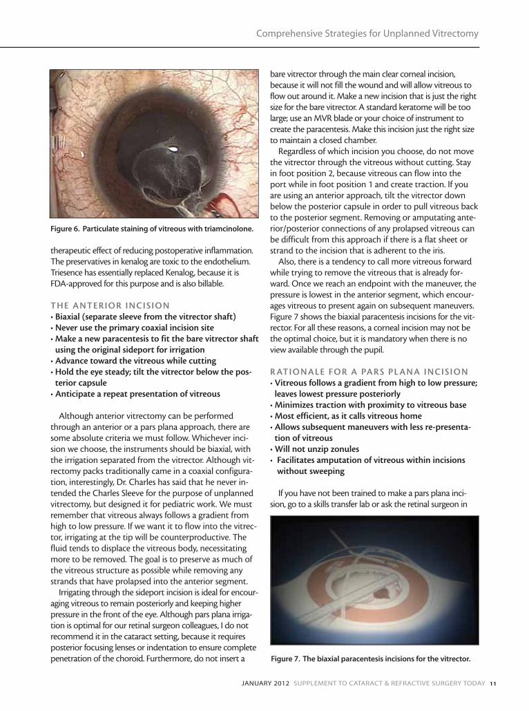
therapeutic effect of reducing postoperative inflammation.The preservatives in kenalog are toxic to the endothelium.Triesence has essentially replaced Kenalog, because it isFDA-approved for this purpose and is also billable.
THE ANTERIOR INCISION• Biaxial (separate sleeve from the vitrector shaft)• Never use the primary coaxial incision site• Make a new paracentesis to fit the bare vitrector shaft
using the original sideport for irrigation• Advance toward the vitreous while cutting• Hold the eye steady; tilt the vitrector below the pos-
terior capsule• Anticipate a repeat presentation of vitreous
Although anterior vitrectomy can be performedthrough an anterior or a pars plana approach, there aresome absolute criteria we must follow. Whichever inci-sion we choose, the instruments should be biaxial, withthe irrigation separated from the vitrector. Although vit-rectomy packs traditionally came in a coaxial configura-tion, interestingly, Dr. Charles has said that he never in-tended the Charles Sleeve for the purpose of unplannedvitrectomy, but designed it for pediatric work. We mustremember that vitreous always follows a gradient fromhigh to low pressure. If we want it to flow into the vitrec-tor, irrigating at the tip will be counterproductive. Thefluid tends to displace the vitreous body, necessitatingmore to be removed. The goal is to preserve as much ofthe vitreous structure as possible while removing anystrands that have prolapsed into the anterior segment.
Irrigating through the sideport incision is ideal for encour-aging vitreous to remain posteriorly and keeping higherpressure in the front of the eye. Although pars plana irriga-tion is optimal for our retinal surgeon colleagues, I do notrecommend it in the cataract setting, because it requiresposterior focusing lenses or indentation to ensure completepenetration of the choroid. Furthermore, do not insert a
bare vitrector through the main clear corneal incision,because it will not fill the wound and will allow vitreous toflow out around it. Make a new incision that is just the rightsize for the bare vitrector. A standard keratome will be toolarge; use an MVR blade or your choice of instrument tocreate the paracentesis. Make this incision just the right sizeto maintain a closed chamber.
Regardless of which incision you choose, do not movethe vitrector through the vitreous without cutting. Stayin foot position 2, because vitreous can flow into theport while in foot position 1 and create traction. If youare using an anterior approach, tilt the vitrector downbelow the posterior capsule in order to pull vitreous backto the posterior segment. Removing or amputating ante-rior/posterior connections of any prolapsed vitreous canbe difficult from this approach if there is a flat sheet orstrand to the incision that is adherent to the iris.
Also, there is a tendency to call more vitreous forwardwhile trying to remove the vitreous that is already for-ward. Once we reach an endpoint with the maneuver, thepressure is lowest in the anterior segment, which encour-ages vitreous to present again on subsequent maneuvers.Figure 7 shows the biaxial paracentesis incisions for the vit-rector. For all these reasons, a corneal incision may not bethe optimal choice, but it is mandatory when there is noview available through the pupil.
R ATIONALE FOR A PAR S PL ANA INCISION• Vitreous follows a gradient from high to low pressure;
leaves lowest pressure posteriorly• Minimizes traction with proximity to vitreous base• Most efficient, as it calls vitreous home• Allows subsequent maneuvers with less re-presenta-
tion of vitreous• Will not unzip zonules• Facilitates amputation of vitreous within incisions
without sweeping
If you have not been trained to make a pars plana inci-sion, go to a skills transfer lab or ask the retinal surgeon in
JANUARY 2012 SUPPLEMENT TO CATARACT & REFRACTIVE SURGERY TODAY 11
Comprehensive Strategies for Unplanned Vitrectomy
Figure 7. The biaxial paracentesis incisions for the vitrector.
Figure 6. Particulate staining of vitreous with triamcinolone.

your area if you can sit in and learn how to do it properly.Practice this incision ahead of time with eye bank or animaleyes, not in the heat of battle.
Again, the rationale for using the pars plana for vitrecto-my is that, because vitreous follows a pressure gradient fromhigh to low, we want to leave the lowest pressure posterior-ly. We also want to minimize traction by removing the vitre-ous closer to its base. If any vitreous follows the instrumentto the incision, it will be right near the vitreous base at thepars plana, rather than up at the corneal incision. This tech-nique is the most efficient, because it calls the vitreoushome. The pars plana technique also allows subsequentmaneuvers while minimizing re-presentation of the vitreous.It will not unzip the zonules when vitreous presents aroundthe lens equator by calling more vitreous forward throughthe defect. Finally, the pars plana approach facilitates ampu-tation of the vitreous within incisions. Although we weretaught to use a sweep from the sideport incision to dragentrapped vitreous away, this actually creates more tractionon the connection through the pupil rather than efficientlyfreeing the vitreous from incarceration in the wound. Istrongly discourage this practice.
VITRECTOMY GOAL S AND PAR A METER S• Particulate identify rather than "Weck" the incision to
test for vitreous loss• Do not sweep the incision (traction), but rather ampu-
tate posterior attachments• Confirm vitrectomy mode: irrigation, then cutting,
then aspiration in foot positions 1, 2, and 3• Maximize the cut rate to minimize traction• Minimize the flow rate (peristaltic pump machine) to
slow the action • Balance the lowest effective vacuum with the lowest
bottle height to maintain normotension• Remove all vitreous from the anterior chamber to
below the capsular plane • Minimally disrupt the posterior-segment vitreous
structure
Vitrectomy Mode: I/Cut/AThe best machine parameters for performing vitrectomy
are those settings that most effectively reduce vitreoretinaltraction and prevent followability. In foot position 1, youwill only be irrigating. In foot position 2, you will be irrigat-ing and cutting. Not until foot position 3 will you be irri-gating, cutting, and sucking, because you do not want toapply any vacuum to the vitreous unless you are cutting itat the same time. You must always follow the sequence ofirrigation, cutting, then aspiration when removing vitre-ous, so be certain not to use the “I/A Cut” setting, wherefoot position 1 is irrigation, foot position 2 is sucking, andfoot position 3 is cutting. This setting will prove usefulwhen followability is desired during removal of the residualcortex, once vitreous is likely out of the way.
Cutting Rate and FlowDr. Charles has coined the term port-based flow limiting,
which describes the goal of achieving the highest cut ratepossible, the lowest effective flow rate, and the lowest vacu-um that generates the removal of vitreous. As the vitreous isengaged, the faster you cut, the more you reduce traction.The highest cut rate possible on some older phacomachines is 400 cuts per minute, but newer models can go
12 SUPPLEMENT TO CATARACT & REFRACTIVE SURGERY TODAY JANUARY 2012
When the Room Gets Quiet
The WhiteStar Signature System (Abbott Medical Optics Inc.) hasadvanced capabilities for anterior vitrectomy. The surgeon has a choice oftwo high-speed guillotine vitrectomy handpieces, the 20-gauge and the 23-gauge vitrector, the latter of which can be paired with a separate irrigator forbimanual clear cornea or pars plana vitrectomy through sutureless incisionsas small as 1.0 mm. Either vitrector may be used with cut rates as high as2,500 cuts per minute for cutting vitreous.
The Signature phaco system offers dual-pump technology whereby thesurgeon may choose to use either a peristaltic or a venturi pump systemduring the anterior vitrectomy. Venturi pump systems are the industry stan-dard for posterior segment surgeons and may offer advantages over peri-staltic pumps during an anterior vitrectomy. Additionally, all the system’s vit-rectomy parameters are fully programmable (ASP, VAC and CPM) for eitherlinear or panel control. Practitioners may use two vitrectomy submodes:ICA (irrigate, cut, aspirate), which is used in the presence of vitreous; andIAC (irrigate, aspirate, cut), in the case of residual cortex or nuclear frag-ments that need to be engaged by the vitrector prior to cutting.
Finally, setup for the Signature vitrector is simple and quick. The scrub technician performs an automated, 5-secondprime cycle to prime and test the cutter before the surgeon enters the eye.
THE WHITESTAR SIGNATURE VITRECTOMY SYSTEM
The WhiteStar 20-gauge high-speed
vitrectomy probe.

as high as 5,000 cuts per minute. Faster cutting leads to lesstraction and a smoother removal of vitreous. Use the fastestcut rate your machine provides. The aspiration rate general-ly should be 15 to 20 cc/min (this is not independently seton Venturi machines, only peristaltic pumps) (see Summaryof Vitrectomy Parameters).
Linear Vs. Fixed Vacuum SettingVitreoretinal surgeons who work in the posterior seg-
ment every day prefer to use linear vacuum for vitrectomy,because they can control the intraoperative environment.Familiarity leads to facility, so that some physicians can usea graduated foot position 3 with ease. Anterior segmentsurgeons who perform vitrectomy rarely, however, are notas adept at these maneuvers and often get nowhere,because we are too timid to even venture into foot posi-tion 3. Staying in foot position 2 will accomplish nothing,because there will be continuous cutting and no suction. Ibelieve that we anterior segment surgeons should use apanel/fixed setting for vacuum instead of a linear vacuumsetting for vitrectomy. We want to find and maintain thelowest level of vacuum that moves the vitreous; there is noreason to use higher or lower vacuum once the vitreous ismoving. Such linearity allows us to either go pedal-to-the-metal in foot position 3 with suction, or come up intofoot position 2 without suction. The vacuum default set-tings on today's phaco machines are usually set at 150mm Hg, ideal for primary three-port vitrectomy. Inunplanned vitrectomy, however, the anterior segment sur-geon is almost always removing vitreous in a sea of disper-sive OVD. I find 150 mm Hg to be inadequate in a vis-coelastic-filled environment; I generally use 250 mm Hgfor 20-gauge and 350 mm Hg for 23-gauge vitrectomy.
IrrigationWe must keep the irrigation bottle moderately high in
order to maintain a normotensive eye. Again, the phaco
machine's default setting places the bottle low. The ap-propriate bottle height depends on the size of cannula weuse. Most anterior segment surgeons opt for a 23-gaugecannula. To control the IOP, I have my scrub nurse standwith one hand on the bottle's button, ready to raise it asneeded, and one hand on the vacuum button. I start thevacuum for 20-gauge vitrectomy at around 200 mm Hg,and I ask the scrub nurse to progress by 10-mm incre-ments to 250 mm Hg or stop once the vitreous begins tomove. As soon as I see movement toward the vitrectorport, I have the scrub nurse raise the bottle to prevent asoft eye. We continue in this manner until we reachhomeostatic normotension.
GET TING STARTEDVitrectomy Mode and Incisions
The first step is to confirm initial vitrectomy mode set-tings and then irrigate any bubbles out of the irrigation linewhile the vitrector is outside the eye. If choosing an anteri-or approach, make a second paracentesis a little less than180º away from the original sideport, large enough to fitthe bare vitrector needle. If you are planning to make a parsplana incision under topical anesthesia, make sure to firstplace a bleb of lidocaine with epinephrine sub-tenons overthe intended area of the incision for the patient's comfort.This step permits a small, fornix-based peritomy to bare thesclera without cautery (Figure 8).
Next, secure the primary wound if there is no vitreousloss. If vitreous is incarcerated in the incision, blocking awatertight closure, then fill the anterior chamber with OVDto approximate normal pressure. Avoid making the scleralincision at 3, 6, 9, and 12 o'clock so as to miss the ciliary ves-sels and nerves. Use a caliper to measure 3.5 mm posteriorto the limbus in the quadrant most convenient to your
JANUARY 2012 SUPPLEMENT TO CATARACT & REFRACTIVE SURGERY TODAY 13
Comprehensive Strategies for Unplanned Vitrectomy
• Less flow = less traction• Select settings to reduce vitreoretinal traction:
- highest cut rate available- flow rate: 20 cc/min for 20-g vitrectomy; 15 cc/min for
23-g- lowest vacuum to remove vitreous: 200 to 250 mm Hg
for 20-g or 350 to 500 for 23-g; use panel setting, notlinear
- irrigation bottle with 23-g cannula to balance for nor-motension, about 80 cm
*Note: These settings are for the Alcon system; settingsmay vary on different machines.
SUMMARY OF VITRECTOMY PARAMETERS*
Figure 8. This image shows the biaxial vitrectomy approach,
with irrigation through a clear corneal incision and the vitrec-
tor entering through a pars plana sclerotomy 3.5 mm posteri-
or to the limbus. A double-bite mattress or X suture with an
8–0 vicryl is used to close the direct-approach sclerotomy.

dominant hand, and avoid perforating vessels. Enter thepars plana with an MVR blade directed toward the opticnerve or the center of the eye until you can visualize theblade in the pupil and withdraw the blade. Insert the irri-gating 23-gauge chamber maintainer or a cannula throughthe original clear corneal incision paracentesis.
VitrectomyInsert the vitrector through the sclerotomy and view it
in the center of the iris through the pupil. Always makesure you can see the port during vitrectomy; the onlyexception is when performing a dry vitrectomy to makespace in a crowded anterior chamber. In this scenario,make sure the port is sideways or backwards to avoidengaging the posterior capsule while removing a smallamount of vitreous.
Remain in foot position 0 to this point. The momentyou can see the vitrector's port, go to foot position 2,which will cause irrigation and activate the cutter with-out allowing vacuum to build. You can now advance thecutter to the center of the pupil below the broken poste-rior capsule and start aspirating vitreous by engagingfoot position 3. Watch for any small bubble or particle toconfirm that the vacuum is high enough to move vitre-ous, and raise the vacuum slowly until you can visuallyconfirm the movement of vitreous into the vitrectorport. If you have set the vacuum on panel or fixed ratherthan linear, you can put the pedal to the metal and askthe scrub nurse to raise the vacuum slowly until it effec-tively begins to aspirate vitreous, usually at approximate-ly 250 mm Hg for 20-gauge or 350 mm Hg for 23-gaugevitrectomy. As soon as you see effective aspiration takingplace, you must raise the bottle to achieve homeostasis
for a normotensive globe. The scrub nurse will also adjustthis parameter on the fly and usually will find homeosta-sis at about 70 to 80 cm, depending on where thepatient's head is relative to the machine's cassette. Asnoted, the flow rate on machines with a peristaltic pumpusually stays at 20 cc/min for 20-gauge and 15 cc/min for23-gauge vitrectomy, and it need not be adjusted.
Completing VitrectomyKeep the pedal to the metal until it is evident that
there is no more prolapsed vitreous flowing to the port.If you must move the position of the vitrector, changefrom foot position 3 to 2 in order to cut with no suction,and then readjust the location of the port to address anyother areas of vitreous prolapse. Do not move the vitrec-tor around through vitreous without being in foot posi-tion 2 to avoid inadvertent traction.
When you think you have reached the end, hold thevitrector still and come to foot position 0. Remove theirrigation cannula, and irrigate some diluted Triesenceinto the anterior chamber. Again place the irrigatorthrough the paracentesis to disperse the Triesence, there-by confirming the complete absence of prolapsed vitre-ous. When extracting the vitrector from the eye, remainin cutting mode (foot position 2) until you have lost visu-alization under the iris. Come up into foot position 1 forthe brief interval that the vitrector is between the edge ofthe iris and when it exits the sclera, but go to foot posi-tion 0 right before you exit the incision, so as not to blowvitreous out of the incision. Retinal surgeons will continueirrigating until they are out of the eye, but that is becausethey have done a total vitrectomy. When cleaning thesclerotomy of any prolapsed vitreous, turn the cutter down
14 SUPPLEMENT TO CATARACT & REFRACTIVE SURGERY TODAY JANUARY 2012
When the Room Gets Quiet
The Stellaris PC Vision Enhancement System (Bausch + Lomb) features
next-generation vitreoretinal surgical technology in the form of an ultra-
high-speed cutter and the familiar lightweight microvit-style handpiece.
With an optimized duty cycle, the port of the handpiece is open at least
50% of the time, even at 5,000 cpm. This allows surgeons to effectively
remove vitreous at even the highest cut rates. Using the lightweight pneu-
matic cutter and 5,000 cpm with an optimized duty cycle, surgeons may
control the flow intuitively via the foot pedal. The programmable foot
pedal is wireless and has Dual-Linear control, which enables the surgeon to
vary the vacuum and cut rate. The vacuum on the Stellaris PC is controlled
by an advanced algorithm that produces accurate, smooth, and linear aspi-
ration that remains predictable at vacuum as low as 2 mm Hg. If the vitrectomy technique involves a pars plana approach,
the trocar tip’s long taper provides sufficient cutting surface. This tip is designed to permit a shallow angle of entry during
tunnel incisions. The trocar’s needle has been redesigned to be solid instead of hollow, which reduces the force of the inser-
tion and displaces less tissue.
THE STELLARIS PC VITRECTOMY SYSTEM
The Stellaris PC ultra-high-speed
vitrectomy probe.

toward the external lips of the wound while irrigating ex-ternally onto the incision, and sharply cut away any exter-nal strands before suturing. Never use a Weck-Cel sponge.
Immediately place a scleral plug or a temporary tiesuture to close the sclerotomy. If you used an anteriorapproach, confirm that the clear corneal incision closes andthat no strand of vitreous has followed your withdrawal ofthe vitrector needle. The eye is now closed and ready forfurther maneuvers, and the vitrectomy site is available incase vitreous presents again.
Wecking and Sweeping IncisionsFor generations, ophthalmic residents were told to use
a cyclodyalisis spatula to sweep from the paracentesis tojust inside the main incision to identify, release, andremove incarcerated vitreous. This practice causes signifi-cant traction on the posterior vitreous and should beabandoned, in my opinion. The preferred technique isparticulate identification and sharp cutting or, ideally,using a vitrector to amputate connections posteriorly atthe pupil margin (thus obviating the need for traction).Once the vitreous sheet either retracts to the posteriorsegment or is severed from the vitreous within thewound, it is safe to remove residual vitreous from theincision with a Weck-Cel sponge. Sponges should neverbe used to remove vitreous that is still attached posteri-orly, however, because they absorb vitreous strands andlead to traction and cause inflammation upon contactwith iris tissue. Any time you touch a sponge to the inci-sion, have scissors ready in the other hand so that, shouldyou discover vitreous, you can cut it without lifting orstretching the strands. Immediately cut off the vitreousat the plane of the sclera so that you do not pull it for-ward out of the wound. Then, you may address the vitre-ous in the optimal manner.
SUTURING OF DIRECT SCLEROTOMY (20-OR 23-GAUGE)
Older-style vitrectors had a flat tip that required anopening slightly larger than their gauge. The vitrector onthe INFINITI Vision System (Alcon Laboratories, Inc.),which I use, has a rounded tip that makes the incisionrequirement true to size and its entry less traumatic.
Closing a 20-gauge sclerotomy requires a two-bite inci-sion. A 23-gauge direct MVR incision may require only onebite to be watertight. Prior to suturing the sclerotomy, irri-gate the lips of the incision while using the vitrector in footposition 2 (cutting) to clear any incarcerated strands of vit-reous. Or, shear any vitreous within the sclerotomy withscissors. Never use a cellulose sponge at a pars plana scle-rotomy. Then, I prefer to make an X or a mattress suture of8-0 Vicryl (Ethicon, Inc.). It may be best to orient the nee-dle from a posterior to anterior position when taking the1/2 thickness bite to ensure that the needle's point does
not come out too far posteriorly. It is not necessary tobury the knot, as it will be covered by the Tenons andconjunctival fornix-based flap that you will suture inplace with the same absorbable suture. Tie the knot onthe inside of the flap for the patient's comfort (Figure 9).The sclerotomy should be watertight, and no blebshould form from pressurizing the globe.
SUTURELE SS VITRECTOMY (23- OR 25-GAUGE TROCAR CANNUL A SYSTE M)
Because we want a noncoincident incision in the con-junctiva and the sclera, it is difficult to make a suturelessincision without the help of a cannula to keep the twoopenings lined up. Finding the openings with the vitrec-tor via an MVR blade-created shelved entry is very chal-lenging. The cannula is a hollow tube that often encasesa sharp MVR blade. You withdraw the blade once youhave completed the incision, but the tube remains as aconduit for the vitrector needle, which you can repeated-ly insert as needed until the case is completed.
A trocar cannula system offers another advantage. Be-cause it protrudes into the vitreous cavity, the vitrectorprobe never gets close to the retinal surface, as it does wheninserted through a bare sclerotomy. This design provides amargin of safety upon entry and exit. As they becomesharper and require less pressure for entry, trocars will bethe entry method of choice.
Using a trocar cannula system requires a firm, intact eyewith closed incisions to handle the pressure applied to theglobe upon entry. If incisions are already present, do notsimply close them with hydration, but suture them closed,because the force of inserting the trocar will likely cause irisprolapse if they are not entirely secure. A soft eye will riskchoroidal detachment or hemorrhage with this procedure.If vitreous has been lost, you will not be able to close theincisions, even with sutures, so a direct MVR entry withsuturing would be safest.
Fill the eye with BSS or OVD until it is firm, whileascertaining that the cataract incisions are closed. Pull the
JANUARY 2012 SUPPLEMENT TO CATARACT & REFRACTIVE SURGERY TODAY 15
Comprehensive Strategies for Unplanned Vitrectomy
Figure 9. A suture under the conjunctival flap.

conjunctiva away from the site of puncture 3.5 mm backfrom the limbus and initiate a partial-thickness scleraltunnel with the trocar parallel to the limbus. Travel 2 mm,then turn the device perpendicular to the sclera andpuncture the sclera in the direction of the optic nerve,driving the trocar cannula system through the eye wall.Once the trocar is in place, remove the MVR blade andleave the seated cannula for the vitrector's insertion.
Dr. Charles does not recommend a 25-gauge vitrectorfor anterior vitrectomy, because he says it is too flexible touse without akinesia (topical anesthesia). My experience iswith 20- and 23-gauge vitrectors only (see Pars PlanaInstrumentation).
RE SIDUAL CORTE X RE MOVAL• Perform a “dry technique” under OVD without irri-gation• Bimanual I/A imparts a small risk of incarcerating
vitreous• Use the vitrector on I/A-Cut mode (not I-Cut-A
default) for followability • Coaxial I/A not recommended unless optic is cap-
tured• Prevent chamber collapse during removal of instru-
ments: vitreous will follow the path of lowest pres-sure and present again
Once you have removed the vitreous and lens particles,you are left with cortex. It is incumbent upon us to cleanthe capsular fornix thoroughly to avoid inflammation,
prevent a poor-quality view with fluffed cortex in the post-operative period, and ultimately to reduce the risk of CME.We have three viable options to accomplish this goal.
The safest way to clean the capsular bag (albeit, notthe most efficient) is to empty it with a dry technique.This means maintaining and expanding the chamber andthe capsular fornix with cohesive viscoelastic and packingin the dispersive agent to cover the capsule or the zonu-lar defect. Without irrigation, use a 3- or 5-mL syringewith a 26-gauge cannula (or a Simcoe cannula system) tosuction out residual cortex. There should be just enoughBSS in the syringe to be able to reflux in case the capsuleor vitreous become incarcerated. When the tip is occlud-ed with cortex, a manual application of suction strips itcleanly away if the anterior leaflet leads. A 23-gauge can-nula is useful for dense cortical or epinuclear material,but we must be careful not to attract the capsule intothe port's opening where it will rip. Replace the cohesiveOVD as needed to keep the capsular fornix expandedand the eye normotensive as cortex removal proceeds.This technique avoids the risk of displacing more vitreouswith irrigation and inviting it forward, or pushing frag-ments to the back of the eye.
Another way to remove residual cortex is via bimanualI/A. Some surgeons prefer this strategy because of its effi-ciency. Bimanual I/A is a much better approach for re-moving residual cortex than coaxial I/A, which involvesirrigating in the same area from where you are trying toremove the cortex. The biaxial approach conducts theirrigation anteriorly, which continues to encourage vitre-ous to stay back. You can direct the separate aspirationinto the cortex and keep it fully occluded. Use a secondsideport paracentesis with this technique so the chamberremains watertight with a closed main clear corneal inci-sion. Although unlikely, if you encounter vitreous, therewill be a tractional event that you can mitigate by imme-diately stopping aspiration while holding the aspirationhandpiece steady, exchanging the irrigation handpiecefor an intraocular scissors, and cutting the incarceratedvitreous strand. This strategy will relieve the tractionbefore you withdraw the aspiration handpiece and haveto deal with the prolapsed vitreous again with vitrector.
A safer technique than bimanual I/A is to use the vit-rector on a different setting. Every phaco machine has adifferent way to switch from irrigation-cut-aspiration,which must always be used for vitreous, to irrigation-aspiration-cut. The point is to discourage followabilitywhen removing vitreous, although followability is neces-sary for removing cortex. Using the vitrector on I-cut-Amode does not work for removing cortex, because itcontinually chops it off and never allows the cortex toflow into the port. Because you cannot strip it out of thesulcus, you end up following the cortex into the sulcusand eating capsule. If you switch to I-A-cut mode, then
16 SUPPLEMENT TO CATARACT & REFRACTIVE SURGERY TODAY JANUARY 2012
When the Room Gets Quiet
Pars Plana Instrumentation• Standard 20-gauge
- Readily available- Conjunctival flap - Sutured incision
• 25-gauge- Too flexible for topical anesthesia
• 23-gauge- Sutureless with excellent sealing if oblique incision- Trocar cannula optional, direct 23-gauge MVR entry
preferred if the globe is not intact
A trocar requires more pressure to insert than a directMVR blade and should only be used when incisions canbe closed with a normotensive or firm eye. For a suture-less incision, a limbal parallel circumferential scleral tunnelprecedes the perpendicular entry into the vitreous cavity.The conjunctiva is pulled aside so the conjunctival entrywound is not coincidental with the scleral entry.
PARS PLANA INSTRUMENTATION
you can remove the cortex in foot position 2 when youare sure the vitreous is out of the way. If you think vitre-ous has emerged, you can immediately go down to footposition 3 and cut it off. This approach is safer than I/A inthat it relieves a tractional event instantly. Finally, makesure to prevent the chamber from collapsing while youremove the instruments after a vitreous prolapse, anopen hyaloid, or even just an open capsule.
IMPL ANTING AN IOLInspect Prior to Implantation
To make sure the anterior segment is cleaned of all cor-tex, you may need to retract the iris to fully view the bagfornix. Reinstill Triesence and then rinse it away to besure there is no vitreous present. Check that the pupil isround and that the incisions are sealed. Verify the statusand size of the capsulorhexis, and evaluate the extent ofthe posterior capsular tear and whether there is a truecontinuous curvilinear tear. Be aware of any retainednuclear fragment below the posterior capsule, and makecertain no fragments are hiding anteriorly under the irisor subincisionally.
IOL Selection and PlacementWhen there is an uncontrolled break in the posterior
capsule but the anterior CCC is intact, I advocate im-planting a three-piece lens in the sulcus and capturingthe optic through the anterior capsulorhexis. Only whenthe posterior tear is converted to a true PCCC will I placeany IOL (ideally, a single-piece one) in the capsular bag.Regardless of its location, shape, or size, an unconvertedrent may extend in response to even minor pressure. Ifthe PCCC is 4 to 5 mm and centrally located, then we canplace a three-piece lens in the bag and perform an opticcapture through the PCCC into Berger's space. This is theideal outcome.
If there was a zonular break in the presence of an intactposterior capsule and an intact anterior capsulorhexis with astable and centered bag, I will use a standard or modifiedcapsular tension ring or a capsular tension segment suturedto the sclera.
If the posterior capsule is intact but there is a break inthe anterior capsulorhexis, I will place a single-piece IOLin the bag (as long as I know the haptics will stay se-questered in the bag and not pop out into the sulcus).
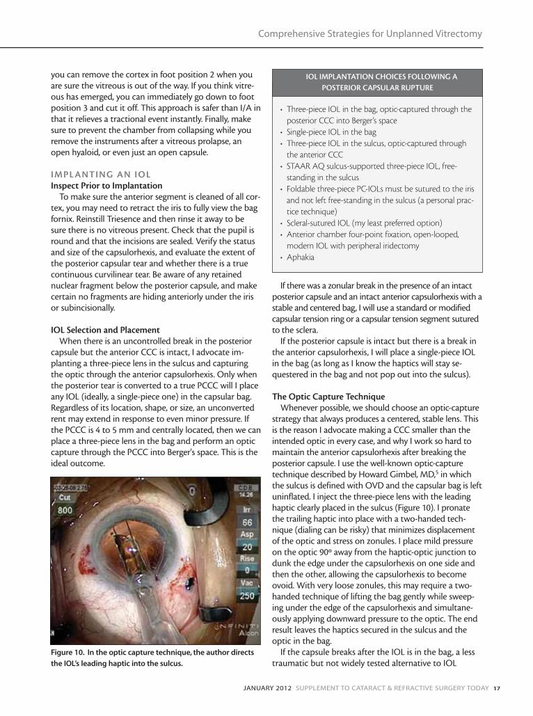
The Optic Capture Technique Whenever possible, we should choose an optic-capture
strategy that always produces a centered, stable lens. Thisis the reason I advocate making a CCC smaller than theintended optic in every case, and why I work so hard tomaintain the anterior capsulorhexis after breaking theposterior capsule. I use the well-known optic-capturetechnique described by Howard Gimbel, MD,5 in whichthe sulcus is defined with OVD and the capsular bag is leftuninflated. I inject the three-piece lens with the leadinghaptic clearly placed in the sulcus (Figure 10). I pronatethe trailing haptic into place with a two-handed tech-nique (dialing can be risky) that minimizes displacementof the optic and stress on zonules. I place mild pressureon the optic 90º away from the haptic-optic junction todunk the edge under the capsulorhexis on one side andthen the other, allowing the capsulorhexis to becomeovoid. With very loose zonules, this may require a two-handed technique of lifting the bag gently while sweep-ing under the edge of the capsulorhexis and simultane-ously applying downward pressure to the optic. The endresult leaves the haptics secured in the sulcus and theoptic in the bag.
If the capsule breaks after the IOL is in the bag, a lesstraumatic but not widely tested alternative to IOL
JANUARY 2012 SUPPLEMENT TO CATARACT & REFRACTIVE SURGERY TODAY 17
Comprehensive Strategies for Unplanned Vitrectomy
Figure 10. In the optic capture technique, the author directs
the IOL’s leading haptic into the sulcus.
• Three-piece IOL in the bag, optic-captured through theposterior CCC into Berger’s space
• Single-piece IOL in the bag• Three-piece IOL in the sulcus, optic-captured through
the anterior CCC• STAAR AQ sulcus-supported three-piece IOL, free-
standing in the sulcus • Foldable three-piece PC-IOLs must be sutured to the iris
and not left free-standing in the sulcus (a personal prac-tice technique)
• Scleral-sutured IOL (my least preferred option)• Anterior chamber four-point fixation, open-looped,
modern IOL with peripheral iridectomy• Aphakia
IOL IMPLANTATION CHOICES FOLLOWING APOSTERIOR CAPSULAR RUPTURE

exchange is the forward optic capture technique. Youmay use the optic-capture concept to your advantage byforward-capturing the optic in front of the intact capsu-lorhexis while leaving the haptics in the bag (a personalpractice technique also described by Howard Gimbel, MD).
If neither the anterior capsulorhexis nor the posteriorcapsule is intact, you must decide between implanting astandalone or sutured sulcus-fixated IOL or a modernanterior-chamber implant. In my opinion, it is impor-tant to secure any IOL that is not a true sulcus-support-ed lens (the only such lens that currently has FDAapproval is the AQ series from STAAR SurgicalCompany). Single-piece IOLs are incompatible with sul-cus fixation. The clinical study of the AcrySof naturalsingle-piece lens was conducted with the lens implanta-tion in the capsular bag only. There are no clinical datato demonstrate its safety and effectiveness for place-ment in the ciliary sulcus, and plenty of evidence that itwill cause pigmentary dispersion glaucoma by chaffingthe posterior iris.6 Standard three-piece IOLs are toosmall for the sulcus. They will move around and causeinflammation, CME, and pigment dispersion in a signifi-cant number of eyes. Liliana Werner, MD, PhD, has shownthat the size of the sulcus varies. It is not proportional toaxial length or white-to-white measurements, and it isoften larger than 12.5 or 13.0 mm, which is the hapticdiameter of a standard three-piece lens. Furthermore, ifthere is a little hole in the posterior capsule or particularlyin the zonules, the sulcus-implanted lens will eventuallyfind its way through it. Having practiced for almost 30years, I have seen this complication on referral many times.You can purchase the STAAR lens (available in the UnitedStates) for inventory. It is made of biocompatible siliconewith a rounded edge. The lens' stiff elastimide haptics are13.5 mm apart, making it our only reliable choice forunsupported sulcus implantation.
It is your choice whether to anchor a standard three-pieceIOL in the sulcus with one or two iris sutures or place aPMMA lens with eyelets as a scleral-sutured lens. (I believethe glued scleral-fixated method has not yet stood the testof time.) In the presence of no intact anterior or posteriorcapsule, especially after a long case, my preference is to placean anterior-chamber lens through a scleral tunnel, havingclosed my clear corneal incision. In this scenario, I make aperipheral iridectomy with the vitrector. After instillingMiochol E, I use I/A/Cut mode with raised vacuum and alowered cut rate to produce a neat peripheral hole in the irisbefore I place the lOL (taking care to not tuck the iris).‘White-to-white plus one' is still the standard for sizing theIOL in this maneuver. A review of the ophthalmic literatureshows that there is no decrease in the quality of long-termoutcomes with implanting IOLs in the anterior chamberversus suturing them in the posterior chamber.
RE MOVING OVD AND CLOSING THE EYEIf you do not capture the optic in an eye that has lost
vitreous, then be very cautious in removing the viscoelas-tic at the end of the case. I usually do this manually inthese eyes by irrigating and then slightly burping the inci-sion to let some of the OVD out. I irrigate some more, andthen I use a manual push/pull technique through theparacenteses to remove all of the viscoelastic that I reason-ably can. Be careful not to lower the pressure in the anteri-or chamber by overzealously removing the viscoelastic,thereby allowing vitreous to re-present. Of course, you canremove the viscoelastic more aggressively if you have usedan optic-capture technique. I prefer not to use Healon5(Abbott Medical Optics Inc.) in complicated cases,because even a little of this OVD left behind can cause asevere rise in IOP in the postoperative period. VISCOATOVD is the most forgiving agent, since it is of a smallermolecular weight. Any OVD that remains behind a proper-ly captured optic is of no concern, as it will not have accessto the trabecular meshwork and will slowly be absorbedwithout incident.
Miochol E (acetylcholine) will provide miosis, protect theIOL's position, and ensure a round pupil with no peak,which would indicate a vitreous wick.
A final minim of Triesence and rigorously closed incisionswith dry tunnels will confirm that no incarcerated vitreousis lurking. Even if the clear corneal incision seals flawlessly, asuture is indicated (Figure 11) if a follow-up retinal proce-dure may be needed due to retained lens material.
Before I exit the eye, I like to make sure the pupil is wellrounded and not stuck anywhere or held back by vitreous.So, I instill Miochol-E and watch the pupil come down (seePharmacology). Then, because a break the vitreous face sig-nificantly increases the risk of endopthalmitis, I always pro-phylax the eye with intracameral moxifloxacin (an off-labeluse).7 Immediately postoperatively, I provide one dose of
18 SUPPLEMENT TO CATARACT & REFRACTIVE SURGERY TODAY JANUARY 2012
When the Room Gets Quiet
Figure 11. A suture is always placed in the direct entry sclero-
tomy. Plan to also suture the cataract incision if there is the
possibility of retained lens material that may necessitate a
secondary retinal procedure.

Avalox (Bayer Corporation), which is a systemic form ofmoxifloxacin (400 mg P.O.) that crosses the blood-retinalbarrier.
Furthermore, patients with open posterior capsules needto use a longer taper of topical steroid than routine patientsand remain on topical NSAIDs for a longer period (off label)to prevent CME. Hypotensive medications are also a goodidea, because we often leave some viscoelastic behind, andthese eyes often experience a postoperative rise in IOP. Iprescribe Diamox (Wyeth Pharmaceuticals) if there is nohistory of sulfa allergy (10% of people who have sulfa allergyare allergic to Diamox), and then I carefully follow thepatient postoperatively.
CONCLUSIONWarn vitrectomy patients to expect floaters postopera-
tively and to call and identify themselves as surgicalpatients if they experience pain or decreasing vision at anytime (see Postoperative Follow-Up). Treat any IOP spikesaggressively and follow them closely, especially over the first48 hours. Teach patients to check their peripheral vision athome. I highly recommend performing a scleral indentedretinal examination and/or a retinal subspecialty consulta-tion within 1 to 2 weeks after the surgery to be absolutelysure there are no holes or tears in the peripheral retina thatneed to be treated. Closely monitor these patients for CMEwith an Amsler grid at home. Also, optical coherencetomography is indicated. Often, vitrectomy patients canachieve 20/20 UCVA on the first postoperative day, and thefloaters will disappear with time. It is critical to inform thesepatients of their increased risk of developing a retinal tearor detachment in the future, in addition to the increasedrisk of glaucoma and CME. Educate these individuals aboutthe signs of a retinal tear or detachment so they may bevigilant for them, and inform them of the need for periodictesting for glaucoma. Finally, if an eye has retained lens
material, then you must refer the patient to a retinal spe-cialist in a timely fashion for a possible definitive treatment.A recent meta-analysis of the literature shows patients farebest if the intervention is from 3 days to 7 days after sur-gery that was complicated by retained lens material.8
When we prepare in advance for a loss of vitreous with acomprehensive strategy and we treat the patient with logicand care, we can often achieve optimal outcomes, even incomplicated cataract cases. ■
Lisa B. Arbisser, MD, is in private practice with EyeSurgeons Assoc. PC, located in the Iowa and Illinois QuadCities. Dr. Arbisser is also an adjunct associate professor at theJohn A. Moran Eye Center of the University of Utah in SaltLake City. She acknowledged no financial interest in the prod-ucts or companies she mentioned. Dr. Arbisser may bereached at (563) 323-2020; [email protected].
The author wishes to thank Steve Charles, MD, for his inspi-ration and guidance and for sharing his vast knowledge. Shealso thanks her partner, Michael Howcroft, MD, for his sup-port and ongoing teaching efforts. Additionally, the authorwishes to thank the following individuals for their time andcontributions to teaching: Amir Arbisser, MD; Brian Berger,MD; Scott Burke, MD; Doug Fanny, PhD; Howard Gimbel,MD; Ricardo Glikin, MD; Rupert Menapace, MD; AnitaNevyas-Wallace, MD; Aron Rose, MD; Kevin Waltz, OD, MD;and Liliana Werner, MD, PhD.
Finally, she thanks her many peers and mentors who havehelped to teach the skills transfer labs: Alan Aker, MD; FrancisApplegate, MD; Rosa Braga-Mele, MD; Jorge Calzada, MD;James A. Davison, MD; Luther L. Fry, MD; Harry B.Grabow, MD; Warren E. Hill, MD; Michael J. Howcroft, MD;Jay I. Lippman, MD; Cathleen M. McCabe, MD; Kevin Miller,MD; Anita Nevyas-Wallace, MD; Kenneth J. Rosenthal, MD;Jack A. Singer, MD (deceased); Michael E. Snyder, MD; DeeStephenson, MD; Jeffrey Whitman, MD; and RichardTipperman, MD.
For further reading:1. Arbisser LB. Anterior vitrectomy for the anterior segment surgeon. Focal points. TheAmerican Academy of Ophthalmology; March 2009.2. Arbisser LB, Charles S, Howcroft M, Werner L. Management of vitreous loss anddropped nucleus during cataract surgery. Ophthalmol Clin North Am. 2006;19(4):495-506.
1. Lincoff H, Zweifach P, Brodie S, et al. Intraocular injection of lidocaine. Ophthalmology.1985;92(11):1587-1591.2. Osher RH. Slow motion phacoemulsification approach. J Cataract Refract Surg.1993;19(5):667.3. Arbisser LB, Charles S, Howcroft M, Werner L. Management of vitreous loss and droppednucleus during cataract surgery. Ophthalmol Clin North Am. 2006;19(4):495-506.4. Spence DJ, Peyman GA. A new technique for the vital staining of the corneal endothe-lium. Invest Ophthalmol. 1976;15(12):1000-1002.5. Gimbel HV, DeBroff BM. Intraocular lens optic capture. J Cataract Refract Surg.2004;30(1):200-206.6. Chang DF, Masket S, Miller KM, et al; ASCRS Cataract Clinical Committee. Complicationsof sulcus placement of single-piece acrylic intraocular lenses: recommendations for backupIOL implantation following posterior capsule rupture. J Cataract Refract Surg.2009;35(8):1445-1458.7. Arbisser LB. Safety of intracameral moxifloxacin for prophylaxis of endophthalmitis aftercataract surgery. J Cataract Refract Surg. 2008;34(7):1114-1120.8. Vanner EA, Stewart MW. Vitrectomy timing for retained lens fragments after surgery forage-related cataracts: a systematic review and meta-analysis. Am J Ophthalmology.2011:152(3):345-357.
JANUARY 2012 SUPPLEMENT TO CATARACT & REFRACTIVE SURGERY TODAY 19
Comprehensive Strategies for Unplanned Vitrectomy
• Warn patients to expect floaters• Prophylax and treat any pressure spikes• If the sclera is indented, a scleral indented retinal exami-
nation and/or retinal consultation are mandatory• Close monitoring for CME• Disclosure to patients
POSTOPERATIVE FOLLOW-UP
• Miochol-E• Antibiotic prophylaxis with intracameral moxifloxacin
and Avalox 400 mg, PO prior to discharge (my choice,though off-label)
• Anti-inflammatory: topical NSAID beyond 1 month anda long taper of steroid
• Hypotensive meds: Diamox (if no sulfa allergy)
PHARMACOLOGY