supplemental figure 1. motor learning is preserved in lrrk2 p.g2019s rats
DESCRIPTION
Supplemental Figure 1. Motor learning is preserved in LRRK2 p.G2019S rats. - PowerPoint PPT PresentationTRANSCRIPT
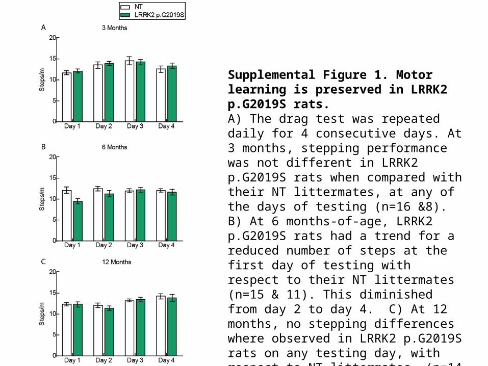
Supplemental Figure 1. Motor learning is preserved in LRRK2 p.G2019S rats.A) The drag test was repeated daily for 4 consecutive days. At 3 months, stepping performance was not different in LRRK2 p.G2019S rats when compared with their NT littermates, at any of the days of testing (n=16 &8). B) At 6 months-of-age, LRRK2 p.G2019S rats had a trend for a reduced number of steps at the first day of testing with respect to their NT littermates (n=15 & 11). This diminished from day 2 to day 4. C) At 12 months, no stepping differences where observed in LRRK2 p.G2019S rats on any testing day, with respect to NT littermates, (n=14 & 11 per group).
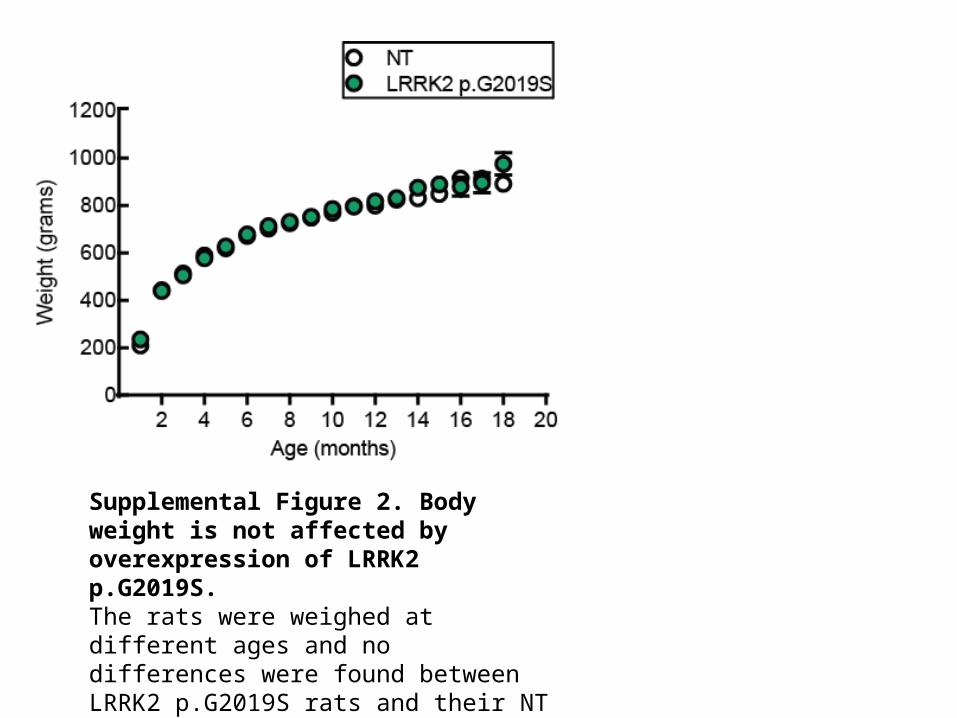
Supplemental Figure 2. Body weight is not affected by overexpression of LRRK2 p.G2019S. The rats were weighed at different ages and no differences were found between LRRK2 p.G2019S rats and their NT littermates, at any age-point up to 12 months (n=9-21 per group).
Supplemental Figure 3. LRRK2 expression.

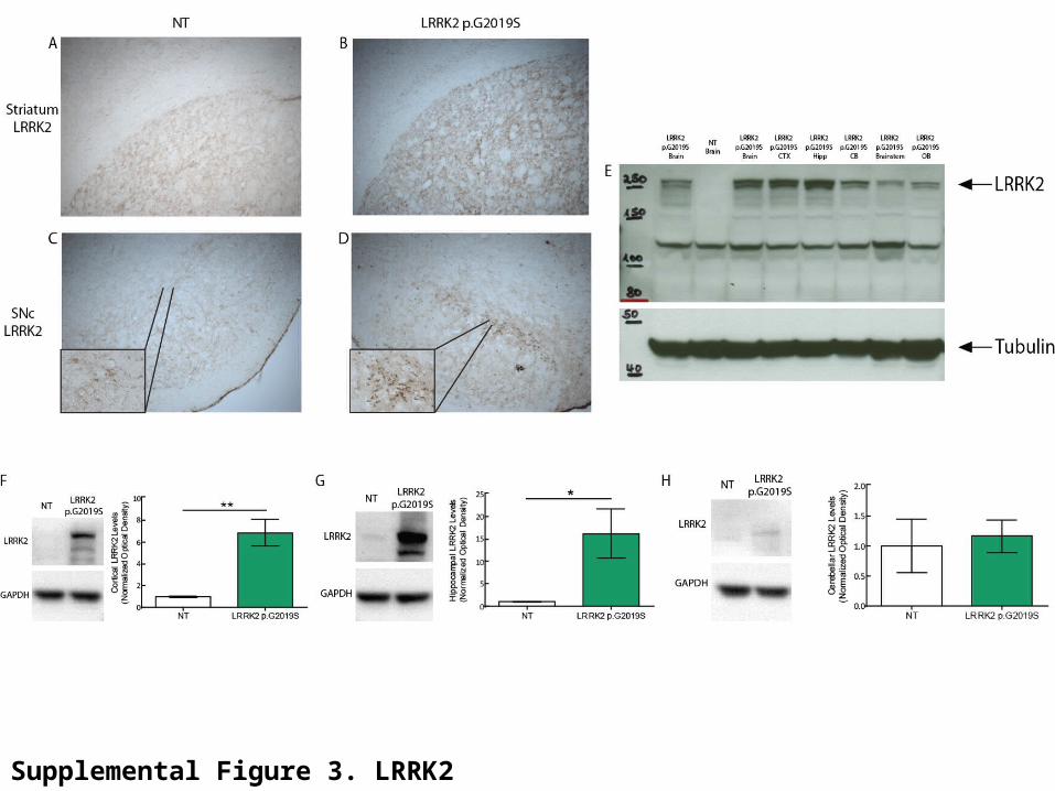
Supplemental Figure 3. LRRK2 expression.A-D) Animals at the age of 3 months were processed for LRRK2 immunohistochemistry. DAB staining appeared denser in the dorsolateral striatum of LRRK2 p.G2019S rats (B) in comparison with their NT littermates (A). In NT animals we detected few LRRK2-positive cells in the substantia nigra compacta (C) while more cells were stained in LRRK2 p.G2019S rats, confirming expression of the protein in this area (D). E) Western blot analysis revealed that 5-month old LRRK2 p.G2019S rats express full-length LRRK2, as probed in whole brain lysates as well as cortex (CTX), hippocampus (Hipp), cerebellum (CB), brainstem and olfactory bulb (OB) tissue samples. F-H) Animals at the age of 12-13 months were sacrificed, different brain regions rapidly microdissected and the tissue subjected to Western blot analysis for measurement of human LRRK2 protein levels. A strong increase in expression is evident in the cortex (A; **p<0.01) and hippocampus (B; *p=0.05) of LRRK2 p.G2019S rats as compared to NT littermates, but not in the cerebellum (n=3 per group, unpaired Student’s t-test).