supplemental information - pnas · supplemental information li tan1,4, ... frin-2 (c) and frin-3...
TRANSCRIPT
S1
Development of covalent inhibitors that can overcome resistance to first generation FGFR kinase
inhibitors
Supplemental Information
Li Tan1,4,†, Jun Wang5†, Junko Tanizaki5,6†, Zhifeng Huang11,12†, Amir R. Aref2,5†, Maria Rusan5,13,
Su-Jie Zhu14, Yiyun Zhang3,10, Dalia Ercan5,6, Rachel G. Liao5,9, Marzia Capelletti5,6, Wenjun
Zhou1,4, Wooyoung Hur1,4,15, NamDoo Kim17, Taebo Sim15,16, Suzanne Gaudet2,4,8, David A.
Barbie5, Jing-Ruey Joanna Yeh3,10, Cai-Hong Yun14, Peter S. Hammerman5,9*, Moosa
Mohammadi11*, Pasi A. Jänne5,6,7*, Nathanael S. Gray1,4*
1Department of Biological Chemistry and Molecular Pharmacology, 2Department of Genetics, 3Department of Medicine, Harvard Medical School, Boston, MA 02115 (USA); 4Department of
Cancer Biology, 5Department of Medical Oncology, 6The Lowe Center for Thoracic Oncology, 7Belfer Institute for Applied Cancer Science, 8Center for Systems Cancer Biology, Dana Farber
Cancer Institute, Boston, MA 02215 (USA); 9Cancer program, Broad Institute of Harvard and MIT,
Cambridge, MA 02141 (USA); 10Cardiovascular Research Center, Massachusetts General
Hospital, Charlestown, MA 02129 (USA); 11Department of Biochemistry and Molecular
Pharmacology, New York University School of Medicine, New York, NY 10016 (USA); 12School
of Pharmacy, Wenzhou Medical University, Wenzhou, 325035 (CN); 13Department of Clinical
Medicine, Aarhus University, Aarhus, 8200 (DK); 14 Peking University Health Science Center,
Beijing, 100191 (CN); 15Chemical Kinomics Research Center, Korea Institute of Science and
Technology, Seoul, 136-791 (KR); 16KU-KIST Graduate School of Converging Science and
Technology, Seoul, 136-713 (KR); 17New Drug Development Center, Daegu-Gyeongbuk Medical
Innovation Foundation, Daegu, 706-010 (KR). †These authors contributed equally.
*Correspondence: [email protected] (PSH),
[email protected] (MM), [email protected] (PAJ),
[email protected] (NSG)
Contents: Figure S1-S8 (page S2-S11); Table S1-S3 (page S12-S15); Synthetic procedures and
spectral data of FIIN-2, FIIN-3, FRIN-2 and FRIN-3 (page S16-20); Supplemental reference:
(page S20).

S2
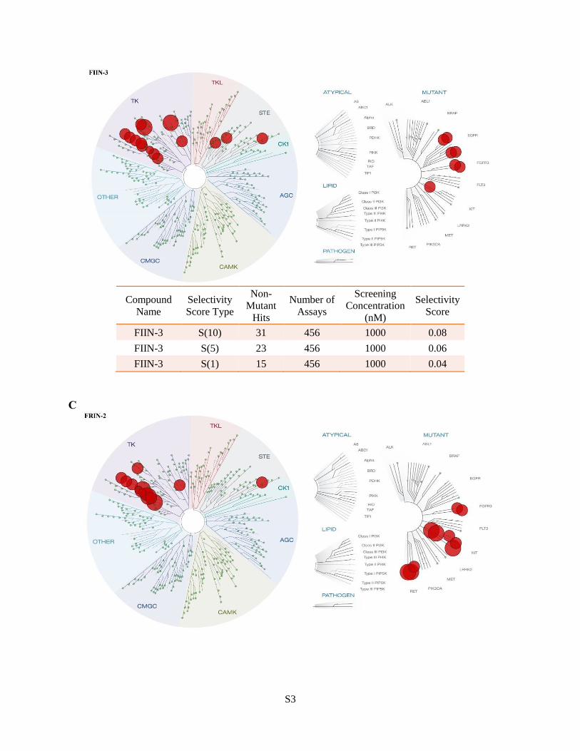
Figure S1. KinomeScan™ kinase selectivity profiles for FIIN2 (A), FIIN3 (B), FRIN-2 (C) and
FRIN-3 (D). All four inhibitors were profiled at a concentration of 1 µM against a diverse panel
of more than 456 kinases (354 non-mutants kinases) by Ambit Biosciences. Scores for primary
screen hits are reported as a percent of the DMSO control (% control). For kinases where no score
is shown, no measurable binding was detected. The lower the score, the lower the Kd is likely to
be, such that scores of zero represent strong hits. Scores are related to the probability of a hit but
are not strictly an affinity measurement. At a screening concentration of 1.0 µM, a score of less
than 10% implies that the false positive probability is less than 20% and the Kd is likely less than
100 nM. A score between 1% and 10% implies that the false positive probability is less than 10%,
although it is difficult to assign a quantitative affinity from a single-point primary screen. A score
of less than 1% implies that the false positive probability is less than 5% and the Kd is most likely
less than 100 nM. (1, 2)
A
Compound
Name
Selectivity
Score Type
Non-
Mutant
Hits
Number of
Assays
Screening
Concentration
(nM)
Selectivity
Score
FIIN-2 S(10) 23 456 1000 0.06
FIIN-2 S(5) 17 456 1000 0.04
FIIN-2 S(1) 10 456 1000 0.03
B
S3
Compound
Name
Selectivity
Score Type
Non-
Mutant
Hits
Number of
Assays
Screening
Concentration
(nM)
Selectivity
Score
FIIN-3 S(10) 31 456 1000 0.08
FIIN-3 S(5) 23 456 1000 0.06
FIIN-3 S(1) 15 456 1000 0.04
C

S4
Compound
Name
Selectivity
Score Type
Non-
Mutant
Hits
Number of
Assays
Screening
Concentration
(nM)
Selectivity
Score
FRIN-2 S(10) 19 456 1000 0.05
FRIN-2 S(5) 17 456 1000 0.04
FRIN-2 S(1) 13 456 1000 0.03
D
Compound
Name
Selectivity
Score Type
Non-
Mutant
Hits
Number of
Assays
Screening
Concentration
(nM)
Selectivity
Score
FRIN-3 S(10) 24 456 1000 0.06
FRIN-3 S(5) 18 456 1000 0.05
FRIN-3 S(1) 10 456 1000 0.03

S5
Figure S2. Inhibition of signaling by BGJ398, FIIN-3 and FIIN-2 in TEL/FGFR WT (A) or
V564M (B) Ba/F3 cells. Cells were treated with indicated inhibitors and doses for 6 h then lysed
and subjected to western blot for phospho-FGFR (Y653/Y654) or total FGFR2.

S6
Figure S3. FIIN-2 and FIIN-3 are covalent, irreversible FGFR inhibitors. WT FGFR2 or C491A
FGFR2 Ba/F3 cells were treated with BGJ398, FIIN-2 or FIIN-3 at 20 nM for 3 h, washed
extensively with PBS, allowed to recover for 4 h, then lysed and subjected to western blot for
phospho-FGFR and total FGFR2 (A). WT FGFR2 or Ba/F3 cells were treated BGJ398, FIIN-2 or
FIIN-3 at 20 nM for 3 h, and the resulting cell lysates were treated with FIIN-1-biotin (5.0 µM, 1
h), followed by pull-down with anti-FGFR2 antibody beads and immunoblotting with streptavidin-
HRP conjugate (B).

S7
Figure S4. Inhibition of signaling by BGJ398, FIIN-3 and FIIN-2 in KATO III and RT112 cells
after treatment at 1.0 µM (A), in H1581 (FGFR V561M) cells after treatment with indicated doses
(B) for 12 h, then lysed and subjected to western-blotted for the indicated proteins or
phosphoproteins.
S8
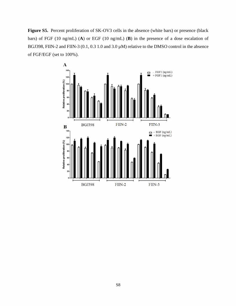
Figure S5. Percent proliferation of SK-OV3 cells in the absence (white bars) or presence (black
bars) of FGF (10 ng/mL) (A) or EGF (10 ng/mL) (B) in the presence of a dose escalation of
BGJ398, FIIN-2 and FIIN-3 (0.1, 0.3 1.0 and 3.0 µM) relative to the DMSO control in the absence
of FGF/EGF (set to 100%).

S9
Figure S6. Inhibition of FGF1-induced signaling by BGJ398, FIIN-2 and FIIN-3 in SKOV-3 cells.
Cells were treated with or without FGR1 (10 ng/mL) and with or without 1.0 µM inhibitor for 15
min then lysed and subjected to western blot for the indicated proteins or phosphoproteins.

S10
Figure S7. No dispersion of SKOV-3 spheroids was detected without growth factor stimulation
(A). The SKOV-3 spheroids were treated with 20 ng/mL of FGF1 (B) or EGF (C) with or without
1.0 µM of FIIN-2, FIIN-3 or FRIN-3 for 48 h, bar-graphs score the percentage of cell dispersion
(B, C). The cell number dispersion of each gel region was determined by drawing a line from the
edge of the spheroids to the tip of the furthest grown sprout or cells are completely separated, using
Image J software (http://rsbweb. nih.gov/ij/). The cell number is quantified by using the inbuilt
Image J function; to measure the cells are separated from spheroids (3).

S11
Figure S8. FIIN-2 and FIIN-3 induce zebrafish embryo developmental defects similar to
AZD4547, BGJ398 and PD173074. Images of representative fish after treatment with inhibitor at
indicated concentration for 48 h. FIIN-2 and FIIN-3 induce Zebrafish embryo developmental
defects similar to other known FGFR inhibitors (A). The bar-graph indicates the scoring of the
posterior phenotypes in 30 fish as normal, mild or severe for the drugs (B).

S12
Table S1. Profiling of FGFR inhibitors for binding against a panel of 456 kinases.
FRIN-2
(1 uM)
FRIN-3
(1 uM)
Score
(%)
IC50
(nM)
Score
(%)
IC50
(nM)Score
IC50
(nM)
Score
(%)
Score
(%)
BLK 0.5 0.45 0.1 1.8 2.6
BTK 100 3.4 1.2 99 94
CSF1R 0.2 0.15 0 0.9 1.6
CSF1R-autoinhibited 2.2 0.95 2.8
DDR1 0.1 0.7 0 0.35 0.45
EGFR 100 57 204 0.35 43.1 85 76
EGFR(E746-A750del) 100 48 0.6 74 84
EGFR(G719C) 100 6.5 0.1 95 87
EGFR(G719S) 100 24 0.15 92 90
EGFR(L747-E749del, A750P) 100 41 0.55 73 75
EGFR(L747-S752del, P753S) 100 37 0.45 90 97
EGFR(L747-T751del,Sins) 100 49 0.65 76 98
EGFR(L858R) 100 24 0.2 76 72
EGFR(L861Q) 100 6 0.1 100 100
EGFR(S752-I759del) 100 56 2.3 62 79
FGFR1 0 9.2 0.25 3.09 2.2 13.1 0.4 0.4
FGFR2 2.1 6.2 2.6 4.3 1.9 21 0.35 0.75
FGFR3 1.8 11.9 1.4 27 4.5 31.4 0.5 0.45
FGFR3(G697C) 0.35 0.7 1.4 0.4 0.1
FGFR4 0.1 189 1.4 45.3 0.1 35.3 1.6 0.3
FLT1 0.3 3.2 0.3 1 1.6
FLT4 0.2 0.3 0.1 0.1 2.2
JAK3(JH1domain-catalytic) 6.4 0 0.15 32 26
KIT 1.2 0.25 0.3 0.05 1.9
KIT(A829P) 1.2 5.4 22
KIT(D816V) 100 3.3 7.2 0.8 13
KIT(L576P) 2.7 1.2 0 0
KIT(V559D) 0.55 0.1 0.1 0 0.65
LCK 0.7 0.95 0.2 0.15 0.35
MAP3K3 0.5 3.8 5.8 2.9 0.85
PDGFRB 2.4 0.05 0 0 0.95
RET 25 0.15 0.6 0 11
RET(M918T) 100 0.2 0.4 0 9.6
RET(V804M) 100 3.6 100 0.35 84
YSK4 100 4.8 5.4 100
FIIN-1 (10 uM) FIIN-2 (1 uM) FIIN-3 (1 uM)
Kinases
Table shows the subset of kinases that exhibited a score of 5 or below (score is percent relative to
DMSO control, smaller numbers indicate stronger binding). Biochemical kinase IC50s (performed
at ATP concentrations equal to the apparent Km) were determined for some kinase targets using
enzymatic assays and are reported in nanomolar. Biochemical assays were performed by Life
technology according to their published procedures. (4)

S13
Table S2. Antiproliferative activity of FGFR and EGFR inhibitors on transformed Ba/F3 cells.
Ba/F3 cell lines EC50 (nM)
FIIN-2 FIIN-3 BIBW2
992
WZ400
2
FGFR2 1 <1 >3300 1216
FGFR2 (V564M) 58 64 >3300 726
RET 196 211
BTK >1000 >1000
FLT1 1000 700
FLT4 240 700
KIT >1000 >1000

S14
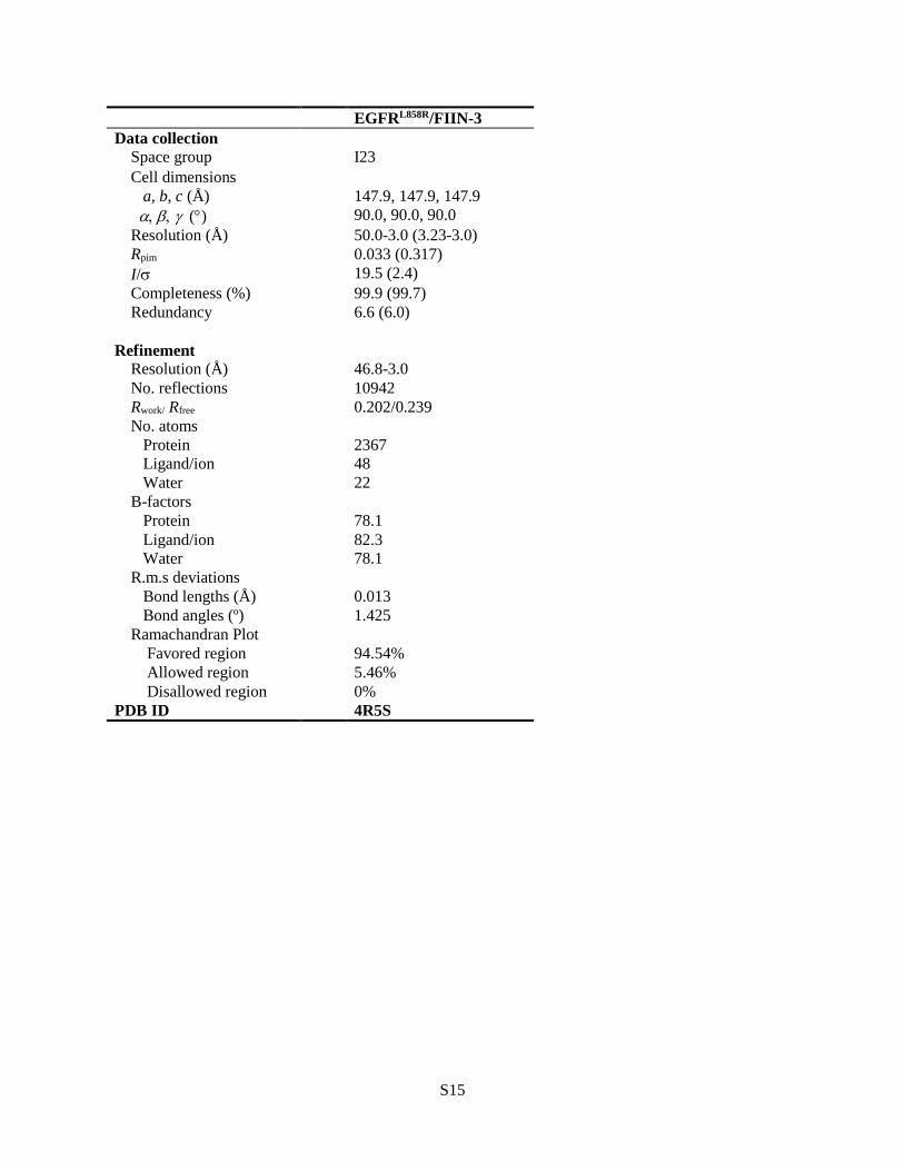
Table S3. Crystallographic Data and Refinement Statistics
Construct FGFR4K-FIIN-2 FGFR4KV550L-FIIN-3
Data Collection
Resolution (Å) 50-2.35 (2.39-2.35) 50-2.35 (2.39-2.35)
Space group R3 R3
Unit Cell Parameters (Å, )
a = 139.83
b = 139.83
c = 49.486
= 90.00
= 90.00
= 120.00
a = 139.38
b = 139.38
c = 50.09
= 90.00
= 90.00
= 120.00
Content of the asymmetric unit 1 1
Measured reflections (#) 172427 43068
Unique Reflections (#) 15298 14984
Data redundancy 11.3 (5.9) 2.9 (2.6)
Data completeness (%) 100 (99.6) 99.6 (98.2)
Rsym (%) 10.6 (40.9) 7.8 (34.5)
I/sig 29.7 (4.0) 12.2 (2.0)
Refinement
R factor/R free 17.4/22.8 17.8/23.7
Number of protein atoms 2138 2110
Number of non-protein/solvent atoms 47 48
Number of solvent atoms 0 10
Rmsd bond length (Å) 0.009 0.008
Rmsd bond angle () 1.19 1.16
PDB ID 4QQC 4R6V
S15
EGFRL858R/FIIN-3
Data collection
Space group I23
Cell dimensions
a, b, c (Å) 147.9, 147.9, 147.9
() 90.0, 90.0, 90.0
Resolution (Å) 50.0-3.0 (3.23-3.0)
Rpim 0.033 (0.317)
I/ 19.5 (2.4)
Completeness (%) 99.9 (99.7)
Redundancy 6.6 (6.0)
Refinement
Resolution (Å) 46.8-3.0
No. reflections 10942
Rwork/ Rfree 0.202/0.239
No. atoms
Protein 2367
Ligand/ion 48
Water 22
B-factors
Protein 78.1
Ligand/ion 82.3
Water 78.1
R.m.s deviations
Bond lengths (Å) 0.013
Bond angles (º) 1.425
Ramachandran Plot
Favored region 94.54%
Allowed region 5.46%
Disallowed region
PDB ID
0%
4R5S
S16
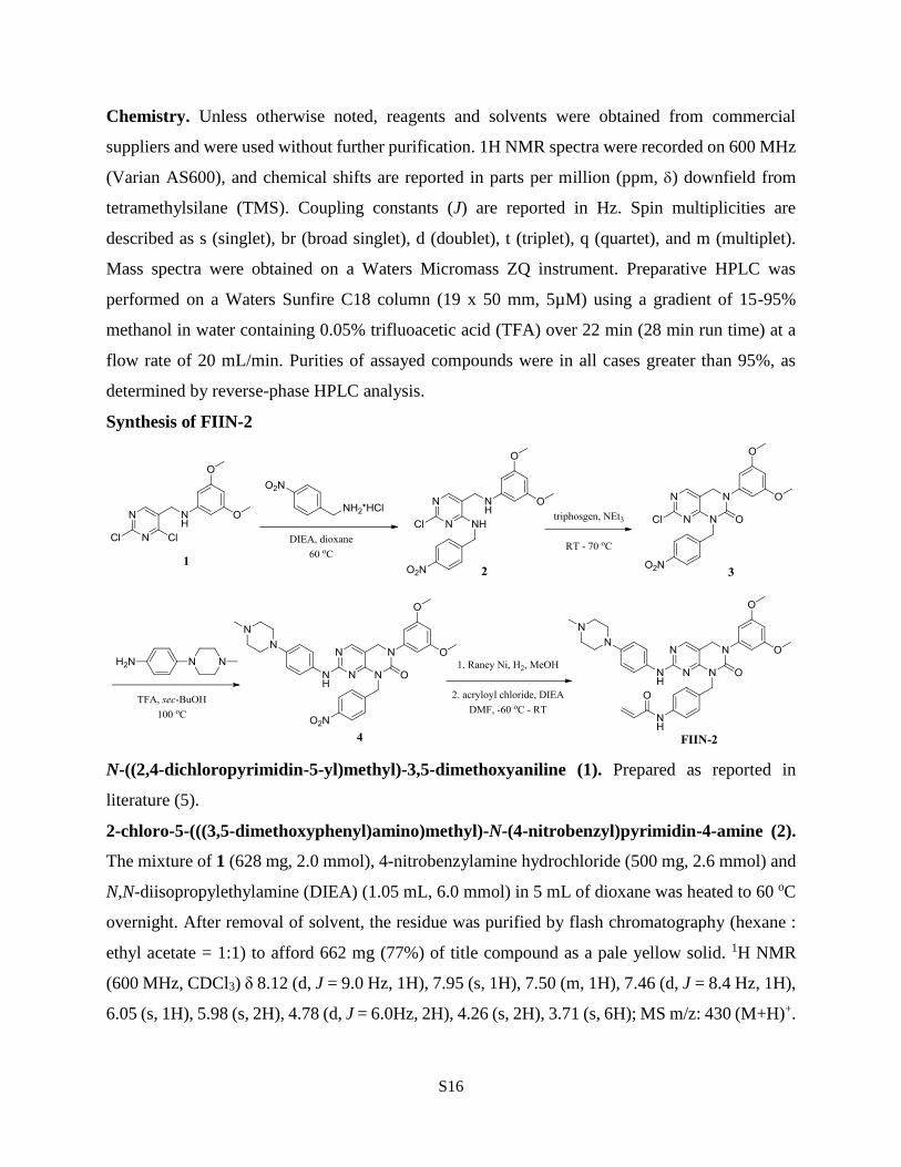
Chemistry. Unless otherwise noted, reagents and solvents were obtained from commercial
suppliers and were used without further purification. 1H NMR spectra were recorded on 600 MHz
(Varian AS600), and chemical shifts are reported in parts per million (ppm, ) downfield from
tetramethylsilane (TMS). Coupling constants (J) are reported in Hz. Spin multiplicities are
described as s (singlet), br (broad singlet), d (doublet), t (triplet), q (quartet), and m (multiplet).
Mass spectra were obtained on a Waters Micromass ZQ instrument. Preparative HPLC was
performed on a Waters Sunfire C18 column (19 x 50 mm, 5µM) using a gradient of 15-95%
methanol in water containing 0.05% trifluoacetic acid (TFA) over 22 min (28 min run time) at a
flow rate of 20 mL/min. Purities of assayed compounds were in all cases greater than 95%, as
determined by reverse-phase HPLC analysis.
Synthesis of FIIN-2
N-((2,4-dichloropyrimidin-5-yl)methyl)-3,5-dimethoxyaniline (1). Prepared as reported in
literature (5).
2-chloro-5-(((3,5-dimethoxyphenyl)amino)methyl)-N-(4-nitrobenzyl)pyrimidin-4-amine (2).
The mixture of 1 (628 mg, 2.0 mmol), 4-nitrobenzylamine hydrochloride (500 mg, 2.6 mmol) and
N,N-diisopropylethylamine (DIEA) (1.05 mL, 6.0 mmol) in 5 mL of dioxane was heated to 60 oC
overnight. After removal of solvent, the residue was purified by flash chromatography (hexane :
ethyl acetate = 1:1) to afford 662 mg (77%) of title compound as a pale yellow solid. 1H NMR
(600 MHz, CDCl3) δ 8.12 (d, J = 9.0 Hz, 1H), 7.95 (s, 1H), 7.50 (m, 1H), 7.46 (d, J = 8.4 Hz, 1H),
6.05 (s, 1H), 5.98 (s, 2H), 4.78 (d, J = 6.0Hz, 2H), 4.26 (s, 2H), 3.71 (s, 6H); MS m/z: 430 (M+H)+.
S17
7-chloro-3-(3,5-dimethoxyphenyl)-1-(4-nitrobenzyl)-3,4-dihydropyrimido[4,5-d]pyrimidin-
2(1H)-one (3). To 2 (430 mg, 1.0 mmol) in 5 ml of anhydrous THF was added triphosgene (119
mg, 0.4 mmol). Triethylamine (TEA) (0.42 mL, 3.0 mmol) was then added slowly. After
completion of addition, the mixture was stirred for 1 h, and heated to 70 oC overnight. After the
reaction was cooled down, it was diluted with ethyl acetate, washed with saturated sodium
bicarbonate. The organic layer was dried over sodium sulfate and concentrated in vacuo. The crude
product was purified through a short pipette column to afford 365 mg (80%) of the title compound
as a pale yellow solid. 1H NMR (600 MHz, MeOH) δ 8.11 (d, J = 9.0 Hz, 1H), 8.09 (s, 1H), 7.61
(d, J = 8.4 Hz, 1H), 6.40 (s, 2H), 6.32 (m, 1H), 5.28 (s,2H), 4.74 (s, 2H), 3.72 (s, 6H); MS m/z:
456 (M+H)+.
3-(3,5-dimethoxyphenyl)-7-((4-(4-methylpiperazin-1-yl)phenyl)amino)-1-(4-nitrobenzyl)-
3,4-dihydropyrimido[4,5-d]pyrimidin-2(1H)-one (4). The mixture of 3 (223 mg, 0.5 mmol), 4-
(4-methylpiperazin-1-yl)aniline (143 mg, 0.75 mmol) and trifluoroacetic acid (TFA) (58 µL, 0.75
mmol) in 5 mL of sec-butanol was heated to 100 oC overnight. After cooling and removal of
solvent, the residue was purified by flash chromatography (dichloromethane : methanol = 15:1) to
afford 428 mg (70%) of title compound as a pale yellow solid. 1H NMR (600 MHz, CDCl3) δ 8.09
(d, J = 9.0 Hz, 1H), 7.98 (s, 1H), 7.48 (d, J = 8.4 Hz, 1H), 7.29 (d, J = 9.0 Hz, 1H), 7.08 (br, 1H),
6.86 (d, J = 9.0 Hz, 1H), 6.47 (s, 2H), 6.39 (m, 1H), 5.30 (s, 2H), 4.68 (s, 2H), 3.78 (s, 6H), 3.30
(m, 4H), 2.83 (m, 4H), 2.52 (s, 3H); MS m/z: 611 (M+H)+.
N-(4-((3-(3,5-dimethoxyphenyl)-7-((4-(4-methylpiperazin-1-yl)phenyl)amino)-2-oxo-3,4-
dihydropyrimido[4,5-d]pyrimidin-1(2H)-yl)methyl)phenyl)acrylamide (FIIN-2). To 4 (122
mg, 0.2 mmol) in MeOH (20 mL) was added 1 mL Raney nickel suspension in MeOH, then the
reaction mixture was stirred for 3 h under 1 atm of hydrogen. The mixture was then filtered through
Celite and the filtrate was concentrated and dried under vacuum to give crude product as a white
solid. To the obtained white solid in DMF (2 mL) was added DIEA (53 µL, 0.3 mmol), the stirred
mixture was then cooled to -60 oC and acryloyl chloride (17.8 µL, 0.22 mmol) was added dropwise.
The reaction was stirred at -60 oC for 10 min and allowed to recover to RT gradually over 30 min,
then purified by reverse phase HPLC to give 108 mg (TFA salt, 72% for 2 steps) of title compound
as a white solid. 1H NMR (600 MHz, DMSO) δ 10.04 (s, 1H), 9.18 (s, 1H), 8.05 (s, 1H), 7.53 (d,
J = 8.4 Hz, 2H), 7.32 (m, 2H), 7.23 (d, J = 7.8 Hz, 2H), 6.74 (d, J = 8.4 Hz, 2H), 6.37 (s, 1H), 6.41
S18
(dd, J = 16.8, 9.6 Hz, 1H), 6.23 (d, J = 16.8 Hz, 1H), 5.72 (J = 11.2 Hz, 1H), 5.15 (s, 2H), 4.74 (s,
1H), 3.75 (s, 6H), 3.09 (m, 4H), 2.61 (m, 4H), 2.34 (s, 3H); MS m/z: 635 (M+H)+.
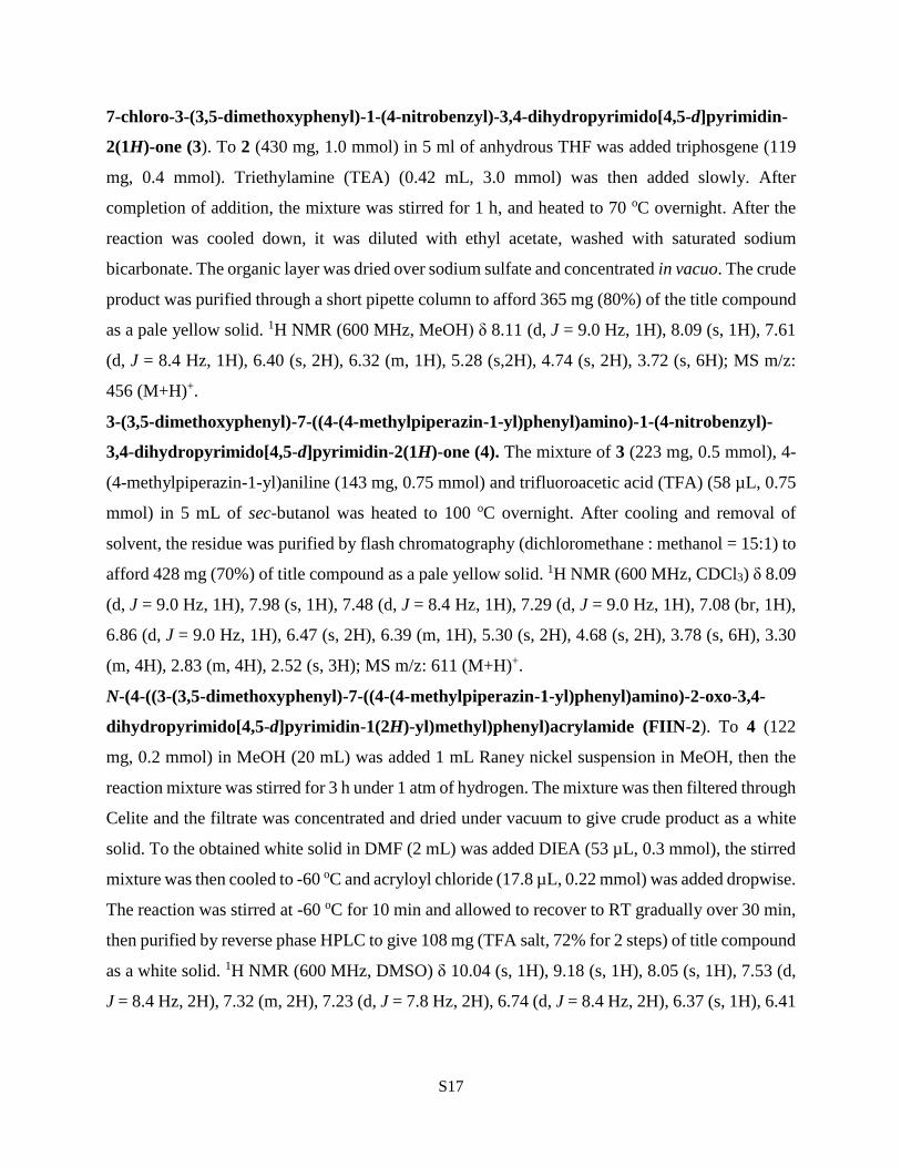
N-(4-((3-(3,5-dimethoxyphenyl)-7-((4-(4-methylpiperazin-1-yl)phenyl)amino)-2-oxo-3,4-
dihydropyrimido[4,5-d]pyrimidin-1(2H)-yl)methyl)phenyl)propionamide (FRIN-2).
Prepared with same method as FIIN-2. 1H NMR (600 MHz, DMSO) ) δ 9.73 (s, 1H), 9.16 (s, 1H),
8.04 (s, 1H), 7.44 (d, J = 8.4 Hz, 2H), 7.31 (m, 2H), 7.17 (d, J = 8.4 Hz, 2H), 6.73 (d, J = 9.0 Hz,
2H), 6.52 (s, 2H), 6.37 (s, 1H), 5.07 (s, 2H), 4.66 (s, 2H), 3.68 (s, 6H), 2.97 (m, 4H), 2.38 (m, 4H),
2.28 (q, J = 7.2 Hz, 2H), 2.22 (s, 3H), 1.05 (t, J = 7.2 Hz, 2H); MS m/z: 637 (M+H)+.
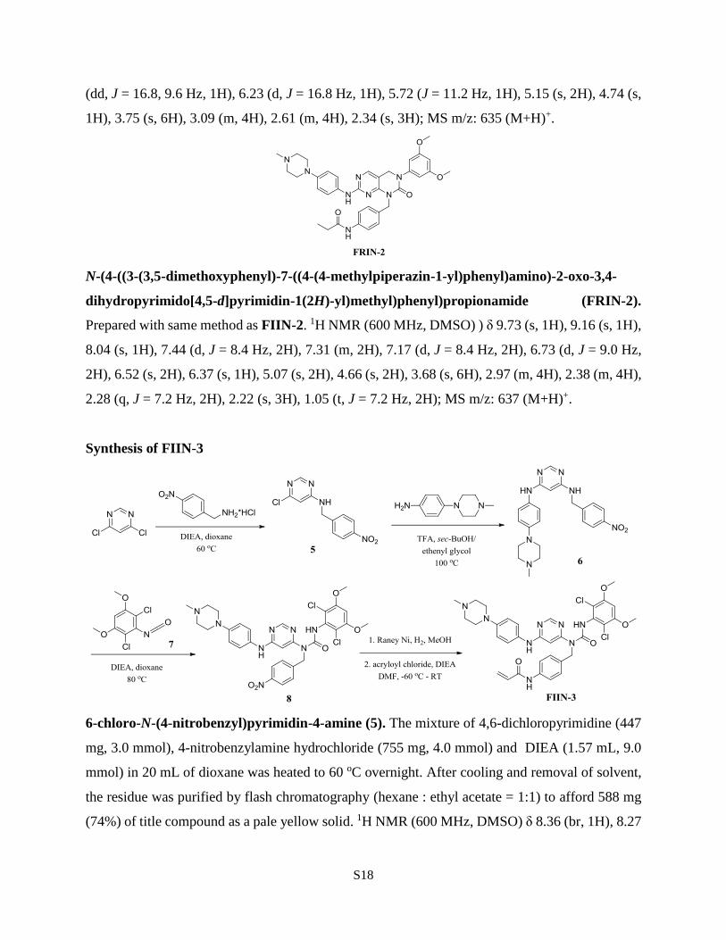
Synthesis of FIIN-3
6-chloro-N-(4-nitrobenzyl)pyrimidin-4-amine (5). The mixture of 4,6-dichloropyrimidine (447
mg, 3.0 mmol), 4-nitrobenzylamine hydrochloride (755 mg, 4.0 mmol) and DIEA (1.57 mL, 9.0
mmol) in 20 mL of dioxane was heated to 60 oC overnight. After cooling and removal of solvent,
the residue was purified by flash chromatography (hexane : ethyl acetate = 1:1) to afford 588 mg
(74%) of title compound as a pale yellow solid. 1H NMR (600 MHz, DMSO) δ 8.36 (br, 1H), 8.27

S19
(s, 1H), 8.19 (d, J = 8.4 Hz, 1H), 7.55 (d, J = 8.4 Hz, 1H), 6.64(s, 1H), 4.69 (br, 2H); MS m/z: 265
(M+H)+.
N4-(4-(4-methylpiperazin-1-yl)phenyl)-N6-(4-nitrobenzyl)pyrimidine-4,6-diamine (6). The
mixture of 5 (530 mg, 2.0 mmol), 4-(4-methylpiperazin-1-yl)aniline (460 mg, 2.4 mmol) and
trifluoroacetic acid (TFA) (184 µL, 2.4 mmol) in 3 mL of sec-butanol and 1 mL of ethylene glycol
was heated to 120 oC overnight. After cooling and removal of solvent, the residue was purified by
flash chromatography (dichloromethane : methanol = 10:1) to afford 670 mg (80%) of title
compound as a pale brown solid. 1H NMR (600 MHz, MeOH) δ 8.21 (d, J = 9.0 Hz, 2H), 8.00 (s,
1H), 7.52 (d, J = 9.0 Hz, 2H), 7.12 (d, J = 8.4 Hz, 2H), 6.94 (d, J = 9.0 Hz, 1H), 5.58 (br, 1H),
4.57 (s, 2H), 3.20 (m, 4H), 2.67 (m, 4H), 2.40 (s, 3H); MS m/z: 420 (M+H)+.
3-(2,6-dichloro-3,5-dimethoxyphenyl)-1-(6-((4-(4-methylpiperazin-1-
yl)phenyl)amino)pyrimidin-4-yl)-1-(4-nitrobenzyl)urea (8). To 2,6-dichloro-3,5-
dimethoxyaniline (330 mg, 1.5 mmol) in 6 ml of anhydrous THF was added triphosgene (178 mg,
0.6 mmol). Triethylamine (TEA) (0.42 mL, 3.0 mmol) was then added slowly. After completion
of addition, the mixture was stirred overnight, then was concentrated. The crude product was
purified through a very short pipette column to afford 186 mg (50%) of 2,4-dichloro-3-isocyanato-
1,5-dimethoxybenzene (7) as a white solid.
To the mixture of 6 (420 mg, 1.0 mmol) and 7 (250 mg, 1.0 mmol) in 5 mL of dioxane was added
DIEA (350 µL, 2.0 mmol), then the reaction mixture was heated to 80 oC for 2 h. After cooling
and removal of solvent, the residue was purified by flash chromatography (dichloromethane :
methanol = 15:1) to afford 454 mg (68%) of title compound as a pale yellow solid. 1H NMR (600
MHz, TFA salt, MeOH) δ 8.41 (s, 1H), 8.23 (d, J = 8.4 Hz, 2H), 7.51 (d, J = 9.0 Hz, 2H), 7.20 (m,
2H), 6.96 (d, J = 9.0 Hz, 2H), 6.85 (s, 1H), 6.06 (s, 1H), 5.29 (s, 2H), 3.98 (s, 6H), 3.84 (m, 2H),
3.66 (m, 2H), 3.33 (m, 2H), 3.06 (m, 2H), 3.02 (s, 3H); MS m/z: 667 (M+H)+.
N-(4-((3-(2,6-dichloro-3,5-dimethoxyphenyl)-1-(6-((4-(4-methylpiperazin-1-
yl)phenyl)amino)pyrimidin-4-yl)ureido)methyl)phenyl)acrylamide (FIIN-3). Prepared with
same method as FIIN-2. 1H NMR (400 MHz, DMSO) ) δ 12.35 (s, 1H), 10.16 (s, 1H), 9.37 (s,
1H), 8.39 (s, 1H), 7.65 (d, J = 8.8 Hz, 2H), 7.20 (d, J = 8.0 Hz, 2H), 7.16 (m, 2H), 6.92 (s, 1H),
6.85 (d, J = 9.2 Hz, 2H), 6.44 (dd, J = 16.8, 10.0 Hz, 1H), 6.20 (s, 1H), 6.26 (d, J = 16.8 Hz, 1H),
5.75 (J = 10.0 Hz, 1H), 5.05 (s, 2H), 3.96 (s, 6H), 3.68 (s, 6H), 3.10 (m, 4H), 2.51 (m, 4H), 2.26
(s, 3H); MS m/z: 691 (M+H)+.

S20
N-(4-((3-(2,6-dichloro-3,5-dimethoxyphenyl)-1-(6-((4-(4-methylpiperazin-1-
yl)phenyl)amino)pyrimidin-4-yl)ureido)methyl)phenyl)propionamide (FRIN-3). Prepared
with same method as FIIN-2. 1H NMR (600 MHz, DMSO) ) δ 12.36 (s, 1H), 9.86 (s, 1H), 9.35 (s,
1H), 8.38 (s, 1H), 7.56 (d, J = 8.4 Hz, 2H), 7.15 (d, J = 8.4 Hz, 2H), 7.14 (m, 2H), 6.92 (s, 1H),
6.84 (d, J = 9.0 Hz, 2H), 6.19 (s, 1H), 5.03 (s, 2H), 3.95 (s, 6H), 3.09 (m, 4H), 2.47 (m, 4H), 2.31
(q, J = 7.8 Hz, 2H), 2.24 (s, 3H), 1.07 (t, J = 7.2 Hz, 2H); MS m/z: 693 (M+H)+.
Reference:
1. Goldstein DM, Gray NS, & Zarrinkar PP (2008) High-throughput kinase profiling as a platform
for drug discovery. Nature reviews. Drug discovery 7(5):391-397.
2. Miduturu CV, et al. (2011) High-throughput kinase profiling: a more efficient approach toward
the discovery of new kinase inhibitors. Chemistry & biology 18(7):868-879.
3. Aref AR, et al. (2013) Screening therapeutic EMT blocking agents in a three-dimensional
microenvironment. Integrative biology : quantitative biosciences from nano to macro 5(2):381-
389.
4. Lebakken CS, et al. (2009) Development and applications of a broad-coverage, TR-FRET-based
kinase binding assay platform. Journal of biomolecular screening 14(8):924-935.
5. Zhou W, et al. (2010) A structure-guided approach to creating covalent FGFR inhibitors.
Chemistry & biology 17(3):285-295.