supplementary figure 1s. antipodal cells show active cdc::h2b:yfp signal in a three day emasculated...
TRANSCRIPT
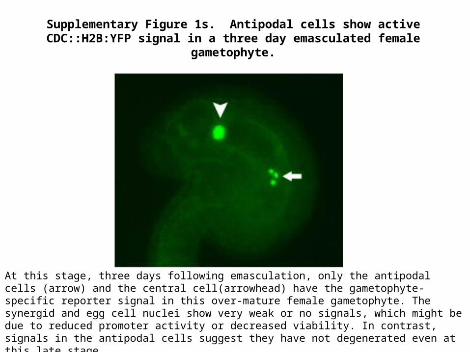
Supplementary Figure 1s. Antipodal cells show active CDC::H2B:YFP signal in a three day emasculated female gametophyte.
At this stage, three days following emasculation, only the antipodal cells (arrow) and the central cell(arrowhead) have the gametophyte-specific reporter signal in this over-mature female gametophyte. The synergid and egg cell nuclei show very weak or no signals, which might be due to reduced promoter activity or decreased viability. In contrast, signals in the antipodal cells suggest they have not degenerated even at this late stage.

2-nuclear 4-nuclear 8-nuclear 16-nuclear0%
25%
50%
75%
100% 94% 96%90%
76%
4%
16%
ratio of seeds that contain all three antipodalsratio of seeds that contain one or two antipodals
Developing stages of seeds indicated by the number of endosperm nuclei
Perc
enta
ge o
f see
ds th
at c
onta
in a
n-tip
odal
s
Seeds at different developing stages are examined in CDC123::H2B:YFP reporter line to check the presence of the antipodal cells. At each stage, n=50 seeds were examined. At 2-nuclear endosperm stage, 47/50 seeds contain all three antipodal cells. 48/50 seeds at 4-nuclear stage show the three antipodal cells. In the 50 seeds at 8-nuclear endosperm stage, 45 seeds contain all three antipodals, and 2 seeds contain 2 antipodal cells. Among the 50 seeds at 16-nuclear stage, three antipodals are observed in 38 seeds, whereas one or two antipodals are observed in 8 seeds, suggestive of degeneration of antipodals initiating around this stage.
Supplementary Figure 2s. Frequency of antipodal cell persistence in developing seeds at different stages

Supplementary Figure 3s. Presence of three antipodal cells at 4-nucleate endosperm stage.
Overlay of YFP (CDC::H2B:YFP, from female gametophyte) and RFP (DD65::NLSmCherry, from pollen) signals in a developing seed. As shown above, 4 nuclei (numbered 1-4) are labeled by both reporters, suggesting they are endosperm; whereas the 3 nuclei at the chalazal pole (arrow) show only the maternal marker, suggesting they are the persisting antipodals instead of fertilization products.
antipodal nuclei endosperm nuclei1
3
5
7
9
11
13
15
Nuclei type in the developing seed
aver
age
cent
rom
eric
dot
s
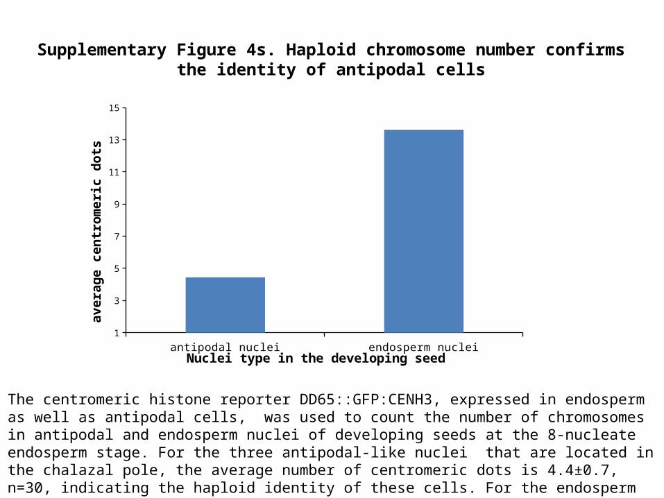
The centromeric histone reporter DD65::GFP:CENH3, expressed in endosperm as well as antipodal cells, was used to count the number of chromosomes in antipodal and endosperm nuclei of developing seeds at the 8-nucleate endosperm stage. For the three antipodal-like nuclei that are located in the chalazal pole, the average number of centromeric dots is 4.4±0.7, n=30, indicating the haploid identity of these cells. For the endosperm nuclei, the average number of centromeric dots is 13.6±1.1, n=48, corresponding to their triploid identity.
Supplementary Figure 4s. Haploid chromosome number confirms the identity of antipodal cells

Supplementary Figure 5s. Antipodal cells persist in mature embryo sac after crossing to Ler ecotype background.
Antipodal cells in one day emasculated mature embryo sac, labeled by CDC123::H2B:YFP reporter in F1 progeny of Columbia ecotype of Arabidopsis crossed with Landsberg erecta ecotype. Similar results were obtained in the F1 plants of the reciprocal cross. Arrow shows the three antipodal cells. Arrowheads show the larger central cell nucleus, and the smaller egg cell nucleus. Synergid cell nuclei are not shown here, as they are out of the focal plane in this picture.