supplementary information for osmotic-pressure-mediated ... · s1 supplementary information for...
TRANSCRIPT

S1
Supplementary Information for
Osmotic-Pressure-Mediated Control of Structural
Colors of Photonic Capsules
Tae Min Choi†, Jin-Gyu Park
‡, Young-Seok Kim
§, Vinothan N. Manoharan
‡, and Shin-Hyun
Kim*†
†Department of Chemical and Biomolecular Engineering and KINC, KAIST, Daejeon, South Korea
‡School of Engineering and Applied Sciences and Department of Physics, Harvard University, Cambridge,
Massachusetts, USA.
§Korea Electronics Technology Institute, Seongnam-si, Gyeonggi-do, South Korea
Correspondence to: Shin-Hyun Kim, [email protected]
S1. Supplementary Figures
S2. Description of Supporting Movies

S2
S1. Supplementary Figures
Figure S1. Time series of optical microscope images showing the shrinkage of double-emulsion droplets which
are incubated at aqueous solutions with osmolarities of (a) 180 mOsml-1, (b) 300 mOsml-1, and (c) 440 mOsml-1.
The incubation time is denoted at each image.
S3
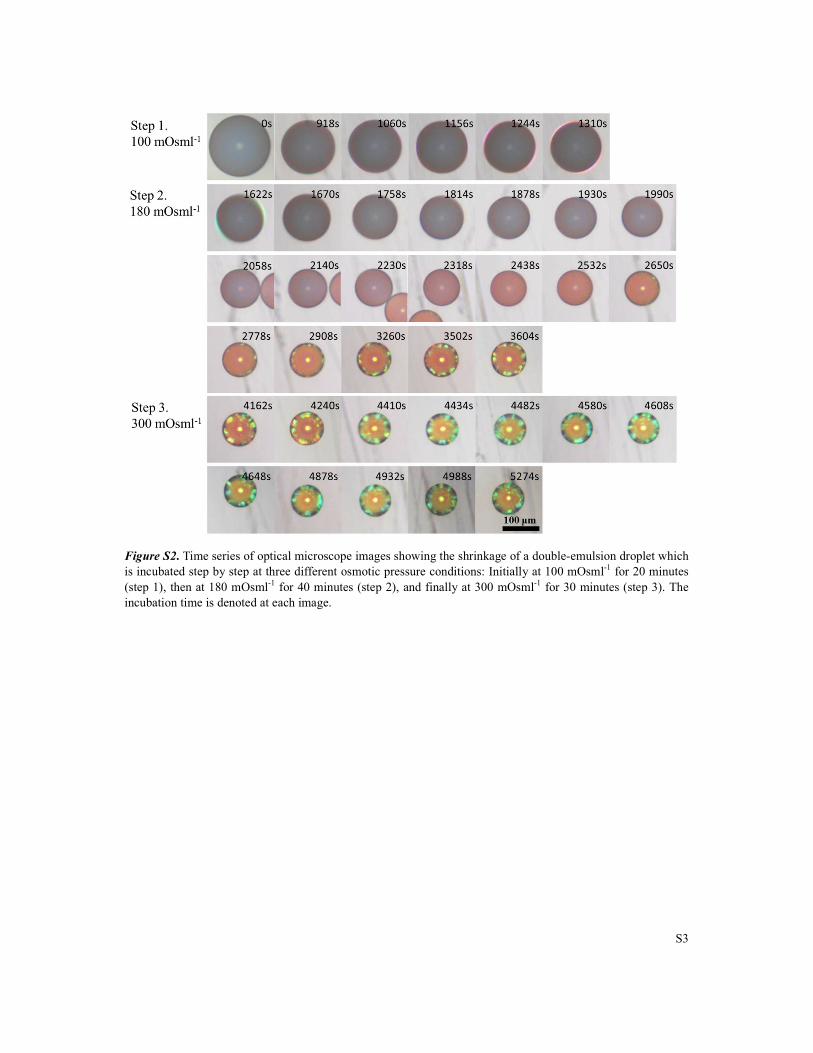
Figure S2. Time series of optical microscope images showing the shrinkage of a double-emulsion droplet which
is incubated step by step at three different osmotic pressure conditions: Initially at 100 mOsml-1 for 20 minutes
(step 1), then at 180 mOsml-1
for 40 minutes (step 2), and finally at 300 mOsml-1
for 30 minutes (step 3). The
incubation time is denoted at each image.

S4
Figure S3. (a-d) Cryo-Scanning electron microscope (Cryo-SEM) images showing internal structures of
photonic capsules prepared by single-step concentration.
Figure S4. (a-d) Cryo-SEM images showing internal structures of photonic capsules prepared by stepwise
concentration.

S5
Figure S5. Sets of optical microscope images and photographs of aqueous suspension of photonic capsules
composed of PS particles with diameters of (a, b) 156 nm and (c, d) 222 nm, where the capsules in (a, c) and (b,
d) are prepared by single-step and stepwise concentration, respectively.
S6
S2. Description of Supporting Movies
Movie S1 shows the generation of double-emulsion drops composed of aqueous core of
colloidal suspension and oil shell of photocurable monomers in a capillary microfluidic
device. Volumetric flow rates of innermost suspension, middle oil, and continuous phases are
set to be 300 µlh-1
, 200 µlh-1
, and 2200 µlh-1
, respectively. This move is taken at 1000 Hz by
high speed camera and played at 30 frames per second: 33 × slower than real time motion.
Movie S2 shows the evolution of double-emulsion drops under two different hypertonic
conditions; osmolarity of incubation solution is set to be 180 mOsml-1
(first part) and 440
mOsml-1
(second part). The incubation time is denoted at each frame.
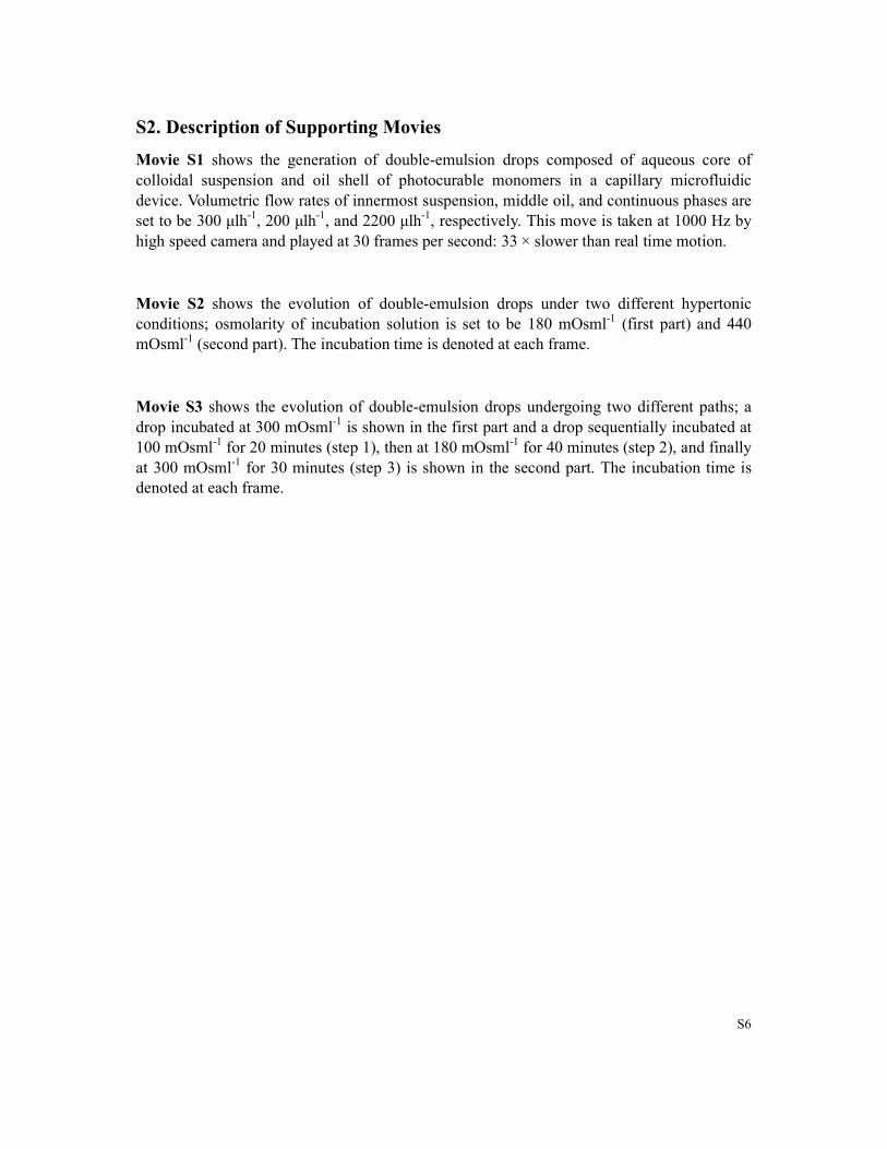
Movie S3 shows the evolution of double-emulsion drops undergoing two different paths; a
drop incubated at 300 mOsml-1
is shown in the first part and a drop sequentially incubated at
100 mOsml-1
for 20 minutes (step 1), then at 180 mOsml-1
for 40 minutes (step 2), and finally
at 300 mOsml-1
for 30 minutes (step 3) is shown in the second part. The incubation time is
denoted at each frame.