supplementary methods - Étsprofs.etsmtl.ca/mmcguffin/research/2015-li-pms2/li-2015-pms2... ·...
TRANSCRIPT

Li et al PMS2 founder mutation with attenuated phenotype – Supplement
1
SUPPLEMENTARY FILE
Supplementary Methods
Study subjects: Kindreds with PMS2 c.2002A>G were identified through
hereditary cancer clinics at hospitals affiliated to McGill University and the
University of Manitoba. Probands were referred to medical genetics clinics by
physicians. Signed informed consent was obtained from all study participants or
their legal representatives. The local ethical committees of McGill University, the
University of Manitoba and other collaborating centers approved this study. Bio-
specimens used in laboratory investigation consisted of patient derived biological
material in the form of peripheral blood lymphocytes, colon or skin biopsies.
Since not every type of material was available for every patient, the number of
samples used in each experimental procedure was determined by availability.
Additional information for PMS2 genotype-phenotype comparisons was retrieved
from the Leiden Open Variation Database (http://www.lovd.nl/3.0/home) and
Online Mendelian Inheritance in Man (http://omim.org/) and was further validated
by reviewing relevant publications.
DNA, RNA and cDNA preparation: DNA and RNA from peripheral blood
lymphocytes were purified using the Gentra Puregene Blood Kit (Qiagen, Toronto,
Canada) and PAXgene Blood RNA Kit IVD (Qiagen, Toronto, Canada),
respectively, according to manufacturer protocols. DNA and RNA concentrations
were quantified using a NanoDrop™ Spectrophotometer (Thermo Scientific,
Ontario, Canada). cDNA synthesis was performed using the QuantiTect reverse

Li et al PMS2 founder mutation with attenuated phenotype – Supplement
2
transcription kit (Qiagen, Toronto, Canada) with 100 ng of total RNA as input
template. cDNA was stored at -20°C for downstream applications.
Mutation identification: Immunohistochemical staining of mismatch repair proteins
MLH1, MSH2, MSH6 and PMS2 was done on formalin-fixed, paraffin-embedded
tissues from affected individuals using standard protocols at treating hospitals.
For the protein truncation test, cDNA fragments of the PMS2 transcript were
amplified from lymphocyte RNA using reverse transcription and long-range PCR
kits (Qiagen, Toronto, Canada), followed by in vitro translation using the TNT
Quick Coupled Transcription / Translation System (Promega, Madison, United
States) and 35S-methionine (Amersham Biosciences, Piscataway, United States).
For cDNA sequencing, transcript derived from the functional PMS2 gene locus
was amplified specifically with a forward primer located in exon 10. For gDNA
sequencing, nested PCR was employed to avoid interference from pseudogenes.
Briefly, the genomic region spanning exons 10, 11 and 12 was amplified using an
Expand Long Template PCR System (Roche, Mississauga, Canada), the long-
range PCR product was then diluted to1:40, and 2 μl of the diluted product was
used as a template for amplification with an internal set of primers. The mutation
was identified by sequencing the final PCR products with a reverse primer
derived from exon 12 using a 3730xl DNA Analyzer (Applied Biosystems, Foster
City, United States).
Prediction Algorithms used: The predicted effect of NM_000535.5:c.2002A>G on
protein function was evaluated using Mutation Taster (http://mutation.taster.org/),
PolyPhen-2 (Polymorphism Phenotyping V2,

Li et al PMS2 founder mutation with attenuated phenotype – Supplement
3
http://genetics.bwh.harvard.edu/pph2/index.shtml) and PROVEAN (Protein
Variation Effect Analyzer, http:/provean.jcvi.org/index.php).1-3
Mutation age calculation: We genotyped 17 microsatellite markers spanning a
region of 8.8 megabases around PMS2. We used methods previously described
in Hamel et al.4 to perform the genotyping and to estimate the age of the
PMS2:c.2002A>G mutation.
Polony Assay: Polony assay relies on solid phase PCR performed with template
molecules separated and immobilized in a thin film of acrylamide gel. Each
individual template molecule is then amplified, forming a homogenous molecule
colony. Polonies are visualized by probe hybridization and single base extension
(SBE) using fluorescein labelled dideoxynucleotides. The primer set for the solid
phase PCR producing the 960 bp product was designed to unbiasedly amplify
both the functional PMS2 and pseudo PMS2CL loci (Figure 2A). The resulting
polonies were identified by two rounds of probe hybridization coupled with SBE:
one round was designed for differentiating the real/pseudo PMS2 transcripts and
the other for detecting the intact/aberrant exon 11-12 junctions (probe/primer
sequences are listed in Table S2). The probes were positioned immediately
before a variant distinguishing each pair of transcripts so each transcript (cDNA)
polony was colored with the fluorescein of the specific dideoxynucleotides added
to the pre-hybridized probe by complementary base-pairing during SBE.
Molecule-specific PCR: Locus specificity was achieved by placing the forward
primer in exon 10, which is unique to the functional PMS2 gene. Each transcript
population was targeted by setting a specific nucleotide at the 3’ end of the

Li et al PMS2 founder mutation with attenuated phenotype – Supplement
4
reverse primer, complementary to either the intact or the aberrant exon 11-12
junction (Figures 2A and S4A). We performed molecule-specific PCR using the
same cDNA template from the Polony assay and used a touch-down protocol.5
The PCR products were then visualized by fragment analysis, which validated
the expression of both the aberrant and intact exon 11-12 junctions from the
mutant allele (Figure 2C).
Western blot and immunoprecipitation: Pierce IP Lysis Buffer (Thermal Scientific,
Rockford, United States) was used for protein extraction and immunoprecipitation.
Protease inhibitors (Roche, Laval, Canada) were added prior to harvesting the
cells. Total cell lysates were obtained from 4 x 10⁷ and 4 x 10⁸ LCLs for protein
analysis by direct western blot and immunoprecipitation of the MLH1-PMS2
complex, respectively. Primary antibody incubation for western blot analysis was
done at a 1:200 dilution for MLH1 (BD Biosciences, Mississauga, Canada,
material # 550838) and 1:500 for PMS2 (BD Biosciences, Mississauga, Canada,
material # 556415). The secondary antibody was horseradish peroxidase-
conjugated goat anti-mouse (Sigma Aldrich, Oakville, Canada) at a dilution of
1:20,000. For each immunoprecipitation experiment, 2 μg MLH1 antibody (same
as above), 30 μl protein A sepharose beads slurry (GE Healthcare,
Buckinghamshire, United Kingdom) and 1 mg cell lysate were incubated in a total
volume of 1 ml in IP lysis buffer. Protein electrophoresis and western blot were
performed by following the protocols for the Mini-PROTEAN® system (Biorad,
Mississauga, Canada) using a 7.5% acrylamide gel. The detection system used
was the Super Signal West Femto kit (Thermo Scientific, Rockford, United States)

Li et al PMS2 founder mutation with attenuated phenotype – Supplement
5
and Amersham HyperfilmTm ECL (GE Healthcare, Buckinghamshire, United
Kingdom). The MLH1-PMS2 complex was collected from the lymphoblastoid
lysate via immunoprecipitation with a primary antibody against MLH1. The
protein complex was then detected by the inclusion of two monocloncal
antibodies (anti-MLH1 at 1:500 dilution and anti-PMS2 at 1:200 dilution) during
the primary incubation of western blot.
Microsatellite Instability (MSI) Assay: A highly sensitive method is required to
detect the rare variant alleles in normal tissues where MSI occurs as somatic
events, a scenario quite different from detecting MSI in tumours where
assessment is based upon the comparison between normal and cancer tissues.6
Tetranucleotide microsatellite markers have been proven to have optimal
sensitivity for DNA repair deficiency in the cellular context of PMS2 null
compared to mononucleotide or dinucleotide markers.7 After pilot experiments
with several markers, we chose D17S1307, a tetranucleotide marker located in
the first intron of the Neurofibromatosis 1 (NF1) gene, to pursue quantitative MSI.

Li et al PMS2 founder mutation with attenuated phenotype – Supplement
6
Figure S1. c.2002A>G is a coding mutation that interferes with RNA splicing.
A. Representative images of immunohistochemical staining of mismatch repair
proteins in the colorectal cancer from individual III-2 from Table S1, 400X
magnification. Images show normal colon crypts (right) and invasive
adenocarcinoma arising in the colon (left). Protein expression is identified as dark
brown staining of the tumor cell nuclei. Counterstain of the nuclei with
haematoxylin is blue. Note the complete absence of staining of both normal and
tumour cells for PMS2.
B. Electropherograms of Sanger sequencing with gDNA and cDNA from
peripheral lymphocytes. Intronic sequences in gDNA are shown in small capital
letters. "//" denotes the junction between exons 11 and 12 in cDNA.
C. c.2002A>G is located at the boundary of exon 11 and intron 11, causing the
substitution of Isoleucine to Valine (reference sequence NM_000535.5). Sanger
sequencing with cDNA from homozygotes detected a five base-pair deletion,
GTAAG, at the exon 11-12 junction of PMS2. The aberrant transcript results
from RNA splicing at the de novo 5' ss generated by the "G" mutant allele.

Li et al PMS2 founder mutation with attenuated phenotype – Supplement
7
Figure S1

Li et al PMS2 founder mutation with attenuated phenotype – Supplement
8
Figure S2. c.2002G>A is a founder mutation in the Inuit population.
A. Geographical distribution of the c.2002A>G mutation. Communities with
cancer patients homozygous for this mutation are marked with a red star. In total,
eight communities were found to harbor this mutation, all located around Hudson
Bay. No homo- or heterozygote individual was identified in a tested series of non-
Nunavik residents (n = 6938), comprising 6435 newborns from Quebec City, 500
unselected individuals from the communities in Inuvialuit (including McKenzie
inlets) and three colon cancer patients from Greenland (data not shown).
B. Shared chromosomal fragment in all c.2002A>G carriers. Genotyping of 17
short tandem repeat markers was done for 5 families where DNA was available
for both heterozygous and homozygous members of the family. Numbers
represent alleles. Each column of numbers represents one haplotype observed in
that family. When two numbers are listed (e.g. 4/1), it indicates inconclusive
status for which allele is linked to the mutation, so both alleles remain
possibilities. X means no genotype information was available for this locus.
Alleles in red are located within the PMS2 gene region. The minimum conserved
region is identified to be ∼581 kilobases and is highlighted using a transparent
box. The maximum conserved region is 1.8 megabases (alleles in bold face).
The age of the mutation was estimated to be approximately 46 generations (95%
CI, 24-80 generations). Assuming a generation time of 18 years for this
population, we estimate the mutation first appeared late in the 11th century,
Common Era.

Li et al PMS2 founder mutation with attenuated phenotype – Supplement
9
Figure S2

Li et al PMS2 founder mutation with attenuated phenotype – Supplement
10
Figure S3. The age of cancer onset in CMMRD patients inversely correlates
with PMS2 expression status
Group I consists of 43 patients carrying bi-allelic PMS2 truncating mutations.
Group II consists of 16 patients carrying bi-allelic PMS2 mutations with laboratory
evidence of residual gene expression. Patients are of various ethnic backgrounds.
The X-axis indicates age and the Y-axis lists the PMS2 genotypes of each
individual. The age at diagnosis of primary (red), secondary (orange) and
additional cancers (dark yellow) for each individual is plotted along the X-axis,
with multiple cancers in the same person being connected by lines. Darkness
indicates death. The difference in age at primary cancer onset between group I
(median = 8, range = 1–16 years) and Group II (median = 20, range = 3–38
years) was statistically significant according to a Mann-Whitney test: U = 591.5,
N1 = 43, N2 = 16, two-tailed P = 2.4 x 10⁻⁵. The observation that the age for
primary cancer onset in Group II is significantly delayed compared to Group I,
supports that residual PMS2 expression (shown in Group II homozygotes)
contributes to genome-guarding function thus delays disease onset.

Li et al PMS2 founder mutation with attenuated phenotype – Supplement
11
Figure S3

Li et al PMS2 founder mutation with attenuated phenotype – Supplement
12
Figure S4. Molecule-specific PCR of the exon 11-12 junction of PMS2
A. Graphical representation of the molecule-specific PCR designed to
differentiate the intact (wild type) and aberrant exon 11-12 junction. A common
forward primer is placed in exon 10 to ensure PMS2-specific amplification; the
reverse primer is located at the exon junction, with the 3’ nucleotide priming for
either the intact (W for wild type) or the aberrant (M for mutant) transcript-
population. W: 1016 bp amplicon with an intact exon 11-12 junction. M: 1011 bp
amplicon with the aberrant exon 11-12 junction of a five-bp deletion. The PCR
products are resolved in an agarose gel after 34 cycles of amplification for M and
48 cycles for W.
B. Validation of the specificity of molecule-specific PCR using cells homozygous
for different PMS2 genotypes. R802X: c.2404C>T, is a truncating mutation
located at exon 14, only W junction is present in the cDNA. p.N412DfsX6
contains a deletion of exons 11-14, both W and M junctions are absent from the
cDNA. These controls demonstrate that the junction anchored, molecule-specific
PCR is highly specific. c.2002A>G, the mutation reported in this study, is a
missense mutation interfering with RNA splicing, both W and M junctions are
present in the cDNA.
C. W-PCR was performed with RNA from subjects homozygous for c.2002A>G
and affected by cancer in their twenties. Lanes 1–4: PCR product with the RNA
of lymphocytes from four patients. Lanes 5–6: PCR product with the RNA of
primary fibroblasts derived from two patients. Lane 7: PCR product with the RNA
of colon biopsy from the same patient as lane 4.

Li et al PMS2 founder mutation with attenuated phenotype – Supplement
13
Figure S4

Li et al PMS2 founder mutation with attenuated phenotype – Supplement
14
Figure S5. Detection of low level PMS2 by Western Blot Analysis
Full-length PMS2 protein is detected in the total lysate from lymphocytes and
fibroblasts. HET: cell line established from a c.2002A>G heterozygote. HOM: cell
line derived from a c.2002A>G homozygotes. The cell lines are derived from two
female patients with cancer diagnosed at the age of 21 and 26 years old,
respectively. CT: cell line from a healthy individual as a control of PMS2 wild type.
TFIIH: Transcription factor II Human. Actin and TFIIH serve as loading controls.

Li et al PMS2 founder mutation with attenuated phenotype – Supplement
15
Figure S6. The MSI molecular phenotype measured with marker D17S1307
The assay is performed with genomic DNA purified from lymphocytes and normal
colon mucosa of CMMRD patients. Individual I is homozygous for PMS2:
c.2002A>G and Individual II is a compound heterozygote, PMS2: c.1221delG +
c.2361delCTTC. The major alleles are determined by conventional genotyping
with 0.1 ng DNA. Rare expansion allele(s) is detected by single-genome assay
with 10 pg of DNA (equivalent to 3 alleles, 1.5 diploid genomes) per reaction.
A. Representative results for individual I. I-1: a typical allele pattern observed in
genotyping with a low input of 10 pg gDNA. I-2: the major alleles of D17S1307 in
individual I detected by conventional genotyping.
B. Representative results for individual II. II-1: The expansion allele (158 bp) is
detected by genotyping with a low input of 10 pg gDNA. II-2: the major alleles of
D17S1307 in individual II detected by conventional genotyping.
C. Summary of the detected expansion alleles at locus D17S1307. Fisher's exact
test showed a significant difference of D17S1307 allele expansion between the
two individuals, P = 0.0037. Detailed information on the expansion alleles
detected in lymphocytes and colon biopsy are presented in Table S5.

Li et al PMS2 founder mutation with attenuated phenotype – Supplement
16
Figure S6

Li et al PMS2 founder mutation with attenuated phenotype – Supplement
17
Table S1: Phenotypical characteristics of the proband’s family
Individual Alive Polyps CALS Cancer
Phenotypes
Age Dx
(yrs)
Immunohistochemistry
MLH1 MSH2 MSH6 PMS2
III-2 N Y Y Colon Ca 16 Present Present Present Absent
Astrocytoma 21 Present Present Present Absent
III-3 Y Y Y Colon Ca 26 Present Present Present Absent
Duodenal ca 40 ND ND ND ND
III-5 N Y Y Duodenal Ca 24 Present Present Present Absent
III-7 N Y Y Gastric Ca 22 ND ND ND ND
CALS = Café-au-lait spots, Dx = diagnosis, Ca = cancer , ND = not determined

Li et al PMS2 founder mutation with attenuated phenotype – Supplement
18
Table S2. Primers used in mutation identification, haplotype analysis, and gene expression characterization.
Protein truncation test (PTT)1
Forward primer codons 1–863 5’-GGATCCTAATACGACTCACTATAGGGAGACCACCATGGAGCGAGCTGAGAGC-3’
Forward primer codons 415–863 5’-GGATCCTAATACGACTCACTATAGGGAGACCACCATGGTGTCCATTTCCAGACTGCG-3’
Forward primer2 codons 332–863 5’-AATACGACTCACTATAGGGAGAGCCACCATGGTTA CTCCAGATAAAAGGCA-3’
Reverse primer (shared) 5’-AGGTTAGTGAAGACTCTGTC-3’
Forward primer codons 1–472 5’-GGATCCTAATACGACTCACTATAGGGAGACCACCATGGAGCGAGCTGAGAGC-3’
Reverse primer codons 1–472 5’-CTGAGGTCTCAGCAGGC-3’
PMS2 cDNA sequencing
Forward primer located in exon 10 5’-TGTTACTCCAGATAAAAGGC-3’
Reverse primer 5’-AGGTTAGTGAAGACTCTGTC-3’
Sequencing primer 5’-TGCAGCATCTCGAAGT-3’
PMS2 gDNA sequencing
Forward primer for long-range PCR
5’-GCGTTGATATCAATGTTACTCCAGA-3’
Reverse primer for long-range PCR
5’-AGTAGTCAGGGTAAAACATTCCAGT-3’
Forward primer for inner PCR 5’-TCACATAAGCACGTCCTCTCACCAT-3’
Reverse primer for inner PCR 5’-GCAACAGAGCAAGACTCTGTCTCAA-3’
Founder Mutation Linkage and Age Analysis
PMS2-MS1 forward primer 5’-TAGGCAGAGCATCACCAAGG-3’
PMS2-MS1 reverse primer 5’-TGCTGAAGCATGAAAACTGC-3’

Li et al PMS2 founder mutation with attenuated phenotype – Supplement
19
D7S1492 forward primer 5’-GCCTCCGAACACACTTCTTC-3’
D7S1492 reverse primer 5’-CCTGAAAATATACGTAACTACACCAA-3’
PMS2-MS2 forward primer 5’-TTCCATATGCAATCCCCATC-3’
PMS2-MS2 reverse primer 5’-GTGCTCCGCCATTTCTGTAT-3’
PMS2-MS3 forward primer 5’-GGCAATGGACAGAGGACAGT-3’
PMS2-MS3 reverse primer 5’-AGGCCAGGAGTTCAACCTG-3’
PMS2-MS18 forward primer 5’-TTTCTTAACCTTTCCTCAGCTT-3’
PMS2-MS18 reverse primer 5’-ATACTGTTGACCAATAACATGG-3’
PMS2-MS15 forward primer 5’-AAGACATTATGTAGACATTGTATGTG-3’
PMS2-MS15 reverse primer 5’-CAAGCGTGAGCTATCACGAG-3’
PMS2-MS6 forward primer 5’-AGGTTGCAGGGAGGCAGAG-3’
PMS2-MS6 reverse primer 5’-CACTTCCATAAATAGGATTGGTC-3’
PMS2-MS10 forward primer 5’-AAGAAAAAGATATGAGGCATAAA-3’
PMS2-MS10 reverse primer 5’-TGCTCATCTTCACGTTTGT-3’
PMS2-MS11 forward primer 5’-GGAACTGGGCTCCTGTTTTT-3’
PMS2-MS11 reverse primer 5’-GGATTCTCCTTCCTCCCAAG-3’
PMS2-MS12 forward primer 5’-GAGGATAAGTGGGGAATGAAA-3’
PMS2-MS12 reverse primer 5’-AACACCAGCTACGACCCATC-3’
PMS2-MS14 forward primer 5’-ATGAAAACCTGGGTGGACTG-3’
PMS2-MS14 reverse primer 5’-GCTGAGATCATGCCGTTGTA-3’
PMS2-MS19 forward primer 5’-CCGTCTCAAACGAGAAAACA-3’

Li et al PMS2 founder mutation with attenuated phenotype – Supplement
20
PMS2-MS19 reverse primer 5’-ATTGAGCTTGAGCTGGGATG-3’
D7S1527 forward primer 5’-CCCTTGGAAAGTCTATACAT-3’
D7S1527 reverse primer 5’-CTGAAATTGTACTGTGCTTCT-3’
D7S2514 forward primer 5’-CATCAGTTGTTAAACTTTGCCAT-3’
D7S2514 reverse primer 5’-CAACCAGCCGTCATCTT-3’
D7S2547 forward primer 5’-CCGGATTTTTTTGGGACTCT-3’
D7S2547 reverse primer 5’-CCTCACATAAGTTGCCATCG-3’
D7S620 forward primer 5’-CAGGGTTCTCCAGAGAGAC-3’
D7S620 reverse primer 5’-TTATGTGAGCCAATTCTCCTC-3’
D7S664 forward primer 5’-AATTCTATCTTTCCAGGATTATCTG-3’
D7S664 reverse primer 5’-GATCAGTGCTGGTATAATAGTAGGT-3’
Polony analysis
Forward primer3 for solid-phase PCR 5’-/acrydite/TGCATGCAGCGGATTTGGAA-AAG-3’
Reverse primer for solid-phase PCR 5’-GTCCGTGGCATGCTGGTCCACTA-3’
Probe4 to differentiate intact & 5-bp deletion transcripts 5’-TCCATTTCTGCAAACATCGTTTTACT-3’
Probe5 to differentiate real & pseudo transcripts 5’-GAGCCCCTGTCCCCTGGGG-3’
Molecule-specific PCR
Forward primer located in exon 10 5’-GTTACTCCAGATAAAAGGCA-3’
Reverse primer for the intact transcript 5’-CTGCAAACATCGTTTTACTTA-3’
Reverse primer for the transcript of the 5 bp deletion 5’-CTGCAAACATCGTTTTACTCT-3’

Li et al PMS2 founder mutation with attenuated phenotype – Supplement
21
1 The forward primers contain a T7 RNA polymerase promoter. 2 For the fragment spanning codons 332–863, the forward
primer is located at the beginning of exon 10, which is specific for the locus of the functional PMS2 gene. 3 The 5’-/acrydite
modification mediates the attachment of primer to the gel matrix thus immobilizing the amplicons in the gel. 4 The probe is
set at the position next to the base pairs involved in alternative splicing. 5 The probe is located next to a variant between
the real PMS2 gene and the transcribed pseudo PMS2.

Li et al PMS2 founder mutation with attenuated phenotype – Supplement
22
Table S3. PMS2 mutations and cancer diagnosis included in the analysis of genotype-phenotype correlation
Central Nervous
Tumour Hematological
Malignancy Lynch Syndrome Related Cancer
Mutation GLI PNE AST ALL NHL OTH STO OV END INS COL X OT Fig Ref
c.2002A>G + c.2002A>G
21 16 35
1&S3 This study
24a 25
22 23
41 26 A
16 16b A
38 39
3 20 A
10c
39 21 A
31 A
11 11 11
21c A
31 A
c.949C>T + c.949C>T
7 16 N S3 8
6 N
c.1239dupA + c.1927C>T
14 15 N 1&S3 9
c.182delA + c.182delA
10 11
S3
10
c.219T>A + c.219T>A
8 3d N
11 c.1306dupA + c.1306dupA
6 16 N 1&S3
9 9

Li et al PMS2 founder mutation with attenuated phenotype – Supplement
23
c.400C>T + c.2184delTC
4 17 13 17
S3
12
13 12 N 13
c.706-?_803+?del + c.706-?_803+?del
8 9
14 15 N
2 N
10 N
c.706-?_2006 +?del + c.706?_2445+?del
12 12 15 15
c.989-1G>T + c.989-1G>T
10 31 26 16 N
16,17 19 26
11 17 17 N
c.1145?_2445+?del + c.1145-?_2445 +?del
9 10 N 18
c.1169ins20 47 47 1
19
c.1169ins20 + c.1169ins20
4 4
1&S3 7 20 N
24 21 23 16 N
c.1927C>T 58e N
1
c.1738A>T 49 N
c.1939A>T 42 N
c.1840A>T 56 N
74 N
c.1831dupA
39 N
60 N
77 N
20 46 N
66 N
42 N
c.1981G>T 36 N 1 21

Li et al PMS2 founder mutation with attenuated phenotype – Supplement
24
c.1221delG + c.2361delCTTC
14 18 18
S3
22 13
f 14
c.1687C>T + c.2446?_2749+?del
13 15 23
c.1768delA + c.1768delA
9 8 N
1&S3 24
1 5
6 7
6 15 15 17
c.1840A>T + c.1840A>T
11 N 25
19 14 21
c.1882C>T
42 N
1 26 55
42g
c.2397_2400del + c.2397_2400del
9 11h N S3 27
c.1164delT + c.1164delT
11 14 1&S3 28 9 3 N
c.543delT + c.543delT
4 6
S3 29
8 N
c.2404C>T + c.2404C>T
10 12
8 N
14 15
4 2 18 N
15 16
15 15
7 6 8
2 2
3 3
6 N
6 7

Li et al PMS2 founder mutation with attenuated phenotype – Supplement
25
Each row records all cancers diagnosed in the same patient. The value in each cell is the age (in years) at the time of
diagnosis. For the outcome column, numbers indicate the age at death. Fig: figure(s). Ref: reference(s). GLI: glioma;
PNE: primitive neuro ectodermal tumors; AST: astrocytoma; ALL: acute lymphoblastic leukemia; NHL: non Hodgkin's
lymphoma; OTH: other types of hematological cancer; STO: stomach; OV: ovary; END: endometrium; INS: intestine;
COL: colon; X: other types of conditions. OT: outcome; N: data not available; A: alive; a: duodenal cancer b: pancreatic
cancer; c: polyps; d: rhabdomyosarcoma; e: colorectal adenoma; f: neuroblastoma; g: adenoma, unspecified; h:
mucoepidermoid carcinoma.

Li et al PMS2 founder mutation with attenuated phenotype – Supplement
26
Table S4. Juxtaposed 5’ splice sites at the exon 11 -intron 11 boundary
mutant allele AGAG//GTAAGgtaaag
wild-type allele AGAGGTAAG//gtaaag
predicting algorithm & related reference
de novo splice sequence
authentic splice sequence
consensus splice sequence
GAG|GTAAGG AAG|GTAAAG CAG|GTAAGT
S&S30 91.82 82.74 100
NNSPLICE31 1.0 0.94 1
HSF32 96.31 84.95 100
MAXENT33 10.28 9.06 10.86
MDD33 13.38 13.78 15.08
MM33 10.08 7.72 12.75
WMM33 10.94 8.32 13.07
ΔG34 -8.7 -5.4 -9.6
H-bonds number34 7 6 9
H-bond score35 17.1 14.1 21.3
The mutation generates a de novo 5’ (donor) splice site (ss) which partially
overlaps with the authentic site at the boundary of exon 11 and intron 11. The
splicing score for both 5’ss are predicted using various algorithms: a higher score
indicates a stronger likelihood of being used by the splicing machinery. S&S:

Li et al PMS2 founder mutation with attenuated phenotype – Supplement
27
Shapiro and Senpathy matrix score (http://esrsearch.tau.ac.il/). NNsplice: neural
network splice site predictor (http://www.fruitfly.org/seq_tools/splice.html). HSF:
human splicing finder (http://www.umd.be/HSF/). MAXENT: maximum entropy
distribution model. MDD: maximum dependence decomposition model. MM:
Markov model. WMM: weight matrix model. Scores of MAXENT, MDD, MM &
WMM are calculated using MaxEntScan (http://genes.mit.edu/burgelab/
maxent/Xmaxent.html). ΔG: free energy; H-bond score: strength of hydrogen
bonding of 5’ss to U1 snRNA. ΔG and H-bond score are calculated with Analyzer
Splice Tool (http://ibis.tau.ac.il/ssat/SpliceSiteFrame.htm). Parameters of
measuring the strength of 5′ ss and U1 snRNA base-pairing are predicted using
the algorithms of OligoArrayAuxo and H-Bond (http://www.uniduesseldorf.de/
rna/html/hbond_score.php).
Li et al PMS2 founder mutation with attenuated phenotype – Supplement
28
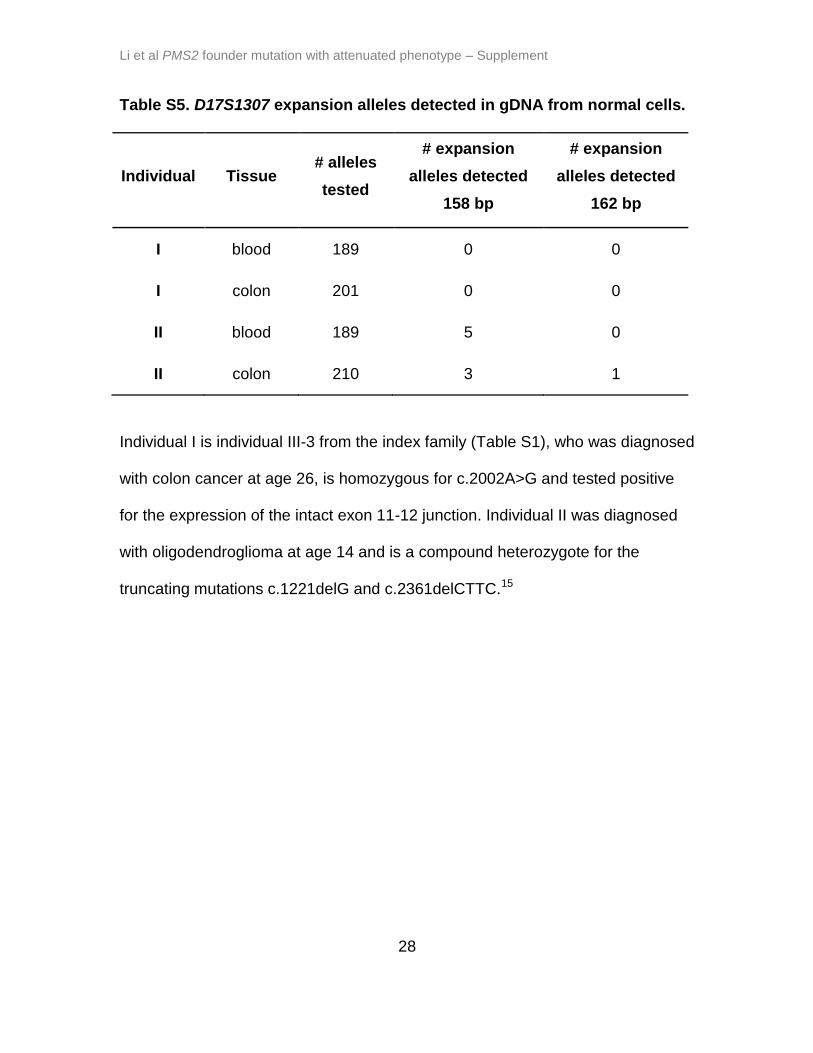
Table S5. D17S1307 expansion alleles detected in gDNA from normal cells.
Individual Tissue # alleles
tested
# expansion
alleles detected
158 bp
# expansion
alleles detected
162 bp
I blood 189 0 0
I colon 201 0 0
II blood 189 5 0
II colon 210 3 1
Individual I is individual III-3 from the index family (Table S1), who was diagnosed
with colon cancer at age 26, is homozygous for c.2002A>G and tested positive
for the expression of the intact exon 11-12 junction. Individual II was diagnosed
with oligodendroglioma at age 14 and is a compound heterozygote for the
truncating mutations c.1221delG and c.2361delCTTC.15

Li et al PMS2 founder mutation with attenuated phenotype – Supplement
29
Web Resources
Leiden Open Variation Database: http://www.lovd.nl/3.0/home
Colon cancer gene variant databases: http://insight-group.org/
Mendelian Inheritance in Man: http://omim.org/
Mutation Taster: http://mutationtaster.org/
PolyPhen-2: http://genetics.bwh.harvard.edu/pph2/index.shtml
PROVEAN: http://provean.jcvi.org/index.php
Nunavik Statistics:
http://www.nunivaat.org/Statistics.aspx/Indicator/Vital_Statistics/
Programs predicting 5’ splicing score:
http://esrsearch.tau.ac.il/
http://www.fruitfly.org/seq_tools/splice.html
http://www.umd.be/HSF/
http://genes.mit.edu/burgelab/maxent/Xmaxent.html
http://ibis.tau.ac.il/ssat/SpliceSiteFrame.htm
http://www.uni-duesseldorf.de/rna/html/hbond_score.php

Li et al PMS2 founder mutation with attenuated phenotype – Supplement
30
References
1. Adzhubei IA, Schmidt S, Peshkin L, Ramensky VE, Gerasimova A, Bork P,
Kondrashov AS, and Sunyaev SR A method and server for predicting
damaging missense mutations. Nat Methods 2010;7:248-249
2. Ng PC and Henikoff S. SIFT: Predicting amino acid changes that affect
protein function. Nucleic Acids Res 2003:31:3812-3814
3. Schwarz JM, Rödelsperger C, Schuelke M, and Seelow D. MutationTaster
evaluates disease-causing potential of sequence alterations. Nat Methods
2010:7:575-576
4. Hamel N, Feng BJ, Foretova L, Stoppa-Lyonnet D, Narod SA, Imyanitov E,
Sinilnikova O, Tihomirova L, Lubinski J, Gronwald J, Gorski B, Hansen TO,
Nielsen FC, Thomassen M, Yannoukakos D, Konstantopoulou I, Zajac V,
Ciernikova S, Couch FJ, Greenwood CM, Goldgar DE, Foulkes WD. On the
origin and diffusion of BRCA1 c.5266dupC (5382insC) in European
populations. Eur J Hum Genet 2011;19:300-306
5. Korbie DJ and Mattick JS. Touchdown PCR for increased specificity and
sensitivity in PCR amplification. Nat Protoc 2008;3:1452-1456
6. Thibodeau SN, Bren G, and Schaid D. Microsatellite instability in cancer of
the proximal colon. Science 1993:260:816-819
7. Shah SN and Eckert KA. Human postmeiotic segregation 2 exhibits biased
repair at tetranucleotide microsatellite sequences. Cancer Res
2009;69:1143-1149

Li et al PMS2 founder mutation with attenuated phenotype – Supplement
31
8. Senter L, Clendenning M, Sotamaa K, Hampel H, Green J, Potter JD,
Lindblom A, Lagerstedt K, Thibodeau SN, Lindor NM, Young J, Winship I,
Dowty JG, White DM, Hopper JL, Baglietto L, Jenkins MA, and de la Chaple
A. The clinical phenotype of Lynch syndrome due to germ-line PMS2
mutations. Gastroenterology 2008;135:419-428
9. Vaughn CP, Robles J, Swensen JJ, Miller CE, Lyon E, Mao R, Bayrak-
Toydemir P, and Samowitz WS. Clinical analysis of PMS2: mutation
detection and avoidance of pseudogenes. Hum Mut 2010;31:588-593
10. Etzler J, Peyrl A, Zatkova A, Schildhaus HU, Ficek A, Merkelbach-Bruse S,
Kratz CP, Attarbaschi A, Hainfellner JA, Yao S, Messiaen L, Slavc I, and
Wimmer K. RNA-based mutation analysis identifies an unusual MSH6
splicing defect and circumvents PMS2 pseudogene interference. Hum Mut
2008;29:299-305
11. Kratz CP, Holter S, Etzler J, Lauten M, Pollett A, Niemeyer CM, Gallinger S,
and Wimmer K. Rhabdomyosarcoma in patients with constitutional
mismatch-repair-deficiency syndrome. J Med Genet 2009;46:418-420
12. De Vos M, Hayward BE, Picton S, Sheridan E, and Bonthron DT. Novel
PMS2 pseudogenes can conceal recessive mutations causing a distinctive
childhood cancer syndrome. Am J Hum Genet 2004;74:954-964
13. Bonthron DT, Hayward BE, De Vos M, and Sheridan E. PMS2 mutations in
childhood cancer. Gut 2005;54:1821

Li et al PMS2 founder mutation with attenuated phenotype – Supplement
32
14. Tan TY, Orme LM, Lynch E, Croxford MA, Dow C, Dewan PA, and Lipton L.
Biallelic PMS2 mutations and a distinctive childhood cancer syndrome. J
Pediatr Hematol Oncol 2008;30254-257
15. Lindsay H, Jubran RF, Wang L, Kipp BR, and May WA. Simultaneous
Colonic Adenocarcinoma and Medulloblastoma in a 12-Year-Old with
Biallelic Deletions in PMS2. J Pediatr 2013;163:601-3
16. Sjursen W, Bjornevoll I, Engebretsen LF, Fjelland K, Halvorsen T, and
Myrvold HE. A homozygote splice site PMS2 mutation as cause of Turcot
syndrome gives rise to two different abnormal transcripts. Fam Cancer
2009;8:179-186
17. Holter S, Pollett A, Zogopoulos G, Kim H, Schwenter F, Asai K, Gallinger S,
Clendenning M, Steinbach G, Jacobson A, and Boycott KM. Hepatic
adenomas caused by somatic HNF1A mutations in children with biallelic
mismatch repair gene mutations. Gastroenterology 2011;140:735-736
18. Peron S, Metin A, Gardes P, Alyanakian MA, Sheridan E, Kratz CP, Fischer
A, and Durandy A. Human PMS2 deficiency is associated with impaired
immunoglobulin class switch recombination. J Exp Med 2008;205:2465-
2472
19. Trimbath JD, Petersen GM, Erdman SH, Ferre M, Luce MC, and Giardiello
FM. Cafe-au-lait spots and early onset colorectal neoplasia: a variant of
HNPCC? Fam Cancer 2001;1:101-105
20. Truninger K, Menigatti M, Luz J, Russell A, Haider R, Gebbers JO,
Bannwart F, Yurtsever H, Neuweiler J, Riehle HM, Cattaruzza MS,

Li et al PMS2 founder mutation with attenuated phenotype – Supplement
33
Heinimann K, Schar P, Jiricny J, and Marra G. Immunohistochemical
analysis reveals high frequency of PMS2 defects in colorectal cancer.
Gastroenterology 2005;128:1160-1171
21. Gulati S, Gustafson S, and Daw H. Lynch Syndrome Associated With PMS2
Mutation: Understanding Current Concepts. Gastrointest. Cancer Res
2011;4:188-190
22. De Rosa M, Fasano C, Panariello L, Scarano MI, Belli G, Iannelli A,
Ciciliano F, and Izzo P. Evidence for a recessive inheritance of Turcot's
syndrome caused by compound heterozygous mutations within the PMS2
gene. Oncogene 2000;19:1719-1723
23. Vaughn CP, Hart KJ, Samowitz WS, and Swensen JJ. Avoidance of
pseudogene interference in the detection of 3' deletions in PMS2. Hum
Mutat 2011;32:1063-1071
24. Gottschling S, Reinhard H, Pagenstecher C, Kruger S, Raedle J, Plotz G,
Henn W, Buettner R, Meyer S, and Graf N. Hypothesis: possible role of
retinoic acid therapy in patients with biallelic mismatch repair gene defects.
Eur J Pediatr 2008;167:225-229
25. Gururangan S, Frankel W, Broaddus R, Clendenning M, Senter L,
McDonald M, Eastwood J, Reardon D, Vredenburgh J, and Quinn J.
Multifocal anaplastic astrocytoma in a patient with hereditary colorectal
cancer, transcobalamin II deficiency, agenesis of the corpus callosum,
mental retardation, and inherited PMS2 mutation. Neuro Oncol 2008;10:93-
97

Li et al PMS2 founder mutation with attenuated phenotype – Supplement
34
26. Hendriks Y, Jagmohan-Changur S, van der Klift HM, Morreau H, van
Puijenbroek M, Tops C, van Os T, Wagner A, Ausems MG, and Gomez E.
Heterozygous Mutations in PMS2 Cause Hereditary Nonpolyposis
Colorectal Carcinoma (Lynch Syndrome). Gastroenterology 2006;130:312-
322
27. Baas AF, Gabbett M, Rimac M, Kansikas M, Raphael M, Nievelstein RA,
Nicholls W, Offerhaus J, Bodmer D, Wernstedt A, Krabichler B, Strasser U,
Nystrom M, Zschocke J, Robertson SP, van Haelst MM, and Wimmer K.
Agenesis of the corpus callosum and gray matter heterotopia in three
patients with constitutional mismatch repair deficiency syndrome. Eur J
Hum Genet 2013;21:55-61
28. Chmara M, Wernstedt A, Wasag B, Peeters H, Renard M, Beert E, Brems H,
Giner T, Bieber I, Hamm H, Sciot R, Wimmer K, and Legius E. Multiple
pilomatricomas with somatic CTNNB1 mutations in children with constitutive
mismatch repair deficiency. Genes Chromosomes Cancer 2013;52:656-664
29. De Vos M, Hayward BE, Charlton R, Taylor GR, Glaser AW, Picton S, Cole
TR, Maher ER, Mckeown CME, Mann JR, Yates JR, Baralle D, Rankin J,
Bonthron DT, and Sheridan E. PMS2 mutations in childhood cancer. J Nat
Cancer Inst 2006;98:358-361
30. Shapiro MB and Senapathy P. RNA splice junctions of different classes of
eukaryotes: sequence statistics and functional implications in gene
expression. Nucleic Acids Res 1987;15:7155-7174

Li et al PMS2 founder mutation with attenuated phenotype – Supplement
35
31. Pertea M, Lin X, and Salzberg SL. GeneSplicer: a new computational
method for splice site prediction. Nucleic Acids Res 2001;29:1185-1190
32. Desmet FO, Hamroun D, Lalande M, Collod-Beroud G, Claustres M, and
Beroud C. Human Splicing Finder: an online bioinformatics tool to predict
splicing signals. Nucleic Acids Res 2009;37(9):e67
33. Yeo G and Burge CB. Maximum entropy modeling of short sequence motifs
with applications to RNA splicing signals. J Comput Biol 2004;11:377-394
34. Carmel I, Tal S, Vig I, and Ast G. Comparative analysis detects
dependencies among the 5G splice-site positions. RNA 2004;10:828-840
35. Freund M, Asang C, Kammler S, Konermann C, Krummheuer J+, Hipp M,
Meyer I, Gierling W, Theiss S, and Preuss T. A novel approach to describe
a U1 snRNA binding site. Nucleic Acids Res 2003;31:6963-6975