supplementary table 1 - media.nature.com · supplementary table 2 lists of hydrogen bonds between...
TRANSCRIPT

Supplementary Table 1 Data collection, phasing and refinement statistics
GA4-GID1A-DELLA
Native
GA3-GID1A-DELLA
Native
GA3-GID1A-DELLA
SeMet
Data collection
Space group P43212 P43212 P43212
Cell dimensions
a, b, c (Å) 81.8, 81.8, 129.9 82.0, 82.0, 130.1 82.1, 82.1, 129.8
Peak
Wavelength (Å) 1.00000 0.99533 0.97938
Resolution (Å) 50-1.8 (1.86-1.80)* 50-1.8 (1.86-1.80) 20-2.0 (2.07-2.00)
Rsym 6.5 (6.8) 5.9 (12.9) 6.4 (11.9)
I/I 71.4 (58.8) 65.8 (28.6) 72.2 (39.7)
Completeness (%) 99.0 (98.2) 100 (98.4) 100 (100)
Redundancy 13.3 (11.2) 13.4 (14.4) 15.4 (15.3)
Refinement
Resolution (Å) 50.0-1.8 50.0-1.8
No. reflections 40,855 39,256
Rwork/ Rfree 19.8/22.1 20.5/22.9
No. atoms
Protein 3,156 3,134
Ligand/ion 24 25
Water 267 216
B-factors
Protein 19.10 22.83
Ligand/ion 11.04 17.75
Water 24.32 27.28
R.m.s deviations
Bond lengths (Å) 0.005 0.005
Bond angles (º) 1.21 1.18
One crystal was used for each data set.
*Values in parenthesis are for highest resolution shell.
www.nature.com/nature 1
SUPPLEMENTARY INFORMATION
doi: 10.1038/nature07519

Supplementary Table 2 Lists of hydrogen bonds between GA and GID1A
GA3-GID1A-DELLA GA4-GID1A-DELLA
GA atom GID1A
residue/atom
distance for GA3 distance for GA4
O3-1 H2O 2.80 Å 2.89 Å
O3-1 Y127/OH 2.83 Å 2.75 Å
O7-1 S116/N 3.01 Å 2.96 Å
O7-1 S116/O 2.68 Å 2.71 Å
O7-1 S191/O 3.28 Å 3.23 Å
O7-2 S191/O 2.95 Å 3.17 Å
O7-2 H2O 2.65 Å 2.71 Å
O19-1 G320/N 2.87 Å 2.90 Å
O13 F238/O 3.25 Å - (GA4 has no O13)
O13 H2O 3.15 Å - (GA4 has no O13)
www.nature.com/nature 2
doi: 10.1038/nature07519 SUPPLEMENTARY INFORMATION

Figure S1 Structural comparison of the GA3- and GA4-GID1A-DELLA complexes. a, Chemical structures of bioactive GA1 and GA7. The bioactive GA molecules conserve
one 7-carboxylate, 3-hydroxyl and one lactone (a cyclic carboxylester) moiety. The
3-hydroxyl group is introduced by GA 3-oxidase at the final step to produce active GA
derivatives.
b, Structural comparison of the GA3- and GA4-GID1A-DELLA complexes. The
GA3-GID1A-DELLA complex is superimposed on the GA4-GID1A-DELLA complex
(grey). The GA3-GID1A-DELLA complex is colored in pink (GAI DELLA domain), light
blue (GID1A) and green (GA3). The GA4-GID1A-DELLA complex is colored in light
green (GAI DELLA domain), yellow-orange (GID1A) and magenta (GA4). The overall
mean rms deviation is extremely small, 0.18 Ǻ, over all the C atom positions of aligned
residues. The GID1A molecules superimpose with an averaged rms deviation of 0.14 Ǻ
over all the C atom positions and the DELLA domains superimpose with an averaged
rms deviation of 0.25 Ǻ.
c, Molecular surface of the GA3-GID1A-DELLA complex with the GAI DELLA domain
(pink), the GID1A N-terminal extension (cyan) and the GID1A / core domain (light
blue).
www.nature.com/nature 3
doi: 10.1038/nature07519 SUPPLEMENTARY INFORMATION
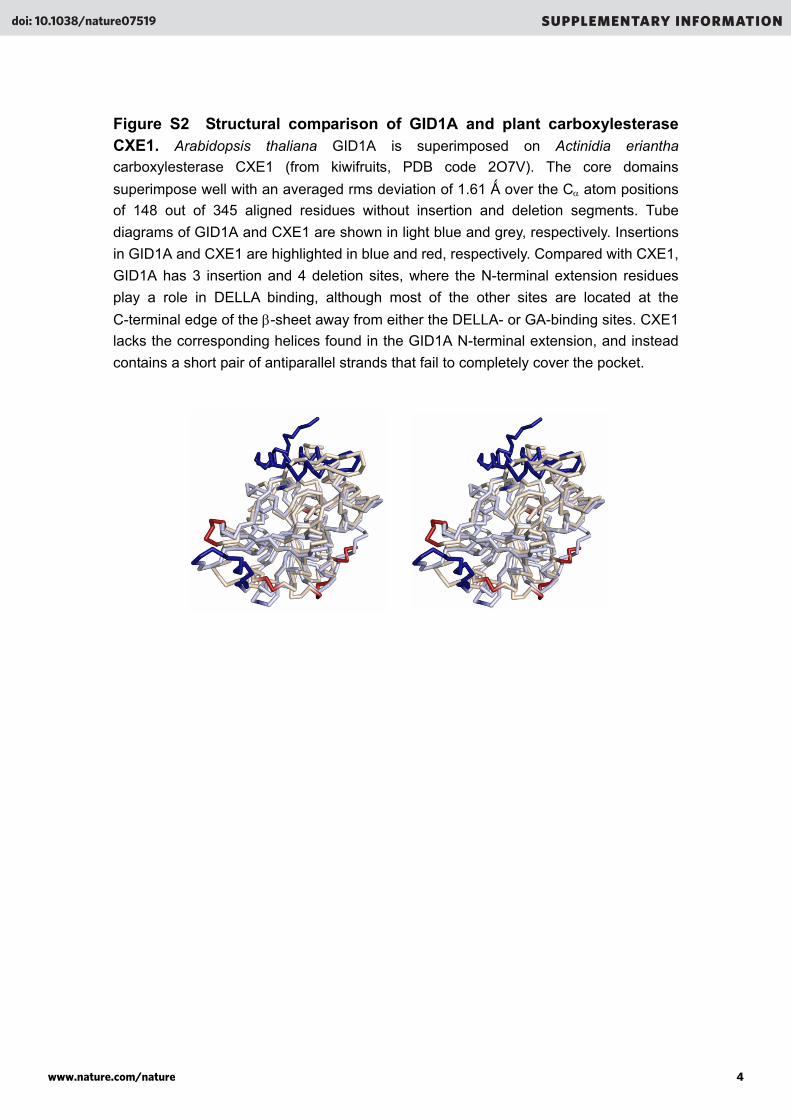
Figure S2 Structural comparison of GID1A and plant carboxylesterase CXE1. Arabidopsis thaliana GID1A is superimposed on Actinidia eriantha
carboxylesterase CXE1 (from kiwifruits, PDB code 2O7V). The core domains
superimpose well with an averaged rms deviation of 1.61 Ǻ over the C atom positions
of 148 out of 345 aligned residues without insertion and deletion segments. Tube
diagrams of GID1A and CXE1 are shown in light blue and grey, respectively. Insertions
in GID1A and CXE1 are highlighted in blue and red, respectively. Compared with CXE1,
GID1A has 3 insertion and 4 deletion sites, where the N-terminal extension residues
play a role in DELLA binding, although most of the other sites are located at the
C-terminal edge of the -sheet away from either the DELLA- or GA-binding sites. CXE1
lacks the corresponding helices found in the GID1A N-terminal extension, and instead
contains a short pair of antiparallel strands that fail to completely cover the pocket.
www.nature.com/nature 4
doi: 10.1038/nature07519 SUPPLEMENTARY INFORMATION

Figure S3 Sequence alignment of GID1s and a plant carboxylesterase. Structural-based sequence alignment of GID1 proteins, A. thaliana GID1A, B and C,
Oryza sativa, (rice) GID1 (Os GID1), and A. eriantha (kiwifruit) carboxylesterase CXE1
(Ae CXE1, PDB code 2O7V). The secondary structures of GID1A are shown at the top
with -helices (yellow bars), 310-helices (orange bars), -strands (green arrows) and
disordered residues (dotted lines). The secondary structures of CXE1 are shown at the
bottom. The N-terminal extension of GID1A comprises three helices, a (residues
9-13),b (18-34) and c (43-49), followed by loopc-1 (residues 51-62). The GID1A
core domain consists of a central eight-stranded mixed -sheet (1-8) and seven
-helices (1-7) and two 310 helices (1 and 2). GID1A lacks three -helices
corresponding to 3, 5, and 8 (grey bars) of CXE1 and one 310-helix corresponding to
1 of CXE1. Instead of 3 of CXE1, GID1A has a 310-helix 1. Bold letters indicate
important residues for GA3 and GA4 binding (blue circles), DELLA binding (black circles),
and the catalytic triad and oxyanion hole found in carboxylesterase (red letters). The
triad residues are Ser191 (loop 5-3), Val319 (loop 8-7) and Asp289 (at loop 7-6).
GA-interacting residues are highlighted in light green and boxed with bold lines.
Identical residues to GID1A are highlighted in pale yellow. Accession numbers: GID1A,
At3g05120; GID1B, At3g63010; GID1C, At5g27320; Os GID1, AB211399. GID1A and
CXE1 share 22.5% sequence identity, whereas no homology at the N-terminal
extension (62 residues) is detected. Among GID1 members, however, the N-terminal
extensions are highly homologous, implying a specific function as the gibberellin
receptor.
www.nature.com/nature 5
doi: 10.1038/nature07519 SUPPLEMENTARY INFORMATION

Figure S3
www.nature.com/nature 6
doi: 10.1038/nature07519 SUPPLEMENTARY INFORMATION

Figure S4 Schematic representation of the interactions. Summary of
intermolecular interactions between GID1A and GA3 in the GA3-GID1A-DELLA complex.
Van der Waals contacts (dotted lines), hydrogen bonds (arrows) and a salt bridge (an
arrow with a dotted line) are shown. Residue side chains that participate in the
interactions are indicated by boxes with bold lines. The N-terminal extension residues of
GID1A are highlighted (cyan). All the interactions are conserved in those with GA4 in the
GA4-GID1A-DELLA complex. Basically, contacts between hydrogen donor and acceptor
atoms are nominated for hydrogen bonds with the typical distances (for example, 2.9 Å
for H-bond between N-H and O) plus/minus 0.2 Å and the angles (for example, C-N---O
and N---O-C) larger than 90.
www.nature.com/nature 7
doi: 10.1038/nature07519 SUPPLEMENTARY INFORMATION

Figure S5 A stereo-view of GA perception in the GA4-GID1A-DELLA complex. GID1A residues that contact GA4 are highlighted in yellow (the core domain)
or cyan (the N-terminal extension) and shown as stick models with van der Waals
surfaces represented by dots. Water molecules are shown as blue spheres. Hydrogen
bonds are indicated with dotted lines. Atom codes are oxygen in red and nitrogen in
blue.
www.nature.com/nature 8
doi: 10.1038/nature07519 SUPPLEMENTARY INFORMATION

Figure S6 Sequence alignment of DELLA domains. The N-terminal DELLA
domains from A. thaliana and O. sativa DELLA proteins are aligned. The secondary
structures of GAI are shown at the top with -helices (yellow bars), loops (solid lines)
and disordered residues (dotted lines). GID1A-binding residues (purple circles) are
indicated with bold letters. Identical residues to GAI are highlighted in pale yellow.
Accession numbers: GAI, At1g14920; RGA, At2g01570; RGL1, At1g66350; RGL2,
At3g03450; RGL3, At5g17490; SLR1, AB262980. GID1A-interacting residues are
highlighted in light green and boxed with bold lines. Identical residues to GID1A are
highlighted in pale yellow.
www.nature.com/nature 9
doi: 10.1038/nature07519 SUPPLEMENTARY INFORMATION

Figure S7 Effects of mutation of the DELLA domain on GID1A binding. Pull-down binding assays of mutated DELLA domains of GAI with GA3-bound GID1A
were performed with single mutations of the DELLA (Leu31, Tyr36 and Val38) and the
LExLE (Met43, Glu51 and Glu54) motifs, and double mutations of Leu31 and Val38,
Tyr36 and Met43, Glu51 and Glu54. Eluted samples analyzed by SDS-PAGE.
www.nature.com/nature 10
doi: 10.1038/nature07519 SUPPLEMENTARY INFORMATION

Figure S8 Conformational flexibility of the GID1A and the DELLA domain of GAI. a, Conformational flexibility of the GID1 N-terminal extension. Proteolysis of GID1A in
the free, GA3-bound and DELLA-bound states. GID1A (residues 14-345) produced by
trypsin-treatment of GST-GID1A was degraded rapidly within 10 min in the GA-unbound
state, whereas resistant over 20-30 min in the GA-bound and DELLA-bound states.
b, Conformational flexibility of the DELLA domain of GAI. CD spectra of the DELLA
domain. No significant changes were observed in the spectra at 20, 37, 50 and 70C.
Secondary structure estimations suggest a 100% random coil structure.
www.nature.com/nature 11
doi: 10.1038/nature07519 SUPPLEMENTARY INFORMATION