supporting information enhanced nonenzymatic … information enhanced nonenzymatic rna copying ......
TRANSCRIPT
Supporting Information
Enhanced Nonenzymatic RNA Copying with 2-AminoimidazoleActivated Nucleotides
Li Li, Noam Prywes†, Chun Pong Tam, Derek K. O’Flaherty, Victor S. Lelyveld, EnverCagri Izgu, Ayan Pal and Jack W. Szostak∗
Howard Hughes Medical Institute, Department of Molecular Biology and Center forComputational and Integrative Biology, Massachusetts General Hospital, Boston,
Massachusetts 02114, United States
∗To whom correspondence should be addressed:CCIB 7215, Simches Research Center
185 Cambridge StreetMassachusetts General HospitalBoston, Massachusetts 02114
E-mail: [email protected]
†Present address: Department of Plant and Environmental Sciences, The Weizmann Institute of Science,
P.O. Box 26, Rehovot 76100, Israel
S1

Contents1 Materials and Methods S5
1.1 Abbreviations . . . . . . . . . . . . . . . . . . . . . . . . . . . . . . . . . . . . . . . . . . . . . S51.2 General information . . . . . . . . . . . . . . . . . . . . . . . . . . . . . . . . . . . . . . . . . S51.3 Procedures for nucleoside-5′-phosphoroimidazolide synthesis . . . . . . . . . . . . . . . . . . . S5
1.3.1 General procedure A for nucleoside-5′-phosphoroimidazolide synthesis . . . . . . . . . S51.3.2 Procedures for nucleoside-5′-phosphoro-(2-aminoimidazolide) synthesis . . . . . . . . . S61.3.3 General procedure B for nucleoside-5′-phosphoroimidazolide synthesis . . . . . . . . . S6
1.4 Measurement of phosphoroimidazolide hydrolysis rate . . . . . . . . . . . . . . . . . . . . . . S61.5 Oligonucleotide synthesis . . . . . . . . . . . . . . . . . . . . . . . . . . . . . . . . . . . . . . S6
1.5.1 Primers, templates, and complementary DNAs or RNAs . . . . . . . . . . . . . . . . . S61.5.2 Trinucleotides activated by 2-aminoimidazole . . . . . . . . . . . . . . . . . . . . . . . S6
1.6 Primer extension reaction . . . . . . . . . . . . . . . . . . . . . . . . . . . . . . . . . . . . . . S71.7 LC-MS analysis of primer extension reaction . . . . . . . . . . . . . . . . . . . . . . . . . . . S81.8 Preparation and characterization of phosphoroimidazolides . . . . . . . . . . . . . . . . . . . S8
1.8.1 Adenosine-5′-phosphoro-(2-aminoimidazole) . . . . . . . . . . . . . . . . . . . . . . . . S81.8.2 Cytidine-5′-phosphoro-(2-aminoimidazole) . . . . . . . . . . . . . . . . . . . . . . . . . S81.8.3 Guanosine-5′-phosphoro-(2-isopropylimidazole) . . . . . . . . . . . . . . . . . . . . . . S81.8.4 Guanosine-5′-phosphoro-(2-phenylimidazole) . . . . . . . . . . . . . . . . . . . . . . . S91.8.5 Guanosine-5′-phosphoro-benzimidazole . . . . . . . . . . . . . . . . . . . . . . . . . . . S91.8.6 Guanosine-5′-phosphoro-(2-methylbenzimidazole) . . . . . . . . . . . . . . . . . . . . . S91.8.7 Guanosine-5′-phosphoro-(2-chloroimidazole) . . . . . . . . . . . . . . . . . . . . . . . . S91.8.8 Guanosine-5′-(2-methyl-1H -pyrrol-3-yl)phosphonate . . . . . . . . . . . . . . . . . . . S101.8.9 Guanosine-5′-phosphoro-(2-aminoimidazole) . . . . . . . . . . . . . . . . . . . . . . . . S121.8.10 Guanosine-5′-phosphoro-(2-methylaminoimidazole) . . . . . . . . . . . . . . . . . . . . S131.8.11 Uridine-5′-phosphoro-(2-aminoimidazole) . . . . . . . . . . . . . . . . . . . . . . . . . S131.8.12 2-thiouridine-5′-phosphoro-(2-aminoimidazole) . . . . . . . . . . . . . . . . . . . . . . S14
2 Supplementary Figures S15
3 Supplementary tables S17
S2

List of FiguresFigure S1 Reaction rates of nonenzymatic primer extension . . . . . . . . . . . . . . . . . . . . S15Figure S2 Hydrolysis rates of activated monomers . . . . . . . . . . . . . . . . . . . . . . . . . . S16
S3
List of TablesTable S1 Observed vs. calculated neutral masses for primer extension products. . . . . . . . . . S17Table S2 Sequences of oligonucleotides used in this study . . . . . . . . . . . . . . . . . . . . . . S18Table S3 Sequences of 2-aminoimidazole activated trinucleotides used in this study . . . . . . . S19
S4
1 Materials and Methods
1.1 AbbreviationsCV, column volumeDCM, dichloromethaneDIAD, diisopropyl azodicarboxylateDIPEA, N,N -diisopropylethylamineDMSO, dimethyl sulfoxideDPDS, 2,2′-dipyridyldisulfideEDC, 1-ethyl-3-(3-dimethylaminopropyl)carbodiimide hydrochlorideEDTA, ethylenediaminetetraacetic acidHEPES, 4-(2-hydroxyethyl)-1-piperazineethanesulfonic acidHPLC, high-performance liquid chromatographyHRMS, high-resolution mass spectrometryLC-MS, liquid chromatography-mass spectrometryNMR, nuclear magnetic resonancePAGE, polyacrylamide gel electrophoresisQ-TOF, quadrupole time-of-flightTEAA, triethylammonium acetateTEAB, triethylammonium bicarbonateTHF, tetrahydrofuranTris, tris(hydroxymethyl)aminomethane
1.2 General informationAll chemicals were purchased from Sigma-Aldrich (St. Louis, MO) unless otherwise noted. All nucleoside-5′-monophosphate free-acid compounds were purchased from Santa Cruz Biotechnology (Dallas, TX). 2-bromoimidazole was purchased from AstaTech (Bristol, PA). 2-thiouridine was purchased from Carbosynth(Compton, Berkshire, UK). 2-aminoimidazole hemisulfate were purchased from Alfa Aesar (Ward Hill, MA).Reverse phase flash chromatography was performed using a prepacked RediSep Rf Gold C18Aq 50 g columnfrom Teledyne Isco (Lincoln, NE).
1H and 13C NMR spectra were acquired at 400 MHz and 100 MHz, respectively, on a Varian OxfordAS-400 NMR spectrometer. Chemical shifts are reported in parts per million. 1H NMR was referenced usingthe solvent resonance as the internal standard (HDO, 4.79 ppm at 25 ◦C and 4.92 ppm at 12 ◦C) 1. 13CNMR was referenced using the solvent resonance (CDCl3, 77.2 ppm) or the CH3 signal of acetone as theinternal standard (30.89 ppm).1 Data are reported as follows: chemical shift, multiplicity (s = singlet, d =doublet, t = triplet, q = quadruplet, m = multiplet), integration, and coupling constants. HRMS was carriedout on an Agilent 6520 QTOF LC-MS.
1.3 Procedures for nucleoside-5′-phosphoroimidazolide synthesis1.3.1 General procedure A for nucleoside-5′-phosphoroimidazolide synthesis
To a solution of nucleoside-5′-monophosphate free acid (1 mmol, 1 equiv.), imidazole (5 mmol, 5 equiv.),triethylamine (4 mmol, 4 equiv.) in DMSO (30 mL) was added triphenylphosphine (9 mmol, 9 equiv.) andDPDS (10 mmol, 10 equiv.) under stirring at room temperature. After 30 min, the reaction mixture wasadded to a pre-chilled solution of acetone/diethyl ether/triethylamine (400/250/30 mL) to which 2 mL ofsaturated NaClO4 solution in acetone had been added. After the precipitate had settled out, the majorityof the supernatant was removed using pipette-suction and the remaining suspension was centrifuged at4,000 rpm for 5 min. The pellets were washed twice with acetone (10 mL). The product was purified byreverse phase flash chromatography using gradient elution between (A) aqueous 25 mM TEAB buffer (pH7.5) and (B) acetonitrile. The sample was eluted between 0% and 15% B over 8 CVs with a flow rate of40 mL/min. Fractions containing products were pooled and lyophilized at -15 ◦C.
S5

1.3.2 Procedures for nucleoside-5′-phosphoro-(2-aminoimidazolide) synthesis
The synthesis of nucleoside-5′-phosphoro-(2-aminoimidazolide) was carried out as described above with theexception that the pH of flash-chromatography fractions containing products were adjusted to 10 using NaOHbefore lyophilization.
1.3.3 General procedure B for nucleoside-5′-phosphoroimidazolide synthesis
An aqueous solution (10 mL) of nucleoside-5′-monophosphate disodium salt (1 mmol, 1 equiv.), EDC (5 mmol,5 equiv.), and imidazole (4 mmol, 4 equiv.) was adjusted to a pH of 7 and allowed to stir for 4 h at roomtemperature. The product was purified by reverse phase flash chromatography using gradient elution between(A) aqueous 25 mM TEAB buffer (pH 7.5) and (B) acetonitrile. The sample was eluted between 0% and15% B over 8 CVs with a flow rate of 40 mL/min. Fractions containing product were pooled and lyophilizedat -15 ◦C.
1.4 Measurement of phosphoroimidazolide hydrolysis rateHydrolysis of phosphoroimidazolides 1a-pG and 1j-pG was performed at 25 ◦C in reaction buffers containing200 mM HEPES (pH 8.0), 50 mM MgCl2, 400 mM NaCl, 0.1% (v/v) trimethyl phosphate, and 10% (v/v)D2O. The hydrolysis reactions were initiated by adding 5 mM 1a-pG or 1j-pG and monitored at 25 ◦Cby 31P NMR spectroscopy on a Varian 400 MHz NMR spectrometer (Oxford AS-400) using previouslypublished procedures.2 The hydrolysis rate constant of each phosphoroimidazolide was calculated from threeindependent experiments.
1.5 Oligonucleotide synthesis1.5.1 Primers, templates, and complementary DNAs or RNAs
All oligonucleotides used in this study are listed in Table S2. Synthetic oligonucleotides were either purchasedfrom Integrated DNA Technologies or prepared by solid-phase synthesis using an Expedite 8909 DNA/RNAsynthesizer with reagents and phosphoramidites purchased from Glen Research (Sterling, VA) and Chemgenes(Wilmington, MA), respectively. Oligonucleotides prepared in-house were deprotected and purified by ion-exchange HPLC on a PA-100 column (Dionex, Sunnyvale, CA) using gradient elution between (A) aqueous25 mM Tris buffer (pH 8.0) and (B) aqueous 25 mM Tris buffer (pH 8.0) with 1 M NaCl with a flow rate of10 mL/min. The collected fractions were desalted and concentrated with Amicon Ultra-15 centrifugal filters(Millipore, Billerica, MA) to a final concentration of 100 µM.
1.5.2 Trinucleotides activated by 2-aminoimidazole
All activated trinucleotides used in this study are listed in Table S3. Trinucleotides were prepared by solid-phase synthesis using a MerMade 6 DNA/RNA synthesizer (Bioautomation, Plano, TX). The trinucleotideswere chemically phosphorylated using bis(2-cyanoethyl)-N,N -diisopropyl phosphoramidite (Chemgenes). The5′-phosphorylated trinucleotides were deprotected and purified by reverse phase flash chromatography usinggradient elution between (A) aqueous 25 mM TEAB buffer (pH 7.5) and (B) acetonitrile. The sample waseluted between 0% and 10% B over 8 CVs with a flow rate of 40 mL/min. The collected fractions were driedin vacuo.
To a solution of trinucleotides (10 µmol, 1 equiv.) and triethylamine (40 equiv.) in DMSO (5 mL) wasadded 2-aminoimidazole hemisulfate (30 equiv.), triphenylphosphine (30 equiv.), and DPDS (30 equiv.) understirring. After 5 h the mixture was added to a pre-chilled mixture of acetone/diethyl ether/triethylamine(80/50/6 mL) to which 0.4 mL of saturated NaClO4 solution in acetone had been added. The precipitatewas pelleted by centrifugation at 4,000 rpm for 5 min and washed twice with acetone (10 mL). The pellet wasdried under vacuum, resuspended in deionized water, and purified by reverse phase HPLC on a 250 mm ×21.2 mm (length × i.d.) Eclipse XDB-C18 PrepHT column with 7 µm particle size (Agilent, Santa Clara, CA)using gradient elution between (A) aqueous 25 mM TEAB buffer (pH 7.5) and (B) acetonitrile. The samplewas eluted between 3% and 12% B over 20 min with a flow rate of 10 mL/min. The fractions containingthe product were combined, and the pH was adjusted to 10. The solution was frozen in liquid nitrogen andlyophilized overnight to yield colorless powder.
S6
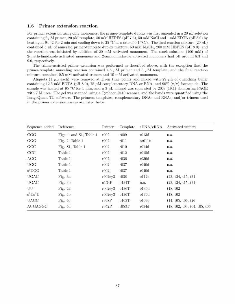
1.6 Primer extension reactionFor primer extension using only monomers, the primer-template duplex was first annealed in a 20 µL solutioncontaining 6 µM primer, 20 µM template, 50 mM HEPES (pH 7.5), 50 mM NaCl and 1 mM EDTA (pH 8.0) byheating at 94 ◦C for 2 min and cooling down to 25 ◦C at a rate of 0.1 ◦C/s. The final reaction mixture (20 µL)contained 5 µL of annealed primer-template duplex mixture, 50 mM MgCl2, 200 mM HEPES (pH 8.0), andthe reaction was initiated by addition of 20 mM activated monomers. The stock solutions (100 mM) of2-methylimidazole activated monomers and 2-aminoimidazole activated monomers had pH around 8.3 and9.6, respectively.
The trimer-assisted primer extension was performed as described above, with the exception that theprimer-template annealing reaction contained 4.8 µM primer and 6 µM template, and the final reactionmixture contained 0.5 mM activated trimers and 10 mM activated monomers.
Aliquots (1 µL each) were removed at given time points and mixed with 29 µL of quenching buffercontaining 12.5 mM EDTA (pH 8.0), 75 µM complementary DNA or RNA, and 90% (v/v) formamide. Thesample was heated at 95 ◦C for 1 min, and a 3-µL aliquot was separated by 20% (19:1) denaturing PAGEwith 7 M urea. The gel was scanned using a Typhoon 9410 scanner, and the bands were quantified using theImageQuant TL software. The primers, templates, complementary DNAs and RNAs, and/or trimers usedin the primer extension assays are listed below.
Sequence added Reference Primer Template cDNA/cRNA Activated trimers
CGG Figs. 1 and S1, Table 1 r002 r009 r013d n.a.GGG Fig. 2, Table 1 r002 r011 or011c n.a.GCC Fig. S1, Table 1 r002 r010 r014d n.a.CCC Table 1 r002 r012 r015d n.a.AGG Table 1 r002 r036 r039d n.a.UGG Table 1 r002 r037 r040d n.a.s2UGG Table 1 r002 r037 r040d n.a.UGAC Fig. 3a r002cy3 r038 o112c t23, t24, t15, t31UGAC Fig. 3b o134P o134T n.a. t23, t24, t15, t31UU Fig. 4a r002cy3 o136T o136d t18, t02s2Us2U Fig. 4b r002cy3 o136T o136d t18, t02UAGC Fig. 4c r098P o103T o103c t14, t05, t06, t26AUGAGGC Fig. 4d r052P r053T r054d t18, t02, t03, t04, t05, t06
S7
1.7 LC-MS analysis of primer extension reactionThe primer extension reaction (30 µL) containing 10 µM primer, 12 µM template, 50 mM MgCl2, 200 mMHEPES (pH 8.0), 0.5 mM of activated trimers, and 10 mM activated monomers was allowed to react for1 day, quenched by adding 3 µL of 0.5 M EDTA (pH 8.0) and 6 µL of 5 M ammonium acetate (pH 5.0),and precipitated by 90 µL of EtOH at -80 ◦C for 2 hours. The precipitant was pelleted by centrifugation at12,000 rpm for 30 minutes, washed twice by 300 µL 67% (v/v) EtOH and resuspended in 25 µL of LC-MSgrade water. The solution was desalted by ion pairing reverse phase (IP-RP) purification on a C18 ZipTippipette tip (Millipore, Billerica, MA). The tip was wetted with acetonitrile and equilibrated with 2 M TEAAprior to sample binding. Extensive washing with 10 mM TEAA was followed by elution in 50% acetonitrile.The eluate was dried under vacuum, followed by resuspension in LC-MS grade water.
The eluted sample was separated and analyzed on an Agilent 1200 HPLC coupled to an Agilent 6230TOF equipped with a solvent degasser, column oven, autosampler, and diode array detector. The sample wasseparated by IP-RP-HPLC on a 100 mm × 1 mm (length × i.d.) Xbridge C18 column with 3.5 µm particlesize (Waters, Milford, MA) using gradient elution between (A) aqueous 200 mM 1,1,1,3,3,3-hexafluoro-2-propanol with 1.25 mM triethylamine, pH 7.0, and (B) methanol, where the sample was eluted between 2.5%and 20% B over 28.5 min with a flow rate of 125 µL/min at 60 ◦C. Samples were analyzed in negative modefrom 239 m/z to 3200 m/z with a scan rate of 1 spectrum/s and the following instrument settings: dryinggas flow, 8 L/min; drying gas temperature, 325 ◦C; nebulizer pressure, 30 psig; capillary voltage, 3500 V;fragmentor, 200 V; and skimmer, 65 V. Extracted ion chromatograms were generated with the Find byFormula algorithm in Agilent’s MassHunter Qualitative Analysis software using a chemical formula databaseof possible primer extension products. The database was generated by calculating the composition of allpossible random RNA extensions beyond the primer sequence up to +8.
1.8 Preparation and characterization of phosphoroimidazolides1.8.1 Adenosine-5′-phosphoro-(2-aminoimidazole)
The adenosine-5′-phosphoro-(2-aminoimidazole) was prepared on a 1-mmol scale using adenosine-5′-mono-phosphate (1 equiv.), 2-aminoimidazole hemisulfate (10 equiv.), triethylamine (13 equiv.), DPDS (10 equiv.),triphenylphosphine (9 equiv.) in DMSO (30 mL) following procedures in section 1.3.2.1H NMR (400 MHz, D2O, 25 ◦C) δ: 8.19 (s, 1H), 8.06 (s, 1H), 6.62 (m, 1H), 6.42 (m, 1H), 5.97 (d, 1H,J = 5.2 Hz), 4.66 (t, 1H, J = 5.2 Hz), 4.35 (t, 1H, J = 4.8 Hz), 4.27 (m, 1H), 4.05 (m, 2H). Peakscorresponding to residual TEAB observed at 3.07 and 1.20 ppm. 13C{1H} NMR (100 MHz, D2O, 12◦C)δ: 155.9, 153.3, 152.4 (d, J = 4.0 Hz), 149.3, 140.1, 124.8 (d, J = 11.3 Hz), 119.0, 115.9 (d, J = 6.0 Hz),87.8, 83.9 (d, J = 8.8 Hz), 74.7, 70.9, 65.8 (d, J = 5.5 Hz). Peaks corresponding to residual TEAB observedat 165.3, 47.1, and 9.0 ppm. HRMS (Q-TOF) m/z : [M − H]− Calcd for C13H16N8O6P 411.0936; Found:411.0940.
1.8.2 Cytidine-5′-phosphoro-(2-aminoimidazole)
The cytidine-5′-phosphoro-(2-aminoimidazole) was prepared on a 1-mmol scale using cytidine-5′-monophos-phate (1 equiv.), 2-aminoimidazole hemisulfate (10 equiv.), triethylamine (13 equiv.), DPDS (10 equiv.),triphenylphosphine (9 equiv.) in DMSO (30 mL) following procedures in section 1.3.2.1H NMR (400 MHz, D2O) δ: 7.79 (d, 1H, J = 7.6 Hz), 6.75 (m, 1H), 6.56 (m, 1H), 6.00 (d, 1H, J = 7.6 Hz),5.90 (d, 1H, J = 4.4 Hz), 4.21 (t, 1H, J = 4.4 Hz), 4.15 (m, 3H), 4.04 (m, 1H). Peaks corresponding toresidual TEAB observed at 3.07 and 1.21 ppm. 13C{1H} NMR (100 MHz, D2O) δ: 166.7, 158.3, 152.5 (d,J = 3.9 Hz), 141.6, 125.0 (d, J = 11.2 Hz), 116.1 (d, J = 6.0 Hz), 97.0, 90.1, 82.9 (d, J = 8.9 Hz), 74.7, 69.9,65.3 (d, J = 5.5 Hz). Peaks corresponding to residual TEAB observed at 166.2, 47.0, and 9.1 ppm. HRMS(Q-TOF) m/z : [M +H]+ Calcd for C12H18N6O7P 389.0969; Found: 389.0993.
1.8.3 Guanosine-5′-phosphoro-(2-isopropylimidazole)
The guanosine-5′-phosphoro-(2-isopropylimidazole) was prepared on a 1-mmol scale using guanosine-5′-mono-phosphate (1 equiv.), 2-isopropylimidazole (4 equiv.) and EDC (5 equiv.) in acetone:water (1:3, v/v, 10 mL)following procedures in section 1.3.3.
S8
1H NMR (400 MHz, D2O, 12 ◦C) δ: 7.89 (s, 1H), 7.08 (s, 1H), 6.78 (s, 1H), 5.83 (d, 1H, J = 5.2 Hz), 4.84(t, 1H, J = 5.2 Hz), 4.47 (t, 1H, J = 5.2 Hz), 4.25 (m, 1H), 4.01 (m, 2H), 3.34 (sep, 1H, J = 6.8 Hz), 1.04(dd, 6H, J = 6.8, 3.2 Hz). 13C{1H} NMR (100 MHz, D2O, 12◦C) δ: 160.3, 158.4 (d, J = 5.9 Hz), 154.9,152.2, 138.1, 126.6 (d, J = 11.1 Hz), 121.9 (d, J = 6.7 Hz), 117.1, 88.1, 83.9 (d, J = 9.6 Hz), 73.6, 71.0,66.3 (d, J = 5.3 Hz), 27.7, 21.9 (d, J = 15.4 Hz). HRMS (Q-TOF) m/z : [M −H]− Calcd for C16H21N7O7P454.1246; Found: 454.1261.
1.8.4 Guanosine-5′-phosphoro-(2-phenylimidazole)
The guanosine-5′-phosphoro-(2-phenylimidazole) was prepared on a 1-mmol scale using guanosine-5′-mono-phosphate (1 equiv.), 2-phenylimidazole (4 equiv.) and EDC (5 equiv.) in acetone:water (1:3, v/v, 10 mL)following procedures in section 1.3.3.1H NMR (400 MHz, D2O, 12 ◦C) δ: 7.76 (s, 1H), 7.58 (d, 2H, J = 7.2 Hz), 7.35 (m, 2H), 7.29 (m, 2H), 7.01(s, 1H), 5.75 (d, 1H, J = 5.6 Hz), 4.67 (t, 1H, J = 5.6 Hz), 4.15 (m, 1H), 4.04 (t, 1H, J = 4.8 Hz), 3.96 (m,2H). 13C{1H} NMR (100 MHz, D2O, 12◦C) δ: 160.4, 154.9, 152.2, 150.7 (d, J = 4.2 Hz), 138.0, 131.3, 129.9,129.6, 128.5, 127.9 (d, J = 11.1 Hz), 124.4 (d, J = 5.9 Hz), 117.1, 88.1, 83.8 (d, J = 9.9 Hz), 73.5, 70.8, 60.1(d, J = 5.3 Hz). HRMS (Q-TOF) m/z : [M −H]− Calcd for C19H19N7O7P 488.1089; Found: 488.1098.
1.8.5 Guanosine-5′-phosphoro-benzimidazole
The guanosine-5′-phosphoro-benzimidazole was prepared on a 1-mmol scale using guanosine-5′-monophos-phate (1 equiv.), benzimidazole (4 equiv.) and EDC (5 equiv.) in acetone:water (1:3, v/v, 10 mL) followingprocedures in section 1.3.3.1H NMR (400 MHz, D2O, 12 ◦C) δ: 8.15 (s, 1H), 7.54 (s, 1H), 7.40 (d, 2H, J = 8.0 Hz), 7.03 (t, 1H,J = 7.2 Hz), 6.93 (t, 1H, J = 8.0 Hz), 5.55 (d, 1H, J = 4.8 Hz), 4.72 (t, 1H, J = 5.2 Hz), 4.38 (t, 1H,J = 4.8 Hz), 4.23 (m, 1H), 4.07 (m, 2H). 13C{1H} NMR (100 MHz, D2O, 12◦C) δ: 159.3, 153.5, 151.2, 146.0(d, J = 6.7 Hz), 143.2 (d, J = 10.6 Hz), 138.7, 133.4 (d, J = 4.9 Hz), 124.0, 123.8, 118.9, 116.9, 114.1,89.1, 84.0 (d, J = 9.8 Hz), 73.0, 70.9, 66.8 (d, J = 5.5 Hz). HRMS (Q-TOF) m/z : [M − H]− Calcd forC17H17N7O7P
– 462.0933; Found: 462.0947.
1.8.6 Guanosine-5′-phosphoro-(2-methylbenzimidazole)
The guanosine-5′-phosphoro-(2-methylbenzimidazole) was prepared on a 1-mmol scale using guanosine-5′-monophosphate (1 equiv.), 2-methylbenzimidazole (4 equiv.) and EDC (5 equiv.) in acetone:water (1:3, v/v,10 mL) following procedures in section 1.3.3.1H NMR (400 MHz, D2O, 12 ◦C) δ: 7.58 (s, 1H), 7.56 (d, 1H, J = 8 Hz), 7.24 (d, 1H, J = 8 Hz), 7.00 (m,1H), 6.91 (m, 1H), 5.59 (d, 1H, J = 4.4 Hz), 4.71 (t, 1H, J = 4.8 Hz), 4.43 (t, 1H, J = 4.8 Hz), 4.24 (dt,1H, J = 4.8, 4.4 Hz), 4.12 (m, 2H), 2.59 (s, 3H). 13C{1H} NMR (100 MHz, D2O, 12◦C) δ: 159.5, 156.5 (d,J = 6.9 Hz), 153.5, 151.2, 141.7 (d, J = 11 Hz), 139.0, 136.0 (d, J = 6.0 Hz), 123.4, 123.1, 117.4, 117.1,114.5, 89.6, 84.2 (d, J = 8.7 Hz), 73.2, 71.0, 67.0 (d, J = 5.2 Hz), 16.4. HRMS (Q-TOF) m/z : [M −H]−
Calcd for C18H19N7O7P 476.1089; Found: 476.1097.
1.8.7 Guanosine-5′-phosphoro-(2-chloroimidazole)
The guanosine-5′-phosphoro-(2-chloroimidazole) was prepared on a 1-mmol scale using guanosine-5′-mono-phosphate (1 equiv.), 2-chloroimidazole (4 equiv.) and EDC (5 equiv.) in water (10 mL) following proceduresin section 1.3.3.1H NMR (400 MHz, D2O, 12 ◦C) δ: 7.85 (s, 1H), 7.16 (s, 1H), 6.74 (s, 1H), 5.80 (d, 1H, J = 5.2 Hz), 4.81(t, 1H, J = 5.2 Hz), 4.47 (t, 1H, J = 4.8 Hz), 4.27 (m, 1H), 4.12 (m, 2H). 13C{1H} NMR (100 MHz, D2O,12◦C) δ: 160.0, 154.7, 152.1, 138.1, 133.7 (d, J = 3.6 Hz), 127.9 (d, J = 10.3 Hz), 124.5 (d, J = 5.8 Hz),116.9, 88.2, 83.8 (d, J = 9.0 Hz), 73.8, 70.9, 66.7 (d, J = 5.5 Hz). HRMS (Q-TOF) m/z : [M −H]− Calcdfor C13H14ClN7O7P 446.0386; Found: 446.0386.
S9

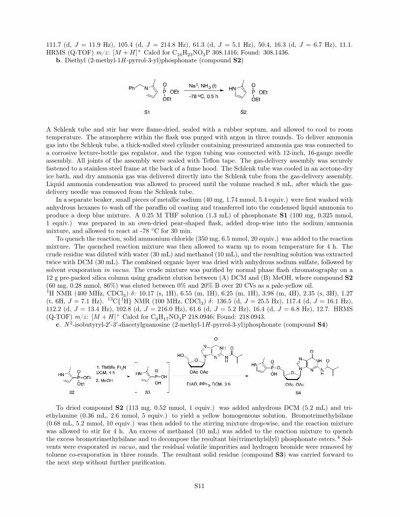
Synthetic scheme of Guanosine-5′-(2-methyl-1H -pyrrol-3-yl)phosphonate. Reaction conditions:(a) [RuCl2(p-cymene)]2, XantPhos, t-BuOK, t-amyl alcohol, 130 ◦C, 16 hr; (b) Na0, NH3 (l), -78 ◦C,30 min; (c) TMSBr, Et3N, DCM, 4 h; then MeOH; (d) DIAD, PPh3, DCM, 3 h; (e) 28% NH3 in H2O, 65 ◦C,2.5 h. [RuCl2(p-cymene)]2, dichloro(p-cymene)ruthenium(II) dimer; XantPhos, 4,5-bis(diphenylphosphino)-9,9-dimethylxanthene; TMSBr, bromotrimethylsilane; DIAD, diisopropyl azodicarboxylate.
1.8.8 Guanosine-5′-(2-methyl-1H -pyrrol-3-yl)phosphonate
a. Diethyl (N -benzyl-2-methyl-3-pyrrolyl)phosphonate (compound S1)
This procedure was adapted from Beller and coworkers.3 To a septa-sealed and oven-dried glass vessel, thefollowing reagents were added: dichloro(p-cymene)ruthenium(II) dimer (92 mg, 0.15 mmol, 0.05 equiv.), 4,5-bis(diphenylphosphino)-9,9-dimethylxanthene (174 mg, 0.3 mmol, 0.1 equiv.), and potassium tert-butoxide(67 mg, 0.6 mmol, 0.2 equiv.), followed by a desiccator-dried magnetic stir bar. After three rounds of argonpurging, tert-amyl alcohol (6 mL), benzylamine (0.48 mL, 4.4 mmol, 1.5 equiv.), ethylene glycol (0.37 mL,6.6 mmol, 2.2 equiv.), and diethyl (2-oxopropyl)phosphonate (0.37 mL, 3 mmol, 1 equiv.) were successivelydelivered to the reaction vessel via needle injections. The reaction vessel was then sealed by a Teflon screwcap, and the reaction was stirred for 16 hours at 130 ◦C. The reaction mixture turned dark brick-red uponhomogenization of solid reagents in tert-amyl alcohol. The solvent of the crude reaction mixture was thenevaporated in vacuo. The crude mixture was purified by normal-phase flash chromatography on a 40 gpre-packed silica column using gradient elution between (A) DCM and (B) MeOH, where compound S1 waseluted between 0% and 10% B over 20 CVs as a dark red oil.1H NMR (400 MHz, CDCl3) δ: 7.32 (m, 3H), 6.97 (d, 2H, J = 6.8 Hz), 6.61 (dd, 1H, J = 4.4, 2.8 Hz), 6.37(dd, 1H, J = 4.4, 2.8 Hz), 5.02 (s, 2H), 4.06 (m, 4H), 2.35 (d, 3H, J = 1.6 Hz), 1.29 (t, 6H, J = 6.8 Hz)13C{1H} NMR (100 MHz, CDCl3) δ: 136.9, 136.4 (d, J = 25.2Hz), 128.8, 127.7, 126.4, 121.7 (d, J = 14.7Hz),
S10
111.7 (d, J = 11.9 Hz), 105.4 (d, J = 214.8 Hz), 61.3 (d, J = 5.1 Hz), 50.4, 16.3 (d, J = 6.7 Hz), 11.1.HRMS (Q-TOF) m/z : [M +H]+ Calcd for C16H23NO3P 308.1416; Found: 308.1436.
b. Diethyl (2-methyl-1H -pyrrol-3-yl)phosphonate (compound S2)
A Schlenk tube and stir bar were flame-dried, sealed with a rubber septum, and allowed to cool to roomtemperature. The atmosphere within the flask was purged with argon in three rounds. To deliver ammoniagas into the Schlenk tube, a thick-walled steel cylinder containing pressurized ammonia gas was connected toa corrosive lecture-bottle gas regulator, and the tygon tubing was connected with 12-inch, 16-gauge needleassembly. All joints of the assembly were sealed with Teflon tape. The gas-delivery assembly was securelyfastened to a stainless steel frame at the back of a fume hood. The Schlenk tube was cooled in an acetone-dryice bath, and dry ammonia gas was delivered directly into the Schlenk tube from the gas-delivery assembly.Liquid ammonia condensation was allowed to proceed until the volume reached 8 mL, after which the gas-delivery needle was removed from the Schlenk tube.
In a separate beaker, small pieces of metallic sodium (40 mg, 1.74 mmol, 5.4 equiv.) were first washed withanhydrous hexanes to wash off the paraffin oil coating and transferred into the condensed liquid ammonia toproduce a deep blue mixture. A 0.25 M THF solution (1.3 mL) of phosphonate S1 (100 mg, 0.325 mmol,1 equiv.) was prepared in an oven-dried pear-shaped flask, added drop-wise into the sodium/ammoniamixture, and allowed to react at -78 ◦C for 30 min.
To quench the reaction, solid ammonium chloride (350 mg, 6.5 mmol, 20 equiv.) was added to the reactionmixture. The quenched reaction mixture was then allowed to warm up to room temperature for 4 h. Thecrude residue was diluted with water (30 mL) and methanol (10 mL), and the resulting solution was extractedtwice with DCM (30 mL). The combined organic layer was dried with anhydrous sodium sulfate, followed bysolvent evaporation in vacuo. The crude mixture was purified by normal phase flash chromatography on a12 g pre-packed silica column using gradient elution between (A) DCM and (B) MeOH, where compound S2(60 mg, 0.28 mmol, 86%) was eluted between 0% and 20% B over 20 CVs as a pale-yellow oil.1H NMR (400 MHz, CDCl3) δ: 10.17 (s, 1H), 6.55 (m, 1H), 6.25 (m, 1H), 3.98 (m, 4H), 2.35 (s, 3H), 1.27(t, 6H, J = 7.1 Hz). 13C{1H} NMR (100 MHz, CDCl3) δ: 136.5 (d, J = 25.5 Hz), 117.4 (d, J = 16.1 Hz),112.2 (d, J = 13.4 Hz), 102.8 (d, J = 216.0 Hz), 61.6 (d, J = 5.2 Hz), 16.4 (d, J = 6.8 Hz), 12.7. HRMS(Q-TOF) m/z : [M +H]+ Calcd for C9H17NO3P 218.0946; Found: 218.0943.
c. N 2-isobutyryl-2′-3′-diacetylguanosine (2-methyl-1H -pyrrol-3-yl)phosphonate (compound S4)
To dried compound S2 (113 mg, 0.52 mmol, 1 equiv.) was added anhydrous DCM (5.2 mL) and tri-ethylamine (0.36 mL, 2.6 mmol, 5 equiv.) to yield a yellow homogeneous solution. Bromotrimethylsilane(0.68 mL, 5.2 mmol, 10 equiv.) was then added to the stirring mixture drop-wise, and the reaction mixturewas allowed to stir for 4 h. An excess of methanol (10 mL) was added to the reaction mixture to quenchthe excess bromotrimethylsilane and to decompose the resultant bis(trimethylsilyl) phosphonate esters.4 Sol-vents were evaporated in vacuo, and the residual volatile impurities and hydrogen bromide were removed bytoluene co-evaporation in three rounds. The resultant solid residue (compound S3) was carried forward tothe next step without further purification.
S11
To a dried flask with crude material (compound S3, 1.1 equiv. assuming 100% conversion) was addedN 2-isobutyryl-2′-3′-diacetylguanosine (204 mg, 0.47 mmol, 1 equiv.), triphenylphosphine (680 mg, 2.6 mmol,5.5 equiv.) and anhydrous DCM (10.4 mL). The mixture was cooled at 0 ◦C for 5 min, and DIAD (0.53 mL,2.60 mmol, 5.5 equiv.) was added to the mixture drop-wise via syringe over five minutes. After the reactionmixture clarified into a clear brown mixture, it was stirred at 0 ◦C for 30 min and then at room temperaturefor 2.5 h. The reaction was stopped by evaporating the solvent in vacuo, followed by normal phase flashchromatography in a pre-packed 40 g silica column. The column was first flushed with 10 CVs of 90:9:1(v/v/v) DCM-MeOH-triethylamine to remove nonpolar impurities, followed by elution with 5 CVs of 99:1(v/v) MeOH-triethylamine. The dried-down crude product was suspended in 9:1 (v/v) water-acetonitrile,and purified with reverse-phase flash chromatography using gradient elution between (A) 25 mM aqueousTEAB buffer (pH 7.5) and (B) acetonitrile, where compound S4 was eluted between 0% and 25% B over25 CVs. The relevant fractions were pooled and lyophilized to afford the title compound as a fluffy whitesolid.1H NMR (400 MHz, CD3OD) δ: 8.18 (s, 1H), 6.53 (dd, 1H, J = 3.5, 3.0 Hz), 6.29 (dd, 1H, J = 3.8, 2.9 Hz),6.11 (m, 2H), 5.59 (dd, 1H, J = 5.2, 2.9 Hz), 4.32 (m, 1H), 4.04 (m, 1H), 3.96 (m, 1H), 2.80 (m, 1H), 2.38(d, 3H, J = 1.6 Hz), 2.11 (s, 3H), 1.98 (s, 3H), 1.20 (m, 6H). Peaks corresponding to residual triethylamineobserved at 2.83 and 1.15 ppm. 13C{1H} NMR (100 MHz, CD3OD) δ: 182.4, 171.5, 170.9, 157.9, 150.8,150.1, 140.8, 134.5 (d, J = 22.7 Hz), 121.9, 116.9 (d, J = 14.8 Hz), 113.4 (d, J = 12.6 Hz), 110.6 (d,J = 203.5 Hz), 87.6, 84.3 (d, J = 9.4 Hz), 74.2, 73.5, 64.5 (d, J = 4.0 Hz), 36.9, 20.7, 20.4, 19.7, 19.5,13.1. Peaks corresponding to residual triethylamine observed at 47.3 and 10.3 ppm. HRMS (Q-TOF) m/z :[M +H]+ Calcd for C23H30N6O10P 581.1761; Found: 581.1786.
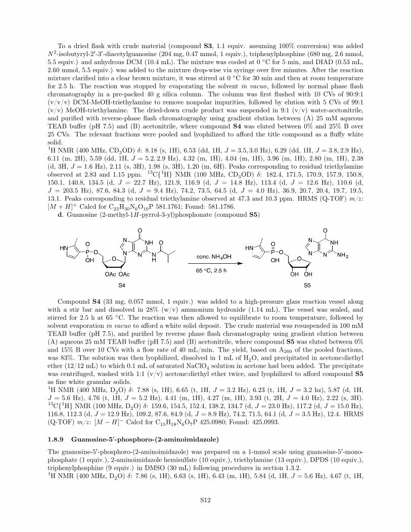
d. Guanosine (2-methyl-1H -pyrrol-3-yl)phosphonate (compound S5)
Compound S4 (33 mg, 0.057 mmol, 1 equiv.) was added to a high-pressure glass reaction vessel alongwith a stir bar and dissolved in 28% (w/v) ammonium hydroxide (1.14 mL). The vessel was sealed, andstirred for 2.5 h at 65 ◦C. The reaction was then allowed to equilibrate to room temperature, followed bysolvent evaporation in vacuo to afford a white solid deposit. The crude material was resuspended in 100 mMTEAB buffer (pH 7.5), and purified by reverse phase flash chromatography using gradient elution between(A) aqueous 25 mM TEAB buffer (pH 7.5) and (B) acetonitrile, where compound S5 was eluted between 0%and 15% B over 10 CVs with a flow rate of 40 mL/min. The yield, based on A260 of the pooled fractions,was 83%. The solution was then lyophilized, dissolved in 1 mL of H2O, and precipitated in acetone:diethylether (12/12 mL) to which 0.1 mL of saturated NaClO4 solution in acetone had been added. The precipitatewas centrifuged, washed with 1:1 (v/v) acetone:diethyl ether twice, and lyophilized to afford compound S5as fine white granular solids.1H NMR (400 MHz, D2O) δ: 7.88 (s, 1H), 6.65 (t, 1H, J = 3.2 Hz), 6.23 (t, 1H, J = 3.2 hz), 5.87 (d, 1H,J = 5.6 Hz), 4.76 (t, 1H, J = 5.2 Hz), 4.41 (m, 1H), 4.27 (m, 1H), 3.93 (t, 2H, J = 4.0 Hz), 2.22 (s, 3H).13C{1H} NMR (100 MHz, D2O) δ: 159.6, 154.5, 152.4, 138.2, 134.7 (d, J = 23.0 Hz), 117.2 (d, J = 15.0 Hz),116.8, 112.3 (d, J = 12.9 Hz), 109.2, 87.6, 84.9 (d, J = 8.9 Hz), 74.2, 71.5, 64.1 (d, J = 3.5 Hz), 12.4. HRMS(Q-TOF) m/z : [M −H]− Calcd for C15H18N6O7P 425.0980; Found: 425.0993.
1.8.9 Guanosine-5′-phosphoro-(2-aminoimidazole)
The guanosine-5′-phosphoro-(2-aminoimidazole) was prepared on a 1-mmol scale using guanosine-5′-mono-phosphate (1 equiv.), 2-aminoimidazole hemisulfate (10 equiv.), triethylamine (13 equiv.), DPDS (10 equiv.),triphenylphosphine (9 equiv.) in DMSO (30 mL) following procedures in section 1.3.2.1H NMR (400 MHz, D2O) δ: 7.86 (s, 1H), 6.63 (s, 1H), 6.43 (m, 1H), 5.84 (d, 1H, J = 5.6 Hz), 4.67 (t, 1H,
S12
J = 5.6 Hz), 4.33 (t, 1H, J = 4.4 Hz), 4.23 (m, 1H), 4.03 (m, 2H). 13C{1H} NMR (100 MHz, D2O): 166.2,159.9, 152.4 (d, J = 4.1 Hz), 152.3, 136.4, 124.8 (d, J = 11.1 Hz), 117.9, 116.0 (d, J = 6.0 Hz), 87.2, 83.8(d, J = 8.8 Hz), 74.1, 71.0, 66.0 (d, J = 5.5 Hz). HRMS (Q-TOF) m/z : [M +H]+ Calcd for C13H18N8O7P429.1031; Found: 429.1050.
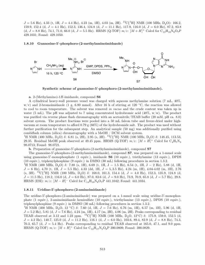
1.8.10 Guanosine-5′-phosphoro-(2-methylaminoimidazole)
Synthetic scheme of guanosine-5′-phosphoro-(2-methylaminoimidazole).
a. 2-(Methylamino)-1H -imidazole, compound S6A cylindrical heavy-wall pressure vessel was charged with aqueous methylamine solution (7 mL, 40%,
w/v) and 2-bromoimidazole (1 g, 6.80 mmol). After 16 h of stirring at 120 ◦C, the reaction was allowedto cool to room temperature. The solvent was removed in vacuo and the crude content was taken up inwater (5 mL). The pH was adjusted to 7 using concentrated hydrobromic acid (48%, w/w). The productwas purified via reverse phase flash chromatography with an acetonitrile/TEAB buffer (20 mM, pH ca. 8.0)solvent system. The product fractions were pooled into a 50 mL falcon tube and freeze-dried under high-vacuum at room temperature to afford 0.79 g (66%) of the hydrobromide salt. The product was used withoutfurther purification for the subsequent step. An analytical sample (50 mg) was additionally purified usingcombiflash column (silica) chromatography with a MeOH / DCM solvent system.1H NMR (400 MHz, D2O) δ: 6.81 (s, 2H), 2.95 (s, 3H). 13C{1H} NMR (100 MHz, D2O) δ: 148.45, 113.53,29.35. Residual MeOH peak observed at 49.45 ppm. HRMS (Q-TOF) m/z : [M + H]+ Calcd for C4H7N398.0713; Found: 98.0721.
b. Preparation of guanosine-5′-phosphoro-(2-methylaminoimidazole), compound S7The guanosine-5′-phosphoro-(2-methylaminoimidazole), compound S7, was prepared on a 1-mmol scale
using guanosine-5′-monophosphate (1 equiv.), imidazole S6 (10 equiv.), triethylamine (13 equiv.), DPDS(10 equiv.), triphenylphosphine (9 equiv.) in DMSO (30 mL) following procedures in section 1.3.2.1H NMR (400 MHz, D2O) δ: 7.88 (s, 1H), 6.69 (t, 1H, J = 1.5 Hz), 6.54 (t, 1H, J = 2 Hz), 5.88 (d, 1H,J = 6 Hz), 4.70 (t, 1H, J = 5.5 Hz), 4.33 (dd, 1H, J = 5, 3.5 Hz), 4.24 (m, 1H), 4.04-4.02 (m, 1H), 2.76(s, 3H). 13C{1H} NMR (100 MHz, D2O) δ: 168.0, 161.3, 154.4 (d, J = 4.0 Hz), 152.3, 135.9, 124.8 (d,J = 11.5 Hz), 118.2, 116.0 (d, J = 6.0 Hz), 87.0, 83.6 (d, J = 9.0 Hz), 73.9, 70.9, 65.8 (d, J = 5.7 Hz), 29.8.HRMS (ESI): m/z : [M −H]− Calcd for C14H19N8O7P 441.1042; Found: 441.1034.
1.8.11 Uridine-5′-phosphoro-(2-aminoimidazole)
The uridine-5′-phosphoro-(2-aminoimidazole) was prepared on a 1-mmol scale using uridine-5′-monophos-phate (1 equiv.), 2-aminoimidazole hemisulfate (10 equiv.), triethylamine (13 equiv.), DPDS (10 equiv.),triphenylphosphine (9 equiv.) in DMSO (30 mL) following procedures in section 1.3.2.1H NMR (400 MHz, D2O, 12 ◦C) δ: 7.60 (d, 1H, J = 7.6 Hz), 6.76 (m, 1H), 6.57 (m, 1H), 5.96 (d, 1H,J = 5.2 Hz), 5.81 (d, J = 7.6 Hz), 4.24 (m, 1H), 4.17 (m, 2H), 4.06 (m, 2H). Peaks corresponding to residualTEAB observed at 3.12 and 1.23 ppm. 13C{1H} NMR (100 MHz, D2O, 12◦C) δ: 175.9, 159.0, 152.5 (d,J = 4.2 Hz), 140.7, 125.0 (d, J = 11.2 Hz), 116.1 (d, J = 6.0 Hz), 103.6, 89.4, 82.9 (d, J = 8.8 Hz), 74.3,70.3, 65.7 (d, J = 5.4 Hz). Peaks corresponding to residual TEAB observed at 165.0, 47.1, and 9.0 ppm.HRMS (Q-TOF) m/z : [M +H]+ Calcd for C12H17N5O8P 390.0809; Found: 390.0829.
S13
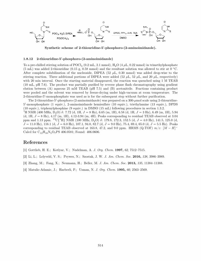
Synthetic scheme of 2-thiouridine-5′-phosphoro-(2-aminoimidazole).
1.8.12 2-thiouridine-5′-phosphoro-(2-aminoimidazole)
To a pre-chilled stirring solution of POCl3 (0.2 mL, 2.1 mmol), H2O (4 µL, 0.22 mmol) in trimethylphosphate(5 mL) was added 2-thiouridine (0.15 g, 0.58 mmol) and the resultant solution was allowed to stir at 0 ◦C.After complete solubilization of the nucleoside, DIPEA (52 µL, 0.30 mmol) was added drop-wise to thestirring reaction. Three additional portions of DIPEA were added (52 µL, 52 µL, and 20 µL, respectively)with 20 min interval. Once the starting material disappeared, the reaction was quenched using 1 M TEAB(10 mL, pH 7.6). The product was partially purified by reverse phase flash chromatography using gradientelution between (A) aqueous 25 mM TEAB (pH 7.5) and (B) acetonitrile. Fractions containing productwere pooled and the solvent was removed by freeze-drying under high-vacuum at room temperature. The2-thiouridine-5′-monophosphate was used as is for the subsequent step without further purification.
The 2-thiouridine-5′-phosphoro-(2-aminoimidazole) was prepared on a 300-µmol scale using 2-thiouridine-5′-monophosphate (1 equiv.), 2-aminoimidazole hemisulfate (10 equiv.), triethylamine (13 equiv.), DPDS(10 equiv.), triphenylphosphine (9 equiv.) in DMSO (15 mL) following procedures in section 1.3.2.1H NMR (400 MHz, D2O) δ: 7.72 (d, 1H, J = 8 Hz), 6.65 (m, 1H), 6.58 (d, 1H, J = 3 Hz), 6.49 (m, 1H), 5.94(d, 1H, J = 8 Hz), 4.17 (m, 1H), 4.12-3.94 (m, 4H). Peaks corresponding to residual TEAB observed at 3.04ppm and 1.13 ppm. 13C{1H} NMR (100 MHz, D2O) δ: 179.8, 172.3, 152.5 (d, J = 4.0 Hz), 141.5, 125.0 (d,J = 11.0 Hz), 116.1 (d, J = 6.0 Hz), 107.1, 94.0, 82.7 (d, J = 9.0 Hz), 75.4, 69.4, 65.0 (d, J = 5.5 Hz). Peakscorresponding to residual TEAB observed at 163.8, 47.2, and 9.0 ppm. HRMS (Q-TOF) m/z : [M − H]−
Calcd for C12H16N5O7PS 406.0581; Found: 406.0606.
References[1] Gottlieb, H. E.; Kotlyar, V.; Nudelman, A. J. Org. Chem. 1997, 62, 7512–7515.
[2] Li, L.; Lelyveld, V. S.; Prywes, N.; Szostak, J. W. J. Am. Chem. Soc. 2016, 138, 3986–3989.
[3] Zhang, M.; Fang, X.; Neumann, H.; Beller, M. J. Am. Chem. Soc. 2013, 135, 11384–11388.
[4] Matulic-Adamic, J.; Haeberli, P.; Usman, N. J. Org. Chem. 1995, 60, 2563–2569.
S14
2 Supplementary Figures
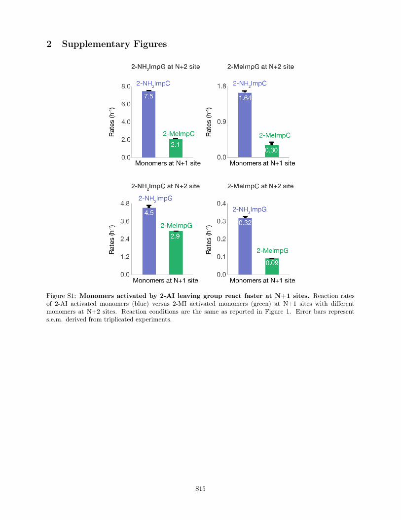
Figure S1: Monomers activated by 2-AI leaving group react faster at N+1 sites. Reaction ratesof 2-AI activated monomers (blue) versus 2-MI activated monomers (green) at N+1 sites with differentmonomers at N+2 sites. Reaction conditions are the same as reported in Figure 1. Error bars represents.e.m. derived from triplicated experiments.
S15
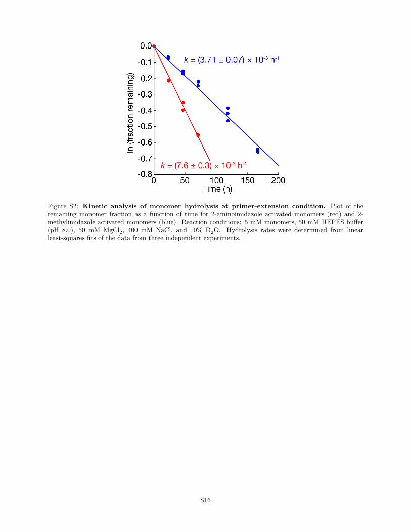
Figure S2: Kinetic analysis of monomer hydrolysis at primer-extension condition. Plot of theremaining monomer fraction as a function of time for 2-aminoimidazole activated monomers (red) and 2-methylimidazole activated monomers (blue). Reaction conditions: 5 mM monomers, 50 mM HEPES buffer(pH 8.0), 50 mM MgCl2, 400 mM NaCl, and 10% D2O. Hydrolysis rates were determined from linearleast-squares fits of the data from three independent experiments.
S16
3 Supplementary tables
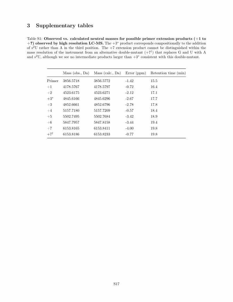
Table S1: Observed vs. calculated neutral masses for possible primer extension products (+1 to+7) observed by high resolution LC-MS. The +3∗ product corresponds compositionally to the additionof s2U rather than A in the third position. The +7 extension product cannot be distinguished within themass resolution of the instrument from an alternative double-mutant (+7†) that replaces G and U with Aand s2U, although we see no intermediate products larger than +3∗ consistent with this double-mutant.
Mass (obs., Da) Mass (calc., Da) Error (ppm) Retention time (min)
Primer 3856.5718 3856.5772 -1.42 15.5+1 4178.5767 4178.5797 -0.72 16.4+2 4523.6175 4523.6271 -2.12 17.1+3∗ 4845.6166 4845.6296 -2.67 17.7+3 4852.6661 4852.6796 -2.78 17.8+4 5157.7180 5157.7209 -0.57 18.4+5 5502.7495 5502.7684 -3.42 18.9+6 5847.7957 5847.8158 -3.44 19.4+7 6153.8165 6153.8411 -4.00 19.8+7† 6153.8186 6153.8233 -0.77 19.8
S17

Table S2: Sequences of oligonucleotides used in this study†
Name R or DNA Sequence (5′→ 3′)
r002 RNA FAM-AGUGAGUAACGCr002cy3 RNA Cy3-AGUGAGUAACGCr009 RNA AACCCGGCGUUACUCACUr010 RNA AAGGGCGCGUUACUCACUr011 RNA AACCCCGCGUUACUCACUr012 RNA AAGGGGGCGUUACUCACUr013d DNA AGTGAGTAACGCCGGGTTr014d DNA AGTGAGTAACGCGCCCTTr015d DNA AGTGAGTAACGCCCCCTTor011c RNA AGUGAGUAACGCGGGGUUr036 RNA AACCCUGCGUUACUCACUr037 RNA AACCCAGCGUUACUCACUr038 RNA AACCGUCAGCGUUACUCACUr039d DNA AGTGAGTAACGCAGGGTTr040d DNA AGTGAGTAACGCTGGGTTo112c RNA AGUGAGUAACGCUGACGGUUo134P RNA AGUGAGUAACGCo134T RNA AACCGUCAGCGUUACUCACUAAAAAo136T RNA CUCAAGCGUUACUCACUo136d DNA AGTGAGTAACGCTTGAGr098P RNA Cy3-AAGGUCACCGo103T RNA UGCGCUACGGUGACCUo103C RNA AGGUCACCGUAGCGCAr052P RNA FAM-CGCUCGACUGr053T RNA GCGCCUCAUCAGUCGAGCGr054d DNA CGCTCGACTGATGAGGCGC
†FAM, fluorescein; Cy3, cyanine 3.
S18

Table S3: Sequences of 2-aminoimidazole activated trinucleotides used in this study
Name Sequence (5′→ 3′)
t02 GAGt03 AGGt04 GGCt05 GCGt06 CGCt14 AGCt15 CGGt18 UGAt23 GACt24 ACGt26 GCAt31 GGU
S19