supporting information for: deoxyfluoro-d-trehalose … · s1 supporting information for:...
TRANSCRIPT

S1
Supporting Information for:
Deoxyfluoro-D-trehalose (FDTre) analogues as potential PET probes for
imaging mycobacterial infection: rapid synthesis and purification,
conformational analysis, and uptake by mycobacteria
Sarah R. Rundell,a,† Zachary L. Wagar,a,† Lisa M. Meints,a Claire D. Olson,a Mara K. O’Neill,a Brent F.
Piligian,a Anne W. Poston,a Robin J. Hood,a Peter J. Woodruff,b and Benjamin M. Swartsa*
aDepartment of Chemistry and Biochemistry, Central Michigan University, Mount Pleasant, MI 48859
(USA). E-mail: [email protected].
bDepartment of Chemistry, University of Southern Maine, Portland, ME (USA).
†These authors contributed equally to this work.
Table of Contents
Supplementary figures __________________________________________________________ S2
Figure S1. EXSIDE spectra for trehalose and 19F-FDTre analogues ___________________ S2
Figure S2. Images of lowest energy conformers of trehalose and 19F-FDTre analogues ____ S3
Figure S3. Chromatograms for uptake of FDTre analogues by M. smegmatis ____________ S4
Figure S4. Data for rapid synthesis, purification, and administration of 19F-FDTre probes _ S5
General experimental for synthesis ________________________________________________ S6
Expression and purification of TreT _______________________________________________ S6
Luminescence glycosyltransferase assay ____________________________________________ S7
Chemoenzymatic synthesis of 19F-FDTre analogues ___________________________________ S7
Chemical synthesis of 19F-4-FDTre ________________________________________________ S8
Conformational analysis of 19F-FDTre analogues _____________________________________ S9
NMR experiments __________________________________________________________ S9
Molecular modeling ________________________________________________________ S10 19F-FDTre uptake analysis in M. smegmatis _________________________________________ S10
Protocol for the rapid synthesis, purification, and administration of FDTre probes ___________ S11
References for supporting information _____________________________________________ S12
NMR spectra for 19F-FDTre analogues _____________________________________________ S13
Electronic Supplementary Material (ESI) for Organic & Biomolecular Chemistry.This journal is © The Royal Society of Chemistry 2016

S2
Supplementary figures
Figure S1. EXSIDE spectra showing long-range heteronuclear couplings across the glycosidic bond for
(A) trehalose, (B) 19F-2-FDTre, (C) 19F-3-FDTre, (D) 19F-4-FDTre, and (E) 19F-6-FDTre. Dashed lines
indicate cross-peaks between H1–C1’ and H1’–C1 for each compound. 3JCOCH constants were measured
from EXSIDE spectra doublets using a scaling factor of 25, as shown.

S3
Figure S2. MM3*-derived lowest energy conformers of (A) trehalose, (B) 19F-2-FDTre, (C) 19F-3-FDTre,
(D) 19F-4-FDTre, and (E) 19F-6-FDTre using NMR-determined glycosidic dihedral angles (see Table 1 in
manuscript) as constraints. The fluorine atom is colored green and marked with an asterisk (*) in (B–E).
Images generated in MacroModel version 11.2.

S4
Figure S3. GC-MS data evaluating SugABC-LpqY-dependent uptake of (A) 19F-2-FDTre, (B) 19F-3-
FDTre, (C) 19F-4-FDTre, and (D) 19F-6-FDTre by M. smegmatis. Standards or cytosolic extracts from
untreated or 19F-FDTre-treated (25 µM) cells were dried, TMS-derivatized, and analyzed by GC-MS
according to the procedure on page S10. (i) Trehalose standard; (ii) 19F-FDTre standard; (iii) untreated M.
smegmatis wild type; (iv) 19F-FDTre-treated M. smegmatis wild type; (v) 19F-FDTre-treated M. smegmatis
ΔsugC mutant; 19F-FDTre-treated M. smegmatis ΔsugC::sugC complement.

S5
Figure S4. Data for rapid synthesis, purification, and administration of 19F-2-FDTre to mycobacteria.
According to the procedure on page S11, TreT catalysis was used to convert 19F-2-FDG to 19F-2-FDTre
(15 min), which was then purified by spin dialysis/ion exchange (45 min) and immediately administered to
M. smegmatis cultures. (A) TLC analysis of the reaction product confirmed quantitative conversion of 19F-
2-FDG to 19F-2-FDTre. Lanes: (i) 19F-2-FDG standard; (ii) TreT reaction product; (iii) co-spot. The TLC
plate was developed in n-butanol/ethanol/water 5:3:2 and stained with 5% H2SO4 in ethanol. Dashed lines
represent the origin (bottom) and solvent front (top). (B) 1H NMR spectrum (500 MHz, D2O) of 19F-2-
FDTre generated through the accelerated synthesis process. (C) GC-MS data of SugABC-LpqY-dependent
uptake of rapidly prepared 19F-2-FDTre by M. smegmatis. Samples were prepared according to the
procedures on pages S10 and S11. (i) Untreated M. smegmatis wild type; (ii) 19F-2-FDTre-treated M.
smegmatis wild type; (iii) 19F-2-FDTre-treated M. smegmatis ΔsugC mutant; (iv) 19F-2-FDTre-treated M.
smegmatis ΔsugC::sugC complement. Retention times for trehalose and 19F-2-FDTre, indicated by dashed
lines, matched those of authentic standards (see Figure S3A).

S6
General experimental for synthesis
Materials and reagents were obtained from commercial sources without further purification unless
otherwise noted. 19F-FDG analogues were obtained from CarboSynth. UDP-Glucose was obtained from
Abcam. Anhydrous solvents were obtained either commercially or from an alumina column solvent
purification system. Chemical synthesis reactions were carried out in oven-dried glassware under inert gas.
Analytical TLC was performed on glass-backed silica 60 Å plates (thickness 250 µm) from Dynamic
Adsorbents and detected by charring with 5% H2SO4 in ethanol. Column chromatography was performed
using flash-grade silica gel 32-63 µm (230-400 mesh) from Dynamic Adsorbents. NMR spectra were
obtained at room temperature on Varian INOVA 500 (1H, 13C) or Varian Mercury 300 (19F) instruments,
with NMR spectra recorded at 500, 125, and 282 MHz, respectively. Coupling constants (J) are reported in
hertz (Hz). See Page S9 for more details on NMR experiments. High-resolution electrospray ionization (HR
ESI) mass spectra were obtained in negative ion mode using a Waters LCT Premier XE with raffinose as
the lock mass for accurate mass determinations.
Expression and purification of TreT
TreT was expressed and purified as described1 with some modifications. Top10 E. coli expressing His-
tagged TreT1 was plated on LB agar containing 100 µg/mL ampicillin. A single colony was picked and
used to inoculate a 3 mL LB/ampicillin culture, which was grown overnight in a shaking incubator at 37
°C. This 3 mL culture was then used to inoculate a 750 mL culture of Terrific Broth containing 100 µg/mL
ampicillin. Once the culture reached mid-log phase, TreT expression was induced by addition of arabinose
to a final concentration of 1 mM. The culture was grown in a shaking incubator overnight at 37 °C. Cells
were centrifuged at 4,000 x g at 4 °C and washed with PBS. After centrifugation at 4,000 x g at 4 °C, the
pellet was resuspended by vortexing in 20 mL of equilibration/lysis/wash buffer (500 mM NaCl, 50 mM
NaH2PO4, 20 mM imidazole, pH 8.0) containing a dissolved EDTA-free protease inhibitor mini tablet
(Pierce). Resuspended pellets were transferred to a beaker and sonicated (3 x 45 s, 75% amplitude). To
clarify the lysate, sonicated cells were centrifuged at 15,000 x g for 30 min at 4 °C and then passed through
a syringe filter (0.45 µm). Next, TreT was purified from lysates using an Akta fast protein liquid
chromatography (FPLC) system equipped with a 5 mL nickel affinity column (GE Healthcare HisTrap HP).
After equilibration of the column with wash buffer (50 mM NaH2PO4, 500 mM NaCl, 20 mM imidazole,
pH 8.0), TreT was eluted using a linear gradient of elution buffer (50 mM NaH2PO4, 500 mM NaCl, 250
mM imidazole, pH 8.0) from 1–100% over 60 min at a flow rate of 1 mL/min. Fractions containing TreT
were collected and dialyzed into Tris-HCl buffer (300 mM NaCl, 50 mM Tris, pH 8.0), analyzed by SDS-

S7
PAGE to verify purity, and assessed for concentration using UV-Vis spectroscopy (NanoDrop 2000). TreT
was stored in Tris-HCl buffer at 4 °C.
Luminescence glycosyltransferase assay
TreT activity was measured using the UDP-Glo glycosyltransferase assay (Promega). For each
experimental condition, three replicates of TreT reaction mixture containing 1 µg TreT, 200 mM NaCl (or
other concentration), 20 mM MgCl2, 10 mM glucose (or analogue), and 0.4 mM UDP-glucose in 25 µL 50
mM Tris-HCl buffer at pH 7.0 (or other buffer/pH) were set up in a white 384-well microplate. The reactions
were incubated for 2 min at room temperature (or other temperature). After equilibrating to room
temperature (if necessary), 25 µL UDP detection reagent were added, which coupled UDP production to a
luciferase reaction. After incubation at room temperature for 60 min, the luminescence signal was recorded
using a microplate reader (Tecan Infinite F200 Pro). The luminescence signal was fitted to a standard curve
made from a dilution series of known UDP concentrations measured in the same 384-well microplate.
Relative light units (RLUs) given by the luminescence reader were converted to percent enzyme activity.
In all experiments, reactions without acceptor substrate were used as negative controls.
Chemoenzymatic synthesis of FDTre analogues
General method for chemoenzymatic synthesis. To a 15 mL conical tube was added 19F-FDG analogue
(0.080 mmol, 14.5 mg), UDP-glucose (0.160 mmol, 97.6 mg), and MgCl2 (0.080 mmol, 16.3 mg). TreT in
Tris-HCl buffer (50 mM Tris, 300 mM NaCl, pH 8.0), plus additional Tris-HCl buffer if needed, were
added to achieve a final volume of 4 mL and a final protein concentration of 10 µM. The reaction was
incubated at 70 oC with shaking at 300 rpm for 1 h, then the tube was cooled by placing it on ice. An Amicon
Ultra-15 centrifugal filter unit (nominal molecular weight limit (NMWL) 10 kDa) was pre-rinsed with 3
mL deionized water three times by centrifugation at 3214 x g for 20 min to remove trace glycerol in the
membrane. After transferring the cooled enzymatic reaction mixture to the pre-rinsed centrifugal filter unit,
it was spun at 3214 x g for 20 min. The upper chamber of the centrifugal filter unit was rinsed two times
with 3 mL of deionized water and centrifuged again using the same speed and time. After discarding the
upper chamber of the centrifugal filter unit, mixed-bed ion-exchange resin (3 g of Bio-Rad Bio-Rex RG
501-X8) was added to the tube and stirred for 1 h at room temperature. Next, the supernatant was decanted
and filtered. The remaining resin was rinsed two times with 5 mL of deionized water and the supernatant
was decanted, filtered, and combined with the rest of the product. TLC was performed using n-
butanol/ethanol/deionized water 5:3:2. The purified product was concentrated by rotary evaporation or
lyophilization.

S8
2-Deoxy-2-fluoro-α,α-D-trehalose (19F-2-FDTre, compound 9). From 14.2 mg of 19F-2-FDG, obtained
21.1 mg 19F-2-FDTre (79%). 1H NMR (500 MHz, D2O): δ 5.39 (d, J = 3.5 Hz, 1 H, H1’), 5.17 (d, J = 3.5
Hz, 1 H, H1), 4.46 (ddd, J = 3.5, 9.5 Hz, JH,F = 49.0 Hz, 1 H, H2’), 4.08 (dt, J = 9.0 Hz, JH,F = 13.0 Hz, 1
H, H3’), 3.88–3.80 (m, 3 H, H5’, H6a/b or H6a/b’), 3.80–3.69 (m, 4 H, H3, H5, H6a/b or H6a/b’), 3.62 (dd, J
= 3.5, 10.0 Hz, 1 H, H2), 3.47 (t, J = 10.0 Hz, 1 H, H4’), 3.41 (t, J = 10.0 Hz, 1 H, H4). 13C NMR (125 Hz,
D2O): δ 93.93 (C1), 91.11 (d, JC,F = 21 Hz, C1’), 89.40 (d, JC,F = 187 Hz, C2’), 72.50 (C5), 72.12 (C5’),
71.01 (d, JC,F = 17 Hz, C3’), 70.86 (C2), 69.47 (C4), 68.98 (C3), 68.92 (C4’), 60.38 (C6 or C6’), 60.18 (C6
or C6’). 19F NMR (282 MHz, D2O): δ –201.8 (dd, JH,F = 13.5, 49.0 Hz). HR ESI MS negative mode: calcd.
for C12H21ClFO10 [M+Cl]- m/z, 379.0807; found, 379.0800.
3-Deoxy-3-fluoro-α,α-D-trehalose (19F-3-FDTre, compound 10). From 15.5 mg of 19F-3-FDG, obtained
21.8 mg 19F-3-FDTre (74%). 1H NMR (500 MHz, D2O): δ 5.23 (t, J = 4.0 Hz, 1 H, H1’), 5.17 (d, J = 3.5
Hz, 1 H, H1), 4.74 (dt, J = 9.5 Hz, JH,F = 54.5 Hz 1 H, H3’), 3.92 (ddd, J = 4.0, 9.5 Hz, JH,F = 13.5 Hz, 1 H,
H2’), 3.87–3.73 (m, 8 H, H3, H5, H6ab, H4’, H5’, H6ab’), 3.63 (dd, J = 4.0, 9.5 Hz, JH,F = 13.5 Hz, 1 H,
H2), 3.44 (t, J = 9.5 Hz, 1 H, H4). 13C NMR (125 Hz, D2O): δ 94.28 (d, JC,F = 178 Hz, C3’), 93.43 (d, JC,F
= 11.4 Hz, C1’), 93.35 (C1), 72.41 (C3), 72.14 (C5), 71.59 (d, JC,F = 6.6 Hz, C5’), 70.88 (C2), 69.54 (C4),
69.45 (d, JH,F = 18.0 Hz, C2’), 67.80 (d, JC,F = 17.1 Hz, C4’), 60.40 (C6 or C6’), 60.00 (C6 or C6’). 19F
NMR (282 MHz, D2O): δ –200.4 (ddt, JH,F = 2.8, 13.0, 54.2 Hz). HR ESI MS negative mode: calcd. for
C12H21ClFO10 [M+Cl]- m/z, 379.0807; found, 379.0794.
6-Deoxy-6-fluoro-α,α-D-trehalose (19F-6-FDTre, compound 12). From 15.6 mg of 19F-6-FDG, obtained
21.9 mg 19F-6-FDG (74%). 1H NMR (500 MHz, D2O): δ 5.21 (d, J = 3.5 Hz, 1 H, H1’), 5.17 (d, J = 4.0 Hz,
1 H, H1), 4.74 (ddd, 1 H, J = 2.5, 10.0 Hz, JH,F = 47.0 Hz, H6a’ or H6b’), 4.67 (dd, 1 H, J = 10.0 Hz, JH,F =
48.0 Hz, H6a’ or H6b’), 3.96 (dd, J = 9.0 Hz, JH,F = 28.0 Hz, 1 H, H5’), 3.87–3.80 (m, 2 H, H5, H6a or H6b),
3.86 (t, J = 10.0 Hz, 1 H, H3’), 3.84 (t, J = 9.5 Hz, 1 H, H3), 3.75 (dd, J = 5.0, 12.5 Hz, 1 H, H6a or H6b),
3.66 (dd, J = 3.5, 9.5 Hz, 1 H, H2’), 3.63 (dd, J = 3.5,10.5 Hz, 1 H, H2), 3.55 (t, J = 9.5 Hz, 1 H, H4’), 3.44
(t, J = 10.0 Hz, 1 H, H4). 13C NMR (125 Hz, D2O): δ 93.50 (C1), 93.45 (C1’), 82.03 (d, JC,F = 168 Hz, C6’),
72.43 (C3’), 72.29 (C3), 72.15 (C5), 70.94 (d, JC,F = 10.5 Hz, C5’), 70.84 (C2 and C2’ overlapping), 69.58
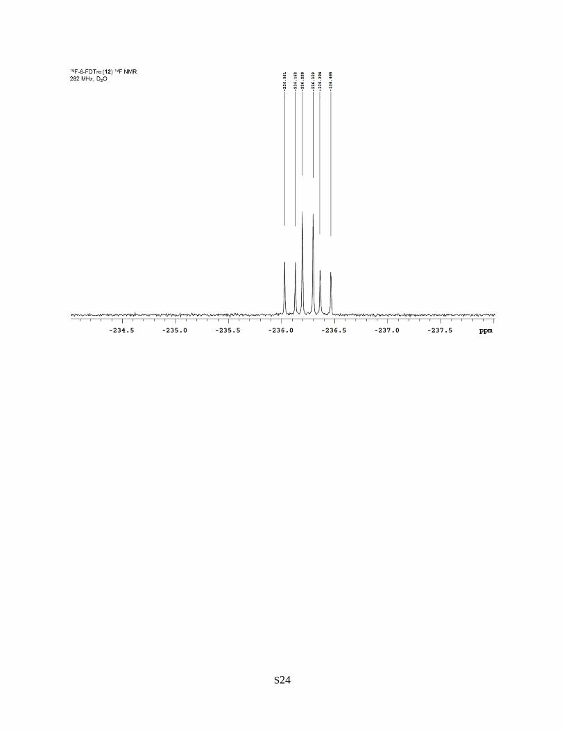
(C4), 68.45 (d, J = 6.6 Hz, C4’), 60.42 (C6). 19F NMR (282 MHz, D2O): δ –236.2 (dt, JH,F = 28.5, 47.1 Hz).
HR ESI MS negative mode: calcd. for C12H21ClFO10 [M+Cl]- m/z, 379.0807; found, 379.0786.
Chemical Synthesis of 19F-4-FDTre
4-Deoxy-4-fluoro-α,α-D-trehalose (19F-4-FDTre, compound 11). To a stirring solution of compound 182
(500 mg, 0.461 mmol) in anhydrous tetrahydrofuran (0.95 mL) under an argon atmosphere at room
temperature was added bis(2-methoxyethyl)aminosulfur trifluoride (BAST, 0.11 mL, 0.500 mmol)

S9
dropwise. The solution was heated to 50 °C and allowed to stir overnight. TLC showed that the reaction
was incomplete. An excess of BAST (0.54 mL, 2.50 mmol) was added dropwise and stirred for an additional
3 h at 50 °C, after which TLC showed completion. The product was diluted with ethyl acetate, then poured
into a separatory funnel and washed sequentially with saturated aqueous NaHCO3 and water. The organic
layer was dried over anhydrous Na2SO4, filtered, and concentrated by rotary evaporation. The crude
material was chromatographed on silica gel (toluene/ethyl acetate 20:1) to give the fluorinated intermediate
(0.371 g, 74%). A portion of the intermediate (0.160 g, 0.149 mmol) was dissolved in anhydrous CH2Cl2
(1 mL), and a freshly prepared solution of NaOCH3 in CH3OH (2 mL) was added dropwise to achieve a
final NaOCH3 concentration of 0.05 M. After stirring overnight, TLC showed complete conversion of the
starting material to a single polar product. Dowex H+ resin was used to neutralize the reaction, after which
the resin was filtered off and the solution was concentrated by rotary evaporation. The crude material was
chromatographed on silica gel (CH2Cl2/CH3OH 2.5:1) to give the desired product, which was re-suspended
in deionized water, passed through a 0.2 µm syringe filter, and dried to give 19F-4-FDTre (0.051 g, 98%;
73% from 18 over 2 steps). 1H NMR (500 MHz, D2O): δ 5.19 (d, J = 4.0 Hz, 1 H, H1’), 5.18 (d, J = 4.0 Hz,
1 H, H1), 4.36 (dt, J = 9.5 Hz, JH,F = 51.5 Hz, 1 H, H4’), 4.14 (dt, J = 10.5 Hz, JH,F = 15.5 Hz, 1 H, H3’),
4.04–3.98 (m, 1 H, H5’), 3.86–3.72 (m, 6 H, H3, H5, H6ab, H6ab’), 3.68 (dd, J = 4.0, 10.5 Hz, 1 H, H2’),
3.64 (dd, J = 4.0, 9.5 Hz, 1 H, H2), 3.44 (t, J = 9.5 Hz, 1 H, H4). 13C NMR (125 Hz, D2O): δ 93.43 (C1),
93.10 (C1’), 89.05 (d, JC,F = 179.3 Hz, C4’), 72.41 (C3 or C5), 72.14 (C3 or C5), 70.90 (C2), 70.67 (d, JC,F
= 8.6 Hz, C3’), 70.42 (d, J = 18.1 Hz, C2’), 69.55 (C4), 69.54 (d, JH,F = 23.9 Hz, C5’) 60.41 (C6 or C6’),
59.74 (C6 or C6’). 19F NMR (282 MHz, D2O): δ –199.0 (dd, JH,F = 15.8, 51.4 Hz). HR ESI MS negative
mode: calcd. for C12H20FO10 [M-H]- m/z, 343.1041; found, 343.1048.
Conformational analysis of FDTre analogues
NMR experiments. Deuterium-exchanged trehalose and 19F-FDTre analogues were dissolved in D2O
(99.8%) to a final concentration of 100 mM. 300 µL of the solution were transferred to a Shigemi NMR
tube matched to D2O. Argon was bubbled through the solution for 1 min and then the tube was sealed with
parafilm. To obtain 1H, 13C, COSY, and HSQC spectra, samples were analyzed on a Varian INOVA 500
instrument at room temperature. For HSQC spectra, in-phase cross-peaks correspond to CH/CH3 carbons,
out-of-phase cross-peaks correspond to CH2 carbons. 1H NMR spectra were referenced to residual HDO
peak at δ 4.78 ppm. To obtain 1H-coupled 19F NMR spectra, samples were analyzed on a Varian Mercury
300 instrument at room temperature. 19F NMR spectra were referenced to trifluoroacetic acid at δ –76.55
ppm. Standard Varian pulse sequences were used for 1D and 2D experiments. NMR data were processed,
analyzed, and plotted using Varian VnmrJ version 4.2 revision A.

S10
For experimental determination of 3JCOCH values, a Varian selexcit experiment with multifrequency
excitation of the anomeric H1 and H1’ protons was set up using a previously acquired 1D proton spectrum
of the appropriate disaccharide. The selexcit experiment was used to configure a 2D excitation-sculptured
indirect-detection NMR experiment (EXSIDE),3 which enabled the measurement of long-range
hetereonuclear coupling constants. Both the selexcit and EXSIDE experiments were part of the standard
Varian VnmrJ software package. EXSIDE spectra were obtained using 18 scans in the F2 (1H) dimension,
256 increments in the F1 dimension (13C), a jnxh setting of 4, and a J scaling factor (N) of 25. Long-range
couplings were observed as in-phase pairs in the F1 dimension of the 2D spectrum. 3JCOCH values were
determined from the EXSIDE spectra using the following equation:
𝐽 = ([𝛿2 − 𝛿1] ∗ 125.7 𝑀𝐻𝑧)
N
where δ2 and δ2 are the chemical shifts of each cross-peak in a pair, 125.7 MHz is the 13C NMR frequency,
and N is the J scaling factor 25. Next, the 3JCOCH values were converted into approximate dihedral angles
using the Karplus equation developed by Tvaroška and co-workers4:
𝐽 = 5.7𝑐𝑜𝑠2(𝜃) − 0.6𝑐𝑜𝑠2(𝜃) + 0.5
Molecular modeling. Molecular mechanics calculations employing the MM3* force field5 were used to
predict the solution conformations of trehalose and the 19F-FDTre analogues. All MM3* calculations were
performed in MacroModel version 11.2 in the Maestro environment (version 10.5) running on the Windows
10 operating system. First, structures for trehalose and the 19F-FDTre analogues were built with the
appropriate stereochemical configurations. The structures were energy-minimized with MM3* using the
Polak-Ribiere Conjugate Gradient (PRCG) method and a maximum of 2500 iterations. Next, these
structures were used to initiate conformational searches using the Monte Carlo Multiple Minimum
(MCMM) torsional sampling algorithm on all rotatable bonds, including ring closure. 1000 structures were
sampled using MCMM. All MM3* calculations were performed with water selected as the solvent. MCMM
conformational searches were performed either without constraints or with glycosidic dihedral angle
constraints determined experimentally by NMR as described above. The lowest-energy conformers were
selected for visual comparison in the manuscript and Supporting Information.
19F-FDTre uptake analysis in M. smegmatis
Cultures of M. smegmatis wild type, ΔsugC mutant, or ΔsugC::sugC complement1 were generated by
inoculating a single colony from a freshly streaked agar plate (with appropriate antibiotic, if needed) into 3
mL Middlebrook 7H9 liquid medium supplemented with ADC (albumin, dextrose, and catalase), 0.5%

S11
glycerol, and 0.05% Tween-80 in a culture tube. Starter cultures were incubated at 37 °C with shaking until
reaching log phase. Cultures were diluted to an OD600 of 0.5, then 19F-FDTre was added from a 1 mM
aqueous stock solution to a final concentration of 25 µM. Control cells received vehicle solution. Cultures
were incubated for 3 h at 37 °C with shaking to achieve a final OD600 of 0.8–1.0. Cells were centrifuged
(6500 rpm, 5 min) and washed three times, twice with PBS and once with deionized water. Cell pellets
were resuspended in deionized water and lysed by boiling at 100 °C for 4 h with shaking (300 rpm).
Insoluble debris from lysed cells was pelleted by centrifugation (3600 rpm, 5 min), after which the
supernatant was transferred to a microcentrifuge tube and dried on a speedvac. The resulting residue, which
contained water-soluble cytosolic metabolites, was trimethylsilyl (TMS)-derivatized by treatment with 50
µL anhydrous pyridine and 50 µL N-methyl-N-(trimethylsilyl) trifluoroacetamide (MSTFA). After
incubation overnight at room temperature, samples were transferred to low-volume vials with embedded
250 µL inserts and analyzed by GC-MS. GC-MS was performed on a Waters GCT Premier with an Agilent
7890A equipped with a CTC Analytics PAL autosampler and an SGE Forte BPX5 capillary column (10 m
length, 0.1 mm inner diameter, 0.1 µm film thickness). A temperature gradient of 100–300 °C (hold at 100
°C for 1 min, linear increase of 10 °C/min for 20 min, hold at 300 °C for 4 min) and a split ratio of 1:50
were used. Chromatograms and spectra collected were analyzed using MassLynx 4.1 software. Standards
of 19F-FDTre and trehalose were derivatized and analyzed identically. Data shown are representative of at
least two independent experiments.
Protocol for the rapid synthesis, purification, and administration of FDTre probes
Cultures of M. smegmatis wild type, ΔsugC mutant, or ΔsugC::sugC complement in 7H9 liquid medium
were incubated at 37 °C with shaking until reaching log phase. Meanwhile, a 1 mL TreT reaction employing
19F-2-FDG as the acceptor substrate was carried out to synthesize 19F-2-FDTre. The general method for
chemoenzymatic synthesis (page S7) was used with some modifications to accelerate the synthesis and
purification processes. After TreT enzyme was added to initiate the reaction, it was incubated with shaking
for 15 min, then transferred to a pre-rinsed Amicon Ultra-15 centrifugal filter unit (NMWL 10 kDa). The
reaction tube was washed two times with 1.5 mL deionized water. The washings were transferred to the
centrifugal filter unit, which was then spun at 3214 x g for 10 min. The upper chamber of the filter unit was
discarded, and mixed-bed ion exchange resin (0.75 g of Bio-Rad Bio-Rex RG 501-X8) was added to the
tube and stirred for 25 min at room temperature. The supernatant was decanted and filtered through a 0.2
µm syringe filter, giving a 4 mL aqueous stock solution of 19F-2-FDTre with an approximate concentration
of 5 mM (the initial substrate concentration of 20 mM was diluted 4x from washing the reaction tube). 15
µL of the 19F-2-FDTre solution were added to each strain of M. smegmatis (adjusted to an OD600 of 0.5) to
achieve a final concentration of 25 µM (the remainder of the 19F-2-FDTre was analyzed by TLC and 1H

S12
NMR as shown in Figure S4A and S4B, respectively). The cultures were incubated for 1 h at 37 °C with
shaking, after which the cells were processed and analyzed by GC-MS as described above. See Figure S4C
for uptake data.
References for Supporting Information
1 B. L. Urbanek, D. C. Wing, K. S. Haislop, C. J. Hamel, R. Kalscheuer, P. J. Woodruff and B. M.
Swarts, ChemBioChem, 2014, 15, 2066–2070.
2 R. W. Bassily, R. I. El-Sokkary, B. A. Silwanis, A. S. Nematalla and M. A. Nashed, Carbohydr.
Res., 1993, 239, 197–207.
3 V. V Krishnamurthy, J. Magn. Reson. A, 1996, 121, 33–41.
4 I. Tvaroška, M. Hricovíni and E. Petráková, Carbohydr. Res., 1989, 189, 359–362.
5 N. L. Allinger, Y. H. Yuh and J. H. Lii, J. Am. Chem. Soc., 1989, 111, 8551–8566.

S13
NMR spectra for 19F-FDTre analogues

S14

S15

S16

S17

S18

S19

S20

S21

S22

S23
S24