supporting information of cyanide ion (cn ) in sol-gel
TRANSCRIPT

S1
Supporting Information
Naphthalimide-linked new pyridylazo phenol derivative for selective sensing of cyanide ion (CN-) in sol-gel medium
Sumit Ghosh,a Palash Janab and Kumaresh Ghosh*a
aDepartment of Chemistry, University of Kalyani, Kalyani-741235, India.Email: [email protected]
bDepartment of Chemical Sciences, Indian Institute of Science Education and Research (IISER) Kolkata, Mohanpur, Nadia, West Bengal 741246, India
Table 1S. Results of gelation test for 1 and 2.
Solvent 1 2DMSO S SDMF S STHF I I
CH3CN I ICH3OH I ICHCl3 S S
Diethyl ether I IHexane I I
Petroleum ether I IDCM S S
Toluene I IDMSO: H2O (1:1, v/v) P PDMF: H2O (1:1, v/v) G (6 mg/mL) P
S = Solution; G = Gel (mgc); I = Insoluble; P = Precipitation. Gelation was primarily investigated by inversion of vial method after 10-15 mins of sample preparation ([Gelator] = 20 mg/mL).
Electronic Supplementary Material (ESI) for Analytical Methods.This journal is © The Royal Society of Chemistry 2020
S2
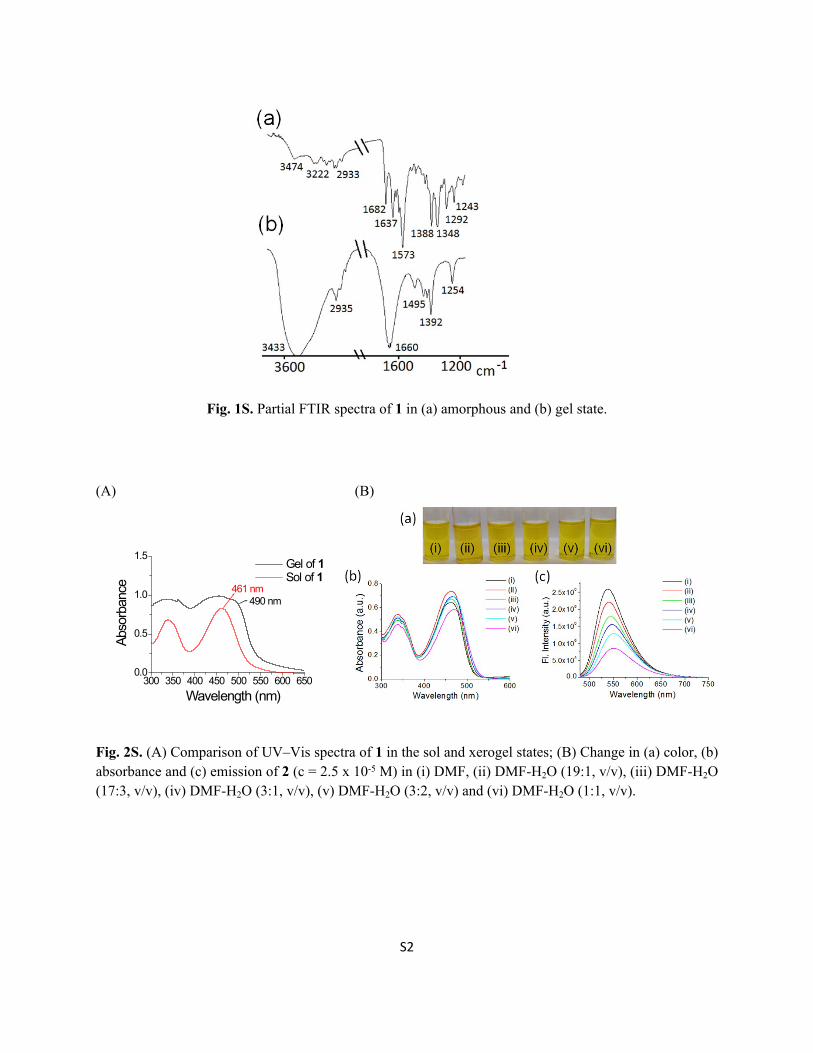
Fig. 1S. Partial FTIR spectra of 1 in (a) amorphous and (b) gel state.
(A) (B)
Fig. 2S. (A) Comparison of UV–Vis spectra of 1 in the sol and xerogel states; (B) Change in (a) color, (b) absorbance and (c) emission of 2 (c = 2.5 x 10⁻⁵ M) in (i) DMF, (ii) DMF-H2O (19:1, v/v), (iii) DMF-H2O (17:3, v/v), (iv) DMF-H2O (3:1, v/v), (v) DMF-H2O (3:2, v/v) and (vi) DMF-H2O (1:1, v/v).
300 350 400 450 500 550 600 6500.0
0.5
1.0
1.5
Wavelength (nm)
490 nm461 nm
Abso
rban
ce
Gel of 1 Sol of 1
S3
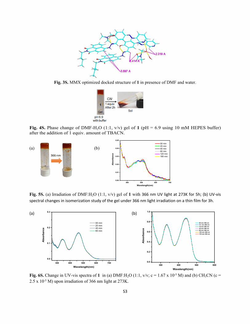
Fig. 3S. MMX optimized docked structure of 1 in presence of DMF and water.
Fig. 4S. Phase change of DMF-H2O (1:1, v/v) gel of 1 (pH = 6.9 using 10 mM HEPES buffer) after the addition of 1 equiv. amount of TBACN.
(a) (b)
Fig. 5S. (a) Irradiation of DMF:H2O (1:1, v/v) gel of 1 with 366 nm UV light at 273K for 5h; (b) UV-vis spectral changes in isomerization study of the gel under 366 nm light irradiation on a thin film for 3h.
(a) (b)
Fig. 6S. Change in UV-vis spectra of 1 in (a) DMF:H2O (1:1, v/v; c = 1.67 x 10⁻⁵ M) and (b) CH3CN (c = 2.5 x 10⁻⁵ M) upon irradiation of 366 nm light at 273K.
400 500 600 7000.00
0.01
0.02
0.03
0.04
0.05
Abs
orba
nce
Wavelength(nm)
00 min 20 min 40 min 60 min 120 min 180 min
300 400 500 600 700
0.0
0.1
0.2
0.3
Abso
rban
ce
Wavelength(nm)
00 min 20 min 40 min 60 min
300 400 500 6000.0
0.2
0.4
0.6
0.8
1.0
Abs
orba
nce
Wavelength(nm)
00 min 366 nm 10 min 366 nm 15 min 366 nm 30 min 366 nm 90 min 366 nm 125 min 366 nm 135 min 366 nm
S4
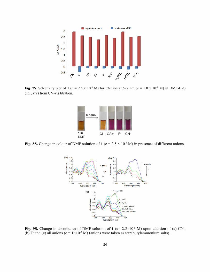
Fig. 7S. Selectivity plot of 1 (c = 2.5 x 10⁻⁵ M) for CN- ion at 522 nm (c = 1.0 x 10⁻³ M) in DMF-H2O (1:1, v/v) from UV-vis titration.
Fig. 8S. Change in colour of DMF solution of 1 (c = 2.5 × 10⁻⁵ M) in presence of different anions.
Fig. 9S. Change in absorbance of DMF solution of 1 (c= 2.5×10⁻⁵ M) upon addition of (a) CN-, (b) F- and (c) all anions (c = 1×10⁻³ M) (anions were taken as tetrabutylammonium salts).
S5
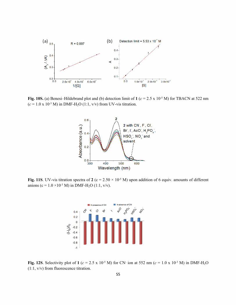
Fig. 10S. (a) Benesi–Hildebrand plot and (b) detection limit of 1 (c = 2.5 x 10⁻⁵ M) for TBACN at 522 nm (c = 1.0 x 10⁻³ M) in DMF-H2O (1:1, v/v) from UV-vis titration.
Fig. 11S. UV-vis titration spectra of 2 (c = 2.50 × 10⁻⁵ M) upon addition of 6 equiv. amounts of different anions (c = 1.0 ×10⁻³ M) in DMF-H2O (1:1, v/v).
Fig. 12S. Selectivity plot of 1 (c = 2.5 x 10⁻⁵ M) for CN- ion at 552 nm (c = 1.0 x 10⁻³ M) in DMF-H2O (1:1, v/v) from fluorescence titration.
S6
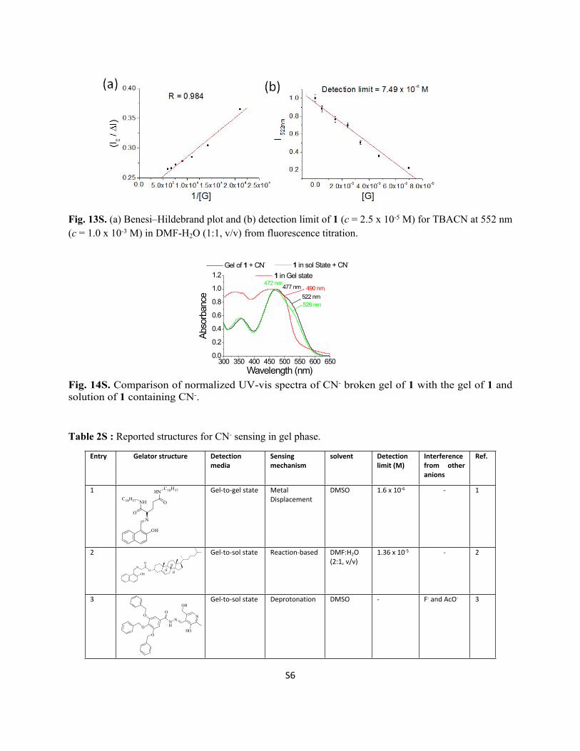
Fig. 13S. (a) Benesi–Hildebrand plot and (b) detection limit of 1 (c = 2.5 x 10⁻⁵ M) for TBACN at 552 nm (c = 1.0 x 10⁻³ M) in DMF-H2O (1:1, v/v) from fluorescence titration.
Fig. 14S. Comparison of normalized UV-vis spectra of CN- broken gel of 1 with the gel of 1 and solution of 1 containing CN-.
Table 2S : Reported structures for CN- sensing in gel phase.
Entry Gelator structure Detection media
Sensing mechanism
solvent Detection limit (M)
Interference from other anions
Ref.
1
OH
NO
NHC18H37
HN
O
C18H37 Gel-to-gel state Metal Displacement
DMSO 1.6 x 10⁻⁶ - 1
2
OH
NO H
H
H
O
Gel-to-sol state Reaction-based DMF:H2O (2:1, v/v)
1.36 x 10⁻⁵ - 2
3
O
O
O
O
NH
NN
HO
OHGel-to-sol state Deprotonation DMSO - F- and AcO- 3
300 350 400 450 500 550 600 6500.0
0.2
0.4
0.6
0.8
1.0
1.2
490 nm
526 nm522 nm
477 nm472 nm
1 in sol State + CN- Gel of 1 + CN-
Abso
rban
ce
Wavelength (nm)
1 in Gel state

S7
4
O
O
HN
HN
HN
O
N
NN
Gel-to-gel state Deprotonation DMSO:H2O (8:2, v/v)
- F-, AcO- and H2PO4
-4
5
OC16H33OC16H33
C16H33O
OHNN
N
Gel-to-gel state Metal Displacement
DMF1.0 x 10-5
1.0 x 10⁻⁷
- 5
6
OC16H33OC16H33
C16H33O
OHNN
Gel-to-gel state Metal Displacement
EtOH 1.0 x 10⁻⁶ - 6
7 Two component gel from citrazinic acid and melamine
Gel-to-sol state Deprotonation Air dried gel
- S2- 7
8
N
CN
CN
O
N
N
Gel-to-sol state Reaction-based CH3CN 9.36 x 10⁻⁶ - 8
9
O
NHOH
O OH
H
H
Gel-to-sol state Deprotonation Toluene F- and AcO- 9
10
O
HCN
O OH
H
H
CNGel-to-sol state Reaction-based Toluene-
MeOH (1:2)4.17 × 10⁻⁶ - 9
11 Gel-to-sol state H-bonding based
DMSO-H2O
(1:1, v/v)1.5 mM No other
anions tested
10
12 Gel-to-sol state Deprotonation DMF:H2O (1:1, v/v)
0.368 mM - 11
13 Gel-to-sol state Deprotonation DMSO 0.4 x 10⁻⁸ No other anions tested
12
S8
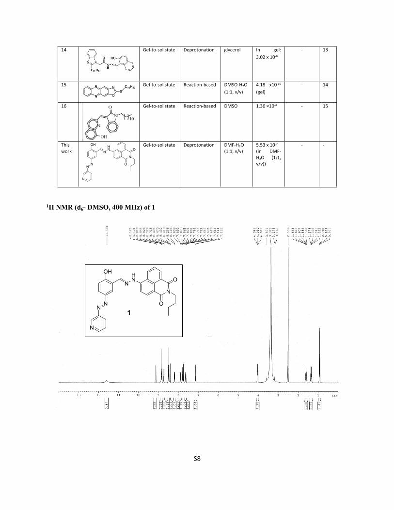
1H NMR (d6- DMSO, 400 MHz) of 1
14 Gel-to-sol state Deprotonation glycerol In gel:3.02 x 10⁻⁶
- 13
15 Gel-to-sol state Reaction-based DMSO-H2O
(1:1, v/v)4.18 x10-10
(gel)- 14
16 Gel-to-sol state Reaction-based DMSO 1.36 ×10⁻⁴ - 15
This work
NN
N
OH
NHN
N
O
O
Gel-to-sol state Deprotonation DMF-H2O
(1:1, v/v)5.53 x 10-7
(in DMF-H2O (1:1, v/v))
- -
NN
N
OH
NHN
N
O
O
1

S9
13C NMR (d6- DMSO, 100 MHz) of 1
NN
N
OH
NHN
N
O
O
1

S10
Mass spectrum of 1.
Calcd. 515.1808 (M+ Na)+, 492.1910 (M)+ Found. 515.1860 (M+ Na)+, 492.1996 (M)+
S11
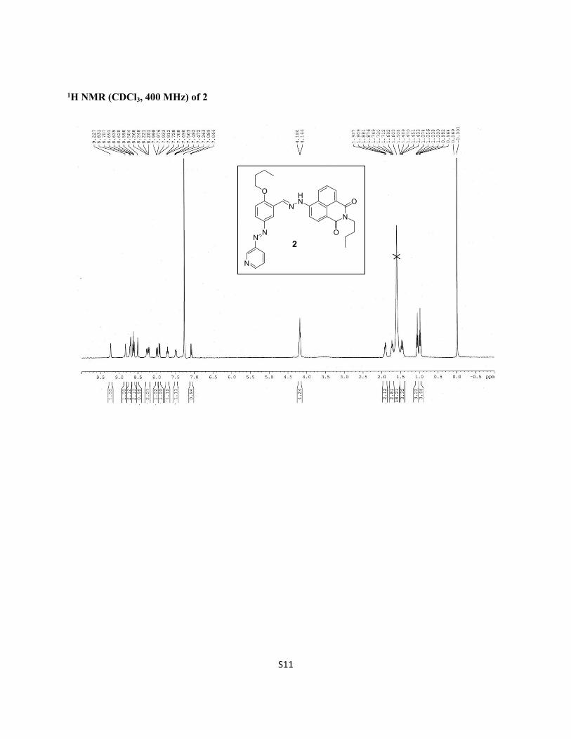
1H NMR (CDCl3, 400 MHz) of 2
NN
N
O
NHN
N
O
O
2
S12
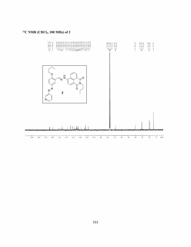
13C NMR (CDCl3, 100 MHz) of 2
NN
N
O
NHN
N
O
O
2
S13
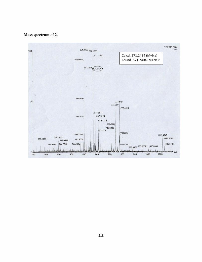
Mass spectrum of 2.
Calcd. 571.2434 (M+Na)+
Found. 571.2404 (M+Na)+

S14
1H NMR (CDCl3, 400 MHz) of 3
NN
N
OH
3
O
H
S15
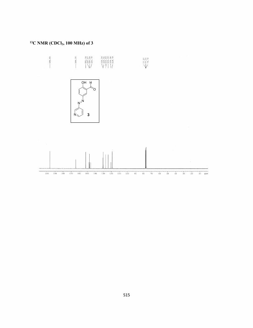
13C NMR (CDCl3, 100 MHz) of 3
NN
N
OH
3
O
H
S16
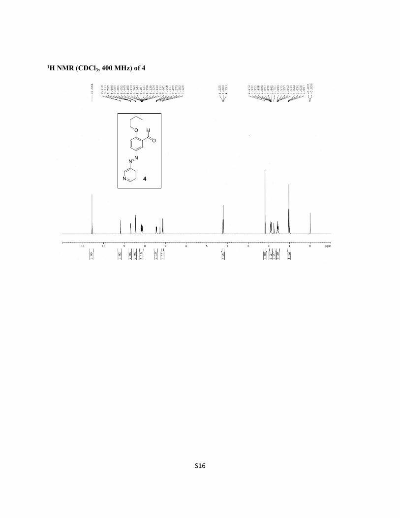
1H NMR (CDCl3, 400 MHz) of 4
NN
N
O
4
O
H
S17
References
1. J. Sun, Y. Liu, L. Jin, T. Chen, B. Yin, Chem. Commun,. 2016, 52,768.2. K. Ghosh, S. Panja, Supramol. Chem., 2017, 29, 350.3. K. Ghosh, C. Pati, Tetrahedron Lett. 2016, 57, 5469.4. A. Ghosh, P. Das, R. Kaushik, K. K. Damodaran, D. A. Jose, RSC Adv., 2016, 6, 83303.5. Q. Lin, B. Sun, Q. P. Yang, Y. P. Fu, X. Zhu, T. B. Wei, Y. M. Zhang, Chem. Eur. J., 2014, 20,
11457.6. Q. Lin, T. T. Lu, X. Zhu, B. Sun, Q. P. Yang, T. B. Wei, Y. M. Zhang, Chem. Commun., 15, 51,
1635.7. S. Sarkar, S. Dutta, C. Ray, B. Dutta, J. Chowdhury and T. Pal, Cryst. Eng. Comm., 2015, 17,
8119.8. A. Panja and K. Ghosh, Chemistry Select, 2018, 3, 1809.9. A. Panja and K. Ghosh, Supramol. Chem., 2019, 31, 239.10. S. Sharma, M. Kumari, N. Singh, Soft Matter, 2020, 16, 6532.11. B. Sarkar, P. Prabakaran, E. Prasad and R. L. Gardas, ACS Sustainable Chem. Eng., 2020, 8,
8327.12. F. Hu, M. Cao, J. Huang, Z. Chen, D. Wu, Z. Xu, S. H. Liu and J. Yin, Dyes and Pigments, 2015,
119, 108.13. H. Yao, J. Wang, S. Song, Y. Q. Fan, X. W. Guan, Q. Zhou, T. B. Wei, Q. Lin and Y. M. Zhang,
New J. Chem., 2018, 42, 18059.14. H. Fang, W. J. Qu, H. H. Yang, J. X. He, H. Yao, Q. Lin, T. B. Wei and Y. M. Zhang, Dyes and
Pigments, 2020, 174, 108066.15. F. Mandegani, H. Z. Boeini, Z. Khayat and R. Scopelliti, Talanta, 2020, 219, 121237.