supporting online material for - sciencemag.org€¦ · supporting online material for ... by dr....
TRANSCRIPT
www.sciencemag.org/cgi/content/full/316/5828/1202/DC1
Supporting Online Material for
Ubiquitin-Binding Protein RAP80 Mediates BRCA1-Dependent
DNA Damage Response
Hongtae Kim, Junjie Chen,* Xiaochun Yu*
*To whom correspondence should be addressed.
E-mail: [email protected] (J.C.); [email protected] (X.Y.)
Published 25 May 2007, Science 316, 1202 (2007)
DOI: 10.1126/science.1139621
This PDF file includes:
Materials and Methods
Figs. S1 to S9
References

1
Supporting Online Material
Materials and methods
Plasmids
Human RAP80 cDNA was purchased from American Type Culture Collection
(Manassas, VA). Full length and deletion mutants of human RAP80 were generated by
PCR and subcloned into a modified pIRES-EGFP mammalian expression vector to create
constructs encoding S-Flag-SBP (Streptavidin Binding Peptide)-tagged (SFB tagged)
full-length or various deletion mutants of RAP80. RAP80 was also subcloned into a
modified pcDNA3.1 vector to generate a construct encoding HA-tagged RAP80 (HA-
RAP80). The point mutants of human RAP80 were created using the QuikChange site-
directed mutagenesis kit (Stratagene). Myc-BRCA1 and Myc-BRCA1 BRCT were also
generated by PCR and subcloned into modified pcDNA3 M-1. RAP80 and RAP80 N-
terminal region were subcloned into a modified pGEX4T-1 vector to generate constructs
encoding GST-tagged RAP80 (GST-RAP80) and RAP80 N-terminal region containing
the double UIMs (GST-RAP80N). Flag-HSJ1A expression plasmid was kindly provided
by Dr. Jorg Hohfeld from the Institute for Cell Biology (Bonn, Germany), and deletion
mutants of human HSJ1A-UIM, BARD1-BRCT and BRCA1-BRCT were generated by
PCR and subcloned into a modified pIRES-EGFP mammalian expression vector. Ubi-
GST construct was kindly provided by Dr. Bruce Horazdovsky from the Mayo Clinic
(Minnesota, USA).
Cell Culture and treatment with ionizing radiation
HeLa, K562 and 293T cells were purchased from American Type Culture Collection
(Manassas, VA) and maintained in RPMI1640 medium supplemented with 10% fetal
bovine serum at 37 °C in 5% CO2 (v/v). MDC-/- and +/+ MEFs (S2), 53BP1-/- and +/+
MEFs (S3), H2AX-/- and +/+ MEFs (S2), NBS1-deficient and reconstituted cells (S4),
ATM-deficient (FT169A) and reconstituted (YZ5) cells (S5), HCC1937 and HCC1937-
BRCA1 (S1) were previously reported. FANCD2-deficient and -reconstituted cells was
kindly provided by Dr. Alan D’Andrea at the Department of Pediatric Oncology at Dana-
Farber Cancer Institute (Boston, MA) and maintained in DMEM medium supplemented
with 15% fetal bovine serum at 37 °C in 5% CO2 (v/v). Cells were irradiated using JL
Spepherd 137
Cs radiation source at indicated doses. The irradiated cells were then
returned to culture conditions and maintained for the indicated periods of time specified
in the figure legends or Methods.
SiRNA
All siRNA duplexes used in this study were purchased from Dharmacon Research
(Lafayette, CO). The sequences of RAP80 SiRNA1 and 2 are
GAAGGAUGUGGAAACUACCdTdT and GCGTAGACTTGAGGATGCAdTdT,
respectively. The sequences of control, CtIP, and BRCA1 SiRNA are
UUCAAUAAAUUCUUGAGGUUU, GCUAAAACAGGAACGAATCdTdT, and

2
UCACAGUGUCCUUUAUGUAdTdT, respectively. SiRNAs were transfected into the
cells using oligofectamine (Invitrogen) according to the manufacturer’s instructions.
Antibodies, Transfection, Immunoprecipitation and Preparation of Chromatin Fractions.
Rabbit anti-Myc and anti-RAP80 antibodies were raised by immunizing rabbits with Myc
peptide CEQKLISEEDL and GST-RAP80 (RAP80 1-354 amino acids) respectively.
These rabbit polyclonal antibodies were affinity-purified using the Sulfolink or
AminoLink Plus Immobilization and Purification Kit (Pierce). MDC1, 53BP1, Chk1,
phospo-Chk1, H2AX and -H2AX antibodies were previously described (S2). Anti-Flag
and -actin antibodies were purchased from Sigma Inc and monoclonal human BRCA1
antibody was purchased from Oncogene Science. Anti-phospho-H3 and monoclonal
ubiquitin antibodies were purchased from Upstate biotechnology Inc. Transient
transfection was performed using Fugene 6 reagent (Roche Inc) according to the
instructions provided by manufacturer. For immunoprecipitation, cells were washed with
ice-cold PBS and then lysed in NETN buffer (0.5% Nonidet P-40, 20 mM Tris [pH 8.0],
50 mM NaCl, 50 mM NaF, 100 M Na3VO4, 1 mM EDTA and 50 g/ml PMSF) at 4 °C
for 10 min. Crude lysates were cleared by centrifugation at 14,000 rpm at 4 °C for 5 min,
and supernatants were incubated with protein A-agarose-conjugated primary antibodies.
The immunocomplexes were washed three times with NETN buffer and subjected to
SDS-PAGE. Western blotting was performed using the antibodies indicated in the figure
legends. The preparation of chromatin fractions was performed as described previously
(S1).
The establishment of stable cell lines and Affinity Purification of S-Flag-SBP(SFB)-
tagged BRCA1-BRCT -containing complexes
To establish cell lines stably expressing epitope-tagged proteins, K562 cells were
transfected with plasmids encoding SFB-BRCA1-BRCT or SFB-BARD1-BRCT. Forty-
eight hours after transfection, the cells were split at the 10:1 ratio and cultured in the
medium containing antibiotics for 3 weeks. The individual antibiotics-resistant colonies
were isolated and screened by Western blotting for the expression of BRCA1-BRCT and
BARD1-BRCT proteins. K562 cells stably expressing SFB-BRCA1 BRCT domain or
SFB-BARD1 BRCT domains were lysed with 4 ml NETN buffer on ice for 10 min.
Crude lysates were cleared by centrifugation at 14,000 rpm at 4 °C for 10 min, and
supernatants were incubated with 300 µl streptavidin-conjugated beads (Amersham). The
immunocomplexes were washed three times with NETN buffer and then bead-bound
proteins were eluted with 500 µl NETN buffer containing 2 mg/ml biotin (Sigma). The
eluted supernatant was incubated with 60 µl S protein beads (Novagen). The
immunocomplexes were washed three times with NETN buffer and subjected to SDS-
PAGE. The silver staining was performed to visualize the protein bands. Specific bands
were excised, digested and the peptides were analyzed by a mass spectrometer.
GST pull-down assay
The GST fusion protein was expressed in Escherichia coli and purified as previously
described (S6). Two g of GST-fusion protein or GST was immobilized on the
glutathione-Sepharose 4B beads and incubated with lysates prepared from cells that were
transiently transfected with plasmids encoding indicated proteins in NETN buffer for 2

3
hours at 4°C. After washing with NETN buffer, the samples were analyzed by Western
blotting analysis. Polyubiquitin chains were purchased from BostonBiochem (Cambridge,
MA)
Immunofluorescence Staining
Cells grown on coverslips were fixed with 3% paraformaldehyde at room temperature for
15 min. Then, cells were permeabilized with PBS containing 0.5% Triton X-100 at room
temperature for 5 min and the coverslips were blocked with PBS containing 5% goat
serum at room temperature for 30 min. The coverslips were incubated with primary
antibodies at room temperature for 20 min. After washing with PBS, cells were incubated
with secondary antibodies, fluorescein isothio-cyanate-conjugated goat anti-mouse IgG,
rhodamine-conjugated goat anti-rabbit IgG, or rhodamine-conjugated goat anti-mouse
IgG (Jackson Immuno-Reserach Laboratories, Inc.) at room temperature for 20 min. 4, 6-
diamidino-2-phenylindole (DAPI) was used to counterstain the nuclei. After a final wash
with PBS, coverslips were mounted with glycerin containing p-phenylenediamine. All
images were obtained with a Nikon ECLIPSE E800 fluorescence microscope.
G2/M cell cycle checkpoint assay
HeLa cells in 35-mm plate were transfected twice with control or RAP80 SiRNAs at the
24 hours time interval. Twenty-four hours after second transfection, the transfected cells
were transferred into 100 mm dishes. The transferred cells were incubated for 24 hours
and then the cells were mock treated or irradiated using a JL Spepherd 137
Cs radiation
source at indicated doses. One hour after irradiation, cells were fixed with 70% ethanol at
-20oC for 24 hours and stained with rabbit anti-phospho-histone H3
antibody (pH3),
followed by incubation with fluorescein isothiocyanate-conjugated
goat anti-rabbit
immunoglobulin secondary antibody. The stained cells were treated with RNaseA,
incubated with propidium iodide, and then analyzed by flow cytometry.
Cell survival assays
HeLa cells in 35-mm plate were transfected twice with control and RAP80 SiRNAs at the
24 hours time intervals. Twenty-four hours after second transfection, cells were splited
and transferred into 60 mm dishes. Cells were incubated for 24 hours before they were
irradiated using a JL Spepherd 137
Cs radiation source at various doses. Eleven days after
irradiation, cells were washed with PBS, fixed and stained with 2% methylene blue, and
the numbers of colonies were determined.
4
Figures
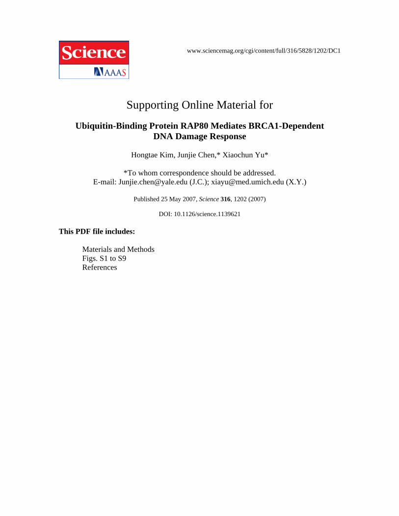
fig. S1. Exogenous RAP80 and BRCA1 associate with each other in vivo. (A) 293T cells
are transfected with plasmids encoding Myc-BRCA1 and HA-RAP80. Cell lysates were
subjected to immunoprecipitation with anti-Myc or control anti-Flag antibodies and
immunoblotting with the indicated antibodies (left panels). The amounts of HA-tagged
RAP80 and Myc-tagged BRCA1 in these lysates were analyzed by immunoblotting and
shown in the right panels.

5
fig. S1. (B) 293T cells are transfected with plasmids encoding Myc-BRCA1 or Myc-
BRCA1 BRCT with those encoding SFB-RAP80. Cell lysates were subjected to
immunoprecipitation with anti-Myc and immunoblotting with the indicated antibodies
(upper panels). The amounts of SFB-tagged RAP80 and Myc-tagged BRCA1 in these
lysates were analyzed by immunoblotting and shown in the lower panels. (C) Binding of
GST-RAP80 fusion protein to endogenous BRCA1 in vitro. GST or GST-RAP80 protein
was incubated with 293T cell lysates. After extensive washing, the proteins bound to the
beads were separated by SDS-PAGE and analyzed by immunoblotting with anti-BRCA1
antibody. The amounts of GST and GST-RAP80 were shown in the lower panel.

6
fig. S2. Ionizing radiation-induced phosphorylation of RAP80. SFB-RAP80 stable cells
were exposed to 0 or 10 Gy of ionizing radiation. One hour after irradiation, cell lysates
were subjected to pull-down with S-beads and immunoblotting with the antibodies to
phospho-Ser101 of RAP80 (upper panel) or RAP80 (lower panel).
7
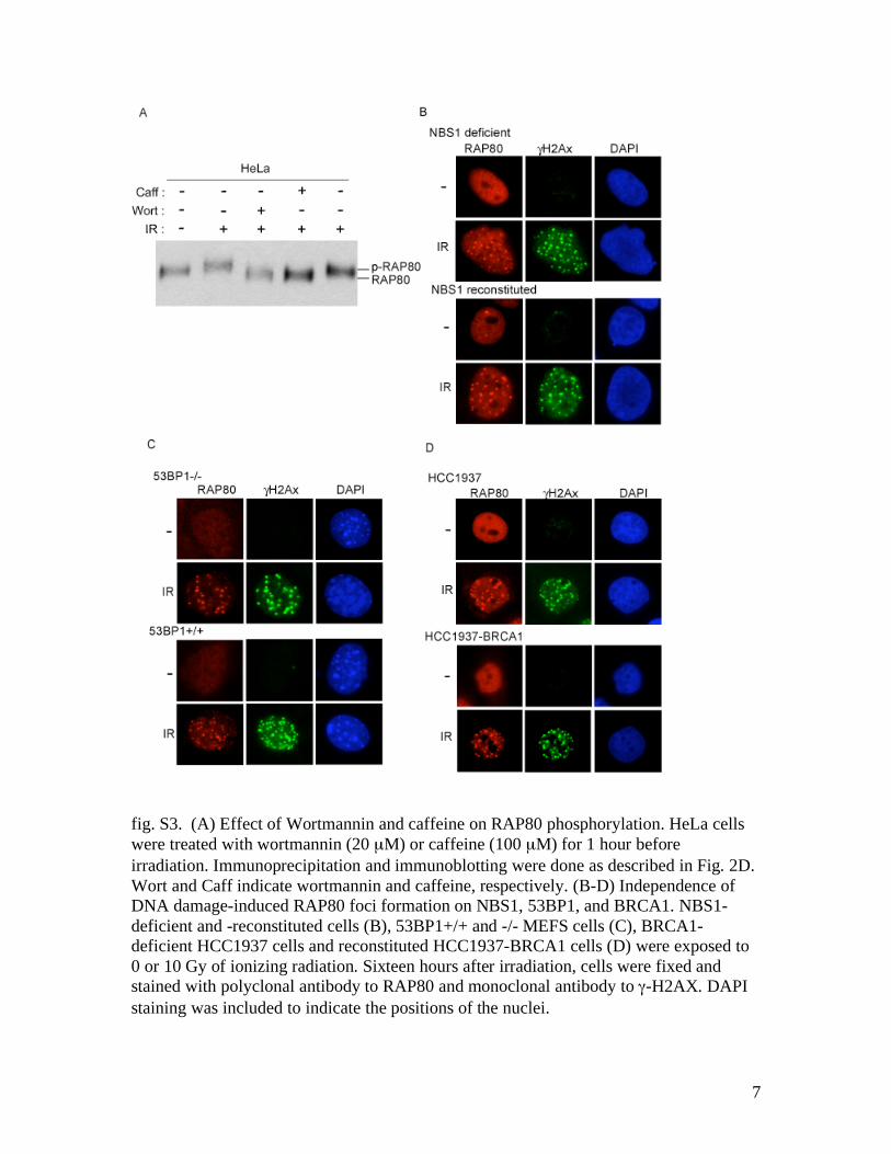
fig. S3. (A) Effect of Wortmannin and caffeine on RAP80 phosphorylation. HeLa cells
were treated with wortmannin (20 µM) or caffeine (100 µM) for 1 hour before
irradiation. Immunoprecipitation and immunoblotting were done as described in Fig. 2D.
Wort and Caff indicate wortmannin and caffeine, respectively. (B-D) Independence of
DNA damage-induced RAP80 foci formation on NBS1, 53BP1, and BRCA1. NBS1-
deficient and -reconstituted cells (B), 53BP1+/+ and -/- MEFS cells (C), BRCA1-
deficient HCC1937 cells and reconstituted HCC1937-BRCA1 cells (D) were exposed to
0 or 10 Gy of ionizing radiation. Sixteen hours after irradiation, cells were fixed and
stained with polyclonal antibody to RAP80 and monoclonal antibody to -H2AX. DAPI
staining was included to indicate the positions of the nuclei.

8
fig. S4. (A) Diagrams of the SFB-tagged wild type and internal serial deletion mutants or
point mutants of RAP80. (B) Alignment of RAP80 UIMs and other UIM regions. UIMs
is an ubiquitin interacting motif which contains many acidic residues followed by highly
conserved PXXAXXXSXXAc core, in which P, X and Ac represent the hydrophobic,
any, and acidic residues, respectively. Identical residues are highlighted in sky blue and
homologous residues are highlighted in blue. (C) HeLa cells were transiently transfected
with Flag-tagged wild type RAP80, HSJ1A, RAP80 UIMs or HSJ1A UIMs. Twenty-four
hours after transfection, cells were exposed to 10 Gy of ionizing radiation. Cells were
fixed 8 hours later and stained with monoclonal antibody to Flag and polyclonal antibody
to -H2AX.

9
fig. S5. Direct binding of RAP80 to lysine 63-linked polyubiquitin chains in vitro. GST
or GST-RAP80 N-terminal fusion proteins (GST-RAP80N) were incubated with lysine
48-linked polyubiquitin chains (Lys-48 Ubs) or lysine 63-linked polyubiquitin chains
(Lys-63 Ubs). After extensive washing, the bound polyubiquitin chains were analyzed by
immunoblotting with monoclonal antibody to ubiquitin. The amounts of GST and GST-
RAP80N were shown in the lower panel.
10
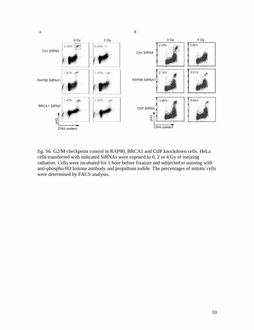
fig. S6. G2/M checkpoint control in RAP80, BRCA1 and CtIP knockdown cells. HeLa
cells transfected with indicated SiRNAs were exposed to 0, 2 or 4 Gy of ionizing
radiation. Cells were incubated for 1 hour before fixation and subjected to staining with
anti-phospho-H3 histone antibody and propidium iodide. The percentages of mitotic cells
were determined by FACS analysis.
11
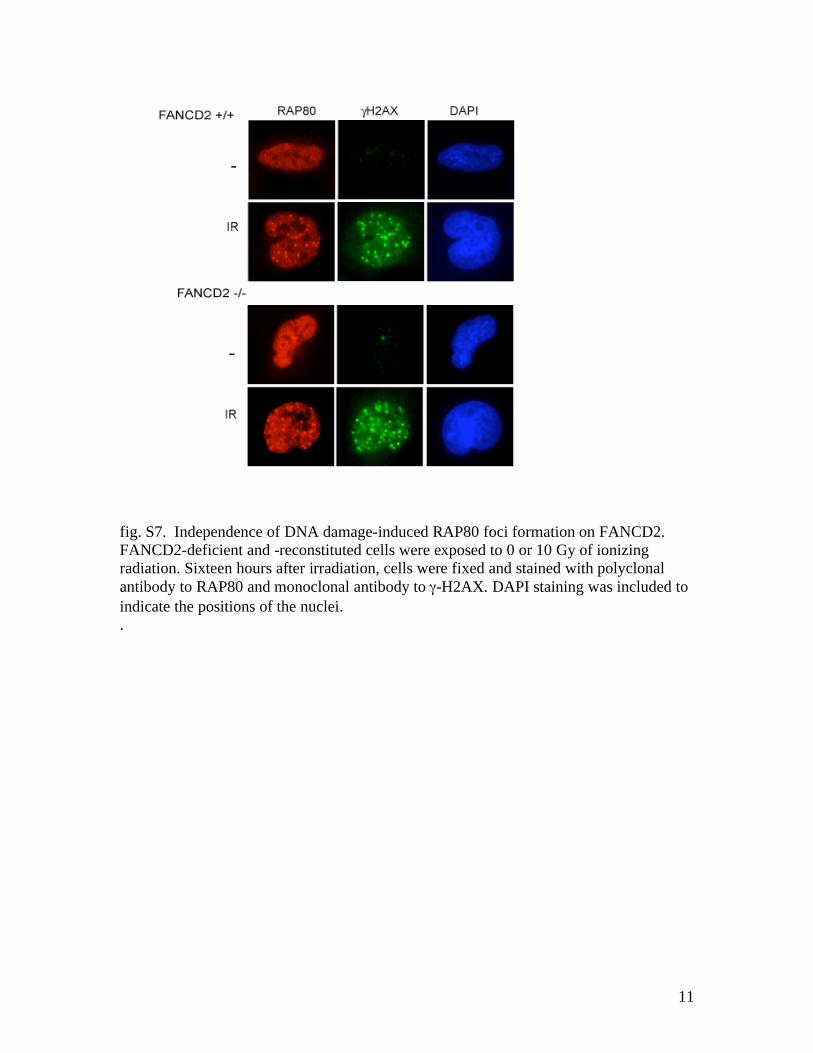
fig. S7. Independence of DNA damage-induced RAP80 foci formation on FANCD2.
FANCD2-deficient and -reconstituted cells were exposed to 0 or 10 Gy of ionizing
radiation. Sixteen hours after irradiation, cells were fixed and stained with polyclonal
antibody to RAP80 and monoclonal antibody to -H2AX. DAPI staining was included to
indicate the positions of the nuclei.
.
12
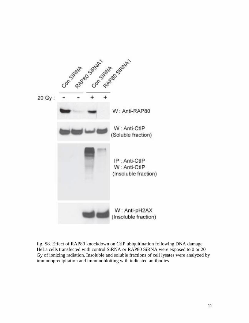
fig. S8. Effect of RAP80 knockdown on CtIP ubiquitination following DNA damage.
HeLa cells transfected with control SiRNA or RAP80 SiRNA were exposed to 0 or 20
Gy of ionizing radiation. Insoluble and soluble fractions of cell lysates were analyzed by
immunoprecipitation and immunoblotting with indicated antibodies

13
fig. S9. Expanded blots including molecular weight size markers from the indicated
selected figures.

14
References
S1. X. Yu, S. Fu, M. Lai, R. Baer, J. Chen, Genes Dev 20, 1721 (Jul 1, 2006).
S2. Z. Lou et al., Mol Cell 21, 187 (Jan 20, 2006).
S3. I. M. Ward, K. Minn, J. van Deursen, J. Chen, Mol Cell Biol 23, 2556 (Apr,
2003).
S4. R. S. Maser, R. Zinkel, J. H. Petrini, Nat Genet 27, 417 (Apr, 2001).
S5. I. Rappold, K. Iwabuchi, T. Date, J. Chen, J Cell Biol 153, 613 (Apr 30, 2001).
S6. B. Hofer, S. Backhaus, K. N. Timmis, Gene 144, 9 (Jun 24, 1994).
Supporting Online Material
www.sciencemag.org
Materials and Methods
Figs. S1 to S9
References