suppressing bacterial interaction with copper surfaces
TRANSCRIPT

Suppressing Bacterial Interaction with Copper Surfaces throughGraphene and Hexagonal-Boron Nitride CoatingsCarolina Parra,*,† Francisco Montero-Silva,‡ Ricardo Henríquez,† Marcos Flores,§ Carolina Garín,†
Cristian Ramírez,∥ Macarena Moreno,† Jonathan Correa,†,⊥ Michael Seeger,‡ and Patricio Haberle†
†Departamento de Física, Universidad Tecnica Federico Santa María, Avenida Espana 1680, Valparaíso, Chile‡Departamento de Química, Universidad Tecnica Federico Santa María, Avenida Espana 1680, Valparaíso, Chile§Departamento de Física, Facultad de Ciencias Físicas y Matematicas, Universidad de Chile, Avenida Blanco Encalada 2008, Santiago,Chile∥Departamento de Ingeniería Química y Ambiental, Universidad Tecnica Federico Santa María, Avenida Espana 1680, Valparaíso,Chile⊥Instituto de Física, Pontificia Universidad Catolica de Valparaíso, Avenida Universidad 330, Curauma, Valparaíso, Chile
*S Supporting Information
ABSTRACT: Understanding biological interaction with gra-phene and hexagonal-boron nitride (h-BN) membranes hasbecome essential for the incorporation of these uniquematerials in contact with living organisms. Previous reportsshow contradictions regarding the bacterial interaction withgraphene sheets on metals. Here, we present a comprehensivestudy of the interaction of bacteria with copper substratescoated with single-layer graphene and h-BN. Our resultsdemonstrate that such graphitic coatings substantially suppressinteraction between bacteria and underlying Cu substrates,acting as an effective barrier to prevent physical contact. Bacteria do not “feel” the strong antibacterial effect of Cu, and thesubstrate does not suffer biocorrosion due to bacteria contact. Effectiveness of these systems as barriers can be understood interms of graphene and h-BN impermeability to transfer Cu2+ ions, even when graphene and h-BN domain boundary defects arepresent. Our results seem to indicate that as-grown graphene and h-BN films could successfully protect metals, preventing theircorrosion in biological and medical applications.
KEYWORDS: graphene coating, biocorrosion, hexagonal boron nitride coating, copper, graphene impermeability
1. INTRODUCTIONPotential applications of graphene, the unique two-dimensional(2D) allotrope of carbon, include electronic devices, sensors,photovoltaics, transistors, biotechnology, and desalination,among others. The incorporation of such technologies inconsumer and industrial products is expected to have asignificant impact on our daily lives. Whereas graphene isgetting closer to mass production, reaching a completeunderstanding of its interaction with biological systems, suchas bacteria and cells, is indeed a priority while consideringexpanding its uses in close contact with live organisms andallow further applications in biomedical products.Bacterial interaction with surfaces is ubiquitous in nature. For
centuries, human civilizations have used metallic copper inmedicine, due to its antibacterial properties, until the advent ofcommercially available antibiotics in 1932. The most popularform of large-area graphene is, in fact, the one grown on copperdue to high quality−cost ratio. Graphene-coated copper foilshave been reported to serve as an ultrathin physical barrier,preventing direct interaction between underlying metal andambient oxygen.1−3 This anticorrosion property has been
recently confirmed to be a short-term effect of graphene coatingwhich, in the end, causes the room-temperature long-termoxidation of copper.4 However, when bacterial interaction witha material is studied, short-term interactions become relevant.The U.S. Environmental Protection Agency (EPA), forexample, defines a material as antimicrobial if it kills 99.9% ofmost bacteria within 2 h.5 Another example of bacterialinteraction is biocorrosion, which is a type of metal corrosionthat occurs when microorganisms present in different environ-ments alter metal−solution interface condition causing a stronginteraction that considerably accelerates mechanical failure ofmetals in a wide range of environments.6,7 This type ofcorrosion induced by microorganisms is a major issue in sectorssuch as metallurgy and construction, which have reported costsof hundreds of millions of dollars in maintenance and repairingdamaged infrastructures.8−10
Received: September 8, 2014Accepted: March 16, 2015Published: March 16, 2015
Research Article
www.acsami.org
© 2015 American Chemical Society 6430 DOI: 10.1021/acsami.5b01248ACS Appl. Mater. Interfaces 2015, 7, 6430−6437

There are many reports of bacterial interaction withgraphene oxide (GO) formed by micro- or nanosized flakesof functionalized graphene in powder or solution,11,12 whichemphasize rupture of the cell membrane as its antibacterialmechanism. Flake size turns out to be a relevant aspect for thereported antibacterial activity of GO.13 However, in the case ofsingle-layer graphene (SLG) sheets grown on Cu, which are 1atom thick as GO, they have surface areas in the squarecentimeter range. Hence, the mechanism of the bacterialinteraction must be different in both cases, and in fact, recentstudies have shown contradictory results regarding in thisregard. Inductively coupled plasma (ICP) measurements haveshown that graphene greatly reduces the available Cu2+ ion14
which, according to the most accepted theory for the bacteriakilling mechanism of copper,15 are responsible of copperantibacterial activity. Reports of graphene being chemicallyinert and impermeable support this claim.16−18 In contract,electron transfer from microbial membranes to graphene hasbeen reported to produce a strong antibacterial effect in thissystem.19
Motivated by this contradiction, in this report, we present acomplete study on the interaction of bacteria with graphene-and hexagonal-boron nitride-coated copper surfaces. We focuson how such graphitic membranes modify (1) the antibacterialeffect of Cu (the way microorganisms “feel” Cu surfaces) and(2) the Cu biocorrosion process (the way Cu “feels” bacteria).For the first scenario, antibacterial activity of Cu foils coatedwith SLG and single layer hexagonal-boron nitride (h-BN) wasevaluated and compared to metallic substrates without suchgraphitic coatings. For the second scenario, biocorrosion ofcoated and uncoated Cu foils in contact to bacteria werestudied. Single-layer h-BN was chosen due to its graphene-typeatomic structure and wide electronic band gap both within andacross the layer.20,21 This is indeed in contrast to SLG, whichdisplays a high electrical conductivity.22 Such differences couldhelp elucidate any connection between electronic propertiesand antibacterial activity of graphitic-like membranes. Inaddition, graphene transferred onto Cu surfaces was includedin this study to establish a comparison between antibacterialperformances of these coatings obtained through differentmethodologies.Our observations demonstrated that an as-grown graphene
coating blocks both the antibacterial activity and biocorrosionof Cu surface, acting as an effective protective membrane thatprevents contact between bacteria and metal. The same resultswere obtained for single layer h-BN grown on Cu. Intrinsic low-quality coating of transferred graphene on Cu foils leads tosimilar bacterial interaction of bare materials. For this study,antibacterial and biocorrosion properties of thermally treatedCu were analyzed. The fact that single-layer h-BN (insulating)is as effective as graphene (conducting) to prevent contact ofbacteria with the underlying substrate emphasizes the lack ofconnection between charge transfer through these 2Dmembranes and their antibacterial activity, as was claimed inprevious studies.19
2. EXPERIMENTAL SECTIONCommercial SLG grown on Cu foil and single-layer h-BN grown onCu were used for this study. Samples of 1 cm2 surface area were usedfor all measurements. Corresponding Cu control substrates (CuT)were prepared treating fresh Cu (CuF) foils (99.8%, Alfa Aesar, 20 μmthickness) under the same temperature and hydrogen pressureconditions used for graphene growth (Supporting Information) but
without the carbon precursor gas. PMMA-assisted transfer method23,24
was used in order to obtain transferred graphene on SiO2 and CuF andCuT, samples (Figure S1, Supporting Information).
A combination of scanning electron microscopy (SEM) (Carl Zeiss,EVO MA-10), scanning tunneling microscopy (UHV-VT Omicron)and X-ray photoelectron spectrometry (XPS; PerkinElmer PHI 1257,Al Kα source, 1486.6 eV) was used to characterize the morphologyand chemical environment of all samples. MicroRaman measurements(Renishaw, 532 nm laser) were used to characterize quality of as-grown and transferred graphene and h-BN. Contact angle measure-ments were performed to characterize surface hydrophobicity ofcoated and uncoated Cu samples. A drop of Milli-Q water (2 μL) wasplaced on the surface of graphene- and h-BN-coated Cu samples, andimages were immediately captured using a high-resolution camera. Thecontact angle was measured using the image processing softwareImageJ.25
To explore the bacterial response of graphene coatings, we usedEscherichia coli MG1655 cultures. Bacterial cultures were grown untilprestationary growth phase in a low ionic strength medium thatcontained meat extract (5 g L−1) and yeast extract (5 g L−1). Thebacterial cultures were concentrated by centrifugation (5000g, 5 min),washed three times with Milli-Q water, and finally resuspended up to aturbidity of 3.0 at 600 nm. The turbidity of this stock dispersion isequivalent to a bacterial concentration of ∼1 × 10⁹ CFU mL−1. Milli-Q water was used as dispersant to avoid bacterial duplication and theforthcoming accumulation of mineral crystals that may interfere withthe collection of microscopy images or cause unwanted chemicalreactions with the samples.
Cell viability (inverse to cell death) was monitored to evaluate theantibacterial activity of coated and uncoated Cu samples. One volume(100 μL) of E. coli MG1655 stock dispersion was placed on eachsample surface in order to obtain a final bacterial density of 60 μL/cm2. Sample+bacteria systems were incubated at 37 °C during 24 h ina humidity chamber to avoid evaporation. Once this incubation periodwas completed, bacteria were recovered with 3 volumes of Milli-Qwater using a standard micropipette. Cell viability at 0 and 24 h wasdetermined using the microdot methodology.26 Each experimental trialwas conducted in triplicate. For SEM characterization, bacteria werefixed on samples with 3% (v/v) glutaraldehyde and dehydrated bywashing with a graded ethanol series (from 10 to 100%), followed bycritical-point drying and gold coating.
Copper release from metallic surfaces was determined by atomicabsorption spectroscopy (AAS) using a Spectraa-800 spectropho-tometer Varian. Coated and uncoated Cu foils were exposed tobacterial cultures using the same experimental parameters previouslydescribed for viability measurements. Control samples (CuT) wereexposed to Milli-Q water without bacteria. After 24 h, bacteria wererecovered, poured into 2.5 mL of 15 μM EDTA solution (pH 10) andcentrifuged at 5000g during 10 min. The supernatant was recovered,and the total Cu concentration was quantified by AAS.
3. RESULTS AND DISCUSSIONMicrometer scale morphological characterization of samplesprior to bacterial contact was supplemented by SEM (FigureS2, Supporting Information). Fresh Cu samples (Figure S2a,bin the Supporting Information) show well-defined stripes acrosstheir surfaces. In contrast, thermally treated Cu foils (CuT;Figure S2c,d, Supporting Information) exhibited a smoothsurface covered with deep grain boundaries (size ∼15 μm),evidence of the irreversible damage to the foil’s microstructureknown as hydrogen embrittlement.27 This process is caused bythe high-temperature treatment of foils in a hydrogenatmosphere before graphene growth. We specifically includedthe influence of this phenomenon in the bacterial interaction bychoosing graphene transferred on untreated Cu foils (CuF).Such systems allowed us the opportunity to explore theperformance of graphitic membranes when transferred onundamaged foils, which is closer to more realistic applications.
ACS Applied Materials & Interfaces Research Article
DOI: 10.1021/acsami.5b01248ACS Appl. Mater. Interfaces 2015, 7, 6430−6437
6431

Scanning electron micrographs of SLG grown on Cu and h-BN grown on Cu showed some contrast that could beidentified as graphene domains. However, as has been reported,there are serious difficulties in clearly distinguishing thegraphitic material using this technique.28 The surface oftransferred SLG onto Cu foil showed micrometric damagesin the graphitic membrane product of the transfer procedure,which leaves some Cu areas exposed (bright areas in FigureS2h, Supporting Information).We have used scanning tunneling microscopy (STM) in
ultrahigh vacuum conditions to visualize SLG grown on Cu, h-BN grown on Cu and SLG transferred to SiO2 with nanoscaleresolution. Characteristic fingerprints of high-quality graphiticmaterials29,30 were observed through atomic-resolved top-ographies. A hexagonal structure with 2.3 Å lattice distance(according to Fourier transform analysis) was observed for SLGgrown on Cu (Figure 1a), in agreement with expected values
for this graphitic material.30 A typical large-scale topography ofh-BN (Figure 1b) shows h-BN-coated Cu terraces, revealingatomic resolved hexagonal lattice for high-resolution image(inset). The absence of electronic coupling to a metallicsubstrate, in the case of SLG transferred to SiO2, allows aclearer visualization of the intrinsic hexagonal structure(honeycomb) of graphene (Figure 1c). A clean surface is acritical aspect for this type of probe microscopy images and, inthe case of transferred graphene sheets, few signs of surfacecontamination were found.To verify the graphitic quality of graphene and h-BN
coatings, we performed microRaman measurements. Multipleareas of each sample were analyzed, and representative spectraare shown in Figure 1. Our SLG on Cu samples (SLG grownon Cu, Figure 1d; SLG transferred on SiO2, Figure 1f; and SLGtransferred on Cu, Figure 1g) typically display sharp G (1584cm−1) and 2D (2680−2693 cm−1) bands, with a small G/2D
Figure 1. STM topographies of graphene- and h-BN-coated Cu samples: (a) Large-scale STM topographic image (100 × 100 nm) of SLG grown onCu showing typical terrace topography (I = 0.1 nA, VBIAS = −0.2 V). Atomically resolved image (2.5 × 2.5 nm) shows a near-hexagonal lattice ofmonolayer graphene (inset) with lattice distance 2.4 Å, according to 2-D Fourier transform analysis. (b) High bias STM image of h-BN grown on Cusamples (I = 0.6 nA, VBIAS = 1.2 V) and the corresponding hexagonal lattice form by B−N atoms (I = 0.6 nA, VBIAS = 1.2 V). (c) Graphenehoneycomb lattice can be clearly resolved in the case of SLG transferred on SiO2 (I = 0.04 nA, VBIAS= −0.15 V). Representative Raman spectra of(d) SLG grown on Cu, (e) SLG transferred on SiO2, (f) SLG transferred on Cu and (g) Ssingle-layer h-BN grown on Cu and transferred to SiO2.Background caused by the luminescence of the copper was subtracted in the case of SLG grown and transferred on Cu.
Figure 2. (a) Cell viability of E. coli MG1685 exposed to different samples after 24 h. Blue columns show results for SLG- and h-BN-coated Cusamples (and corresponding control, SiO2 and SLG tr on SiO2). Typical photographs of cultivated E. coli colonies on agar plates are shown in FigureS2 (Supporting Information). Images of contact angle measurements using Milli-Q water in contact with (b) CuT, (c) SLG grown on Cu, (d) and h-BN grown on Cu.
ACS Applied Materials & Interfaces Research Article
DOI: 10.1021/acsami.5b01248ACS Appl. Mater. Interfaces 2015, 7, 6430−6437
6432

ratio (0.25, 0.29, and 0.28 respectively). These results areconsistent with SLG, according to values reported inliterature.31−33 In our single-layer h-BN grown on Cu sample(Figure 1h), the Raman peak occurs at ∼1369 cm−1 but withintensity ∼5 times smaller than that for graphene’s G peakunder the same measurement conditions.Figure 2 summarizes cell viability results on E. coli after 24 h
of incubation on graphene-coated Cu samples, h-BN-coated Cuand control. Cell viability percentage (viability%) was calculatedcomparing CFU at 24 h and at t = 0 (Figure S3, SupportingInformation). After 1 day of incubation, both h-BN and SLGgrown on Cu samples show a viability% of 100 and 118%,respectively. This indicates no interaction between Cu ions andbacteria after 24 h. In contrast, cell viability for CuF and CuTwas zero, in agreement with the expected contact killing ofbacteria on Cu.34
Antibacterial effect for CuT and CuF was monitored at 2 h toquantify their differences as a function of time (Figure S4,Supporting Information). The corresponding antibacterialefficiencies (100% viability%) after 2 h for CuF was 30%,whereas 87% was obtained for CuT, indicating a higherbactericidal activity for treated Cu. The same antimicrobialefficiency of bare Cu at 24 h (100%) was obtained fortransferred graphene on Cu foil, which is probably connected tothe intrinsic defects of graphene coatings on these samples(Figure S2h, Supporting Information).
When bacterial interaction with surfaces is studied, bacterialadhesion need to be considered as a relevant parameter. Uponapproach of a surface, microorganisms will be attracted orrepelled, depending on the resultant of the different nonspecificinteraction forces.35 Among these, hydrophobic force is one ofthe most important properties involved in the adhesion processand is determined by physicochemical surface properties.36
Bacteria are more prone to attach to the hydrophobic surfacesrather than hydrophilic37 surfaces, and hydrophobicity of thecell surface, like the reported for E. coli,37 tends to increaseadhesion.38
Contact angle measurements were performed using 2 μL ofMilli-Q water on graphene-coated and uncoated Cu todetermine the influence of possible hydrophobic characteristicsof metallic substrate surfaces over bacterial adhesion processand, in consequence, over bacterial interaction with suchsurfaces. In the case of copper, there is a clear transition fromhydrophilic surface (contact angle of ∼82°) for treated coppersubstrate (Figure 2b) to a hydrophobic surface when copper iscovered by graphitic membranes, such as SLG and h-BN(contact angle of ∼98° and ∼103°, Figure 2c,d). Our contactangle measurements show that, considering bacterial adhesionis promoted from a physicochemical point of view byhydrophobic metal and cell surface, graphitic coatings areexpected to increase physical contact between bacteria andcoated Cu surface.
Figure 3. SEM images of E. coli after 24 h of incubation on different samples (scale bars correspond to 1 μm in all cases except in the left panel,where it corresponds to 2 μm). (a) SiO2, (b) SLG grown on Cu, (c) single-layer h-BN grown on Cu, (d) treated Cu (CuT), and (e) fresh Cu (CuF).After 1 day of incubation on CuT and CuF, bacteria exhibit highly damaged membranes, irregular shapes, and rough surfaces, a clear sign of cell lysis.Similar damage was found in bacteria incubated on graphene transferred on CuF and CuT samples (Figure S5b,c, Supporting Information). Incontrast, intact and smooth bacteria surface were observed when incubated on graphene- and h-BN-coated Cu foils, indicating such coatingssubstantially decrease the toxicity of the Cu substrate to bacteria.
ACS Applied Materials & Interfaces Research Article
DOI: 10.1021/acsami.5b01248ACS Appl. Mater. Interfaces 2015, 7, 6430−6437
6433
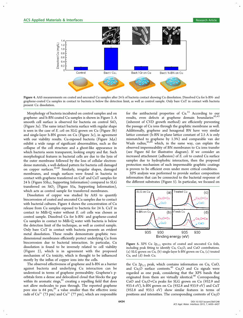
Morphology of bacteria incubated on control samples and ongraphene- and h-BN-coated Cu samples is shown in Figure 3. Asmooth cell surface is observed for bacteria on control SiO2(Figure 3a). The same intact bacteria surface with regular shapeis seen in the case of E. coli on SLG grown on Cu (Figure 3b)and single-layer h-BN grown on Cu (Figure 3c), in agreementwith our viability results. Cu-exposed bacteria (Figure 3d,e)exhibit a wide range of significant abnormalities, such as thecollapse of the cell structure and a ghost-like appearance inwhich bacteria seem transparent, looking empty and flat. Suchmorphological features in bacterial cells are due to the lysis ofthe outer membrane followed by the loss of cellular electron-dense materials, a well-defined pattern for bacteria cell damagedon copper surfaces.39,40 Similarly, irregular shapes, damagedmembranes, and rough surfaces were found in bacteria incontact with graphene transferred on CuF and CuT samples for24 h (Figure S5b,c, Supporting Information) compared to SLGtransferred on SiO2 (Figure S5a, Supporting Information),which acts as control sample for transferred membranes.Dissolution of copper was studied by AAS to quantify
biocorrosion of coated and uncoated Cu samples due to contactwith bacterial cultures. Figure 4 shows the concentration of Curelease from Cu samples exposed to bacteria for 24 h. CuT incontact to Milli-Q water without E. coli cells was chosen ascontrol sample. Dissolved Cu for h-BN- and graphene-coatedCu samples in contact to Milli-Q water with bacteria is belowthe detection limit of the technique, as well as control sample.Only bare CuT in contact with bacteria presents an evidentmetal dissolution. These results demonstrate graphitic two-dimensional membranes efficiently protect underlying Cu frombiocorrosion due to bacterial interaction. In particular, Cudissolution is found to be inversely related to cell viability(Figure 2), which is in agreement with the proposedmechanism of Cu toxicity, which is thought to be influencedmostly by the influx of copper ions into the cells.The observed effectiveness of graphene and h-BN as a barrier
against bacteria and underlying Cu interaction can beunderstood in terms of graphene permeability. Graphene’s p-orbitals form a dense and delocalized cloud that blocks the gapwithin its aromatic rings,41 creating a repelling field that doesnot allow molecules to pass through. The reported graphenepore size is 64 pm,20 a value smaller than the effective ionicradii of Cu2+ (73 pm) and Cu1+ (77 pm), which are responsible
for the antibacterial properties of Cu.15 According to ourresults, even defects at graphene domain boundaries42,43
(inherent of CVD growth method) are efficiently preventingthe passage of Cu ions through the graphitic membrane as well.Additionally, graphene and hexagonal BN have very similarlattice constant (h-BN in-plane lattice constant of 2.5 A is onlymismatched to graphene by 1.3%) and comparable van derWaals radius,43,44 which, in the same way, can explain theobserved impermeability of BN membranes to Cu ions transfer(see Figure 6d for illustrative diagram). If we consider anincreased attachment (adhesion) of E. coli to coated Cu surfacesamples due to hydrophobic interaction, then the proposedprotection mechanism of such impermeable graphitic coatingsis proven to be efficient even under more critical conditions.XPS analysis was performed to provide surface composition
information that can be connected to the bacterial response ofthe different substrates (Figure 5). In particular, we focused on
the Cu 2p3/2 peak, which contains information on Cu, CuO,and Cu2O surface contents.45 Cu2O and Cu signals wereregarded as one peak, considering that the XPS bands thatoriginated from them are virtually identical.46 CorrespondingCuO and Cu2O+Cu peaks for SLG grown on Cu (932.9 and933.4 eV), h-BN grown on Cu (932.8 and 933.9 eV) and CuT(932.8 and 933.5 eV) show similar features in terms ofpositions and intensities. The corresponding contents of Cu2O
Figure 4. AAS measurements on coated and uncoated Cu samples after 24 h of bacteria contact showing Cu dissolution. Dissolved Cu for h-BN- andgraphene-coated Cu samples in contact to bacteria is below the detection limit, as well as control sample. Only bare CuT in contact with bacteriapresent Cu dissolution.
Figure 5. XPS Cu 2p3/2 spectra of coated and uncoated Cu foils,including peak fitting to identify Cu, Cu2O, and CuO contributions.(a) SLG grown on Cu, (b) single-layer h-BN grown on Cu, (c) treatedCu, and (d) fresh Cu.
ACS Applied Materials & Interfaces Research Article
DOI: 10.1021/acsami.5b01248ACS Appl. Mater. Interfaces 2015, 7, 6430−6437
6434

+Cu were 71.6% for SLG grown on Cu, 75.6% for h-BN grownon Cu, and 65.4% for CuT, indicating a strong metallic signalfor all these samples.Such a result is expected, considering the oxide removal
thermal treatment to which the samples were subjected prior tographene growth. Additionally, this measurement clearlyindicates that CuT and not CuF is, in fact, the “real controlsample” of graphene-coated Cu foils for antibacterial studiesdue to the similarity of Cu+Cu2O content in both samples. Onthe other hand, CuF has a stronger CuO signal (62.9%), anoxide species with lower bactericide activity.5 This result is inagreement with the diminished 51.3% antibacterial efficiencyobserved after 2 h for this sample (Figure S4, SupportingInformation).Figure 6 summarizes the interaction between bacteria and
graphene- and h-BN-coated Cu substrates, inferred from ourstudy. Figure 6a shows the typical “contact killing” phenom-enon observed in the case of Cu substrate (fresh or treated)which proceeds by successive membrane damage, copper influxinto the cells, oxidative damage and cell death. Our AASmeasurements are in agreement with this antibacterialmechanism. When metallic substrate is covered by an as-grown graphitic membrane, regardless of its electrical proper-ties, we observed inhibition of all Cu antibacterial activity andCu biocorrosion, leading to (1) intact bacteria, as our viabilitymeasurements and SEM micrographs have confirmed, and (2)absence of Cu dissolution, as our AAS measurements haveshown. Such behavior indicates that there is no physical orelectrical interaction between underlying Cu and bacteria(Figure 6b,c). This effect is presumably connected toimpermeability of graphene and h-BN membranes to Cu ionstransfer (Figure 6d).
4. CONCLUSION
In conclusion, we studied the interaction between bacteria andgraphene- and h-BN-coated Cu samples. Our findings clearlyshow both graphene and h-BN coating substantially suppressinteraction between bacteria and underlying Cu substrate. Fromthe bacteria perspective, metal toxicity (connected to Cu ionsinflux into cells) is suppressed and with regard to the effect onthe Cu substrate, biocorrosion, due to bacteria in contact tometal surface, is prevented. The fact that both a conducting(graphene) and an insulating (h-BN) membrane, with almostthe same lattice constant, equally suppressed antibacterial
properties of Cu suggests that a connection between chargetransfer from metal to bacteria through these 2D systems andtheir antimicrobial activity is less likely. On the other hand,impermeability of the graphene and h-BN membranes is strongenough to prevent all exchange of Cu ions through it, evenconsidering typical grain boundary defects and the hydrophobicproperties of cells and coatings. At the end, this effectdetermines the lack of antibacterial activity and the consequentabsence of biocorrosion of coated Cu foils. Our results indicateas-grown graphene and h-BN films could successfully protectmetals and prevent their corrosion in biological environmentslinked to medical applications.
■ ASSOCIATED CONTENT*S Supporting InformationDetailed description of PMMA-assisted graphene transfermethod and Cu foils thermal treatment; morphologicalcharacterization of samples prior to bacterial contact; typicalphotographs of cultivated E. coli on agar plates for differentsubstrates; 2 h viability results for fresh and treated copper;SEM images of E. coli after 24 h incubation on SLG transferredon SiO2, SLG transferred on CuF, and SLG transferred onCuT. This material is available free of charge via the Internet athttp://pubs.acs.org.
■ AUTHOR INFORMATIONCorresponding Author*E-mail: [email protected] authors declare no competing financial interest.
■ ACKNOWLEDGMENTSThis work was financially supported by Conicyt de Insercionnos. 791220009 and 791100037, Fondecyt grant nos. 1110935and 1110992, Proyecto Interno DGIP nos. 111469 and 111470,PIA anillo ACT 1117. FMS gratefully acknowledges thesupport of CONICYT Ph.D., Mecesup FSM1204, andUTFSM-PIIC fellowships.
■ REFERENCES(1) Chen, S. S.; Brown, L.; Levendorf, M.; Cai, W. W.; Ju, S. Y.;Edgeworth, J.; Li, X. S.; Magnuson, C. W.; Velamakanni, A.; Piner, R.D.; et al. Oxidation Resistance of Graphene-Coated Cu and Cu/NiAlloy. ACS Nano 2011, 5, 1321−1327.
Figure 6. Schematic illustration of bacteria-coated copper interaction inferred from our study. (a) Bare Cu (fresh or treated) exhibits antibacterialactivity producing membrane damage, copper influx into the cells, oxidative damage, and cell death. (b) For graphene-coated Cu, there is nointeraction between bacteria and underlying substrate, leading to inhibition of Cu antibacterial activity due to this highly conductive nanoscalecoating. In addition, absence of biocorrosion of Cu is observed. (c) In the case of h-BN-coated Cu, the same lack of interaction between metal andbacteria is observed, even when considering insulating properties of such coating. (d) Pore size of graphene (similar to corresponding for h-BN)determines impermeability to Cu ions, which are responsible of its antibacterial activity. Both graphitic coatings suppress interaction betweenbacteria and metal, protecting bacteria from antibacterial properties of Cu and Cu from biocorrosion due to bacteria contact.
ACS Applied Materials & Interfaces Research Article
DOI: 10.1021/acsami.5b01248ACS Appl. Mater. Interfaces 2015, 7, 6430−6437
6435

(2) Nilsson, L.; Andersen, M.; Balog, R.; Laegsgaard, E.; Hofmann,P.; Besenbacher, F.; Hammer, B.; Stensgaard, I.; Hornekaer, L.Graphene Coatings: Probing the Limits of the One Atom ThickProtection Layer. ACS Nano 2012, 6, 10258−10266.(3) Topsakal, M.; Sahin, H.; Ciraci, S. Graphene Coatings: AnEfficient Protection from Oxidation. Phys. Rev. B 2012, 85, 155445−155451.(4) Zhou, F.; Li, Z.; Shenoy, G. J.; Liand, L.; Liu, H. EnhancedRoom-Temperature Corrosion of Copper in the Presence ofGraphene. ACS Nano 2013, 7, 6939−6947.(5) Hans, M.; Erbe, A.; Mathews, S.; Chen, Y.; Solioz, M.; Mucklich,F. Role of Copper Oxides in Contact Killing of Bacteria. Langmuir2013, 29, 16160−16166.(6) Rao, T. Microbial Fouling and Corrosion: Fundamentals andMechanisms. In Operational and Environmental Consequences of LargeIndustrial Cooling Water Systems; Rajagopal, S., et al., Eds.; Springer:New York, 2012; pp 95−126.(7) Stoodley, P.; Sauer, K.; Davies, D. G.; Costerton, J. W. Biofilms asComplex Differentiated Communities. Annu. Rev. Microbiol. 2002, 56,187−209.(8) Li, K.; Whitfield, M.; Van Vliet, K. J. Beating the Bugs: Roles ofMicrobial Biofilms in Corrosion. Corros. Rev. 2013, 31, 73−84.(9) Sreekumari, K. R.; Sato, Y.; Kikuchi, Y. Antibacterial Metals. AViable Solution for Bacterial Attachment and MicrobiologicallyInfluenced Corrosion. Mater. Trans. 2005, 46, 1636−1645.(10) Keevil, C. W. The Physico-Chemistry of Biofilm-MediatedPitting Corrosion of Copper Pipe Supplying Potable Water. Water Sci.Technol. 2010, 49, 91−98.(11) Yu, L.; Zhang, Y.; Zhang, B.; Liu, J.; Zhang, H.; Song, C.Preparation and Characterization of HPEI-GO/PES UltrafiltrationMembrane with Antifouling and Antibacterial Properties. J. Membr. Sci.2013, 447, 452−462.(12) Yu, L.; Zhang, Y.; Zhang, B.; Liu, J. Enhanced AntibacterialActivity of Silver Nanoparticles/Halloysite Nanotubes/GrapheneNanocomposites with Sandwich-Like Structure. Sci. Rep. 2014, 4,4551−4555.(13) Al-Thani, R. F.; Patan, N. K.; Al-Maadeed, M. A. GrapheneOxide as Antimicrobial Against Two Gram-positive and Two Gram-Negative Bacteria in Addition to One Fungus, OnLine. J. Biol. Sci.2014, 14, 230−239.(14) Zhang, W.; Lee, S.; McNear, K. L.; Chung, T. F.; Lee, S.; Lee,K.; Crist, S. A.; Ratliff, T. L.; Zhong, Z.; Chen, Y. P.; Yang, C. Use ofGraphene as Protection Film in Biological Environments. Sci. Rep.2014, 4, 4097−4105.(15) Grass, G.; Rensing, C.; Solioz, M. Metallic Copper as anAntimicrobial Surface. Appl. Environ. Microbiol. 2011, 77, 1541−1547.(16) Bunch, J. S.; Verbridge, S. S.; Alden, J. S.; van der Zande, A. M.;Parpia, J. M.; Craighead, H. G.; McEuen, P. L. Impermeable AtomicMembranes from Graphene Sheets. Nano Lett. 2008, 8, 2458−2462.(17) Cho, J.; Gao, L.; Tian, J.; Cao, H.; Wu, W.; Yu, Q.; Yitamben, E.N.; Fisher, B.; Guest, J. R.; Chen, Y. P.; Guisinger, N. P. Atomic-ScaleInvestigation of Graphene Grown on Cu Foil and the Effects ofThermal Annealing. ACS Nano 2011, 5, 3607−3613.(18) Lee, K. H.; Shin, H. J.; Lee, J.; Lee, I.; Kim, G.; Choi, J.; Kim, S.Large-Scale Synthesis of High-Quality Hexagonal Boron NitrideNanosheets for Large-Area Graphene Electronics. Nano Lett. 2012,12, 714−718.(19) Li, J.; Wang, G.; Zhu, H.; Zhang, M.; Zheng, X.; Di, Z.; Liu, X.;Wang, X. Antibacterial Activity of Large-Area Monolayer GrapheneFilm Manipulated by Charge Transfer. Sci. Rep. 2014, 4, 4359−4366.(20) Berry, V. Impermeability of Graphene and its Applications.Carbon 2013, 62, 1−10.(21) Wu, J.; Wang, B.; Wei, Y.; Yang, R.; Dresselhaus, M. Mechanicsand Mechanically Tunable Band Gap in Single-Layer HexagonalBoron-Nitride. Mater. Res. Lett. 2013, 1, 200−206.(22) Geim, A. K. Graphene: Status and Prospects. Science 2009, 324,1530−1534.(23) Reina, A.; Son, H.; Jiao, L.; Fan, B.; Dresselhaus, M. S.; Liu, Z.;Kong, J. Transferring and Identification of Single- and Few-Layer
Graphene on Arbitrary Substrates. J. Phys. Chem. C 2008, 112, 17741−17744.(24) Jia, C.; Jiang, J.; Gan, L.; Guo, X. Direct Optical Character-ization of Graphene Growth and Domains on Growth Substrates. Sci.Rep. 2012, 2, 707−712.(25) Stalder, A. F.; Kulik, G.; Sage, D.; Barbieri, L.; Hoffmann, P. ASnake-Based Approach to Accurate Determination of Both ContactPoints and Contact Angles. Colloids Surf. 2006, 286, 92−103.(26) Chirino, B.; Strahsburger, E.; Agullo, L.; Gonzalez, M.; Seeger,M. Genomic and Functional Analyses of the 2-Aminophenol CatabolicPathway and Partial Conversion of Its Substrate into Picolinic Acid inBurkholderia Xenovorans LB400. PloS One 2013, 8 (10), e75746.(27) Shin, Y. C.; Kong, J. Hydrogen-Excluded Graphene SynthesisVia Atmospheric Pressure Chemical Vapor Deposition. Carbon 2013,59, 439−447.(28) Kirkland, N. T.; Schiller, T.; Medhekar, N.; Birbilis, N.Exploring Graphene as a Corrosion Protection Barrier. Corros. Sci.2012, 56, 1−4.(29) Park, J.; Lee, J.; Liu, L.; Clark, K. W.; Durand, C.; Park, C.;Sumpter, B. G.; Baddorf, A. P.; Mohsin, A.; Yoon, M.; Gu, G.; Li, A. P.Spatially Resolved One-dimensional Boundary States in Graphene−Hexagonal Boron Nitride Planar Heterostructures. Nat. Commun.2014, 5, 5403.(30) Yu, Q.; Jauregui, L. A.; Wu, W.; Colby, R.; Tian, J.; Su, Z.; Cao,H.; Liu, Z.; Pandey, D.; Wei, D.; Chung, T. F.; Peng, P.; Guisinger, N.P.; Stach, E. A.; Bao, J.; Pei, S. S.; Chen, Y. P. Control andCharacterization of Individual Grains and Grain Boundaries inGraphene Grown by Chemical Vapour Deposition. Nat. Mater.2011, 10, 443−449.(31) Li, X.; Cai, W.; An, J.; Kim, S.; Nah, J.; Yang, D.; Piner, R.;Velamakanni, A.; Jung, I.; Tutuc, E.; Banerjee, S. K.; Colombo, L.;Ruoff, R. S. Large-Area Synthesis of High-Quality and UniformGraphene Films on Copper Foils. Science 2009, 324, 1312−1314.(32) Ferrari, A. C.; Meyer, J. C.; Scardaci, V.; Casiraghi, C.; Lazzeri,M.; Mauri, F.; Piscanec, S.; Jiang, D.; Novoselov, K. S.; Roth, S.; Geim,A. K. Raman Spectrum of Graphene and Graphene Layers. Phys. Rev.Lett. 2006, 97, 187401−187405.(33) Reina, A.; Jia, X.; Ho, J.; Nezich, D.; Son, H.; Bulovic, V.;Dresselhaus, M. S.; Kong, J. Large Area, Few-Layer Graphene Films onArbitrary Substrates by Chemical Vapor Deposition. Nano Lett. 2008,9, 30−35.(34) Espirito Santo, D.; Elowsky, C. G.; Quaranta, D.; Domaille, D.W.; Chang, C. J.; Grass, G. Bacterial Killing by Dry Metallic CopperSurfaces. Appl. Environ. Microbiol. 2011, 77, 794−802.(35) Gottenbos, B.; Busscher, H. J.; Van Der Mei, H. C.;Nieuwenhuis, P. Pathogenesis and Prevention of Biomaterial CenteredInfections. J. Mater. Sci.: Mater. Med. 2002, 13, 717−722.(36) Busscher, H. Specific and Non-specific Interactions in BacterialAdhesion to Solid Substrata. FEMS Microbiol. Lett. 1987, 46, 165−173.(37) Mendez-Vilas, A. Microbial Pathogens and Strategies forCombating Them: Science, Technology and Education; FormatexResearch Center: Badajoz, Spain, 2013.(38) Donlan, R. M. Biofilms: Microbial Life on Surfaces. EmergingInfect. Dis. 2002, 8, 881−890.(39) Shina, S. Y.; Bajpaia, V. K.; Kimb, H. R.; Kanga, S. C.Antibacterial Activity of Eicosapentaenoic Acid (EPA) Against FoodBorne and Dood Spoilage Microorganisms. LWTFood Sci. Technol.2007, 40, 1515−1519.(40) Warnes, S. L.; Keevil, C. W. Mechanism of Copper SurfaceToxicity in Vancomycin-Resistant Enterococci Following Wet or DrySurface Contact. Appl. Environ. Microbiol. 2011, 77, 6049−6059.(41) Sreeprasad, T. S.; Berry, V. How Do the Electrical Properties ofGraphene Change with Its Functionalization? Small 2012, 9, 341−50.(42) Biro, L. P.; Lambin, P. Grain Boundaries in Graphene Grown byChemical Vapor Deposition. New J. Phys. 2013, 15, 035024−035062.(43) Hod, O. Graphite and Hexagonal Boron-Nitride Have the SameInterlayer Distance. Why? J. Chem. Theory Comput. 2012, 8, 1360−1369.
ACS Applied Materials & Interfaces Research Article
DOI: 10.1021/acsami.5b01248ACS Appl. Mater. Interfaces 2015, 7, 6430−6437
6436

(44) Dean, C. R.; Young, A. F.; Meric, I.; Lee, C.; Wang, L.;Sorgenfrei, S.; Watanabe, K.; Taniguchi, T.; Kim, P.; Shepard, K. L.;Hone, J. Boron Nitride Substrates for High-Quality GrapheneElectronics. Nat. Nanotecnol. 2010, 5, 722−726.(45) Biesingera, M. C.; Laua, L. W. M.; Gersonb, A. R.; Smart, R.St.C. Resolving Surface Chemical States in XPS Analysis of First RowTransition Metals, Oxides and Hydroxides: Sc, Ti, V, Cu and Zn. Appl.Surf. Sci. 2010, 257, 887−898.(46) Platzman, I.; Brener, R.; Haick, H.; Tannenbaum, R. Oxidationof Polycrystalline Copper Thin Films at Ambient Conditions. J. Phys.Chem. C 2008, 112, 1101−1108.
ACS Applied Materials & Interfaces Research Article
DOI: 10.1021/acsami.5b01248ACS Appl. Mater. Interfaces 2015, 7, 6430−6437
6437