suppressing thyroid hormone signaling preserves cone ... · suppressing thyroid hormone signaling...
TRANSCRIPT
Suppressing thyroid hormone signaling preservescone photoreceptors in mouse models ofretinal degenerationHongwei Maa, Arjun Thapaa, Lynsie Morrisa, T. Michael Redmondb, Wolfgang Baehrc, and Xi-Qin Dinga,1
aDepartment of Cell Biology, University of Oklahoma Health Sciences Center, Oklahoma City, OK 73104; bLaboratory of Retinal Cell and Molecular Biology,National Eye Institute, Bethesda, MD 20892; and cJohn A. Moran Eye Center, University of Utah, Salt Lake City, UT 84132
Edited by Jeremy Nathans, Johns Hopkins University, Baltimore, MD, and approved January 24, 2014 (received for review September 9, 2013)
Cone phototransduction and survival of cones in the humanmacula is essential for color vision and for visual acuity. Pro-gressive cone degeneration in age-related macular degeneration,Stargardt disease, and recessive cone dystrophies is a major causeof blindness. Thyroid hormone (TH) signaling, which regulates cellproliferation, differentiation, and apoptosis, plays a central rolein cone opsin expression and patterning in the retina. Here, weinvestigated whether TH signaling affects cone viability in inheritedretinal degeneration mouse models. Retinol isomerase RPE65-deficient mice [a model of Leber congenital amaurosis (LCA) withrapid cone loss] and cone photoreceptor function loss type 1 mice(severe recessive achromatopsia) were used to determine whethersuppressing TH signaling with antithyroid treatment reduces conedeath. Further, cone cyclic nucleotide-gated channel B subunit-deficient mice (moderate achromatopsia) and guanylate cyclase2e-deficient mice (LCA with slower cone loss) were used todetermine whether triiodothyronine (T3) treatment (stimulatingTH signaling) causes deterioration of cones. We found that conedensity in retinol isomerase RPE65-deficient and cone photorecep-tor function loss type 1 mice increased about sixfold followingantithyroid treatment. Cone density in cone cyclic nucleotide-gated channel B subunit-deficient and guanylate cyclase 2e-deficient mice decreased about 40% following T3 treatment. Theeffect of THsignaling on cone viability appears to be independent ofits regulation on cone opsin expression. This work demonstratesthat suppressing TH signaling in retina dystrophy mouse models isprotective of cones, providing insights into cone preservation andtherapeutic interventions.
Rod and cone photoreceptors degenerate under a variety ofpathological conditions, including a wide array of hereditary
retinal diseases, such as retinitis pigmentosa, macular degeneration,and cone–rod dystrophies. Defects in a large number of genesare linked to inherited retinal degenerative disorders (www.sph.uth.tmc.edu/RetNet/disease.htm), including those encoding en-zymes involved in the recycling of 11-cis retinal in the retinalpigment epithelium (RPE), retinoid isomerase (RPE65), andlecithin retinol acyltransferase (LRAT), and the phototransduction-associated proteins (opsins, subunits of transducin, cGMP phos-phodiesterase PDE6, guanylate cyclase, and cyclic nucleotide-gated channel). There are currently no treatments for humanretinal dystrophies. Despite a high genetic heterogeneity, the degen-erating photoreceptors show common cellular disorder features,including oxidative damage (1, 2), endoplasmic reticulum stress(3, 4), and apoptosis (5, 6).Thyroid hormone (TH) signaling regulates cell proliferation,
differentiation, and apoptosis. The role of TH signaling in retinaregarding its regulation of cone opsin expression and patterninghas been well documented (7, 8). Most mammals possess di-chromatic color vision that is mediated by two opsins with peaksensitivities to medium-long (M, green) and short (S, blue) wave-lengths of light (9, 10). In mouse, M- and S-opsins are expressedin opposing gradients such that varying amounts of both opsinsare coexpressed in cones in midretinal regions, whereas M-opsin
predominates in dorsal (superior) regions and S-opsin predom-inates in ventral (inferior) regions (10, 11) (Fig. S1). Duringdevelopment and in the adult postmitotic retina, TH signalingvia its receptor type β2 (TRβ2) suppresses expression of S-opsin,induces expression of M-opsin, and promotes the dorsal–ventralopsin patterning (7, 8). Importantly, TH signaling has been asso-ciated with cone viability. Triiodothyronine (T3) treatment wasshown to cause cone death in mice and this effect was reversed bydeletion of TRβ2 gene (12). Excessive TH signaling was alsoshown to induce auditory defects and cochlear degeneration inmice (13). TH signaling has been associated with apoptosis ofa variety of human cell lines, including lymphocytes (14), breastcancer cells (15), HeLa cells (16), and pituitary tumor cells (17),and TH signaling has been well documented in apoptotic tissueremodeling during anuran metamorphosis (18, 19). To determinewhether TH signaling affects cone viability in inherited retinaldegeneration, we investigated cone death/survival in retinal de-generation mouse models following TH signaling suppressionand stimulation. Retinol isomerase RPE65-deficient (Rpe65−/−) (amodel of Leber congenital amaurosis, LCA) (20, 21) and conephotoreceptor function loss type 1 (cpfl1) mice (PDE6C mutation,a model of achromatopsia) (22), displaying fast and severe conedegeneration, were used to determine whether suppressing THsignaling with antithyroid treatment reduces cone degeneration.Cone cyclic nucleotide-gated channel B subunit-deficient (Cngb3−/−)(a model of achromatopsia) (23) and guanylate cyclase 2e-deficient(Gucy2e−/−) (another model of LCA) mice (24), displaying rela-tively slow progressive and moderate cone degeneration, wereused to determine whether stimulating TH signaling (with T3
Significance
Photoreceptors degenerate in a wide array of hereditary reti-nal diseases and age-related macular degeneration. There iscurrently no treatment available for retinal degenerations.While outnumbered roughly 20:1 by rods in the human retina,it is the cones that mediate color vision and visual acuity, andtheir survival is critical for vision. In this communication, weinvestigate whether thyroid hormone (TH) signaling affectscone viability in retinal degeneration mouse models. TH sig-naling is known to be important for cone pigment expressionand patterning, but excess TH signaling causes death of cones.We demonstrate that suppressing TH signaling in mousemodels of cone–rod dystrophy preserves cones. This findingmay lead to a novel and substantially different approach forretinal degeneration management.
Author contributions: H.M. and X.-Q.D. designed research; H.M., A.T., and L.M. performedresearch; H.M., A.T., and L.M. analyzed data; and H.M., T.M.R., W.B., and X.-Q.D. wrotethe paper.
The authors declare no conflict of interest.
This article is a PNAS Direct Submission.1To whom correspondence should be addressed. E-mail: [email protected].
This article contains supporting information online at www.pnas.org/lookup/suppl/doi:10.1073/pnas.1317041111/-/DCSupplemental.
3602–3607 | PNAS | March 4, 2014 | vol. 111 | no. 9 www.pnas.org/cgi/doi/10.1073/pnas.1317041111
treatment) deteriorates cones. We report here that cone survivalwas greatly improved in Rpe65−/− and cpfl1 mice following THsignaling suppression, whereas cone degeneration was signifi-cantly increased in Cngb3−/− and Gucy2e−/− mice following THsignaling stimulation, demonstrating a protective role of sup-pressing TH signaling in cones.
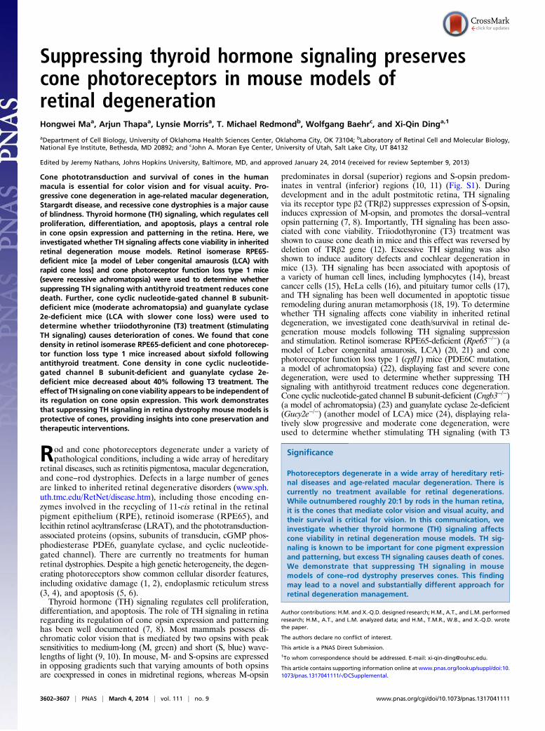
ResultsSuppressing TH Signaling Preserves Cones in Rpe65−/− Mice. Rpe65−/−,a severe cone degeneration model, displays rapid ventral andcentral cone loss, whereas dorsal cones are preserved relativelywell (20, 21). Our cone density evaluation by peanut agglutinin(PNA) and cone arrestin (CAR) labeling in Rpe65−/− mice showeda degeneration pattern similar to that reported previously (20, 25),i.e., the ventral and central retina shows early onset, fast conedegeneration (about 10% of the wild-type level remained atpostnatal day 30, P30), whereas the peripheral dorsal retinadegenerated more slowly (about 50% of the wild-type levelremained at P30) (Fig. 1 A and B). Rpe65−/− mice received anti-thyroid drug (methimazole and sodium perchlorate monohydrate)treatment for 30 d, beginning on P1. The antithyroid treatmentreduced serum T3 levels by about 30% in the treated mice,compared with untreated controls, when measured on the last day
of the treatment (Table S1). This treatment significantly increasedcone density. Cone density evaluation showed that the number ofPNA-labeled cones in the ventral and dorsal retinas in antithyroid-treated Rpe65−/− mice increased about 6- and 1.3-fold, respec-tively, compared with untreated controls (Fig. 1 A and B). Simi-larly, CAR-labeled cones in the ventral retinas increased about4-fold. The increased cone density in antithyroid-treated Rpe65−/−
mice was also shown by PNA and CAR labeling on retinal cross-sections (Fig. S2). These results demonstrate a protective role ofsuppressing TH signaling in Rpe65−/− cones.The effect of TH signaling on cone opsin expression has been
well characterized (7, 8). We examined M- and S-cone density bylabeling M- and S-opsin with specific antibodies. In agreementwith reported information in wild-type mice, M-opsin–labeledcones (M cones) were significantly reduced in Rpe65−/− micefollowing antithyroid treatment, whereas S-opsin–labeled cones(S cones) were greatly increased, compared with untreated con-trols (Fig. 1 A and B and Fig. S2). Cone preservation in Rpe65−/−
mice following TH signaling suppression was also evaluated byexamining the expression levels of cone specific proteins. CAR andcone transducin α-subunit [guanine nucleotide-binding protein G(t)subunit alpha-2 (GNAT2)] expression levels in antithyroid-treatedRpe65−/− mice increased by 30–40%, compared with untreated
Fig. 1. Suppressing TH signaling preserves cones inRpe65−/− mice. Rpe65−/− mice received antithyroidtreatment for 30 d, beginning on P1. At the end ofthe treatment, cone density was evaluated by im-munofluorescence labeling on retinal whole mounts,and cone-specific protein expression was evaluatedby Western blotting. (A) Representative confocalimages of immunofluorescence labeling of PNA, CAR,M-opsin, and S-opsin in hypothyroid and untreatedRpe65−/− mice and wild-type (WT) mice. (Scale bar,10 μm.) (B) Correlating quantitative analysis of theimmunofluorescence labeling. (C) Shown are repre-sentative images of the Western blot detection ofCAR, GNAT2, M-opsin, and S-opsin, and the corre-lating quantifications. Data are represented asmean ± SEM of three to four assays using eyes/retinasfrom four mice. Unpaired Student t test was usedto determine significance between hypothyroidand untreated Rpe65−/− mice (*P < 0.05, **P <0.01, and ***P < 0.001).
Ma et al. PNAS | March 4, 2014 | vol. 111 | no. 9 | 3603
NEU
ROSC
IENCE
controls (Fig. 1C). Similar to S-opsin labeling of cones, S-opsinexpression levels were increased about 5-fold. By quantitative(q)RT-PCR, the mRNA levels of S-opsin and M-opsin in an-tithyroid-treated Rpe65−/− mice increased about 3.8-fold anddecreased by about 20%, respectively (Fig. S3). This finding isconsistent with the previously demonstrated effect of TH sig-naling on cone opsin expression (7, 8).
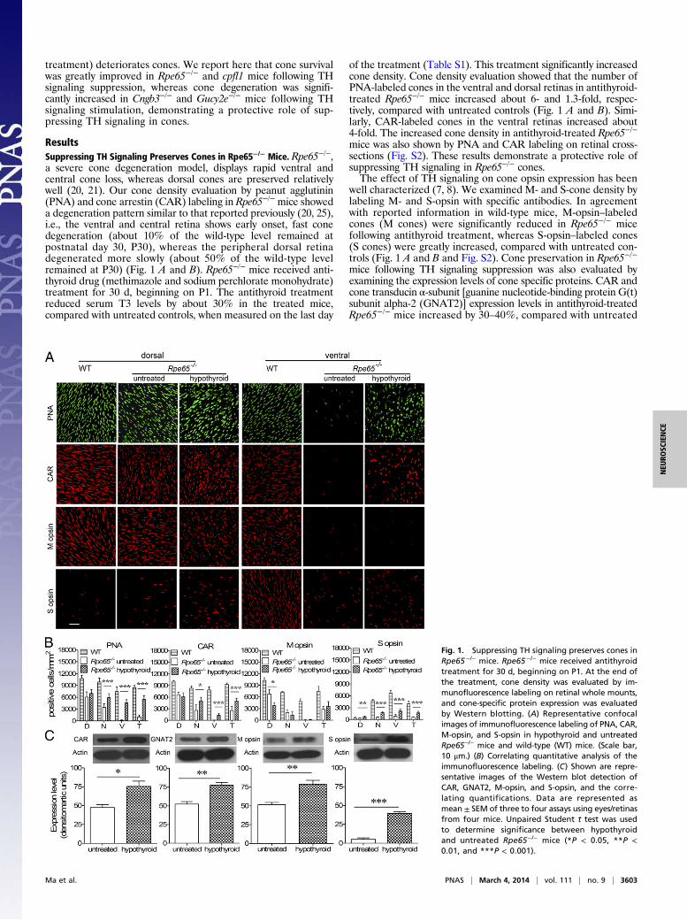
Suppressing TH Signaling Preserves Cones in Cpfl1 Mice. Cpfl1, anaturally occurring cone degeneration mouse line, is another fastand severe cone degeneration model (26, 27). Cpfl1 mice re-ceived antithyroid drug (methimazole and sodium perchloratemonohydrate) treatment for 30 d, beginning on P1. The anti-thyroid treatment reduced serum T3 levels in the treated mice byabout 50%, compared with untreated controls, when measuredon the last day of the treatment (Table S1). This treatment sig-nificantly improved cone survival. Fig. 2 shows representativeimages of PNA, GNAT2, M-opsin, and S-opsin labeling of reti-nal cross sections (Fig. 2A) and their quantifications (Fig. 2B) inP30 antithyroid-treated and untreated cpfl1 mice. Cone densityin cpfl1 mice was about 50% of the wild-type level. Followingantithyroid treatment, cone density was increased to about 70–80% of the wild-type level (Fig. 2 A and B). Similar to the obser-vations in Rpe65−/− mice, M cones were significantly reduced,whereas S cones were greatly increased (about a 0.7- to 5.0-foldincrease, depending on the retinal areas, with the maximal in-crease observed in the dorsal retina) (Fig. 2 and Fig. S4). qRT-PCR analysis showed a 3.8-fold increase in S-opsin mRNA levelsand a 50% decrease of M-opsin mRNA level in cpfl1 mice afterantithyroid treatment (Fig. S5).To determine whether suppressing TH signaling affects post-
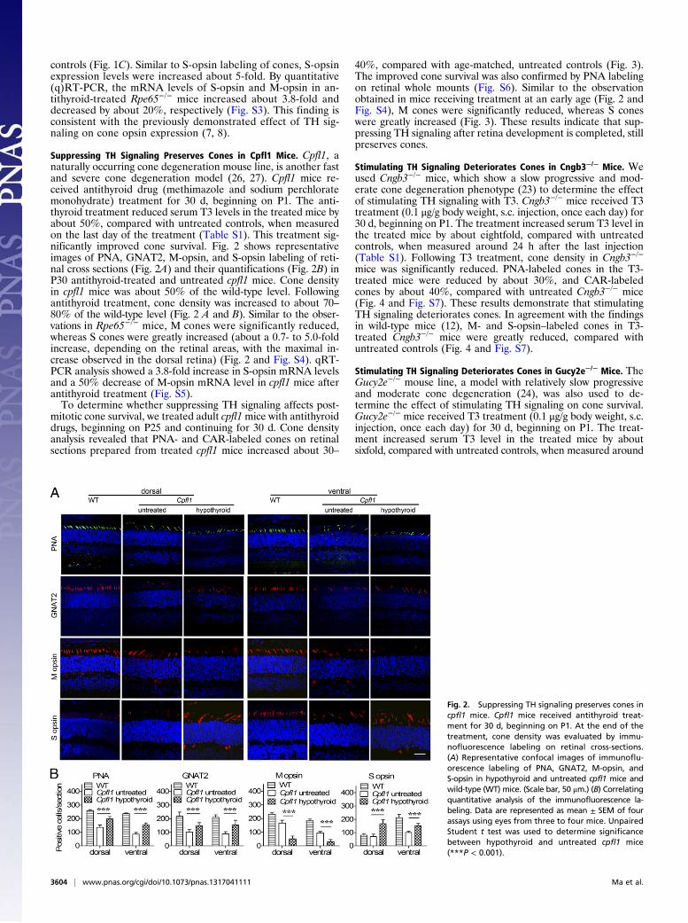
mitotic cone survival, we treated adult cpfl1mice with antithyroiddrugs, beginning on P25 and continuing for 30 d. Cone densityanalysis revealed that PNA- and CAR-labeled cones on retinalsections prepared from treated cpfl1 mice increased about 30–
40%, compared with age-matched, untreated controls (Fig. 3).The improved cone survival was also confirmed by PNA labelingon retinal whole mounts (Fig. S6). Similar to the observationobtained in mice receiving treatment at an early age (Fig. 2 andFig. S4), M cones were significantly reduced, whereas S coneswere greatly increased (Fig. 3). These results indicate that sup-pressing TH signaling after retina development is completed, stillpreserves cones.
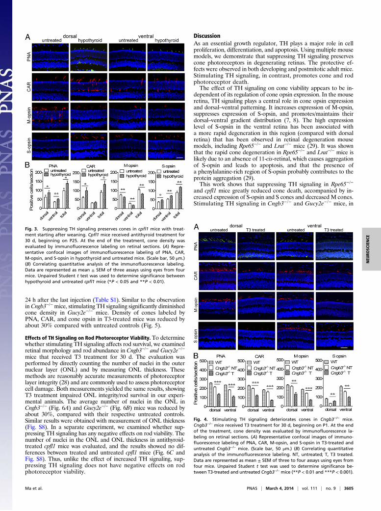
Stimulating TH Signaling Deteriorates Cones in Cngb3−/− Mice. Weused Cngb3−/− mice, which show a slow progressive and mod-erate cone degeneration phenotype (23) to determine the effectof stimulating TH signaling with T3. Cngb3−/− mice received T3treatment (0.1 μg/g body weight, s.c. injection, once each day) for30 d, beginning on P1. The treatment increased serum T3 level inthe treated mice by about eightfold, compared with untreatedcontrols, when measured around 24 h after the last injection(Table S1). Following T3 treatment, cone density in Cngb3−/−
mice was significantly reduced. PNA-labeled cones in the T3-treated mice were reduced by about 30%, and CAR-labeledcones by about 40%, compared with untreated Cngb3−/− mice(Fig. 4 and Fig. S7). These results demonstrate that stimulatingTH signaling deteriorates cones. In agreement with the findingsin wild-type mice (12), M- and S-opsin–labeled cones in T3-treated Cngb3−/− mice were greatly reduced, compared withuntreated controls (Fig. 4 and Fig. S7).
Stimulating TH Signaling Deteriorates Cones in Gucy2e−/− Mice. TheGucy2e−/− mouse line, a model with relatively slow progressiveand moderate cone degeneration (24), was also used to de-termine the effect of stimulating TH signaling on cone survival.Gucy2e−/− mice received T3 treatment (0.1 μg/g body weight, s.c.injection, once each day) for 30 d, beginning on P1. The treat-ment increased serum T3 level in the treated mice by aboutsixfold, compared with untreated controls, when measured around
Fig. 2. Suppressing TH signaling preserves cones incpfl1 mice. Cpfl1 mice received antithyroid treat-ment for 30 d, beginning on P1. At the end of thetreatment, cone density was evaluated by immu-nofluorescence labeling on retinal cross-sections.(A) Representative confocal images of immunoflu-orescence labeling of PNA, GNAT2, M-opsin, andS-opsin in hypothyroid and untreated cpfl1 mice andwild-type (WT) mice. (Scale bar, 50 μm.) (B) Correlatingquantitative analysis of the immunofluorescence la-beling. Data are represented as mean ± SEM of fourassays using eyes from three to four mice. UnpairedStudent t test was used to determine significancebetween hypothyroid and untreated cpfl1 mice(***P < 0.001).
3604 | www.pnas.org/cgi/doi/10.1073/pnas.1317041111 Ma et al.
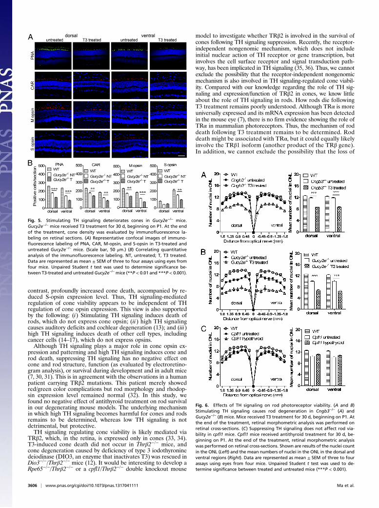
24 h after the last injection (Table S1). Similar to the observationin Cngb3−/−mice, stimulating TH signaling significantly diminishedcone density in Gucy2e−/− mice. Density of cones labeled byPNA, CAR, and cone opsin in T3-treated mice was reduced byabout 30% compared with untreated controls (Fig. 5).
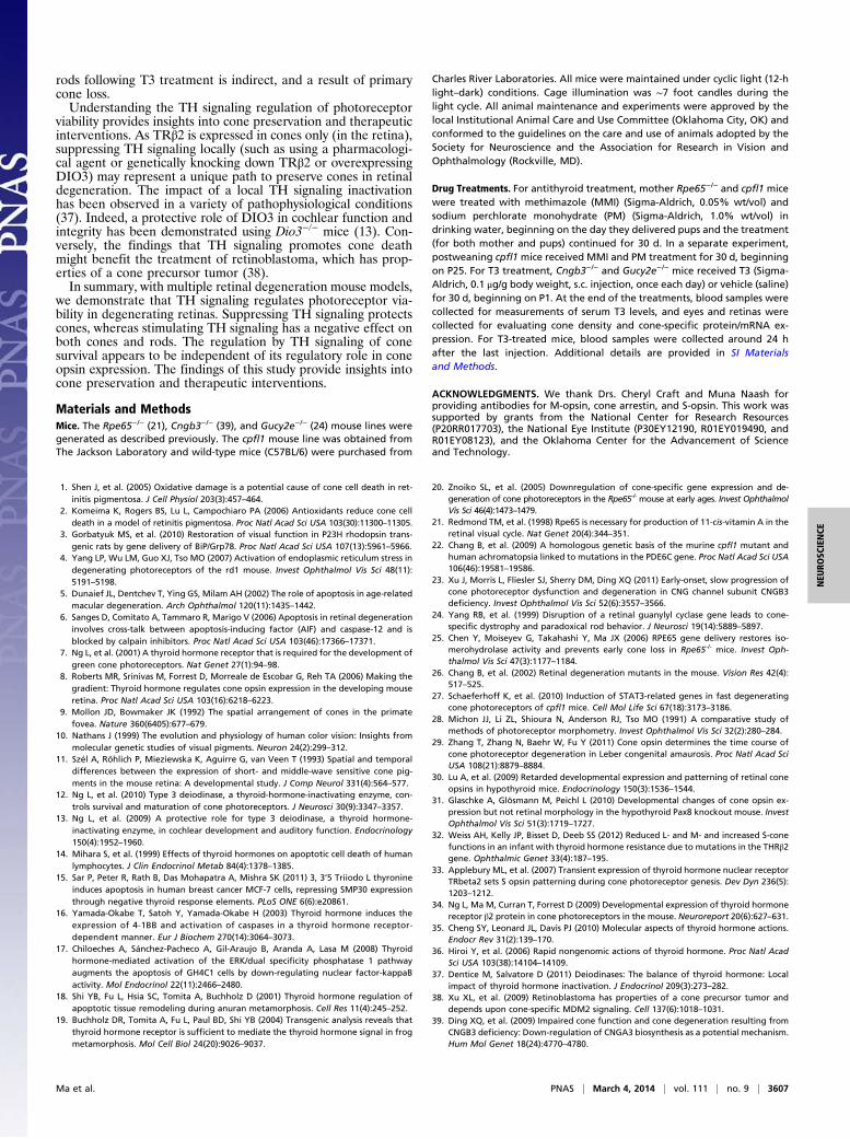
Effects of TH Signaling on Rod Photoreceptor Viability. To determinewhether stimulating TH signaling affects rod survival, we examinedretinal morphology and rod abundance in Cngb3−/− and Gucy2e−/−
mice that received T3 treatment for 30 d. The evaluation wasperformed by directly counting the number of nuclei in the outernuclear layer (ONL) and by measuring ONL thickness. Thesemethods are reasonably accurate measurements of photoreceptorlayer integrity (28) and are commonly used to assess photoreceptorcell damage. Both measurements yielded the same results, showingT3 treatment impaired ONL integrity/rod survival in our experi-mental animals. The average number of nuclei in the ONL inCngb3−/− (Fig. 6A) and Gucy2e−/− (Fig. 6B) mice was reduced byabout 30%, compared with their respective untreated controls.Similar results were obtained with measurement of ONL thickness(Fig. S8). In a separate experiment, we examined whether sup-pressing TH signaling has any negative effects on rod viability. Thenumber of nuclei in the ONL and ONL thickness in antithyroid-treated cpfl1 mice was evaluated, and the results showed no dif-ferences between treated and untreated cpfl1 mice (Fig. 6C andFig. S8). Thus, unlike the effect of increased TH signaling, sup-pressing TH signaling does not have negative effects on rodphotoreceptor viability.
DiscussionAs an essential growth regulator, TH plays a major role in cellproliferation, differentiation, and apoptosis. Using multiple mousemodels, we demonstrate that suppressing TH signaling preservescone photoreceptors in degenerating retinas. The protective ef-fects were observed in both developing and postmitotic adult mice.Stimulating TH signaling, in contrast, promotes cone and rodphotoreceptor death.The effect of TH signaling on cone viability appears to be in-
dependent of its regulation of cone opsin expression. In the mouseretina, TH signaling plays a central role in cone opsin expressionand dorsal–ventral patterning. It increases expression of M-opsin,suppresses expression of S-opsin, and promotes/maintains theirdorsal–ventral gradient distribution (7, 8). The high expressionlevel of S-opsin in the ventral retina has been associated witha more rapid degeneration in this region (compared with dorsalretina) that has been observed in retinal degeneration mousemodels, including Rpe65−/− and Lrat−/− mice (29). It was shownthat the rapid cone degeneration in Rpe65−/− and Lrat−/− mice islikely due to an absence of 11-cis-retinal, which causes aggregationof S-opsin and leads to apoptosis, and that the presence ofa phenylalanine-rich region of S-opsin probably contributes to theprotein aggregation (29).This work shows that suppressing TH signaling in Rpe65−/−
and cpfl1 mice greatly reduced cone death, accompanied by in-creased expression of S-opsin and S cones and decreased M cones.Stimulating TH signaling in Cngb3−/− and Gucy2e−/− mice, in
Fig. 3. Suppressing TH signaling preserves cones in cpfl1 mice with treat-ment starting after weaning. Cpfl1 mice received antithyroid treatment for30 d, beginning on P25. At the end of the treatment, cone density wasevaluated by immunofluorescence labeling on retinal sections. (A) Repre-sentative confocal images of immunofluorescence labeling of PNA, CAR,M-opsin, and S-opsin in hypothyroid and untreated mice. (Scale bar, 50 μm.)(B) Correlating quantitative analysis of the immunofluorescence labeling.Data are represented as mean ± SEM of three assays using eyes from fourmice. Unpaired Student t test was used to determine significance betweenhypothyroid and untreated cpfl1 mice (*P < 0.05 and **P < 0.01).
Fig. 4. Stimulating TH signaling deteriorates cones in Cngb3−/− mice.Cngb3−/− mice received T3 treatment for 30 d, beginning on P1. At the endof the treatment, cone density was evaluated by immunofluorescence la-beling on retinal sections. (A) Representative confocal images of immuno-fluorescence labeling of PNA, CAR, M-opsin, and S-opsin in T3-treated anduntreated Cngb3−/− mice. (Scale bar, 50 μm.) (B) Correlating quantitativeanalysis of the immunofluorescence labeling. NT, untreated; T, T3 treated.Data are represented as mean ± SEM of three to four assays using eyes fromfour mice. Unpaired Student t test was used to determine significance be-tween T3-treated and untreated Cngb3−/− mice (**P < 0.01 and ***P < 0.001).
Ma et al. PNAS | March 4, 2014 | vol. 111 | no. 9 | 3605
NEU
ROSC
IENCE
contrast, profoundly increased cone death, accompanied by re-duced S-opsin expression level. Thus, TH signaling-mediatedregulation of cone viability appears to be independent of THregulation of cone opsin expression. This view is also supportedby the following: (i) Stimulating TH signaling induces death ofrods, which do not express cone opsin; (ii) high TH signalingcauses auditory deficits and cochlear degeneration (13); and (iii)high TH signaling induces death of other cell types, includingcancer cells (14–17), which do not express opsins.Although TH signaling plays a major role in cone opsin ex-
pression and patterning and high TH signaling induces cone androd death, suppressing TH signaling has no negative effect oncone and rod structure, function (as evaluated by electroretino-gram analysis), or survival during development and in adult mice(7, 30, 31). This is in agreement with the observations in a humanpatient carrying TRβ2 mutations. This patient merely showedred/green color complications but rod morphology and rhodop-sin expression level remained normal (32). In this study, wefound no negative effect of antithyroid treatment on rod survivalin our degenerating mouse models. The underlying mechanismin which high TH signaling becomes harmful for cones and rodsremains to be determined, whereas low TH signaling is notdetrimental, but protective.TH signaling regulating cone viability is likely mediated via
TRβ2, which, in the retina, is expressed only in cones (33, 34).T3-induced cone death did not occur in Thrβ2−/− mice, andcone degeneration caused by deficiency of type 3 iodothyroninedeiodinase (DIO3, an enzyme that inactivates T3) was rescued inDio3−/−/Thrβ2−/− mice (12). It would be interesting to develop aRpe65−/−/Thrβ2−/− or a cpfl1/Thrβ2−/− double knockout mouse
model to investigate whether TRβ2 is involved in the survival ofcones following TH signaling suppression. Recently, the receptor-independent nongenomic mechanism, which does not includeinitial nuclear action of TH receptor or gene transcription, butinvolves the cell surface receptor and signal transduction path-way, has been implicated in TH signaling (35, 36). Thus, we cannotexclude the possibility that the receptor-independent nongenomicmechanism is also involved in TH signaling-regulated cone viabil-ity. Compared with our knowledge regarding the role of TH sig-naling and expression/function of TRβ2 in cones, we know littleabout the role of TH signaling in rods. How rods die followingT3 treatment remains poorly understood. Although TRα is moreuniversally expressed and its mRNA expression has been detectedin the mouse eye (7), there is no firm evidence showing the role ofTRα in mammalian photoreceptors. Thus, the mechanism of roddeath following T3 treatment remains to be determined. Roddeath might be associated with TRα, but it could equally likelyinvolve the TRβ1 isoform (another product of the TRβ gene).In addition, we cannot exclude the possibility that the loss of
Fig. 5. Stimulating TH signaling deteriorates cones in Gucy2e−/− mice.Gucy2e−/− mice received T3 treatment for 30 d, beginning on P1. At the endof the treatment, cone density was evaluated by immunofluorescence la-beling on retinal sections. (A) Representative confocal images of immuno-fluorescence labeling of PNA, CAR, M-opsin, and S-opsin in T3-treated anduntreated Gucy2e−/− mice. (Scale bar, 50 μm.) (B) Correlating quantitativeanalysis of the immunofluorescence labeling. NT, untreated; T, T3 treated.Data are represented as mean ± SEM of three to four assays using eyes fromfour mice. Unpaired Student t test was used to determine significance be-tween T3-treated and untreated Gucy2e−/−mice (**P < 0.01 and ***P < 0.001).
Fig. 6. Effects of TH signaling on rod photoreceptor viability. (A and B)Stimulating TH signaling causes rod degeneration in Cngb3−/− (A) andGucy2e−/− (B) mice. Mice received T3 treatment for 30 d, beginning on P1. Atthe end of the treatment, retinal morphometric analysis was performed onretinal cross-sections. (C) Suppressing TH signaling does not affect rod via-bility in cpfl1 mice. Cpfl1 mice received antithyroid treatment for 30 d, be-ginning on P1. At the end of the treatment, retinal morphometric analysiswas performed on retinal cross-sections. Shown are results of the nuclei countin the ONL (Left) and the mean numbers of nuclei in the ONL in the dorsal andventral regions (Right). Data are represented as mean ± SEM of three to fourassays using eyes from four mice. Unpaired Student t test was used to de-termine significance between treated and untreated mice (***P < 0.001).
3606 | www.pnas.org/cgi/doi/10.1073/pnas.1317041111 Ma et al.
rods following T3 treatment is indirect, and a result of primarycone loss.Understanding the TH signaling regulation of photoreceptor
viability provides insights into cone preservation and therapeuticinterventions. As TRβ2 is expressed in cones only (in the retina),suppressing TH signaling locally (such as using a pharmacologi-cal agent or genetically knocking down TRβ2 or overexpressingDIO3) may represent a unique path to preserve cones in retinaldegeneration. The impact of a local TH signaling inactivationhas been observed in a variety of pathophysiological conditions(37). Indeed, a protective role of DIO3 in cochlear function andintegrity has been demonstrated using Dio3−/− mice (13). Con-versely, the findings that TH signaling promotes cone deathmight benefit the treatment of retinoblastoma, which has prop-erties of a cone precursor tumor (38).In summary, with multiple retinal degeneration mouse models,
we demonstrate that TH signaling regulates photoreceptor via-bility in degenerating retinas. Suppressing TH signaling protectscones, whereas stimulating TH signaling has a negative effect onboth cones and rods. The regulation by TH signaling of conesurvival appears to be independent of its regulatory role in coneopsin expression. The findings of this study provide insights intocone preservation and therapeutic interventions.
Materials and MethodsMice. The Rpe65−/− (21), Cngb3−/− (39), and Gucy2e−/− (24) mouse lines weregenerated as described previously. The cpfl1 mouse line was obtained fromThe Jackson Laboratory and wild-type mice (C57BL/6) were purchased from
Charles River Laboratories. All mice were maintained under cyclic light (12-hlight–dark) conditions. Cage illumination was ∼7 foot candles during thelight cycle. All animal maintenance and experiments were approved by thelocal Institutional Animal Care and Use Committee (Oklahoma City, OK) andconformed to the guidelines on the care and use of animals adopted by theSociety for Neuroscience and the Association for Research in Vision andOphthalmology (Rockville, MD).
Drug Treatments. For antithyroid treatment, mother Rpe65−/− and cpfl1 micewere treated with methimazole (MMI) (Sigma-Aldrich, 0.05% wt/vol) andsodium perchlorate monohydrate (PM) (Sigma-Aldrich, 1.0% wt/vol) indrinking water, beginning on the day they delivered pups and the treatment(for both mother and pups) continued for 30 d. In a separate experiment,postweaning cpfl1mice received MMI and PM treatment for 30 d, beginningon P25. For T3 treatment, Cngb3−/− and Gucy2e−/− mice received T3 (Sigma-Aldrich, 0.1 μg/g body weight, s.c. injection, once each day) or vehicle (saline)for 30 d, beginning on P1. At the end of the treatments, blood samples werecollected for measurements of serum T3 levels, and eyes and retinas werecollected for evaluating cone density and cone-specific protein/mRNA ex-pression. For T3-treated mice, blood samples were collected around 24 hafter the last injection. Additional details are provided in SI Materialsand Methods.
ACKNOWLEDGMENTS. We thank Drs. Cheryl Craft and Muna Naash forproviding antibodies for M-opsin, cone arrestin, and S-opsin. This work wassupported by grants from the National Center for Research Resources(P20RR017703), the National Eye Institute (P30EY12190, R01EY019490, andR01EY08123), and the Oklahoma Center for the Advancement of Scienceand Technology.
1. Shen J, et al. (2005) Oxidative damage is a potential cause of cone cell death in ret-initis pigmentosa. J Cell Physiol 203(3):457–464.
2. Komeima K, Rogers BS, Lu L, Campochiaro PA (2006) Antioxidants reduce cone celldeath in a model of retinitis pigmentosa. Proc Natl Acad Sci USA 103(30):11300–11305.
3. Gorbatyuk MS, et al. (2010) Restoration of visual function in P23H rhodopsin trans-genic rats by gene delivery of BiP/Grp78. Proc Natl Acad Sci USA 107(13):5961–5966.
4. Yang LP, Wu LM, Guo XJ, Tso MO (2007) Activation of endoplasmic reticulum stress indegenerating photoreceptors of the rd1 mouse. Invest Ophthalmol Vis Sci 48(11):5191–5198.
5. Dunaief JL, Dentchev T, Ying GS, Milam AH (2002) The role of apoptosis in age-relatedmacular degeneration. Arch Ophthalmol 120(11):1435–1442.
6. Sanges D, Comitato A, Tammaro R, Marigo V (2006) Apoptosis in retinal degenerationinvolves cross-talk between apoptosis-inducing factor (AIF) and caspase-12 and isblocked by calpain inhibitors. Proc Natl Acad Sci USA 103(46):17366–17371.
7. Ng L, et al. (2001) A thyroid hormone receptor that is required for the development ofgreen cone photoreceptors. Nat Genet 27(1):94–98.
8. Roberts MR, Srinivas M, Forrest D, Morreale de Escobar G, Reh TA (2006) Making thegradient: Thyroid hormone regulates cone opsin expression in the developing mouseretina. Proc Natl Acad Sci USA 103(16):6218–6223.
9. Mollon JD, Bowmaker JK (1992) The spatial arrangement of cones in the primatefovea. Nature 360(6405):677–679.
10. Nathans J (1999) The evolution and physiology of human color vision: Insights frommolecular genetic studies of visual pigments. Neuron 24(2):299–312.
11. Szél A, Röhlich P, Mieziewska K, Aguirre G, van Veen T (1993) Spatial and temporaldifferences between the expression of short- and middle-wave sensitive cone pig-ments in the mouse retina: A developmental study. J Comp Neurol 331(4):564–577.
12. Ng L, et al. (2010) Type 3 deiodinase, a thyroid-hormone-inactivating enzyme, con-trols survival and maturation of cone photoreceptors. J Neurosci 30(9):3347–3357.
13. Ng L, et al. (2009) A protective role for type 3 deiodinase, a thyroid hormone-inactivating enzyme, in cochlear development and auditory function. Endocrinology150(4):1952–1960.
14. Mihara S, et al. (1999) Effects of thyroid hormones on apoptotic cell death of humanlymphocytes. J Clin Endocrinol Metab 84(4):1378–1385.
15. Sar P, Peter R, Rath B, Das Mohapatra A, Mishra SK (2011) 3, 3’5 Triiodo L thyronineinduces apoptosis in human breast cancer MCF-7 cells, repressing SMP30 expressionthrough negative thyroid response elements. PLoS ONE 6(6):e20861.
16. Yamada-Okabe T, Satoh Y, Yamada-Okabe H (2003) Thyroid hormone induces theexpression of 4-1BB and activation of caspases in a thyroid hormone receptor-dependent manner. Eur J Biochem 270(14):3064–3073.
17. Chiloeches A, Sánchez-Pacheco A, Gil-Araujo B, Aranda A, Lasa M (2008) Thyroidhormone-mediated activation of the ERK/dual specificity phosphatase 1 pathwayaugments the apoptosis of GH4C1 cells by down-regulating nuclear factor-kappaBactivity. Mol Endocrinol 22(11):2466–2480.
18. Shi YB, Fu L, Hsia SC, Tomita A, Buchholz D (2001) Thyroid hormone regulation ofapoptotic tissue remodeling during anuran metamorphosis. Cell Res 11(4):245–252.
19. Buchholz DR, Tomita A, Fu L, Paul BD, Shi YB (2004) Transgenic analysis reveals thatthyroid hormone receptor is sufficient to mediate the thyroid hormone signal in frogmetamorphosis. Mol Cell Biol 24(20):9026–9037.
20. Znoiko SL, et al. (2005) Downregulation of cone-specific gene expression and de-generation of cone photoreceptors in the Rpe65-/- mouse at early ages. Invest OphthalmolVis Sci 46(4):1473–1479.
21. Redmond TM, et al. (1998) Rpe65 is necessary for production of 11-cis-vitamin A in theretinal visual cycle. Nat Genet 20(4):344–351.
22. Chang B, et al. (2009) A homologous genetic basis of the murine cpfl1 mutant andhuman achromatopsia linked to mutations in the PDE6C gene. Proc Natl Acad Sci USA106(46):19581–19586.
23. Xu J, Morris L, Fliesler SJ, Sherry DM, Ding XQ (2011) Early-onset, slow progression ofcone photoreceptor dysfunction and degeneration in CNG channel subunit CNGB3deficiency. Invest Ophthalmol Vis Sci 52(6):3557–3566.
24. Yang RB, et al. (1999) Disruption of a retinal guanylyl cyclase gene leads to cone-specific dystrophy and paradoxical rod behavior. J Neurosci 19(14):5889–5897.
25. Chen Y, Moiseyev G, Takahashi Y, Ma JX (2006) RPE65 gene delivery restores iso-merohydrolase activity and prevents early cone loss in Rpe65-/- mice. Invest Oph-thalmol Vis Sci 47(3):1177–1184.
26. Chang B, et al. (2002) Retinal degeneration mutants in the mouse. Vision Res 42(4):517–525.
27. Schaeferhoff K, et al. (2010) Induction of STAT3-related genes in fast degeneratingcone photoreceptors of cpfl1 mice. Cell Mol Life Sci 67(18):3173–3186.
28. Michon JJ, Li ZL, Shioura N, Anderson RJ, Tso MO (1991) A comparative study ofmethods of photoreceptor morphometry. Invest Ophthalmol Vis Sci 32(2):280–284.
29. Zhang T, Zhang N, Baehr W, Fu Y (2011) Cone opsin determines the time course ofcone photoreceptor degeneration in Leber congenital amaurosis. Proc Natl Acad SciUSA 108(21):8879–8884.
30. Lu A, et al. (2009) Retarded developmental expression and patterning of retinal coneopsins in hypothyroid mice. Endocrinology 150(3):1536–1544.
31. Glaschke A, Glösmann M, Peichl L (2010) Developmental changes of cone opsin ex-pression but not retinal morphology in the hypothyroid Pax8 knockout mouse. InvestOphthalmol Vis Sci 51(3):1719–1727.
32. Weiss AH, Kelly JP, Bisset D, Deeb SS (2012) Reduced L- and M- and increased S-conefunctions in an infant with thyroid hormone resistance due to mutations in the THRβ2gene. Ophthalmic Genet 33(4):187–195.
33. Applebury ML, et al. (2007) Transient expression of thyroid hormone nuclear receptorTRbeta2 sets S opsin patterning during cone photoreceptor genesis. Dev Dyn 236(5):1203–1212.
34. Ng L, Ma M, Curran T, Forrest D (2009) Developmental expression of thyroid hormonereceptor β2 protein in cone photoreceptors in the mouse. Neuroreport 20(6):627–631.
35. Cheng SY, Leonard JL, Davis PJ (2010) Molecular aspects of thyroid hormone actions.Endocr Rev 31(2):139–170.
36. Hiroi Y, et al. (2006) Rapid nongenomic actions of thyroid hormone. Proc Natl AcadSci USA 103(38):14104–14109.
37. Dentice M, Salvatore D (2011) Deiodinases: The balance of thyroid hormone: Localimpact of thyroid hormone inactivation. J Endocrinol 209(3):273–282.
38. Xu XL, et al. (2009) Retinoblastoma has properties of a cone precursor tumor anddepends upon cone-specific MDM2 signaling. Cell 137(6):1018–1031.
39. Ding XQ, et al. (2009) Impaired cone function and cone degeneration resulting fromCNGB3 deficiency: Down-regulation of CNGA3 biosynthesis as a potential mechanism.Hum Mol Genet 18(24):4770–4780.
Ma et al. PNAS | March 4, 2014 | vol. 111 | no. 9 | 3607
NEU
ROSC
IENCE