suppression of aml1/eto via sirna...
TRANSCRIPT

SUPPRESSION OF AML1/ETO VIA siRNA MEDIATED
GENE KNOCKDOWN AND ITS EFFECTS ON FOXO3
AND c-MYC EXPRESSION IN AML t (8,21) CELLS
By
ONG SI MIN
ADVANCED MEDICAL AND DENTAL INSTITUTE
(AMDI)
UNIVERSITI SAINS MALAYSIA
2017

SUPPRESSION OF AML1/ETO VIA siRNA MEDIATED GENE KNOCKDOWN AND
ITS EFFECTS ON FOXO3 AND c-MYC EXPRESSION IN AML t (8,21) CELLS
By
ONG SI MIN
Dissertation Submitted In Partial Fulfilment Of The Requirements For The Degree Of
Master Of Science (Transfusion Science)
ADVANCED MEDICAL AND DENTAL INSTITUTE (AMDI)
UNIVERSITI SAINS MALAYSIA
JUNE 2017

ii
DECLARATION
Here, I declare that this research has been sent to Universiti Sains Malaysia for degree of
Master of Science. It is also not be send to any other Universities. With that, this research
might be used for consultation and can be photocopied as reference.
ONG SI MN
(901117-07-5646)

iii
ACKNOWLEDGEMENT
I would like to acknowledge and extend my heartfelt gratitude to the following persons who
have made the completion of this project possible. First, I would like to take this opportunity
to offer my profoundest gratitude to my supervisor, Dr. Emmanuel Jairaj Moses for all his
guidance, valuable and constructive suggestions and advices during planning and
development of this project. Without his kind and patient instruction, it is impossible for me
to finish this project successfully. I would also like to give my gratitude to my co-supervisor,
Prof Dr. Narazah Mohd. Yusoff for her advices and support and not to forget Mr. Muhammad
Adam Bin Azlan for his assistance in completing this project.
I would also like to express my deepest gratitude to the members and staffs of Animal
Research Complex (ARC), AMDI, IPPT for their great support and cooperation in allowing
me to use the equipment and performing lab work at ARC. Great deals appreciation to Dr
Nur Arzuar Abdul Rahim and Dr Muhammad Azrul Zabidi for their suggestion, guidance,
and discussion about completing this dissertation. My thanks and appreciations also goes to
my colleague Miss Kasturi Purushodman in developing the project and people who have
willingly helped me out with their abilities.
Most thanks especially to my parents and sibling for their unconditional love, support,
blessing, wishes and encouragement for the successful of this project. Last but not least,

iv
thanks to all of those who have supported me in any aspects during the completion of this
dissertation especially all my fellow course mates. Above all, I would like to thank God, who
made all things possible.

v
TABLE OF CONTENT
PAGE
DECLARATION ii
ACKNOWLEDGEMENT iii-iv
TABLE OF CONTENT v-viii
LIST OF TABLES ix
LIST OF FIGURES x-xi
LIST OF SYMBOLS, ABBREVIATIONS, AND ACRONYMS xii-xiii
ABSTRAK xiv-xv
ABSTRACT xvi-xvii
CHAPTER 1 INTRODUCTION 1-4
1.1 Problem Statement 5
1.2 Objectives 5
1.2.1 General objectives 5
1.2.2 Specific objectives 5
1.3 Hypothesis 5
1.3.1 Hypothesis 1 5
1.3.2 Hypothesis 2 5
CHAPTER 2 LITERATURE REVIEW 6
2.1 Acute Myeloid Leukemia (AML) 6-7
2.1.1 AML Classification 7-9

vi
2.1.2 Clinical and Hematological Characterization of AML 10-11
2.1.3 Molecular Genetics of AML t (8,21) 12
2.1.4 Epidemiology of AML in Malaysia 13-16
2.2 AML1/ETO Gene 17-19
2.2.1 Classical Model of Leukemogenesis by AML1/ETO 20
2.3 A Model Cell Line for AML t (8,21) 20-21
2.4 FOXO3 21-23
2.5 c-MYC 23-25
2.6 Small Interfering RNA (siRNA) 25
2.6.1 Mechanism of siRNA 25-27
CHAPTER 3 MATERIALS AND METHODS 28
3.1 Overview of Study 28-29
3.2 Materials 30-32
3.3 Methodology 33
3.3.1 Preparation of Culture Medium 33
3.3.2 Kasumi-1 Cell Lines Maintenance 33
3.3.3 Sub-culture of Cells 34
3.3.4 Cell Cryopreservation 34
3.3.5 Revival of Frozen Kasumi-1 Cells 35
3.3.6 Cells counting using Tryphan Blue Exclusion Assay
(TBEA)
35-36
3.4 Small interfering RNA (siRNA) Mediated Knockdown via
Electroporation
36-37

vii
3.5 RNA Analysis 37
3.5.1 RNA Extraction 37-38
3.5.2 RNA Concentration Determination 38
3.6 Real Time Polymerase Chain Reaction (qPCR) Experiment 39
3.6.1 cDNA Conversion 39-40
3.6.2 Real Time Polymerase Chain Reaction (qPCR) 41-42
3.7 Statistical Analysis 43
CHAPTER 4 RESULTS 44
4.1 Morphology of Kasumi-1 Cells 44
4.1.1 Morphology of Kasumi-1 Cells before siRNA Mediated
Silencing of AML1/ETO
45
4.1.2 Morphology of Kasumi-1 Cells 0 Hour after siRNA
Mediated Silencing of AML1/ETO
46-47
4.1.3 Morphology of Kasumi-1 Cells 24 Hours after siRNA
Mediated Silencing of AML1/ETO
48-49
4.1.4 Morphology of Kasumi-1 Cells 48 Hours after siRNA
Mediated Silencing of AML1/ETO
50-51
4.1.5 Morphology of Kasumi-1 Cells 72 Hours after siRNA
Mediated Silencing of AML1/ETO
52-53
4.2 Expression analysis of AML1/ETO by Real Time PCR 54
4.3 Expression analysis of FOXO3 by Real Time PCR 55
4.4 Expression analysis of c-MYC by Real Time PCR 56
CHAPTER 5 DISCUSSION 57-61

viii
CHAPTER 6 CONCLUSION AND FUTURE WORK 62
REFERENCES 63-73
APPENDICES 74-83

ix
LIST OF TABLES
Table 2.1: French-American-British (FAB) classification of AML.
Table 2.2: WHO classification of Acute myeloid leukemia
Table 2.3: Ten most common cancers in Malaysia population.
Table 3.1: List of Apparatus and instruments
Table 3.2: List of cell lines
Table 3.3: List of chemicals and reagents used in this study
Table 3.4: List of siRNAs used
Table 3.5: List of commercial kits used in the study
Table 3.6: Composition of Genomic DNA elimination
Table 3.7 Composition of reverse transcription reaction
Table 3.8: List of primers
Table 3.9: Composition of q-PCR reaction.
Appendix J Table of Results for Paired Samples T-Test

x
LIST OF FIGURES
Figure 2.1: Acute Myeloid Leukemia with t (8,21) (q22; q22).
Figure 2.2 Leukemia types: Age-specific incidence rate, females, Malaysia, 2007-2011.
Figure 2.3 Leukemia types: Age-specific incidence rate, males, Malaysia, 2007-2011.
Figure 2.4: Translocation of t (8,21) (q22; q22) resulting in formation of fused gene
AML1/ETO.
Figure 2.5: Mechanism of gene silencing mediated by siRNA
Figure 3.1: Flowchart of the study.
Figure 4.1: Morphology of Kasumi-1 cells before the knockdown of AML1/ETO gene
under 400x magnification.
Figure 4.2: Morphology of Kasumi-1 cell 0 hour after knockdown by mismatch siRNA
(siAGF-6) under 400x magnification.
Figure 4.3: Morphology of Kasumi-1 cell 0 hour after knockdown by target siRNA
(siAGF-1) under 400x magnification.
Figure 4.4: Morphology of Kasumi-1 cell line 24 hours after knockdown by mismatch
siRNA (siAGF-6) under 400x magnification.
Figure 4.5: Morphology of Kasumi-1 cell line 24 hours after knockdown by target
siRNA (siAGF-1) under 400x magnification.
Figure 4.6: Morphology of Kasumi-1 cell line after 48 hours of knockdown by
mismatch siRNA (siAGF-6) under 400x magnification.
Figure 4.7: Morphology of Kasumi-1 cell line after 48 hours knockdown by target
siRNA (siAGF-1) under 400x magnification.
Figure 4.8: Morphology of Kasumi-1 cell line after 72 hours of knockdown by
mismatch siRNA (siAGF-6) under 400x magnification
Figure 4.9: Morphology of Kasumi-1 cell line after 72 hours of knockdown by target
siRNA (siAGF-1) under 400x magnification
Figure 4.10: Expression of AML1/ETO gene after the suppression by siRNA for three
time periods.
Figure 4.11: Expression of FOXO3 gene after the suppression of AML1/ETO gene by
siRNA for three time periods.

xi
Figure 4.12: Expression of c-MYC gene after the suppression of AML1/ETO gene by
siRNA for three time periods
Appendix A MELT CURVE FOR AML1/ETO REPLICATION 1
Appendix B MELT CURVE FOR AML1/ETO REPLICATION 2
Appendix C MELT CURVE FOR AML1/ETO REPLICATION 3
Appendix D MELT CURVE FOR FOXO3 REPLICATION 1
Appendix E MELT CURVE FOR FOXO3 REPLICATION 2
Appendix F MELT CURVE FOR FOXO3 REPLICATION 3
Appendix G MELT CURVE FOR c-MYC REPLICATION 1
Appendix H MELT CURVE FOR c-MYC REPLICATION 2
Appendix I MELT CURVE FOR c-MYC REPLICATION 3

xii
LIST OF SYMBOLS, ABBREVIATIONS AND ACRONYMNS
AML Acute Myeloid Leukemia
ALL Acute Lymphoid Leukemia
AGO2 Argonaute 2
⁰C Degree Celsius
CBFβ Core binding factor β
cDNA Complementary DNA
cm2 centimeter square
CO2 Carbon Dioxide
CLL Chronic Lymphoid Leukemia
CML Chronic Myeloid Leukemia
DMSO Dimethyl sulfoxide
dsDNA Double stranded deoxyribonucleic acid
FAB French-American-British
FBS Fetal bovine serum
g Gravity
LAC Lung adenocarcinoma
µL Microlitre
mL Mililitre
msec Milisecond
mm Milimeter
mRNA Messenger Ribonucleic acid
miRNA Micro Ribonucleic acid
ng Nanogram
nm Nanometer
nM Nanomolar

xiii
NPM Nucleophosmin
NSCLC Non-small cell lung cancer
OD Optical density
OGG1 8-Oxyguanine DNA glycosylase
PCR Polymerase chain reaction
PBS Phosphate buffered saline
RISC RNA-induced silencing complex
RNA Ribonucleic acid
RPMI Roswell Park Memorial Institute
rpm Revolution per minute
siRNA small interfering Ribonucleic acid
v/v Volume/volume
w/v Weight/volume
SD Standard deviation
TRAIL Tumor necrosis factor related apoptosis that induce ligand
WHO World Health Organization
USA United States of America
V Voltage
% Percentage
xiv
ABSTRAK
Akut mieloid leukemia (AML) dengan t (8,21) translocation adalah salah satu kelainan
karyotypic yang paling kerap diperhatikan dalam AML yang menghasilkan gen bergabung
onko-protein, AML1/ETO. siRNA adalah alat penggangguan RNA (RNAi) yang biasa
digunakan untuk mendorong pembubaran jangka pendek gen pengekodan protein. Terdapat
kajian menunjukkan bahawa pembubaran AML1/ETO akan menaikkan atau mengurangkan
regulasi dalam pelbagai gen yang bertanggungjawab untuk apoptosis, percambahan, dan
pembaharuan diri sel. FOXO3 bertindak sebagai gen penindas tumor melalui pencetusan
apoptosis dengan menaikkan regulasi sementara c-MYC adalah proto-onkogene yang
didapati dinaikkan regulasi dalam banyak jenis kanser. Walau bagaimanapun, hubungan
antara ketiga-tiga gen ini tidak diketahui. Oleh itu, objektif kajian ini adalah untuk mengkaji
supresi gen AML1/ETO melalui kaedah perencatan gen dengan menggunakan siRNA dan
kesannya terhadap ekspresi gen FOXO3 dan c-MYC dalam sel-sel t (8,21) Kasumi-1. Ia telah
dihipotesiskan bahawa kaedah perencatan gen menggunakan siRNA adalah cekap dalam
supres gen AML1/ETO dan terdapat korelasi antara gen AML1/ETO, FOXO3 dan c-MYC.
Hipotesis ini diuji secara eksperimen menggunakan perencatan gen AML1/ETO melaui
electroporasi siRNA diikuti oleh kajian tahap ekspresi gen FOXO3 dan c-MYC menggunakan
qPCR. Hasil daripada siRNA yang diantarkan eksperimen supresi AML1/ETO dengan

xv
menggunakan siAGF1 menunjukkan bahawa terdapat penurunan sebanyak 52% pada 24 jam
(p=0.005), 65% (p=0.002) pada 48 jam dan 85% (p=0.018) pada 72 jam. Sedangkan
keputusan tahap ekspresi gen FOXO3 dikurangkan sebanyak 3% pada 24 jam (p=0.860), 48
jam menunjukkan 50% peningkatan (p=0.153) dan 72 jam menunjukkan 46% (p=0.154).
Ekspresi c-MYC meningkat 11% pada 24 jam (p=0.821), 48 jam menunjukkan 83%
meningkat (p=0.232) manakala 72 jam menunjukkan 25% penurunan ekspresi (p=0.209).
Keputusan ekspresi gen setelah perencatan gen tidak menunjukkan korelasi konklusif atau
signifikan antara AML1/ETO, FOXO3 dan c-MYC. Perencatan gen berpanjangan disarankan
untuk mengesahkan lagi hasil carian.

xvi
SUPPRESSION OF AML1/ETO VIA siRNA MEDIATED GENE KNOCKDOWN
AND ITS EFFECTS ON FOXO3 AND c-MYC EXPRESSION IN AML t (8,21)
CELLS
ABSTRACT
Acute Myeloid Leukemia (AML) with t (8,21) translocation is one of the most frequent
karyotypic abnormalities observed in AML that results in formation of fusion onco-protein,
AML1/ETO. siRNA is a commonly used RNA interference (RNAi) tool to induce short-term
silencing of protein coding genes. There are studies shows that silencing of AML1/ETO
results in up- or down-regulation of various gene responsible for apoptosis, proliferation, and
self-renewal of cells. FOXO3 act as a tumor suppressor gene by triggering apoptosis with its
upregulation while c-MYC is a proto-oncogene which is found to be upregulated in many
types of cancers. Nonetheless, the correlation between these three genes is not known.
Therefore, the objective of this study was to study the suppression of AML1/ETO gene via
siRNA mediated knockdown and its effect on FOXO3 and c-MYC gene expression in AML
t (8,21) positive Kasumi-1 cell. It was hypothesized that siRNA mediated knockdown is
efficient in suppressing AML1/ETO gene and there is a correlation between AML1/ETO,
FOXO3 and c-MYC gene. This hypothesis was experimentally tested using a siRNA

xvii
mediated gene knockdown by electroporation of AML1/ETO followed by the study of gene
expression level of FOXO3 and c-MYC using qPCR. The result of siRNA mediated
AML1/ETO gene knockdown experiments using siAGF1 reveal that there is a knockdown of
52% at 24 hours (p=0.005), 65% (p=0.002) at 48 hours and 85% (p=0.018) at 72 hours.
Whereas the result of gene expression level of FOXO3 reduced by 3 % at 24 hours (p=0.860),
48 hours showed a 50% of increase (p=0.153) and 72 hours showed a 46% (p=0.154) of
decrease in expression. Expression of c-MYC increased by 11% at 24 hours (p=0.821), 48
hours showed 83% of increased (p=0.232) while 72 hours showed 25% of decreased in
expression (p=0.209). The results of gene expression after gene knockdown does not show
conclusive or significant correlation between AML1/ETO, FOXO3 and c-MYC which a
prolong knockdown is suggested to further verify the finding.

1
CHAPTER 1 INTRODUCTION
Leukemia is a disease involving bone marrow and blood which consists of four types of
leukemia namely Chronic Myeloid Leukemia (CML), Chronic Lymphocytic Leukemia
(CLL), Acute Lymphoblastic Leukemia (ALL) as well as Acute Myeloid Leukemia (AML).
Among the four types of leukemia, The most commonly occur acute leukemia among adults
is AML and the occurrence will increase with age (Deschler and Lubbert, 2006).
As stated by French-American-British (FAB) classification, t (8,21) (q22; q22) is the most
general karyotypic abnormalities encounter in M2 subtype of AML (LeBeau and Rowley,
1984). Runt-related transcription factor RUNX1/AML1 which is a key regulator that involve
in process of hematopoiesis is the most common target of chromosomal translocations occur
in Acute Myeloid Leukemia. During the translocation, AML1 gene located at chromosome
21 will fuses with ETO gene which is located at chromosome 8 resulting in the construction
of AML1/ETO fusion protein (Sweetser et al., 2005).
Sweetser et al,. at 2005 suggest that cells which loss the expression of AML1/ETO will
caused the increased in proliferation and cell survival thus giving a good prognosis for AML
patients (Sweetser et al., 2005). However, AML1/ETO gene singly may be insufficient to
induce leukemia (Rhoades et al., 2000) . There is a lack of study regarding the roles of
AML1/ETO involve in process of inducing leukemogenesis (Heidenreich et al., 2003) .
According to Heidenreich et al., 2003, an approach which is through the selective depletion
of AML1/ETO gene in leukemic cells can give a rough idea on the role of the gene and it can
also study the effects of AML1/ETO depletion on gene expression (Heidenreich et al., 2003) .
2
A study by Spirin et al., in 2014 that used RNA-interference (RNAi) specifically short hairpin
RNA (shRNA) as a method for explanation regarding the functional consequences on the
suppression of AML1/ETO in AML t (8,21) Kasumi-1 cell line shows a notable decrease in
expression of KIT accompanied by growth inhibition and increase in apoptosis of the
leukemic cells (Spirin et al., 2014).
FOXO transcription factors functions in regulation of growth, cells differentiation, cells
survival, stress, cell cycle, metabolism, and tumor suppression pathways. The function of
FOXO factors involve in development of tumor was primarily proposed through studies that
three of the FOXO genes were found at chromosomal breakpoints in various category of
cancer which are rhabdomyosarcomas for FOXO1, and Acute Myeloid Leukemia for FOXO3
and FOXO4 (Barr, 2001).
While in some studies, FOXO3 was found to be removed in carcinogen-induced human lung
adenocarcinoma (LAC) of mice and in human non-small cell lung cancer (NSCLC) cell lines.
The findings from this study show that the loss of FOXO3 causes the pathogenesis of NSCLC
(Herzog et al., 2009, Blake et al.). FOXO3 gene was recognize as one of the novel target for
deletion in human lung adenocarcinoma. Homozygous or bi-allelic removal of FOXO3 was
identified mostly in early-stage LAC among smokers which made up of 24.2% (Mikse et al.,
2010). Inactivation of FOXOs occurs in most of human cancers, due to over activation of
PI3K/Akt pathway and the latter is caused by mutations that occur in RAS, PTEN or PI3K
(Dansen and Burgering, 2008). These studies suggest that FOXOs can act as a tumor
suppressors factor that may be use as therapeutic purposes (Jin et al., 2004).
Posttranslational modifications (PTMs) play a crucial part in regulating the activity of FOXO
transcription factors which resulting in changes of the subcellular localization of these

3
proteins (Brunet et al., 1999) . Based on a study by Chapuis et al., on 2010 the importance of
PTMs on FOXO3 activity and the involvement of FOXO3 in tumor-associated chromosomal
aberrations in AML had assumed that the loss of FOXO3 tumor suppressive function may
represent a common feature in AML cells and could contribute to leukemogenesis. The
killing of leukemic cells can be induced through restoring of FOXO3 tumor suppressive
functions and could thus represent an attractive perspective in AML therapy (Chapuis et al.,
2010).
The c-MYC gene has appear as central oncogenic switch in various types of cancers. The
discovery that human cancers that often show altered expression of human c-MYC gene had
drawn attention to the importance on the function of this gene in development of human
cancers (Evan et al., 1992). The ability of c-MYC protein as a transcription factors that are
able to regulate gene expression both positively and negatively plays a major part in cell
growth and proliferation (Adhikary and Eilers, 2005, Eilers and Eisenman, 2008). The most
common events related with cancer are the amplification, mutation, or activation of MYC
oncogene family (Eilers and Eisenman, 2008). In AML, activation of the c-MYC gene is
commonly occur and the activation has play a major part in triggering the event of leukemia
(Hoffman et al., 2002, Renneville et al., 2008).
RNAi has been proven to be a very useful research tool that allows a much more rapid
characterization on the function of certain known genes (Mocellin and Provenzano, 2004).
Currently, RNA interference is a popular method in gene silencing or gene knockdown where
it is easy to perform, relatively nontoxic, affordable, and it is highly specific. RNA
interference (RNAi) which consists of siRNA, miRNA and shRNA has known as a natural
mechanism for gene expression silencing over the past decades. siRNA technology is

4
successfully used in genomic studies to study between the genes functions and their
interaction, and also serve as a method in discovery for new pharmaceutical preparations
(Rulina et al., 2010). RNA interference is a promising option as a base for new biomedical
approach against different diseases which also include tumors (Rulina et al., 2010).
However, RNAi has its own limitations despite the facts that significant efforts have been
made as compared to previous practice done. Not all sequence can achieve its target, with
most of the researchers only reports about one in three of a success rate. In addition, although
the effects of RNAi are generally assumed to be highly sequence-specific, there are some
doubts that still remain as to whether some of the effects observed are "off target"(Mocellin
and Provenzano, 2004).
As all three AML1/ETO, FOXO3 and c-MYC plays a role in affecting Acute Myeloid
Leukemia individually, however, there is a lack of study in understanding the correlation
between these three genes during the process of leukemogenesis in Acute Myeloid Leukemia.
Therefore, a study on the relationship between AML1/ETO, FOXO3 and c-MYC can be an
interesting knowledge gap to fill. Besides that, this study also aims to experiment on the
efficiency of siRNA in suppressing AML1/ETO gene.

5
1.1 Problem Statement:
AML1/ETO, FOXO3 and c-MYC genes are involved in the progression of cancer respectively,
however the correlation between these three genes in AML are not well established. Hence,
the study serves to explore the association between AML1/ETO, FOXO3 and c-MYC genes
in AML cells.
1.2 Objectives:
1.2.1 General objective:
To study the expression among AML1/ETO, FOXO3 and c-MYC genes in AML t (8,21) cells
1.2.2 Specific objectives:
i) To suppress AML1/ETO gene in Kasumi-1 cell line using siRNA
ii) To measure the gene expression level of FOXO3 and c-MYC after suppression of
AML1/ETO in Kasumi-1 cells using Real Time PCR
1.3 Hypothesis
1.3.1 Hypothesis 1:
siRNA is efficient in suppressing AML1/ETO gene in AML t (8,21) cells.
1.3.2 Hypothesis 2:
There is correlation between AML1/ETO, FOXO3 and c-MYC genes in AML t (8,21) cells.

6
CHAPTER 2 LITERATURE REVIEW
2.1 Acute Myeloid Leukemia (AML)
Leukemia is one of the well-known oncological disease that is distinguished by an increased
in morphologically immature blood cells which are known as blasts, where the blast cells are
unable to perform normal functions of mature cells and instead it will exclude normal
precursors because of the active proliferation (Rulina et al., 2010). In clinical definition,
leukemias that start acutely and display rapid progression are known as acute leukemias.
Delayed in or lack of treatment will cause death within a few months’ time. Acute leukemia
is a form of heterogenous group of malignant diseases of the hematopoietic system
characterized by the existence of large number of blast cells in blood of the patient which
will cause specific damage to the blood marrow (Rulina et al., 2010).
AML is one of the cancerous disease of blood which commonly occur among the adults that
arise from the transformation and differentiation of hematopoietic cells of the myeloid cell
lines (Rubnitz et al., 2008). AML can be distinguished by a rise in numbers of myeloid cells
in bone marrow and halt in the progression of the precursor cells which usually results in
insufficiency of hematopoietic cells such as thrombocytopenia, anemia or granulocytopenia,
with or without the occurrence of leukocytosis (Lowenberg et al., 1999). In the absence of
treatment within one year, the failure of myeloid precursor cells to differentiate will lead to
fatal infection, bleeding or organ infiltration (Estey and Döhner, 2006).
AML patients with cytogenetic abnormalities of t (8,21) will present with specific biological
and clinical characteristics and the criteria of diagnosis varies from different AML patients
where the biological features that the leukemia showed are atypical in other AML subtype

7
and the prognosis after an intensive chemotherapy shows significant improvement in these
patients compared to the majority of AML patients (Campo et al., 2011).
2.1.1 AML Classification
Two different classification systems that are frequently used in staging Acute Myeloid
Leukemia which are the French-American-British (FAB) classification system (Table 2.1)
that define a specific immunotypes based on the cells morphology observed and the World
Health Organization (WHO) classification system(Table 2.2) that analyze the chromosome
translocation and evidence of dysplasia (Rulina et al., 2010).
In FAB classifications, AML is subdivides into eight different subclasses which is M0-M7
and the patients will have different prognoses and receive different treatment depending on
the subclasses (Bennett et al., 1976). While WHO classification of AML was established take
into consideration of the FAB system. WHO classification is more conveniently use for
clinical application because it take into account the most significant prognostic signs of the
disease found in AML (Vardiman et al., 2002).

8
Table 2.1. French-American-British (FAB) classification of AML. (Tenen, 2003)
FAB
subtype
Description Comments
M0 Undifferentiated Myeloperoxidase negative; myeloid marker
positive
M1 Myeloblastic without maturation Some evidence of granulocytic differentiation
M2 Myeloblastic with maturation Maturation at or beyond the promyelocytic
stage of differentiation; can be divided into
those with t (8,21) AML1-ETO fusion and those
without
M3 Promyelocytic APL; most cases have t (5,17) PML-RARα or
another translocation involving RARα
M4 Myelomonocytic
M4EO Myelomonocytic with bone-marrow
eosinophilia
Characterized by inversion of chromosome 16
involving CBFβ, which normally forms a
heterodimer with AML1
M5 Monocytic
M6 Erythroleukaemia
M7 Megakaryoblastic GATA1 mutations in those associated with
Down’s syndrome

9
Table 2.2. WHO classification of Acute Myeloid Leukemia. (Arber et al., 2016)
Acute myeloid leukemia (AML) and related neoplasms
AML with recurrent genetic abnormalities
AML with t (8,21) (q22; q22.1); RUX1-RUNX1T1
AML with inv (16) (p13.1q22) or t (16;16) (p13.1; q22); CBFB-MYH11
AML with PML-RARA
AML with t (9,11) (p21.3; q23.3); MLLT3-KMT2A
AML with t (6;9) (p23; q34.1); DEK-NUP214
AML with inv (3) (q21.3q26.2) or t (3;3) (q21.3; q26.2); GATA2, MECOM
AML (megakaryoblastic) with t (1;22) (p13.3; q13.3); RBM15-MKL1
Provisional entity: AML with mutated RUNX1
AML with myelodysplasia-related changes
Therapy-related myeloid neoplasms
AML, NOS
AML with minimal differentiation
AML without maturation
AML with maturation
AML myelomonocytic leukemia
Acute monoblastic/monocytic leukemia
Pure erythroid leukemia
Acute megakaryoblastic leukemia
Acute basophilic leukemia
Acute panmyelosis with myelofibrosis
Myeloid sarcoma
Myeloid proliferations related to Down syndrome
Transient abnormal myelopoiesis (TAM)
Myeloid leukemia associated with Down syndrome

10
2.1.2 Clinical and Hematological Characterization of AML
The appearance and development of leukemias during the initial stage is generally
asymptomatic. Usually, the patients will feel themselves in good health until the spread of
cancer cells occur throughout the hematopoietic system. Acute leukemia can be diagnosed
through morphological examination of bone marrow or peripheral blood of patient for
presence of blast tumor cells. The morphology of AML cells in bone marrow and peripheral
blood shown in Figure 2.1 is characterized by large myeloblasts with plenty of basophilic
cytoplasm and often containing azurophilic granules. Replacing normal hematopoietic stem
cells with neoplastic cells will cause a decrease in range of blood cells such as anemia,
thrombocytopenia, and neutropenia, the replacement will also leads to the manifestation of
frequent infectious disease and mucosal ulcerations (Vorob’ev and Lorie, 2002).
In acute myeloid leukemia, presence of blast cells and a “leukemic gap’’ can be seen in the
peripheral blood leukocytosis. The ‘’leukemic gap’’ is characterized by a drastic elevation in
the number of blast cells and unique mature elements that exist along with the lack of
transitional maturing forms. While in few cases of AML, the leukocyte number will remain
the same but the blast cells will consistently present and if the blast cells are incapable of
leaving the bone marrow, the number of blast cells will be rather high (Vorob’ev and Lorie,
2002). According to Rubnitz et al., on 2008, AML can be diagnosed depend on the existence
of over 30% of myeloblasts present in the bone marrow (Rubnitz et al., 2008).

11
Figure 2.1. Acute Myeloid Leukemia with t (8,21) (q22; q22). Bone marrow blasts showing
abundant granular cytoplasm with perinuclear clearing and large orange-pink granules.
(1000x magnification)

12
2.1.3 Molecular Genetics of AML t (8,21)
Separation of cooperating mutations of different genes that results in the manifestation of
AML will increase the cells proliferative ability and survival which have an impact on the
normal differentiation and apoptosis process of cells precursors that incudes myeloid,
erythroid, megakaryocytic and also monocytic lineage. Acute leukemias are made up of
clonal cell line that arise from either a partially differentiated or a very early cell precursor
towards different hematopoietic series in a single mutant hematopoietic cell. The clinical
course of acute leukemia, therapy programs and its effectiveness can be determine through
the relation between a definite hematopoietic series of blast cells and their level of
differentiation (Grimwade et al., 1998).
Among the types of mutations involve in AML, Class II mutations are the mutations that
affect the genes of transcription factors and cause alterations in the functions or activity of
the genes and affects cell differentiation. The most frequent class II mutation that occurs in
AML is the translocation of t (8;21) (q22; q22) and inversion (16) (p13; q22) and this
translocation results in the formation of fused proteins AML1-ETO and CBFβ/MYH11 on
the functional level (Wakita et al., 2011). Class II mutation is inadequate for cancer
transformation therefore class I mutations which is a secondary genetic rearrangement is
required to obtain the tumor phenotype. Class I mutation happens in the tyrosine kinases gene
which cause the essential activation and thus cell proliferation and cell survival. Among the
tyrosine kinases genes, tyrosine kinases KIT and FLT3 genes are the genes that the AML
mutations most commonly occur (Kelly and Gilliland, 2002).

13
2.1.4 Epidemiology of AML in Malaysia
According to forecast made by the World Health Organization (WHO), oncologic diseases
may surpass cardiovascular diseases and become the leading killer disease in near future
(Rulina et al., 2010). Cancer has become one of the major cause of death in Malaysia
nowadays. As reported by the Malaysian National Cancer Registry Report (MNCR), there
are a total of 103,507 new cancer cases were diagnosed in Malaysia during the duration of
2007 to 2011, of which 46,794 or 45.2% were reported in males and 56, 713 or 54.8% were
reported in females. As per the report, the chances of males getting cancer was 1 in 10 while
there is a risk of 1 in 9 for females.
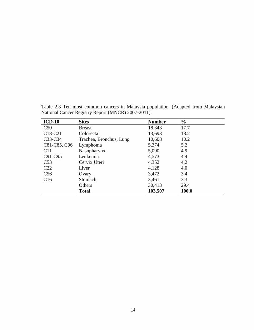
Among the cancers reported shown in Table 2.3, from 2007 to 2011 leukemia is the sixth
most common cancers occur in Malaysia population with the number of 4, 573 out of total
cases of 103,507 or 4.4% of which 2, 549 or 5.4% were reported in males and 2, 024 or 3.65%
were reported in females. It is the most common cancer among 0-14 years old with Malay
had the highest rate followed by Chinese and Indian in both sexes. The lifetime risk for males
was 1 in 275 and 1 in 348 for females.
According to the data collected by the Malaysian National Cancer Registry, AML is
commonly occurring among elderly people as compared to other types of leukemia and as
shown in Figure 2.2 and Figure 2.3, the average age of incidence of AML among females
and males are between 60 to 75 years old. The statistical data also showed that AML is more
commonly occur in males compare to females with males has a prevalence rate of 7 cases in
every 100,000 people while females has a prevalence rate of 6 cases in every 100,000 people
in population.
14
Table 2.3 Ten most common cancers in Malaysia population. (Adapted from Malaysian
National Cancer Registry Report (MNCR) 2007-2011).
ICD-10 Sites Number %
C50 Breast 18,343 17.7
C18-C21 Colorectal 13,693 13.2
C33-C34 Trachea, Bronchus, Lung 10,608 10.2
C81-C85, C96 Lymphoma 5,374 5.2
C11 Nasopharynx 5,090 4.9
C91-C95 Leukemia 4,573 4.4
C53 Cervix Uteri 4,352 4.2
C22 Liver 4,128 4.0
C56 Ovary 3,472 3.4
C16 Stomach 3,461 3.3
Others 30,413 29.4
Total 103,507 100.0

15
Figure 2.2 Leukemia types: Age-specific incidence rate, females, Malaysia, 2007-
2011(Adapted from Malaysian National Cancer Registry Report (MNCR) 2007-2011).

16
Figure 2.3 Leukemia types: Age-specific incidence rate, males, Malaysia, 2007-
2011(Adapted from Malaysian National Cancer Registry Report (MNCR) 2007-2011).

17
2.2 AML1/ETO Gene
There are around 40% of M2 AML cases is associated with t (8,21) karyotypic abnormalities
(Bitter et al., 1987) and this translocation has the most frequent chromosomal anomaly that
can be found in 18-20% of leukemia cases (Look, 1997, Mitelman and Heim, 1992). The
combination of AML1 gene that located at chromosome 21q22 with ETO gene located at
chromosome 8q22 results in formation of AML1/ETO chimeric protein as shown in Figure
2.4 which carries a transcriptional activity (Rulina et al., 2010). This fusion protein of
AML1-ETO encodes an initial 177 of AML1 amino acids that links to the ETO sequences
(Erickson et al., 1992, Miyoshi et al., 1991). AML1/ETO is identify in blood and also in
marrow sample of AML patient who achieve long-term complete remissions after treated
with chemotherapy or hematopoietic stem cell transplantation (Miyamoto et al., 1995).
AML1/ETO fusion protein solely is inadequate in causing leukemia, however the
downregulation in the expression of enzyme 8- oxoguanine DNA glycosylase (OGG1) which
is involved in the DNA repair might cause the addition of genetic abnormalities that leads
to development of AML (Liddiard et al., 2010). There are some experimental and clinical
studies that draw attention to the importance of secondary mutations in AML1-ETO that
mediates the development of leukemia. For instance, expression of AML1/ETO gene in mice
will require an exposure to mutagen such as N-ethyl-N-nitrosourea or through a co-
expression of constitutively active tyrosine such as TEL-PDGFRβ fusion for the
development of AML (Higuchi et al., 2002, Grisolano et al., 2003).
Similarly, the clinical data also show high prevalence of secondary genetic alteration that
will affects the tyrosine kinase signal transduction pathways in AML patient present with t
(8,21) translocation. Based on the observation done by Wang et al., in 2005, approximately

18
50% of the involved AML patients that present with t (8,21) had point mutations in the
tyrosine kinase gene C-KIT receptor (Wang et al., 2005). The hyperexpression of tyrosine
kinase KIT receptor only occur in approximately 5% of AML patient but with the presence
of AML1/ETO which results from the t(8,21) translocation frequency of mutation has shown
an increased up to 30% (Peterson and Zhang, 2004) . The association between the two
oncogenes was known for a certain of time. It was assumed that the presence of two activated
oncogenes in cells for example transcription factor AML1-ETO and tyrosine kinase c-kit will
function to promote their malignization.
Besides that, Wang et al., also discovered that majority of leukemia with t (8,21)
translocation are more likely to overexpress c-Kit despite the mutational status. While in
other study by Schessl et al., at the same year of 2005 found that more than one third of AML
patient with t (8,21) translocation to conceal activated mutations in either FLT3, C-KIT or
NRAS (Schessl et al., 2005). Furthermore, the study also showed AML1/ETO will cooperate
with mutated FLT3 to trigger the development of leukemia in transplanted mice and this
emphasizes the importance of cooperation between expression of AML1/ETO and abnormal
tyrosine kinase signaling in the development of leukemia (Schessl et al., 2005).

19
Figure 2.4. Translocation of t (8,21) (q22; q22) resulting in formation of fused gene
AML1/ETO. (Modified from Rulina et al., 2010).

20
2.2.1 Classical Model of Leukemogenesis by AML1/ETO
In a corresponding model, RUNX 1 will commonly functioned as transcriptional activator
that upregulate target genes from specific lineage such as myeloperoxidase to promotes
differentiation of granulocytes. RUNX1 will go through a metamorphosis process of
switching from activator to suppressor which will downregulate all of the target genes that
involved in differentiation of cells from granulocytic lineage through the combination of
RUNX 1 with ETO which result from the t (8,21) translocation. (Elagib and Goldfarb, 2007)
2.3 A Model Cell Line for AML t (8,21)
The Kasumi-1 cells is a human cells line that was obtained from peripheral blood sample of
a 7-year old Japanese boy present with AML that has relapsed after a bone marrow transplant.
The Japanese boy was diagnosed with M2 subtype AML at Matsuyama Red Cross Hospital
(Matsuyama, Japan). The first incident of leukemia relapsed occurred after a complete
remission that was achieved through chemotherapy. On the following relapsed, a bone
marrow transplant was done on this boy at Hiroshima Red Cross Hospital (Hiroshima, Japan)
collected from his HLA-matched sibling at the second complete remission after introduced
by mitxantrone and cytosine arabinoside. The relapsed occurred 98 days after the
transplantation where the engraftment was achieved and further application of chemotherapy
failed. The patient died due to disease progression.
During the whole clinical course, there was no leukemic cells with cancer formation observed
outside the marrow cavity. The blood sample was collected when the patient’s leukocyte
count was 99,800/µL with 93% blasts using a heparinized syringe. The Kasumi-1 cells were