surface antigens of the embryonic chick myoblast: expression on freshly trypsinized cells
TRANSCRIPT

Journal of Supramolecular Structure 7: 323-338 ( I 977) Cell Surface Carbohydrates and Biological Recognition 295-31 0
Surface Antigens of the Embryonic Chick Myoblast: Expression on Freshly Trypsinized Cells Martin Friedlander and Donald A. Fischman Departments of Anatom y and Biology and the Committee on Developmental Biology, University of Chicago, Chicago, Illinois 60637
Using an antiserum raised in rabbits against embryonic chick skeletal myoblasts (Anti- M-24), we have examined the trypsin and neuraminidase sensitivity and physiological expression of myogenic cell surface antigens. It was found that trypsin-released muscle cells more effectively inhibited, on a cell t o cell basis, the cytotoxicity of Anti-M-24 for 24-h-old myoblast monolayers than did identical cells that had re- ceived a 3-4 h suspension culture recovery period from trypsinization. There was no such difference in absorptive capacities observed for any other embryonic chick tissue tested (e.g. brain, retina, liver, heart, and red blood cells) when freshly tryp- sinized cells were compared t o ones which were given a 3-4 h culture period. If freshly trypsinized muscle cells were treated with high concentrations (30,000 international units (IU)/O.l ml packed cells) of trypsin or with neuraminidase (30,000 IU/ml packed cells), there was a selective loss of myoblast-specific surface antigens. When single cells that had been in suspension culture for 3.5 h were re- exposed t o low concentrations ( 1 0,000 1U/O. 1 ml packed cells) of trypsin, more antigenic sites were revealed o n their surfaces as detected by an increased absorptive capacity in removing myoblast-binding antibodies from Anti-M-24. This increase in antigenic expression was time-dependent and inversely related to the length of culture time after trypsinization. Immunofluorescence studies revealed that tissue specific myoblast cell surface antigens are present on both muscle cells that were freshly dissociated and those that had been in suspension culture for 3-4 h. Further- more, freshly trypsinized myoblasts possessed cell surface components that were highly antigenic; antiserum to such cells reacted extensively with both trypsinized and recovered muscle cells as detected by complement-dependent 51 Cr release cyto- toxicity assays and immunofluorescence. We conclude that embryonic chick myo- blasts possess surface antigens that may be selectively removed by neuraminidase or high concentrations of trypsin. These antigens may be progressively masked, with increasing time of culture after protease-dissociation, by molecules that are sensitive t o low concentrations of trypsin. Such masking of tissue-specific cell surface antigens could result in the display of molecular mosaics which may play a role in facilitating intercellular recognition and subsequent differentiation and histogenesis.
Key words: embryonic muscle, cell surface antigens, myogenesis, cytotoxicity assays
Martin Friedlander is presently at Rockefeller University, New York, New York 10021. Donald A. Fischman is presently at the Department of Anatomy and Cell Biology, SUNY, Downstate Medical Center, Brooklyn, New York 1 1 203.
Received April 28, 1977; accepted June 28, 1977.
0 1977 Alan R. Liss, Inc., 150 Fifth Avenue, New York, NY 1001 1

324:JSS Friedlander and Fischman
Recently we reported the immunological detection of tissue and developmental stage specific surface antigens in embryonic chick skeletal muscle in vitro (1-3). These studies were initiated to detect subtle compositional changes at the cell surface which might accompany differentiation of a particular embryonic tissue. We chose to examine developing embryonic muscle because this tissue provides an excellent system for studying the relationship between cell surface modifications and the nuclear events which underlie the phenotypic modifications we term differentiation. Cell recognition prior t o myotube formation is highly specific (4, S), both prefusion myoblasts and multinucleated myotubes exhibit cellular migration and motility (6), and cell-cell and cell-substratum adhesions clearly influence the course of myodifferentiation (7, 8). The aggregation of myogenic cells and the formation of intercellular junctions at interfacing plasma membranes prior to cell fusion (9, 10) are poorly understood, but probably significant, steps in muscle differentia- tion. In addition, there are well established methods available for culturing muscle which produce a developing tissue that faithfully mimics in vivo myogenesis (1 1-1 3).
Antiserum to the prefusion myoblast (Anti-M-24) was produced in rabbits and ex- tensively characterized by serological and immunohistochemical methods (3, 14). The results of these experiments are summarized in Table I. Such criteria have also been used to identify tissue specific surface antigens in other embryonic (1 5-1 S), transformed (19-22), or adult (23, 24) tissues. If the molecular basis for such operationally defined specificities is to be understood, we felt it would be necessary to examine the biochemical properties of the myoblast cell surface antigens. As a preliminary step towards immuno- chemically isolating and characterizing these antigens, we have examined their physiological expression and protease sensitivity. A number of proteases have been used in the isolation and characterization of histocompatibility antigens from mouse (25) and human (26) tissues. Chymotrypsin may be used to selectively expose tumor associated antigens on infected avian cell surfaces (27), while the major surface glycoprotein of fibroblasts is markedly trypsin sensitive (28,29). Trypsin has also been used to explore the physiological expression (30, 31) and membrane fluidity (32-34) of lectin binding sites on embryonic cell surfaces. Others have used neuraminidase to examine the expression of a number of cell surface antigens (35, 36). While such enzymatic treatments of living cells is known to have marked effects on a number of cell properties (37, 38), it is still of interest to con- sider their use in the analysis of potentially cryptic, or masked, membrane antigens.
the protease and neuraminidase sensitivity and the physiological expression of myoblast surface antigens. These data show that myoblast antigens may be selectively removed from the cell surface after treatment with neuraminidase or high concentrations of trypsin. Freshly trypsinized muscle cells apparently have greater quantities of these antigens on their surface than do cells permitted a period of culture after trypsinization; mild retrypsi- nization of such cells reveals the same amount of antigen as found on freshly dissociated cells. Parallel studies have involved the immunochemical analysis of detergent -extracted antigens by radioimmunoprecipitation and sodium dodecyl sulfate-polyacrylamide gel electrophoresis. The results of those investigations will be reported elsewhere (39).
In this communication we wish to present new data we have obtained concerning
MATERIALS AND METHODS
Cell Culture
chicks by methods routinely used in our laboratory (40). All visible nervous, vascular, and 296:CSCBR
Muscle was obtained from the hindlimbs of 12-day-old embryonic White Leghorn

Chick Myoblas t Cell S u r f a c e Antigens JSS: 325
TABLE I. Summary of the Characteristics of Antiserum (Anti-M-24) Prepared in Rabbits Against Embryonic Chick Prefusion Myoblasts
1. Complement-mediated 51Cr release cytotoxicity experiments indicate: A. Absorption of Anti-M-24 with live cell monolayers, live cell suspensions, acetone powders, or
freshly homogenized tissue from embryonic red blood cells, liver, brain, retina, or heart or adult skeletal muscle will remove all cytotoxicity of this antiserum for these tissues while lowering the cytotoxicity for embryonic muscle by only 20-60%. We interpret these results to demonstrate TISSUE SPECIFICITY.
B. Absorption of Anti-M-24 with myotubes or skeletal muscle fibroblasts removes all cytotoxicity of this antiserum for these cell types at a point where there is still significant cytotoxicity remaining for myoblasts. We interpret these results to demonstrate DEVELOPMENTAL STAGE SPECIFICITY.
C. Light microscopical observations of cultures treated with appropriately absorbed antiserum and guinea pig complement confirm the serological data; in such cultures only the spindle-shaped myoblasts are lysed.
2. Immunohistochemical staining (indirect immunofluorescence or immunoperoxidase) of monolayers or single cell suspensions further demonstrates that, after appropriate absorption, Anti-M-24 will stain only prefusion myoblasts. Shared, muscle tissue-specific, antigens may also be detected on embryonic muscle at all stages of differentiation.
3. Myoblast-specific antigens are cell surface components: 1) intact, viable cells inhibit cytotoxicity of Anti-M-24 for myoblasts and 2) observations by indirect immunofluorescence demonstrate that, under appropriate conditions, these antigens are mobile within the plasma membrane.
4. Myoblast cell surface antigens detected with Anti-M-24 are not an artefact of tissue culture: 1) fresh, homogenized chick tissues will absorb reactivity of Anti-M-24 with cultured cells or cells freshly released from the embryo and 2) Anti-M-24 binds to frozen sections of fresh embryonic hindlimb muscle as detected by indirect immunofluorescence.
connective tissue was removed from the limbs by careful dissection and the remaining muscle finely minced with scissors. After washing the tissue with calcium- and magnesium- free Tyrodes solution (CMF), it was dissociated into single cells by a 50 min incubation period at 37°C in 0.3% trypsin (Armour Pharmaceuticals, St. Louis, Missouri, “Tryptar”) in CMF. Proteolytic dispersion of the cells was stopped by washing the cell suspension 3 times with Eagle’s basal medium containing 10% heat inactivated (30 min at 56°C) horse serum (Grand Island Biologicals, Grand Island, New York), 1% glutamine, 1% penicillin (E-HS) and soybean trypsin inhibitor (10 kg/ml). After passing the cell suspension through a metal mesh filter, single cells suspended in E-HS with 5% embryo extract (complete medium, CM) were plated at varying densities on gelatin coated plastic culture dishes (Falcon Plastics, Oxnard, California). Such primary muscle cell cultures contained approxi- mately 90% myoblasts as assessed by morphologically scoring 24-h-old monolayers established from comparable cell suspensions (47,48). Other embryonic chick tissues (e.g. heart, brain, liver, retina) were obtained in similar fashion. Red blood cells were collected from the chorioallantoic vessels of the embryo, washed with phosphate-buffered saline, and used immediately for absorptions or cytotoxic assays.
Antiserum
for 3.5 h by placing 5-10 X lo6 cells/4.0 ml E-HS into 25-ml Ehrlenmeyer flasks and rotating the flasks at 110 rpm on a New Brunswick Gyratory Shaker. Such a culture period has been operationally defined as “preaggregation” to emphasize the observation that during the 3-4 h time period multicellular aggregates do not form and greater than 95%
Freshly dissociated embryonic muscle cells were cultured after trypsin dissociation
C S C B R : 2 9 7

326: JSS Friedlander and Fischman
of the cells remain as single, viable cells. To monitor the state of aggregation during such a time period, parallel suspension cultures were established and samples taken at 1-h in- tervals from 0 to 6 h of culture. For each time point, the cells were pelleted by centrifuga- tion, resuspended with a known volume of E-HS, and observed in a light microscope. Representative fields were scored for the number of single cells and multicellular aggregates. By 2-3 h small clusters of cells were observed, but these were easily dissociated into single cells by gentle pipetting. Only after 4-5 h were larger multicellular aggregates present that could not be easily dispersed into single cells by pipetting. Such aggregates rapidly increased in number from 6-24 h of culture, even at gyratory shaker speeds of 120 rpm. Such a period of suspension culture presumably permitted the renewal of cell surface materials which may have been denuded by tryptic treatment during the dissociation procedure (1 5,41).
After 3.5 h of suspension culture, cells were collected, washed several times with Tyrodes solution, and injected into rabbits. The immunization protocol and bleeding schedule has been reported elsewhere and will not be detailed here (3, 14). Antiserum raised in rabbits against prefusion embryonic chick myoblasts will be referred to as Anti- M-24 and represents serum pooled from 3 individual rabbits.
Cytotoxicity Assay
The complement-dependent 51 Cr release cytotoxicity assay is a modification of that described by Wigzell(42) and Sanderson (43). Target cells were plated at 1-5 X lo4 cells/ 0.1 ml CM/well on Falcon microtiter plates. Six hours later, 1 .O pCi of 51 Cr (New England Nuclear Corporation, Boston, Massachusetts) in 0.1 ml E-HS was added to each well and after an additional 12 h incubation at 37"C, the excess 51 Cr was removed by several washes of the wells with E-HS. Antibody (0.05 ml) and guinea pig complement (0.05 ml) diluted to a final concentration of 1/10 (Beckman Diagnostics, Fullerton, California) was added and the cultures incubated for an additional 50 min. The reaction was stopped by adding 0.1 ml of cold Tyrodes solution to each well. Following a 10 min centrifugation at 2,800 rpm to remove cellular debris, 0.1 ml samples from each of tripli- cate wells were taken to be counted on a Searle Autogamma Counter. The results were calculated as a percent cytotoxicity:
x 100 cpm 51 Cr released by antibody - background cpm 51 Cr released by TX-I 00 - background Percent Cytotoxicity =
For each set of triplicate samples the mean and standard deviation were determined. The variability of replicates in a single experiment was never greater than 5 10% cytotox- icity and generally was between 5 and 8. All experiments were repeated in their entirety at least twice and most were repeated 3-5 times. For Figs. 2-7, the mean values from 2 to 5 experiments were averaged and presented as a single data point.
E-HS was absorbed with increasing numbers of cells overnight on a wrist shaker at 4°C. Cells used for absorption were removed by centrifugation and the absorbed serum used immediately. For trypsinization of absorbing muscle cells, lo8 freshly dissociated or pre- aggregated myoblasts were suspended in 4.0 ml of trypsin-CMF (0.05-0.3%), rotated at 110 rpm for 50 min in a gyratory shaker bath at 37"C, washed extensively with E-HS and soybean trypsin inhibitor (10 pg/ml), and then used to absorb antiserum as described above. Neuraminidase treatment was conducted under identical conditions as for trypsinization;
For inhibition of cytotoxicity assays, 0.5 ml of antiserum diluted 1/10 or 1/100 with
298:CSCBR
Chick Myoblast Cell Surface Antigens JSS: 327
30,000 IU/ml of neuraminidase Type I from C1. perfinigens (Sigma Chemical Company, St. Louis, Missouri) was used.
I mmunof luorescence
Cells to be stained by immunofluorescence were either live or fixed with 2.0% para- formaldehyde for 10 min at 50°C. All subsequent procedures were conducted on ice (for live cells) or at room temperature (for fixed cells). Five million cells were washed with E-HS, treated for 15 min with Anti-M-24 or preimmune serum from the same rabbit diluted 1/10 with E-HS, washed 3 times with E-HS, incubated for an additional 15 min with goat antirabbit IgG conjugated to fluorescein isothiocyanate (GAR-FITC) (Cappel Laboratories, Downingtown, Pennsylvania), washed 2 times with E-HS, mounted in glycerol under a glass coverslip and observed under an Apo 40X oil immersion lens mounted to a Zeiss epifluorescent microscope. Photomicrographs were taken after a 30 sec exposure on Kodak Tri-X film and subsequently developed in Diafine developer. All prints were processed under identical conditions.
RESULTS
Anti-M-24 was prepared by immunizing rabbits with myoblasts that had been allowed a 3-4 h culture period after primary trypsin-dissociation under the assumption that such a period of time was necessary for the cells to renew surface components that may have been denuded by the protease dissociation procedure (1 5,41). To further test this assumption, we decided to compare the ability of freshly dissociated and preaggregated myoblasts to inhibit cytotoxicity of Anti-M-24 for 24-h-old myoblast monolayers. At this point, 2 observations should be emphasized: 1) muscle cultures consist of 2 predominant cell types, myoblasts and fibroblasts, the former accounting for approximately 90% of the cells present at 24 h of monolayer culture (40,44) and 2) on a cell to cell basis, fibroblasts take up 3-4 times the amount of ” Cr as do myoblasts (14). These are important considera- tions when attempting to interpret data obtained from ’’ Cr-release experiments. For example, while myoblasts account for nearly 90% of the total cell population in a 24-h-old muscle culture, they only contain 40-60% of the ’’ Cr bound within the cells of such a culture.
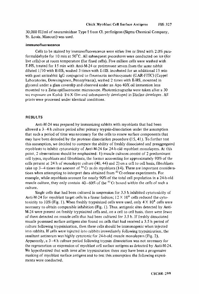
Anti-M-24 for myoblast target cells in a linear fashion; 12 X lo6 cells reduced the cyto- toxicity to 10% (Fig. 1). When freshly trypsinized cells were used, only 4 X lo6 cells were necessary to obtain comparable inhibition (Fig. 1). Thus, antigenic sites detected by Anti- M-24 were present on freshly trypsinized cells and, on a cell to cell basis, there were fewer of them detected on muscle cells that had been cultured for 3.5 h. If freshly dissociated muscle possessed surface antigens also found on cells that had received a 3.5 h period of culture following trypsinization, then these cells should be immunogenic when injected into rabbits. If cells were injected into rabbits immediately following trypsinization, the resultant antiserum was highly cytotoxic for 24-h-old muscle monolayers (Fig. 2). Apparently, a 3-4 h culture period following trypsin dissociation was not necessary for the regeneration or expression of myoblast cell surface antigens as detected by Anti-M-24. We hypothesized that with time after trypsinization there may have been a progressive masking of myoblast surface antigens and to test this assumption the following experi- ments were conducted.
Single cells that had been cultured in suspension for 3.5 h inhibited cytotoxicity of
CSCBR:299

328: JSS Friedlander and F i s c h m a n
2 0 6 8 10 12 IWR of ABSORBING CELLS
( x 1VbI
Fig. 1. Inhibition of cytotoxicity of Anti-M-24 for myoblast target cells after absorption with in- creasing numbers of suspension cultured (0-0) or freshly trypsinized (x- - -x) myoblasts. One-half milliliter of antiserum was diluted 1/100 with E-HS and absorbed overnight o n a shaker a t 4°C with either 2, 4, 6 , 8, 10, or 12 million cells from 12-day-old embryonic chick hindlimb muscle that had been freshly dissociated with 0.3% Tryptar/ml of minced muscle or with identical cells that had been cultured for 3.5 h at 110 rprn on a gyratory shaker. The absorbed antisera were tested, in the presence of guinea pig complement, for cytotoxicity on 24-h-old muscle monolayers. Twice as many suspension cultured cells as freshly trypsinized ones were needed to remove all cytotoxicity of the antiserum for myoblast target cells. Vertical lines a t each point represent the range of 1 standard deviation from the mean as calculated for 12 replicate samples obtained from 4 independent experiments.
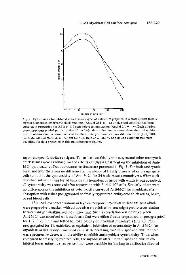
Cells which had been cultured for 3.5 h following trypsinization (“preaggregated” cells) were treated with high (30,000 IU/O.l ml packed cells) or low (10,000 IU/O.1 ml packed cells) concentrations of trypsin and then assessed for their ability to inhibit cytotoxicity of Anti-M-24 for 24-h-old muscle monolayers. Exposure of preaggregated cells to high concentrations of trypsin apparently removed most of the antigens while a comparable group of cells that had been exposed to low concentrations of this protease were as effective in the inhibition of cytotoxicity as freshly dissociated cells (Fig. 3). It should be emphasized that a single group of cells was used for the data obtained in Figs. 1 and 3; Anti-M-24 was absorbed with either the freshly dissociated cells or cells from the same dissociation which were then preaggregated or preaggregated and retrypsinized. Identical results were obtained when the experiment was repeated.
single cells were greatly reduced in the ability to inhibit cytotoxicity of Anti-M-24 for muscle monolayers (Fig. 4). However, if such absorbed antiserum was tested on pure fibro- blast monolayers, cytotoxicity for these target cells was removed entirely after absorption with 8 X lo6 cells. These results suggested that trypsin may have selectively removed the
If freshly dissociated muscle was reexposed to higher concentrations of trypsin, the
300:CSCBR
Chick Myoblast Cell Surface Antigens JSS:329
1 20 80 320 I2&
DILUTION OF MTISERUM
Fig. 2. Cytotoxicity for 24-h-old muscle monolayers of antiserum prepared in rabbits against freshly trypsin-dissociated embryonic chick hindlimb (Anti-M-24T, x- - -x) or identical cells that had been cultured in suspension for 3.5 h at 110 rpm before immunization (Anti-M-24, 0-0). Each dilution curve represents pooled serum obtained from 2-3 rabbits. Preimmune serum from identical rabbits used to obtain immune serum induced less than 10% cytotoxicity at any dilution tested (1-1/800). See Materials and Methods in the text for discussion of variability of data and experimental repro- ducibility for data presented in this and subsequent figures.
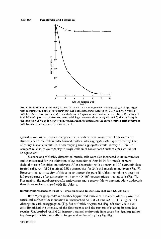
myoblast-specific surface antigens. To further test this hypothesis, several other embryonic chick tissues were examined for the effects of trypsin treatment on the inhibition of Anti- M-24 cytotoxicity. Two representative tissues are presented in Fig. 5 . For both embryonic brain and liver there was no difference in the ability of freshly dissociated or preaggregated cells to inhibit the cytotoxicity of Anti-M-24 for 24-h-old muscle monolayers. When such absorbed antiserum was tested back on the homologous tissue with which it was absorbed, all cytotoxicity was removed after absorption with 2-4 X lo6 cells. Similarly, there were no differences in the inhibition of cytotoxicity curves of Anti-M-24 for myoblasts after absorption with either preaggregated or freshly trypsinized embryonic chick retina, heart, or red blood cells.
were progressively masked with culture after trypsinization, one might predict a correlation between antigen masking and the culture time. Such a correlation was observed when Anti-M-24 was absorbed with myoblasts that were either freshly trypsinized or preaggregated for 1 , 2 , 3 , or 3.5 h and tested for cytotoxicity on myoblast monolayers (Fig. 6). Cells preaggregated for 1 h exhibited an equivalent inhibition of cytotoxicity in Anti-M-24 for myoblasts as did freshly dissociated cells. With increasing time in suspension culture there was a progressive decrease in the ability to inhibit antimyoblast cytotoxicity. Thus, when compared to freshly trypsinized cells, the myoblasts after 3 h in suspension culture ex- hibited fewer antigenic sites per cell that were available for binding to antibodies directed
If indeed low concentrations of trypsin reexposed myoblast surface antigens which
CSCBR:301
330: JSS Friedlander and Fischman
0 2 4 6 8 10 12
NUFLIER OF LgSORBlNG ELLS ( x 1 0 - 6 )
Fig. 3. Inhibition of cytotoxicity of Anti-M-24 for 24-h-old muscle cell monolayers after absorption with increasing numbers of myoblasts that had been suspension cultured for 3.5 h and then treated with high (x- - -x) or low (0-0) concentrations of trypsin as described in the text. Note 1) the lack of inhibition of cytotoxicity after treatment with high concentrations of trypsin and 2) the similarity in the inhibition curve of the low trypsin concentration treatment and the curve obtained after absorption with freshly dissociated cells as seen in Fig. 1 .
against myoblast cell surface components. Periods of time longer than 3.5 h were not studied since these cells rapidly formed multicellular aggregates after approximately 4 h of rotary suspension culture. These varying sized aggregates would be very difficult to compare in absorption capacity to single cells since the exposed surface areas would not be equivalent.
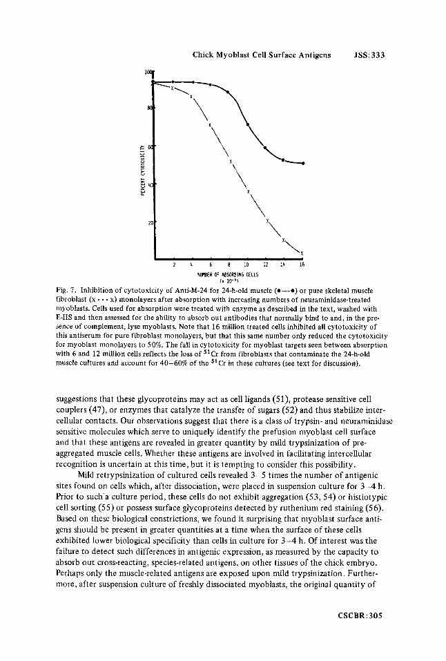
and then assessed for the inhibition of cytotoxicity of Anti-M-24 for muscle or pure skeletal muscle fibroblast monolayers. After absorption with as many as 1 O7 neuraminidase- treated cells, Anti-M-24 retained 75% cytotoxicity for 24-h-old muscle monolayers (Fig. 7). However, the cytotoxicity of this same antiserum for pure fibroblast monolayers began to fall precipitously after absorption with only 4 X lo6 neuraminidase-treated cells (Fig. 7). Presumably, the myoblast-specific antigens are more susceptible to neuraminidase hydrolysis than those antigens shared with fibroblasts.
lrnrnunofluorescence of Freshly Trypsinized and Suspension Cultured Muscle Cells
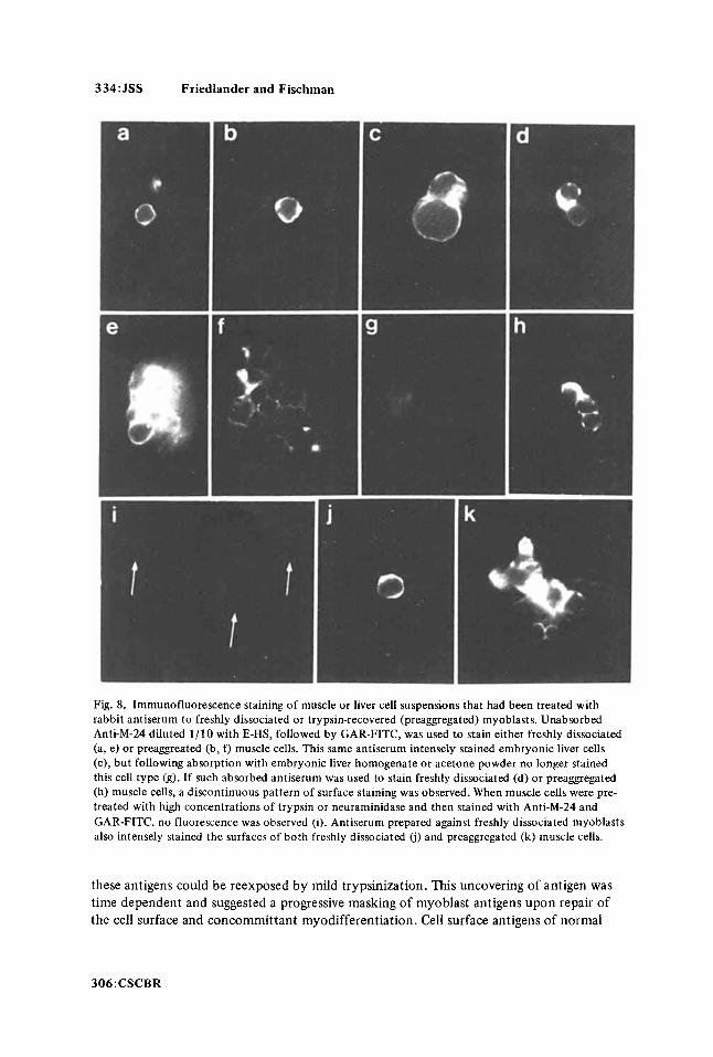
entire cell surface after incubation in unabsorbed Anti-M-24 and GAR-FITC (Fig. 8a-d). Absorption with preaggregated (Fig. 8e) or freshly trypsinized (Fig. 80 embryonic liver cells diminished the intensity of the fluorescence and the pattern of staining became less regular. Unabsorbed Anti-M-24 intensely stained embryonic liver cells (Fig. 8g), but follow- ing absorption with liver cells no longer stained hepatocytes (Fig. 8h).
302: CSCBR
Suspensions of freshly dissociated muscle cells were also incubated in neuraminidase
Both “preaggregated” and freshly trypsinized muscle cells stained intensely over the

Chick Myoblas t Cell Surface Ant igens JSS:331
2 4 6 8 10
NUMBER OF ABSORBING CELLS ( x 10-9
Fig. 4. Inhibition of cytotoxicity of Anti-M-24 for 24-h-old muscle (e-0) or pure skeletal muscle fibroblast (x- - -x) monolayers after absorption with increasing numbers of freshly dissociated myo- blasts that had been retrypsinized. Muscle was dissociated into single cells by a 50 min incubation period at 37°C in 0.3% trypsin, washed 4 times with E-HS and soybean trypsin inhibitor, and then retrypsinized with a comparable amount of trypsin for an additional 50 min at 37°C in a gyratory shaker water bath. After 4 more washes with E-HS and soybean trypsin inhibitor, the cells were used to absorb Anti-M-24. Absorption with 8 X lo6 treated cells removed all cytotoxicity of this antiserum for fibroblasts while as many as lo7 only lowered the cytotoxicity for myoblasts to 50%.
Myoblasts incubated in neuraminidase or high concentrations of trypsin and sub- sequently stained with Anti-M-24 and GAR-FITC failed to demonstrate specific immuno- fluorescence (Fig. 8i). When freshly dissociated (Fig. 8j) or preggregated (Fig. 8k) myoblasts were stained with unabsorbed antiserum raised against freshly dissociated cells, a uniformly intense fluorescence pattern was observed around their periphery. Preimmune serum failed to stain any of the cell types used in this study (not illustrated). All immuno- fluorescence experiments were conducted on paraformaldehyde-fixed cells or on live cells at 4°C so the patterns of fluorescence could not be attributed to capping phenomena which occur as a result of mobile surface receptors (45). If cells were stained at 37"C, capping did occur, followed by the complete removal of surface antigen-antibody com- plexes (Friedlander, unpublished observations).
DISCUSSION
The role of surface glycoproteins as recognition molecules on developing embryonic cells has been explored in several systems and it is clear that such macromolecules are responsible for, or at least associated with, tissue specific aggregation phenomena of embryonic chick retina (46,47) and the promotion of differentiation in cartilage (48), muscle (49), and glia (50). The manner in which such macromolecules exert their influence on cellular interactions and the promotion of differentiation is not clear; there have been
CSCBR:303
332:JSS Friedlander and Fischman
> c - u P X
0 c > U
I-
?: V E Y 0
I I 0 2 4 6 8 10
NUMBER OF ABSORBIN6 CELLS [xlO-'l Fig. 5. Inhibition of cytotoxicity of Anti-M-24 for 24-h-old muscle monolayers after absorption with increasing numbers of 12day-old embryonic brain or liver cells that had been freshly dissociated (0 ) or suspension cultured for 3.5 h (0). There was no significant difference in the rate a t which freshly dis- sociated or preaggregated cells from liver or brain inhibited cytotoxicity.
MWBER ff ABsoSlC CEUS (x 1LV)
Fig. 6 . Inhibition of cytotoxicity of Anti-M-24 for 24-huld muscle cell monolayers after absorption with increasing numbers of rnyoblasts that were either freshly dissociated with trypsin (x- - -x) or cultured in suspension for 1 (m - - - m), 2 (0-o), 3 (A-A), or 3.5 (0-0) h. Cells to be used for absorption were divided into aliquots of 2 ,4 , 6 , 8, 10, or 12 million and shaken gently overnight with 0.5 ml of antiserum diluted 1/100 with E-HS. After removal of the cells by centrifugation, the serum was assessed for cytotoxicity, in the presence of guinea pig complement, on myoblast target cells. Note that the cytotoxicity curves obtained after absorption of antiserum with cells preaggregated for 2 and 3 h were intermediate to those obtained with serum absorbed with cells preaggregated for 0 and 3.5 h.
304:CSCBR
Chick Myoblast Cell Surface Antigens JSS: 333
r I
2 4 6 8 10 12 14 16
NUMBER of !&SORBING CELLS ( x 10-6 )
Fig. 7. Inhibition of cytotoxicity of Anti-M-24 for 24-h-old muscle (0-0) or pure skeletal muscle fibroblast (x - - - x) monolayers after absorption with increasing numbers of neuraminidase-treated myoblasts. Cells used for absorption were treated with enzyme as described in the text, washed with E-HS and then assessed for the ability to absorb out antibodies that normally bind to and, in the pre- sence of complement, lyse myoblasts. Note that 16 million treated cells inhibited all cytotoxicity of this antiserum for pure fibroblast monolayers, but that this same number only reduced the cytotoxicity for myoblast monolayers to 50%. The fall in cytotoxicity for myoblast targets seen between absorption with 6 and 12 million cells reflects the loss of 51Cr from fibroblasts that contaminate the 24-h-old muscle cultures and account for 40-60% of the "Cr in these cultures (see text for discussion).
suggestions that these glycoproteins may act as cell ligands (51), protease sensitive cell couplers (47), or enzymes that catalyze the transfer of sugars (52) and thus stabilize inter- cellular contacts. Our observations suggest that there is a class of trypsin- and neuraminidase sensitive molecules which serve to uniquely identify the prefusion myoblast cell surface and that these antigens are revealed in greater quantity by mild trypsinization of pre- aggregated muscle cells. Whether these antigens are involved in facilitating intercellular recognition is uncertain at this time, but it is tempting to consider this possibility.
sites found on cells which, after dissociation, were placed in suspension culture for 3-4 h. Prior to such a culture period, these cells do not exhibit aggregation (53, 54) or histiotypic cell sorting (55) or possess surface glycoproteins detected by ruthenium red staining (56). Based on these biological constrictions, we found it surprising that myoblast surface anti- gens should be present in greater quantities at a time when the surface of these cells exhibited lower biological specificity than cells in culture for 3-4 h. Of interest was the failure to detect such differences in antigenic expression, as measured by the capacity to absorb out cross-reacting, species-related antigens, on other tissues of the chick embryo. Perhaps only the muscle-related antigens are exposed upon mild trypsinization. Further- more, after suspension culture of freshly dissociated myoblasts, the original quantity of
Mild retrypsinization of cultured cells revealed 3-5 times the number of antigenic
CSCBR:305
334:JSS Friedlander and Fischman
Fig. 8. Immunofluorescence staining of muscle or liver cell suspensions that had been treated with rabbit antiserum to freshly dissociated or trypsin-recovered (preaggregated) myoblasts. Unabsorbed Anti-M-24 diluted 1/10 with E-HS, followed by GAR-FITC, was used to stain either freshly dissociated (a, e) or preaggreated (b, f) muscle cells. This same antiserum intensely stained embryonic liver cells (c), but following absorption with embryonic liver homogenate or acetone powder no longer stained this cell type (g). If such absorbed antiserum was used to stain freshly dissociated (d) or preaggregated (h) muscle cells, a discontinuous pattern of surface staining was observed. When muscle cells were pre- treated with high concentrations of trypsin or neuraminidase and then stained with Anti-M-24 and GAR-FITC, no fluorescence was observed (i). Antiserum prepared against freshly dissociated myoblasts also intensely stained the surfaces of both freshly dissociated (i) and preaggregated (k) muscle cells.
these antigens could be reexposed by mild trypsinization. This uncovering of antigen was time dependent and suggested a progressive masking of myoblast antigens upon repair of the cell surface and concommittant myodifferentiation. Cell surface antigens of normal
306:CSCBR

Chick Myoblast Cell Surface Antigens JSS:335
and trypsinized myotubes must be compared before a statement can be made concerning the developmental significance of the trypsin effect. Such a study is in progress. Pre- liminary observations indicate that more muscle-specific antigens, as detected by inhibition of Anti-M-24 for myoblast monolayers, are exposed on mildly trypsinized myotubes than on untrypsinized cells (Friedlander, unpublished observations).
Our conclusion that only myoblast-specific surface antigens were enhanced by mild trypsinization, but eliminated by neuraminidase or extensive trypsinization, was based on experiments which warrant additional discussion. Myoblast-specific antiserum, as opera- tionally defined, retained high cytotoxicity for 24-h-old muscle monolayers at levels of absorption where cytotoxicity was lost for other embryonic chick tissues. Thus, Anti-M- 24 exhibited 40-60% cytotoxicity for myoblast monolayers after absorption, a 30-40% reduction from values obtained with unabsorbed antiserum. Although myoblasts account for 80-90% of the cells in 24-h-old monolayers, these cells only account for approxi- mately 50% of the ’’ Cr taken up in such cultures. Based on this observation and the morphological scoring of cell types that were specifically lysed in such heterologous cultures, we concluded that specifically absorbed Anti-M-24 binds predominantly to spindle-shaped myoblasts (3). Similarly, Fig. 4 and 7 are more readily interpreted when the heterogeneity of 24-h-old muscle cultures is considered. If myoblast-specific antigens were selectively removed with high levels of trypsin or neuraminidase, why was there an initial decline, then a plateau in the inhibition of cytoxicity of Anti-M-24 for myoblast monolayers after absorption with protease- or neuraminidase-treated muscle cell suspen- sions? Since the inhibition of cytoxicity plateaued between 40 and 60% cytoxicity in both series of experiments, and since myoblasts only account for 40-60% of the 51 Cr in such cultures, it seemed reasonable to conclude that the fibroblasts in such cultures were lysed by the antiserum and complement. In other words, absorption of Anti-M-24 with trypsin- or neuraminidase-treated muscle cell suspensions failed to remove antibodies reactive with both myoblasts and fibroblasts. However, we cannot be certain if the myoblast-specific antigens were totally removed by enzymatic treatment. Similarly, the shared antigens might also be removed upon prolonged exposure of the cells to sialidase or trypsin. To better understand the relative labilities of these 2 classes of antigens, enzymatic treatment for longer periods of time and with higher concentrations of trypsin and neuraminidase will be re- quired. Furthermore, absorption with higher cell numbers will be required to establish if the observed differences in absorptive capacity of enzymatically treated cells are qualitative or quantitative phenomena. If cells after 3-4 h of suspension culture were incubated in high concentrations of trypsin, all surface antigens were apparently removed since these cells failed to inhibit the cytotoxicity of Anti-M-24 for myoblast targets. It is conceivable that residual trypsin, present during the preaggregation periods that preceded retrypsiniza- tion, rendered the cell surface more labile to a subsequent trypsin digestion. Such residual activity might explain the difference in the cytotoxicity curves observed after absorption of Anti-M-24 with myoblasts treated with high concentrations of trypsin either immedi- ately following tissue dissociation (Fig. 4) or after 3.5 h of suspension culture (Fig. 3).
embryonic cells surfaces become preferentially masked or unmasked, thus creating cell surface mosaics which are tissue and developmental stage specific (57, 58). Our data are consistent with the existence of protease-sensitive, developmentally regulated, cell surface mosaics. At this time we are uncertain as to how protease treatment preferentially exposes myoblast cell surface antigens; whether trypsin unmasks “cryptic” antigens or somehow
It has been suggested that with progressive differentiation various antigenic sites on
CSCBR: 307

336:JSS Friedlander and Fischman
alters the organization of these molecules within the membrane, and thus affects their expression, remains unanswered. Our observation of differential staining patterns obtained with unabsorbed and absorbed Anti-M-24 suggest that there is a mosaicism to the distribu- tion of the antigens detected by this antiserum. We previously reported that myoblasts possess surface antigens not detected on muscle of later stages of development (1,2), but at that time could not rule out the possibility that these sites were simply masked or present in greatly diminished quantities on myotubes. We now present evidence that myo- blast surface antigens were revealed in greater quantities by mild proteolysis and there are preliminary indications that similar treatment of myotubes makes these cells more effec- tive inhibitors of cytotoxicity of Anti-M-24 for myoblast and myotube monolayers.
We conclude from these experiments that neuraminidase-sensitive antigens, which are also removed by high concentrations of trypsin, but revealed in greater quantities by mild proteolysis, niay serve to distinguish the myoblast cell surface from other embryonic chick tissues and muscle at later stages of development. The potential biological roles for such developmental stage specific markers are many: intercellular recognition, the regula- tion of cellular proliferation, or the initiation of myofibrillogenesis by providing sub- plasmalemmal insertion sites for contractile filaments. All of these phenomena characterize the myogenic developmental program and probably involve surface associated events (59, 60). While the data presented here do not directly support any such role for the antigens we have detected, the information we now have about their neuraminidase and protease sensitivity should prove valuable in designing procedures for isolating and charac- terizing these molecules. We are currently exploring the immunochemical nature of these antigens, their fluidity within the cell membrane, and their possible relationship to cyto- plasmic structures and physiological activities of the muscle cell.
ACKNOWLEDGMENTS
The authors gratefully acknowledge the invaluable technical assistance of Lovenia Williams, Marian Daniels, and Furman Davis. We would also like to thank Dr. Frank Fitch for the generous use of certain laboratory facilities and Sally Hoskins for her assistance in the preparation of the antisera used in this study. Our discussions with Eric Beyer and Sheila Fallon and Drs. Linda Marton, Theodore Steck, Michael Edidin, Frank Fitch, and Hewson Swift were most helpful and greatly appreciated. Martin Friedlander was a post- doctoral fellow of the Muscular Dystrophy Association of America and was also supported by an institutional allowance from American Cancer Society grant 1 N-41-P. This research was also funded by grants from the University of Chicago Cancer Research Center and the Harry Levine Memorial Foundation, and United States Public Health Service grant NHLI- 13505.
REFERENCES
1. Friedlander M, Fischman DA: J Cell Biol 63: 105a, 1974. 2. Friedlander M, Fischman DA: J Cell Biol67:124a, 1975. 3. Fischman DA, Doering J, Friedlander M: In Marois A (ed): “Tests of Teratogenicity in vitro.”
4. Bischoff R, Holtzer H: J Cell Biol41:188, 1969. 5. Yaffe D: Exp Cell Res 66:33, 1971. 6. Powell JA: Exp Cell Res 80:251, 1973. 7. Hauschka SD, Konigsberg IR: Proc Natl Acad Sci USA 55:119, 1966.
308:CSCBR
Amsterdam: North Holland Publishing Company, 1976, p 233.

Chick Myoblast Cell Surface Antigens JSS: 337
8. Yaffe D: Curr Top Dev Biol4:37, 1969. 9. Rash JE, Staehelin LA: Dev Biol36:455, 1974.
10. Kalderon N, Gilula NB: Neurosci Abstr 2:412, 1976. 11. Stockdale FE, Holtzer H: Exp Cell Res 24:508, 1961. 12. Konigsberg IR: Science 140:1273, 1963. 13. Shimada Y, Fischman DA, Moscona AA: J Cell Biol35:445, 1967. 14. Friedlander M, Fischman DA: Manuscript submitted. 15. Goldschneider I, Moscona AA: J Cell Biol 53:435, 1972. 16. Wiley LD, Calarco PG: Dev Biol47:407, 1975. 17. Edidin M, Gooding LR, Johnson M: In “Karolinska Symposia on Research Methods in Reproduc-
18. O’Rand MG, Romrell LJ: Dev Biol55:347, 1977. 19. Kurth R, Bauer H: Virology 47:426,1972. 20. Phillips ER, Perdue JF: J Supra mol Struct 4:27, 1976. 21. Artzt K, DuBois P, Bennett D, Condamine H, Babinet C, Jacob F: Proc Natl Acad Sci USA
22. Akeson R, Henchman HR: Proc Natl Acad Sci USA 71: 187, 1974. 23. Lloyd KO, DarnuleTV: J Immunol 112:311,1974. 24. Croissile Y: In Marois A (ed): “Tests of Teratogenicity in vitro.” Amsterdam: North Holland
25. Nathenson SG, Cullen SE: Biochim Biophys Acta 344: 1, 1974. 26. Springer TA, Strominger JL, Mann D: Proc Natl Acad Sci USA 71:1539, 1974. 27. Kurth R: Personal communication. 28. Robbins PW, Wickus GG, Branton PE, Gaffney BJ, Hirschberg CB, Fuchs P, Blumberg PM: Cold
Spring Harbor Symp Quant Biol 39:1173, 1975. 29. Hynes RO, Wyck JA, Bye JM, Humphreys KC, Pearlstein ES: In Reich E, Rifkin DB, Shaw E (eds):
“Proteases and Biological Control.” New York: Cold Spring Harbor Laboratories, 1975, p 931. 30. Burger MM: Proc Natl Acad Sci USA 62:994, 1969. 31. Kleinschuster SJ, Moscona AA: Exp Cell Res 70:397, 1972. 32. Noonan KD, Burger MM: J Cell Biol59: 134, 1973. 33. Martinozzi M, Moscona AA: Exp Cell Res 94:253, 1975. 34. Singer SJ: In Meints RH, Davies E (eds): “Control Mechanisms in Development.” New York:
35. Parham P, Humphreys RE, Turner MJ, Strominger JL: Proc Natl Acad Sci USA 71:3998, 1974. 36. Ostrand-Rosenberg S, Edidin M, Jewett M: Manuscript submitted. 37. Weiss L: Exp Cell Res 14:80, 1958. 38. Maslow DE: In Poste G, Nicolson GL (eds): “The Cell Surface in Animal Embryogenesis and
Development.” Amsterdam: Elsevier/North Holland Publishing Company, 1976, p 697. 39. Marton L, van der Westhuyzen D, Friedlander M: Manuscript in preparation. 40. Friedlander M, Beyer E, Fischman DA: Manuscript submitted. 41. Shimada Y, Fischman DA: In Lieberman M, Sano T (eds): “Developmental and Physiological
42. Wigzell H: Transplantation 3:423, 1965. 43. Sanderson AR: Br J Exp Pathol45:398, 1964. 44. Friedlander M: PhD thesis, University of Chicago, 1976. 45. Schreiner GF, Unanue ER: Adv Immunol24:37, 1976. 46. Hausman RE, Moscona AA: Proc Natl Acad Sci USA 72:916, 1975. 47. Rutishauser U, Thiery J-P, Brackenbury R, Sela B, Edelman GM: Proc Natl Acad Sci USA
73:577, 1976. 48. Solursh M, Meier S: Dev Biol 30:279, 1973. 49. Doering J: PhD thesis, University of Chicago, 1975. 50. Lim R, Turrif DE, Troy SS, Kato T: In Federoff S (ed): “Cell, Tissue and Organ Cultures in
Neurobiology.” New York: Academic Press (In press). 51. Moscona AA: In Moscona AA (ed): “The Cell Surface in Development.” New York: John Wiley
and Sons, 1976, p 67. 52. Roth S, McGuire EJ, Roseman S: J Cell Biol51:536, 1971. 53. Roth S, Dev Biol 18:602, 1968. 54. Moscona AA: In Bittar EE (ed): “Cell Biology in Medicine.” New York: Wiley-Medical, 1973,
tive Endocrinology.” 7th Symposium, Immunological Approaches to Fertility Control, 1974, p 336.
70:2988, 1973.
Publishing Company, 1976, p 149.
Plenum Press, 1976, p 181.
Correlates of Cardiac Muscle.” New York: Raven Press, 1975, p 81.
p 571.
CSCBR: 309
338:JSS Friedlander and Fischman
55. Steinberg MS, Granger RE: Am Zoo1 6:337a, 1966. 56. Caravita S , Zachei AM: J Embryo1 Exp Morphol 32:25, 1974. 57. Bennett D, Boyse EA, Old LJ: In Silvestri LG (ed): “Cell Interactions.” Amsterdam: North
Holland Publishing Company, 1972, p 247. 58. Singer SJ: In Bradshaw RA, Frazier WA, Merrell RC, Gottlieb DI, Hogue-Angeletti RA (eds):
“Surface Membrane Receptors.” New York: Plenum Press, 1976, p 1. 59. Edidin M: In Poste, G Nicolson GL (eds): “The Cell Surface in Animal Embryogenesis and
Development.” Amsterdam: Elsivier/North Holland Publishing Company, 1976, p 127. 60. Edelman GM: Science 192:218, 1976.
3 10: CSCBR