surface passivation for molecular motor protein assays
TRANSCRIPT
IntroductionSurface passivation is crucial for kinesin and microtubule experiments. Without passivation, gliding motility assays will fail. The current beliefs as to why failure occur include1:
• No structural support for kinesin.• Kinesin denatures when it hits glass.• Motor domains may inactivate when theycontact glass.
To run experiments, a protein called casein is used to passivate the glass. There are many issues with using casein, some of them include:
• It is designed to carry calcium phosphate to infant mammals. This calcium could aid in depolymerizing microtubules• No crystal structure exists.• Its use is based on legacy.
Surface Passivation for Molecular Motor Protein AssaysAndy Maloney, Brigette Black, Anthony Salvagno, Larry Herskowitz,
Brian Josey, Steve Koch, University of New Mexico
Glass slideCasein
Kinesin Microtubule
Gliding Motility Assay
MotivationKinesin and microtubules have been proposed as components for chemical and biological sensors. In order to fully understand the dynamics of kinesin and microtubules, fundamental questions must be answered related to how we observe those interactions. One way to observe their interactions is by using a gliding motility assay which can easily be visualized as a molecular "crowd surfing" for microtubules. In order for kinesin to support microtubule crowd surfing, the glass the kinesin is on must be functionalized in order to passivate the surface. Understanding the interactions of kinesin with this passivation is crucial to understanding novel ways to engineer MEMS devices for the detection of chemical and biological materials.
Casein2
• Globular protein.• Makes up 80% of the protein content in bovine milk.• Consists of 3 major subunits, α, β, κ.• Carries calcium phosphate to infant mammals.* Amphiphilic.
An issue that makes casein unattractive as a surface passivator is that it is designed to deliver calcium to infant mammals. Calcium is a known depolymerizing agent for microtubules. Its use is also based on legacy which has not been fully investigated. To fully elucidate the physics behind kinesin and microtubule interactions a simpler, cleaner surface passivator must be found. Knowing the physics behind the interactions will greatly expedite the design of novel sensors.
References1. Verma, Vivek, William Hancock, and Jeffrey Catchmark. “The role of casein in supporting the operation of surface bound kinesin.” Journal of Biological Engineering 2, no. 1 (2008): 14.2. http://www.food-info.net/uk/protein/milk.htm3. Kam, Lance, and Steven G. Boxer. “Spatially Selective Manipulation of Supported Lipid Bilayers by Laminar Flow: Steps Toward Biomembrane Microfluidics†.” Langmuir 19, no. 5 (March 1, 2003): 1624-1631. doi:doi: 10.1021/la0263413.4. Mennicke, Ulrike, and Tim Salditt. “Preparation of Solid-Supported Lipid Bilayers by Spin-Coating.” Langmuir 18, no. 21 (October 1, 2002): 8172-8177. doi:doi: 10.1021/la025863f.5. HHMI Report 2005.6. CINT is a user facility. More information can be found here, http://cint.lanl.gov/7. Guydosh, Nicholas R, and Steven M Block. “Direct observation of the binding state of the kinesin head to the microtubule.” Nature 461, no. 7260 (September 3, 2009): 125-128. doi:10.1038/nature08259. Supplemental information.* Images false colored in ImageJ.
Kinesin
Protofilament
MicrotubuleDepolymerization β Tubulin
+ α Tubulin
Tubulin
α casein β casein κ caseinWhole casein
"Kiney"
Preliminary resultsUse of phosphatidylcholine (PC) on both hydrophobic and hydrophilic slides have given negative results. While a lipid layer can be formed on the substrate, no motility was observed.
Future work with lipids and heavy water
• Phosphatidylethanolomine. PE has a higher melting temperature than PC and thus, may give kinesin a solid support for motility.• Charged lipids. There exists a possibility that kinesin is attracted to a charged group on the casein. Determining what charge, positive or negative, is easily done by functionalized lipids.• Cysteine functionalized lipid head groups. Casein has many cysteine residues on it. There is a possibility that the reason kinesin sustains motility with casein is because it sticks to the cysteine residues and prevents the motor domains of kinesin from sticking to the glass.
In parallel with this lipids work, we have been studying the affects of heavy water and osmotic stress on kinesin motility. Preliminary data to the right shows a distinct effect of D2O on gliding speed, errors are SEM. We have also seen a long-term stabilization of the gliding assay. It is possible that heavy water will improve passivation and stabilization of kinesin, and we will explore its effects in combination with studies of surface functionalization by caseins, lipids, and other molecules.
For more information about the experiments, please see Maloney's open notebook at:http://www.openwetware.org/wiki/User:Andy_Maloney
Acknowledgments• This work was supported by the DTRA CB Basic Research Program under Grant No. HDTRA1-09-1-008.• Susan Atlas. PI of the DTRA project.• Haiqing Liu (CINT)• Matt Goertz (CINT)• Gabriel Montano (CINT)• Partial funding from the IGERT INCBN.
Cargo
HeadsNeck linker
Microtubule
Stalk
Tails
HHMI report5 .
Kinesin• Dimer that consists of two heavy chains and two light chains.• The heavy chains form the "head" group or the motor domains.• The light chains form the "tail" group where cargo binds. The kinesin supplied to us from Dr. Liu does not have light chains. It is a truncated heavy chain Drosophila kinesin-1.• The chains are connected by a "neck linker" and an intertwined stalk region.• Uses ATP to generate motion.• Please see the poster on Wednesday (W236) for more information on kinesin's kinetic cycle.
Microtubules• Heterodimer of tubulin subunits α and β. One α and one β subunit together is called tubulin• Tubulin forms polymers called protofilaments.• Microtubules are made from 13 - 17 protofilaments.• They are hollow and are an average of 25 nm in width.• Calcium causes depolymerization.
Phosphate groups
Calcium Phosphate
k-casein with carbohydrates
Casein protein complex
PC functionalized
15 µmKochLab data*
Lipid bilayer
HydrophilicHead
HydrophobicTails
Surface passivation with lipids• Lipids could be a cleaner method of surface passivation.• Crystallizable, functionalizable, and can be synthetic.• Spin coating lipids can make single to multiple lipid layers.• Liposome bilayer formation.
There are many different types of lipids available. They range from size, to charge, to natural or synthetic. You can also functionalize the head and tail groups with just about anything you want. The best part about using lipids is that you can pattern them on photoresist to make any structure you want.
The photoresist barrier appears yellow in this image, as AZ 5214is fluorescent in both TR and NBD channels (as confirmed bymicroscopy in the absence of fluorescently labeled lipids). Figure1D illustrates a further evolution of this strategy to a higherresolution, three-component system (as detailed in SupportingInformation). Electron-beam lithography was used to define a 100-nm-wide barrier of chromium (which appear dark) on glass thatalso incorporates rectangular regions topologically isolated fromthe rest of the pattern and subsequently located under the centralstream of a laminar flow system. Bilayers formed inside theseisolated regions (green) did not mix with other areas, while thosein the central flow but outside the isolated rectangle were dilutedinto the adjacent bilayers, yielding the three-component system.Systems containing four or more membrane types can be readilyenvisioned (see Supporting Information).
We next demonstrate the use of our platform in presentingspatially segregated, micropatterned ligands to the T cell surfaceproteins TCR and LFA-1. A barrier (S1805 photoresist) was usedto create a two-component, interdigitated system (Figure 2A)consisting of 1,2-dioleoyl-sn-glycero-3-phosphocholine (DOPC,Avanti) lipids supplemented with 0.02 mol % Biotinyl-Cap-PE(Avanti) on one side and 6 mol % of DOGS-NTA (Avanti) on theother. The S1805 photoresist exhibits fluorescence in the far-redspectrum, allowing better visualization of the resultant surface. Thesurface was blocked with BSA and incubated with Alexa-488conjugated streptavidin (Molecular Probes), washed with PBS, andincubated with monobiotinylated-OKT3 (an antibody directedagainst and which activates TCR) and Cy5-labeled ICAM-1-6His(ICAM-1 is the natural ligand of LFA-1). The diffusion coefficientof ICAM-1 was determined to be 0.39 µm/s2 while that ofstreptavidin before and after linkage with biotin-OKT3 was 0.37and 0.23 µm/s2, respectively.
Human CD4+ T cell blasts clustered the tethered ligands withinminutes of introduction to the surface, spanning across up to threelipid bilayer stripes (Figure 2C); of 121 cells observed across twoindependent experiments, 51 interacted with a single stripe contain-ing OKT3, 44 interacted with two bilayer stripes (one of OKT3and a second of ICAM-1), while 26 spanned across three stripes
(Figure 2C). All cells formed clusters of OKT3 and ICAM-1, whichwere present only on the corresponding type of bilayer; cells werenot able to pull ligands over the barriers over the 2 h observationtime. Receptor clusters appeared to be directed toward the cellcenter, accumulating against barriers when this motion would beimpeded or under the cell center in the absence of barriers. Cellsoverlying three stripes (Figure 2C) were able to form clusters ofOKT3 or ICAM-1 in the central region, away from the bilayeredges. Notably, the ability of these cells to form ICAM-1 clusterswhich did not localize to the periphery of the IS suggests that thesegregation observed in the normal IS is dependent on theconcurrent presentation of TCR clusters in the same region; oursystem provides a new glimpse into this crosstalk, which will beinvestigated in subsequent studies. Cells were not able to attach tosurfaces without tethered OKT3 and ICAM-1, showing that thebarriers did not directly promote cell interaction.
In conclusion, the complex interplay between lateral mobilityand spatial organization of signaling complexes is an emerging areaof research. We introduce a new strategy for combining multiple,spatially separated SLBs on a single surface, with applicationspecifically in the context of T cells.
Acknowledgment. We thank G. Vasiliver-Shamis, D. Blair,and M. L. Dustin (New York University School of Medicine)for CD4+ T cells and protein reagents. This work was supportedby the National Institutes of Health, EY016586 and EB008199.
Supporting Information Available: Experimental procedures. Thismaterial is available free of charge via the Internet at http://pubs.acs.org.
References
(1) Grakoui, A.; Bromley, S. K.; Sumen, C.; Davis, M. M.; Shaw, A. S.; Allen,P. M.; Dustin, M. L. Science 1999, 285, 221–7.
(2) Groves, J. T.; Dustin, M. L. J. Immunol. Methods 2003, 278, 19–32.(3) Mossman, K. D.; Campi, G.; Groves, J. T.; Dustin, M. L. Science 2005,
310, 1191–3.(4) Pautot, S.; Lee, H.; Isacoff, E. Y.; Groves, J. T. Nat. Chem. Biol. 2005, 1,
283.(5) Perez, T. D.; Nelson, W. J.; Boxer, S. G.; Kam, L. Langmuir 2005, 21,
11963–68.(6) Oliver, A. E.; Ngassam, V.; Dang, P.; Sanii, B.; Wu, H.; Yee, C. K.; Yeh,
Y.; Parikh, A. N. Langmuir 2009, 25, 6992–96.(7) Dori, Y.; Bianco-Peled, H.; Satija, S. K.; Fields, G. B.; McCarthy, J. B.;
Tirrell, M. J. Biomed. Mater. Res. 2000, 50, 75–81.(8) Stroumpoulis, D.; Zhang, H.; Rubalcava, L.; Gliem, J.; Tirrell, M. Langmuir
2007, 23, 3849–56.(9) Yokosuka, T.; Kobayashi, W.; Sakata-Sogawa, K.; Takamatsu, M.; Hash-
imoto-Tane, A.; Dustin, M. L.; Tokunaga, M.; Saito, T. Immunity 2008,29, 589–601.
(10) Dustin, M. L. Arthritis Res. 2002, 4, S119–25.(11) Dustin, M. L.; Tseng, S. Y.; Varma, R.; Campi, G. Curr. Opin. Immunol.
2006, 18, 512–6.(12) Tseng, S. Y.; Liu, M.; Dustin, M. L. J. Immunol. 2005, 175, 7829–36.(13) Tseng, S. Y.; Waite, J. C.; Liu, M.; Vardhana, S.; Dustin, M. L. J. Immunol.
2008, 181, 4852–63.(14) Doh, J.; Irvine, D. J. Proc. Natl. Acad. Sci. U.S.A. 2006, 103, 5700–5.(15) Schwarzenbacher, M.; Kaltenbrunner, M.; Brameshuber, M.; Hesch, C.;
Paster, W.; Weghuber, J.; Heise, B.; Sonnleitner, A.; Stockinger, H.; Schutz,G. J. Nat. Methods 2008, 5, 1053–60.
(16) Shen, K.; Thomas, V. K.; Dustin, M. L.; Kam, L. C. Proc. Natl. Acad. Sci.U.S.A. 2008, 105, 7791–6.
(17) Kam, L.; Boxer, S. G. Langmuir 2003, 19, 1624–1631.(18) Tsai, J.; Sun, E.; Gao, Y.; Hone, J. C.; Kam, L. C. Nano. Lett 2008, 8,
425–30.(19) Kam, L.; Boxer, S. G. J. Am. Chem. Soc. 2000, 122, 12901–12902.(20) Yoshina-Ishii, C.; Boxer, S. G. J. Am. Chem. Soc. 2003, 125, 3696–7.(21) Yang, T.; Simanek, E. E.; Cremer, P. Anal. Chem. 2000, 72, 2587–9.(22) Jackson, B. L.; Groves, J. T. J. Am. Chem. Soc. 2004, 126, 13878–9.(23) Lenhert, S.; Sun, P.; Wang, Y.; Fuchs, H.; Mirkin, C. A. Small 2007, 3,
71–5.(24) Shi, J.; Chen, J.; Cremer, P. S. J. Am. Chem. Soc. 2008, 130, 2718–9.(25) Kung, L. A.; Kam, L.; Hovis, J. S.; Boxer, S. G. Langmuir 2000, 16, 6773–
6776.(26) Shi, J.; Yang, T.; Cremer, P. S. Anal. Chem. 2008, 80, 6078–84.
JA904721H
Figure 2. (A) Interdigitating bilayers used for capture of TCR and LFA-1ligands. The barrier consists of S1805 photoresist (blue). DOPC SLBs weresupplemented with biotinyl-Cap-PE or DOGS-NTA lipids. After overnightdiffusion, streptavidin (green) and GFP-6His (red) were captured onto thebilayer surface. (B) Schematic illustration of T cell interaction with aninterdigitated lipid bilayer presenting separated TCR activating and ICAM-1adhesion signals. The arrows indicate the direction which T cells pull theirligands. (C) Human CD4+ T cell blasts interact with spatially separatedligands, forming altered IS configurations on the interdigitating bilayer.These representative images show 30-min interaction of T cells with thebilayers.
J. AM. CHEM. SOC. 9 VOL. 131, NO. 37, 2009 13205
C O M M U N I C A T I O N S
Kam and Boxer's image of lipid structures made by photoresists3.
Lipid slide preparation• Spin coating.• Bilayer formation.
Depending on what chemistry we are interested in looking at, we can either spin coat lipids4 on either a hydrophobic or hydrophilic glass slide and make lamellar bilayers of lipids. By varying the amount of lipids spin coated on a slide, we can generate a single layer of lipid molecules that support aqueous environments. Liposome
Used for making bilayer substrates.
Glass surfacesSilanized glass.
AFM of spin coated lipidsIndicates we can make lamellar structures
from spin coating.
!"#$%&'()*$"' ' ' ' ' +#,"-.*,-'
'''''''' '
/%*-"'()*$"' ' ' ' ' 0)12#&34"'5"66768'()*$"'
''''''''''''' '
'
'
'
'
'
'
'
'
'
KochLab datafrom CINT6
Spin Master MicroDIY evaporator made from a DC motor controller, switch, and a model train motor. For more information on DIY bio,
and links to other DIY projects, please see the
Wikipedia article on DIYbio.
Spin Coater 3000Transforming the Spin Master Micro into
a spin coater.
This work was supported by the DTRA CB Basic Research Program under Grant No. HDTRA1-09-1-008.We would also like to thank Susan Atlas, PI of the DTRA project.
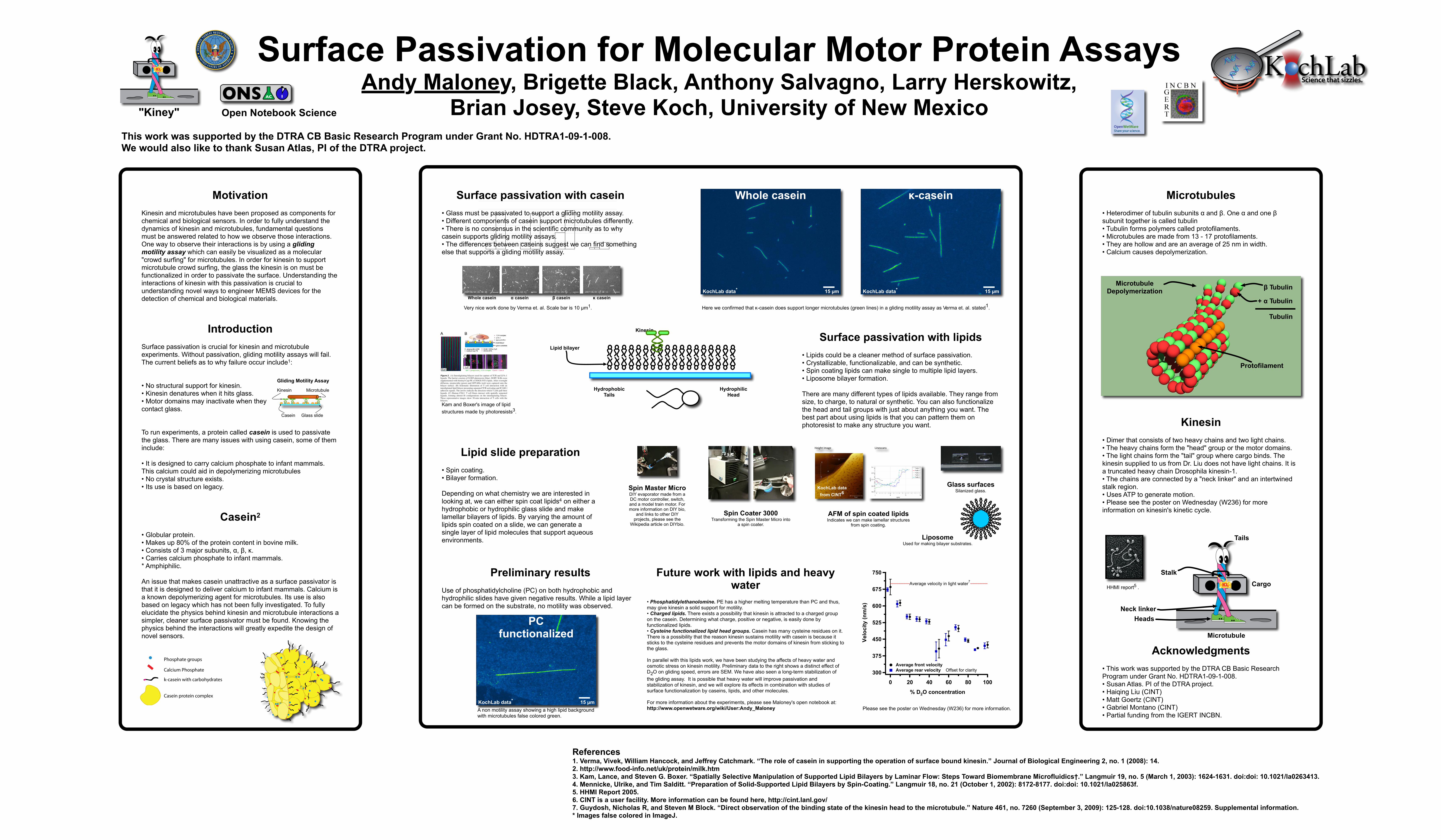
Surface passivation with casein• Glass must be passivated to support a gliding motility assay.• Different components of casein support microtubules differently.• There is no consensus in the scientific community as to why casein supports gliding motility assays. • The differences between caseins suggest we can find something else that supports a gliding motility assay.
!"#$%&'(")(*+"'",+-&'(.%,+%//$+%,!"##$%!!&'( )**+&,,---./0123456.276,825*45*,",','(
9:64!;!2<!'#
01&,/(%#23/$(%"4()"$(-+4&4+"%(1#$1"5/56
by fluorescence microscopy. Glass coverslips were coatedwith rhodamine labeled whole, !, " and # casein, washedin an antifade solution containing BRB80, 20 mM D-glu-cose, 20 $g/ml glucose oxidase, 8 $g/ml catalase and 0.5%"-merceptoethanol to reduce rhodamine bleaching, andvisualized using epi-fluorescence microscopy. A secondset of glass cover slips were first coated with unlabeledcasein, then exposed to fluorescently-labeled casein, andfinally antifade solution was washed in. The intensity ofthe rhodamine fluorescence on the surface was quantifiedfor each case and the intensity corrected for the back-ground signal in the absence of any fluorescent label. Datafor the two conditions are presented in figure 4. Rhodam-ine labeling extent of casein and its subunits was takeninto consideration. UV spectrophotometer analysis wasperformed on rhodamine labeled caseins and the fluores-
cence intensity was normalized accordingly. When rhod-amine labeled whole casein or casein subunits wasexposed to the clean glass surface, a tightly bound layer ofcasein adsorbed to the surface that was not washed awayby replacing the protein solution with the antifade solu-tion. However, when unlabeled casein was first intro-duced into the flow cell followed by the labeled casein,the measured intensity decreased in all cases. In the caseof whole casein and " casein, this decrease was substan-tial, representing a reduction of 48% and 34%, respec-tively. These data suggest that a bi-layer structure isformed for the whole casein and " casein, and that the sec-ond layer is more loosely bound. The very small differencein the measured intensities for # casein again suggests adifference in how this casein interacts with the glass sur-face and kinesin and provides no evidence for formation
Comparing the effectiveness of different casein subunits on kinesin functionFigure 3Comparing the effectiveness of different casein subunits on kinesin function. (a) The average number of microtu-bules was observed in a standard motility assay at low (0.8 $g/ml) and high (8 $g/ml) motor concentrations where casein was included in the surface blocking, kinesin adsorption and microtubule motility steps. For different casein subunits, (!, " and #) all solutions contained only that specific subunit. (b) Screen shots showing microtubule density on glass surface for different casein and high kinesin. Microtubules less than 1 $m were not counted. Scale bar is 10 $m.
!"#
0
10
20
30
40
50
60
70
Whole Alpha Beta Kappa
Nu
mb
er
of
Mic
rotu
bu
les
Kinesin 0.8ug/ml Kinesin 8ug/ml
$"#
## %&'()#*!+),-#!-.#
&,/�,-)+,-##*!+),-#!-.#&,/�,-)+,-#
#*!+),-#!-.#&,/�,-)+,-#
##*!+),-#!-.#&,/�,-)+,-#
# Very nice work done by Verma et. al. Scale bar is 10 µm1.
Whole casein
15 µmKochLab data*
κ-casein
15 µmKochLab data*
Here we confirmed that κ-casein does support longer microtubules (green lines) in a gliding motility assay as Verma et. al. stated1.
Please see the poster on Wednesday (W236) for more information.
750
675
600
525
450
375
300
Velo
city
(nm
/s)
100806040200
% D2O concentration
Average front velocityAverage rear velocity Offset for clarity
Average velocity in light water7
A non motility assay showing a high lipid background with microtubules false colored green.
Open Notebook Science