surfaces with bio-inspired, hierarchical structures time...
TRANSCRIPT

Time-Dependent Wetting Behavior of PDMS Surfaces with Bio-Inspired, Hierarchical Structures
KAUSTRepository
Item type Article
Authors Mishra, Himanshu; Schrader, Alex M.; Lee, Dong Woog;Gallo, Adair; Chen, Szu-Ying; Kaufman, Yair; Das,Saurabh; Israelachvili, Jacob N.
Citation Time-Dependent Wetting Behavior of PDMS Surfaceswith Bio-Inspired, Hierarchical Structures 2015 ACSApplied Materials & Interfaces
Eprint version Post-print
DOI 10.1021/acsami.5b10721
Publisher American Chemical Society (ACS)
Journal ACS Applied Materials & Interfaces
Rights This document is the Accepted Manuscript version of aPublished Work that appeared in final form in ACSApplied Materials & Interfaces, copyright © AmericanChemical Society after peer review and technical editingby the publisher. To access the final edited and publishedwork see http://pubs.acs.org/doi/10.1021/acsami.5b10721.
Downloaded 6-Jun-2018 18:32:51
Link to item http://hdl.handle.net/10754/592756

Subscriber access provided by King Abdullah University of Science and Technology Library
ACS Applied Materials & Interfaces is published by the American Chemical Society.1155 Sixteenth Street N.W., Washington, DC 20036Published by American Chemical Society. Copyright © American Chemical Society.However, no copyright claim is made to original U.S. Government works, or worksproduced by employees of any Commonwealth realm Crown government in the courseof their duties.
Article
Time-Dependent Wetting Behavior of PDMSSurfaces with Bio-Inspired, Hierarchical Structures
Himanshu Mishra, Alex M. Schrader, Dong Woog Lee, Adair Gallo, Szu-Ying Chen, Yair Kaufman, Saurabh Das, and Jacob N. Israelachvili
ACS Appl. Mater. Interfaces, Just Accepted Manuscript • DOI: 10.1021/acsami.5b10721 • Publication Date (Web): 28 Dec 2015
Downloaded from http://pubs.acs.org on January 3, 2016
Just Accepted
“Just Accepted” manuscripts have been peer-reviewed and accepted for publication. They are postedonline prior to technical editing, formatting for publication and author proofing. The American ChemicalSociety provides “Just Accepted” as a free service to the research community to expedite thedissemination of scientific material as soon as possible after acceptance. “Just Accepted” manuscriptsappear in full in PDF format accompanied by an HTML abstract. “Just Accepted” manuscripts have beenfully peer reviewed, but should not be considered the official version of record. They are accessible to allreaders and citable by the Digital Object Identifier (DOI®). “Just Accepted” is an optional service offeredto authors. Therefore, the “Just Accepted” Web site may not include all articles that will be publishedin the journal. After a manuscript is technically edited and formatted, it will be removed from the “JustAccepted” Web site and published as an ASAP article. Note that technical editing may introduce minorchanges to the manuscript text and/or graphics which could affect content, and all legal disclaimersand ethical guidelines that apply to the journal pertain. ACS cannot be held responsible for errorsor consequences arising from the use of information contained in these “Just Accepted” manuscripts.

1
Time-Dependent Wetting Behavior of PDMS
Surfaces with Bio-Inspired, Hierarchical Structures
Himanshu Mishra1‡†*
, Alex M. Schrader2‡
, Dong Woog Lee2, Adair Gallo Jr.
3, Szu-Ying Chen
2,
Yair Kaufman2, Saurabh Das
2, Jacob N. Israelachvili
2,4*
1California NanoSystems Institute, University of California, Santa Barbara, Santa Barbara, CA
93106, USA
2Department of Chemical Engineering, University of California, Santa Barbara, Santa Barbara,
CA 93106, USA
3CAPES Foundation, Ministry of Education of Brazil, Brasilia – DF, 70.040-020, Brazil
4Materials Department, University of California Santa Barbara, Santa Barbara, CA 93106, USA
KEYWORDS
Biomimicry; Wettability; Superhydrophobic; Cassie-Baxter; Wenzel; Cassie-impregnated;
Sand dollar
Page 1 of 27
ACS Paragon Plus Environment
ACS Applied Materials & Interfaces
123456789101112131415161718192021222324252627282930313233343536373839404142434445464748495051525354555657585960

2
ABSTRACT
Wetting of rough surfaces involves time-dependent effects, such as surface deformations,
non-uniform filling of surface pores within or outside the contact area, and surface chemistries,
but the detailed impact of these phenomena on wetting is not entirely clear. Understanding these
effects is crucial for designing coatings for a wide range of applications, such as membrane-
based oil-water separation and desalination, waterproof linings/windows for automobiles,
aircrafts, and naval vessels, and antibiofouling. Herein, we report on time-dependent contact
angles of water droplets on a rough polydimethylsiloxane (PDMS) surface that cannot be
completely described by the conventional Cassie-Baxter or Wenzel models or the recently
proposed Cassie-impregnated model. Shells of sand dollars (Dendraster excentricus) were used
as lithography-free, robust templates to produce rough PDMS surfaces with hierarchical,
periodic features ranging from 10-7
-10-4
m. Under saturated vapor conditions, we found that in
the short-term (<1 min), the contact angle of a sessile water droplet on the templated PDMS,
θSDT = 140° ± 3°, was accurately described by the Cassie-Baxter model (predicted θSDT = 137°);
however, after 90 min, θSDT fell to 110°. Fluorescent confocal microscopy confirmed that the
initial reduction in θSDT to 110° (the Wenzel limit) was primarily a Cassie-Baxter to Wenzel
transition during which pores within the contact area filled gradually, and more rapidly for
ethanol-water mixtures. After 90 min, the contact line of the water droplet became pinned,
perhaps caused by viscoelastic deformation of the PDMS around the contact line, and a
significant volume of water began to flow from the droplet to pores outside the contact region,
causing θSDT to decrease to 65° over 48 h on the rough surface. The system we present here to
explore the concept of contact angle time dependence (dynamics) and modeling of natural
surfaces provides insights into the design and development of long- and short-lived coatings.
Page 2 of 27
ACS Paragon Plus Environment
ACS Applied Materials & Interfaces
123456789101112131415161718192021222324252627282930313233343536373839404142434445464748495051525354555657585960

3
1. Introduction
Biomimicry translates design principles in nature to address technological and scientific
challenges. For instance, a variety of textured coatings across the animal and plant kingdoms
have evolved to prevent wetting, especially from water. Commonly observed examples in nature,
including leaves of lotus, rose petals, and duck feathers, motivate the engineering of inexpensive
non-wetting surfaces/coatings via biomimicry. While the simplest way to mimic the texture of a
surface is to use it as a template for other materials, the structural and/or chemical fragility of
naturally non-wetting surfaces prevents them from direct applications. As a result, micro-/nano-
fabrication techniques have been employed to develop bio-inspired topographical features.1–5
Here, we employed sand dollars (Dendraster excentricus) as robust templates for creating
superhydrophobic polydimethylsiloxane (PDMS). Sand dollars are sea urchins (echinoderms)
from the order Clypeasteroida.6 As marine calcifiers, they crystallize striking endoskeletons
(called ‘tests’) with interconnected porosity by precipitating aqueous Ca2+
, Mg2+
, and CO32-
species into magnesium-calcite (exact ionic concentrations vary with geographies and
genomes).6 When the organism is living, tests are covered with fuzzy bristles that have finer
cilia, which participate in locomotion, prevent biofouling, and help catch and ferry food to the
centrally located mouth.6 In addition to the unique appearance of their flattened tests, some sand
dollar larvae are known to asexually clone themselves under predatory threat.7 The sand dollar
tests used in this study were nonliving and had no bristles. Electron microscopy of sand dollar
tests revealed hierarchical features in the range of 0.1-100 µm, which is typical for topography-
enabled hydrophobicity (Figure 1).8,9
The features appeared to be somewhat ordered, and thus
potentially amenable to mathematical modeling. In addition to the hierarchical surface features,
Page 3 of 27
ACS Paragon Plus Environment
ACS Applied Materials & Interfaces
123456789101112131415161718192021222324252627282930313233343536373839404142434445464748495051525354555657585960

4
the mechanical robustness of sand dollars inspired us to employ them as templates for PDMS; it
would be quite difficult to fabricate such textures via microfabrication techniques.
Figure 1. Scanning electron micrographs of a typical sand dollar: (a) top view shows the
repetitive, ordered topography, and (b) cross section of a sand dollar show the porosity of the
test’s center.
Indeed, various researchers have harnessed biomimicry to develop specific surface
properties. For example, wings of beetles have inspired the darkest material in the visible and
infra-red regime,10
butterfly wings (Morpho aega)11
and the compound eyes of house flies12
were
used to create antireflection coatings via atomic layer deposition of alumina; biofouling-resistant
Page 4 of 27
ACS Paragon Plus Environment
ACS Applied Materials & Interfaces
123456789101112131415161718192021222324252627282930313233343536373839404142434445464748495051525354555657585960

5
PDMS films were inspired by the Nepenthes pitcher plant,13
shark skins,14
and bristles of
echinoderms;15
cell-infused sand dollars (Clypeaster subdepressus) were used as scaffolds for
bone regeneration,16,17
and sea urchin bioskeletons have been exploited to create macroporous
gold.18
Techniques of microfabrication have also been employed to create bio-inspired surfaces,
however, non-orthogonal hierarchical features in three dimensions are very difficult to achieve
and scale up.
2. Stability of Contact Angles on Rough Surfaces
When a liquid droplet is placed on a rough surface, a layer of air could be trapped
between the liquid and the solid depending on the intrinsic contact angle, θo, and the surface
texture. The resulting apparent contact angle, θr, or θSDT in the present work, depends on the real
contact areas between the solid and the liquid, ALS, and between the liquid and the vapor, ALV. In
these scenarios, theoretical models proposed by Cassie and Baxter19
(with trapped air) and
Wenzel20
(without trapped air) are often employed to described wetting behaviors. Further, for
surfaces where the intrinsic contact angle of liquids, θo < 90°, pores outside the contact area can
be partially filled at thermodynamic equilibrium – a state described by the Cassie-impregnated,
or “hemi-wicking”,21
model.22-24
Using the sand-dollar-templated PDMS (henceforth referred to
as SDT-PDMS), we present a time-dependent wetting behavior that at short times (~ 1 min) is
accurately described by the Cassie-Baxter model, at intermediate times (~ 90 min) by the Wenzel
model, and at long times (~ 48 h) qualitatively resembles the Cassie-impregnated state.
Additional scenarios not accounted for in the Cassie-Baxter, Wenzel, and Cassie-impregnated
models, such as cavity sizes that are non-negligible compared to the size of the drop, a non-
constant droplet volume, surface deformations, and capillary condensation are addressed as well.
Figure 2 shows schematically the time-dependent wetting behavior of water on SDT-PDMS
Page 5 of 27
ACS Paragon Plus Environment
ACS Applied Materials & Interfaces
123456789101112131415161718192021222324252627282930313233343536373839404142434445464748495051525354555657585960

6
(documented in detail in the Results and Discussion section), but the behavior is likely general to
many natural, biomimetic, and engineered surfaces. If the initial Cassie-Baxter state is
metastable, meaning that pores fill over time (from panel (a) to (b) and eventually to (c), see
below), a single “static,” but metastable, contact angle can no longer accurately describe the
equilibrium (thermodynamic) state or the dynamics of the system. For example, natural surfaces
(such as rose petals25
and sand dollars) contain micro- and nano-channels which serve as
conduits for the flow of liquid, either into cavities beneath the droplet or outside the contact
region (panel (c)). Furthermore, when a droplet rests on a surface, the unresolved normal
component of the liquid surface tension might deform the surface viscoelastically, potentially
causing pinning or drastic changes in the apparent (macroscopic) contact angle over time.26
We
found that when water droplets are applied to SDT-PDMS, these effects have substantial short-
term and long-term effects on the contact angle.
Page 6 of 27
ACS Paragon Plus Environment
ACS Applied Materials & Interfaces
123456789101112131415161718192021222324252627282930313233343536373839404142434445464748495051525354555657585960

7
Figure 2. Schematics illustrating the decrease in the contact angle of water on SDT-PDMS. (a)
After 1 min on the surface, the drop has a contact angle of ~140°. (b) After ~90 min, pores
beneath the droplet have filled, producing a smaller contact angle and a larger contact diameter.
(c) After 48 h, the contact line becomes pinned, and water flows from the droplet into the pores
outside of the contact region, forming a Cassie-impregnated-like state and resulting in an even
smaller contact angle.
3. Experimental Section
Formulation of sand-dollar-templated PDMS. The Dow Corning’s Sylgard®184
silicone polymer and cross-linker were mixed for 10 min in a 10:1 ratio by mass and poured over
Page 7 of 27
ACS Paragon Plus Environment
ACS Applied Materials & Interfaces
123456789101112131415161718192021222324252627282930313233343536373839404142434445464748495051525354555657585960

8
a water-rinsed and dried sand dollar test. After degassing the PDMS with a mechanical pump for
10 min, the sample was cured in a convection oven at 80 °C and ambient pressure for 1 h. After,
the SDT-PDMS chips were peeled off of the sand dollar template, rinsed with water, and used
for measurement.
Contact angle studies. With the exception of the advancing and receding studies, all
contact angle measurements were conducted in a hermetically sealed glass chamber saturated
with water vapor. Liquid droplets of 1 µL were gently placed on the surface and the needle
withdrawn prior to image capture. The advancing and receding measurements at a rate of 0.1-0.5
µL/min were taken on a DataPhysics OCA 15Pro system using an automatic elliptical fitting
program.
Water-immersion fluorescent confocal microscopy. The SDT-PDMS was placed
underneath the objective lens of an Olympus FluoView 1000MPE confocal microscope and
roughly 1 mL of water was added in the gap. For observation purposes, the water was saturated
with fluorescein isothiocyanate (FITC - green), and the SDT-PDMS was doped with Rhodamine
B by soaking it in a saturated dye solution for 2 days. A number of images were taken at ~20
different focal planes from the bottom to the top of the features to confirm full pore filling.
4. Results and Discussion
4.1 The topography of sand-dollar-templated PDMS
PDMS (Dow Corning’s Sylgard®184) was chosen as a model polymer because of its
extensive applications across natural and applied sciences. After peeling the SDT-PDMS chips
from sand dollar surfaces, analysis showed regular ring-like microscopic (10-100 µm) features
Page 8 of 27
ACS Paragon Plus Environment
ACS Applied Materials & Interfaces
123456789101112131415161718192021222324252627282930313233343536373839404142434445464748495051525354555657585960

9
with convex edges decorated with smaller (0.1-10 µm) spherical hierarchical features (Figure
3a, 3b). The ring-shaped structures were separated from each other and organized in a somewhat
hexagonal lattice. We considered that such a surface may give rise to high contact angles of
water due to its texture and the intrinsic hydrophobicity of PDMS.
Figure 3. The sand-dollar-templated PDMS (SDT-PDMS) surface was approximated to be
composed of circular rings arranged in a hexagonal lattice: (a) a zoomed-in scanning electron
Page 9 of 27
ACS Paragon Plus Environment
ACS Applied Materials & Interfaces
123456789101112131415161718192021222324252627282930313233343536373839404142434445464748495051525354555657585960
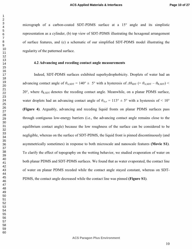
10
micrograph of a carbon-coated SDT-PDMS surface at a 15° angle and its simplistic
representation as a cylinder, (b) top view of SDT-PDMS illustrating the hexagonal arrangement
of surface features, and (c) a schematic of our simplified SDT-PDMS model illustrating the
regularity of the patterned surface.
4.2 Advancing and receding contact angle measurements
Indeed, SDT-PDMS surfaces exhibited superhydrophobicity. Droplets of water had an
advancing contact angle of θA,SDT = 140° ± 5° with a hysteresis of ∆θSDT (= θA,SDT – θR,SDT) <
20°, where θR,SDT denotes the receding contact angle. Meanwhile, on a planar PDMS surface,
water droplets had an advancing contact angle of θA,o = 113° ± 5° with a hysteresis of < 10°
(Figure 4). Arguably, advancing and receding liquid fronts on planar PDMS surfaces pass
through contiguous low-energy barriers (i.e., the advancing contact angle remains close to the
equilibrium contact angle) because the low roughness of the surface can be considered to be
negligible, whereas on the surface of SDT-PDMS, the liquid front is pinned discontinuously (and
asymmetrically sometimes) in response to both microscale and nanoscale features (Movie S1).
To clarify the effect of topography on the wetting behavior, we studied evaporation of water on
both planar PDMS and SDT-PDMS surfaces. We found that as water evaporated, the contact line
of water on planar PDMS receded while the contact angle stayed constant, whereas on SDT-
PDMS, the contact angle decreased while the contact line was pinned (Figure S1).
Page 10 of 27
ACS Paragon Plus Environment
ACS Applied Materials & Interfaces
123456789101112131415161718192021222324252627282930313233343536373839404142434445464748495051525354555657585960

11
Figure 4. Advancing and receding contact angles for deionized, pH 6 water on SDT-PDMS and
on planar PDMS using a constant volumetric flow rate of 0.5 µL/min and a stainless steel needle
with an outer diameter of 0.55 mm. Solid circles represent advancing measurements, and unfilled
circles represent receding measurements. Advancing measurements began within 1 s of the
water touching the surface, and receding measurements began immediately after advancing
began and ended when the fitted contact angle began dropping sharply due to syringe needle
effects (typically around droplet volumes ~0.05 µL). Measurements at 0.1 and 0.3 µL/min (not
shown) gave very similar contact angles.
To measure the extent to which the surface texture of SDT-PDMS would prevent wetting,
1 min after the deposition of mixed water-ethanol droplets (0-100% by volume), contact angles,
θSDT, were measured (Figure 5). We found that θSDT ≥ 90° for ethanol volume fractions up to Cv
= 60% (surface tension ≥ 33 mN/m) (Figure S2), in comparison to a contact angle of < 90° on
planar PDMS for Cv > 10%.
Page 11 of 27
ACS Paragon Plus Environment
ACS Applied Materials & Interfaces
123456789101112131415161718192021222324252627282930313233343536373839404142434445464748495051525354555657585960

12
Figure 5. Short-term contact angles of water-ethanol mixtures on (a) SDT-PDMS and (b) planar
PDMS, and (c) the fraction of pores, p, on the SDT-PDMS which were fully filled, as calculated
from our analytical model. Contact angles were measured 1 min after depositing a 1 µL droplet
and have characteristic error of ± 4°. At ethanol concentrations > 50 vol%, a significant fraction
of the pores are filled within 1 min. The solid blue lines show predictions of the Cassie-Baxter
(p=0) and Wenzel (p=1) models.
Page 12 of 27
ACS Paragon Plus Environment
ACS Applied Materials & Interfaces
123456789101112131415161718192021222324252627282930313233343536373839404142434445464748495051525354555657585960

13
4.3 Model predictions
The apparent contact angle, θSDT, is determined by the ratios of the real liquid-vapor and
liquid-solid areas (ALV, ALS) to the projected area (AP) such that φLV (=ALV/AP), φLS (=ALS/AP),
and �� (intrinsic contact angle, as conventionally defined by the Young equation) via the
equation19
cos���� = � �cos���� − � �, (1)
which can predict both metastable and stable contact angles; where � � +� � ≥ 1, � � ≥ 0,
and � �,��� ��⁄ ≥ � � ≥ 0. When all pores are fully filled with liquid, � � = 0 and Equation 1
reduces to the Wenzel equation, where � � is typically denoted as r.
To understand how the texturing of SDT-PDMS affects its wettability, we used scanning
electron microscopy (SEM) (Figure 3 and S3) to measure the key dimensions and distributions
of features on the SDT-PDMS surfaces. We found the average inner and outer radii of the rings
and the height to be 50, 70, and 20 µm, respectively. We ignored the surface areas of slopes and
smaller hierarchical features in this model. Next, we assumed a hexagonal lattice of rings
separated by a distance, l = 20 µm, as representative of the surface of SDT-PDMS. Some ring-
shaped features can either be in a partially wetting state (Cassie), wherein the liquid remains at
the top of the features, or in a fully wetting state (Wenzel). We approximated the fraction of
pores fully filled with liquid, p, using a simple analytical model (detailed calculations and
diagrams are presented in Section S1 and Figure S4). Using the model SDT-PDMS surface, as
shown in Figure 3c, the values for the partially filled state (p = 0) were calculated as � � = 0.66
and � � = 0.34, and the corresponding values for the fully filled state (p = 1) (Figure S4) were
calculated as � � = 0and � � = 1.68. Thus, to determine the fraction of fully filled unit cells,
Page 13 of 27
ACS Paragon Plus Environment
ACS Applied Materials & Interfaces
123456789101112131415161718192021222324252627282930313233343536373839404142434445464748495051525354555657585960
14
p, we set � � = �1 − #� × 0.66 and � � = �1 − #� × 0.34 + # × 1.68. When �� is known, p
can be determined as a function of ���.
The short-term (1 min) contact angle of water-ethanol mixtures is shown in Figure 5
along with fitted p values and predicted contact angles for p=0 (fully non-wetting) and for p=1
(fully wetting). As the surface tension of water-ethanol mixtures decreased with the increasing
ethanol content (Table S1), we intuitively expected for the fraction of filled pores to increase.
We found p to be zero for ethanol volume fractions < 60%, but p increased at higher ethanol
volume fractions. Given the knowledge of the time dependence of ��� (described below), we
infer that ethanol-water mixtures simply fill the pores faster than does pure water. It is worth
noting that the viscosity of ethanol-water mixtures increases up to Cv ~ 60% and decreases when
Cv exceeds 60%,27
which indicates that interfacial energies, rather than viscosity, dominate pore
filling kinetics in our ethanol-water studies. The short-term wetting scenario for low ethanol
concentrations would be represented schematically by Figure 2a, whereas higher ethanol
concentrations correspond to Figure 2b.
4.4 Effects of waiting time on the stability of contact angles
While investigating the time-dependence of contact angles, we noted that the apparent
contact angle of water on SDT-PDMS reduced from ���~140° to ���~65° after 2 days,
while no change was observed on planar PDMS (both were maintained under a saturated vapor
environment) (Figure 6); a similar decrease was observed with canola oil droplets (Figure S5
and S6). Such a dramatic reduction in the apparent contact angle could be a practical limitation
for textured surfaces that rely on metastable Cassie-states. We considered several possible
Page 14 of 27
ACS Paragon Plus Environment
ACS Applied Materials & Interfaces
123456789101112131415161718192021222324252627282930313233343536373839404142434445464748495051525354555657585960

15
explanations, including (1) pore filling due to inertia/weight of the liquid or capillary
condensation, (2) change in surface chemistries over time, (3) mechanical deformation of the
triple-phase contact line due to an unresolved normal component of the surface tension of water,
and (4) contact line pinning and subsequent reduction in droplet volume through flow of liquid
into pores outside the triple-phase contact line.
Figure 6. Time-dependent changes in contact angles and droplet volumes of sessile water
droplets on SDT-PDMS and planar PDMS over 3000 min (50 h). The contact angle on the
surface of SDT-PDMS decreased from ~140° to ~65° while on the surface of planar PDMS it
remained at �� = 102° )2°. Both surfaces were kept in the same chamber during the
measurements.
Page 15 of 27
ACS Paragon Plus Environment
ACS Applied Materials & Interfaces
123456789101112131415161718192021222324252627282930313233343536373839404142434445464748495051525354555657585960

16
Using fluorescently labeled water and PDMS, pore filling over 70 min was directly
observed with fluorescent confocal microscopy (Figure 7). Within 4 min of droplet deposition,
~10% of the pore volume was filled with water, and after 70 min, ~60% was filled, essentially a
transition from the wetting state in Figure 2a to that in Figure 2b. Note that the unit cell volume
includes both the volume within the ring structures (the pores) and that within the connected
valleys between pores. First, we consider the weight of the liquid drop as a potential cause for
the filling. This invokes the concept of capillary length, which is the characteristic length scale
where surface tension dominates over weight, given by * = +, -.⁄ , where , is the surface
tension (72 mN-m-1
), - is the density of water (1000 kg-m-3
), and . is the acceleration due to
gravity (9.8 ms-2
). For water, the capillary length is approximately 2.7 mm. Since the diameters
of sessile droplets employed in these experiments were ≤ 2 mm, the prospect of a water drop
filling air pockets due it its own weight is ruled out. However, capillary condensation, or vapor
penetration, is a possibility, given the high degree of roughness of the SDT-PDMS surface;
however, due to the intrinsic hydrophobicity of the PDMS, it is unlikely that pores would fill up
primarily with condensate. The alternative mechanism to capillary condensation is liquid
penetration, or flow of bulk liquid from the droplet into the pore. Confocal microscopy showed
that once penetration of a given cavity was initiated, full filling was attained in <1 min, releasing
large air bubbles, indicative of rapid filling from the liquid above (though not due to gravity),
rather than condensation slowly filling the cavity below the droplet. Moreover, when the SDT-
PDMS was allowed to sit in saturated vapor for 48 hr prior to droplet deposition, no difference
was observed in the wetting behavior. From pore filling observations and from our model
(Section S1), we can deduce that the fully filled wetting state is energetically favorable.
Page 16 of 27
ACS Paragon Plus Environment
ACS Applied Materials & Interfaces
123456789101112131415161718192021222324252627282930313233343536373839404142434445464748495051525354555657585960

17
However, mechanisms in addition to the liquid penetration of pores beneath the droplet must be
involved because this could only explain a decrease in ��� down to 110°, the Wenzel limit
(Section S1).
Figure 7. Images captured using fluorescent confocal microscopy at a representative focal plane,
where pore filling is displayed over time. Green regions correspond to water, red to PDMS, and
black to vapor. Pores and regions between pores which become filled are labeled with white
arrows. Pores are indicated with dashed circles on the image taken 4 min after adding the water.
For observation purposes, the water was saturated with fluorescein isothiocyanate (FITC -
green), and the SDT-PDMS was doped with Rhodamine B by soaking in a saturated dye solution
for 2 days. A number of images were taken at ~20 different focal planes from the bottom to the
top of the features to confirm full pore filling, but are not shown here.
Although changes in the surface chemistry of PDMS28
in contact with water could lead to
a reduction in the contact angle, this seems unlikely because there is no change in the contact
Page 17 of 27
ACS Paragon Plus Environment
ACS Applied Materials & Interfaces
123456789101112131415161718192021222324252627282930313233343536373839404142434445464748495051525354555657585960
18
angle of water on the planar PDMS (Figure 6). Moreover, the contact angle of sessile water
droplets on perfluorotridecyltrichlorosilane (FDTS)-coated SDT-PDMS surfaces decreased in a
similar fashion to the uncoated SDT-PDMS (Figure S6 and S7, and SI Experimental Section).
We also consider that the unresolved vertical component of the surface tension of water at the
triple-phase contact line might bend/flex topographical features on the SDT-PDMS, which could
appear as a smaller apparent contact angle. In fact, researchers have recently observed
mechanical deformations of the contact line formed between a silicone gel (CY52-276A/B, Dow
Corning Toray) and glycerol that could explain our observations.29
We observed this event on
planar PDMS using optical profilometry (Figure S8), but on SDT-PDMS, this was difficult due
to the roughness of the surface. It is likely that when a droplet is placed on the planar PDMS,
which is elastic, the deformation forms within a fraction of a second and remain constant as long
as the droplet is on the surface. In the case of the SDT-PDMS, the gradual pore penetration and
spreading (see below) that occur may give rise to deformations which change over time. Lastly,
fluorescent confocal microscopy and contact angle studies (Figure S9) showed that the contact
line advanced during the first hour that the droplet was on the surface, and was pinned thereafter,
perhaps caused by a deformation of the surface. Subsequently, water flowed from the droplet to
areas outside of the contact region (Figure 2c and S10), resulting in a lower ��� by a
combination of pinning and a substantial loss of droplet volume. In the final state, when some
surface cavities outside the droplet were filled with the liquid (i.e., the Cassie-impregnated-like
state), the total volume of the liquid inside the cavities is not negligible compared to the droplet
volume (Figure 6 shows a 80% decrease in droplet volume over 50 h). In fact, this violates the
fundamental assumption of conservation of volume of the liquid drop (and negligible volume of
cavities in comparison) on which all the aforementioned theoretical models are based.30
Thus,
Page 18 of 27
ACS Paragon Plus Environment
ACS Applied Materials & Interfaces
123456789101112131415161718192021222324252627282930313233343536373839404142434445464748495051525354555657585960

19
the apparent angle in the long-time regime might fall out of scope of any of the theoretical
models. Bormashenko and co-workers recently investigated transitions from the Cassie to
Wenzel and then to Cassie-impregnated state by vertically oscillating liquid drops on textured
PDMS, polystyrene, polyethylene, and polyetherimide at frequency, f = 36 Hz and amplitude, A=
1.1 mm, and further claimed the Cassie-impregnated state to be the thermodynamic
minimum.22,23
They proposed that the transition to the Cassie-impregnated state is only possible
when the “local” angle22
(intrinsic angle, θo, in the present work) is less than 90°; however,
external vibrations can transiently reduce the local angle. Because no such energy input was
applied in our studies and θo was larger than 90°, we concluded that θSDT at t > 90 min cannot be
explained by the Cassie-impregnated model. We posit that the liquid drainage outside the droplet
takes place via flow through the connected valleys between the pores, where microscale channels
may act as conduits for the liquid. Flows in comparably sized channels have been directly
observed and studied in detail for textured hydrophobic polymer surfaces,31,32
and including
PDMS, but further discussion of the fluid dynamics is beyond the scope of this work. In
summary, it appears that the primary mechanism by which the contact angle decreases occurs on
two time scales (Figure 2): pore filling, which happens within ~90 min of droplet deposition,
and pinning and volume drainage, which begins to occur thereafter and can presumably progress
indefinitely.
5. Conclusions
We found that tests of sand dollars, which are hydrophilic by nature, could act as
physically and chemically robust templates for imparting non-wetting topographical features to
many thermally- or photo-setting polymer surfaces. This biomimicking approach is simple,
quick, and inexpensive and elucidates how both topographical and chemical modifications can
Page 19 of 27
ACS Paragon Plus Environment
ACS Applied Materials & Interfaces
123456789101112131415161718192021222324252627282930313233343536373839404142434445464748495051525354555657585960
20
be combined to engineer non-wetting materials; for example, SDT-PDMS exhibited contact
angles ≥ 90° for liquids with surface tensions ≥ 33mN/m. Scanning electron microscopy of
SDT-PDMS allowed us to develop a simple model, which agreed well between measured short-
term contact angles and the predictions of the Cassie-Baxter and Wenzel equations. Next, we
investigated the time-dependence of contact angles on soft polymeric surfaces. The apparent
contact angle of water on SDT-PDMS decreased from ~140° to ~65° over the course of 2 days,
while on planar PDMS no change in contact angle with time was observed. Our contact angle
and confocal microscopy experiments indicated that a combination of pore filling beneath the
droplet (Figure 2b) and contact line pinning followed by flow of liquid outside of the contact
region (Figure 2c) are responsible for the decrease in the contact angle. The dramatic time-
dependence is particularly surprising given that the intrinsic contact angle, ��, was larger than
90°. For rough surfaces where �� is less than 90°, one would expect qualitative and quantitative
differences from the time-dependent behavior shown here, in particular that Equation 1 may no
longer apply, as pores outside the contact region eventually become filled with condensate at
thermodynamic equilibrium.33
Lastly, if the volume of liquid within the pores is non-negligible
compared to the droplet volume, none of the aforementioned models can be applied to fully
describe the wetting behavior. The concepts of contact angle stability applied to this simple bio-
inspired model system should provide insight for the design and development of durable
omniphobic coatings.
ASSOCIATED CONTENT
Supporting Information.
Page 20 of 27
ACS Paragon Plus Environment
ACS Applied Materials & Interfaces
123456789101112131415161718192021222324252627282930313233343536373839404142434445464748495051525354555657585960

21
The Supporting Information contains 10 additional figures, 2 tables, 1 movie, and derivations
which elaborate upon arguments made succinctly in the manuscript. This material is available
free of charge via the Internet at http://pubs.acs.org.
AUTHOR INFORMATION
Corresponding Author
*To whom correspondence should be addressed:
Dr. Himanshu Mishra: [email protected]; Ph. 966-54-808-2110
Dr. Jacob N. Israelachvili: Jacob@[email protected]; Ph. 805-893-8407
Present Addresses
† Water Desalination and Reuse Center, Biological and Environmental Science and Engineering
Division, King Abdullah University of Science and Technology, Thuwal 23955-6900, Saudi
Arabia
Author Contributions
‡These authors contributed equally.
The manuscript was written through contributions of all authors. All authors have given approval
to the final version of the manuscript.
Funding Sources
This work was supported by a grant from the Procter & Gamble Company. H. M. was funded by
the Elings Prize Fellowship in Experimental Science of the California NanoSystems Institute at
the University of California, Santa Barbara.
Page 21 of 27
ACS Paragon Plus Environment
ACS Applied Materials & Interfaces
123456789101112131415161718192021222324252627282930313233343536373839404142434445464748495051525354555657585960

22
ACKNOWLEDGMENT
This work was supported by a grant from the Procter & Gamble Company. H. M. was funded by
the Elings Prize Fellowship in Experimental Science of the California NanoSystems Institute at
the University of California, Santa Barbara. We acknowledge the use of the NRI-MCDB
Microscopy Facility at UC Santa Barbara, and we thank Dr. Mary Raven for assistance with
confocal microscopy. The MRL Shared Experimental Facilities (used for SEM imaging) are
supported by the MRSEC Program of the NSF under Award No. DMR 1121053; a member of
the NSF-funded Materials Research Facilities Network.
REFERENCES
(1) Wen, L. P.; Tian, Y.; Jiang, L. Bioinspired Super-Wettability from Fundamental Research to
Practical Applications Angew. Chem., Int. Ed. 2015, 54, 3387–3399.
(2) Leslie, D. C.; Waterhouse, A.; Berthet, J. B.; Valentin, T. M.; Watters, A. L.; Jain, A.; Kim,
P.; Hatton, B D.; Nedder, A.; Donovan, K.; Super, E. H.; Howell, C.; Johnson, C. P.; Vu, T. L.;
Bolgen, D. E.; Rifai, A.; Hansen, A. R.; Aizenberg, M.; Super, M.; Aizenberg, J.; Ingber, D. E..
A Bioinspired Omniphobic Surface Coating on Medical Devices Prevents Thrombosis and
Biofouling Nat. Biotechnol. 2014, 32, 1134–1140.
(3) Grinthal, A.; Aizenberg, J. Mobile Interfaces: Liquids as a Perfect Structural Material for
Multifunctional Antifouling Surfaces Chem. Mater. 2014, 26, 698–708.
Page 22 of 27
ACS Paragon Plus Environment
ACS Applied Materials & Interfaces
123456789101112131415161718192021222324252627282930313233343536373839404142434445464748495051525354555657585960

23
(4) Ebert, D.; Bhushan, B. Wear-Resistant Rose Petal-Effect Surfaces with Superhydrophobicity
and High Droplet Adhesion using Hydrophobic and Hydrophilic Nanoparticles J. Colloid
Interface Sci. 2012, 384, 182–188.
(5) Ralston, E.; Swain, G. Bioinspiration-the Solution for Biofouling Control? Bioinspiration
Biomimetics 2009, 4, 1–9.
(6) Campbell, D. D. F. (2015) "Dendraster excentricus" Encyclopedia of Life, available from
http://eol.org/pages/460427. Accessed 15 Jan 2014.
(7) Vaughn, D.; Strathmann, R. R. Predators Induce Cloning in Echinoderm Larvae Science
2008, 319, 1503.
(8) Nosonovsky, M.; Bhushan, B. Biomimetic Superhydrophobic Surfaces: Multiscale Approach
Nano Lett. 2007, 7, 2633–2637.
(9) Chhatre, S. S.; Choi, W.; Tuteja, A.; Park, K. C.; Mabry, J. M.; McKinley, G. H.; Cohen,
R.E. Scale Dependence of Omniphobic Mesh Surfaces Langmuir 2010, 26, 4027–2035.
(10) Huang, J. Y.; Liu, C.; Zhu, Y.; Masala, S.; Alarousu, E.; Han, Y.; Fratalocchi, A.
Harnessing Structural Darkness in the Visible and Infrared Wavelengths for a New Source of
Light Nat. Nanotechnol. 2015, DOI: 10.1038/nnano.2015.228.
(11) Huang, J. Y.; Wang, X. D.; Wang, Z. L. Controlled Replication of Butterfly Wings for
Achieving Tunable Photonic Properties Nano Lett. 2006, 6, 2325–2331.
(12) Huang, J. Y.; Wang, X. D.; Wang, Z. L. Bio-Inspired Fabrication of Antireflection
Nanostructures by Replicating Fly Eyes Nanotechnology 2008, 19, 1–6.
Page 23 of 27
ACS Paragon Plus Environment
ACS Applied Materials & Interfaces
123456789101112131415161718192021222324252627282930313233343536373839404142434445464748495051525354555657585960

24
(13) Epstein, A. K.; Wong, T.-S.; Belisle, R. A.; Boggs, E. M.; Aizenberg, J. Liquid-Infused
Structured Surfaces with Exceptional Anti-Biofouling Performance Proc. Nat. Acad. Sci. U. S.
A. 2012, 109, 13182–13187.
(14) Schumacher, J. F.; Carman, M. L.; Estes, T. G.; Feinberg, A. W.; Wilson, L. H.; Callow, M.
E.; Callow, J. A.; Finlay, J. A.; Brennan, A. B. Engineered Antifouling Microtopographies -
Effect of Feature Size, Geometry, and Roughness on Settlement of Zoospores of the Green Alga
Ulva Biofouling 2007, 23, 55–62.
(15) Epstein, A. K.; Hong, D.; Kim, P; Aizenberg, J. Biofilm Attachment Reduction on
Bioinspired, Dynamic, Micro-Wrinkling Surfaces New J. Phys. 2013, 15, 1–13.
(16) Barreiro, A. M.; Recouvreux, D. O. S.; Hotza, D.; Porto, L. M.; Rambo, C. R. Sand Dollar
Skeleton as Templates for Bacterial Cellulose Coating and Apatite Precipitation J. Mater. Sci.
2010, 45, 5252–5256.
(17) Petite, H.; Viateau, V.; Bensaid, W.; Meunier, A.; de Pollack, C.; Bourguignon, M.;
Ouidina, K.; Sedel, L.; Guillemin, G. Tissue-Engineered Bone Regeneration Nat. Biotechnol.
2000, 18, 959–963.
(18) Seshadri, R.; Meldrum, F. C. Bioskeletons as Templates for Ordered, Macroporous
Structures Adv. Mater. 2000, 12, 1149–1151.
(19) Cassie, A. B. D.; Baxter, S. Wettability of Porous Surfaces Trans. Faraday Soc. 1944, 40,
546–551.
(20) Wenzel, R. N. Resistance of Solid Surfaces to Wetting by Water Ind. Eng. Chem. 1936, 28,
988–994.
Page 24 of 27
ACS Paragon Plus Environment
ACS Applied Materials & Interfaces
123456789101112131415161718192021222324252627282930313233343536373839404142434445464748495051525354555657585960

25
(21) Bico, J.; Thiele, U.; Quere, D. Wetting of Textured Surfaces Colloids Surf., A 2002, 206,
41–46.
(22) Bormashenko, E.; Pogreb, R.; Stein, T.; Whyman, G.; Erlich, M.; Musin, A.; Machavariani,
V.; Aurbach, D. Characterization of Rough Surfaces with Vibrated Drops Phys. Chem. Chem.
Phys. 2008, 10, 4056–4061.
(23) Bormashenko, E. Progress in Understanding Wetting Transitions on Rough Surfaces Adv.
Colloid Interface Sci. 2015, 222, 92–103.
(24) Boreyko, J. B.; Baker, C. H.; Poley, C. R.; Chen, C.-H. Wetting and Dewetting Transitions
on Hierarchical Superhydrophobic Surfaces Langmuir 2011, 27, 7502–7509.
(25) Feng, L.; Zhang, Y.; Xi, J.; Zhu, Y.; Wang, N.; Xia, F.; Jiang, L. Petal Effect: A
Superhydrophobic State with High Adhesive Force Langmuir 2008, 24, 4114–4119.
(26) Style, R. W.; Hyland, C.; Boltyanskiy, R.; Wettlaufer, J. S.; Dufresne, E. R. Surface Tension
and Contact with Soft Elastic Solids Nat. Commun. 2013, 4, 1–6.
(27) Khattab, I. S.; Bandarkar, F.; Fakhree, M. A. A.; Jouyban, A. Density, Viscosity, and
Surface Tension of Water+Ethanol Mixtures from 293 to 323 K Korean J. Chem. Eng. 2012, 29,
812–817.
(28) Delamarche, E.; Schmid, H.; Michel, B.; Biebuyck, H. Stability of Molded
Polydimethylsiloxane Microstructures Adv. Mater. 1997, 9, 741–746 .
(29) Style, R. W.; Boltyanskiy, R.; Che, Y.; Wettlaufer, J.S.; Wilen, L. A.; Dufresne, E. R.
Universal Deformation of Soft Substrates Near a Contact Line and the Direct Measurement of
Solid Surface Stresses Phys. Rev. Lett. 2013, 110, 066103-1–066103-5.
Page 25 of 27
ACS Paragon Plus Environment
ACS Applied Materials & Interfaces
123456789101112131415161718192021222324252627282930313233343536373839404142434445464748495051525354555657585960

26
(30) Whyman, G.; Bormashenko, E.; Stein, T. The Rigorous Derivation of Young, Cassie-Baxter
and Wenzel Equations and the Analysis of the Contact Angle Hysteresis Phenomenon Chem.
Phys. Lett. 2008, 450, 355–359.
(31) Sbragaglia, M.; Peters, A. M.; Pirat, C.; Borkent, B. M.; Lammertink, R. G. H.; Wessling,
M.; Lohse, D. Spontaneous Breakdown of Superhydrophobicity Phys. Rev. Lett. 2007, 99,
156001-1–156001-4.
(32) Peters, A. M.; Pirat, C.; Sbragaglia, M.; Borkent, B. M.; Wessling, M.; Lohse, D.;
Lammertink, R. G. Cassie-Baxter to Wenzel State Wetting Transition: Scaling of the Front
Velocity Eur. Phys. J. E: Soft Matter 2009, 29, 391–397.
(33) This assumes a supersaturated vapor environment, which is necessary to prevent
evaporation of the droplet.
Page 26 of 27
ACS Paragon Plus Environment
ACS Applied Materials & Interfaces
123456789101112131415161718192021222324252627282930313233343536373839404142434445464748495051525354555657585960

27
Table of Contents Entry
Sand-dollar-templated (SDT) PDMS is a simple, lithography-free surface. Shown is a droplet of
water on the SDT-PDMS with an advancing contact angle, θA,SDT = 140°, and a scanning
electron micrograph of a characteristic feature on the SDT-PDMS surface.
Page 27 of 27
ACS Paragon Plus Environment
ACS Applied Materials & Interfaces
123456789101112131415161718192021222324252627282930313233343536373839404142434445464748495051525354555657585960