surgical anatomy of the liver
TRANSCRIPT

Dr MHA Bemelmans,
ESCAM (European Surgical Center Aachen Maastricht)
Surgical Anatomy of the liver
Uniklinik RWTH Aachen

Uniklinik
RWTH Aachen
ESCAM
Disclosures
Uniklinik RWTH Aachen ESCAM
Potential conflict of interest None
Potentially relevant company relationships in
connection with event
None

Uniklinik RWTH Aachen ESCAM
Marc

Uniklinik RWTH Aachen ESCAM

Uniklinik RWTH Aachen ESCAM
The cell lineage steps during hepatic development (red) from
uncommitted endoderm to functional adult hepatocytes and
biliary epithelium

Time line mouse liver development
Uniklinik RWTH Aachen ESCAM

Adult liver
Uniklinik RWTH Aachen ESCAM

Galen (circa 130–200 BC) was one of the
first who described the liver. He thought that
the liver was five-lobed. Such opinion
dominated until the 15th century.
Uniklinik RWTH Aachen ESCAM

Vesalius founder modern anatomy (1514-1564)
Uniklinik RWTH Aachen ESCAM

1st book on human anatomy Vesalius
Uniklinik RWTH Aachen ESCAM
In 1654, F. Glisson (1597–1677) studied the
liver. He discussed topography of the
intrahepatic vessels and surrounding
connective tissue. Even today this is
referred to as Glisson’s capsule and the triad
(portal vein, biliary duct and hepatic artery)
is called portal pedicle or Glisson’s pedicle
Uniklinik RWTH Aachen ESCAM

Francois Glisson
Uniklinik RWTH Aachen ESCAM
1597-1677

N. A. Goldsmith and R. T. Woodburne
supported division of the liver into four
segments, each having two subsegments
with second order of portal vein branches
(1957)
Uniklinik RWTH Aachen ESCAM

C. Couinaud suggested that the liver should
be divided into eight segments, based on
third order portal vein distribution (1957)
Uniklinik RWTH Aachen ESCAM

Claude Couinaud
Uniklinik RWTH Aachen ESCAM

A good knowledge of the anatomy
is a prerequisite for modern surgery
of the liver.”
H. Bismuth
Uniklinik RWTH Aachen ESCAM

Henri Bismuth
Uniklinik RWTH Aachen ESCAM

Porto Allegre Brasil 2016
Uniklinik RWTH Aachen ESCAM

Bismuth’s classification. H. Bismuth brought
together the Couinaud’s cadaveric system in
situ and the system of Goldsmith and
Woodburn in vivo
Uniklinik RWTH Aachen ESCAM

He distinguished three planes (scissurae),
hosting the hepatic veins and a transverse
plane passing through the right and left
portal branches. Additionally, H. Bismuth
described the caudate lobe as a separate
segment I.
Uniklinik RWTH Aachen ESCAM

Anatomy according to Bismuth
Uniklinik RWTH Aachen ESCAM

Anatomy 1
Uniklinik RWTH Aachen ESCAM

Anatomy 2
Uniklinik RWTH Aachen ESCAM

Anatomy 3
Uniklinik RWTH Aachen ESCAM

Anatomy 4
Uniklinik RWTH Aachen ESCAM

Anatomy 5
Uniklinik RWTH Aachen ESCAM

Anatomy 6
Uniklinik RWTH Aachen ESCAM

Anatomy 7
Uniklinik RWTH Aachen ESCAM

Anatomy 9
Uniklinik RWTH Aachen ESCAM

VIDEO???
Uniklinik RWTH Aachen ESCAM

Cirrhosis
Uniklinik RWTH Aachen ESCAM

Anatomy 10
Uniklinik RWTH Aachen ESCAM

No comment
Uniklinik RWTH Aachen ESCAM

Uniklinik RWTH Aachen ESCAM
Uniklinik RWTH Aachen ESCAM

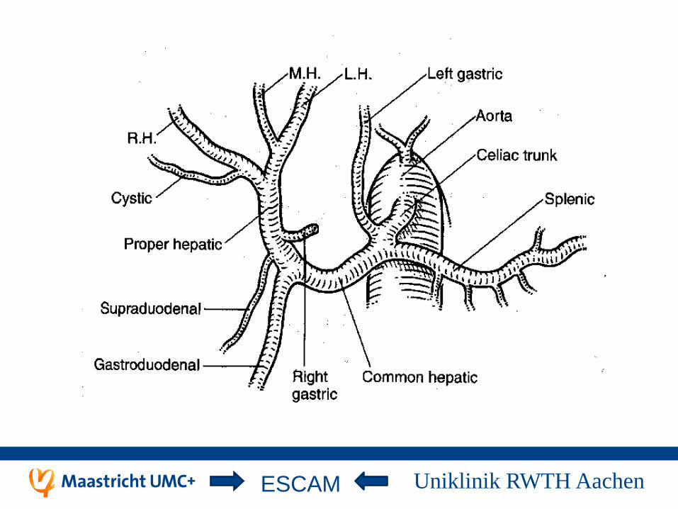
Arterial anatomy
Uniklinik RWTH Aachen ESCAM

Uniklinik RWTH Aachen ESCAM

Portal Anatomy
Uniklinik RWTH Aachen ESCAM

Uniklinik RWTH Aachen ESCAM

Biliary Anatomy
Uniklinik RWTH Aachen ESCAM

Uniklinik RWTH Aachen ESCAM

Uniklinik RWTH Aachen ESCAM
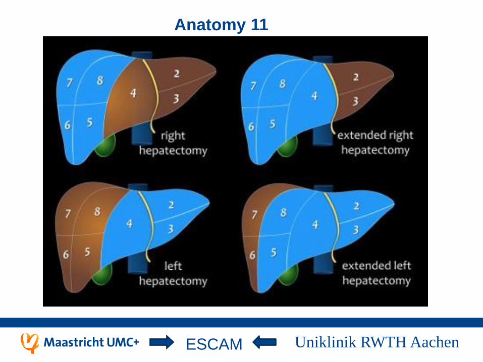
Anatomy 11
Uniklinik RWTH Aachen ESCAM

Anatomy 12
Uniklinik RWTH Aachen ESCAM

Anatomy 13
Uniklinik RWTH Aachen ESCAM

Anatomy 14
Uniklinik RWTH Aachen ESCAM

Uniklinik RWTH Aachen ESCAM
Majno et all j Hepatol 2014
Uniklinik RWTH Aachen ESCAM

Uniklinik RWTH Aachen ESCAM

Uniklinik RWTH Aachen ESCAM