surgical indications for pituitary tumors during pregnancy
TRANSCRIPT

HAL Id: hal-02468520https://hal.archives-ouvertes.fr/hal-02468520
Submitted on 12 May 2021
HAL is a multi-disciplinary open accessarchive for the deposit and dissemination of sci-entific research documents, whether they are pub-lished or not. The documents may come fromteaching and research institutions in France orabroad, or from public or private research centers.
L’archive ouverte pluridisciplinaire HAL, estdestinée au dépôt et à la diffusion de documentsscientifiques de niveau recherche, publiés ou non,émanant des établissements d’enseignement et derecherche français ou étrangers, des laboratoirespublics ou privés.
Surgical indications for pituitary tumors duringpregnancy: a literature review
Thomas Graillon, Thomas Cuny, Frederic Castinetti, Blandine Courbiere,Marie Cousin, Frédérique Albarel, Isabelle Morange, Nicolas Bruder, Thierry
Brue, Henry Dufour
To cite this version:Thomas Graillon, Thomas Cuny, Frederic Castinetti, Blandine Courbiere, Marie Cousin, et al.. Surgi-cal indications for pituitary tumors during pregnancy: a literature review. Pituitary, Springer Verlag,2020, 23 (2), pp.189-199. �10.1007/s11102-019-01004-3�. �hal-02468520�

Vol.:(0123456789)1 3
Pituitary (2020) 23:189–199 https://doi.org/10.1007/s11102-019-01004-3
Surgical indications for pituitary tumors during pregnancy: a literature review
Thomas Graillon1,2 · Thomas Cuny2,3 · Frédéric Castinetti2,3 · Blandine Courbière4 · Marie Cousin5 · Frédérique Albarel3 · Isabelle Morange3 · Nicolas Bruder6 · Thierry Brue2,3 · Henry Dufour1,2
Published online: 6 November 2019 © Springer Science+Business Media, LLC, part of Springer Nature 2019
AbstractPurpose Surgical indications for pituitary tumors during pregnancy are rare, and are derived from a balance between expected benefits, particularly for maternal benefits, and anesthetic/surgical risks.Methods A literature review was performed to define the optimal surgical indications for pituitary adenomas (PA) and other pituitary tumors during pregnancy.Results Main benefits are expected in case of critical visual impairment and/or life-threatening endocrine disturbances. Multidisciplinary patient management is systematically required although nonobstetric surgery presents a reasonable risk during pregnancy. The risks of congenital malformation during the first trimester and those of premature birth during the third trimester make the second trimester the optimal period for surgery. In prolactin-secreting, nonsecreting, GH- and TSH-secreting PAs, transsphenoidal surgery (TS) is recommended in cases involving severe visual impairment, characterized by severe visual field deficit, visual acuity impairment, and abnormal optical coherence tomography findings, and when no other medical alternatives are possible and/or sufficient. Uncontrolled and severe Cushing’s disease (CD) during pregnancy increases both maternal and fetal morbimortality, thus justifying TS or sometimes dopamine agonist therapy as a safer alterna-tive. Finally, metyrapone, ketoconazole, or bilateral adrenalectomy could be recommended in certain cases after the failure of medical therapies and/or TS. Surgery is also required for suprasellar meningiomas, craniopharyngiomas, and pituitary cysts in the case of severe visual deficit.Conclusion Surgical indications for pituitary tumors are rare during pregnancy; therefore, surgery should be avoided when possible. Further, the second trimester should be considered as the optimal surgical period. Severe visual disturbance and uncontrolled CD are the main surgical indications during pregnancy.
Keywords Pituitary adenoma · Pregnancy · Surgery · Meningioma · Craniopharyngioma · Pituitary cyst
Introduction
The diagnosis of pituitary adenomas (PAs) and other pitui-tary tumors during pregnancy is uncommon, which explains the scarcity of the available data in the literature to guide the treatment using a case-by-case approach. Surgical indi-cations and medical alternatives are actively discussed by expert pituitary teams. In usual neurosurgical practice, sur-gery during pregnancy is exceptional, mainly reported for lumbar disc herniation and/or neurovascular pathologies. Regarding our team, we have reported only two cases of pituitary tumor surgery in the last 3 years. Surgical indi-cations are estimated while balancing between anesthetic/surgical risks and potential surgical benefits. Between these, the latter is principally related to the mother, whereas the
* Thomas Graillon [email protected]
1 Neurosurgery Department, Aix-Marseille Univ, APHM, CHU Timone, La Timone Hospital, 264 rue Saint-Pierre, 13005 Marseille, France
2 Aix-Marseille Univ, INSERM, MMG, Marseille, France3 Endocrinology Department, Aix-Marseille Univ, APHM,
CHU Conception, Marseille, France4 Centre Clinico-Biologique d’AMP, Pôle
Femmes-Parents-Enfants, Hôpital de La Conception, AP-HM, Marseille/Aix Marseille Univ, Avignon Univ, CNRS, IRD, IBME, Marseille, France
5 Cabinet d’Ophtalmologie, Saint-Rémy de Provence, France6 Anesthesiology-Intensive Care Department, Aix-Marseille
Univ, APHM, CHU Timone, Marseille, France

190 Pituitary (2020) 23:189–199
1 3
risks are shared by the mother and fetus; further, they could exceptionally be vital but more likely functional in cases of visual loss or endocrine disturbances. Here, a literature review is provided to assess the anesthetic and surgical risks as well as the benefits expected for the mother and fetus. Then, we detail surgical strategies depending on dif-ferent pituitary tumors given the current literature data and knowledge.
Surgical indications
For vital benefit
A surgery performed for pituitary tumors for vital risks in a pregnant patient is an exceptional situation. Indeed, it is almost always characterized by the occurrence of life-threat-ening pituitary apoplexy, which could also be the cause of severe and acute corticotroph deficiency [1].
For visual benefit
Visual improvement is the main functional benefit expected from surgery. Visual benefit is proportional to preoperative severity of visual loss. The definition of severe visual loss, which indicates surgery without delay, remains unclear. It could be practically defined as a com-bination of severe visual impairment and related handicap, potential of recovery, and a high risk of definitive visual sequelae. Therefore, surgery is likely required when the delay until delivery may lead to irreversible damages to optic pathways. Complete visual examination includes examination of visual acuity (VA), visual field (VF), and the retinal nerve fiber layer (RNFL); fundus examination; and optical coherence tomography (OCT) for the ganglion cell complex. Visual prognostic factors are well described in the general population: patient aged ≥ 50 years, optic nerve compression lasting for ≥ 1 year, tumor type and vol-ume, preoperative VF and acuity, and optic nerve atrophy [2]. In pregnant women, young age and short-term preop-erative compression are the frequent favorable prognostic factors. In the meta-analysis conducted by Muskens et al., which included patients with PAs responsible for visual disturbances, postoperative VF improvement occurred in approximately 80% of patients, which included only 67.5% of patients with VA impairment [3]. The degree of preop-erative VF impairment is a strong prognostic factor for postoperative visual outcome. In patients with complete postoperative VF recovery, preoperative VF impairment was significantly less pronounced than in those with only partial VF recovery. More specifically, Gnanalingham et al. showed that the severity of preoperative temporal VF deficit was a strong prognostic factor for postoperative
visual outcome. The mean preoperative quantitative VF as per the Humphrey field was − 20 dcb in the upper tem-poral VF in the case of partial recovery and − 10 dcb in the case of complete recovery [4]. The mean preoperative quantitative VF was − 16 dcb in the lower temporal VF in the case of partial recovery and − 8 dcb in the case of complete recovery. In the study by Barzaghi et al., the mean preoperative quantitative VF was − 15.6 ± 0.8 dcb in the case of partial recovery and − 8.5 ± 0.6 dcb in the case of complete recovery [5]. This study also underlined the relevance of preoperative VA to predict visual outcome. Among patients with preoperative VA deficit, one-third of patients had complete recovery, one-third had partial recovery, and the remaining did not show any postopera-tive improvement [5]. In this study, the mean preopera-tive VA was 0.9 for patients with complete recovery and 0.6 for those with partial recovery. Abnormal OCT find-ings of RNFL thickness are other prognostic factors, e.g., alterations in RNFL found on OCT impair the potential of optic nerve recovery and are prognostic factors for central and peripheral VF recovery [6–8]. The threshold for the optic nerve fiber diameter for complete recovery was found to be ≥ 85 µm [8]. Blanch et al. highlighted the higher sensitivity of RNFL and ganglion cell complex (GCC) alteration observed on OCT compared to classical VF assessment [9]. Moreover, sensitivity of GCC-OCT may be higher than RNFL-OCT to assess chiasmal compression [9, 10].Therefore, severe visual impairment that could lead to definitive visual sequelae and that require immediate surgery includes severe VF impairment with mean VF of − 15 to − 20 dcb, VA impairment, and severe RNFL and GCC-OCT deterioration (Table 1).
For endocrine functions
In cases of hormone hypersecretion, surgery is the most beneficial in Cushing’s disease (CD) [11]. In GH-, PRL-, or TSH-secreting PAs, surgical relevance remains limited given the efficacies of different medical therapies, and it still raises the concerns of the risk of pituitary deficiencies.
Table 1 Preoperative situations with severe visual disturbance lead-ing to a high risk of nonrecoverable optic nerve lesion and definitive visual sequelae
Severe visual impairment requiring non-delayed surgerySevere VF impairment (VF quantitative measure between − 15 and
− 20 dcb)VA impairmentSevere RNFL thickness and GCC loss detected by OCT

191Pituitary (2020) 23:189–199
1 3
Maternal‑ and fetal‑related risks of anesthesia and pituitary surgery during pregnancy
Main principles of surgical management
It is of upmost importance that surgical management will be conducted using a multidisciplinary approach at an expert pituitary center. Preoperative assessment by an obstetrician is also strongly recommended [12–15]. All anesthetic drugs cross the placenta. Teratogenic anesthetic agents should be avoided, but no anesthetic agent cur-rently used has been associated with teratogenic effects on humans. The maximal risk of teratogenicity likely occurs between 13 and 60 days after gestation. During the third trimester, neurotoxicity and abnormal neural tube closure should be considered, but their incidence is probably rare. In animal studies, anesthetic agents have shown to induce neuronal apoptosis, disruption of brain circuit formation, and impairment of neurogenesis and synaptogenesis, leading to morphological and functional alterations in the brain and long-term cognitive dysfunction after a single exposure to anesthesia [16, 17]. However, clinical stud-ies did not demonstrate any adverse effects on the cogni-tive functions of children anesthetized in the first year of age [18, 19]. Considering the uncertainty of the effects of anesthetic agents on the developing brain, surgery should be avoided whenever possible during pregnancy. To assess the fetal risk related to premature birth, a recent study on 6696 births in 24–34 gestational week (GW) showed 59.1% fetal survival at 25 GW, 75.3% at 26 GW, 93.6% between 27 and 31 GW, and 98.9% between 32 and 34 GW. In the same study, the rate of hospital discharge without severe neonatal pathologies was 0% at 23 GW, 11.6% at 24 GW, 30.0% at 25 GW, 47.5% at 26 GW, 81.3% between 27 and 31 weeks, and 96.8% between 32 and 34 GW [20]. Therefore, the risk related to premature birth is minimal between 27 and 31 GW, and birth is safe from 32 GW.
Preoperative and postoperative imaging should strictly be limited to necessity. Brain MRI without gadolinium enhancement is sufficient in most cases for diagnosis and operational planning. Gadolinium enhancement-caused fetal toxicity remains undemonstrated and uncertain, but gadolinium enhancement is rarely required. CT should be avoided considering X-ray toxicity [21].
Orotracheal intubation and patient installation
Difficult orotracheal intubation is more frequent during the second and third trimesters, and it is related to swelling and friability of the oropharyngeal airway mucosa. From
the second trimester, high abdominal pressure increases the risk of active reflux, regurgitation, and aspiration (Mendelson’s syndrome), leading to pulmonary infection or acute respiratory failure. Compression of the inferior vena cava because of gravid uterus should be avoided from the second trimester. Reduced venous return to the heart causes hypotension, placental insufficiency, or decreased cerebral blood flow during surgery. Patients should be positioned in the supine position with a chopping block under the right hip to tilt the body to the left or in the lateral decubitus (“park bench”) position. Deep vein thrombosis should be prevented. From 18 to 24 GW, fetal monitoring should be performed before and after surgery [22–25]. After 25 GW, continuous fetal monitoring during anesthesia remains particularly debated given the lack of proven benefits.
During the surgery
Moderate hypotension classically used in endonasal surgery should be avoided. Hypothermia, arterial hypotension, pre-operative hyperventilation, hypoxia, acidosis, and dehydra-tion should also be avoided. The use of diuretic drugs is not recommended. Electrolyte and hemoglobin concentrations should be tightly monitored. Hematocrit should be main-tained at ≥ 28%, and abdominal pressure should be avoided to limit inferior vena cava compression. Local vasocon-strictive injection and administration of hemostatic agents with fibrin are allowed. Prophylactic anticonvulsant agents should be avoided when not necessary and are associated with adverse effects on the fetus. Tocolytics, prophylactic agents, could be administrated during the third trimester in the case of contractions and cervical modifications. Mannitol (0.5–1 g/kg) can be administrated in the case of high intrac-ranial pressure, but if not required, mannitol should also be avoid given the risk of fetal dehydration [13, 26].
During the postoperative period
A close monitoring of natremia and the hormonal status is required for the prevention of maternal dehydration, even more in the case of diuretic or mannitol use. Eventual hor-monal ante and/or neurohypophysis deficit should be rapidly substituted. Endonasal surgeries cause moderate postopera-tive pain, for which paracetamol is usually sufficient. The use of nonsteroidal anti-inflammatory drugs and prophy-lactic anticonvulsant agents should be avoided. A rapid decrease in the dose of postoperative corticosteroids is also recommended [27]. Early mobilization is required to pre-vent venous thrombosis. Moreover, the risk of postoperative cerebrospinal fluid leak should be closely monitored given high maternal abdominal pressure. Overall, obstetric and

192 Pituitary (2020) 23:189–199
1 3
fetal monitoring should be continued during the postopera-tive period.
Risk assessment of nonobstetric surgery during pregnancy
During nonobstetric surgery, maternal mortality is excep-tional, whereas the risk of congenital malformations is con-sidered limited. During the initial 15 days after gestation, this risk is considered “all or nothing,” whereas it is con-sidered maximal from 13 to 60 days after gestation [28]. The proportion of major malformations is estimated to be 2% during the entire pregnancy and 3.9% during the first trimester. The possibility of neurotoxicity and abnormality of neural tube closure is considered maximal during the third trimester [29]. The risk of miscarriage is estimated to be 5.8% during the entire pregnancy and 10.5% during the first trimester, with the risk of fetal loss being 2.5% [30]. There-fore, anesthetic and surgical risks during pregnancy appear reasonable when surgery is needed, but the risks are still difficult to assess with precision. Whenever possible, surgi-cal indications during the first trimester should be delayed to the second trimester, with the latter being the optimal period for surgery. When surgery is considered during the third trimester, Lynch et al. have recommended to delay surgery after 30 GW given that fetal survival is 50–70% at 26–27 GW and 90% after 27 GW [31]. Finally, general anesthesia during pregnancy should be performed only when required without any valuable medical alternatives [14, 24, 28, 30].
Management of PAs during pregnancy
Microprolactinomas and macroprolactinomas
Prolactinomas are the most frequent type of PAs in pregnant women. An increase in the tumor volume is well known during pregnancy, secondary to estrogen-induced pituitary lactotroph hyperplasia, but it rarely leads to symptoms. Indeed, an increase in symptomatic prolactinomas likely occurs in 1.3% of cases of microprolactinomas and 23.2% of macroprolactinomas [32]. Moreover, it has clearly been demonstrated that this increase was more frequent in undiag-nosed macroprolactinomas without pre-pregnancy treatment [33]. Follow-up and management of microprolactinomas and macroprolactinomas during pregnancy are well described and well established [34, 35]. The available data on dopa-mine agonists demonstrate a good safety profile, particu-larly for cabergoline, and good overall antitumor efficacy [36]. Therefore, surgical indications in the case of prolac-tinomas are mainly represented by macroadenomas with a symptomatic tumor volume increase and severe visual defect
when dopamine agonists fail or are not tolerated by patients (Fig. 1).
Cushing’s disease (CD)
Cases of CD diagnosed during pregnancy are also particu-larly rare. Approximately 200 cases of Cushing’s syndromes (CS) are reported in the literature; however, in these cases, corticotroph adenomas represent only 15–40% of cases of CS, whereas cortisol-secreting adrenal adenoma represent the most frequent etiology [37, 38]. Related data strongly suggest the necessity to control CS during pregnancy. In the meta-analysis conducted by Bronstein et al., which included 150 women with uncontrolled hypercortisolemia, arterial blood hypertension was observed in 68% of the cases, dia-betes or glucose intolerance in 25%, pre-eclampsia occurred in 14%, cardiac disease in 3%, and psychiatric disorders 4%. Moreover, fetal prematurity occurred in 43% of cases, birth death in 6%, intrauterine growth restriction in 21%, and fetal loss in 5% when CS was uncontrolled [39]. Likewise, in the study conducted by Caimari et al. (263 pregnancy in 220 patients), metabolic and obstetric comorbidities were more prevalent in patients with uncontrolled CS than in those with controlled CS, i.e., arterial hypertension (50% vs. 2.3%), diabetes mellitus (36.9% vs. 2.3%), pre-eclampsia (26.3% vs. 2.3%), fetal loss (23.6% vs. 8.5%), and fetal morbidity (33.3% vs. 4.9%) [40, 41].
During pregnancy, TS is considered the preferred treat-ment for CD [11]. It should be performed during the sec-ond trimester and could be recommended during the third with an increased risk of prematurity. The reported cases of TS for CD during pregnancy are limited, but they have resulted in a safe and efficient outcome, so that the surgical risk can be considered reasonable [42–47]. Remarkably,
Fig. 1 Therapeutic strategy for prolactinomas during pregnancy
193Pituitary (2020) 23:189–199
1 3
many cases of adrenalectomy for adrenal adenomas have also been reported with a good outcome, particularly dur-ing the second trimester, suggesting this therapeutic alter-native for severe and uncontrolled CD [48–50].
Medical alternatives that have to be discussed include DA, particularly cabergoline [a dopamine subtype 2 recep-tor (D2R) agonist]. D2R is expressed in approximately 80% of cases of corticotroph PA, and it reportedly controls 30% of cases of CD in the long term [51–53]. For example, Ferriere et al. reported 20–25% of control cases of CD during pregnancy [54]. Some cases are reported during pregnancy with favorable outcomes [55, 56]. Data on DA adverse effects and teratogenicity during pregnancy sug-gest it to be safe. Cases of CD treated with metyrapone during pregnancy are also reported with interesting effi-cacy but with adverse effects such as blood pressure ele-vation and pre-eclampsia [57, 58]. Ketoconazole admin-istration during pregnancy has also been reported with demonstrated efficiency. Adverse effects such as fetal cor-ticotroph deficiency at childbirth are reported, but they remain uncertain and undefined [59–62].
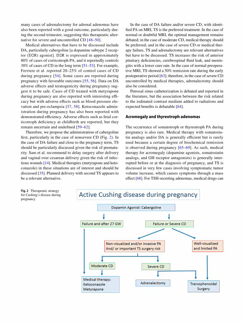
Therefore, we propose the administration of cabergoline first, particularly in the case of nonsevere CD (Fig. 2). In the case of DA failure and close to the pregnancy term, TS should be particularly discussed given the risk of prematu-rity. Sam et al. recommend to delay surgery after delivery and vaginal over cesarean delivery given the risk of infec-tious wounds [14]. Medical therapies (metyrapone and keto-conazole) in these situations are of interest and should be discussed [35]. Planned delivery with second TS appears to be a relevant alternative.
In the case of DA failure and/or severe CD, with identi-fied PA on MRI, TS is the preferred treatment. In the case of normal or doubtful MRI, the optimal management remains debated; in the case of moderate CD, medical therapy should be preferred; and in the case of severe CD or medical ther-apy failure, TS and adrenalectomy are relevant alternatives but have to be discussed. TS increases the risk of anterior pituitary deficiencies, cerebrospinal fluid leak, and menin-gitis with a lower cure rate. In the case of normal preopera-tive MRI, TS showed a 50% remission rate during the early postoperative period [63]; therefore, in the case of severe CD uncontrolled by medical therapies, adrenalectomy should also be considered.
Petrosal sinus catheterization is debated and reported in the literature, but the association between the risk related to the iodinated contrast medium added to radiations and expected benefits is debatable [64].
Acromegaly and thyreotroph adenomas
The occurrence of somatotroph or thyreotroph PA during pregnancy is also rare. Medical therapy with somatosta-tin analogs and/or DA is generally efficient but is rarely used because a certain degree of biochemical remission is observed during pregnancy [65–69]. As such, medical therapy for acromegaly (dopamine agonists, somatostatin analogs, and GH receptor antagonists) is generally inter-rupted before or at the diagnosis of pregnancy, and TS is discussed in very few cases involving symptomatic tumor volume increase, which causes symptoms through a mass effect [68]. For TSH-secreting adenomas, medical drugs can
Fig. 2 Therapeutic strategy for Cushing’s disease during pregnancy

194 Pituitary (2020) 23:189–199
1 3
antagonize thyroid hormone effect, and only one case of SSA-resistant thyreotroph PA with visual impairment requir-ing TS has been reported with good outcome. Therefore, sur-gical indications are mainly proposed in the case of medical therapy failure and severe visual impairment.
Nonsecreting adenomas
Surgical indications are considered in the case of severe vis-ual impairment and apoplexy. Apoplexy of nonfunctioning PA (NFPA) during pregnancy is rare, and macroprolactino-mas are essentially concerning. In the study by Enfer-Vat-taut, only 2/9 cases of NFPA presented symptomatic tumor volume increase (Thèse de Médecine, Toulouse, 2005). Volume increase was also observed in 1/7 case of NFPA diagnosed before conception and 3/5 cases diagnosed during pregnancy [35]. Bromocriptine was efficient in one case and surgery was required in the second case. Their surgical indi-cations and strategies are similar to those of prolactinomas during pregnancy (Fig. 1).
Pituitary apoplexy
Pituitary apoplexy during pregnancy remains an exceptional condition with < 50 cases currently described in the litera-ture and an estimated prevalence of 1/10,000 pregnancy at term [70]. Its mean gestational age of occurrence is 24 GW [28]. Notably, 11% occurred during the postpartum period. Macroprolactinomas are mainly concerning. The clinical symptoms of pituitary apoplexy are similar to those
of nonpregnant patients. Headaches are present in 95% of patients, visual disturbances in 59%, and nausea in 35% [70]. Pregnancy is considered a risk factor given intratu-moral vascular and hormonal changes, blood hypertension, and pregnancy stress. Surgical indications remain limited to vital necessity and severe visual disturbances. In the review published by Grand’maison et al. only 42% of cases of pitui-tary apoplexy required surgery during pregnancy. Hormone replacement was required in 61% of cases. Remarkably, dopamine agonists were administrated to 31% of patients. In this series, adverse effects on the mother and fetus seemed limited with only one case of fetal loss. Therefore, surgical tumor removal should not be systematically recommended when pituitary apoplexy occurs during pregnancy. Surgical indications in the case of apoplexy during pregnancy include deteriorating level of consciousness or a significant or pro-gressive neuro-ophthalmological deficit as described in the case illustrated in Fig. 3 [71].
Management of other intra or suprasellar tumors during pregnancy
Meningiomas
Meningiomas are the most frequent intracranial tumors in adults with a female-to-male ratio of 2/1. They express receptors of progesterone (70–90%) [72, 73], prolactin (50%–60%) [74, 75], and estrogen (8%–40%) [76, 77]. The hormone-dependent status of meningiomas, particularly in
Fig. 3 Macroprolactinoma apoplexy at 28 gestational week. The case of a 32-year-old pregnant woman (28 gestational weeks) who pre-sented with intermittent headaches and left visual loss for 2 weeks. Regarding the left eye, visual acuity was 6/10 with an altered visual field. Visual acuity and field were normal for the right side. Prol-actin was considered in the normal range for the pregnancy term (450 ng/ml). No diplopia was observed, and MRI revealed apoplexy
of a pituitary adenoma with intratumoral fluid–fluid level. The tumor was removed using a transsphenoidal approach. Histological results concluded the tumor to be of prolactinoma grade 1a according to the classification reported by Trouillas et al. Vaginal delivery occurred at 40 + 2 GW with excellent outcome, and breastfeeding was possible without dopamine agonist therapy. Visual findings returned to normal levels at postpartum 3 months

195Pituitary (2020) 23:189–199
1 3
the case of progestin-related meningiomas, is well demon-strated [78]. Meningiomas diagnosed during pregnancy are mainly located on the skull base [27, 79]. An increase in the tumor volume is usual during pregnancy, whereas a decrease is usual in postpartum [80, 81]. Several mechanisms may be involved: hormonal impregnation involving progesterone rather than prolactin or estrogen with tumoral turgescence; hemodynamic changes; higher growth factor secretion and tumor growth acceleration; increase in the volume of the normal pituitary gland (in suprasellar meningiomas) as described in the following clinical case (Fig. 4) [82, 83].
Meningiomas with nonsymptomatic growth do not require surgery during pregnancy; close follow-up is required, and radiologic assessment is indicated in postpar-tum. Surgery is considered depending on postpartum neu-rological and visual outcomes, but suprasellar meningiomas are an exception. Proximity with visual pathways explains that limited tumor growth rapidly leads to visual impair-ment. Kanaan et al. reported 18 patients with meningiomas
diagnosed during pregnancy [27], among which 12 patients presented visual impairment. All 7 operated patients had visual impairment. Laviv et al. compared the management of 104 patients with suprasellar meningiomas operated dur-ing (40%) or after (60%) pregnancy [84]. A similar rate of premature birth (37% and 39%, respectively) was observed; however, maternofetal morbimortality was higher in the group operated during pregnancy with two maternal mor-tality and two fetal mortality. Despite OR being 14.7, no significant differences were observed in terms of mater-nofetal mortality; we hypothesize that most cases of severe meningiomas were operated during pregnancy. After 27 GW, the authors concluded that a delayed surgery during the post-partum period was preferable when possible [80]. If not, two alternatives could be considered: a planned Caesarian deliv-ery followed by surgery for the meningioma or surgery fol-lowed by vaginal delivery. No data are currently available to determine the optimal alternative. When possible, delivery should be delayed to 32 GW. We suggest the consideration
Fig. 4 Suprasellar meningi-oma during pregnancy. The case of a 31-year-old pregnant woman at 32 GW presented for left visual loss (visual acuity: 6/10) with altered visual field. Cesarean delivery was planned before tumor removal, but the patient consulted 3 days later for worsening of visual disturbances. At this point, left visual acuity was at 3/10; therefore, transcranial surgical removal was performed. Com-plete recovery was observed at postoperative 4 days. Preopera-tive MRI displayed a typical suprasellar meningioma with optic pathway compression with a considerable increase in the pituitary volume compared with that observed on postoperative 3-month MRI, suggesting the involvement of the increase in the pituitary gland volume

196 Pituitary (2020) 23:189–199
1 3
of surgery first between 27 and 32 GW and planned delivery after 32 GW. The surgical strategy is summarized in Fig. 5.
Surgical considerations include epilepsy risk manage-ment. Epilepsy prevention should not be systematical. Blood loss and carotid lesion must be avoided. Prophylactic corti-costeroids are indicated for fetal lung maturation, but pro-longed postoperative corticosteroid administration should be avoided to limit fetal toxicity. Histologically, meningiomas excised during pregnancy appear mostly benign WHO grade I tumors [79, 82, 85].
Considering the possibility and the management of a pregnancy in a patient bearing a meningioma, literature data are poor. There is no formal contraindication, but concerns remain, particularly in case of proximity with the optic path-way. Case-by-case management should be proposed.
Craniopharyngiomas and pituitary cysts
The diagnosis of craniopharyngiomas or pituitary cysts dur-ing pregnancy remains exceptional. Eight cases of crani-opharyngiomas diagnosed during pregnancy are currently published [86]. Among these, 6 cases had visual disturbance at diagnosis. The relation of volume increase with pregnancy is highly uncertain. No progesterone, estrogen, or prolactin receptors were detected. In craniopharyngiomas, as in differ-ent pituitary cysts, surgery is required in the case of severe visual deficit (Fig. 5). Good outcome has been reported for surgical cases in the literature.
Conclusion
Nonobstetric surgery is reasonable during pregnancy if its benefits outweigh the risks for the mother and fetus. The risks of congenital malformations during the first
trimester and those of premature birth during the third trimester lead to a preference for the second trimester to consider surgery. Although limited, surgical risks remain difficult to assess, and surgery should be proposed only when safer medical alternatives are lacking.
In macroprolactinomas, nonsecreting as well as GH- and TSH-secreting PAs, TS should be recommended only in the case of failure of medical therapies with severe visual impairment. Surgery is also required in suprasellar meningiomas, craniopharyngiomas, and pituitary cysts in the case of severe visual deficit. Severe visual impairment could be defined by severe VF deficit, VA impairment, and abnormal OCT findings for optic fiber thickness.
Uncontrolled CD during pregnancy increases mater-nofetal morbimortality. The preferred treatment is TS, but cabergoline appears as a safer alternative in the first-line treatment. Metyrapone and ketoconazole could be recom-mended in specific situations as adrenalectomy in the case of severe CD and failure of medical therapy and TS.
Acknowledgements Club Français de l’Hypophyse.
Funding No Funding.
Compliance with ethical standards
Conflict of interest The authors declare that they have no conflict of interest.
Ethical approval All procedures performed in studies involving human participants were in accordance with the 1964 Helsinki declaration and its later amendments.
Informed consent Informed consent was obtained from all participants included in the study.
Fig. 5 Therapeutic strategy for suprasellar meningiomas, pituitary cysts, and craniophar-yngiomas

197Pituitary (2020) 23:189–199
1 3
References
1. Witek P, Zielinski G, Maksymowicz M, Zgliczynski W (2012) Transsphenoidal surgery for a life-threatening prolactinoma apo-plexy during pregnancy. Neuro Endocrinol Lett 33(5):483–488
2. Lee J, Kim SW, Kim DW, Shin JY, Choi M, Oh MC, Kim SM, Kim EH, Kim SH, Byeon SH (2016) Predictive model for recov-ery of visual field after surgery of pituitary adenoma. J Neuroon-col 130(1):155–164. https ://doi.org/10.1007/s1106 0-016-2227-5
3. Muskens IS, Zamanipoor Najafabadi AH, Briceno V, Lamba N, Senders JT, van Furth WR, Verstegen MJT, Smith TRS, Mekary RA, Eenhorst CAE, Broekman MLD (2017) Visual outcomes after endoscopic endonasal pituitary adenoma resection: a sys-tematic review and meta-analysis. Pituitary 20(5):539–552. https ://doi.org/10.1007/s1110 2-017-0815-9
4. Gnanalingham KK, Bhattacharjee S, Pennington R, Ng J, Men-doza N (2005) The time course of visual field recovery following transphenoidal surgery for pituitary adenomas: predictive factors for a good outcome. J Neurol Neurosurg Psychiatry 76(3):415–419. https ://doi.org/10.1136/jnnp.2004.03557 6
5. Barzaghi LR, Medone M, Losa M, Bianchi S, Giovanelli M, Mor-tini P (2012) Prognostic factors of visual field improvement after trans-sphenoidal approach for pituitary macroadenomas: review of the literature and analysis by quantitative method. Neurosurg Rev 35(3):369–378. https ://doi.org/10.1007/s1014 3-011-0365-y (discussion 378–369)
6. Jacob M, Raverot G, Jouanneau E, Borson-Chazot F, Perrin G, Rabilloud M, Tilikete C, Bernard M, Vighetto A (2009) Predicting visual outcome after treatment of pituitary adenomas with optical coherence tomography. Am J Ophthalmol 147(1):64–70. https ://doi.org/10.1016/j.ajo.2008.07.016
7. Danesh-Meyer HV, Wong A, Papchenko T, Matheos K, Stylli S, Nichols A, Frampton C, Daniell M, Savino PJ, Kaye AH (2015) Optical coherence tomography predicts visual outcome for pituitary tumors. J Clin Neurosci 22(7):1098–1104. https ://doi.org/10.1016/j.jocn.2015.02.001
8. Garcia T, Sanchez S, Litre CF, Radoi C, Delemer B, Rousseaux P, Ducasse A, Arndt C (2014) Prognostic value of retinal nerve fiber layer thickness for postoperative peripheral visual field recovery in optic chiasm compression. J Neurosurg 121(1):165–169. https ://doi.org/10.3171/2014.2.JNS13 1767
9. Blanch RJ, Micieli JA, Oyesiku NM, Newman NJ, Biousse V (2018) Optical coherence tomography retinal ganglion cell complex analysis for the detection of early chiasmal compres-sion. Pituitary 21(5):515–523. https ://doi.org/10.1007/s1110 2-018-0906-2
10. Tieger MG, Hedges TR 3rd, Ho J, Erlich-Malona NK, Vuong LN, Athappilly GK, Mendoza-Santiesteban CE (2017) Ganglion cell complex loss in chiasmal compression by brain tumors. J Neu-roophthalmol 37(1):7–12. https ://doi.org/10.1097/WNO.00000 00000 00042 4
11. Brue T, Amodru V, Castinetti F (2018) Management of endocrine disease: management of Cushing’s syndrome during pregnancy: solved and unsolved questions. Eur J Endocrinol 178(6):R259–R266. https ://doi.org/10.1530/EJE-17-1058
12. Cohen-Gadol AA, Friedman JA, Friedman JD, Tubbs RS, Munis JR, Meyer FB (2009) Neurosurgical management of intracranial lesions in the pregnant patient: a 36-year institutional experience and review of the literature. J Neurosurg 111(6):1150–1157. https ://doi.org/10.3171/2009.3.JNS08 1160
13. Carvalho CS, Resende F, Centeno MJ, Ribeiro I, Moreira J (2013) Anesthetic approach of pregnant woman with cerebral arterio-venous malformation and subarachnoid hemorrhage during preg-nancy: case report. Braz J Anesthesiol 63(2):223–226. https ://doi.org/10.1016/j.bjane .2012.05.004
14. Sam S, Molitch ME (2003) Timing and special concerns regard-ing endocrine surgery during pregnancy. Endocrinol Metab Clin North Am 32(2):337–354
15. Xia Y, Ma X, Griffiths BB, Luo Y (2018) Neurosurgical anesthe-sia for a pregnant woman with macroprolactinoma: a case report. Medicine (Baltimore) 97(37):e12360. https ://doi.org/10.1097/MD.00000 00000 01236 0
16. Wagner M, Ryu YK, Smith SC, Patel P, Mintz CD (2014) Review: effects of anesthetics on brain circuit formation. J Neurosurg Anesthesiol 26(4):358–362. https ://doi.org/10.1097/ANA.00000 00000 00011 8
17. Vutskits L, Xie Z (2016) Lasting impact of general anaesthe-sia on the brain: mechanisms and relevance. Nat Rev Neurosci 17(11):705–717. https ://doi.org/10.1038/nrn.2016.128
18. Graham MR (2017) Clinical update regarding general anesthesia-associated neurotoxicity in infants and children. Curr Opin Anaes-thesiol 30(6):682–687. https ://doi.org/10.1097/ACO.00000 00000 00052 0
19. Perna RB, Loughan AR, Le JA, Hertza J (2015) Prenatal and perinatal anesthesia and the long-term cognitive sequelae: a review. Appl Neuropsychol Child 4(1):65–71. https ://doi.org/10.1080/21622 965.2013.77927 5
20. Ancel PY, Goffinet F, Kuhn P, Langer B, Matis J, Hernandorena X, Chabanier P, Joly-Pedespan L, Lecomte B, Vendittelli F, Dreyfus M, Guillois B, Burguet A, Sagot P, Sizun J, Beuchee A, Rouget F, Favreau A, Saliba E, Bednarek N, Morville P, Thiriez G, Marpeau L, Marret S, Kayem G, Durrmeyer X, Granier M, Baud O, Jarreau PH, Mitanchez D, Boileau P, Boulot P, Cambonie G, Daude H, Bedu A, Mons F, Fresson J, Vieux R, Alberge C, Arnaud C, Vayssiere C, Truffert P, Pierrat V, Subtil D, D’Ercole C, Gire C, Simeoni U, Bongain A, Sentilhes L, Roze JC, Gondry J, Leke A, Deiber M, Claris O, Picaud JC, Ego A, Debillon T, Poulichet A, Coline E, Favre A, Flechelles O, Samperiz S, Ramful D, Branger B, Benhammou V, Foix-L’Helias L, Marchand-Martin L, Kaminski M (2015) Survival and morbidity of preterm children born at 22 through 34 weeks’ gestation in France in 2011: results of the EPIPAGE-2 cohort study. JAMA Pediatr 169(3):230–238. https ://doi.org/10.1001/jamap ediat rics.2014.3351
21. Committee on Obstetric Practice (2017) No. 723: guidelines for diagnostic imaging during pregnancy and lactation. Obstet Gynecol 130(4):e210–e216. https ://doi.org/10.1097/AOG.00000 00000 00235 5
22. Horrigan TJ, Villarreal R, Weinstein L (1999) Are obstetrical per-sonnel required for intraoperative fetal monitoring during nonob-stetric surgery? J Perinatol 19(2):124–126
23. Kendrick JM, Neiger R (2000) Intraoperative fetal monitoring during nonobstetric surgery. J Perinatol 20(4):276–277
24. Reitman E, Flood P (2011) Anaesthetic considerations for non-obstetric surgery during pregnancy. Br J Anaesth 107(Suppl 1):i72–78. https ://doi.org/10.1093/bja/aer34 3
25. ACOG committee opinion (2003) Nonobstetric surgery in preg-nancy. Int J Gynaecol Obstet 83(1):135
26. Tuncali B, Aksun M, Katircioglu K, Akkol I, Savaci S (2006) Intraoperative fetal heart rate monitoring during emergency neurosurgery in a parturient. J Anesth 20(1):40–43. https ://doi.org/10.1007/s0054 0-005-0359-4
27. Kanaan I, Jallu A, Kanaan H (2003) Management strategy for meningioma in pregnancy: a clinical study. Skull Base 13(4):197–203. https ://doi.org/10.1055/s-2004-81769 5
28. Tuchmann-Duplessis H (1983) The teratogenic risk. Am J Ind Med 4(1–2):245–258
29. Mazze RI, Kallen B (1989) Reproductive outcome after anesthesia and operation during pregnancy: a registry study of 5405 cases. Am J Obstet Gynecol 161(5):1178–1185
30. Cohen-Kerem R, Railton C, Oren D, Lishner M, Koren G (2005) Pregnancy outcome following non-obstetric

198 Pituitary (2020) 23:189–199
1 3
surgical intervention. Am J Surg 190(3):467–473. https ://doi.org/10.1016/j.amjsu rg.2005.03.033
31. Lynch JC, Gouvea F, Emmerich JC, Kokinovrachos G, Pereira C, Welling L, Kislanov S (2011) Management strategy for brain tumour diagnosed during pregnancy. Br J Neurosurg 25(2):225–230. https ://doi.org/10.3109/02688 697.2010.50884 6
32. Maiter D (2016) Prolactinoma and pregnancy: from the wish of conception to lactation. Ann Endocrinol (Paris) 77(2):128–134. https ://doi.org/10.1016/j.ando.2016.04.001
33. Molitch ME (1999) Management of prolactinomas during preg-nancy. J Reprod Med 44(12 Suppl):1121–1126
34. Melmed S, Casanueva FF, Hoffman AR, Kleinberg DL, Montori VM, Schlechte JA, Wass JA (2011) Diagnosis and treatment of hyperprolactinemia: an Endocrine Society clinical practice guideline. J Clin Endocrinol Metab 96(2):273–288. https ://doi.org/10.1210/jc.2010-1692
35. Lambert K, Rees K, Seed PT, Dhanjal MK, Knight M, McCance DR, Williamson C (2017) Macroprolactinomas and nonfunc-tioning pituitary adenomas and pregnancy outcomes. Obstet Gynecol 129(1):185–194. https ://doi.org/10.1097/AOG.00000 00000 00174 7
36. Molitch ME (2015) Endocrinology in pregnancy: management of the pregnant patient with a prolactinoma. Eur J Endocrinol 172(5):R205–213. https ://doi.org/10.1530/EJE-14-0848
37. Lindsay JR, Jonklaas J, Oldfield EH, Nieman LK (2005) Cush-ing’s syndrome during pregnancy: personal experience and review of the literature. J Clin Endocrinol Metab 90(5):3077–3083. https ://doi.org/10.1210/jc.2004-2361
38. Buescher MA, McClamrock HD, Adashi EY (1992) Cushing syndrome in pregnancy. Obstet Gynecol 79(1):130–137
39. Bronstein MD, Paraiba DB, Jallad RS (2011) Management of pituitary tumors in pregnancy. Nat Rev Endocrinol 7(5):301–310. https ://doi.org/10.1038/nrend o.2011.38
40. Caimari F, Corcoy R, Webb SM (2018) Cushing’s disease: major difficulties in diagnosis and management during pregnancy. Minerva Endocrinol 43(4):435–445. https ://doi.org/10.23736 /S0391 -1977.18.02803 -1
41. Caimari F, Valassi E, Garbayo P, Steffensen C, Santos A, Cor-coy R, Webb SM (2017) Cushing’s syndrome and pregnancy outcomes: a systematic review of published cases. Endocrine 55(2):555–563. https ://doi.org/10.1007/s1202 0-016-1117-0
42. Abbassy M, Kshettry VR, Hamrahian AH, Johnston PC, Dobri GA, Avitsian R, Woodard TD, Recinos PF (2015) Surgical man-agement of recurrent Cushing’s disease in pregnancy: a case report. Surg Neurol Int 6(Suppl 25):S640–645. https ://doi.org/10.4103/2152-7806.17047 2
43. Casson IF, Davis JC, Jeffreys RV, Silas JH, Williams J, Belchetz PE (1987) Successful management of Cushing’s disease dur-ing pregnancy by transsphenoidal adenectomy. Clin Endocrinol (Oxf) 27(4):423–428
44. Mellor A, Harvey RD, Pobereskin LH, Sneyd JR (1998) Cush-ing’s disease treated by trans-sphenoidal selective adenomec-tomy in mid-pregnancy. Br J Anaesth 80(6):850–852
45. Ross RJ, Chew SL, Perry L, Erskine K, Medbak S, Afshar F (1995) Diagnosis and selective cure of Cushing’s disease dur-ing pregnancy by transsphenoidal surgery. Eur J Endocrinol 132(6):722–726
46. Verdugo C, Alegria J, Grant C, Briano E, Gonzalez MI, Meza H, Amthauer N, Paiva O, Vigueras R, Madariaga J (2004) Cush-ing’s disease treatment with transsphenoidal surgery during pregnancy. Rev Med Chil 132(1):75–80
47. Jolly K, Darr A, Arlt W, Ahmed S, Karavitaki N (2019) Sur-gery for Cushing’s disease in pregnancy: our experience and a literature review. Ann R Coll Surg Engl 101(1):e26–e31. https ://doi.org/10.1308/rcsan n.2018.0175
48. Wang W, Yuan F, Xu D (2015) Cushing’s syndrome during preg-nancy caused by adrenal cortical adenoma: a case report and lit-erature review. Front Med 9(3):380–383. https ://doi.org/10.1007/s1168 4-015-0407-x
49. Sammour RN, Saiegh L, Matter I, Gonen R, Shechner C, Cohen M, Ohel G, Dickstein G (2012) Adrenalectomy for adrenocorti-cal adenoma causing Cushing’s syndrome in pregnancy: a case report and review of literature. Eur J Obstet Gynecol Reprod Biol 165(1):1–7. https ://doi.org/10.1016/j.ejogr b.2012.05.030
50. Martinez Garcia R, Martinez Perez A, del Pozo CD, Sospedra Ferrer R (2016) Cushing’s syndrome in pregnancy. Laparoscopic adrenalectomy during pregnancy: the mainstay treatment. J Endocrinol Invest 39(3):273–276. https ://doi.org/10.1007/s4061 8-015-0345-0
51. Pivonello R, De Martino MC, Cappabianca P, De Leo M, Fag-giano A, Lombardi G, Hofland LJ, Lamberts SW, Colao A (2009) The medical treatment of Cushing’s disease: effective-ness of chronic treatment with the dopamine agonist cabergoline in patients unsuccessfully treated by surgery. J Clin Endocrinol Metab 94(1):223–230. https ://doi.org/10.1210/jc.2008-1533
52. Godbout A, Manavela M, Danilowicz K, Beauregard H, Bruno OD, Lacroix A (2010) Cabergoline monotherapy in the long-term treatment of Cushing’s disease. Eur J Endocrinol 163(5):709–716. https ://doi.org/10.1530/EJE-10-0382
53. Sek KS, Deepak DS, Lee KO (2017) Use of cabergoline for the management of persistent Cushing’s disease in pregnancy. BMJ Case Rep. https ://doi.org/10.1136/bcr-2016-21785 5
54. Ferriere A, Cortet C, Chanson P, Delemer B, Caron P, Chabre O, Reznik Y, Bertherat J, Rohmer V, Briet C, Raingeard I, Castinetti F, Beckers A, Vroonen L, Maiter D, Cephise-Velayoudom FL, Nunes ML, Haissaguerre M, Tabarin A (2017) Cabergoline for Cushing’s disease: a large retrospective multicenter study. Eur J Endocrinol 176(3):305–314. https ://doi.org/10.1530/EJE-16-0662
55. Nakhleh A, Saiegh L, Reut M, Ahmad MS, Pearl IW, Shechner C (2016) Cabergoline treatment for recurrent Cushing’s disease during pregnancy. Hormones (Athens) 15(3):453–458. https ://doi.org/10.14310 /horm.2002.1685
56. Woo I, Ehsanipoor RM (2013) Cabergoline therapy for Cushing disease throughout pregnancy. Obstet Gynecol 122(2 Pt 2):485–487. https ://doi.org/10.1097/AOG.0b013 e3182 9e398 a
57. Lim WH, Torpy DJ, Jeffries WS (2013) The medical manage-ment of Cushing’s syndrome during pregnancy. Eur J Obstet Gynecol Reprod Biol 168(1):1–6. https ://doi.org/10.1016/j.ejogr b.2012.12.015
58. Close CF, Mann MC, Watts JF, Taylor KG (1993) ACTH-inde-pendent Cushing’s syndrome in pregnancy with spontaneous resolution after delivery: control of the hypercortisolism with metyrapone. Clin Endocrinol (Oxf) 39(3):375–379
59. Prebtani AP, Donat D, Ezzat S (2000) Worrisome striae in preg-nancy. Lancet 355(9216):1692
60. Amado JA, Pesquera C, Gonzalez EM, Otero M, Freijanes J, Alva-rez A (1990) Successful treatment with ketoconazole of Cushing’s syndrome in pregnancy. Postgrad Med J 66(773):221–223
61. Berwaerts J, Verhelst J, Mahler C, Abs R (1999) Cushing’s syn-drome in pregnancy treated by ketoconazole: case report and review of the literature. Gynecol Endocrinol 13(3):175–182. https ://doi.org/10.3109/09513 59990 91675 52
62. Zieleniewski W, Michalak R (2017) A successful case of preg-nancy in a woman with ACTH-independent Cushing’s syndrome treated with ketoconazole and metyrapone. Gynecol Endocrinol 33(5):349–352. https ://doi.org/10.1080/09513 590.2017.12900 70
63. Yamada S, Fukuhara N, Nishioka H, Takeshita A, Inoshita N, Ito J, Takeuchi Y (2012) Surgical management and outcomes in patients with Cushing disease with negative pituitary magnetic resonance imaging. World Neurosurg 77(3–4):525–532. https ://doi.org/10.1016/j.wneu.2011.06.033

199Pituitary (2020) 23:189–199
1 3
64. Pinette MG, Pan YQ, Oppenheim D, Pinette SG, Blackstone J (1994) Bilateral inferior petrosal sinus corticotropin sampling with corticotropin-releasing hormone stimulation in a pregnant patient with Cushing’s syndrome. Am J Obstet Gynecol 171(2):563–564
65. Muhammad A, Neggers SJ, van der Lely AJ (2017) Pregnancy and acromegaly. Pituitary 20(1):179–184. https ://doi.org/10.1007/s1110 2-016-0740-3
66. Hannon AM, Frizelle I, Kaar G, Hunter SJ, Sherlock M, Thomp-son CJ, O’Halloran DJ (2019) Octreotide use for rescue of vision in a pregnant patient with acromegaly. Endocrinol Diabetes Metab Case Rep. https ://doi.org/10.1530/edm-19-0019
67. Hannon AM, O’Shea T, Thompson CA, Hannon MJ, Dineen R, Khattak A, Gibney J, O’Halloran DJ, Hunter S, Thompson CJ, Sherlock M (2019) Pregnancy in acromegaly is safe and is asso-ciated with improvements in IGF-1 concentrations. Eur J Endo-crinol 180(4):K21–K29. https ://doi.org/10.1530/EJE-18-0688
68. Chanson P, Vialon M, Caron P (2019) An update on clini-cal care for pregnant women with acromegaly. Expert Rev Endocrinol Metab 14(2):85–96. https ://doi.org/10.1080/17446 651.2019.15719 09
69. Perdomo CM, Arabe JA, Idoate MA, Galofre JC (2017) Manage-ment of a pregnant woman with thyrotropinoma: a case report and review of the literature. Gynecol Endocrinol 33(3):188–192. https ://doi.org/10.1080/09513 590.2016.12601 10
70. Grand’Maison S, Weber F, Bedard MJ, Mahone M, Godbout A (2015) Pituitary apoplexy in pregnancy: a case series and literature review. Obstet Med 8(4):177–183. https ://doi.org/10.1177/17534 95X15 59891 7
71. Rajasekaran S, Vanderpump M, Baldeweg S, Drake W, Reddy N, Lanyon M, Markey A, Plant G, Powell M, Sinha S, Wass J (2011) UK guidelines for the management of pituitary apo-plexy. Clin Endocrinol (Oxf) 74(1):9–20. https ://doi.org/10.1111/j.1365-2265.2010.03913 .x
72. Baxter DS, Orrego A, Rosenfeld JV, Mathiesen T (2014) An audit of immunohistochemical marker patterns in meningi-oma. J Clin Neurosci 21(3):421–426. https ://doi.org/10.1016/j.jocn.2013.06.008
73. Bozzetti C, Camisa R, Nizzoli R, Manotti L, Guazzi A, Naldi N, Mazza S, Nizzoli V, Cocconi G (1995) Estrogen and progesterone receptors in human meningiomas: biochemical and immunocyto-chemical evaluation. Surg Neurol 43(3):230–233 (discussion 234)
74. Muccioli G, Ghe C, Faccani G, Lanotte M, Forni M, Ciccarelli E (1997) Prolactin receptors in human meningiomas: characteriza-tion and biological role. J Endocrinol 153(3):365–371
75. Carroll RS, Schrell UM, Zhang J, Dashner K, Nomikos P, Fahl-busch R, Black PM (1996) Dopamine D1, dopamine D2, and prolactin receptor messenger ribonucleic acid expression by the polymerase chain reaction in human meningiomas. Neurosurgery 38(2):367–375
76. Carroll RS, Zhang J, Black PM (1999) Expression of estrogen receptors alpha and beta in human meningiomas. J Neurooncol 42(2):109–116
77. Hsu DW, Efird JT, Hedley-Whyte ET (1997) Progesterone and estrogen receptors in meningiomas: prognostic consid-erations. J Neurosurg 86(1):113–120. https ://doi.org/10.3171/jns.1997.86.1.0113
78. Bernat AL, Oyama K, Hamdi S, Mandonnet E, Vexiau D, Pocard M, George B, Froelich S (2015) Growth stabilization and regres-sion of meningiomas after discontinuation of cyproterone acetate: a case series of 12 patients. Acta Neurochir (Wien) 157(10):1741–1746. https ://doi.org/10.1007/s0070 1-015-2532-3
79. Priddy BH, Otto BA, Carrau RL, Prevedello DM (2018) Man-agement of skull base tumors in the obstetric population: a case series. World Neurosurg 113:e373–e382. https ://doi.org/10.1016/j.wneu.2018.02.038
80. Chacko JG, Miller JL, Angtuaco EJ (2010) Spontaneous post-partum resolution of vision loss caused by a progesterone recep-tor-positive tuberculum sellae meningioma. J Neuroophthalmol 30(2):132–134. https ://doi.org/10.1097/WNO.0b013 e3181 da9d5 9
81. Smith JS, Quinones-Hinojosa A, Harmon-Smith M, Bollen AW, McDermott MW (2005) Sex steroid and growth factor profile of a meningioma associated with pregnancy. Can J Neurol Sci 32(1):122–127
82. Lusis EA, Scheithauer BW, Yachnis AT, Fischer BR, Chicoine MR, Paulus W, Perry A (2012) Meningiomas in pregnancy: a clinicopathologic study of 17 cases. Neurosurgery 71(5):951–961. https ://doi.org/10.1227/NEU.0b013 e3182 6adf6 5
83. Ebner FH, Bornemann A, Wilhelm H, Ernemann U, Honegger J (2008) Tuberculum sellae meningioma symptomatic during preg-nancy: pathophysiological considerations. Acta Neurochir (Wien) 150(2):189–193. https ://doi.org/10.1007/s0070 1-007-1417-5 (dis-cussion 193)
84. Laviv Y, Bayoumi A, Mahadevan A, Young B, Boone M, Kasper EM (2018) Meningiomas in pregnancy: timing of surgery and clinical outcomes as observed in 104 cases and establishment of a best management strategy. Acta Neurochir (Wien) 160(8):1521–1529. https ://doi.org/10.1007/s0070 1-017-3146-8
85. Moscovici S, Fraifeld S, Cohen JE, Dotan S, Elchalal U, Shoshan Y, Spektor S (2014) Parasellar meningiomas in pregnancy: surgi-cal results and visual outcomes. World Neurosurg 82(3–4):e503–512. https ://doi.org/10.1016/j.wneu.2013.06.019
86. Zoia C, Cattalani A, Turpini E, Custodi VM, Benazzo M, Pagella F, Carena P, Lovati E, Lucotti P, Gaetani P (2014) Haemorrhagic presentation of a craniopharyngioma in a preg-nant woman. Case Rep Neurol Med 2014:435208. https ://doi.org/10.1155/2014/43520 8
Publisher’s Note Springer Nature remains neutral with regard to jurisdictional claims in published maps and institutional affiliations.