surgical microanatomy of the mu¨ller muscle-conjunctival … · 2017-08-25 · cally, we examined...
TRANSCRIPT

ORIGINAL ARTICLE
Surgical Microanatomy of the Muller Muscle-ConjunctivalResection Ptosis Procedure
Marcus M. Marcet, M.D.*†, Pete Setabutr, M.D.‡, Bradley N. Lemke, M.D.§, Megan E. Collins, M.D.*,James C. Fleming, M.D.�, Ralph E. Wesley, M.D.¶, Jayant M. Pinto, M.D.*, and Allen M. Putterman, M.D.‡
Departments of *Surgery and †Pathology, University of Chicago; ‡Department of Ophthalmology, University ofIllinois, Chicago, Illinois; §Department of Ophthalmology, University of Wisconsin, Madison, Wisconsin; �Hamilton
Eye Institute, University of Tennessee, Memphis; and ¶Vanderbilt University Eye Institute, Nashville,Tennessee, U.S.A.
Purpose: To assess for alterations in the microscopic anat-omy that occur as a result of the Muller muscle-conjunctivalresection (MMCR) ptosis procedure and to better understandthe mechanisms by which MMCR elevates the eyelid.
Methods: Sixteen orbits from 8 fresh frozen Caucasiancadaver heads, ranging from 38 to 100 years of age were used.For each head, MMCR was performed on one side. The contralat-eral, unoperated orbit served as an anatomic control. Each exen-terated orbital contents and excised MMCR specimen was evalu-ated. The histopathology of the eyelids and MMCR specimenswere studied microscopically by staining with hematoxylin-eosin,elastic, and Verhoeff-Masson trichrome.
Results: Muller muscle and conjunctiva were present in all 8of the excised MMCR specimens. Elastic fibers consistent withMuller muscle tendon or among the smooth muscle fibers wereseen within all excised MMCR specimens. The levator apo-neurosis was intact in 8 of 8 operated eyelids; however, theaponeurosis was plicated in all. The accessory lacrimal glandtissues were intact in all of the operated and unoperated eyelids.
Conclusions: MMCR works by shortening the posteriorlamella, which results in advancement of the levator palpebraesuperioris muscle and plication of the levator aponeurosis.Plication of the levator aponeurosis likely contributes to theincreased volumetric effect seen clinically after MMCR. Phen-ylephrine testing can help in fine-tuning the amount of resec-tion, but given the mechanism of action of MMCR, adequatelevator muscle function remains a critical factor in the successof the surgery. Moreover, MMCR preserves accessory lacrimalgland tissues.
(Ophthal Plast Reconstr Surg 2010;26:360–364)
F irst described by Putterman and Urist1 in 1975, the Mullermuscle-conjunctival resection (MMCR) ptosis procedure
represents a posterior approach to the correction of ptosis. The
exact mechanism by which the surgery lifts the eyelid has beenunclear.2–4 It has been postulated to be secondary to a numberof factors, including resection and advancement of Mullermuscle (MM) and advancement of the levator aponeurosis.2,5
This study uniquely uses a cadaveric model to charac-terize the microanatomic alterations produced with the MMCRprocedure. By visualizing the altered microscopic anatomy, wehoped to gain insight into the mechanism of action of thesurgery. In seeking a visually integrated depiction of collagen,muscle, and elastic fibers for the histopathologic evaluation, acombined Verhoeff-Masson trichrome stain was included aspart of the study.
METHODSSixteen orbits from 8 fresh frozen Caucasian cadaver heads (2
male and 6 female) ranging in age from 38 to 100 years (average age,78 years) were used for the study. In compliance with the University ofChicago policy on research for decedents, the study was registered withthe institutional review board and conformed to the principles of theDeclaration of Helsinki. An MMCR procedure was performed on oneupper eyelid of each cadaver head (4 right upper eyelids and 4 leftupper eyelids). The amount of MMCR ranged from 8 to 10 mm(average resection, 9.25 mm). The contralateral, nonoperated side wasused for anatomic comparison. The procedure was performed as pre-viously described,1,2 including the use of the specially designed clamp(Bausch & Lomb Storz Company, Manchester, MO, U.S.A.).6 A 6-0polyglactin suture was used rather than plain gut to facilitate a durableclosure throughout the formalin fixation period. In addition, a secondrunning suture, consisting of 10-0 nylon, paralleling the course of the firstsuture was placed as a histopathologic marker. Care was taken to avoidfurther plication of tissues with the placement of the second suture.
The excised segment of posterior eyelid tissue was oriented flaton a piece of filter paper and placed in 10% neutral buffered formalin.After the MMCR, both orbits were immediately exenterated and fixedwith 10% neutral buffered formalin for 3 weeks. After fixation, thelower eyelid tissue and orbital fat were removed to facilitate histologicprocessing and sectioning. The orbits were then sectioned parasagittallythrough the central eyelid along the axis of the orbit. The eyelidspecimens were examined grossly under surgical loupe magnification(3.5X-EF, Designs for Vision, Ronkonkoma, NY, U.S.A.).
On the 8 eyelids status post-MMCR, the running 6-0 polygla-ctin suture was removed, and the 10-0 nylon marking suture was left inplace in one half of the parasagittal central eyelid sections. The 6-0polyglactin suture was removed, because solid foreign material such assutures cause streaks in the paraffin-embedded tissue block during theprocess of sectioning.7
For the other half of the parasagittal central eyelid section of theoperated eyelids, both the 10-0 and 6-0 sutures were removed to allow
Accepted for publication November 11, 2009.This manuscript was accepted by the ASOPRS Thesis Committee as
submitted May 1, 2009 and subsequently presented as a thesis at theASOPRS 40th Annual Fall Scientific Symposium, San Francisco, CA,October 22, 2009.
The authors have no financial or proprietary conflicts of interest to reportrelated to this study.
Address correspondence and reprint requests to Marcus M. Marcet, M.D.,University of Chicago, 5841 S. Maryland Ave, MC 2114, Chicago, IL60637, U.S.A. E-mail: [email protected]
DOI: 10.1097/IOP.0b013e3181cb79a2
Ophthal Plast Reconstr Surg, Vol. 26, No. 5, 2010360

for examination of the uninvolved bed of tissue superior to the site ofresection. The eyelid specimens were again studied grossly as de-scribed (Fig. 1A, B). In 4 of the orbits, the newly visible surface (afterrelease of the sutures) superior to the segment of excised tissue was inkedwith black histologic tissue paint (#3408-3, Davidson Marking System,Bradley Products, Bloomington, MN, U.S.A.) to further enhance micro-scopic study of the surgical site (Fig. 1C, D). The excised tissue from theMMCR surgery was cut in a bread loaf fashion.8
The 20 specimens of central eyelid tissue (8 unoperated eyelids,8 eyelids status post-MMCR, and 4 operated eyelids with completesuture release) and 8 specimens of excised MMCR tissue were pro-cessed in paraffin and cut in serial 6-�m sections. All 28 specimenswere stained with hematoxylin-eosin, elastic, and Verhoeff-Masson(Masson trichrome counterstained with Verhoeff-Van Gieson elasticstain).9 Masson trichrome stains muscle red and collagen blue, whereasVerhoeff stains elastic fibers black. The microanatomic eyelid changessecondary to the MMCR surgery were studied histologically. Specifi-cally, we examined the anatomy of the MM and the levator palpebraesuperioris muscle (LPS), the course and integrity of the levator apo-neurosis, the presence of accessory lacrimal tissue, and the appearanceof the tarsal plate.
RESULTS
Gross Examination. An intact levator aponeurosis was present in alloperated eyelids. In addition, plication of the levator aponeurosis wasseen in the operated eyelids (Fig. 1A–D).
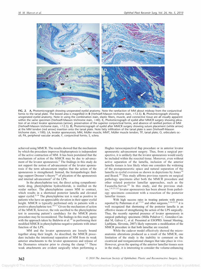
Histopathologic Examination. In the unoperated eyelids, verticalbifurcation of the striated muscle fibers of the LPS was seen near thelevel of Whitnall superior transverse ligament. Rarefaction of the MMwas observed (Fig. 2A, B). MM and conjunctiva were present in all theexcised MMCR tissue pathology specimens. Elastic fibers consistent
with MM tendon or among the MM fibers were seen in all excisedMMCR specimens.
The levator aponeurosis was intact in all operated eyelids (Fig.2C). Levator aponeurosis and LPS were absent in MMCR pathologyspecimens. Minimal or no residual MM tendon was present on theoperated eyelids. Suture closure occurred at the insertion of MM tendonon the tarsal plate (Fig. 2D). Plication of the levator aponeurosis wasseen in the operated eyelids (Fig. 2C). Accessory lacrimal gland tissue ofthe glands of Wolfring and Krause were observed, respectively, in thetarsus and superior fornix of the operated and unoperated eyelids. Thesuperior conjunctival fornix was intact in the operated and unoperatedeyelids. The findings seen on Verhoeff-Masson stain were confirmed bythe hematoxylin-eosin and elastic stains.
DISCUSSIONHistorically, the levator aponeurosis has been considered
the main transmitter of the retracting force the LPS exerts onthe eyelid. Although early works, such as Berke and Wads-worth,10 describe the role of MM in providing the primaryupward pull on the tarsal plate, conceptually ptosis repair forthe last half century has been shaped by an age of aponeuroticawareness.11 Within this setting, it is not surprising thatMMCR has been postulated to work by advancing the levatoraponeurosis.2,5 More recently, there has been renewed intereston the pulling effect that the LPS wields on the tarsal platethrough the posterior lamella.12–15
This study demonstrates that the MMCR procedure ad-vances the LPS by shortening the posterior lamella. The ante-rior lamella, including the levator aponeurosis, is plicated as aresult of the resection of the posterior lamellar tissues (Fig.3A, B). Glatt et al.5 reported a series of 6 patients with Hornersyndrome in which successful correction of the ptosis was
FIG. 1. Gross examination photograph of exenterated orbital contents seen after MMCR, parasagittal view along the axis of the or-bit. A, Superior border of the tarsal plate and cut edge of conjunctiva and MM are held in place by suture closure. B, Superior borderof the tarsal plate (asterisk) and cut edge of conjunctiva and MM are noted after suture release and the eyelid placed on traction. Theintact levator aponeurosis is also seen superior to the surgical site (arrow). C, After the MMCR suture was released, the undersurfaceof the levator aponeurosis (arrow) was inked for better visualization. The superior border of the tarsal plate (dotted white line) andcut edge of conjunctiva and MM (dotted red line) can be seen. D, Plication of the levator aponeurosis (arrow) is demonstrated. Notethe formalin fixed tissue without tension resumes the original position.
Ophthal Plast Reconstr Surg, Vol. 26, No. 5, 2010 Surgical Microanatomy of the MMCR
© 2010 The American Society of Ophthalmic Plastic and Reconstructive Surgery, Inc. 361
achieved using MMCR. The results showed that the mechanismby which the procedure improves blepharoptosis is independentof the active contraction of MM. It has been postulated that themechanism of action of the MMCR may be due to advance-ment of the levator aponeurosis.2 The findings in this study donot support the notion of advancement of the levator aponeu-rosis if the term advancement implies that the action of theaponeurosis is strengthened. Instead, the histopathologic find-ings support Dresner’s theory16 of plication of the aponeurosisand internal advancement2 of the LPS.
In the phenylephrine test, the direct acting sympathomi-metic drug, phenylephrine hydrochloride, is instilled on theocular surface. The phenylephrine causes MM to contract,which results in a shortened posterior lamella and elevatedupper eyelid.2,14 The phenylephrine test is read as positive inpatients who have an appreciable elevation in their upper eyelidheight. MMCR is typically performed only in patients with apositive phenylephrine test.16,17 Given the mechanism of actionof the MMCR, however, the role ascribed to the phenylephrinetest in assessing patient’s candidacy for the MMCR ptosisprocedure may be reconsidered. The findings in this study agreewith the approach taken by Baldwin et al.14 in performing theirmodified MMCR in phenylephrine-negative patients with intactfunction of the LPS.
MM and the levator aponeurosis are loosely boundtogether along their length. As described, the MMCR proce-dure includes the intentional separation of MM from its weakanterior attachments to the levator aponeurosis and release ofthe Desmarres retractor prior to closing the clamp.1,2 Theseweak attachments are evident surgically when performing a
Hughes tarsoconjunctival flap procedure or in anterior levatoraponeurotic advancement surgery. Thus, from a surgical per-spective, it is unlikely that the levator aponeurosis would easilybe included within the resected tissue. Moreover, even withoutactive separation of the lamella, inclusion of the anteriorlamella tissues is less likely when one considers the wideningof the postaponeurotic space and natural separation of thelamella in eyelid eversion as shown in depictions by Jones18
and Beard.19 This study affirms previous reports on surgicalpathology specimens after both the MMCR procedure andother related posterior lamellar approaches, such as theFasanella-Servat.20 In this study, and the previous stud-ies,1,8,21,22 levator aponeurosis has been absent from pathol-ogy specimens consistent with a resection of the posteriorlamellar tissues.
With high success rates in treating patients with ptosisreported by Putterman et al.17,21 and other surgeons,3,16,22,23 it iswell recognized that shortening of the posterior lamella is aneffective means of strengthening the action of the LPS surgically.Thus, the recently reported presence of levator aponeurosis insurgical pathology specimens (Milla Penalver C, Gonzalez-Can-dial M, Galvez C, et al. Presented at ESOPRS Annual Meeting,Ljubljana, Slovenia, 2007) likely represents a modification of theMMCR procedure in that both lamellae are resected.
While the cadaver model effectively showed the micro-anatomic alterations produced as a result of the MMCR, onelimitation of the study is the inability to comment on thecicatricial and reorganizational changes that take place in vivo.However, given the sparing of the anterior lamellar tissues seenin the study, it is interesting to consider the volume-enhancing
FIG. 2. A, Photomicrograph showing unoperated eyelid anatomy. Note the rarefaction of MM about midway from the conjunctivalfornix to the tarsal plate. The boxed area is magnified in B (Verhoeff-Masson trichome stain, �12.5). B, Photomicrograph showingunoperated eyelid anatomy. Note in using the combination stain, elastic fibers, muscle, and connective tissue are all visually apparentwithin the same specimen (Verhoeff-Masson trichrome stain, �40). C, Photomicrograph of eyelid after MMCR surgery showing plica-tion of an intact levator aponeurosis (arrow), preservation of the superior conjunctival fornix, and absence of rarefied portion of MM(Verhoeff-Masson trichome stain, �12.5). D, Photomicrograph of eyelid after MMCR surgery showing suture placement (white arrow)at the MM tendon (red arrow) insertion onto the tarsal plate. Note fatty infiltration of the tarsal plate is seen (Verhoeff-Massontrichome stain, �100). LA, levator aponeurosis; MM, Muller muscle; MMT, Muller muscle tendon; TP, tarsal plate; O, orbicularis oc-uli; PA, peripheral vascular arcade; F, conjunctival fornix; S, sclera.
M. M. Marcet et al. Ophthal Plast Reconstr Surg, Vol. 26, No. 5, 2010
362 © 2010 The American Society of Ophthalmic Plastic and Reconstructive Surgery, Inc.

effects that are seen clinically after MMCR surgery (Fig.4A, B).4,24,25 The plicated levator aponeurosis may contributeto volume enhancement similar to that described for the plica-tion of the orbicularis oculi muscle seen in soft tissue sparingcosmetic blepharoplasty.25 The eyelid crease may lower due toa raised eyelid height relative to the plicated overlying levatoraponeurosis. This reduced vertical pull of the anterior levatorinsertions in the orbicularis oculi muscle and skin may alsoallow the eyelid crease to sag lower. Thus, it is possible that inan involutionally ptotic eyelid, weak or rarefied posterior la-mellae (MM and the levator aponeurotic tarsal insertion) resultin an anterior muscle-skin aponeurotic insertion that is undergreater strain to provide more of the lift for eyelid, whichmanifests clinically as a higher eyelid crease (Fig. 4A).
Although the MMCR ptosis procedure has a low rate ofcomplications,17,21–23 concerns have been raised about pa-tient’s developing dry eyes after the surgery based on theexcision of healthy conjunctival tissues.11 This study demon-strates preservation of the superior conjunctival fornix and theglands of Krause. In addition, the accessory lacrimal glands ofWolfring were seen in the operated eyelids. These microana-tomic findings establish a clinicopathologic correlation, withpublished clinical reports describing the lack of any effect ontear production by MMCR procedure.8,21,26,27
Previous work by Collin et al.28 found that MM and itstendon normally became thinner and elongated with increasing
age. Findings by Buckman et al.8 correlated the earlier descrip-tion and suggested that MM migrates away from the superiortarsal border over time. Cahill et al.29 described fatty infiltra-tion of MM. In this study, the rarefied portions of the MM (Fig.2A, B) were excised, which resulted in the anatomic reapproxi-mation of thicker, more posterior portions of the MM with theinsertion of its tendon onto the tarsal plate (Fig. 2C, D).
The surgical microscopic anatomy of the suture closurein the MMCR ptosis procedure was observed using the 10-0nylon marking suture. By removing the 6-0 polyglactin sutureand leaving only the smaller diameter 10-0 nylon suture inplace, the complication of streaks in the paraffin-embeddedtissue block was avoided. The study demonstrated that thetarsal aspect of the MMCR takes place at the insertion of theMM tendon on the tarsus. It is interesting to consider thatsutures in levator aponeurosis advancement ptosis repair areplaced in the upper third of the tarsal plate.30 Given that thelevator aponeurosis attaches to the lower third of the tarsus,31
it is likely that traditional levator aponeurotic advancementrepairs the action of the posterior lamellar elevation of thetarsus by the LPS. It follows that the eyelid crease is re-formedwhere the natural functional anterior attachments32 of thelevator aponeurosis are reconstructed.
The Verhoeff’s elastic and Masson trichrome stains havebeen commonly used for anatomic study. To our knowledge,however, this study is the first to use the combination Verhoeff-
FIG. 3. Illustration showing eyelid anatomy preoperatively (A) and post-MMCR (B). A, Note planned area of excision of the poste-rior lamellar tissues preoperatively (cross hatched). B, Plication of the levator aponeurosis is seen after MMCR surgery.
Ophthal Plast Reconstr Surg, Vol. 26, No. 5, 2010 Surgical Microanatomy of the MMCR
© 2010 The American Society of Ophthalmic Plastic and Reconstructive Surgery, Inc. 363

Masson stain9 for examining eye or ocular adnexal anatomy.Combined stains use less material and save costs. However,most importantly, in this study the Verhoeff-Masson stainfacilitated histopathologic assessment by providing a visuallyintegrated depiction of collagen, muscle, and elastic fibers.
In this study, a cadaver model was uniquely used todemonstrate the microanatomic alterations produced by theMMCR surgery. Aesthetically, the spared anterior lamellartissues and plicated levator aponeurosis favorably contributeto the volume-enhancing effect of the MMCR ptosis procedure.The MMCR procedure corrects blepharoptosis by shorteningthe posterior lamella and thus internally advancing the LPS.Given the mechanism of action, any patient with normal levatormuscle function is potentially a candidate for the MMCR ptosisprocedure.
REFERENCES1. Putterman AM, Urist MJ. Muller muscle-conjunctiva resection.
Technique for treatment of blepharoptosis. Arch Ophthalmol 1975;93:619–23.
2. Mercandetti M, Putterman AM, Cohen ME, et al. Internal levatoradvancement by Muller’s muscle-conjunctival resection: techniqueand review. Arch Facial Plast Surg 2001;3:104–10.
3. Ayala E, Galvez C, Gonzalez-Candial M, Medel R. Predictabilityof conjunctival- Mullerectomy for blepharoptosis repair. Orbit2007;26:217–21.
4. Yip CC, Foo FY. The role of Muller’s muscle-conjunctiva resec-tion (MCR) in the treatment of ptosis. Ann Acad Med Singapore2007;36(Suppl):22–6.
5. Glatt HJ, Putterman AM, Fett DR. Muller’s muscle-conjunctivalresection procedure in the treatment of ptosis in Horner’s syn-drome. Ophthalmic Surg 1990;21:93–6.
6. Putterman AM. A clamp for strengthening Muller’s muscle in the
treatment of ptosis. Modification, theory, and clamp for the Fasanella-Servat ptosis operation. Arch Ophthalmol 1972;87:665–7.
7. Carson FL. Histotechnology. 2nd ed. Chicago, IL: ASCP Press,1997:50–2.
8. Buckman G, Jakobiec FA, Hyde K, et al. Success of the Fasanella-Servat operation independent of Muller’s smooth muscle excision.Ophthalmology 1989;96:413–8.
9. O’Connor WN, Valle S. A combination Verhoeff’s elastic andMasson’s trichrome stain for routine histology. Stain Technol1982;57:207–10.
10. Berke RN, Wadsworth JA. Histology of levator muscle in congen-ital and acquired ptosis. AMA Arch Ophthalmol 1955;53:413–28.
11. Anderson RL. Age of Aponeurotic Awareness. Ophthal PlastReconstr Surg 1985;1:77–9.
12. Bang YH, Park SH, Kim JH, et al. The role of Muller’s musclereconsidered. Plast Reconstr Surg 1998;101:1200–4.
13. Kakizaki H, Zako M, Nakano T, et al. The levator aponeurosisconsists of two layers that include smooth muscle. Ophthal PlastReconstr Surg 2005;21:379–82.
14. Baldwin HC, Bhagey J, Khooshabeh R. Open sky Muller muscle-conjunctival resection in phenylephrine test-negative blepharopto-sis patients. Ophthal Plast Reconstr Surg 2005;21:276–80.
15. Khooshabeh R, Baldwin HC. Isolated Muller’s muscle resectionfor the correction of blepharoptosis. Eye 2008;22:67–72.
16. Dresner SC. Further modifications of the Muller’s muscle-conjunc-tival resection procedure for blepharoptosis. Ophthal Plast Recon-str Surg 1991;7:114–22.
17. Putterman AM, Fett DR. Muller’s muscle in the treatment of uppereyelid ptosis: a ten-year study. Ophthalmic Surg 1986;17:354–60.
18. Jones LT. The Anatomy of the upper eyelid and its relation toptosis surgery. Am J Ophthalmol 1964;57:943–59.
19. Beard C. Blepharoptosis repair by modified Fasanella-Servat op-eration. Am J Ophthalmol 1970;69:850–7.
20. Fasanella RM, Servat J. Levator resection for minimal ptosis:another simplified operation. Arch Ophthalmol 1961;65:493–6.
21. Putterman AM, Urist MJ. Muller’s muscle-conjunctival resectionptosis procedure. Ophthalmic Surg 1978;9:27–32.
22. Weinstein GS, Buerger GF Jr. Modification of the Muller’s mus-cle-conjunctival resection operation for blepharoptosis. Am J Oph-thalmol 1982;93:647–51.
23. Guyuron B, Davies B. Experience with the modified Puttermanprocedure. Plast Reconstr Surg 1988;82:775–80.
24. Putterman AM, Fagien S. Muller muscle-conjunctival resection-ptosis procedure combined with upper blepharoplasty. In: FagienS, ed. Putterman’s Cosmetic Oculoplastic Surgery. 4th ed. St.Louis, MO: Elsevier, 2008:123–33.
25. Fagien S. Advanced rejuvenative upper blepharoplasty: enhancingaesthetics of the upper periorbita. Plast Reconstr Surg 2002;110:278–91; discussion 292.
26. Dailey RA, Saulny SM, Sullivan SA. Muller muscle-conjunctivalresection: effect on tear production. Ophthal Plast Reconstr Surg2002;18:421–5.
27. Shields M, Putterman A. Re: “Muller muscle-conjunctival resec-tion: effect on tear production”. Ophthal Plast Reconstr Surg2003;19:254–5; author reply 255.
28. Collin JR, Beard C, Wood I. Experimental and clinical data on theinsertion of the levator palpebrae superioris muscle. Am J Oph-thalmol 1978;85:792–801.
29. Cahill KV, Buerger GJ Jr, Johnson BL. Ptosis associated with fattyinfiltration of Muller’s muscle and levator muscle. Ophthal PlastReconstr Surg 1986;2:213–7.
30. Callahan M, Beard C. Beard’s Ptosis. 4th ed. Birmingham, AL:Aesculapius Publishing Company, 1990:186–208.
31. Kikkawa DO, Lemke BN. Orbital and eyelid anatomy. In:Dortzbach RK, ed. Ophthalmic Plastic Surgery: Prevention andManagement of Complications. New York, NY: Raven Press,1994:1–30.
32. Stasior GO, Lemke BN, Wallow IH, Dortzbach RK. Levatoraponeurosis elastic fiber network. Ophthal Plast Reconstr Surg1993;9:1–10.
FIG. 4. Clinical patient photograph showing volume-enhancingeffect of MMCR. A, The patient is seen in the upper panel preop-eratively with 2 mm of ptosis on the right eyelid and dermatocha-lasis on the left eyelid. The patient only wanted the ptosis re-paired. B, The postoperative appearance is seen 2 months afterundergoing an 8-mm MMCR of the right upper eyelid. Note su-perior sulcus defect preoperatively and upper eyelid soft tissuevolume enhancement postoperatively. The patient subsequentlyelected to undergo bilateral upper eyelid blepharoplasty.
M. M. Marcet et al. Ophthal Plast Reconstr Surg, Vol. 26, No. 5, 2010
364 © 2010 The American Society of Ophthalmic Plastic and Reconstructive Surgery, Inc.