surgical neuraxial analgesia/anesthesia: … · kingston general hospital surgical neuraxial...
TRANSCRIPT

KINGSTON GENERAL HOSPITAL
SURGICAL NEURAXIAL ANALGESIA/ANESTHESIA:
EPIDURAL/PARAVERTEBRAL/ INTRATHECAL/PERIPHERAL
REGIONAL, INTERMITTENT OR CONTINUOUS
ANALGESIA/ANESTHESIA
LEARNING GUIDE
Prepared by: Nursing Education Date: 2011 December Edition Revised: 2012 February

Page 2
This learning guide has been developed by
Kingston General Hospital Staff
Copyright© 2011, Kingston General Hospital All rights reserved.

Page 3
TABLE OF CONTENTS PAGE
Neuraxial Analgesia/Anesthesia: An Introduction 5
Authorization 7
Review of Related Anatomy and Physiology 9
Pain Transmission 10
The effects of neuraxial analgesia/anesthesia on pain transmission 13
Epidural/Paravertebral Analgesia/Anesthesia 18
Care of the Patient Receiving Continuous/Intermittent Epidural or Paravertebral Analgesia/Anesthesia
21
Table 1: Continuous Infusions of Opioid Analgesic Agents and/or Bolus Doses of Opioid Analgesic Agents
22
Table 2: Continuous Epidural/Paravertebral Infusions and/or Intermittent Boluses of Local Anesthetic Agents or Local Anesthetic /Opioid Analgesic Agent Combinations
23
Care of the Patient Receiving Intermittent Epidural Analgesia/Anesthesia: RN Administered OR/ICU/PACU
26
Care of the Patient Who Has Received Intermittent (Intraoperative) Intrathecal (Spinal) Analgesia/Anesthesia
29
Continuous Intrathecal Analgesia/Anesthesia 30
Side Effects of Opioid Administration (Epidural/Paravertebral/Intrathecal) 32
Side Effects of Local Anesthetic Administration (Epidural/Paravertebral/Intrathecal)
35
Complications of Epidural/Paravertebral/Intrathecal Analgesia/Anesthesia 39
Continuous/Intermittent Peripheral Regional Analgesia/Anesthesia 43
Care of the Patient Receiving Continuous/Intermittent Peripheral Regional Analgesia/Anesthesia
48
Table 3: Continuous Infusion and Single Shot Peripheral Nerve Block Of Local Anesthetic Agents
50
Naloxone Administration Guidelines 53
Patient Education 53
Assessment Tools 55
Authorization Test: Surgical Neuraxial Analgesia/Anesthesia 57
Test Answer Sheet 65
References 67
Evaluation of Learning Guide 69

Page 4
Note: This learning guide contains information current at the time of distribution. Policies and
procedures are frequently revised. Please refer to related policies and procedures contained in the Nursing Policy and Procedure Manual for ongoing current information.

Page 5
NEURAXIAL ANALGESIA/ANESTHESIA: AN INTRODUCTION Pain is an unpleasant sensory and emotional experience associated with actual or potential tissue damage. Pain is always subjective and its interpretation is affected by previous painful experiences, whether physical or emotional. It is a critical symptom that affects quality of life and other patient outcomes in both acute and chronic illness. Acute pain, which is most often intense, short-lived, and reversible, is usually the result of a surgical or medical emergency, and should subside as the injury heals. However, it is not acceptable for patients to experience severe pain at any stage of their health care experience as few other symptoms can be as physically, emotionally and spiritually distressing and unpleasant.
The primary nurse is with the patient more than any other health care team member and is in the best position to frequently assess pain and respond to the effectiveness of pain management interventions. Along with the four classic vital signs, pulse, blood pressure, respiration and temperature, pain can be thought of as the ‘fifth vital sign’. Pain assessment aims to detect pain, quantify its severity, and assess response to treatment.
Traditionally post-operative pain has been treated with intermittent intravenous, intramuscular or subcutaneous opioids, or oral analgesics. The disadvantages in these methods include peaks and valleys in pain relief and increased nursing workload. Neuraxial analgesia/anesthesia refers to the administration of medications, specifically opioids and/or local anesthetics, via continuous epidural analgesia (CEA), patient controlled epidural analgesia (PCEA), paravertebral injections/infusions, intrathecal injections/infusions, and peripheral regional injections/infusions to block pain impulses in nerves and the spinal cord. These modalities may be used for anesthesia, as well as analgesia. Staff nurses are responsible for assessing the patient’s response to neuraxial analgesia/anesthesia and to intervene as necessary to maintain patient safety and comfort.
Benefits Neuraxial analgesia/anesthesia offers many important benefits for both patients and care-givers; these include: 1. more effective analgesia than with parenteral routes;
- longer duration of action; - less opioid required;
2. decreased systemic effects, e.g., sedation, compared to parenteral medications; 3. site-specific pain relief, i.e., segmental analgesia vs. systemic analgesia; 4. improved pulmonary function:
- less respiratory depression than with parenteral opioids; - deep breathing and coughing facilitated postoperatively due to
effective pain management; 5. early mobilization and ambulation:
- decrease incidence of DVT and thromboembolism; 6. use of local anesthetics may:
- decrease autonomic stress response to surgery; - decrease the amount of opioid required for analgesia; - delay the development of tolerance to opioid agents;

Page 6
- contribute to pain relief by eliminating secondary muscle spasm and vasoconstriction; - provide enhanced blood flow to areas of surgical trauma; - no significant effect on sensation (except in intended analgesic zone);
7. decreased time spent by caregivers in the administration of analgesics.
Risks Risks associated with neuraxial analgesia/anesthesia include: 1. early OR late onset respiratory depression; 2. infection; 3. migration of the epidural catheter into either the subarachnoid (intrathecal) space or the
epidural veins; 4. migration of paravertebral catheter into the epidural space; 5. pneumothorax with insertion of paravertebral needle; 6. hypotension potentiated by hypovolemia with local anesthetic use; and 7. post-dural puncture headache (intrathecal).
Multi-modal Approach Also known as balanced analgesia, this is the use of a combination of medications and therapies to obtain analgesia. The goal is to use smaller doses of multiple analgesics of varying classes, via various routes to create analgesia while minimizing side effects. Common medications used at KGH include: NSAIDs, acetaminophen, opioids and local anesthetics.
Acute Pain Management Service The Acute Pain Management Service (APMS) is an interdisciplinary team that works with the primary nurse and the attending service to ensure that consulted patients receive the most appropriate and effective pain management. The APMS is made up of a clinical pharmacist, 2 Nurse Practitioners, anesthesia residents and a small group of anesthesiologists who rotate on the APMS. Patients who are receiving neuraxial analgesia/anesthesia are always followed by APMS until cessation of neuraxial therapy. The team rounds in the morning and again in the afternoon as necessary but is available by pager throughout the day. On-call anesthesiology staff and residents cover APMS patients during off-hours. The APMS has pre-printed orders for the administration of neuraxial analgesia. Orders written by APMS do not have to be co-signed by the attending service, unless specified as “Suggest” orders.
*Precaution* While under APMS care, give only those analgesics, antiemetics, anti-histamines, neuropathic pain agents or sedatives authorized by the APMS. While under APMS care the Anticoagulant Therapy Warning is in Effect, meaning no unfractionated heparin over 5000 units SC q12h, low molecular weight heparin, warfarin, platelet-aggregates, direct thrombin inhibitors, thrombolytic agents or other anticoagulants are to be administered to any patient undergoing neuraxial analgesia without permission from APMS. These warnings and a more detailed list of specific medications can be found on the APMS pre-printed orders.

Page 7
AUTHORIZATION Neuraxial Analgesia/Anesthesia:
Epidural /Paravertebral/Intrathecal/Peripheral Regional, Intermittent or Continuous Analgesia/Anesthesia: Patient Care and Monitoring (Added Nursing Skill)
Only authorized Registered Nurses may care for and monitor patients receiving any of the above modalities. EXCEPTION: Registered Practical Nurses (RPNs) may care for and monitor patients after they have received a peri-operative intrathecal opioid analgesic and/or local anesthetic. EXCEPTION: RNs and RPNs working in Obstetrics (Labour and Delivery & Postpartum Care) require additional education specific to management of care for Neuraxial Analgesia in the Obstetrical Population and care of patients post epidural, post vaginal delivery. Specific policies and procedures for Intrapartum Obstetrical Analgesia and Anesthesia can be found in the Obstetrics/Gynecology Program Policy & Procedure Manual; Post Epidural, Post Vaginal Delivery: Patient Care and Monitoring: Advanced Competency (AC) for Nurses (Registered Nurses and Registered Practical Nurses) can be found in both the Kingston General Hospital Nursing Policy and Procedure Manual and the Obstetrics/Gynecology Program Policy & Procedure Manual.
AUTHORIZATION PROCESS
Authorization for the Care and Monitoring of Patients Receiving Neuraxial Analgesia/Anesthesia:
The authorization process involves:
review of the Surgical Neuraxial Analgesia/Anesthesia Learning Guide;
review of related policies and procedures in the Nursing Policy and Procedure Manual;
successful completion of the written test (80% pass) relating to the specific skills required;
successful performance and documentation, as determined by the Clinical Educator, Nursing Manager or delegate, of the assessment of a patient receiving the above modalities.
The authorized nurse will NOT:
administer the first dose; NOTE: The initial dose is defined as the first dose the patient receives, including the dose the patient receives in the Operating Room. The initial dose is always administered by the attending anesthesiologist or resident.
mix opioid and/or local anesthetic combinations (Pharmacy Services and/or APMS prepare these combinations);
change the epidural/paravertebral/intrathecal/peripheral regional dressing or tubing.

Page 8
Expected Competencies for the Learner Following authorization, the Registered Nurse will care for patients receiving neuraxial modalities as follows:
1. Assess patient status and responses at appropriate intervals. 2. Ensure patient safety and comfort throughout the course of the therapy. 3. Recognize, respond to, and communicate assessment of complications.
4. Identify patient care needs (nursing diagnoses) for patients receiving or who
have received neuraxial analgesia/anesthesia.
5. Implement and update patient care plans based on the information gathered in assessment/monitoring of patients receiving or who have received neuraxial analgesia/anesthesia.
6. Document monitoring data, care provided and response to care accurately and
thoroughly. 7. Evaluate the outcomes of the nursing care provided for the patients.
8. Communicate relevant patient information to other members of the health care
team.

Page 9
REVIEW OF RELATED ANATOMY AND PHYSIOLOGY
To clearly understand the use of neuraxial analgesia/anesthesia, it is helpful to review the anatomy and physiology of the spinal column and of pain transmission.
Vertebral Column
The vertebral column is composed of the bony outer structures that house and protect the spinal cord. It consists of 33 vertebrae:
Cervical: 7 Lumbar: 5 Coccygeal: 3-4 Thoracic: 12 Sacral: 5
The intervertebral discs are pads of cartilage that lie between the vertebrae, serving as shock absorbers and acting as an axis for movement.
Each vertebra consists of a:
body, central portion that gives it strength;
laminae, bony spines that project laterally and posteriorly;
transverse process, (a key landmark for paravertebral nerve block) is a bony spine that extends laterally on each side of the vertebra; and
spinal foramen, or central canal, that surrounds the spinal cord.
The ligamentum flavum is the ligamentous covering of the interlaminar foramen, the gap between the vertebrae. It is an important landmark used to locate the epidural space and the cerebrospinal fluid (CSF) filled subarachnoid space.
Spinal Meninges
Three layers of membranous coverings known as meninges surround the spinal cord:
pia mater: · innermost layer; · adheres directly to the spinal cord; · contains many blood vessels to supply the spinal cord.
arachnoid: · middle transparent layer; · separated from the pia mater by the CSF-filled
subarachnoid space (also known as the spinal or intrathecal space)
dura mater: · strong, tough outer layer; · consists of dense, fibrous connective tissue.

Page 10
Epidural Space The epidural space is a potential space that lies between the dura mater (outer layer of spinal meninges) and the ligamentum flavum. It acts as a protective cushion for the cord. It consists of fat, loose connective tissue, arteries, veins and lymphatics. The spinal nerves traverse this area.
Paravertebral Space The paravertebral space is the area beside the vertebrae. Nerves leave the spinal cord and exit through a space between the individual vertebrae. The transverse process is the principal landmark for placement.
Spinal Nerves
There are 31 pairs of spinal nerves containing motor, sensory, and sympathetic nerve fibres that travel from the spinal cord through the intervertebral foramina to the periphery. Near the cord, spinal nerves split into anterior and posterior roots that connect with the anterior (ventral) and posterior (dorsal) horns within the spinal cord. The posterior root carries sensory fibres into the cord, and the anterior root carries motor fibres out from the cord. Sympathetic fibres also traverse the posterior root.
Skin segments innervated by specific sensory roots of the spinal nerves are called dermatomes (Figure 2). Dermatomes are significant in determining the level of analgesia when local anesthetic agents are administered by the epidural, paravertebral, intrathecal, and peripheral regional routes. Epidural, paravertebral, and peripheral regional interventions are placed to provide analgesia at a specific dermatome range.
Pain Transmission There are three major types of pain:
1. Acute pain; 2. Chronic malignant (cancer) pain; and 3. Chronic non-malignant pain.
Acute pain is mainly nociceptive and is either somatic (arising from bone, joints, muscle, skin or connective tissue) or visceral (arising from visceral organs) in nature. Neuropathic pain arises from nerves and can be generated from both central and peripheral nervous tissue. Pain transmission is called nociception and can be described as a four step process. This process translates a noxious mechanical, chemical, thermal or electromagnetic stimulus to a conscious pain experience. Nociceptive pain is transmitted by nociceptors (sensory receptors) found in subcutaneous tissue, bone, muscle, viscera, connective tissue and blood vessels. Figure 1 provides pictorial representation of the nociceptive process.
1. Transduction
Damaged cells release substances that activate and sensitize nociceptors. This activation leads to the generation of an action potential. An action potential is

Page 11
generated when there is a change in charge across a nerve membrane and sensitizing substances are released. The change in charge occurs when Na+ moves across the nerve membrane along with the transfer of other ions.
2. Transmission
The action potential continues from the damaged tissue to the spinal cord then up to higher brain centres (brain stem, thalamus and cortex).
3. Perception of Pain
Conscious experience of pain. 4. Modulation
Inhibition of pain impulses happen with the release of endogenous opioids, serotonin and norepinephrine.

Page 12
Figure 1: Transmission of Pain - Nociception

Page 13
The Effects of Neuraxial Analgesics/Anesthetics on Pain Transmission
Agents used in neuraxial analgesia/anesthesia include both opioids and local anesthetic agents. Each may be given individually or in combination. Opioid Agents: Mechanism of Action
Substance P found in the dorsal horn of the spinal cord and at the site of tissue injury is a neurotransmitter that facilitates pain transmission. Other neurochemicals knows as enkephalins are thought to act by decreasing the release of substance P; this inhibits the transmission of painful impulses. Opioids administered epidurally or intrathecally bind with opioid receptor sites in the dorsal horn facilitating the release of enkephalins, thus decreasing the release of substance P. Both the perception and emotional response to pain are altered by this mechanism.
Opioid agents given into the epidural space result in analgesia by the following mechanisms:
Agents diffuse across the spinal meninges, through the subarachnoid space and CSF, and into the neural tissue of the spinal cord to act directly on the opioid receptors of the dorsal horn.
Agents are also absorbed systemically by the epidural vasculature, which increases the plasma drug concentration.
Agents may be taken up by the epidural fat and slowly released.
Approximately one third of the intravenous dose of analgesic is required in the epidural space to achieve equivalent pain relief.
Opioid agents given into the intrathecal space spread via the CSF to act directly upon the opioid receptors in the dorsal horn of the spinal cord. Approximately one tenth of the dose used in the epidural space is required in the intrathecal space to achieve equivalent pain relief. Because the medication spreads directly into the CSF, complications from intrathecal analgesia will occur sooner and with smaller doses than compared to epidural administration. The lipid solubility of the drug injected is a major determining factor affecting the onset and duration of analgesia. The more lipid soluble (lipophylic) the drug, i.e., fentanyl, the more rapidly it passes through the meninges and tissue layers of the cord, and is therefore absorbed more rapidly. Compared to fentanyl, morphine and hydromorphone are less lipophilic. Drugs that are less lipophilic (hydrophilic) have a slower rate of absorption and may persist longer in the CSF which offers a more widespread analgesia.

Page 14
Local Anesthetic Agents: Mechanism of Action
Local anesthetic agents administered neuraxially block impulse conduction within the spinal cord itself (intrathecal & epidural), across nerve roots (epidural & paravertebral), and individual nerves or branches (peripheral regional).
The effects of local anesthetics agents administered follow a predictable cascade. The sensitivity of a nerve fibre to local anesthetic is dependent upon its size. The smaller diameter
fibres (A and C) that carry pain impulses are more easily blocked than the larger fibres that regulate non-painful sensory and motor function. The goal of neuraxial local anesthetic administration is to achieve adequate sensory blockade for pain management with minimal or no motor or sympathetic effect. Lower concentrations of local anesthetics (bupivacaine 0.1%) usually block the transmission of pain and sometimes temperature along sensory fibres without blocking the transmission of touch, pressure or motor impulses. Higher concentrations (bupivacaine 0.75%) are used if a complete block (sympathetic, sensory and motor) is desired for anesthetic purposes.
Local anesthetic agents administered neuraxially provide segmental analgesia (in specific dermatomes see Figure 2 Dermatomes). The zone of analgesia provided is dependent upon the volume of drug provided: the higher the volume the greater the segmental spread. Conversely, the density of the sensory blockade is related to the concentration of local anesthetic administered. Frequently a combination of opioid and local anesthetic is used, reducing the total amount of each agent required to relieve pain. Using less drug decreases the side effects associated with each of these agents. The most commonly used local anesthetic agents are lidocaine, bupivacaine, and ropivacaine. A combination of bupivacaine with hydromorphone is standard at KGH for epidural or paravertebral infusions. Sequence of Nerve Fibre Block
pain and sympathetic
temperature discrimination
proprioception
tactile (touch) stimulation
motor function
Local Anesthetic Agents: Patient Assessment An effective way to assess analgesic zones is to use a dermatome chart. Each spinal nerve receives sensory information from a specific area of the skin known as a dermatome. Dermatomes are used to landmark the sensory function of specific spinal nerves. ICE should be used to assess sensory blockade through temperature discrimination because generally

Page 15
when the nerves that carry temperature discrimination are blocked, ie. cold ice feels “less cold”, so too are the nerves that carry pain sensations.
When using ice to assess the analgesic zone, the ice feels very cold on unaffected areas and less cold on affected areas. Monitoring the level at which an altered perception of temperature is first perceived by the patient and continues to the level where perception of icy coldness returns illustrates the analgesic zone. Assess both right and left sides as level of the temperature discrimination may differ. The zone of analgesia is expressed as one dermatome below the upper sensation of icy cold and one dermatome above the lower sensation of icy cold. The intended zone of analgesia should cover the operative site although the anesthesiologist may specify the desired upper and lower block in the APMS Preprinted Orders. It is important to note that many patients may not demonstrate a sensory block to ice but may be pain free. This can be due to subjectivity in patient perception of cold or from a neuraxial block of pain/sympathetic sensation only (see Sequence of Nerve Fibre Block).
ANATOMICAL REGION
SPINAL SEGMENT
Arms and Shoulders C2 – T1
Thorax T2 – T6
Upper Abdomen T6 – T8
Lower Abdomen and Upper Legs T8 – L4
Lower Legs and Perineum L4 – S5

Page 16
Figure 2: Dermatomes

Page 17
Dosing Guidelines: All medications must be preservative-free for neuraxial administration.
Epidural Opioid Agents
MORPHINE FENTANYL HYDROMORPHONE (Most Common at
KGH)
Solubility “water” “lipid” “lipid/water"
Bolus dose (epidural) 1 - 5 mg 50 - 100 mcg 0.5 - 3.0 mg
Onset of Action 30 min. 4 - 10 min. 5 - 10 min.
Peak Effectiveness 60 min. 20 min. 20 min.
Duration of Effect 12 - 24 h 3 - 5 h 6 - 18 h
Infusion Rate 0.5 - 2.0 mg/h 30 - 80 mcg/h 0.15 - 0.3 mg/h
Epidural Local Anesthetic Agents
LIDOCAINE BUPIVACAINE (Most Common at KGH)
Concentration 1% ( = 10 mg/mL) 0.08 - 0.5% (= 0.8 - 5 mg/mL)
Bolus Dose 5 - 12 mL 4 - 10 mL
Onset of Action 2 - 5 min. 7 - 10 min.
Peak Effectiveness 20 min. 30 min.
Duration of Effect ¾ - 1½ h 1 - 3 h
Infusion Rate 6 - 15 mL/h 6 - 15 mL/h
Epidural Combination Agents
BUPIVACAINE 1MG – 2 MG/ML +
HYDROMORPHONE 10-20 MCG/ML
(Most Common at KGH)
BUPIVACAINE 1MG - 2.5 MG/ML +
FENTANYL 2.5 – 10 MCG/ML
Bolus Dose 2 - 10 mL 5 - 10 mL
Onset of Action 10 min. 10 min.
Peak Effectiveness 30 min. 30 min.
Duration of Effect 6 - 12 h 1 - 3 h
Infusion Rate 4 - 10 mL/h 4 - 15 mL/h
Paravertebral/Peripheral Regional Anesthetic Agents
LIDOCAINE BUPIVACAINE (+/- Epinephrine) (Most Common at KGH)
Concentration 1- 2% (= 10 - 20 mg/mL) 0.2 - 0.5% (= 2 - 5 mg/mL)
Bolus Dose 5 - 20 mL 5 - 20 mL
Onset of Action 1 - 5 min. 3 - 10 min.
Peak Effectiveness 20 min. 30 min.
Duration of Effect 1 h 2 - 4 h
Infusion Rate 2 - 10 mL/h 2 - 10 mL/h

Page 18
EPIDURAL AND PARAVERTEBRAL ANALGESIA/ANESTHESIA INTRODUCTION
Epidural analgesia/anesthesia involves the administration, either intermittently or continuously, of local anesthetic agents and/or opioid agents into the epidural space. Local anesthetics provide bilateral segmental analgesia/anesthesia (analgesia zone) when administered epidurally. In general, patients who have surgical procedures requiring incisions in areas affected by the mechanics of respiratory function or rehabilitative activities benefit from epidural analgesia (i.e., abdominal perineal resections, thoracotomy, nephrectomy). Paravertebral administration of local anesthetic agents provides unilateral segmental analgesia by acting directly upon nerve roots to inhibit pain transmission. One advantage of paravertebral analgesia is that coagulation status is not a limiting factor for placement. Patients who may require analgesia that is unilateral may benefit from paravertebral local anesthetic administration (i.e. thoracotomy, nephrectomy, hip and knee replacement).
Indications for Use of Epidural Analgesia or Paravertebral Analgesia/Anesthesia
Epidural catheters are used to:
provide postoperative analgesia for patients following surgery, and for selected patients experiencing post-traumatic pain;
manage chronic pain;
obstetrical pain relief
Contraindications of Epidural Analgesia or Paravertebral Analgesia/Anesthesia
Absolute Contraindications
hemorrhage, shock or hypovolemia;
infection: local or systemic;
inadequate monitoring capability and/or lack of resuscitative equipment and drugs;
coagulopathy; not applicable to paravertebral nerve block
anticoagulant therapy;
allergy or history of adverse reaction to agents used for epidural analgesia or paravertebral nerve blocks;
patient refusal.
Relative Contraindications
increased intracranial pressure;
skeletal or spinal abnormalities;
unstable spinal fracture;
previous laminectomy with opening of the dura;
back pain;
some cardiac conditions, e.g., cardiac valvular stenosis/right to left intraventricular shunt;
pre-existing neurological disease.

Page 19
Components of an Epidural/Paravertebral Catheter
An epidural/paravertebral catheter is a thin nylon or teflon catheter with metal reinforcement fitted with a connector at the hub of the catheter. It can be placed in the epidural, intrathecal, paravertebral space, or near a peripheral nerve. A 22 micron particulate filter is attached to the catheter connector which is in turn connected to a microbore tubing set through which a continuous infusion of medications is administered. CADD infusion pumps are used at KGH for neuraxial infusions. The catheter should be attached securely to blue and black connector using the taping technique shown in Figure 3 to prevent the catheter from pulling out of the connector and compromising sterility. If a continuous epidural is no longer required, the filter can be fitted with a cap for intermittent boluses as necessary. The cap is removed by the anesthesiologist/APMS in order to administer the intermittent dose. Patient-controlled epidural analgesia (PCEA) is a method of neuraxial pain control similar to patient controlled intravenous analgesia (PCA-IV), the difference being that there is a basal rate of epidural solution running continuously with the patient having the option of additional intermittent self-administered boluses via a hand-held button.
Figure 3: CADD pump, PCEA button, proper taping technique

Page 20
Catheter Placement Epidural catheters are preferably placed with the patient sitting upright on the side of the bed and leaning over a bedside table. They may also be placed while the patient is positioned on his/her side with knees bent towards the chest, if possible, in the fetal position. These positions extend the spaces between the vertebrae, facilitating insertion. The catheter is introduced into the epidural space through a needle placed between the spinous processes of the vertebrae (fig 4). Epidural catheters are commonly placed in the thoracic region, but can be placed in the lumbar or caudal region at the interspace central to the area requiring analgesia. The specific site of surgery or trauma governs the level at which analgesia is required, and therefore, the level at which the epidural catheter is inserted.
Anti-coagulation is an absolute contradiction for epidural catheter placement due to the risk of epidural hematoma. For epidural catheter placement, the anesthesiologist will write orders to hold anticoagulants when required.
Figure 4: A Section of the Vertebral Column Showing Epidural Needle/Catheter Similar to epidural catheter placement, paravertebral catheter placement involves introduction of the catheter into the paravertebral space via a needle placed lateral to the spinous processes of the vertebrae. The transverse process is located approximately 3-5cm deep from the skin in an average adult. Once the transverse process has been located, lateral to the spinous process, the anesthesiologist advances the needle either just above or below the transverse process by approximately 1-2cm after contact with the bone is lost. The catheter is threaded into place, thus bringing the tip of the catheter into close opposition with the nerve.

Page 21
Once the catheter is placed by the anesthesiologist or resident a test dose of local anesthetic, usually containing epinephrine is given via the epidural to ensure correct catheter placement. The epinephrine would cause a rise in heart rate if the catheter was positioned in a vein or artery. A slight drop in blood pressure may indicate that the catheter is in the epidural space as the local anesthetic causes vasodilation by blocking sympathetic nerve conduction (called a sympathectomy). A rapid loss of motor function and sensation could indicate that the catheter is in the intrathecal space or there has been a tear in the dura. Ideally, a loss of sensation using the ice test should be noted in order to deem the epidural or paravertebral successful.
CARE OF THE PATIENT RECEIVING CONTINUOUS/INTERMITTENT EPIDURAL OR PARAVERTEBRAL ANALGESIA/ANESTHESIA The epidural and paravertebral catheters and initial medication will be established by the anesthesiologist or anesthesia resident in a bed equipped with cardiac monitoring.
Nursing Actions 1. Maintain the head of the bed elevated 30 degrees for patients receiving continuous
Epidural infusions.
2. When the patient is receiving a continuous infusion of opioid analgesic agents and/or bolus doses of opioid analgesic agents, assess the patient according to the guidelines in Table 1.
3. When the patient is receiving a continuous infusion and/or intermittent boluses of local
anesthetic agents or local anesthetic/opioid analgesic agent combinations, assess the patient according to the guidelines in Table 2.

Page 22
Table 1
Continuous Infusions of Opioid Analgesic Agents and/or Bolus Doses of Opioid Analgesic Agents
Assessment Schedule Monitor Indications for PRN Catheter Site Assessments
At start of infusion or immediately after rate increase, or with bolus infusion
Respiratory rate (RR) & effort
Heart rate (HR) & blood pressure (BP)
Pain & level of sedation (LOS)
Catheter site
Change in pain, development of sensory and/or motor block.
Presence of fluid at catheter site.
Patient report of pain at catheter site.
Presence of elevated temperature.
Change in primary nursing responsibility.
At 30 minutes RR & effort
At 1 & 2 hours RR & effort
Pain & LOS
At 3 hours RR & effort
At 4 hours RR & effort
HR & BP
Pain & LOS
At 6, 10, 14, 18, 22 hours RR & effort
Pain & LOS
At 8, 12, 16, 20, 24 hours RR & effort
Pain & LOS
HR & BP
Then Q4 h for duration of therapy
RR & effort
HR & BP
Pain & LOS
Q12 h and prn Catheter site

Page 23
Table 2
Continuous Epidural/Paravertebral Infusions and/or Intermittent Boluses of Local Anesthetic Agents or Local Anesthetic/Opioid Analgesic Agent Combinations
Assessment Schedule Monitoring Activity Indications for PRN Assessments
At the start of the infusion or at the start of a bolus
RR & effort
Pain & LOS
HR & BP
Catheter site
5, 10, 15 minutes RR & effort
HR & BP
30 minutes RR & effort
Pain & LOS
HR & BP
Sensory block
Motor block
1 hour RR & effort
Pain & LOS
HR & BP
2 hours RR & effort
Pain & LOS
HR & BP
3 hours RR & effort
Pain & LOS
HR & BP
4 hours RR & effort
Pain & LOS
HR & BP
Sensory Block
Motor Block
Q4H and prn (hours 8, 12, 16, 20, 24, etc.) for duration of infusion
HR & BP
RR & effort
Pain & LOS
Q12H and prn for duration of infusion
Sensory block
Motor block
Catheter site
4. Assess for presence of motor block before assisting the patient to weight-bear. Ensure patient can lift legs up off the bed.
Indications for PRN Sensory/Motor Block
Assessments:
Pain
Patient report of numbness
Presence of extremity paresthesia
Presence of extremity weakness
Sudden bowel or bladder incontinence
Urinary retention
Change in primary nursing responsibility.
Indications for PRN Catheter Site Assessments:
Change in pain, development of sensory and/or motor block.
Presence of fluid at catheter site
Patient report of pain at catheter site
Presence of elevated temperature
Change in primary nursing responsibility

Page 24
5. When therapy has been completed, assess the patient’s respiratory status every 4 hours:
x 1 post-administration of fentanyl;
x 1 post-administration of bupivacaine;
x 5 post-administration of hydromorphone;
x 6 post-administration of the last dose of morphine. 6. The patient will have established IV access for the duration of the therapy and that IV access will remain unless otherwise ordered by the physician for:
4 hours post-administration of fentanyl;
6 hours post-administration of bupivacaine;
20 hours post-administration of hydromorphone; and
24 hour post-administration of morphine. 7. A vial of Naloxone 0.4 mg IV, a 10 mL vial of normal saline, and a 10 mL syringe/needle will be available** during and after therapy for:
4 hours post-administration of fentanyl;
20 hours post-administration of hydromorphone; and
24 hours post-administration of morphine. (see Naloxone administration guidelines section).
8. When the patient is receiving a local anesthetic agent, a vial of ephedrine 50 mg IV (anticipate prescribed bolus doses of ephedrine 5 mg) and a vial of atropine 0.6 mg IV, two 10 mL vials of normal saline, as well as two 10 mL syringes will be available** during and up to 6 hours after completion of therapy.
** “available” denotes either on the unit in the medication room or via the RACE or CODE 99 team. Note: Prophylactic doses of Heparin may be given 2 hours after the epidural/paravertebral catheter has been removed. Recording and Reporting
1. Discontinue the infusion and notify APMS STAT if you observe or suspect:
respiratory rate <10 breaths per minute or dyspnea; NOTE: report variations from normal in infant and pediatric patients
systolic blood pressure <90mm Hg; NOTE: report variations from normal in infant and pediatric patients
heart rate <50 beats per minute;
convulsions;
sedation score of 5; NOTE: report variations from normal in infant and pediatric patients
development of motor block, or sensory block increases by more than 3 dermatomes above the desired upper level and the patient is compromised;
age appropriate, central nervous system changes such as numbness on tongue or lips, vertigo, tinnitus, feeling restless or jittery, difficulty in focusing, slurred speech
leaking of fluid from the catheter site;
catheter migration; or

Page 25
disconnection of the infusion tubing from the epidural catheter.
2. Notify APMS if you observe:
inadequate pain relief or new onset of pain in back, or complaints of headache or backache that are not relieved with prescribed analgesia;
temperature greater than 38.5o C;
side effects that are not alleviated with nursing interventions;
problems with the infusion pump; or
a written order for anticoagulant therapy (e.g. IV heparin, oral warfarin, dalteparin) [not applicable to patients who have received intraoperative (intermittent) intrathecal (spinal) analgesia/anesthesia].
Developing motor block, or sensory block more than 3 dermatomes above the desired upper level.
3. Document on the Analgesia Flow Sheet:
date and time;
respiratory effort and rate;
patient’s subjective description of pain (0-10 at rest and with activity);
level of sedation;
level of sensory block (if any);
level of motor block (if any); DO NOT MOBLIZE PATIENTS WITH A MOTOR BLOCK
cumulative amount given, and number of demands, number of doses (if applicable);
indications for and findings of prn sensory/motor block or catheter site assessment;
rate changes; and
bag changes
4. Document on the Medication Administration Record:
epidural solution;
order date, time, route;
start date and time; and
discontinuation date and time.
5. Document in the Progress Notes or unit-specific flow sheet:
alterations in respiratory status;
condition of catheter and insertion site;
evidence of side effects/complications and actions taken to manage those;
evaluation of patient response to interventions; and
communication with APMS staff. Policies and Procedures: N-100, N-101

Page 26
CARE OF THE PATIENT RECEIVING INTERMITTENT EPIDURAL ANALGESIA/ANESTHESIA: RN ADMINISTERED OR/ICU/PACU Introduction Intermittent infusions of opioid agents and/or anesthetic agents are used to reinforce existing epidural analgesia. The anesthesiologist is responsible for establishing the initial epidural analgesia in a bed equipped with cardiac monitoring. The authorized Registered Nurse will monitor the patient, and may administer intermittent boluses and adjust the rate, as ordered. Authorization for Administration of Intermittent Epidural Analgesia (Added Nursing Skill) Authorized Registered Nurses in the Operating Room (OR), Intensive Care Unit (ICU) and the Post Anesthetic Care Unit (PACU) may:
administer intermittent bolus doses of epidural opioids and/or local anesthetic agents, as ordered, into a capped epidural catheter that has been established and tested by the anesthesiologist;
adjust epidural infusion rates, as prescribed; and
administer intermittent bolus doses of opioids and/or local anesthetic agents, as ordered, during an epidural infusion.
Nursing Actions 1. Confirm that the order specifies medication, route, strength, and dose/rate.
NOTE: A new order is required to re-start an infusion or intermittent doses if the nurse discontinues administration or withholds an intermittent dose.
2. Prior to each administration of an intermittent dose, assess:
vital signs;
pain intensity at rest and with activity (0 - 10 Pain Scale);
level of sensory block and existence of motor block, if any;
absence/presence of complications; and
time of most recent dose. NOTE: If it has been less than one hour since the last reinforcement and the patient
requires more medication, consult the anesthesiologist.
3. Position the patient as follows: For all continuous infusions and intermittent opioid administration:
Maintain the head of the bed elevated at 30. For intermittent local anesthetic administration:
Prior to administration of a bolus dose of local anesthetic agent, place the patient in a supine position and maintain this position for 10 minutes after completion of the intermittent infusion.
Then return the head of the bed to elevation of 30. 4. Prepare or obtain epidural bolus agents.

Page 27
If the patient has a continuous epidural infusion running and the bolus will not be given via the epidural CADD pump, stop the pump.
5. Disconnect the epidural catheter from the extension tubing or cap.
Cap the extension tubing with the sheathed sterile blunt needle.
6. Aspirate with a 3mL syringe to assess for correct placement of the epidural catheter prior to intermittent administration.
NOTE: If blood or free flow of fluid is aspirated do not administer the dose. Notify the anesthesiologist/APMS immediately. 7. Administer intermittent dose, total volume not to exceed 12mL, as follows:
Give only 3mL at first, over 1 minute.
Assess blood pressure, respiratory status and HR immediately, and q1 minute X 3 following each injection of local anesthetic agent; then q5 minutes X 4.
After 3 minutes, administer remainder of dose, in 3 mL increments q3 minutes, unless contraindicated by patient condition.
Monitor vital signs as described above after each 3 mL bolus.
OR give epidural bolus using the epidural CADD pump.
8 Reconnect the extension tubing to the epidural catheter; turn on the pump and adjust the infusion rate as prescribed, if applicable.
OR re-cap the epidural catheter. 9. Resume assessment of the patient according to the schedule outlined in Tables 1 and 2.
10. Assess for presence of motor block before assisting the patient to weight-bear. Ensure patient can lift legs up off the bed. 11. The patient will be monitored as per tables 1 and 2 for the following time frames after the
last bolus dose has been administered, regardless of whether the catheter has been removed: 11.1 4 hours post-administration of fentanyl; 11.2 20 hours post-administration of hydromorphone; and 11.3 24 hour post-administration of morphine.
12. The patient will have established IV access for the duration of the therapy, and that IV
access will remain unless otherwise ordered by the physician for: 12.1 4 hours post-administration of fentanyl; 12.2 6 hours post-administration of local anesthetic; 12.3 20 hours post-administration of hydromorphone; and 12.4 24 hour post-administration of morphine.
13. A vial of naloxone 0.4 mg IV, a 10 mL vial of 0.9% sodium chloride, and a 10 mL
syringe/needle will be available** during and after epidural analgesia for: 13.1 4 hours post-administration of fentanyl; 13.2 20 hours post-administration of hydromorphone; and 13.3 24 hours post-administration of morphine.

Page 28
NOTE: Naloxone 0.4 mg should be diluted in 9 mL of 0.9% sodium chloride and administered in increments of 1 mL (0.04 mg) as ordered.
14. When the patient is receiving a local anesthetic agent, a vial of ephedrine 50 mg IV and a
vial of atropine 0.6 mg IV, two 10 mL vials of 0.9% sodium chloride, as well as two 10 mL syringes/needles, will be available** during and up to 6 hours after the infusion.
** “available” denotes either on the unit in the medication room or via the RACE or CODE 99 team.
Recording and Reporting
Please refer to Tables 1 and 2 and sections Nursing Actions and Recording and Reporting in Care of the Patient Receiving Continuous Epidural or Paravertebral Analgesia/Anesthesia
Policies and Procedures: N-100, N-102

Page 29
CARE OF THE PATIENT WHO HAS RECEIVED INTERMITTENT (INTRAOPERATIVE) INTRATHECAL (SPINAL) ANALGESIA/ANESTHESIA
Introduction
Opioids and local anesthetics administered intrathecally have the advantage of rostral (upward) spread within the CSF and close proximity to spinal cord structures. As previously mentioned, intrathecal dosing is 1/10 that of epidural dosing. Local anesthetics may be used intrathecally to provide sensory and motor blockade for operative procedures instead of or in addition to general anesthetics. The anesthesiologist or resident determines and administers the necessary dose of local anesthetic prior to surgery; the duration of the effect is dose-dependent.
The anesthesiologist/resident may administer a small dose of opioid intrathecally to provide analgesia for 8-20 hours post-operatively. Fentanyl is more fat soluble so will work faster with a shorter duration of action, whereas morphine and hydromorphone are water soluble and thus are slower to act but have a longer duration of action. Contraindications for the Use of Intraoperative Intrathecal Analgesia/ Anesthesia
Intrathecal analgesia/anesthesia is contraindicated in the following situations:
Presence of local and systemic infection
Inadequate monitoring capability and/or lack of resuscitative equipment and medications
Coagulopathy or anti-coagulant therapy
History of adverse reaction to proposed agent. Other relative contraindications include:
Increased intracranial pressure
Skeletal or spinal abnormalities
Prior laminectomy with opening of the dura. In order for a patient who has received intrathecal analgesia/anesthesia to be discharged from PACU, the patient must meet the following criteria:
there must be regression of the sensory block by 2 segments, and the sensory block must be lower than T10
there must be some movement of the lower extremities
there must be no evidence of orthostatic hypotension (decrease in systolic blood pressure of > 20 mmHg or decrease in diastolic blood pressure of > 10 mmHg) when the patient is moved from the supine to sitting position
Nursing Actions
1. For the 24 hours post intrathecal analgesia/anesthesia monitor the respiratory rate q4 and prn.

Page 30
2. For the duration of motor and sensory blockage post intrathecal analgesia./anesthesia:
2.1 Monitor the level of sedation and pain q4 h; 2.2 Elevate the head of the bed 30 degrees; 2.3 maintain IV access; 2.4 Assess motor block on a scale of 0-3, using the scale on the Analgesia Flow Sheet, q1 h until return of full motor function (or pre procedure/surgery motor function) DO NOT MOBILIZE PATIENTS WITH A MOTOR BLOCK; and, 2.5 Assess the level of sensory block with ice, using the dermatome chart on the Analgesia Flow sheet, q1 h until return of full sensation (or pre procedure/surgery sensation).
3. Outpatient monitoring: 3.1 Post intrathecal bupivicaine (only), monitor per #2 above.
3.2 For the 5 hours post bupivicaine and fentanyl, monitor per #2 above and respiratory rate q30 minutes for 2 hours then q 2 h.
4. Outpatient discharge: 4.1 Outpatients who have received intrathecal bupivicaine +/- fentanyl may be discharged home once full return of sensation (or pre procedure/surgery sensation) has been achieved and all relevant opioid monitoring is complete. 4.2 Outpatients who have received epimorphine may not be discharged home until 24 hours post injection. 4.3 Assess for presence of motor block before assisting the patient to weight-bear. Ensure patient can lift legs up off the bed.
5. Keep a vial of naloxone 0.4 mg IV, a 10 mL vial of 0.9% sodium chloride, and a 10 mL syringe/needle available** during, and for 24 hours after the administration of opioid analgesics. 6. When the patient is receiving a local anesthetic agent, a vial of ephedrine 50 mg IV and a vial of atropine 0.6 mg IV, two 10 mL vials of 0.9% sodium chloride, as well as two 10 mL syringes/needles will be available** during and up to 6 hours after the infusion. ** “available” denotes either on the unit in the medication room or via the RACE or CODE 99 team. Recording and Reporting Please refer to section Recording and Reporting in Care of the Patient Receiving Continuous Epidural or Paravertebral Analgesia/Anesthesia Policies and Procedures: N-100, N-103
CONTINUOUS INTRATHECAL ANALGESIA/ANESTHESIA Introduction

Page 31
At KGH, patients with chronic pain that is refractory to traditional methods of pain control such as parenteral and oral opioids may be eligible for the insertion of a continuous intrathecal analgesic pump. A trial is performed prior to the expensive pump being surgically placed to ensure that this mode of delivery is effective in controlling the patient’s pain. The trial involves placing an epidural catheter into the intrathecal (subarachnoid) space and delivering a combination of local anesthetic and/or opioid in very small doses and titrating the medications to achieve adequate pain control. The trial lasts approximately 3 or 4 days, after which time the catheter is removed, the patient is restarted on his/her previous pain medications and the patient is placed on the waiting list to receive a permanently implanted pump. Nursing care and side effects of continuous intrathecal analgesia/anesthesia are similar to those for patients receiving continuous epidural or paravertebral analgesia/anesthesia. Of note, occasionally intrathecal catheters may be tunneled, whereby the catheter is run under the skin in the subcutaneous tissue to an exit point further from the actual insertion site on the spinal column. This is done to decrease the likelihood of infection because the distance from the point of catheter exit from the skin to the intrathecal space is greater; therefore the bacteria must travel a longer distance in order to contaminate the catheter insertion site or intrathecal space. Nursing Actions
1. Maintain the head of the bed elevated 30 degrees for patients receiving continuous intrathecal infusions. EXCEPTION: Patients suffering from post-dural puncture headache, which is more common with intrathecal catheter placement because a hole is made in the dura mater causing a CSF leak, should be positioned supine to relieve the headache.
2. When the patient is receiving a continuous infusion of opioid analgesic agents and/or bolus doses of opioid analgesic agents, assess the patient according to the guidelines in Table 1.
3. When the patient is receiving a continuous infusion and/or intermittent boluses of local
anesthetic agents or local anesthetic/opioid analgesic agent combinations, assess the patient according to the guidelines in Table 2.
4. Assess for presence of motor block before assisting the patient to weight-bear. Ensure
patient can lift legs up off the bed.
5. A vial of Naloxone 0.4 mg IV, a 10 mL vial of normal saline, and a 10 mL syringe/needle will be available** during, and for 24 hours after, the administration of opioid analgesics.
6. If the intrathecal infusion contains local anesthetic, a vial of ephedrine 50 mg IV, a vial of
atropine 0.6 mg IV, two 10 mL vials of 0.9% sodium chloride, and two 10 mL syringes are available**e during and up to 6 hours after the infusion.
** “available” denotes either on the unit in the medication room or via the RACE or CODE 99 team.

Page 32
7. The patient has an established intravenous (IV) access for the duration of intrathecal
therapy and that IV access remains unless otherwise ordered by the physician. Recording and Reporting Please refer to Tables 1 and 2 and the section Recording and Reporting in Care of the Patient Receiving Continuous Epidural or Paravertebral Analgesia/Anesthesia Policies and Procedures: N-100, N-104

Side Effects of Opioid Administration (Epidural /Paravertebral/Intrathecal) (Not all side effects are applicable to every neuraxial modality- think critically)
Side Effects Etiology Nursing Assessment Prevention/Therapy
1. Respiratory Depression · initial present-
ation is often increasing level of sedation (LOS) · depth of respir- ation may first become shallow with little change in rate
Due to vascular absorption of drug.
Most serious but least frequent effect
More likely to occur if: · patient is >70 and receives large dose · History of impaired
respiratory function · parenteral opioids, sedatives or antiemetics administered concurrently · medications given during surgery · patient lying flat · patient is obese
More likely with morphine and hydromorphone than with fentanyl
Latent respiratory effects may occur after intrathecal morphine/hydromorphone due to drug circulating in CSF around respiratory centre in brain
Patient assessment includes monitoring of respiratory depth and rate, LOS.
Assess for opioid toxicity (<1%, rare):
· drowsiness; · mental clouding; · pinpoint pupillary constriction; · coma; · respiratory depression; · with fentanyl, may see muscle rigidity, laryngospasm.
Frequency and extent of the assessment varies according to the specific analgesic therapy that is being/has been administered.
Encourage deep breathing.
Elevate head of bed 30o. Patient
may lie flat for linen changes, physiotherapy.
ensure patient has IV access
Administer NO OTHER OPIOIDS, sedatives, antiemetics, without orders from anesthesiologist/APMS.
Encourage early, assisted ambulation.
Notify Anesthesia/APMS if adult RR<10 and/or LOS = 5.
Administer PRN O2
If patient is somnolent (LOS = 5): · attempt to rouse; · assess RR; · stop continuous infusion; · call APMS Administer naloxone (0.4mg/mL available) IV as ordered (see naloxone administration guidelines). Call Code 99 for respiratory arrest

Page 34
Side Effects Etiology Nursing Assessment Prevention/Therapy
2. Pruritus Tends to develop on face, trunk and/or upper extremities
Not an allergy
No rash or redness
May be related to histamine release or central effect
Observe for itching. Administer small doses of naloxone as ordered.
3. Urinary Retention
Due to opioid effect on spinal cord & spinal nerves innervating the bladder
Occurs more frequently in men
More common when epidural opioids are administered in lumbar area
Monitor input and output, and continue 24 h post-removal.
Palpate abdomen prn for distended bladder.
Observe for symptoms of dis- comfort, frequency and urgency.
Catheterize intermittently as ordered
Administer small doses of naloxone as ordered.
NOTE: foley catheters are not required for patients with thoracic epidural analgesia however patients with lumbar epidural analgesia may require foley catheterization.
4. Nausea/ Vomiting
Due to vascular absorption of opioid
Latent effects may occur 6-10 h after intrathecal morphine due to drug circulating in CSF around vomiting centre in brain
Monitor for nausea and vomiting.
Provide support measures.
Administer antiemetic as ordered.
A second agent may be required. If ineffective, call APMS to have medications reassessed.
On rare occasions, naloxone may be administered in low dosage to relieve nausea without loss of analgesia.

Page 35
Side Effects of Local Anesthetic Administration (Epidural/Paravertebral/Intrathecal) (Not all side effects are applicable to every neuraxial modality- think critically)
Side Effect Etiology Nursing Assessment Treatment
1. Hypotension Due to sympathetic nerve block which causes vasodilation (degree of blockade is dose dependent)
At high concentrations or rapid infusion rates, more sympathetic fibres become blocked and hypotension can occur, accentuated by hypovolemia
Assess for positional hypo- tension.
Assess patient’s fluid balance: HR, BP, urine output, CVP, etc.
Assess patient for dizziness.
Call APMS.
Administer fluid boluses prn as ordered.
If BP drops > 20 mmHg and patient does not respond to fluid boluses, stop the epidural infusion
(nursing judgment to turn pump off), elevate legs, anticipate the need for
ephedrine (vasoconstrictor).
Teach the patient to move slowly from a prone to a sitting or standing position.
Encourage PO fluid intake.
2. Bradycardia Cardioaccelerator sympathetic block dependent on the concentration of local anesthetic being administered.
May become apparent with sensory losses above T4
Assess HR, sensory losses. Call APMS.
Anticipate infusion rate reduction or decrease in concentration of local anesthetic.
Anticipate the need for atropine (an anticholinergic or parasympatholytic).
3. Sensory Loss
Related to dose and concentration of drug
Progression of sensory loss:
- pain sensation
- temp. discrimination
- proprioception
- vibration sense
- pressure sensation
Monitor for signs of a rising and more dense level of sensory block unrelated to dose changes.
Note if pain free, but bothered by sensory loss or numbness.
Assess for potential skin break- down.
If the sensory block increases 3 or more dermatomes above the desired upper level, notify an anesthesiologist/APMS.
If the sensory block increases 4 or more dermatomes above the desired upper level and the patient is compromised, notify an anesthesiologist/APMS STAT.
If sensory loss is a problem, the

Page 36
Side Effect Etiology Nursing Assessment Treatment
infusion rate will likely be decreased.
Provide measures to prevent skin breakdown.
Side Effect Etiology Nursing Assessment Treatment
4. Motor Loss Motor nerve blockade is more likely with higher concentration of drug (e.g., 0.25% vs. 0.1% bupivacaine and with lumbar
epidural/ paravertebral catheters)
Prior to any ambulation assess motor blockage by using the motor strength impairment scale to check the patient’s ability to flex the feet and knees. (Inability to bend at the knees may indicate a need to reduce or stop the infusion.) DO NOT MOBILIZE PATIENTS WITH A MOTOR BLOCK
Check ankle flexion & extension or ability to move toes in orthopedic patients when large casts or immobilization dressings are preventing bending at knees.
Call APMS.
Anticipate stopping the infusion to rule out epidural hematoma.
Goal is to control pain, but not have motor loss (some sensory loss may be expected).
Ensure head of bed elevated 30º. (May be flat for linen changes and physiotherapy.)
Protect patient from injury until return of motor function.
5. Nausea and Vomiting
Usually will occur only if the patient also experiences hypotension
Monitor/question patient about nausea and vomiting, ie. Is it associated with movement?
Antiemetics may be ordered (e.g., prochlorperazine, ondansetron, dimenhydrinate).
Encourage PO fluid intake, when able, to treat hypotension.
6. Urinary Retention
Due to sensory blockade of nerve fibres innervating the bladder
Usually occurs in first 24-48 hrs
Monitor input and output, and continue 24 h post-removal.
Palpate abdomen prn for distended bladder.
Observe for symptoms of
Perform intermittent in and out catheterization as ordered.
Encourage frequent voiding NOTE: foley catheters are not required for patients with thoracic epidural

Page 37
Side Effect Etiology Nursing Assessment Treatment
4. Motor Loss Motor nerve blockade is more likely with higher concentration of drug (e.g., 0.25% vs. 0.1% bupivacaine and with lumbar
epidural/ paravertebral catheters)
Prior to any ambulation assess motor blockage by using the motor strength impairment scale to check the patient’s ability to flex the feet and knees. (Inability to bend at the knees may indicate a need to reduce or stop the infusion.) DO NOT MOBILIZE PATIENTS WITH A MOTOR BLOCK
Check ankle flexion & extension or ability to move toes in orthopedic patients when large casts or immobilization dressings are preventing bending at knees.
Call APMS.
Anticipate stopping the infusion to rule out epidural hematoma.
Goal is to control pain, but not have motor loss (some sensory loss may be expected).
Ensure head of bed elevated 30º. (May be flat for linen changes and physiotherapy.)
Protect patient from injury until return of motor function.
discomfort, frequency and urgency.
analgesia however patients with lumbar epidural analgesia may require foley catheterization.

Toxic Effects of Local Anesthetic Agents
Toxicity, although rare, is a potential side effect most commonly associated with more than the maximum recommended doses of bupivacaine and lidocaine or with the injection of the medication into a blood vessel or CSF. With an overdose of intrathecal local anesthetic (as caused by catheter migration), the result could be a “total spinal”, manifested by complete sympathetic blockade, respiratory paralysis, and unconsciousness. This would require complete physiologic support until the anesthetic effect has dissipated. If the catheter has migrated intravenously into an epidural vein, injection of a large quantity of local anesthetic could result in local anesthetic toxicity. This may be manifested by neurotoxic signs such as numbness of tongue or lips, feeling jittery, confusion, slurred speech, ringing in the ears, metallic taste, or nausea. Generalized seizures may occur. Local anesthetics also have cardiotoxic properties that may cause severe bradydysrhythmias and, ultimately, cardiac arrest. The bedside nurse who is aware of all potential side effects can help to ensure safety of the patient. Early detection of these complications is essential

COMPLICATIONS OF EPIDURAL/PARAVERTEBRAL/INTRATHECAL ANALGESIA/ANESTHESIA
COMPLICATIONS ETIOLOGY NURSING ASSESSMENT PREVENTION/TREATMENT
1. Inadequate Analgesia
The spread of epidural/paravertebral analgesia is somewhat gravity dependent. Thus, if the patient is lying on his/her side, the nerve roots on that side may receive more of the epidural medication, causing the different sides of the body to have different levels of analgesia.
There may be insufficient dose or volume of drug provided.
There may be catheter malpositon such as kinking, compression, breakage, or migration.
Assess patient’s pain at rest and with activity to obtain most accurate information.
Assess sensory block with ice to determine presence/absence of temperature discrimination.
Assess catheter site for damage or leakage at insertion site. Ensure that the catheter is secure.
Notify APMS if analgesia is inadequate.
Check tubing connections.
Encourage change of patient’s position q2h.
Assist with early ambulation.
Consult APMS and the attending service to rule out complications, as necessary.
2. Infection Rare with short-term neuraxial analgesia.
When it does occur, it is likely due to hematogenous spread from infections in other organs (septicemia) or to introduction of organisms from the skin at time of catheter placement or bolus administration.
Observe catheter insertion site for purulent drainage, redness, or swelling.
Assess temperature and vital signs.
Notify APMS if nuchal rigidity (posterior neck pain) or back pain at insertion site develops.
Ensure that catheter connections are secure and the occlusive dressing over the insertion site is intact.
Report fevers > 38.5 ° C to APMS.
Anticipate catheter removal; tip may be sent for C&S.

Page 40
COMPLICATIONS ETIOLOGY NURSING ASSESSMENT PREVENTION/TREATMENT
3. Catheter Migration The catheter in the epidural/paravertebral space could potentially migrate to:
~ An area outside the epidural/paravertebral space (ie. Catheter inadvertenly pulled out) ~ The epidural vein, resulting in accidental IV administration of an opioid or local anesthetic agent. ~ The subarachnoid space, resulting in accidental administration of opioid or local
Assess if catheter is secure and dressing intact.
Loss of analgesic effect.
Loss of demonstrable sensory block.
For opioid administration assess for: o Increased analgesic effect; o Nausea and vomiting; o Hypotension; o LOS; o Respiratory depression,
progressing to arrest.
For local anesthetic administration assess for:
o Circumoral numbness (numb tongue and lips);
o Jittery feeling; o Tinnitus; o Confusion; o Muscle twitching; o Hypotension; o Bradycardia; o Respiratory depression; o Seizures; o Cardiac arrest (will usually be seen
within 5-10 min of administration).
For opioid administration assess for: o Increase in analgesic effect;
See specific modality for details regarding frequency of monitoring respiratory rate, LOS, etc.
Assess catheter insertion site and dressing q12h and prn for drainage, redness, secureness, etc.
Provide supportive therapy.
APMS will aspirate catheter if they suspect migration in order to verify catheter placement; there should be no aspirate.

Page 41
anesthetic into CSF. ~ The catheter positioned in the paravertebral space could migrate into the epidural space.
o LOS; o Respiratory depression,
progressing to arrest; o Headache; o Hypotension
For local anesthetic administration assess for:
o Sudden onset of motor weakness/loss;
o Ascending sensory loss; o Respiratory depression,
progressing to arrest; o Hypotension
Due to increased concentration of local anesthetic used (bupivacaine 1.25mg/ml or 2.5mg/ml), assess for:
o Increased sensory loss; o Development of motor block; o Change of unilateral block to
bilateral block;
Assess if catheter secure and dressing intact.
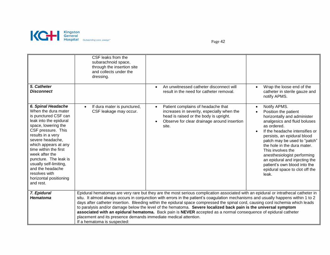
4. Wet or Loose Dressing
May be due to leakage from epidural space over time, or from accumulation of perspiration.
May be due to catheter migration.
If the dura mater was punctured during insertion,
Assess for development of spinal headache (see below).
Assess catheter site. Note: A small amount of serosanguinous drainage at the catheter site is normal and may be due to localized edema.
If dressing wet, cover site with sterile gauze and notify APMS.
Page 42
CSF leaks from the subarachnoid space, through the insertion site and collects under the dressing.
5. Catheter Disconnect
An unwitnessed catheter disconnect will result in the need for catheter removal.
Wrap the loose end of the catheter in sterile gauze and notify APMS.
6. Spinal Headache When the dura mater is punctured CSF can leak into the epidural space, lowering the CSF pressure. This results in a very severe headache, which appears at any time within the first week after the puncture. The leak is usually self-limiting, and the headache resolves with horizontal positioning and rest.
If dura mater is punctured, CSF leakage may occur.
Patient complains of headache that increases in severity, especially when the head is raised or the body is upright.
Observe for clear drainage around insertion site.
Notify APMS.
Position the patient horizontally and administer analgesics and fluid boluses as ordered.
If the headache intensifies or persists, an epidural blood patch may be used to “patch” the hole in the dura mater. This involves the anesthesiologist performing an epidural and injecting the patient’s own blood into the epidural space to clot off the leak.
7. Epidural Hematoma
Epidural hematomas are very rare but they are the most serious complication associated with an epidural or intrathecal catheter in situ. It almost always occurs in conjunction with errors in the patient’s coagulation mechanisms and usually happens within 1 to 2 days after catheter insertion. Bleeding within the epidural space compressed the spinal cord, causing cord ischemia which leads to paralysis and/or damage below the level of the hematoma. Severe localized back pain is the universal symptom associated with an epidural hematoma. Back pain is NEVER accepted as a normal consequence of epidural catheter placement and its presence demands immediate medical attention. If a hematoma is suspected:

Page 43
~ Stop the epidural infusion. ~ Immobilize the patient. ~ Notify APMS STAT. An MRI may be completed and surgical removal of the hematoma (laminectomy) may be performed. Therapeutic heparin doses must be held for 6 to 12 hours, INR must be less than 1.5 and the platelet count must be > 80,000 to safely remove an epidural catheter. Heparin can be resumed 2 hours post catheter removal, if ordered.

Page 44
CONTINUOUS/INTERMITTENT PERIPHERAL REGIONAL ANALGESIA/ANESTHESIA Introduction Peripheral regional analgesia/anesthesia techniques can be used to provide analgesia, as well as anesthesia for surgical procedures. Regional techniques used at KGH are most often “single shot” applications, usually providing 8-12 hours of analgesia. Many of these techniques can be done with a larger needle that allows an infusion catheter to be place inside or along a nerve sheath. A continuous infusion of local anesthetic can be provided through this catheter allowing for continuous analgesia for an indefinite amount of time. Indications Post-operative pain management following orthopedic, vascular and plastic surgery. Continuous sympathetic blockade in the management of vascular and plastic surgery. Conditions where opioid sparing pain management required i.e. sleep apnea, past drug
abuse Adjuvant to opioid therapy for post-operative pain management. Adjuvant to opioid therapy in selected chronic pain syndromes i.e. Chronic Regional Pain
Syndrome I & II Contraindications Patient refusal Systemic infection or local infection at the site of injection. Underlying nerve damage or disease. Relative Contraindications
Coagulopathy or concomitant anti-coagulant therapy Risk for compartment syndrome
Sites for Continuous Regional Analgesia Brachial Plexus (Upper Extremity) Blocks: Continuous or single shot blocks of the nerves of the brachial plexus are useful for pain management following surgical procedures or injury of the shoulder, arm or hand. There are three major types of brachial plexus blocks: axillary; infraclavicular; and interscalene. Patients who receive any brachial plexus block can be expected to experience sensory and motor block in the affected nerves as well as proprioceptive loss. The distribution of nerves affected by each type of brachial plexus block is presented below. AXILLARY: Indications: forearm and hand surgery

Page 45
Location of Injection/Catheter: Infiltration of the nerves is achieved by placement of the needle or catheter as illustrated below. Continuous catheters will be secured with steri-strips and tape or an occlusive dressing. Figure 5: Placement of catheter during axillary nerve block
Nerve distribution affected: all upper limb innervation EXCEPT axillary nerve and intercostobrachial nerve (see fig. 6a & 6b) Note – the innervation to the triceps muscle is blocked while often the biceps innervation is not, leaving the action of the biceps muscle group unopposed. Patients may find any movement of the arm difficult as a result.
Figure 6a: Nerve Distribution Upper Extremity with Arm Supinated

Page 46
Figure 6b: Nerve Distribution Upper Extremity with Arm Pronated
INTERSCALENE AND INFRACLAVICULAR: Indications: Used in shoulder surgery - especially useful in shoulder replacement and rotator cuff repair. Location of Injection/Catheter: Infiltration of the nerves is achieved by placement of the needle or catheter as illustrated below. Continuous catheters will be secured with steri-strips and tape or an occlusive dressing.

Page 47
Figure 7: Interscalene Block Placement
Figure 8: Infraclavicular Block Placement
Needle/catheter insertion site
Needle/catheter insertion site

Page 48
Nerve distribution affected: Interscalene - all upper limb innervation EXCEPT ulnar nerve (see fig. 6a & 6b). Infraclavicular – all upper limb innervation (see fig. 6a & 6b).
**Note: Continuous and single shot interscalene blocks have several potential complications: development of Horner’s syndrome (vocal chord paralysis); transient
hearing loss on the affected side; and diaphragmatic paralysis/paresis in most patients that may cause some respiratory difficulty in patients with pre-existing respiratory
disease. All of these potential complications are reversed with the cessation of therapy. The APMS must be made aware if any of these complications arise.
FEMORAL (LOWER LIMB) BLOCK: Indications: Continuous or single shot block of the femoral nerve is useful for pain management following surgical procedures or injury of the knee or anterior thigh or ankle. Patients who receive any femoral nerve block can be expected to experience sensory and motor block in the affected nerves as well as proprioceptive loss. Functionally, this equates to quadriceps weakness and hip flexor motor block but knee flexion, hip extension and dorsi/plantar flexion of the foot should be unaffected. Location of Injection/Catheter: Infiltration of the nerve is achieved by placement of the needle or catheter as illustrated below. Continuous catheters will be secured with steri-strips and tape or an occlusive dressing.
Figure 9: Femoral Nerve Block Catheter/Needle Placement

Page 49
Nerve distribution affected: Femoral nerve block affects the distribution of the following: femoral nerve; lateral femoral cutaneous nerve; obturator nerve; and saphenous nerve (see fig. 10). Figure 10: Nerve distribution of lower extremities
CARE OF THE PATIENT RECEIVING CONTINUOUS/INTERMITTENT PERIPHERAL REGIONAL ANALGESIA/ANESTHESIA Introduction Patients will have limited motor control of the affected limb as well as limited sensation. Limbs must be protected from trauma, burns, bumps or pressure. Patients with femoral blocks should be cautious when weight bearing on the blocked side because of the potential for distal motor function impairment. However, ambulation is not restricted during therapy when necessary precautions are taken. Motor and proprioceptive function must be assessed prior to any ambulation to prevent falls or other untoward events. Nursing Actions

Page 50
1. When the patient is receiving a continuous infusion and/or a single shot peripheral nerve
block of local anesthetic agents assess the patient according to the guidelines in Table 3.
2. If single shot blocks are performed on cardiac monitor-equipped nursing units, the
physician has a responsibility to communicate the need for the block and a full report of the procedure to the primary nurse.
3. It is important to note that any patient for who compartment syndrome would be a post-
operative risk (e.g. tibial plateau surgery, orthopedic trauma) could potentially have the pain of a developing compartment syndrome masked by a regional block; therefore, it is especially important to assess for the signs and symptoms of compartment syndrome in these patients.
4. When the patient has received single bolus dose of local anesthetic agents assess the
patient according to the guidelines in Table 3 until the sensory/motor deficit has resolved EXCEPTION: Patients who are discharged home before the sensory/motor block has resolved must receive both verbal and written teaching instructions prior to discharge home.
5. When the patient is receiving a continuous peripheral nerve block infusion or has
received a single bolus of a local anesthetic agent, a vial of ephedrine 50 mg IV and a vial of atropine 0.6 mg IV, two 10 mL vials of normal saline, as well as two 10 mL syringes will be available** during and up to 6 hours after the infusion or after the single bolus.
** “available” denotes either on the unit in the medication room or via the RACE or CODE 99 team.
6. If the patient has a lower extremity single-shot or continuous peripheral nerve block
infusion, assess for presence of motor block before assisting the patient to weight-bear. Ensure patient can lift legs up off the bed. DO NOT MOBILIZE PATIENTS WITH A MOTOR BLOCK
7. Infusions may be stopped and the catheter capped with a saline lock prior to
physiotherapy to facilitate movement. A sterile Interlink connector must be placed on the end of the disconnected infusion tubing to maintain asepsis. The infusion tubing can be reconnected following return from physiotherapy and the infusion restarted. When the infusion is re-started, follow monitoring guidelines in Table 3.

Page 51
Table 3
Continuous Infusion and/or Single Shot Peripheral Nerve Block of Local Anesthetic Agents
Assessment Schedule Monitoring Activity Indications for PRN Assessments
At the start of the infusion Or
At the time of single bolus dose
Pain
BP & HR
Catheter site if applicable
Sensory/motor function
Indications for PRN Catheter Site Assessments:
Change in pain
Presence of fluid at catheter site
Patient report of pain at catheter site
Presence of elevated temperature
Change in primary nursing responsibility
Signs of Local Anesthetic Toxicity Early signs: numbness/tingling around lips, metallic taste, dizziness, blurred vision, tinnitus, decreased hearing, restlessness, tremor. Late signs: decrease in cardiac electrical excitability, conduction, arrhythmia, bradycardia or tachycardia, seizures, hypotension, cardiac collapse.
15 and 30 minutes Pain
BP & HR
Signs and symptoms of local anesthetic toxicity
1 hour Pain
BP & HR
Signs and symptoms of local anesthetic toxicity
Then Q4H and prn for duration of infusion/sensory and motor deficit and until the resumption of previous sensory/motor function
Pain & LOS
BP & HR
Signs and symptoms of local anesthetic toxicity
Sensory/motor function
Q12H and prn for duration of infusion/ sensory and motor deficit and until the resumption of previous sensory/motor function
Catheter site
Paresthesia/sensory/motor function
8. When peripheral nerve block therapy has been completed, assess the patient’s
respiratory status every 4 hours: 8.1 x1 post-administration of bupivacaine.
9. The patient will have established IV access for the duration of the therapy and that IV access will remain unless otherwise ordered by the physician for:
9.1 6 hours post-administration of bupivacaine; Recording and Reporting: 1. Discontinue the infusion and notify APMS STAT if you observe or suspect:
1.1 systolic blood pressure <90mm Hg; NOTE: report variations from normal in infant and pediatric patients
1.2 convulsions;

Page 52
1.3 local anesthetic toxicity; 1.4 leaking of fluid from the catheter site; 1.5 catheter migration; 1.6 disconnection of the infusion line from the peripheral nerve catheter.
2. Notify APMS if you observe: 2.1 inadequate pain relief; 2.2 temperature greater than 38.5o C; 2.3 side effects that are not alleviated with nursing interventions; 2.4 problems with the infusion pump; 2.5 paresthesia/sensory/motor block that is distressing to patient.
3. Document on the Analgesia Flow Sheet:
3.1 date and time; 3.2 patient’s subjective description of pain (0-10 at rest and with activity); 3.3 indications for and findings of prn catheter site assessment; 3.4 sensory and motor function; 3.5 rate changes; 3.6 catheter disconnection and reconnection; 3.7 patient controlled function demands and deliveries, if applicable; and 3.8 solution and reservoir changes.
NOTE: Assessment and documentation of items in Section 3 will be performed for the duration of sensory and motor deficits in both single bolus or infusion interventions except patients with planned discharge home before sensory/motor blockade has resolved. These patients will be followed-up by APMS. NOTE: If the patient is receiving both a continuous regional infusion and Intravenous Patient Controlled Analgesia (PCA-IV), a separate Analgesia Flowsheet must be used and documentation must be appropriate to each individual modality. 4. Document on the Medication Administration Record (MAR):
4.1 peripheral nerve block solution; 4.2 order date, time, route; 4.3 start date and time; and 4.4 discontinuation date and time.
5. Document in the Progress Notes or on the unit-specific flow sheet: 5.1 condition of catheter and insertion site if applicable;
5.2 evidence of side effects/complications and actions taken to manage those; 5.3 evaluation of patient response to interventions; and 5.4 communication with APMS staff.

Page 53
Complications Single Shot Blocks The period of risk associated with single shot blocks is immediately following injection. Potential complications that may occur are intravenous/intra-arterial injection of local anesthetic causing seizure or cardiac arrhythmia, pneumothorax (brachial plexus blocks only) and nerve injury. Most patients who receive a single shot block will be well beyond this risk period when transferred to the nursing unit. Analgesia associated with these blocks may last 8-12 hours. Continuous Regional Analgesia Potential complications of Continuous Regional Analgesia are consistent with those associated with central neuraxial analgesia (epidural/ paravertebral). Policies and Procedures: N-100, N-105 Please refer to the following sections of the Surgical Neuraxial Analgesia/Anesthesia Learning Guide:
Review of related anatomy and physiology
Toxic Effects of Local Anesthetic Agents and Side Effects of Local Anesthetic Administration
Complications of Epidural/Paravertebral/Intrathecal Analgesia/Anesthesia {except intrathecal migration, spinal headache, epidural hematoma}
Care of the Patient Receiving Continuous Epidural or Paravertebral Analgesia/Anesthesia
Assessment tools

Page 54
NALOXONE ADMINISTRATION GUIDELINES Naloxone is a short acting parenteral opioid antagonist. It is used to antagonize significant opioid-induced respiratory depression and somnolence, whether caused by excessive doses given therapeutically or by self-administered overdose. Reversal of respiratory depression caused by excessive opioid while maintaining adequate pain control in the postoperative patient demands a cautious approach to the administration of naloxone. To assist in this process, guidelines for dilution and dosing of naloxone are as follows:
1) dilute 1 ampoule (naloxone 0.4mg/ml) with 9 mL 0.9% Sodium Chloride (to yield a final
naloxone concentration of 0.04 mg/mL) 2) administer 1 mL of diluted solution every two minutes until desired response is achieved or
to a maximum or 0.8 mg (20 mL) as ordered by the physician.
PATIENT EDUCATION If health care providers anticipate that a patient will have a neuraxial analgesia/anesthesia modality it is important to ensure that the patient understands the procedure and rationale for the use of these methods of pain relief. The patient should also be aware of possible side effects. The answers to the following commonly asked questions provide a guide for the nurse when explaining these modalities to the patient. How does an epidural or paravertebral control my pain? An analgesic is a medicine that controls your feeling of pain. You may hear the names of these medicines, such as bupivacaine or hydromorphone. These are just two of many medicines that can be used for pain relief. When you go to a dentist, you may be given an anesthetic into the area around a tooth being filled. Sometimes we call this ‘a local’ or ‘freezing’. It stops the nerves near that tooth from making you feel pain. Pain relieving medicines may be used in many other areas of the body. They work in much the same way. You may get your pain medicine by ‘epidural’. This means that your doctor gives the medicine by a needle or small tube called a catheter, into the area in your back around your spinal cord. This prevents your spinal cord from sending pain messages. You may not feel pain in any part of your body below the epidural needle or catheter. You can still move and feel touch in that part of your body. Your doctor will start this treatment. The catheter or small tube may be left in place so medicine can be given as long as you need it to keep you comfortable. You may get pain medicine by a paravertebral. This means that your doctor gives the medicine by a needle or small tube into an area in your back around a group of spinal nerves. Spinal nerves come and go from all parts of your body and connect to your spinal cord. These nerves

Page 55
send pain messages to your brain from your body. If they are ‘blocked’, they do not carry the messages. For example, if the nerves going to and from your arm are blocked, you will not feel pain in your arm. If the nerves going to and from your leg are blocked, you will not feel pain in your leg. Your doctor decides which nerves to block. A nerve block usually blocks pain on one side of your body. Sometimes your doctor may use a block on both sides. The small tube may be left in place so medicine can be given continuously or as you need it. Why is this method used to relieve my pain instead of a pill or a ‘needle’? When you have a pill or a ‘needle’, the pain relief may wear off between doses. Medicine given by epidural or paravertebral keeps you comfortable all the time. It lets you get up and move about. Being up and mobile is very important, as it helps your body to work as usual. You can do exercises that you may need to do after surgery. Pain relief makes your recovery easier and more pleasant.
What happens when they are starting an epidural or paravertebral block? The epidural or paravertebral analgesic is placed through a tiny tube (catheter) into your back, near your spine. Your doctor (an anesthesiologist) will ask you either to curl up on your side or to sit at the side of the bed. This position makes it easier for your doctor to put the catheter (tube) in place. Your doctor will explain what is being done. Once the catheter is in place, it is kept there with a see-through covering. Are there any side effects, and how will I know if I have them? As with any medicine, there may be side effects. You may not notice any changes, but your nurse will check your breathing, temperature, blood pressure and pulse. The nurse will also check the dressing around the tube. It is important to let your nurse know if you have any sign of headache or dizziness when you get up out of bed. Let your nurse know if you feel nauseated or if your skin feels itchy; these side effects can be treated. Your nurse will check your urine output to make sure that you are emptying your bladder if you do not have a tube (catheter) to empty your bladder for you. It is important to know if your bladder is empty after going to the bathroom. What you drink and the amount of urine you make will be written down or recorded. You can help keep track.
Tell your nurse about any pain you feel. Your nurse will want to make sure that you are comfortable.
You may notice that the medicine may cause numbness in the area being treated. This effect is brief. It lasts only until the local anesthetic (freezing) wears off. Your nurse or doctor may use ice on your skin to test your feeling. How long will I have the tube (catheter) in place? Your doctor will remove the catheter when you no longer need pain relief. This will likely be in 2-7 days for an epidural or paravertebral. The effects of the last dose may last up to 24 hours. How long you have your epidural or paravertebral will depend on your surgery or your condition.

Page 56
When you are allowed to drink, the Acute Pain Management Service staff may start giving your pain medication by mouth. You may have a gradual change from one type of pain relief medicine to another.
ASSESSMENT TOOLS:
Motor Block
0 Free movements of legs and
Feet 1 Just able to flex knees. Free movements of feet. 2 Unable to flex knees. Free movement of feet. 3 Unable to move legs or feet.
Sedation Scale
1. Wide Awake 2. Drowsy 3. Dozing Intermittently 4. Mostly Sleeping 5. Only Awakens When Stimulated
(somnolent)
Pain Assessment Scales Numeric Rating Scale 0 = No pain at all 10 = Worst pain imaginable "What number is your pain when you are still? What number is your pain when you move?” Present Pain Intensity/Verbal Descriptor Scale Anchor Word No pain Mild Discomforting Distressing Horrible
Documentation 0 2 4 6 8
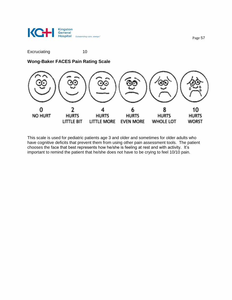
Page 57
Excruciating
10
Wong-Baker FACES Pain Rating Scale
This scale is used for pediatric patients age 3 and older and sometimes for older adults who have cognitive deficits that prevent them from using other pain assessment tools. The patient chooses the face that best represents how he/she is feeling at rest and with activity. It’s important to remind the patient that he/she does not have to be crying to feel 10/10 pain.

Page 58
AUTHORIZATION TEST: SURGICAL NEURAXIAL ANALGESIA/ANESTHESIA Circle the appropriate letters on the answer sheet provided at the end of the test to indicate the phrases that best complete the following statements: 1. The medication used to reverse opioid-induced respiratory depression is:
a. atropine b. naloxone c. ephedrine d. fentanyl
2. The risk of respiratory depression caused by epidural opioids is greater when:
1. The patient is lying flat 2. The patient’s head is raised 3. Epidural and IM opioids are combined 4. The patient has COPD
a. 1,3,4 b. 2,3,4 c. 1 only d. 3 only
3. The most serious complication of a longstanding indwelling epidural catheter is:
a. tinnitus b. ineffectual analgesia c. infection d. respiratory depression
4. Substance P is:
1. released in the dorsal horn of the spinal cord 2. released at the site of tissue injury 3. a neurotransmitter that facilitates pain transmission 4. an endogenous opioid a. 1,3 b. 1,4 c. 1,2,3 d. 1 only

Page 59
5. If an epidural catheter becomes infected, there may be:
a. a rise in the patient’s temperature b. localized redness and tenderness over the catheter site c. posterior neck pain d. all of the above
6. Drugs that can be ordered by the surgical team while the patient has an epidural catheter
are: a. acetaminophen with codeine 30 mg (Tylenol #3) b. morphine c. lorazepam d. none of the above
7. Epidural catheters are most commonly placed in the thoracic region for:
a. thoracotomies b. AAA repairs c. Bowel resections d. All of the above
8. From the list below, choose the signs and symptoms that a patient would exhibit if an
epidural infusion of hydromorphone 10 ug/mL and bupivacaine 1 mg/mL had migrated into the subarachnoid space:
1. respiratory depression 2. loss of analgesic effect 3. hypotension 4. drowsiness a. 1,2,4 b. 2,3,4 c. 1,3,4 d. 1,4
9. Hypotension in patients with epidural or paravertebral analgesia is most often
potentiated by:
a. pain relief b. hypovolemia c. opioid administration d. nausea

Page 60
10. Which patient would be considered the best candidate for epidural analgesia?
a. a trauma patient with severe head injuries and increased intracranial pressure (ICP)
b. a febrile patient with generalized systemic infection c. pre-operative patient with disseminated intravascular coagulation (DIC) d. post-operative patient with chronic obstructive pulmonary disease (COPD)
11. An autologous blood patch is a treatment for which of the following complications:
a. hematoma b. post-dural puncture headache c. paresthesia d. motor loss
Mrs. C had an Abdominal Aortic Aneurysm (AAA) repair yesterday. She was transferred from the Intensive Care Unit (ICU) to a regular floor bed this morning. Mrs. C has an epidural catheter at T8-9. A continuous epidural infusion of hydromorphone 10 ug/mL and bupivacaine 1 mg/mL is running at 8 mL/hr. You have come on shift at 1500 and do your first assessment. You find that Mrs. C is rating her pain 5/10 at rest and 9/10 with activity and can demonstrate a sensory block to ice from T10-T12. Her vital signs are HR 120; RR 24; BP 148/92; O2 Sat 94% on 50% Ventimask (questions 12-14 pertain to Mrs. C’s case): 12. Mrs. C’s current pain status may affect which of the following activities:
a. breathing b. ambulation c. bathing d. all of the above
13. Why does Mrs. C have so much pain?
a. Mrs. C had major surgery and should expect to have severe pain b. the zone of analgesia produced by the epidural is not large enough c. Mrs. C is moving around too much d. Mrs. C’s bowel function is returning causing crampy pain
14. What should you do to address Mrs. C’s pain problem?
a. reassure her that some pain is to be expected b. obtain an order from the vascular service for IM morphine c. call the APMS and suggest Mrs. C’s infusion be decreased d. call the APMS and suggest Mrs. C’s infusion be increased

Page 61
Mr. T is a patient who had a left thoracotomy this morning for lung cancer. He has an epidural catheter in at T2-3 through which hydromorphone 10 ug/mL and bupivacaine 1 mg/mL is infusing at 10 mL/hr. He has been complaining of difficulty getting his breath, his HR is 64, RR is 20/minute and O2 Sat is 94% on 3L NP. He rates his pain as 0/10 rest and activity but complains that his arms are numb and feel ‘heavy’. He is able to demonstrate a sensory block to ice from C4-T8 (questions 15 & 16 pertain to Mr. T’s case): 15. What is the most likely cause of Mr. T’s shortness of breath?
a. Mr. T has developed pneumonia b. Mr. T needs to have his O2 increased c. Mr. T has opioid induced respiratory depression d. Mr. T’s zone of analgesia is too large
16. What should you do to address this problem?
a. call thoracic surgery and suggest a chest x-ray b. call the APMS and suggest a visit to decrease Mr. T’s epidural infusion c. call the APMS and suggest Mr. T needs naloxone d. perform passive ROM on Mr. T’s upper extremities
17. Unlike epidural analgesia, intrathecal analgesia involves the placement of the agent:
a. directly into the subarachnoid space b. between the dura mater and the ligamentum flavum c. directly into the spinal cord d. into an epidural vein
18. Local anesthetic agents given into the epidural space block nerve impulses in the
following order:
a. pain, touch, motor b. motor, pain, touch c. touch, pain, motor d. motor, touch, pain
19. Ask a patient receiving an epidural infusion containing local anesthetic to wiggle her
toes, bend her knees, lift her legs off the bed and to squeeze her buttocks to assess:
a. lower extremity sensory deficit b. local anesthetic toxicity c. lower extremity muscle weakness d. respiratory depression

Page 62
20. Which of the following statements are correct regarding opioid agents administered in
the epidural space?
1. Duration of action is longer than IM/IV opioids 2. Agents can be absorbed systemically by the epidural vasculature 3. Motor nerve blockage may occur 4. Agents should be preservative free 5. Agents that are more water soluble have a slower rate of absorption and may reach
the respiratory centre of the brain 6-24 hours post-administration 6. An intermittent bolus injection is administered with the patient lying flat a. 1,3,4,6 b. 2,3,4,6 c. 1,2,4,5 d. 1,2,3,4,5 e. 1,2,3,4,5,6
21 Which of the following statements are correct regarding local anesthetic agents
administered in the epidural space:
1. Local anesthetic agents are administered in a low dose to produce analgesia 2. The specific anesthetic agent, dose and concentration contribute to the actual
analgesic effect 3. Local anesthetic agents always cause sensory and motor blockade 4. Local anesthetic agents are frequently combined with opioid agents, but may be
administered alone.
a. 1,2,3,4 b. 2,3,4 c. 2,3 d. 1,2,4 e. 1,2,3,4
22. A paravertebral catheter is positioned:
a. inside the vertebral column b. in the epidural space c. in the subarachnoid space d. 2-3 cm lateral to the spinous process
It is 1600 and you have just admitted two post-operative hip replacement patients. Patient A received 100 ug of epimorph intrathecally at 1300 in addition to spinal anesthesia and patient B received spinal anesthesia only at 1230. Both patients are comfortable, report pain scores of 2/10 at rest and can demonstrate a sensory block to ice from T12-L3 (Questions 23 & 24 pertain to this scenario):

Page 63
23. Patient A will likely have analgesia for longer than patient B because intrathecal epimorph
can provide analgesia for up to 24 hours
a. true b. false
24. Patient A will need to be monitored for which serious complication of intrathecal opioids
for 24hours:
a. pruritis b. nausea c. respiratory depression d. post-dural puncture headache
Match the following signs/symptoms with the appropriate cause by circling the appropriate letter on the answer sheet for each question. Each answer can only be used once. 25. localized back pain and progressive motor loss 26. sudden onset of motor loss 27. respiratory arrest 28. seizures and cardiac changes 29. loss of analgesia
Causes of signs/ symptoms a. epidural dose of opioid administered into the subarachnoid space b. development of epidural hematoma c. IV administration of an epidural dose of local anesthetic agent d. Epidural dose of local anesthetic agent administered into the subarachnoid space e. Catheter out of the epidural space
Please indicate on the answer sheet whether the follow statements are true of false:
30. The onset of action of hydromorphone is similar to that of morphine.
a. True b. False
31. All patients receiving continuous epidural analgesia require a foley catheter.
a. True b. False

Page 64
32. It is unnecessary to assess for the presence of motor block in patients who have or are
receiving a local anesthetic agent prior to ambulation.
a. True b. False
33. Patients receiving unilateral continuous paravertebral analgesia should have a sensory
block to ice on both sides.
a. True b. False
34. The first signs of respiratory depression may be an increasing level of sedation and a decreasing depth of respiration.
a. True b. False
35. Absence of a sensory block to ice, even if the patient is pain-free, is an indicator that an
epidural or paravertebral is not functioning.
a. True b. False
Mrs. B had an elective repair of an AAA yesterday. She arrived in your unit at 1100 POD #1. She has a T8 epidural catheter in place and is receiving a continuous infusion of hydromorphone 10 ug/mL and bupivacaine 1 mg/mL at 10 mL/hr. Upon assessment, Mrs. B was 2/5 on the sedation scale and her pain was 2/10 Rest and 4/10 Activity. Her vital signs were stable (Questions 36-40 pertain Mrs. B’s case): Which of the following nursing actions are appropriate? Answer yes or no on the answer sheet provided. 36. Head of bed should be elevated to at least 30 degrees. 37. Patient should be maintained on bedrest. 38. Assess and document the level of sensory block by determining the dermatome level where
loss of sensation occurs using ice q1h. At 1330, Mrs. B became restless, her HR and BP increased. She described her pain as 8/10 R and 10/10 A. Her sensory block is found to be T10-T12. 39. Call the APMS to reassess Mrs. B’s epidural infusion rate

Page 65
The APMS team arrives and administers a 5 mL bolus of pump solution and increases the infusion rate to 12 mL/hr. 40. The frequency for monitoring Mrs. B increases for 4 hours then the previous monitoring schedule resumes.

Page 66
Surgical Neuraxial Analgesia/Anesthesia Test Answer Sheet Name ___________________________________ Date _______________
1. a b c d 2. a b c d 3. a b c d 4. a b c d 5. a b c d 6. a b c d
7. a b c d 8. a b c d 9. a b c d 10. a b c d 11. a b c d 12. a b c d 13. a b c d 14. a b c d 15. a b c d 16. a b c d 17. a b c d 18. a b c d 19. a b c d 20. a b c d e

Page 67
21. a b c d e 22. a b c d 23. a b 24. a b c d 25. a b c d e 26. a b c d e 27. a b c d e 28. a b c d e 29. a b c d e 30. a b 31. a b 32. a b 33. a b 34. a b 35. a b 36. Yes __ No __ 37. Yes __ No __ 38. Yes __ No __ 39. Yes __ No __ 40. Yes __ No __

Page 68
REFERENCES
American Society of Anesthesiologists Task Force on infectious complications associated with neuraxial techniques (2010). Practice advisory for the prevention, diagnosis, and management of infectious complications associated with neuraxial techniques: a report by the American Society of Anesthesiologists Task Force on infectious complications associated with neuraxial techniques. Anesthesiology. 112(3), 530-545.
AORN Recommended Practice, C. (2007). Recommended practices for managing the patient
receiving local anesthesia. AORN Journal, 85(5), 965-971. Banks, A. (2007). Innovations in postoperative pain management: Continuous infusion of local
anesthetics. AORN Journal, 85(5), 904-914. Basse, L., Werner, M. & Kehlet, H. (2000). Is urinary drainage necessary during continuous
epidural analgesia after colonic resection? Regional Anesthesia and Pain Medicine, 25, 498-501.
Brown, D. (2010). Atlas of regional anesthesia (4th ed.). Philadelphia: W.B. Saunders. Cole, P., Craske, D. & Wheatley, R. (2000). Efficacy and respiratory effects of low dose spinal
morphine for post-operative analgesia following knee arthroplasty. British Journal of Anaesthesia, 85(2), 233-237.
Coleman, R. & Ohrling, D. (2005). Independent learning package: Caring for the post operative
patient who has received intrathecal/epidural morphine. Collingwood General & Marine Hospital: Collingwood, ON.
Curatolo, M., et al. (1999). Block of pinprick and cold sensation poorly correlate with relief of
post-operative pain during epidural analgesia. The Clinical Journal of Pain, 15, 6-12. Faculty of Pain Medicine of the Royal College of Anaesthetists (2010). Best practice in the
management of epidural analgesia in the hospital setting. London: (http://www.rcoa.ac.uk/docs/epiduralanalgesia2010.pdf)
Gwirtz, K., et al. (1999). The safety and efficacy of intrathecal opioid analgesia for acute
postoperative pain: Seven years’ experience with 5969 surgical patients at Indiana University Hospital. Anaesthesia and Analgesia, 88, 599-604.
Hoklocker, T., et al. (2009). Practice guidelines for the prevention, detection, and management
of respiratory depression associated with neuraxial opioid administration. Anesthesiology. 110(2), 218-230.
Kreger, C. (2002). Staff development special: Spinal anesthesia and analgesia. Nursing
Management. 33(1), 31-37.

Page 69
McCaffery, M. & Pasero, C. (1999) Pain: Clinical Manual (2nd ed.) Toronto: Mosby. McCamant, K.L. (2006). Peripheral nerve blocks: Understanding the nurse’s role. Journal of
PeriAnesthesia Nursing, 21(1), 16-23. Pasero, C. (2005). Improving postoperative outcomes with epidural analgesia. Journal of
PeriAnesthesia Nursing. 20(1), 51-55. Simpson, T., DeTraglia, M., Speck, E., Wahl, G., & Taylor, D. (1992). The effects of epidural
versus parental opioid analgesia on postoperative pain and pulmonary function in adults who have undergone thoracic and abdominal surgery: A research critique. Heart and Lung, 21(2), 125-127.
Slack, J.F., & Faut-Callahan, M. (1991). Efficacy of epidural analgesia for pain management of
critically ill patients and the implications for nursing care. ACCN Clinical Issues, 2, 729-740.
Stillwell, S. (2000). When you suspect epidural hematoma. American Journal of Nursing, 100(9),
69-74. Walkom, D. (2006). Pain Management with Neuraxial Narcotic Administration: Self directed
learning package. Huron Perth Healthcare Alliance: Clinton, St. Mary’s, Seaforth, Stratford, ON.
White, J., Stevens, R., Beardsley, D., Teague, P. & Kao, T. (1994). Differential epidural block:
Does the choice of local anesthetic matter? Regional Anesthesia, 19(5), 335-338.
White, J., Stevens, R. & Kao, T. (1998). Differential sensory block: Spinal vs epidural with lidocaine. Canadian Journal of Anaesthesia, 45, 1049-1053.
Wild, L., & Coyne, C. (1992). The basics and beyond: Epidural analgesia. American Journal of
Nursing, 92(4), 26-33.

Page 70
EVALUATION OF LEARNING GUIDE
Your feedback and comments are most appreciated. Thank you for your time in responding to this questionnaire. It will help us in planning/revising learning materials. Please circle appropriate response. Strongly
Agree Strongly disagree
1. The content was clear and easy to understand. 1 2 3 4 5
Comments:
2. The content was relevant. 1 2 3 4 5
Comments:
3. My learning needs were met. 1 2 3 4 5
Comments:
4. This guide will help me to meet the knowledge/ Skill requirements relating to the care of patients receiving epidural analgesia/anesthesia and paravertebral nerve blocks.
1 2 3 4 5
Comments:
Additional comments/suggestions:
Please return the completed evaluation to your Clinical Instructor.
Thank you