surgical resection for hepatocellular carcinoma with
TRANSCRIPT
3265
□ CASE REPORT □
Surgical Resection for Hepatocellular Carcinomawith Cardiac Cirrhosis after the Fontan Procedure
Yoshitaka Takuma 1, Yuji Fukada 1, Shota Iwadou 1, Hirokazu Miyatake 1, Shuji Uematsu 1,
Ryoichi Okamoto 1, Daisuke Sato 2, Hiroyoshi Matsukawa 2, Shigehiro Shiozaki 2,
Masahiro Kamada 3, Toshiaki Morito 4 and Yasuyuki Araki 1
Abstract
A 29-year-old woman who underwent the Fontan procedure at 10 years of age had an incidental finding of
liver masses on abdominal ultrasonography. Subsequent gadolinium ethoxybenzyl diethylenetriamine pen-
taacetic acid magnetic resonance imaging showed a 15 mm hypervascular mass with washout in the hepato-
biliary phase in liver segment 4 (S4), and an 18 mm hypervascular mass without washout in the hepatobiliary
phase in liver segment 2 (S2). The S2 liver mass was pathologically diagnosed to be a regenerative nodule by
an ultrasound-guided needle biopsy, and the S4 liver mass was pathologically diagnosed as a poorly differen-
tiated hepatocellular carcinoma after partial hepatectomy.
Key words: hepatocellular carcinoma (HCC), Fontan, cardiac cirrhosis, surgical resection
(Intern Med 55: 3265-3272, 2016)(DOI: 10.2169/internalmedicine.55.6869)
Introduction
The Fontan procedure is used to separate the systemic
and pulmonary circulations in patients with various forms of
functionally univentricular hearts. In the Fontan circulation,
systemic venous return is sent to the pulmonary arteries
without passage through a ventricle. The Fontan procedure
can result in various late complications, including central ve-
nous hypertension, diminished oxygen delivery, reduced car-
diac output, venous thrombosis, and arrhythmia (1, 2).
These complications caused by central venous hypertension
lead to parenchymal injury, fibrosis, and cirrhosis of the
liver (2, 3). Cardiac cirrhosis is a serious late complication
of congenital heart disease and can cause hepatocellular car-
cinoma (HCC) (4, 5). However, the prevalence and risk fac-
tors of cirrhotic changes and HCCs have not been clearly
identified. Furthermore, non-invasive diagnostic tools for he-
patic fibrosis and the management of HCC in patients after
undergoing the Fontan procedure have not yet been clearly
established.
We herein report a case of HCC with cardiac cirrhosis
treated with surgical resection.
Case Report
A 29-year-old woman with a history of a univentricular
heart had undergone a Fontan operation 10 years of age.
She also had situs inversus. She was followed-up at the
department of pediatric cardiology in our hospital, and
regularly underwent blood examinations without alpha-
fetoprotein (AFP) at 2- or 3-month intervals. The results
showed that the transaminase level was within the normal
range. On a routine follow-up day, she experienced slight
abdominal discomfort and received abdominal ultrasonogra-
phy (US). She had an incidental finding of liver masses and
was referred to our department.
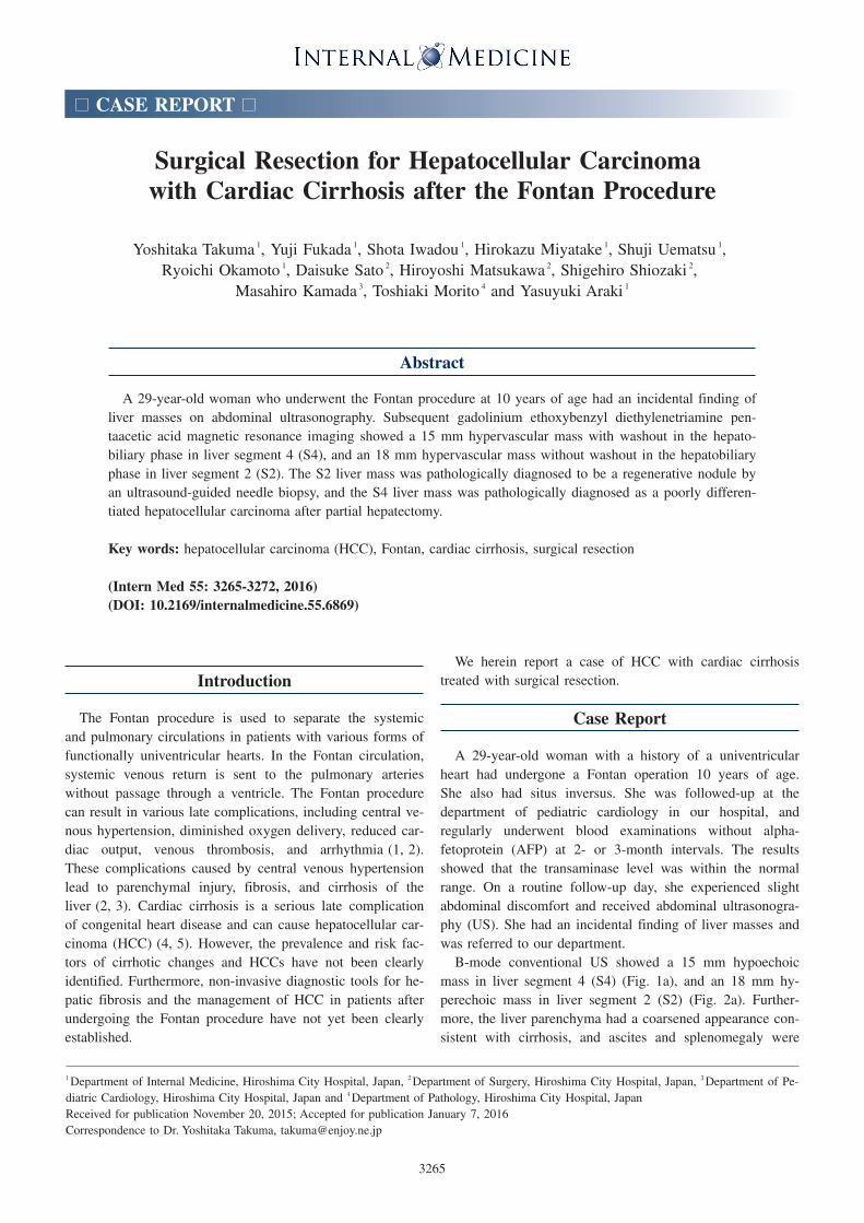
B-mode conventional US showed a 15 mm hypoechoic
mass in liver segment 4 (S4) (Fig. 1a), and an 18 mm hy-
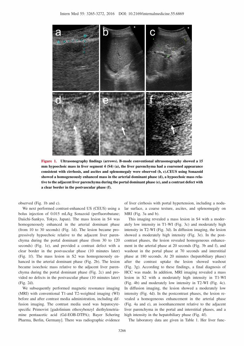
perechoic mass in liver segment 2 (S2) (Fig. 2a). Further-
more, the liver parenchyma had a coarsened appearance con-
sistent with cirrhosis, and ascites and splenomegaly were
1Department of Internal Medicine, Hiroshima City Hospital, Japan, 2Department of Surgery, Hiroshima City Hospital, Japan, 3Department of Pe-
diatric Cardiology, Hiroshima City Hospital, Japan and 4Department of Pathology, Hiroshima City Hospital, Japan
Received for publication November 20, 2015; Accepted for publication January 7, 2016
Correspondence to Dr. Yoshitaka Takuma, [email protected]
Intern Med 55: 3265-3272, 2016 DOI: 10.2169/internalmedicine.55.6869
3266
Figure 1. Ultrasonography findings (arrows). B-mode conventional ultrasonography showed a 15 mm hypoechoic mass in liver segment 4 (S4) (a), the liver parenchyma had a coarsened appearance consistent with cirrhosis, and ascites and splenomegaly were observed (b, c).CEUS using Sonazoid showed a homogeneously enhanced mass in the arterial dominant phase (d), a hypoechoic mass rela-tive to the adjacent liver parenchyma during the portal dominant phase (e), and a contrast defect with a clear border in the postvascular phase (f).
d f
a
e
b c
observed (Fig. 1b and c).
We next performed contrast-enhanced US (CEUS) using a
bolus injection of 0.015 mL/kg Sonazoid (perfluorobutane;
Daiichi-Sankyo, Tokyo, Japan). The mass lesion in S4 was
homogeneously enhanced in the arterial dominant phase
(from 10 to 30 seconds) (Fig. 1d). The lesion became pro-
gressively hypoechoic relative to the adjacent liver paren-
chyma during the portal dominant phase (from 30 to 120
seconds) (Fig. 1e), and provided a contrast defect with a
clear border in the postvascular phase (10 minutes later)
(Fig. 1f). The mass lesion in S2 was homogeneously en-
hanced in the arterial dominant phase (Fig. 2b). The lesion
became isoechoic mass relative to the adjacent liver paren-
chyma during the portal dominant phase (Fig. 2c) and pro-
vided no defects in the postvascular phase (10 minutes later)
(Fig. 2d).
We subsequently performed magnetic resonance imaging
(MRI) with conventional T1-and T2-weighted imaging (WI)
before and after contrast media administration, including dif-
fusion imaging. The contrast media used was hepatocyte-
specific Primovist [gadolinium ethoxybenzyl diethylenetria-
mine pentaacetic acid (Gd-EOB-DTPA); Bayer Schering
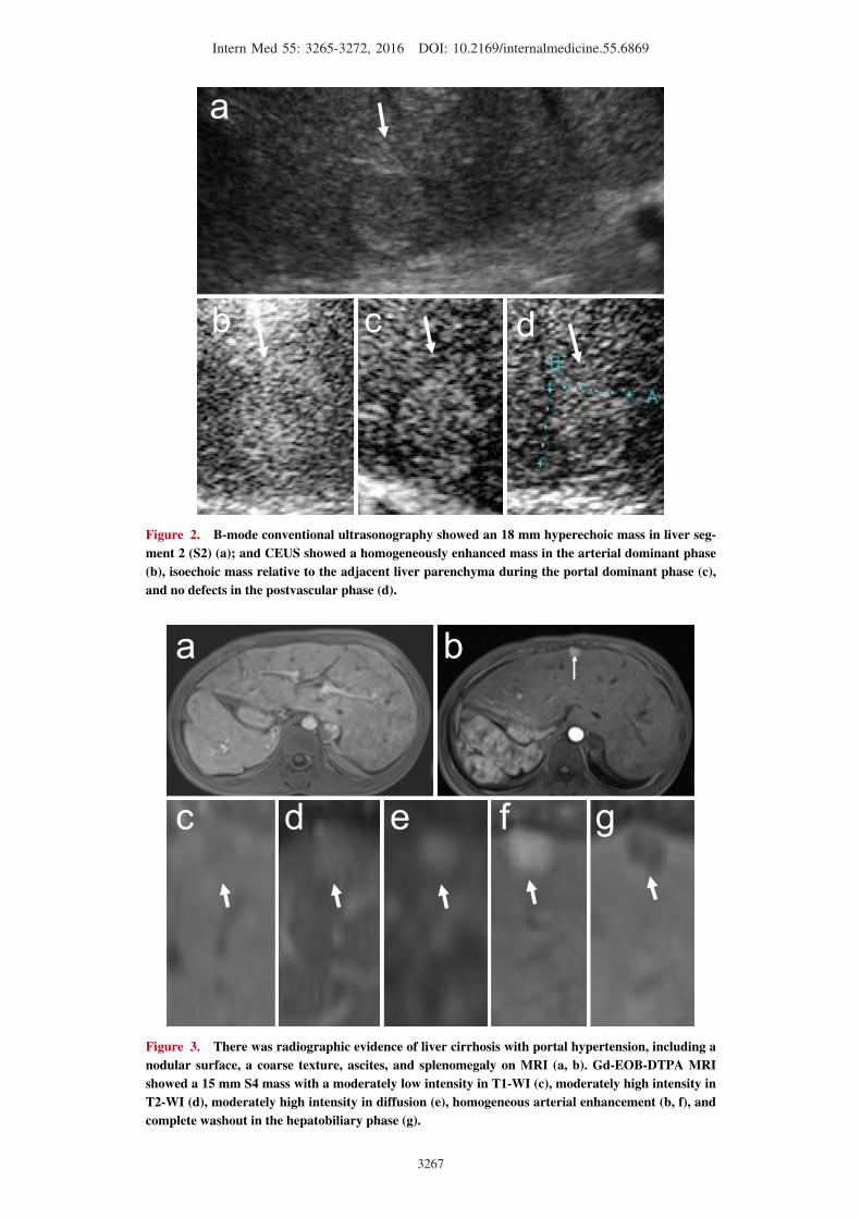
Pharma, Berlin, Germany]. There was radiographic evidence
of liver cirrhosis with portal hypertension, including a nodu-
lar surface, a coarse texture, ascites, and splenomegaly on
MRI (Fig. 3a and b).
This imaging revealed a mass lesion in S4 with a moder-
ately low intensity in T1-WI (Fig. 3c) and moderately high
intensity in T2-WI (Fig. 3d). In diffusion imaging, the lesion
showed a moderately high intensity (Fig. 3e). In the post-
contrast phases, the lesion revealed homogeneous enhance-
ment in the arterial phase at 20 seconds (Fig. 3b and f), and
washout in the portal phase at 70 seconds and interstitial
phase at 180 seconds. At 20 minutes (hepatobiliary phase)
after the contrast uptake the lesion showed washout
(Fig. 3g). According to these findings, a final diagnosis of
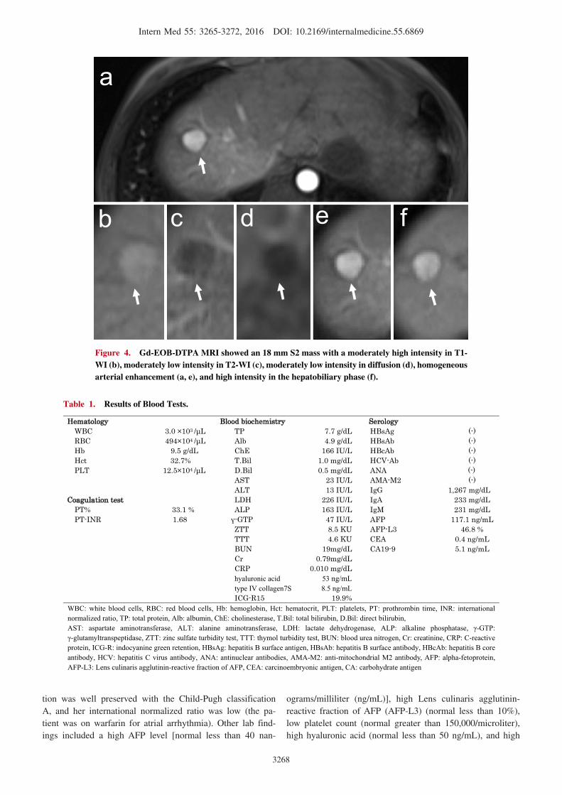
HCC was made. In addition, MRI imaging revealed a mass
lesion in S2 with a moderately high intensity in T1-WI
(Fig. 4b) and moderately low intensity in T2-WI (Fig. 4c).
In diffusion imaging, the lesion showed a moderately low
intensity (Fig. 4d). In the postcontrast phases, the lesion re-
vealed a homogeneous enhancement in the arterial phase
(Fig. 4a and e), an isoenhancement relative to the adjacent
liver parenchyma in the portal and interstitial phases, and a
high intensity in the hepatobiliary phase (Fig. 4f).
The laboratory data are given in Table 1. Her liver func-
Intern Med 55: 3265-3272, 2016 DOI: 10.2169/internalmedicine.55.6869
3267
Figure 2. B-mode conventional ultrasonography showed an 18 mm hyperechoic mass in liver seg-ment 2 (S2) (a); and CEUS showed a homogeneously enhanced mass in the arterial dominant phase (b), isoechoic mass relative to the adjacent liver parenchyma during the portal dominant phase (c), and no defects in the postvascular phase (d).
b d
a
c
Figure 3. There was radiographic evidence of liver cirrhosis with portal hypertension, including a nodular surface, a coarse texture, ascites, and splenomegaly on MRI (a, b). Gd-EOB-DTPA MRI showed a 15 mm S4 mass with a moderately low intensity in T1-WI (c), moderately high intensity in T2-WI (d), moderately high intensity in diffusion (e), homogeneous arterial enhancement (b, f), and complete washout in the hepatobiliary phase (g).
c d e f g
a b
Intern Med 55: 3265-3272, 2016 DOI: 10.2169/internalmedicine.55.6869
3268
Figure 4. Gd-EOB-DTPA MRI showed an 18 mm S2 mass with a moderately high intensity in T1-WI (b), moderately low intensity in T2-WI (c), moderately low intensity in diffusion (d), homogeneous arterial enhancement (a, e), and high intensity in the hepatobiliary phase (f).
b c d e f
a
Table 1. Results of Blood Tests.
WBC: white blood cells, RBC: red blood cells, Hb: hemoglobin, Hct: hematocrit, PLT: platelets, PT: prothrombin time, INR: international normalized ratio, TP: total protein, Alb: albumin, ChE: cholinesterase, T.Bil: total bilirubin, D.Bil: direct bilirubin, AST: aspartate aminotransferase, ALT: alanine aminotransferase, LDH: lactate dehydrogenase, ALP: alkaline phosphatase, -GTP: -glutamyltranspeptidase, ZTT: zinc sulfate turbidity test, TTT: thymol turbidity test, BUN: blood urea nitrogen, Cr: creatinine, CRP: C-reactive
protein, ICG-R: indocyanine green retention, HBsAg: hepatitis B surface antigen, HBsAb: hepatitis B surface antibody, HBcAb: hepatitis B core antibody, HCV: hepatitis C virus antibody, ANA: antinuclear antibodies, AMA-M2: anti-mitochondrial M2 antibody, AFP: alpha-fetoprotein, AFP-L3: Lens culinaris agglutinin-reactive fraction of AFP, CEA: carcinoembryonic antigen, CA: carbohydrate antigen
Hematology Blood biochemistry SerologyWBC 3.0 ×10³ / L TP 7.7 g/dL HBsAg (-)RBC 494×104 / L Alb 4.9 g/dL HBsAb (-)Hb 9.5 g/dL ChE 166 IU/L HBcAb (-)Hct 32.7% T.Bil 1.0 mg/dL HCV-Ab (-)PLT 12.5×10⁴ / L D.Bil 0.5 mg/dL ANA (-)
AST 23 IU/L AMA-M2 (-)ALT 13 IU/L IgG 1,267 mg/dL
Coagulation test LDH 226 IU/L IgA 233 mg/dLPT% 33.1 % ALP 163 IU/L IgM 231 mg/dLPT-INR 1.68 -GTP 47 IU/L AFP 117.1 ng/mL
ZTT 8.5 KU AFP-L3 46.8 %TTT 4.6 KU CEA 0.4 ng/mLBUN 19mg/dL CA19-9 5.1 ng/mLCr 0.79mg/dLCRP 0.010 mg/dLhyaluronic acid 53 ng/mLtype IV collagen7S 8.5 ng/mLICG-R15 19.9%
tion was well preserved with the Child-Pugh classification
A, and her international normalized ratio was low (the pa-
tient was on warfarin for atrial arrhythmia). Other lab find-
ings included a high AFP level [normal less than 40 nan-
ograms/milliliter (ng/mL)], high Lens culinaris agglutinin-
reactive fraction of AFP (AFP-L3) (normal less than 10%),
low platelet count (normal greater than 150,000/microliter),
high hyaluronic acid (normal less than 50 ng/mL), and high
Intern Med 55: 3265-3272, 2016 DOI: 10.2169/internalmedicine.55.6869
3269
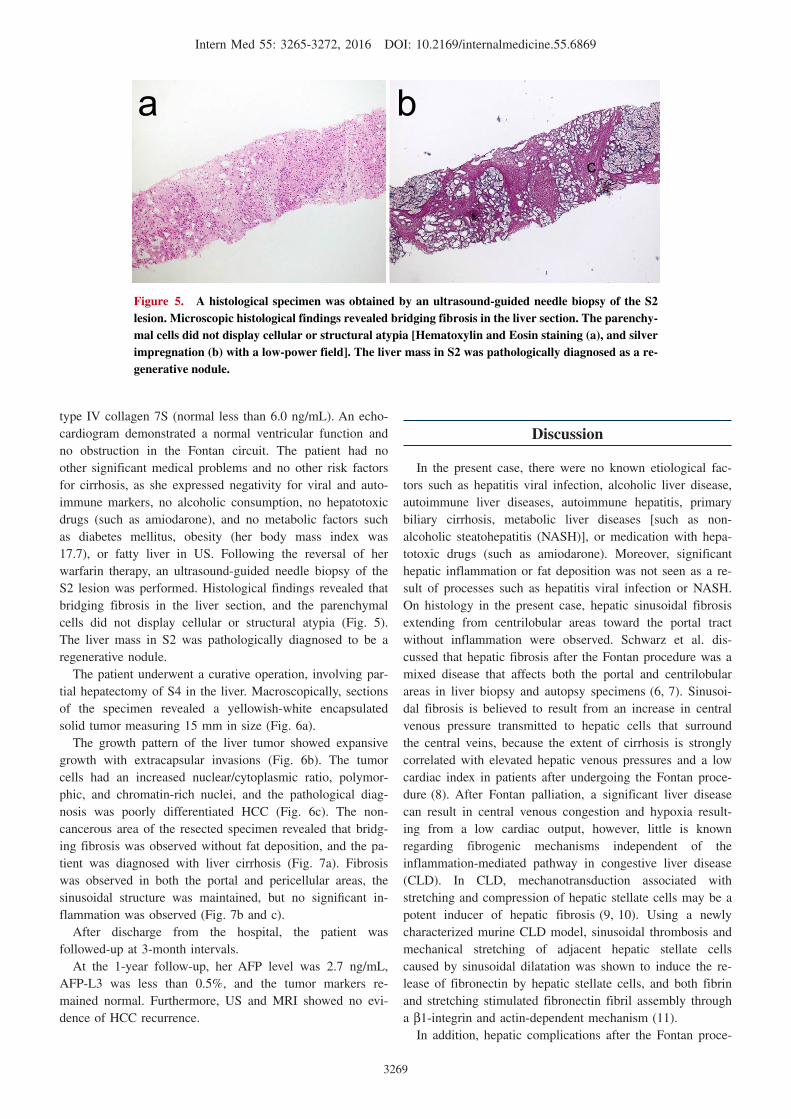
Figure 5. A histological specimen was obtained by an ultrasound-guided needle biopsy of the S2 lesion. Microscopic histological findings revealed bridging fibrosis in the liver section. The parenchy-mal cells did not display cellular or structural atypia [Hematoxylin and Eosin staining (a), and silver impregnation (b) with a low-power field]. The liver mass in S2 was pathologically diagnosed as a re-generative nodule.
a b c
type IV collagen 7S (normal less than 6.0 ng/mL). An echo-
cardiogram demonstrated a normal ventricular function and
no obstruction in the Fontan circuit. The patient had no
other significant medical problems and no other risk factors
for cirrhosis, as she expressed negativity for viral and auto-
immune markers, no alcoholic consumption, no hepatotoxic
drugs (such as amiodarone), and no metabolic factors such
as diabetes mellitus, obesity (her body mass index was
17.7), or fatty liver in US. Following the reversal of her
warfarin therapy, an ultrasound-guided needle biopsy of the
S2 lesion was performed. Histological findings revealed that
bridging fibrosis in the liver section, and the parenchymal
cells did not display cellular or structural atypia (Fig. 5).
The liver mass in S2 was pathologically diagnosed to be a
regenerative nodule.
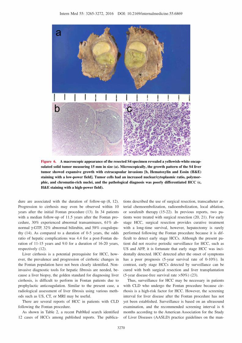
The patient underwent a curative operation, involving par-
tial hepatectomy of S4 in the liver. Macroscopically, sections
of the specimen revealed a yellowish-white encapsulated
solid tumor measuring 15 mm in size (Fig. 6a).
The growth pattern of the liver tumor showed expansive
growth with extracapsular invasions (Fig. 6b). The tumor
cells had an increased nuclear/cytoplasmic ratio, polymor-
phic, and chromatin-rich nuclei, and the pathological diag-
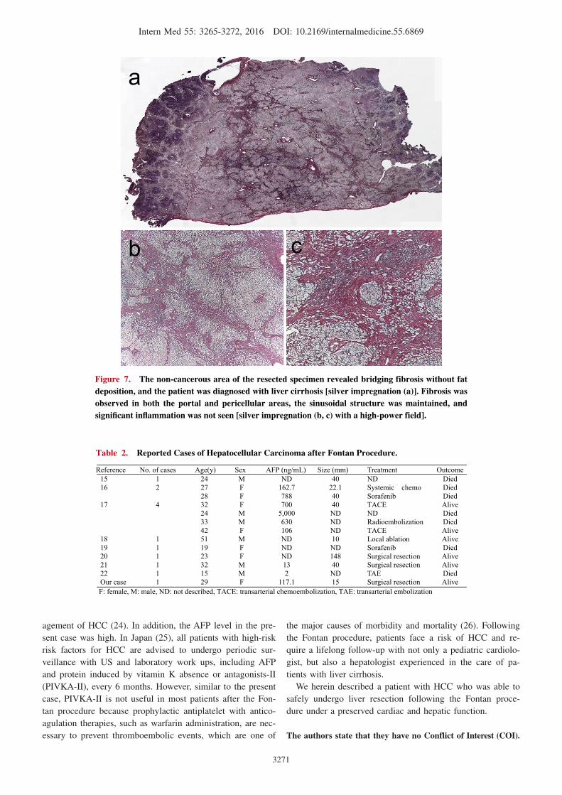
nosis was poorly differentiated HCC (Fig. 6c). The non-
cancerous area of the resected specimen revealed that bridg-
ing fibrosis was observed without fat deposition, and the pa-
tient was diagnosed with liver cirrhosis (Fig. 7a). Fibrosis
was observed in both the portal and pericellular areas, the
sinusoidal structure was maintained, but no significant in-
flammation was observed (Fig. 7b and c).
After discharge from the hospital, the patient was
followed-up at 3-month intervals.
At the 1-year follow-up, her AFP level was 2.7 ng/mL,
AFP-L3 was less than 0.5%, and the tumor markers re-
mained normal. Furthermore, US and MRI showed no evi-
dence of HCC recurrence.
Discussion
In the present case, there were no known etiological fac-
tors such as hepatitis viral infection, alcoholic liver disease,
autoimmune liver diseases, autoimmune hepatitis, primary
biliary cirrhosis, metabolic liver diseases [such as non-
alcoholic steatohepatitis (NASH)], or medication with hepa-
totoxic drugs (such as amiodarone). Moreover, significant
hepatic inflammation or fat deposition was not seen as a re-
sult of processes such as hepatitis viral infection or NASH.
On histology in the present case, hepatic sinusoidal fibrosis
extending from centrilobular areas toward the portal tract
without inflammation were observed. Schwarz et al. dis-
cussed that hepatic fibrosis after the Fontan procedure was a
mixed disease that affects both the portal and centrilobular
areas in liver biopsy and autopsy specimens (6, 7). Sinusoi-
dal fibrosis is believed to result from an increase in central
venous pressure transmitted to hepatic cells that surround
the central veins, because the extent of cirrhosis is strongly
correlated with elevated hepatic venous pressures and a low
cardiac index in patients after undergoing the Fontan proce-
dure (8). After Fontan palliation, a significant liver disease
can result in central venous congestion and hypoxia result-
ing from a low cardiac output, however, little is known
regarding fibrogenic mechanisms independent of the
inflammation-mediated pathway in congestive liver disease
(CLD). In CLD, mechanotransduction associated with
stretching and compression of hepatic stellate cells may be a
potent inducer of hepatic fibrosis (9, 10). Using a newly
characterized murine CLD model, sinusoidal thrombosis and
mechanical stretching of adjacent hepatic stellate cells
caused by sinusoidal dilatation was shown to induce the re-
lease of fibronectin by hepatic stellate cells, and both fibrin
and stretching stimulated fibronectin fibril assembly through
a β1-integrin and actin-dependent mechanism (11).
In addition, hepatic complications after the Fontan proce-
Intern Med 55: 3265-3272, 2016 DOI: 10.2169/internalmedicine.55.6869
3270
Figure 6. A macroscopic appearance of the resected S4 specimen revealed a yellowish-white encap-sulated solid tumor measuring 15 mm in size (a). Microscopically, the growth pattern of the S4 liver tumor showed expansive growth with extracapsular invasions [b, Hematoxylin and Eosin (H&E) staining with a low-power field]. Tumor cells had an increased nuclear/cytoplasmic ratio, polymor-phic, and chromatin-rich nuclei, and the pathological diagnosis was poorly differentiated HCC (c, H&E staining with a high-power field).
a
b c
dure are associated with the duration of follow-up (8, 12).
Progression to cirrhosis may even be observed within 10
years after the initial Fontan procedure (13). In 34 patients
with a median follow-up of 11.5 years after the Fontan pro-
cedure, 30% experienced abnormal transaminases, 61% ab-
normal γ-GTP, 32% abnormal bilirubin, and 58% coagulopa-
thy (14). As compared to a duration of 0-5 years, the odds
ratio of hepatic complications was 4.4 for a post-Fontan du-
ration of 11-15 years and 9.0 for a duration of 16-20 years,
respectively (12).
Liver cirrhosis is a potential prerequisite for HCC, how-
ever, the prevalence and progression of cirrhotic changes in
the Fontan population have not been clearly identified. Non-
invasive diagnostic tools for hepatic fibrosis are needed, be-
cause a liver biopsy, the golden standard for diagnosing liver
cirrhosis, is difficult to perform in Fontan patients due to
prophylactic anticoagulation. Similar to the present case, a
radiological assessment of liver fibrosis using various meth-
ods such as US, CT, or MRI may be useful.
There are several reports of HCC in patients with CLD
following the Fontan procedure.
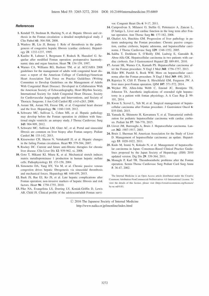
As shown in Table 2, a recent PubMed search identified
12 cases of HCCs among published reports. The publica-
tions described the use of surgical resection, transcatheter ar-
terial chemoembolization, radioembolization, local ablation,
or sorafenib therapy (15-22). In previous reports, two pa-
tients were treated with surgical resection (20, 21). For early
stage HCC, surgical resection provides curative treatment
with a long-time survival, however, hepatectomy is rarely
performed following the Fontan procedure because it is dif-
ficult to detect early stage HCCs. Although the present pa-
tient did not receive periodic surveillance for HCC, such as
US and AFP, it is fortunate that early stage HCC was inci-
dentally detected. HCC detected after the onset of symptoms
has a poor prognosis (5-year survival rate of 0-10%). In
contrast, early stage HCCs detected by surveillance can be
cured with both surgical resection and liver transplantation
(5-year disease-free survival rate >50%) (23).
Thus, surveillance for HCC may be necessary in patients
with CLD who undergo the Fontan procedure because cir-
rhosis is a high-risk factor for HCC. However, the screening
interval for liver disease after the Fontan procedure has not
yet been established. Surveillance is based on an ultrasound
examination, and the recommended screening interval is 6
months according to the American Association for the Study
of Liver Diseases (AASLD) practice guidelines on the man-
Intern Med 55: 3265-3272, 2016 DOI: 10.2169/internalmedicine.55.6869
3271
Figure 7. The non-cancerous area of the resected specimen revealed bridging fibrosis without fat deposition, and the patient was diagnosed with liver cirrhosis [silver impregnation (a)]. Fibrosis was observed in both the portal and pericellular areas, the sinusoidal structure was maintained, and significant inflammation was not seen [silver impregnation (b, c) with a high-power field].
b c
a
Table 2. Reported Cases of Hepatocellular Carcinoma after Fontan Procedure.
F: female, M: male, ND: not described, TACE: transarterial chemoembolization, TAE: transarterial embolization
Reference No. of cases Age(y) Sex AFP (ng/mL) Size (mm) Treatment Outcome1516
17
1819202122Our case
12
4
111111
2427 28 32 24 33 42 51 19 23 32 15 29
MF F F M M F M F F M M F
ND162.7 788 700
5,000 630 106 NDNDND13 2
117.1
4022.1 40 40 NDNDND10 ND148 40 ND15
NDSystemic chemoSorafenib TACENDRadioembolization TACELocal ablationSorafenib Surgical resectionSurgical resectionTAESurgical resection
DiedDiedDiedAliveDiedDiedAliveAliveDiedAliveAliveDiedAlive
agement of HCC (24). In addition, the AFP level in the pre-
sent case was high. In Japan (25), all patients with high-risk
risk factors for HCC are advised to undergo periodic sur-
veillance with US and laboratory work ups, including AFP
and protein induced by vitamin K absence or antagonists-II
(PIVKA-II), every 6 months. However, similar to the present
case, PIVKA-II is not useful in most patients after the Fon-
tan procedure because prophylactic antiplatelet with antico-
agulation therapies, such as warfarin administration, are nec-
essary to prevent thromboembolic events, which are one of
the major causes of morbidity and mortality (26). Following
the Fontan procedure, patients face a risk of HCC and re-
quire a lifelong follow-up with not only a pediatric cardiolo-
gist, but also a hepatologist experienced in the care of pa-
tients with liver cirrhosis.
We herein described a patient with HCC who was able to
safely undergo liver resection following the Fontan proce-
dure under a preserved cardiac and hepatic function.
The authors state that they have no Conflict of Interest (COI).
Intern Med 55: 3265-3272, 2016 DOI: 10.2169/internalmedicine.55.6869
3272
References
1. Kendall TJ, Stedman B, Hacking N, et al. Hepatic fibrosis and cir-
rhosis in the Fontan circulation: a detailed morphological study. J
Clin Pathol 61: 504-508, 2008.
2. Wanless IR, Liu JJ, Butany J. Role of thrombosis in the patho-
genesis of congestive hepatic fibrosis (cardiac cirrhosis). Hepatol-
ogy 21: 1232-1237, 1995.
3. Kaulitz R, Luhmer I, Bergmann F, Rodeck B, Hausdorf G. Se-
quelae after modified Fontan operation: postoperative haemody-
namic data and organ function. Heart 78: 154-159, 1997.
4. Warnes CA, Williams RG, Bashore TM, et al. ACC/AHA 2008
guidelines for the management of adults with congenital heart dis-
ease: a report of the American College of Cardiology/American
Heart Association Task Force on Practice Guidelines (Writing
Committee to Develop Guidelines on the Management of Adults
With Congenital Heart Disease). Developed in Collaboration With
the American Society of Echocardiography, Heart Rhythm Society,
International Society for Adult Congenital Heart Disease, Society
for Cardiovascular Angiography and Interventions, and Society of
Thoracic Surgeons. J Am Coll Cardiol 52: e143-e263, 2008.
5. Asrani SK, Asrani NS, Freese DK, et al. Congenital heart disease
and the liver. Hepatology 56: 1160-1169, 2012.
6. Schwartz MC, Sullivan L, Cohen MS, et al. Hepatic pathology
may develop before the Fontan operation in children with func-
tional single ventricle: an autopsy study. J Thorac Cardiovasc Surg
143: 904-909, 2012.
7. Schwartz MC, Sullivan LM, Glatz AC, et al. Portal and sinusoidal
fibrosis are common on liver biopsy after Fontan surgery. Pediatr
Cardiol 34: 135-142, 2013.
8. Kiesewetter CH, Sheron N, Vettukattill JJ, et al. Hepatic changes
in the failing Fontan circulation. Heart 93: 579-584, 2007.
9. Rockey DC. Current and future anti-fibrotic therapies for chronic
liver disease. Clin Liver Dis 12: 939-962, xi, 2008.
10. Goto T, Mikami KI, Miura K, et al. Mechanical stretch induces
matrix metalloproteinase 1 production in human hepatic stellate
cells. Pathophysiology 11: 153-158, 2004.
11. Simonetto DA, Yang HY, Yin M, et al. Chronic passive venous
congestion drives hepatic fibrogenesis via sinusoidal thrombosis
and mechanical forces. Hepatology 61: 648-659, 2015.
12. Baek JS, Bae EJ, Ko JS, et al. Late hepatic complications after
Fontan operation; non-invasive markers of hepatic fibrosis and risk
factors. Heart 96: 1750-1755, 2010.
13. Pike NA, Evangelista LS, Doering LV, Koniak-Griffin D, Lewis
AB, Child JS. Clinical profile of the adolescent/adult Fontan survi-
vor. Congenit Heart Dis 6: 9-17, 2011.
14. Camposilvan S, Milanesi O, Stellin G, Pettenazzo A, Zancan L,
D’Antiga L. Liver and cardiac function in the long term after Fon-
tan operation. Ann Thorac Surg 86: 177-182, 2008.
15. Ghaferi AA, Hutchins GM. Progression of liver pathology in pa-
tients undergoing the Fontan procedure: Chronic passive conges-
tion, cardiac cirrhosis, hepatic adenoma, and hepatocellular carci-
noma. J Thorac Cardiovasc Surg 129: 1348-1352, 2005.
16. Saliba T, Dorkhom S, O’Reilly EM, Ludwig E, Gansukh B,
Abou-Alfa GK. Hepatocellular carcinoma in two patients with car-
diac cirrhosis. Eur J Gastroenterol Hepatol 22: 889-891, 2010.
17. Asrani SK, Warnes CA, Kamath PS. Hepatocellular carcinoma af-
ter the Fontan procedure. N Engl J Med 368: 1756-1757, 2013.
18. Elder RW, Parekh S, Book WM. More on hepatocellular carci-
noma after the Fontan procedure. N Engl J Med 369: 490, 2013.
19. Rajoriya N, Clift P, Thorne S, Hirschfield GM, Ferguson JW. A
liver mass post-Fontan operation. QJM 107: 571-572, 2014.
20. Weyker PD, Allen-John Webb C, Emond JC, Brentjens TE,
Johnston TA. Anesthetic implications of extended right hepatec-
tomy in a patient with fontan physiology. A A Case Rep 2: 99-
101, 2014.
21. Kwon S, Scovel L, Yeh M, et al. Surgical management of hepato-
cellular carcinoma after Fontan procedure. J Gastrointest Oncol 6:
E55-E60, 2015.
22. Yamada K, Shinmoto H, Kawamura Y, et al. Transarterial emboli-
zation for pediatric hepatocellular carcinoma with cardiac cirrho-
sis. Pediatr Int 57: 766-770, 2015.
23. Llovet JM, Burroughs A, Bruix J. Hepatocellular carcinoma. Lan-
cet 362: 1907-1917, 2003.
24. Bruix J, Sherman M; American Association for the Study of Liver
D. Management of hepatocellular carcinoma: an update. Hepatol-
ogy 53: 1020-1022, 2011.
25. Kudo M, Izumi N, Kokudo N, et al. Management of hepatocellu-
lar carcinoma in Japan: Consensus-Based Clinical Practice Guide-
lines proposed by the Japan Society of Hepatology (JSH) 2010
updated version. Dig Dis 29: 339-364, 2011.
26. Monagle P, Karl TR. Thromboembolic problems after the Fontan
operation. Semin Thorac Cardiovasc Surg Pediatr Card Surg Annu
5: 36-47, 2002.
The Internal Medicine is an Open Access article distributed under the Creative
Commons Attribution-NonCommercial-NoDerivatives 4.0 International License. To
view the details of this license, please visit (https://creativecommons.org/licenses/
by-nc-nd/4.0/).
Ⓒ 2016 The Japanese Society of Internal Medicine
http://www.naika.or.jp/imonline/index.html