surgical suturing of articular cartilage induces osteoarthritis-like changes
TRANSCRIPT
Osteoarthritis and Cartilage (2008) 16, 1067e1073
ª 2008 Osteoarthritis Research Society International. Published by Elsevier Ltd. All rights reserved.doi:10.1016/j.joca.2008.01.009
InternationalCartilageRepairSociety
Surgical suturing of articular cartilage induces osteoarthritis-likechanges1
E. B. Hunziker M.D.* and A. Stahli D.M.D.DST Research Center, Department of Clinical Research, University of Bern, Murtenstrasse 35,P.O. Box 54, 3010 Bern, Switzerland
Summary
Introduction: In clinical tissue-engineering-based approaches to articular cartilage repair, various types of flap are frequently used to retain animplanted construct within the defect, and they are usually affixed by suturing. We hypothesize that the suturing of articular cartilage is asso-ciated with a loss of chondrocytes from, and osteoarthritis-like changes within, the perisutural area.
Materials and methods: We established a large, partial-thickness defect model in the femoral groove of adult goats. The defects were filledwith bovine fibrinogen to support a devitalized flap of autologous synovial tissue, which was sutured to the surrounding articular cartilage withsingle, interrupted stitches. The perisutural and control regions were analyzed histologically, histochemically and histomorphometrically shortlyafter surgery and 3 weeks later.
Results: Compared to control regions, chondrocytes were lost from the perisutural area even during the first few hours of surgery. During theensuing 3 weeks, the numerical density of cells in the perisutural area decreased significantly. The cell losses were associated with a loss ofproteoglycans from the extracellular matrix. Shortly after surgery, fissures were observed within the walls of the suture channels. By the thirdweek, their surface density had increased significantly and they were filled with avascular mesenchymal tissue.
Conclusions: The suturing of articular cartilage induces severe local damage, which is progressive and reminiscent of that associated with theearly stages of osteoarthritis. This damage could be most readily circumvented by adopting an alternative mode of flap affixation, such asgluing with a biological adhesive.ª 2008 Osteoarthritis Research Society International. Published by Elsevier Ltd. All rights reserved.
Key words: Surgical suturing, Articular cartilage, Osteoarthritic fissures, Proteoglycan loss.
Introduction
Structural lesioning of the articular cartilage layer is a com-mon consequence of traumatic or pathological events inlarge human joints. Such lesions are frequently associatedwith pain, diminished joint functionality and reduced life-quality1,2. The symptoms tend to be most severe whenthe lesions arise pathologically during the course of osteo-arthritis3e6. It is generally believed that these debilitatingsymptoms can be best relieved by inducing the lesions toheal. With this aim in view, diverse surgical strategieshave been elaborated to induce a spontaneous tissue re-pair response. These include surgical stimulation of thesubchondral bone marrow by, for example, Pridie drilling,microfracturing or abrasive chondroplasty (for review, seeRef. 7). These interventions are reasonably successful inthe short- and mid-terms (months to a few years)8,9, butnot in the long-run10. Against this background, great effortshave been made to improve the repair of articular cartilagelesions by novel means, such as tissue engineering. One
1This study was supported by the University of Bern, Switzerlandand by a grant from the NIH, USA (NIAMS, grant number 1 R01 AR52766-01A1).
*Address correspondence and reprint requests to: ProfessorErnst B. Hunziker, DST Research Center, Department of ClinicalResearch, University of Bern, Murtenstrasse 35, P.O. Box 54,3010 Bern, Switzerland. Tel: 41-31-632-8685; Fax: 41-31-632-4955; E-mail: [email protected]
Received 9 October 2007; revision accepted 14 January 2008.
1067
such approach that has been avidly adopted in clinical prac-tice is the autologous chondrocyte implantation tech-nique11,12. This procedure involves the suturing of anautologous periosteal flap to the border of the articular carti-lage lesion, into which a suspension of autologous chondro-cytes is then injected12. The introduction of this methodologyinto clinical practice has popularized the surgical suturing ofdiverse types of flap to articular cartilage lesions that havebeen variously treated to induce their repair. After morethan a decade’s experience with the autologous chondrocyteimplantation technique, it is now evident that it confers noadvantage over conventional bone-marrow-stimulating ap-proaches, such as microfracturing10. To improve the resultsyielded by tissue-engineering approaches, efforts mustnow be made not only to improve the therapeutic principlesupon which they are based, but also to minimize iatrogenicdamage and to eliminate surgical manocuvres that compro-mise the repair response of the articular cartilage tissue.
The surgical suturing of articular cartilage is known to bea delicate task, owing to the tissue’s avascular, aneural andalymphatic nature, and to its poor healing capacity. Sutur-ing involves the insertion of a surgical needle into the tis-sue, thereby creating a channel which, in effect, isa partial-thickness lesion. We hypothesize that these chan-nel-like defects are associated with damage to the adjacenttissue, and that the lesions not only fail to heal but enlargewith time and joint usage.
To test this hypothesis, we established a large, partial-thickness defect model in the trochlear femoral groove of
1068 E. B. Hunziker and A. Stahli: Suturing of articular cartilage
adult goats. The defects were filled with bovine fibrinogen tosupport a thin flap of devitalized synovial tissue, which wassutured to the surrounding articular cartilage. The animalswere killed either 2e3 h after surgery or 3 weeks later.The perisutural area and control regions were analyzedhistologically, histochemically (metachromatic staining ofthe extracellular matrix with Toluidine Blue to assess theproteoglycan status) and histomorphometrically (numericaland volume densities of chondrocytes, and their meanvolume; surface and volume densities of fissures withinthe walls of the suture channels).
Materials and methods
EXPERIMENTAL OUTLINE
The purpose of this study was to investigate the effects of surgical sutur-ing on normal hyaline articular cartilage tissue within the synovial knee-jointsof adult goats, both at the time of surgery and 3 weeks later. These effectswere analyzed histologically, histochemically and histomorphometrically.
SURGICAL PROCEDURE
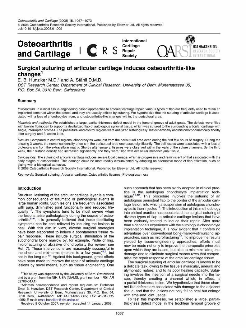
Sixteen adult Saanen goats were used for this investigation, eight ofwhich were sacrificed 2e3 h after surgery and the other eight 3 weeks later.The study was approved by the local ethical committee for animal experi-mentation and conducted in accordance with its regulations. General anaes-thesia was induced by an intravenous injection of butorphenol anddiazepam and maintained by ketamine. In one leg per animal, the trochlearfemoral groove was exposed layer by layer. One purely chondral defect,5 mm in width and 10 mm in length (in the sagittal plane), was createdwithin the lateral facet of the patellar groove using a custom-made cuttingtool (see Ref. 13). Bovine fibrinogen was deposited within the defect to sup-port the flap of synovial tissue that was sutured to its roof. This material wasexcised from the lateral wall of the synovial joint. Its surface area slightly ex-ceeded that of the defect. Prior to placement, it was devitalized by threefreezingethawing cycles, and then trimmed to precisely match the defectarea. It was sutured to the lesion borders with single stitches of Vicryl7-0 thread (Fig. 1). The wound was closed layer by layer.
Eight of the goats were killed within 2e3 h of surgery, and the other eight3 weeks later. In the latter category, a soft cast was applied to the treated leg
Fig. 1. Macroscopic view of a synovial tissue flap sutured to theborders of a chondral defect. Bar¼ 2 mm.
[to hinder the loss of flaps (see Ref. 14)]. This cast restricted movement ofthe knee to 3e4�, but permitted full loading of the joint. The animals wereable to stand up and lie down of their own accord (see Ref. 15).
After the 3-week period had elapsed, the goats were anaesthetized (asdescribed above) and infused with an overdose of potassium chloride toinduce cardiac arrest.
The treated joints were excised and transferred to a moist chamber for theremoval of soft tissues and the preparation of tissue blocks that comprisedthe entire defect area.
TISSUE PROCESSING
The tissue blocks were prefixed in 4% buffered formaldehyde solution(Merck, Darmstadt, Germany) for 1e2 h at ambient temperature. Theywere then cut into smaller units using an Exact saw. Tissue fixation was con-tinued for 5 days at ambient temperature. The blocks were then rinsed in tapwater, dehydrated in ethanol (Alco Suisse, Bern, Switzerland), and embed-ded in methylmethacrylate (Merck, Darmstadt, Germany).
TISSUE-SAMPLING PROTOCOL
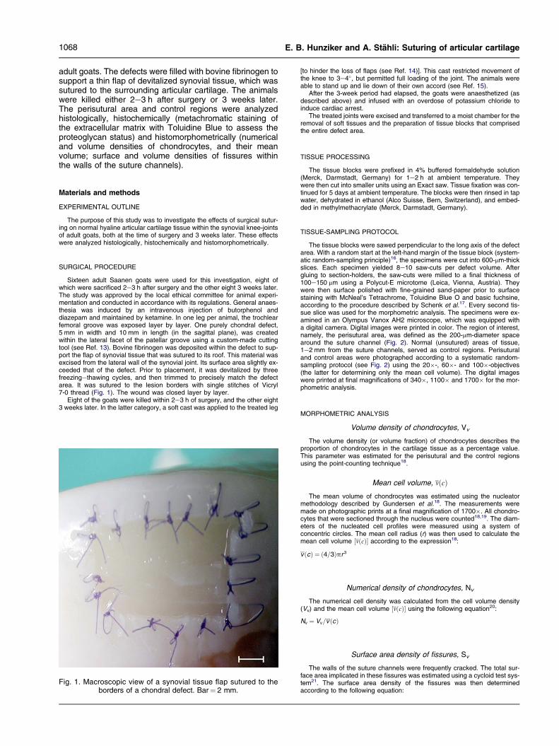
The tissue blocks were sawed perpendicular to the long axis of the defectarea. With a random start at the left-hand margin of the tissue block (system-atic random-sampling principle)16, the specimens were cut into 600-mm-thickslices. Each specimen yielded 8e10 saw-cuts per defect volume. Aftergluing to section-holders, the saw-cuts were milled to a final thickness of100e150 mm using a Polycut-E microtome (Leica, Vienna, Austria). Theywere then surface polished with fine-grained sand-paper prior to surfacestaining with McNeal’s Tetrachrome, Toluidine Blue O and basic fuchsine,according to the procedure described by Schenk et al.17. Every second tis-sue slice was used for the morphometric analysis. The specimens were ex-amined in an Olympus Vanox AH2 microscope, which was equipped witha digital camera. Digital images were printed in color. The region of interest,namely, the perisutural area, was defined as the 200-mm-diameter spacearound the suture channel (Fig. 2). Normal (unsutured) areas of tissue,1e2 mm from the suture channels, served as control regions. Perisuturaland control areas were photographed according to a systematic random-sampling protocol (see Fig. 2) using the 20�-, 60�- and 100�-objectives(the latter for determining only the mean cell volume). The digital imageswere printed at final magnifications of 340�, 1100� and 1700� for the mor-phometric analysis.
MORPHOMETRIC ANALYSIS
Volume density of chondrocytes, Vv
The volume density (or volume fraction) of chondrocytes describes theproportion of chondrocytes in the cartilage tissue as a percentage value.This parameter was estimated for the perisutural and the control regionsusing the point-counting technique18.
Mean cell volume, vðcÞThe mean volume of chondrocytes was estimated using the nucleator
methodology described by Gundersen et al.18. The measurements weremade on photographic prints at a final magnification of 1700�. All chondro-cytes that were sectioned through the nucleus were counted18,19. The diam-eters of the nucleated cell profiles were measured using a system ofconcentric circles. The mean cell radius (r) was then used to calculate themean cell volume ½vðcÞ� according to the expression18:
vðcÞ ¼ ð4=3Þpr 3
Numerical density of chondrocytes, Nv
The numerical cell density was calculated from the cell volume density(Vv) and the mean cell volume ½vðcÞ� using the following equation20:
Nv ¼ Vv=vðcÞ
Surface area density of fissures, Sv
The walls of the suture channels were frequently cracked. The total sur-face area implicated in these fissures was estimated using a cycloid test sys-tem21. The surface area density of the fissures was then determinedaccording to the following equation:
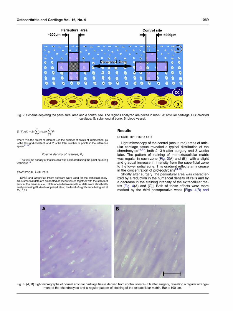
Fig. 2. Scheme depicting the perisutural area and a control site. The regions analyzed are boxed in black. A: articular cartilage; CC: calcifiedcartilage; S: subchondral bone; B: blood vessel.
1069Osteoarthritis and Cartilage Vol. 16, No. 9
SvðY ; refÞ ¼ 2xXn
i¼1
Ii=I=pxXn
i¼1
Pi
where Y is the object of interest, Ii is the number of points of intersection, pxis the test-grid constant, and Pi is the total number of points in the referencespace20,21.
Volume density of fissures, Vv
The volume density of the fissures was estimated using the point-countingtechnique18.
STATISTICAL ANALYSIS
SPSS and GraphPad Prism software were used for the statistical analy-sis. Numerical data are presented as mean values together with the standarderror of the mean (S.E.M.). Differences between sets of data were statisticallyanalyzed using Student’s unpaired t-test, the level of significance being set atP< 0.05.
Fig. 3. (A, B) Light micrographs of normal articular cartilage tissue derivedment of the chondrocytes and a regular pattern of sta
Results
DESCRIPTIVE HISTOLOGY
Light microscopy of the control (unsutured) areas of artic-ular cartilage tissue revealed a typical distribution of thechondrocytes22,23, both 2e3 h after surgery and 3 weekslater. The pattern of staining of the extracellular matrixwas regular in each zone [Fig. 3(A) and (B)], with a slightand gradual increase in intensity from the superficial zoneto the lower radial zone. This gradient reflects an increasein the concentration of proteoglycans24,25.
Shortly after surgery, the perisutural area was character-ized by a reduction in the numerical density of cells and bya decrease in the staining intensity of the extracellular ma-trix [Fig. 4(A) and (C)]. Both of these effects were moremarked by the third postoperative week [Figs. 4(B) and
from control sites 2e3 h after surgery, revealing a regular arrange-ining of the extracellular matrix. Bar¼ 100 mm.
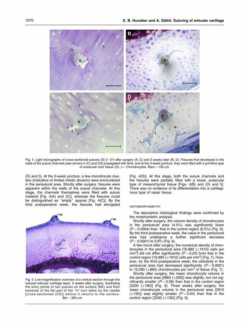
Fig. 4. Light micrographs of cross-sectioned sutures (S) 2e3 h after surgery (A, C) and 3 weeks later (B, D). Fissures that developed in thewalls of the suture channels [see arrows in (C) and (D)] propagated with time, and at the 3-week juncture, they were filled with a primitive type
of avascular scar tissue (D). c¼Chondrocytes. Bars¼ 100 mm.
1070 E. B. Hunziker and A. Stahli: Suturing of articular cartilage
(D) and 5]. At the 3-week juncture, a few chondrocyte clus-ters (indicative of limited mitotic division) were encounteredin the perisutural area. Shortly after surgery, fissures wereapparent within the walls of the suture channels. At thisstage, the channels themselves were filled with suturematerial [Fig. 4(A) and (C)], whereas the fissures couldbe distinguished as ‘‘empty’’ spaces [Fig. 4(C)]. By thethird postoperative week, the fissures had elongated
Fig. 5. Low-magnification overview of a vertical section through thesutured articular cartilage layer, 3 weeks after surgery, illustratingthe entry points of two sutures on the surface (NE) and theirtraversal of the flat part of the ‘‘U’’-turn taken by the needle[cross-sect ioned (CS)] before i t returns to the surface.
Bar¼ 300 mm.
[Fig. 4(D)]. At this stage, both the suture channels andthe fissures were partially filled with a loose, avasculartype of mesenchymal tissue [Figs. 4(B) and (D) and 5].There was no evidence of its differentiation into a cartilagi-nous type of repair tissue.
HISTOMORPHOMETRY
The descriptive histological findings were confirmed bythe morphometric analysis.
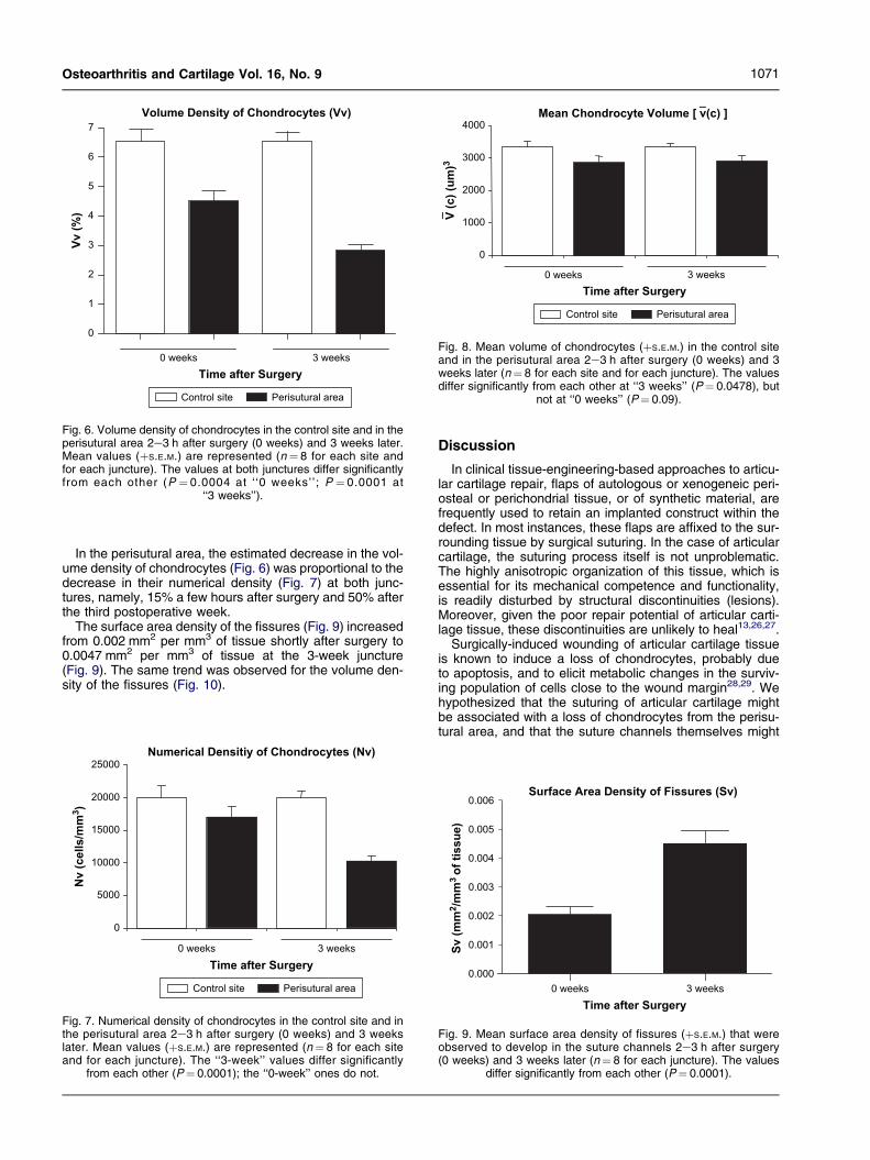
Shortly after surgery, the volume density of chondrocytesin the perisutural area (4.5%) was significantly lower(P¼ 0.0004) than that in the control region (6.5%) (Fig. 6).By the third postoperative week, the value in the perisuturalarea had undergone a further significant decrease(P< 0.0001) to 2.8% (Fig. 6).
A few hours after surgery, the numerical density of chon-drocytes in the perisutural area [16,980 (�1610) cells permm3] did not differ significantly (P¼ 0.23) from that in thecontrol region [19,990 (�1910) cells per mm3] (Fig. 7). How-ever, by the third postoperative week, the cellularity in theperisutural area had decreased significantly (P< 0.0001)to 10,290 (�860) chondrocytes per mm3 of tissue (Fig. 7).
Shortly after surgery, the mean chondrocyte volume inthe perisutural area [2860 (�200)] was slightly, but not sig-nificantly smaller (P¼ 0.09) than that in the control region[3330 (�180)] (Fig. 8). Three weeks after surgery, themean chondrocyte volume in the perisutural area [2910(�150)] was slightly smaller (P¼ 0.04) than that in thecontrol region [3330 (�130)] (Fig. 8).
Time after Surgery
0 weeks 3 weeks
Vv (%
)
Volume Density of Chondrocytes (Vv)
0
1
2
3
4
5
6
7
Control site Perisutural area
Fig. 6. Volume density of chondrocytes in the control site and in theperisutural area 2e3 h after surgery (0 weeks) and 3 weeks later.Mean values (þS.E.M.) are represented (n¼ 8 for each site andfor each juncture). The values at both junctures differ significantlyfrom each other (P ¼ 0.0004 at ‘ ‘0 weeks’’; P ¼ 0.0001 at
‘‘3 weeks’’).
0
1000
2000
3000
4000
0 weeks 3 weeks
V (c) (u
m)3
Mean Chondrocyte Volume [ v(c) ]
Time after Surgery
Control site Perisutural area
Fig. 8. Mean volume of chondrocytes (þS.E.M.) in the control siteand in the perisutural area 2e3 h after surgery (0 weeks) and 3weeks later (n¼ 8 for each site and for each juncture). The valuesdiffer significantly from each other at ‘‘3 weeks’’ (P¼ 0.0478), but
not at ‘‘0 weeks’’ (P¼ 0.09).
1071Osteoarthritis and Cartilage Vol. 16, No. 9
In the perisutural area, the estimated decrease in the vol-ume density of chondrocytes (Fig. 6) was proportional to thedecrease in their numerical density (Fig. 7) at both junc-tures, namely, 15% a few hours after surgery and 50% afterthe third postoperative week.
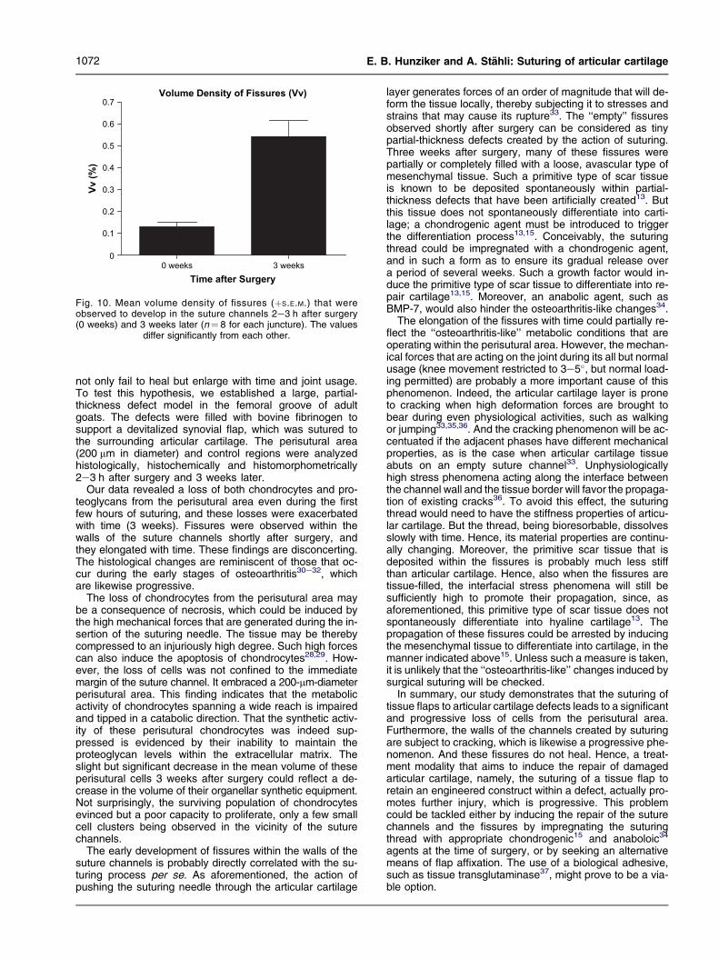
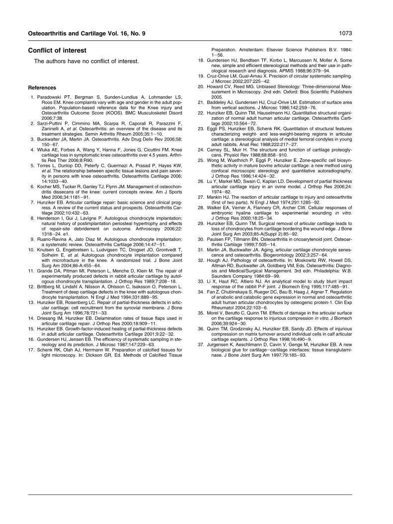
The surface area density of the fissures (Fig. 9) increasedfrom 0.002 mm2 per mm3 of tissue shortly after surgery to0.0047 mm2 per mm3 of tissue at the 3-week juncture(Fig. 9). The same trend was observed for the volume den-sity of the fissures (Fig. 10).
Numerical Densitiy of Chondrocytes (Nv)
0
5000
10000
15000
20000
25000
Nv (cells/m
m3)
0 weeks 3 weeksTime after Surgery
Control site Perisutural area
Fig. 7. Numerical density of chondrocytes in the control site and inthe perisutural area 2e3 h after surgery (0 weeks) and 3 weekslater. Mean values (þS.E.M.) are represented (n¼ 8 for each siteand for each juncture). The ‘‘3-week’’ values differ significantly
from each other (P¼ 0.0001); the ‘‘0-week’’ ones do not.
Discussion
In clinical tissue-engineering-based approaches to articu-lar cartilage repair, flaps of autologous or xenogeneic peri-osteal or perichondrial tissue, or of synthetic material, arefrequently used to retain an implanted construct within thedefect. In most instances, these flaps are affixed to the sur-rounding tissue by surgical suturing. In the case of articularcartilage, the suturing process itself is not unproblematic.The highly anisotropic organization of this tissue, which isessential for its mechanical competence and functionality,is readily disturbed by structural discontinuities (lesions).Moreover, given the poor repair potential of articular carti-lage tissue, these discontinuities are unlikely to heal13,26,27.
Surgically-induced wounding of articular cartilage tissueis known to induce a loss of chondrocytes, probably dueto apoptosis, and to elicit metabolic changes in the surviv-ing population of cells close to the wound margin28,29. Wehypothesized that the suturing of articular cartilage mightbe associated with a loss of chondrocytes from the perisu-tural area, and that the suture channels themselves might
0.000
0.001
0.002
0.003
0.004
0.005
0.006Surface Area Density of Fissures (Sv)
0 weeks 3 weeksTime after Surgery
Sv (m
m2/m
m3 o
f tissu
e)
Fig. 9. Mean surface area density of fissures (þS.E.M.) that wereobserved to develop in the suture channels 2e3 h after surgery(0 weeks) and 3 weeks later (n¼ 8 for each juncture). The values
differ significantly from each other (P¼ 0.0001).
Volume Density of Fissures (Vv)
0 weeks 3 weeksTime after Surgery
Vv (%
)
0
0.1
0.2
0.3
0.4
0.5
0.6
0.7
Fig. 10. Mean volume density of fissures (þS.E.M.) that wereobserved to develop in the suture channels 2e3 h after surgery(0 weeks) and 3 weeks later (n¼ 8 for each juncture). The values
differ significantly from each other.
1072 E. B. Hunziker and A. Stahli: Suturing of articular cartilage
not only fail to heal but enlarge with time and joint usage.To test this hypothesis, we established a large, partial-thickness defect model in the femoral groove of adultgoats. The defects were filled with bovine fibrinogen tosupport a devitalized synovial flap, which was sutured tothe surrounding articular cartilage. The perisutural area(200 mm in diameter) and control regions were analyzedhistologically, histochemically and histomorphometrically2e3 h after surgery and 3 weeks later.
Our data revealed a loss of both chondrocytes and pro-teoglycans from the perisutural area even during the firstfew hours of suturing, and these losses were exacerbatedwith time (3 weeks). Fissures were observed within thewalls of the suture channels shortly after surgery, andthey elongated with time. These findings are disconcerting.The histological changes are reminiscent of those that oc-cur during the early stages of osteoarthritis30e32, whichare likewise progressive.
The loss of chondrocytes from the perisutural area maybe a consequence of necrosis, which could be induced bythe high mechanical forces that are generated during the in-sertion of the suturing needle. The tissue may be therebycompressed to an injuriously high degree. Such high forcescan also induce the apoptosis of chondrocytes28,29. How-ever, the loss of cells was not confined to the immediatemargin of the suture channel. It embraced a 200-mm-diameterperisutural area. This finding indicates that the metabolicactivity of chondrocytes spanning a wide reach is impairedand tipped in a catabolic direction. That the synthetic activ-ity of these perisutural chondrocytes was indeed sup-pressed is evidenced by their inability to maintain theproteoglycan levels within the extracellular matrix. Theslight but significant decrease in the mean volume of theseperisutural cells 3 weeks after surgery could reflect a de-crease in the volume of their organellar synthetic equipment.Not surprisingly, the surviving population of chondrocytesevinced but a poor capacity to proliferate, only a few smallcell clusters being observed in the vicinity of the suturechannels.
The early development of fissures within the walls of thesuture channels is probably directly correlated with the su-turing process per se. As aforementioned, the action ofpushing the suturing needle through the articular cartilage
layer generates forces of an order of magnitude that will de-form the tissue locally, thereby subjecting it to stresses andstrains that may cause its rupture33. The ‘‘empty’’ fissuresobserved shortly after surgery can be considered as tinypartial-thickness defects created by the action of suturing.Three weeks after surgery, many of these fissures werepartially or completely filled with a loose, avascular type ofmesenchymal tissue. Such a primitive type of scar tissueis known to be deposited spontaneously within partial-thickness defects that have been artificially created13. Butthis tissue does not spontaneously differentiate into carti-lage; a chondrogenic agent must be introduced to triggerthe differentiation process13,15. Conceivably, the suturingthread could be impregnated with a chondrogenic agent,and in such a form as to ensure its gradual release overa period of several weeks. Such a growth factor would in-duce the primitive type of scar tissue to differentiate into re-pair cartilage13,15. Moreover, an anabolic agent, such asBMP-7, would also hinder the osteoarthritis-like changes34.
The elongation of the fissures with time could partially re-flect the ‘‘osteoarthritis-like’’ metabolic conditions that areoperating within the perisutural area. However, the mechan-ical forces that are acting on the joint during its all but normalusage (knee movement restricted to 3e5�, but normal load-ing permitted) are probably a more important cause of thisphenomenon. Indeed, the articular cartilage layer is proneto cracking when high deformation forces are brought tobear during even physiological activities, such as walkingor jumping33,35,36. And the cracking phenomenon will be ac-centuated if the adjacent phases have different mechanicalproperties, as is the case when articular cartilage tissueabuts on an empty suture channel33. Unphysiologicallyhigh stress phenomena acting along the interface betweenthe channel wall and the tissue border will favor the propaga-tion of existing cracks36. To avoid this effect, the suturingthread would need to have the stiffness properties of articu-lar cartilage. But the thread, being bioresorbable, dissolvesslowly with time. Hence, its material properties are continu-ally changing. Moreover, the primitive scar tissue that isdeposited within the fissures is probably much less stiffthan articular cartilage. Hence, also when the fissures aretissue-filled, the interfacial stress phenomena will still besufficiently high to promote their propagation, since, asaforementioned, this primitive type of scar tissue does notspontaneously differentiate into hyaline cartilage13. Thepropagation of these fissures could be arrested by inducingthe mesenchymal tissue to differentiate into cartilage, in themanner indicated above15. Unless such a measure is taken,it is unlikely that the ‘‘osteoarthritis-like’’ changes induced bysurgical suturing will be checked.
In summary, our study demonstrates that the suturing oftissue flaps to articular cartilage defects leads to a significantand progressive loss of cells from the perisutural area.Furthermore, the walls of the channels created by suturingare subject to cracking, which is likewise a progressive phe-nomenon. And these fissures do not heal. Hence, a treat-ment modality that aims to induce the repair of damagedarticular cartilage, namely, the suturing of a tissue flap toretain an engineered construct within a defect, actually pro-motes further injury, which is progressive. This problemcould be tackled either by inducing the repair of the suturechannels and the fissures by impregnating the suturingthread with appropriate chondrogenic15 and anaboloic34
agents at the time of surgery, or by seeking an alternativemeans of flap affixation. The use of a biological adhesive,such as tissue transglutaminase37, might prove to be a via-ble option.
1073Osteoarthritis and Cartilage Vol. 16, No. 9
Conflict of interest
The authors have no conflict of interest.
References
1. Paradowski PT, Bergman S, Sunden-Lundius A, Lohmander LS,Roos EM. Knee complaints vary with age and gender in the adult pop-ulation. Population-based reference data for the Knee injury andOsteoarthritis Outcome Score (KOOS). BMC Musculoskelet Disord2006;7:38.
2. Sarzi-Puttini P, Cimmino MA, Scarpa R, Caporali R, Parazzini F,Zaninelli A, et al. Osteoarthritis: an overview of the disease and itstreatment strategies. Semin Arthritis Rheum 2005;35:1e10.
3. Buckwalter JA, Martin JA. Osteoarthritis. Adv Drug Deliv Rev 2006;58:150e67.
4. Wluka AE, Forbes A, Wang Y, Hanna F, Jones G, Cicuttini FM. Kneecartilage loss in symptomatic knee osteoarthritis over 4.5 years. Arthri-tis Res Ther 2006;8:R90.
5. Torres L, Dunlop DD, Peterfy C, Guermazi A, Prasad P, Hayes KW,et al. The relationship between specific tissue lesions and pain sever-ity in persons with knee osteoarthritis. Osteoarthritis Cartilage 2006;14:1033e40.
6. Kocher MS, Tucker R, Ganley TJ, Flynn JM. Management of osteochon-dritis dissecans of the knee: current concepts review. Am J SportsMed 2006;34:1181e91.
7. Hunziker EB. Articular cartilage repair: basic science and clinical prog-ress. A review of the current status and prospects. Osteoarthritis Car-tilage 2002;10:432e63.
8. Henderson I, Gui J, Lavigne P. Autologous chondrocyte implantation:natural history of postimplantation periosteal hypertrophy and effectsof repair-site debridement on outcome. Arthroscopy 2006;22:1318e24. e1.
9. Ruano-Ravina A, Jato Diaz M. Autologous chondrocyte implantation:a systematic review. Osteoarthritis Cartilage 2006;14:47e51.
10. Knutsen G, Engebretsen L, Ludvigsen TC, Drogset JO, Grontvedt T,Solheim E, et al. Autologous chondrocyte implantation comparedwith microfracture in the knee. A randomized trial. J Bone JointSurg Am 2004;86-A:455e64.
11. Grande DA, Pitman MI, Peterson L, Menche D, Klein M. The repair ofexperimentally produced defects in rabbit articular cartilage by autol-ogous chondrocyte transplantation. J Orthop Res 1989;7:208e18.
12. Brittberg M, Lindahl A, Nilsson A, Ohlsson C, Isaksson O, Peterson L.Treatment of deep cartilage defects in the knee with autologous chon-drocyte transplantation. N Engl J Med 1994;331:889e95.
13. Hunziker EB, Rosenberg LC. Repair of partial-thickness defects in artic-ular cartilage: cell recruitment from the synovial membrane. J BoneJoint Surg Am 1996;78:721e33.
14. Driesang IM, Hunziker EB. Delamination rates of tissue flaps used inarticular cartilage repair. J Orthop Res 2000;18:909e11.
15. Hunziker EB. Growth-factor-induced healing of partial-thickness defectsin adult articular cartilage. Osteoarthritis Cartilage 2001;9:22e32.
16. Gundersen HJ, Jensen EB. The efficiency of systematic sampling in ste-reology and its prediction. J Microsc 1987;147:229e63.
17. Schenk RK, Olah AJ, Herrmann W. Preparation of calcified tissues forlight microscopy. In: Dickson GR, Ed. Methods of Calcified Tissue
Preparation. Amsterdam: Elsevier Science Publishers B.V. 1984:1e56.
18. Gundersen HJ, Bendtsen TF, Korbo L, Marcussen N, Moller A. Somenew, simple and efficient stereological methods and their use in path-ological research and diagnosis. APMIS 1988;96:379e94.
19. Cruz-Orive LM, Gual-Arnau X. Precision of circular systematic sampling.J Microsc 2002;207:225e42.
20. Howard CV, Reed MG. Unbiased Stereology: Three-dimensional Mea-surement in Microscopy. 2nd edn. Oxford: Bios Scientific Publishers2005.
21. Baddeley AJ, Gundersen HJ, Cruz-Orive LM. Estimation of surface areafrom vertical sections. J Microsc 1986;142:259e76.
22. Hunziker EB, Quinn TM, Hauselmann HJ. Quantitative structural organi-zation of normal adult human articular cartilage. Osteoarthritis Carti-lage 2002;10:564e72.
23. Eggli PS, Hunziker EB, Schenk RK. Quantitation of structural featurescharacterizing weight- and less-weight-bearing regions in articularcartilage: a stereological analysis of medial femoral condyles in youngadult rabbits. Anat Rec 1988;222:217e27.
24. Carney SL, Muir H. The structure and function of cartilage proteogly-cans. Physiol Rev 1988;68:858e910.
25. Wong M, Wuethrich P, Eggli P, Hunziker E. Zone-specific cell biosyn-thetic activity in mature bovine articular cartilage: a new method usingconfocal microscopic stereology and quantitative autoradiography.J Orthop Res 1996;14:424e32.
26. Lu Y, Markel MD, Swain C, Kaplan LD. Development of partial thicknessarticular cartilage injury in an ovine model. J Orthop Res 2006;24:1974e82.
27. Mankin HJ. The reaction of articular cartilage to injury and osteoarthritis(first of two parts). N Engl J Med 1974;291:1285e92.
28. Walker EA, Verner A, Flannery CR, Archer CW. Cellular responses ofembryonic hyaline cartilage to experimental wounding in vitro.J Orthop Res 2000;18:25e34.
29. Hunziker EB, Quinn TM. Surgical removal of articular cartilage leads toloss of chondrocytes from cartilage bordering the wound edge. J BoneJoint Surg Am 2003;85-A(Suppl 2):85e92.
30. Paulsen FP, Tillmann BN. Osteoarthritis in cricoarytenoid joint. Osteoar-thritis Cartilage 1999;7:505e14.
31. Martin JA, Buckwalter JA. Aging, articular cartilage chondrocyte senes-cence and osteoarthritis. Biogerontology 2002;3:257e64.
32. Hough AJ. Pathology of osteoarthritis. In: Moskowitz RW, Howell DS,Altman RD, Buckwalter JA, Goldberg VM, Eds. Osteoarthritis: Diagno-sis and Medical/Surgical Management. 3rd edn. Philadelphia: W.B.Saunders Company 1984:69e99.
33. Li X, Haut RC, Altiero NJ. An analytical model to study blunt impactresponse of the rabbit P-F joint. J Biomech Eng 1995;117:485e91.
34. Fan Z, Chubinskaya S, Rueger DC, Bau B, Haag J, Aigner T. Regulationof anabolic and catabolic gene expression in normal and osteoarthriticadult human articular chondrocytes by osteogenic protein-1. Clin ExpRheumatol 2004;22:103e6.
35. Morel V, Berutto C, Quinn TM. Effects of damage in the articular surfaceon the cartilage response to injurious compression in vitro. J Biomech2006;39:924e30.
36. Quinn TM, Grodzinsky AJ, Hunziker EB, Sandy JD. Effects of injuriouscompression on matrix turnover around individual cells in calf articularcartilage explants. J Orthop Res 1998;16:490e9.
37. Jurgensen K, Aeschlimann D, Cavin V, Genge M, Hunziker EB. A newbiological glue for cartilageecartilage interfaces: tissue transglutami-nase. J Bone Joint Surg Am 1997;79:185e93.