surgical technique for palsy of superior division of
TRANSCRIPT
Surgical Technique for Palsy of Superior Division of Oculomotor Nerve
Address for correspondence: Birsen Gokyigit, MD. Beyoglu Goz Egitim ve Arastirma Hastanesi, Bereketzade Cami Sokak,34421 Beyoglu, Istanbul, Turkey
Phone: +90 532 246 37 99 E-mail: [email protected] Date: July 23, 2017 Accepted Date: August 23, 2017 Available Online Date: October 16, 2017©Copyright 2017 by Beyoglu Eye Training and Research Hospital - Available online at www.beyoglueye.com
Introduction
The diagnosis and management of third nerve dysfunction varies according to the age of the patient, characteristics of the third nerve palsy, and the presence of associated symp-toms and signs. Third nerve palsy may be partial or complete, congenital or acquired, isolated or accompanied by signs of more extensive neurological involvement. It can result from lesions located anywhere from the oculomotor nucleus to the termination of the third nerve in the extra-ocular mus-cles within the orbit. The superior division runs forward in-traconally to innervate first the superior rectus first and then the levator above on its inferior surface.
Isolated paralysis of the superior rectus muscle is most com-monly congenital, but may also be secondary to trauma (1). The paralyzed eye is affected primarily in abduction and elevation, and is hypotropic in the primary position.
Surgical Technique
This study was approved by the institutional review board. Written informed consent was obtained from the patient and the study adhered to the tenets of the Declaration of Helsinki.
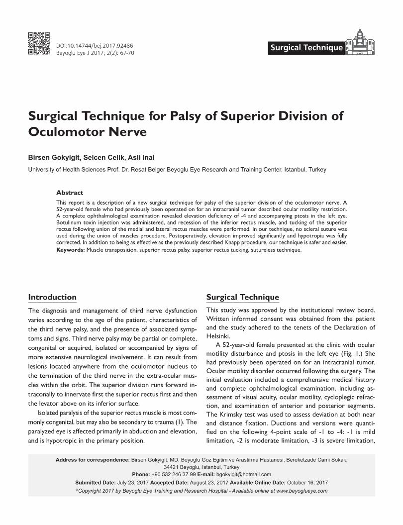
A 52-year-old female presented at the clinic with ocular motility disturbance and ptosis in the left eye (Fig. 1.) She had previously been operated on for an intracranial tumor. Ocular motility disorder occurred following the surgery. The initial evaluation included a comprehensive medical history and complete ophthalmological examination, including as-sessment of visual acuity, ocular motility, cycloplegic refrac-tion, and examination of anterior and posterior segments. The Krimsky test was used to assess deviation at both near and distance fixation. Ductions and versions were quanti-fied on the following 4-point scale of -1 to -4: -1 is mild limitation, -2 is moderate limitation, -3 is severe limitation,
This report is a description of a new surgical technique for palsy of the superior division of the oculomotor nerve. A 52-year-old female who had previously been operated on for an intracranial tumor described ocular motility restriction. A complete ophthalmological examination revealed elevation deficiency of -4 and accompanying ptosis in the left eye. Botulinum toxin injection was administered, and recession of the inferior rectus muscle, and tucking of the superior rectus following union of the medial and lateral rectus muscles were performed. In our technique, no scleral suture was used during the union of muscles procedure. Postoperatively, elevation improved significantly and hypotropia was fully corrected. In addition to being as effective as the previously described Knapp procedure, our technique is safer and easier.Keywords: Muscle transposition, superior rectus palsy, superior rectus tucking, sutureless technique.
Birsen Gokyigit, Selcen Celik, Asli InalUniversity of Health Sciences Prof. Dr. Resat Belger Beyoglu Eye Research and Training Center, Istanbul, Turkey
Abstract
DOI:10.14744/bej.2017.92486Beyoglu Eye J 2017; 2(2): 67-70
Surgical Technique
Gokyigit et al., Superior rectus palsy68
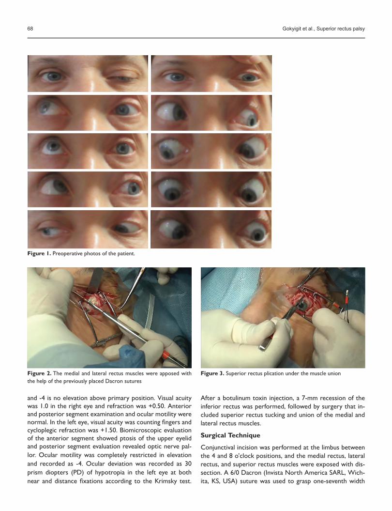
Figure 2. The medial and lateral rectus muscles were apposed with the help of the previously placed Dacron sutures
Figure 3. Superior rectus plication under the muscle union
and -4 is no elevation above primary position. Visual acuity was 1.0 in the right eye and refraction was +0.50. Anterior and posterior segment examination and ocular motility were normal. In the left eye, visual acuity was counting fingers and cycloplegic refraction was +1.50. Biomicroscopic evaluation of the anterior segment showed ptosis of the upper eyelid and posterior segment evaluation revealed optic nerve pal-lor. Ocular motility was completely restricted in elevation and recorded as -4. Ocular deviation was recorded as 30 prism diopters (PD) of hypotropia in the left eye at both near and distance fixations according to the Krimsky test.
After a botulinum toxin injection, a 7-mm recession of the inferior rectus was performed, followed by surgery that in-cluded superior rectus tucking and union of the medial and lateral rectus muscles.
Surgical Technique
Conjunctival incision was performed at the limbus between the 4 and 8 o’clock positions, and the medial rectus, lateral rectus, and superior rectus muscles were exposed with dis-section. A 6/0 Dacron (Invista North America SARL, Wich-ita, KS, USA) suture was used to grasp one-seventh width
Figure 1. Preoperative photos of the patient.

Gokyigit et al., Superior rectus palsy 69
of muscle 10 mm posterior to the insertions of the medi-al and lateral rectus muscles. The muscles were both then passed with partial and full thickness suture bites. Sutures were locked and locking bite was also secured with a twist lock. Approximately 8 to 10 mm posterior to the insertion of the superior rectus muscle, the medial and lateral rectus muscles were apposed with the help of the previously placed 6/0 Dacron sutures, tied and locked 3 times (Fig. 2). Scleral suture was not used at this stage. Then, 8 mm posterior to the insertion, one-third width of the superior rectus muscle was grasped centrally using partial and full thickness bites of 6/0 Dacron sutures, plication anteriorly, and sutured to the sclera 1 mm anterior to the insertion (Fig. 3). Conjunctiva was closed with 8/0 Vicryl (Ethicon, Inc., Somerville, NJ, USA) suture.
Postoperatively, elevation improved significantly and hy-potropia was fully corrected (Fig. 4).
Discussion
Surgical treatment of superior branch of cranial third nerve palsy depends on the type and severity of the paralysis and the presence of other associated factors (2).In the presence of superior rectus palsy, the preferred pro-cedure is a Knapp transposition.
Knapp reported of 15 patients for whom correction of hypotropia ranged from 21 to 55 PD, with a mean of 38 PD.
Good results were obtained in 14 of 15 patients (3). Other authors have found similar rates of correction (4-9).
Mims reported 3 cases of isolated superior rectus palsy and performed 8 to 9-mm recessions of the antagonist infe-rior rectus muscle for abnormal head tilt (10). Either Knapp procedure or augmented modifications (11) eliminate the hypotropia on effected eye similarly vertical rectus proce-dures on the fellow eye. But all these procedures needed many scleral suturations.
In this case, we preferred to perform muscle union sur-gery prior to the superior rectus application. While per-forming the transposition and union of the medial and lateral rectus muscles, we did not use a scleral suture. In addition to being just as effective as the Knapp procedure with or with-out augmentation we observed that this technique is easier and safer than Knapp procedure.
DisclosuresPeer-review: Externally peer-reviewed.
Conflict of Interest: None declared.
Authorship Contributions: Involved in design and conduct of the study (B.G.,S.C.); preparation and review of the study (B.G.,S.C.,A.I.); data collection (A.I.,S.C.).
References
1. von Noorden GK. Binocular Vision and Ocular Motility. Mosby: 2002.
Figure 4. Postoperative photos of the patient.

Gokyigit et al., Superior rectus palsy70
2. Singh A, Bahuguna C, Nagpal R, Kumar B. Surgical management of third nerve palsy. Oman J Ophthalmol 2016;9:80–6. [CrossRef]
3. Knapp P. The surgical treatment of double-elevator paralysis. Trans Am Ophthalmol Soc 1969;67:304–23.
4. Barsoum-Homsy M. Congenital double elevator palsy. J Pediatr Ophthalmol Strabismus 1983;20:185–91.
5. Watson AG. A new operation for double elevator paresis. Trans Can Ophthalmol Soc 1962;25:182–7.
6. Cooper EL, Greenspan G. Operation for double elevator palsy. J Paediatr Ophthalmol Strabismus 1971;8:8–14.
7. Scott WE, Jackson OB. Double elevator palsy: the significance of inferior rectus restriction. Am Orthopt J 1977;27:5–10.
8. Caldeira JA. Vertical transposition of the horizontal rectus muscles for congenital/early onset "acquired" double elevator
palsy: a retrospective long term study of 10 consecutive pa-tients. Binocul Vis Strabismus Q 2000;15:29–38.
9. Kamlesh, Dadeya S. Surgical management of unilateral ele-vator deficiency associated with horizontal deviation using a modified Knapp's procedure. Ophthalmic Surg Lasers Imaging 2003;34:230–5.
10. Mims JL 3rd. Three Similar But Unique Cases Of Isolated Su-perior Rectus ExtraOcular Muscle (EOM) Palsy Strabismus, Presenting With Large Abnormal Head Tilts (Postures, AHP). Binocul Vis Strabolog Q Simms Romano 2011;26:154–69.
11. Snir M, Friling R, Kalish-Stiebel H, Bourla D, Weinberger D, Ax-er-Siegel R. Combined rectus muscle transposition with poste-rior fixation sutures for the treatment of double-elevator palsy. Ophthalmology 2005;112:933–8. [CrossRef]