surgical tutorial 1: tissue extraction and retrieval
TRANSCRIPT

Sponsored by
AAGLAdvancing Minimally Invasive Gynecology Worldwide
Surgical Tutorial 1: Tissue Extraction and Retrieval
PROGRAM CHAIR
Kimberly A. Kho, MD, MPH, MSCR
Christopher S. Awtrey, MD Sarah L. Cohen, MD, MPH

Professional Education Information Target Audience This educational activity is developed to meet the needs of residents, fellows and new minimally invasive specialists in the field of gynecology. Accreditation AAGL is accredited by the Accreditation Council for Continuing Medical Education to provide continuing medical education for physicians. The AAGL designates this live activity for a maximum of 1.0 AMA PRA Category 1 Credit(s)™. Physicians should claim only the credit commensurate with the extent of their participation in the activity. DISCLOSURE OF RELEVANT FINANCIAL RELATIONSHIPS As a provider accredited by the Accreditation Council for Continuing Medical Education, AAGL must ensure balance, independence, and objectivity in all CME activities to promote improvements in health care and not proprietary interests of a commercial interest. The provider controls all decisions related to identification of CME needs, determination of educational objectives, selection and presentation of content, selection of all persons and organizations that will be in a position to control the content, selection of educational methods, and evaluation of the activity. Course chairs, planning committee members, presenters, authors, moderators, panel members, and others in a position to control the content of this activity are required to disclose relevant financial relationships with commercial interests related to the subject matter of this educational activity. Learners are able to assess the potential for commercial bias in information when complete disclosure, resolution of conflicts of interest, and acknowledgment of commercial support are provided prior to the activity. Informed learners are the final safeguards in assuring that a CME activity is independent from commercial support. We believe this mechanism contributes to the transparency and accountability of CME.

Table of Contents
Course Description ........................................................................................................................................ 1 Disclosure ...................................................................................................................................................... 2 The Morcellation Issue and Minimimally Invasive Approaches to Safe Tissue Extraction K.A. Kho ........................................................................................................................................................ 3 Contained Power Morcellation: What Do We Know, What's New and What's on the Horizon? S.L. Cohen ..................................................................................................................................................... 7 A Gynecologic Oncologist's Perspective on Safe Tissue Extraction C.S. Awtrey ................................................................................................................................................. 11 Cultural and Linguistics Competency ......................................................................................................... 15

Surgical Tutorial 1: Tissue Extraction and Retrieval
Kimberly A. Kho, Chair
Faculty: Christopher S. Awtrey, Sarah L. Cohen This session features subject matter experts who will review the most recent data on this important and timely topic. Speakers will use surgical videos to present their thoughts and illustrate techniques on Safe Tissue Extraction and Retrieval. Minimally invasive options for uterine, myoma, and adnexal mass removal will be discussed. Each video will be no more than 15 minutes to allow for a 15-‐minute question and answer segment at the end of the session, taking questions from the audience. Learning Objectives: At the conclusion of this course, the clinician will be able to: 1) Review the risks and benefits of various methods of tissue extraction; and 2) describe methods for tissue extraction in minimally invasive gynecology.
Course Outline 11:00 Welcome, Introductions and Course Overview K.A. Kho
11:05 The Morcellation Issue and Minimimally Invasive Approaches to Safe Tissue Extraction K.A. Kho
11:20 Contained Power Morcellation: What Do We Know, What's New and What's on the Horizon? S.L. Cohen
11:35 A Gynecologic Oncologist's Perspective on Safe Tissue Extraction C.S. Awtrey
11:50 Questions & Answers All Faculty
12:00 Adjourn
1

PLANNER DISCLOSURE The following members of AAGL have been involved in the educational planning of this workshop and have no conflict of interest to disclose (in alphabetical order by last name). Art Arellano, Professional Education Manager, AAGL* Amber Bradshaw Speakers Bureau: Myriad Genetics Lab Other: Proctor: Intuitive Surgical Erica Dun* Frank D. Loffer, Medical Director, AAGL* Linda Michels, Executive Director, AAGL* Johnny Yi* SCIENTIFIC PROGRAM COMMITTEE Arnold P. Advincula Consultant: Intuitive Royalty: CooperSurgical Sarah L. Cohen* Jon I. Einarsson* Stuart Hart Consultant: Covidien Speakers Bureau: Boston Scientific, Covidien Kimberly A. Kho Contracted/Research: Applied Medical Other: Pivotal Protocol Advisor: Actamax Matthew T. Siedhoff Other: Payment for Training Sales Representatives: Teleflex M. Jonathon Solnik Consultant: Z Microsystems Other: Faculty for PACE Surgical Courses: Covidien FACULTY DISCLOSURE The following have agreed to provide verbal disclosure of their relationships prior to their presentations. They have also agreed to support their presentations and clinical recommendations with the “best available evidence” from medical literature (in alphabetical order by last name). Christopher Awtrey* Sarah L. Cohen* Kimberly A. Kho Contracted/Research: Applied Medical Pivotal Protocol Advisor: Actamax Asterisk (*) denotes no financial relationships to disclose.
2

The Morcellation Issue and Minimimally Invasive Approaches to
Safe Tissue ExtractionKimberly Kho, MD, MPH
Gynecology Director, Southwestern Center for Minimally Invasive Surgery University of Texas Southwestern Medical Center
Dallas, TX
Disclosures
• Contracted/Research: Applied Medical
• Other: Pivotal Protocol Advisor: Actamax
Objectives
• Describe methods for preoperative riskassessment to mitigate morcellation of occultmalignancies
• Discuss techniques for minimally invasive tissueextraction
– Transvaginally
– Mini‐lap
– Enclosed power morcellation
Hysterectomies: Controversy grows over rare cancer riskJennifer Levity, February 26, 2014Wall Street Journal
When a hysterectomy can be a death sentenceKaren Weintraub, February 18, 2014, USA TODAY,
Evaluating the Risks of Electric Uterine Morcellation Kimberly A. Kho, MD, MPH1; Ceana H. Nezhat, MD2
JAMA. 2014;311(9):905-906. doi:10.1001/jama.2014.1093.
Cancer Risks from Uterine Morcellation ExaminedFebruary 7, 2014New England Journal of Medicine
Critics of Fibroid Removal Procedure Question Risks It May Pose for Women With Undetected Uterine Cancer
Tracy Hampton, PhD JAMA. 2014;311(9):891-893. doi:10.1001/jama.2014.27.
Uterine Surgical Technique Is Linked to Abnormal Growths and Cancer SpreadDenise Grady, February 6, 2014The New York Times
Temple restricts controversial gynecological procedureMarie McCullough, February 22, 2014Philadelphia Inquirer
Concerns about intracorporeal EMM• Tissue dissemination
– Benign tissue (parasitic myomas, disseminatedleiomyomatosis, endometriosis, adenomyosis, splenosis)
– Occult malignancy (sarcomas, endometrial,cervical, ovarian pathology)
• Histopathologic challenges
• Potential worsening of prognosis and need forre‐intervention
• Organ injury
– FDA MAUDE review (‘03‐’13 55 complications, 6 deaths)
– Associated with user inexperience
Al Talib A, Tulundi T, et al. GOI, 2010.
Risk estimates for occult malignancy• FDA: 1:352 risk of sarcoma in women undergoingmyomectomy/hysterectomy for suspected fibroids
• 1:458 risk of occult LMS
• Wright: 0.27% incidence of uterine malignancy inwomen undergoing morcellation during MIS
• 1.01% incidence of endometrial hyperplasia
• Manhert: 2.7% unexpected malignancy in hysterectomypts; 0.22% incidence of occult sarcoma
• Kho: 9 sarcomas: 10,119 benign hysterectomy (0.09%)• 5 LMS (0.05%)
No methods have been proven to reduce the risk of cellular dispersion
3

Preoperative considerations• Informed consent process
• Preoperative evaluation– Thorough history and physical, including family hx
– Endometrial evaluation –• longitudinal review of imaging
– Up to date cervical cancer screening
– Consideration of risk factors for sarcoma• Tamoxifen use, pelvic XRT, HLRCC Sx
• New symptoms or growth in postmenopausal woman
• Tissue extraction techniques– Enclosed vs non‐enclosed
– Intra‐corporeal vs extra‐corporeal
Utilization of cutting edge MRI modalities
• Diffusion weighed imaging (DWI) measures random movement of water in tissue– Increased intensity due to restricted water movement in highly cellular areas
– Calculate ADC – quantitative assessment of cellularity
• Dynamic contrast enhancement (DCE)‐ for quantification of tissue perfusion & permeability– Rapid accumulation and washout may suggest malignancy
How do we safely remove large tissue masses while still performing MIS?
• Utilize techniques during– Hysterectomy (TLH, SCH)
– Myomectomy
– Cystectomy, Oophorectomy
• Routes for tissue extirpation– Mini‐laparotomy
– Vaginally through colpotomy
– Laparoscopic power morcellation within containment bag
Goal: To minimize risks while maximizing benefits
Retrieval Bags
• Endoscopic bags – Ethicon Endopouch: 4x6 in, 224 cc
– Covidien EndoCatch: 10mm‐ 6 cm d (220 cc), 15mm‐ 12.7 cm d (1000cc)
– Applied Contained Extraction System: 17cm d (6500 cc)
• Isolation (Lahey) bags – transparent PVC– 3M, Isodrape – 50 x 50 cm
• Containment bag – rip stop nylon– Cook LapSac: 4x6, 5x8, 8x10 cm (1500cc)
– BERT bags: 30 x35 cm (3000 cc)
– Eco Sac: 3100 cc
Mini‐laparotomy Extraction
• Use a circumferential wound retractor
– New systems combining containment bag with abdominal wound retractor
• 2‐3 cm incision
– Suprapubic vs Umbilical
• Lahey thyroid clamps
• 10 or 11 blade
• Peeling technique
Mini‐laparotomy Extraction during Laparoscopic Myomectomy
4

Vaginal tissue extraction
• Naturally occurring, accomodating orifice
• Maintain existing port sites without extension
• Consider this a clean contaminated procedure
• Clinical trials compared to trans‐abdominal extraction have not shown inferior outcomes:
– Conversion
– Postoperative pain
– Dyspareunia
– Infection
– Adhesions
Transvaginal morcellation
• Coring
• Bi‐valving
• Wedge resection
• Myomectomy
• Posterior retractor
• Tenaculums to maintain specimen orientation
Enclosed transvaginal morcellation after TLH In bag manual morcellation through posterior colpotomy
• Creation of posterior colpotomy
– Create colpotomy laparoscopically
• Moist sponge stick for delineation
• Monopolar hook, scissors, harmonic scalpel
– Create colpotomy vaginally
• Introduce containment bag
• Vaginal extraction and enclosed morcellation
• Vaginal closure
Enclosed morcellation of 18 cm adnexal mass via posterior colpotomy
Enclosed morcellation of 18 cm adnexal mass via posterior colpotomy
5

Multiport power morcellation in insufflated bag
• Technique not FDA‐approved, currently being used in clinical trials
• Use large containment bag
• Insert bag via 15 mm umbilical incision
• Open bag in abdominal cavity and place specimen within bag
– Use reverse Trendelenburg positioning
• Insufflate bag intra‐abdominally
• Concerns regarding bag integrity and need for puncture of bag
Multiport power morcellation in insufflated bag
On the horizon
• Innovation of instrumentation and techniques
• Prospective evaluation of safety and outcomes
• Systematic reporting of AE’s for accurate ascertainment of risks
• Development of preoperative diagnostic tools for risk stratification
AAGL practice report: morcellation during uterine tissue extraction. J Minim Invasive Gynecol. 2014 Jul‐Aug;21(4):517‐30. Kho KA, Nezhat C. Parasitic myomas. Obstetrics and gynecology. 2009;114(3):611‐5.Nezhat C, Kho K. Iatrogenic myomas: new class of myomas? Journal of minimally invasive gynecology. 2010;17(5):544‐50.Donnez O, Squifflet J, Leconte I, Jadoul P, Donnez J. Posthysterectomy pelvic adenomyoticmasses observed in 8 cases out of a series of 1405 laparoscopic subtotal hysterectomies. Journal of minimally invasive gynecology. 2007;14(2):156‐Hilger WS, Magrina JF. Removal of pelvic leiomyomata and endometriosis five years after supracervical hysterectomy. Obstetrics and gynecology. 2006;108(3 Pt 2):772‐4.Kho KA, Anderson TL, Nezhat CH. Intracorporeal practice report: Morcellation electromechanical tissue morcellation: a critical review and recommendations for clinical practice. Obstet Gynecol. 2014 Oct;124(4):787‐93.Al‐Talib A, Tulandi T. Pathophysiology and possible iatrogenic cause of leiomyomatosis peritoneals disseminata. Gynecol Obstet Invest. 2010;69(4):239‐44.Mahnert N, Morgan D, Campbell D, Johnston C, As‐Sanie S. Unexpected gynecologic malignancy diagnosed after hysterectomy performed for benign indications. Obstet Gynecol. 2015 Feb;125(2):397‐405.Wright JD, Tergas AI, Burke WM, Cui RR, Ananth CV, Chen L, HershmanDL. Uterine pathology in women undergoing minimally invasive hysterectomy using morcellation. JAMA. 2014 Sep 24;312(12):1253‐5. Nezhat F, Brill AI, Nezhat CH, Nezhat C. Adhesion formation after endoscopic posterior colpotomy. The Journal of reproductive medicine. 1993;38(7):534‐6.Uccella S, Cromi A, Bogani G, Casarin J, Serati M, Ghezzi F. Transvaginal specimen extraction at laparoscopy without concomitant hysterectomy: our experience and systematic review of the literature. Journal of minimally invasive gynecology. 2013;20(5):583‐90.Goto A, Takeuchi S, Sugimura K, Maruo T. Usefulness of Gd‐DTPA contrast‐enhanced dynamic MRI and serum determination of LDH and its isozymes in the differential diagnosis of leiomyosarcoma from degenerated leiomyoma of the uterus. International journal of gynecological cancer : official journal of the International Gynecological Cancer Society. 2002;12(4):354‐61.Sato K, Yuasa N, Fujita M, Fukushima Y. Clinical application of diffusion‐weighted imaging for preoperative differentiation between uterine leiomyoma and leiomyosarcoma. Am J Obstet Gynecol. Dec 22 2013.Cornfeld D, Israel G, Martel M, Weinreb J, Schwartz P, McCarthy S. MRI appearance of mesenchymal tumors of the uterus. European journal of radiology. Apr 2010;74(1):241‐249.Thomassin‐Naggara I, Dechoux S, Bonneau C, et al. How to differentiate benign from malignant myometrial tumours using MR imaging. European radiology. Aug 2013;23(8):2306‐2314.Cohen SL, Einarsson JI, Wang KC, Brown D, Boruta D, Scheib SA, Fader AN, Shibley T. Contained power morcellation within an insufflated isolation bag. Obstet Gynecol. 2014 Sep;124(3):491‐7
References
Thank you!
Email: [email protected]
6
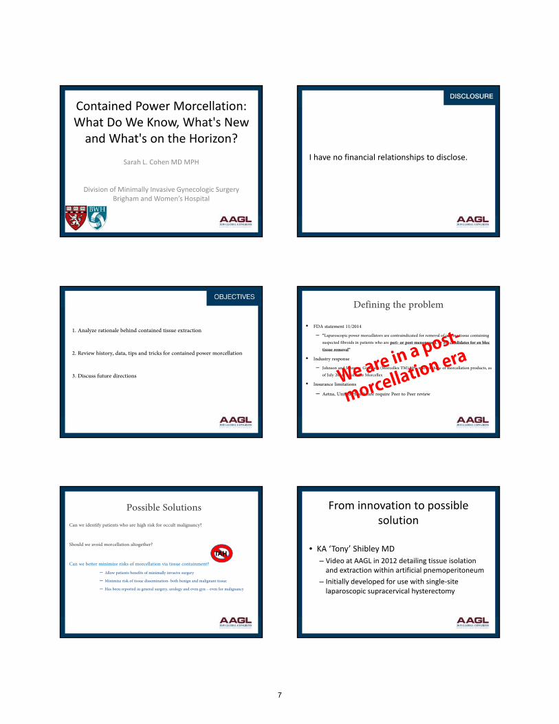
Contained Power Morcellation:What Do We Know, What's New and What's on the Horizon?
Sarah L. Cohen MD MPH
Division of Minimally Invasive Gynecologic SurgeryBrigham and Women’s Hospital
I have no financial relationships to disclose.
1. Analyze rationale behind contained tissue extraction
2. Review history, data, tips and tricks for contained power morcellation
3. Discuss future directions
Defining the problem
• FDA statement 11/2014
– “Laparoscopic power morcellators are contraindicated for removal of uterine tissue containing suspected fibroids in patients who are peri- or post-menopausal, or are candidates for en bloc tissue removal”
• Industry response
– Johnson and Johnson: Gynecare (Morcellex TM) 80% market share of morcellation products, as of July 2014- recalls the Morcellex
• Insurance limitations
– Aetna, United Healthcare require Peer to Peer review
Possible SolutionsCan we identify patients who are high risk for occult malignancy?
Should we avoid morcellation altogether?
Can we better minimize risks of morcellation via tissue containment?
– Allow patients benefits of minimally invasive surgery
– Minimize risk of tissue dissemination- both benign and malignant tissue
– Has been reported in general surgery, urology and even gyn – even for malignancy
TAH
From innovation to possible solution
• KA ‘Tony’ Shibley MD
– Video at AAGL in 2012 detailing tissue isolation and extraction within artificial pnemoperitoneum
– Initially developed for use with single‐site laparoscopic supracervical hysterectomy
7

Single port Shibley VideoInitial Technique: Cohen et al. Obstet Gynecol. 2014.
Collaboration between BWH, MGH, JHH, KA Shibley
Jan 2013-April 2014: 73 patients, robotic and conventional LSC
–Hysterectomy: Mulitport 29, single site 32
–Myomectomy: Multiport 11, single site 1
Cohen et al. Obstet Gynecol. 2014.
–2/3 had prior abdominal surgery
–Median operative time 114 min (32-380 min)
–Median EBL 50 mL (10-500 mL)
–Median specimen weight 257 gm (53-1,481 gms)
–No conversions, readmissions, or reoperation; 78% discharged home same day
Cohen et al. Obstet Gynecol. 2014.
Limitations: lack objective measure bag integritysmall observational studyspecialized high volume surgeons
Contained Power Morcellation: Multi-port approach Videos
Contained Power Morcellation Emerging Data
Vargas et al. JMIG. 2015
– Compared OR time 3 months before and after implementing in bag power morcellation
– 36 IBM, 49 open morcellation; IBM added 26 minutes to OR time
8

Contained Power Morcellation Emerging Data
Cohen et al. JMIG 2014
– Pilot study: Morcellation of 500g beef tongue in simulation lab
– Compared 3 techniques: Nylon bag mulitport, Isolation bag multiport, Isolation bag single port
– Evaluated spill with indigo carmine and by cytology
– Blue dye spilled in 1 of 12 trials (from seam of bag upon insufflation)- visible and on cytology
Contained Power Morcellation Emerging Data
Submitted for publication:
Prospective study across 7 sites in BostonMulti-port approach, varying bags usedPrimary outcome: leakage of tissue or blue dye
Enrollment goal 400, early stop at 89 patients due to leakage events
7 cases of dye or tissue leakage on post morcellation survey
Contained Power Morcellation - Data
Submitted for publication:
76 cases successful3 specimen too large10 morcellation not necessary
No bag tears during morcellation
Avg morcellation time 30.2 minutes (+ 22.4)1 patient with STUMP on final pathology1 complication of large EBL
Contained Power Morcellation- Variations
• Akdemir et al. Obstet Gynecol. 2015
– Enclosed morcellation using insufllated size 8.5 surgical glove
Contained Power Morcellation- Variations
• Review literature for any new developments/publications since summer 2015
Contained Tissue Extraction: Future Directions
Espiner sleeved Ecosac prototypeIsakov et al. J Med Devices 8(1), 2014.
9

•U.S. Food and Drug Administration. UPDATED Laparoscopic Uterine Power Morcellation in Hysterectomy and Myomectomy: FDA Safety Communication. November 14, 2014.
•Park JY, Park SK, Kim DY, Kim JH, Kim YM, Kim YT, Nam JH. The impact of tumor morcellation during surgery on the prognosis of patients with apparently early uterine leiomyosarcoma. Gynecol Oncol. 2011; 122(2):255-9.
•Perri T, Korach J, Sadetzki S, Oberman B, Fridman E, Ben-Baruch G. Uterine leiomyosarcoma: does the primary surgical procedure matter? Int J Gynecol Cancer. 2009;19(2):257-60.
•George S, Barysauskas C, Serrano C, Oduyebo T, Rauh-Hain JA, Del Carmen MG, Demetri GD, Muto MG. Retrospective cohort study evaluating the impact of intraperitoneal morcellation on outcomes of localized uterine leiomyosarcoma. Cancer. 2014;120(20):3154-8.
•Einarsson, JI, Cohen SL, Fuchs-Weizman N, Wang KC. In bag morcellation. J Minim Invasive Gynecol. 2014; 21(5):951-3.
•Cohen SL, Einarsson JI, Wang KC, Brown DN, Boruta D, Scheib SA, Fader AN, Shibley KA. Contained power morcellation within an insufflated isolation bag. ObstetGynecol. 2014; 124(3):491-7.
•Cohen SL, Greenberg JA, Wang KC, Srouji SS, Gargiulo AR, Pozner CN, Hoover N, Einarsson JI. Risk of leakage and tissue dissemination with various contained tissue extraction (CTE) techniques: an in vitro pilot study. J Minim Invasive Gynecol. 2014; 21(5):935-9.
•Vargas MV, Cohen SL, Fuchs-Weizman N, Wang KC, Manoucheri E, Vitonis AF, Einarsson JI. Open power morcellation versus contained power morcellation within an insufflated isolation bag: comparison of perioperative outcomes. J Minim Invasive Gynecol. 2015 ;22(3):433-8.
•Akdemir A, Taylan E, Zeybek B, Ergenoglu AM, Sendag F. Innovative technique for enclosed morcellation using a surgical glove. Obstet Gynecol. 2015 ;125(5):1145-9.
10

A Gynecologic Oncologist's Perspective on Safe Tissue
Extraction
Christopher S. Awtrey, M.D.
Director, Division of Gynecologic OncologyBeth Israel Deaconess Medical Center
• I have no financial relationships to disclose.
1. Explain the potential oncologic concerns with electromechanical morcellation.
2. Review importance of conversation and consent with patients and their families.
3. Discuss the opportunity for advancement of our field.
The newspapers pick up a tragic story!
• Amy Reed, an anesthesiologist at BIDMC has 6 children and had been followed thru her last pregnancy for a fibroid uterus
• She underwent laparoscopic hysterectomy and electromechanical morcellation
• Discovered postoperatively that she had a LMS
• Went on to have re‐exploration, Heated IP chemotherapy
11

Mark Twain
• If you don’t read newspapers you are uninformed. If you read the newspaper, you’re mis‐informed.
Morcellated specimens, the risks• Injury to surrounding structures
EMM-rotating blade
Vaginal / Abdominal with blade
• Dissemination of benign tissue
• Diffuse benign leiomyomatosis
• Endometriosis
• Dissemination of malignant tissue
• LMS, ESS
• Carcinosarcoma, endometrial cancers
• FT/Ovary cancer
• Inability to identify or characterize pathology due to fragmentation
Gynecologic Cancer: New CasesUnited States, 2015
Cervix 12,900Vulva 5,150Other 4,070
Corpus 54,870Ovary 21,290
Siegel RL et al, CA Cancer J Clin;65;5-29; 2015
5%13%
56%
22%4%
Ovary
Corpus
CervixVulvaOther
Corpus Cancer
• Majority of corpus cancer is endometrial (92%)
• Corpus Mesenchymal Tumors (8%) 4,390 pts in 2015
– Sarcomas 43% (1,888 patients in 2015) ****
– Carcinosarcomas 39% (1,712 patients in 2015)
– Endometrial stromal sarcomas 15% (659 patients in 2015)
**** Doesn’t include STUMP tumors, cellular leiomyomas, atypical myomas
12

Uterine Pathology in Women Undergoing Minimally Invasive Hysterectomy Using Morcellation
(Wright et al. JAMA 2014)
• Database 232,882 patients underwent MIS surgery 2006‐2012
• 36,470 (15.7%) Morcellation
– 27/10,000 cancers in morcellated uteri (3/1000)
– Increased with age
•Prevalence ratio comp to younger than 40 years old
•Greater than 65 prevalence ratio 35.97
Unexpected gynecologic malignancy discovered after hysterectomy performed for
bengin indicationsMahnert et al Ob & Gyn 2015
•Aim: to determine frequency of unexpected malignancy in patients having a hysterectomy for benign indications
•Statewide Database data collected for 2013
•3,360 patients had a hysterectomy for benign indications
•Unexpected path 172 pts (2.7%)
•Ovary 1.08%
•Endometrial 1.02
•Uterine sarcoma 0.2%
•Cervical 0.17%
So now what….?• Currently at BIDMC EMM is not utilized unless cleared by a expert review panel
• Alternative extraction methods are being developed and some are being dusted off
• In‐bag hand morcellation
• Vaginal surgery
• Mini‐laparotomy
• More laparotomies are likely being done
In Bag Morcellation
In Bag Morcellation• Established technique in MIS removal of kidneys, ovaries and spleen
• Gynecologic specimens, uteri, fibroids may be large
– Specialized bags
• It’s a challenge to hand carve the specimens
– Specialized tools
• Still challenges for our pathologist due to fragmented specimen
Our work
13

AAGL Practice Report
• Collaboration with device makers to innovate new contained morcellation systems
• More rigorous system for mandatory adverse events reporting
• Stressed the importance of an informed consent process
The Future
• New technologic advances are needed
• Contained morcellation systems
• Improvements in detection, dx Sarcoma
• Serum Profile?
• MRI?
• Nationally we need to improve in understanding how best to advocate for patients
• National registry, monitoring and discussion of adverse events
14

CULTURAL AND LINGUISTIC COMPETENCY Governor Arnold Schwarzenegger signed into law AB 1195 (eff. 7/1/06) requiring local CME providers, such as
the AAGL, to assist in enhancing the cultural and linguistic competency of California’s physicians
(researchers and doctors without patient contact are exempt). This mandate follows the federal Civil Rights Act of 1964, Executive Order 13166 (2000) and the Dymally-Alatorre Bilingual Services Act (1973), all of which
recognize, as confirmed by the US Census Bureau, that substantial numbers of patients possess limited English proficiency (LEP).
California Business & Professions Code §2190.1(c)(3) requires a review and explanation of the laws
identified above so as to fulfill AAGL’s obligations pursuant to California law. Additional guidance is provided by the Institute for Medical Quality at http://www.imq.org
Title VI of the Civil Rights Act of 1964 prohibits recipients of federal financial assistance from
discriminating against or otherwise excluding individuals on the basis of race, color, or national origin in any of their activities. In 1974, the US Supreme Court recognized LEP individuals as potential victims of national
origin discrimination. In all situations, federal agencies are required to assess the number or proportion of LEP individuals in the eligible service population, the frequency with which they come into contact with the
program, the importance of the services, and the resources available to the recipient, including the mix of oral
and written language services. Additional details may be found in the Department of Justice Policy Guidance Document: Enforcement of Title VI of the Civil Rights Act of 1964 http://www.usdoj.gov/crt/cor/pubs.htm.
Executive Order 13166,”Improving Access to Services for Persons with Limited English
Proficiency”, signed by the President on August 11, 2000 http://www.usdoj.gov/crt/cor/13166.htm was the genesis of the Guidance Document mentioned above. The Executive Order requires all federal agencies,
including those which provide federal financial assistance, to examine the services they provide, identify any
need for services to LEP individuals, and develop and implement a system to provide those services so LEP persons can have meaningful access.
Dymally-Alatorre Bilingual Services Act (California Government Code §7290 et seq.) requires every
California state agency which either provides information to, or has contact with, the public to provide bilingual
interpreters as well as translated materials explaining those services whenever the local agency serves LEP members of a group whose numbers exceed 5% of the general population.
~
If you add staff to assist with LEP patients, confirm their translation skills, not just their language skills.
A 2007 Northern California study from Sutter Health confirmed that being bilingual does not guarantee competence as a medical interpreter. http://www.pubmedcentral.nih.gov/articlerender.fcgi?artid=2078538.
US Population
Language Spoken at Home
English
Spanish
AsianOther
Indo-Euro
California
Language Spoken at Home
Spanish
English
OtherAsian
Indo-Euro
19.7% of the US Population speaks a language other than English at home In California, this number is 42.5%
15