susie bowell ba (hons) rgn heart failure specialist nurse rgn ba (hons)
TRANSCRIPT
HEART FAILURE
Susie Bowell BA (Hons) RGN
Heart Failure Specialist NurseRGN BA (Hons)
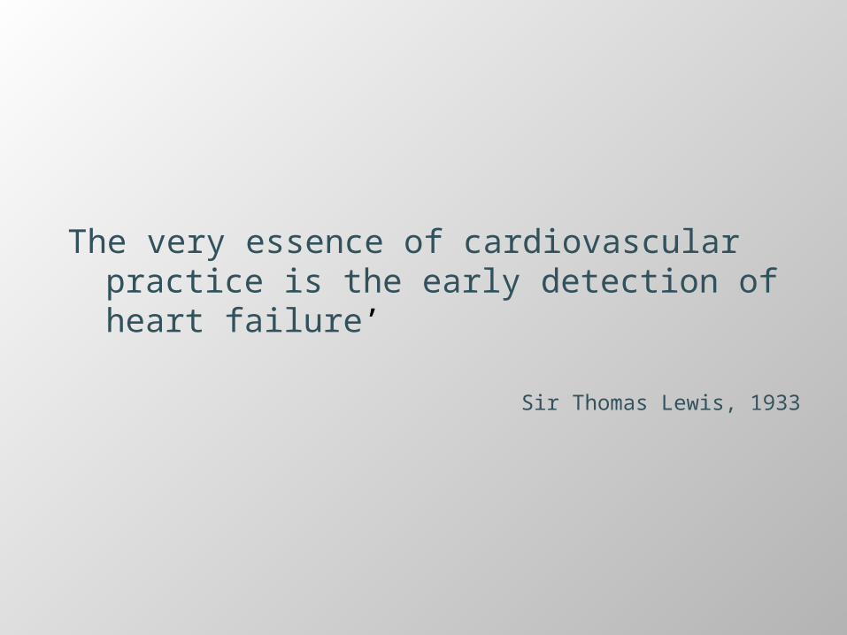
The very essence of cardiovascular practice is the early detection of heart failure’
Sir Thomas Lewis, 1933

Heart Failure Definition
Heart failure (HF) is a complex clinical syndrome of symptoms and signs that suggest impairment of the heart as a pump supporting physiological circulation.
It is caused by structural or functional abnormalities of the heart.
The demonstration of objective evidence of these cardiac abnormalities is necessary for the diagnosis of heart failure to be made.
NICE GUIDELINES, 2010

Heart Failure
30-40% of newly diagnosed will die within one year
After the first year there is a less than10% per year mortality rate
NICE 2010
Incidence and hospital admissions are predicated to rise by 50% over the next 25 years due to ageing demographics and the success in treating Ischaemic Heart Disease.
Mean age of HF patient 74yearsPrevalence approx. 8‐10% in over 75s have heart failure
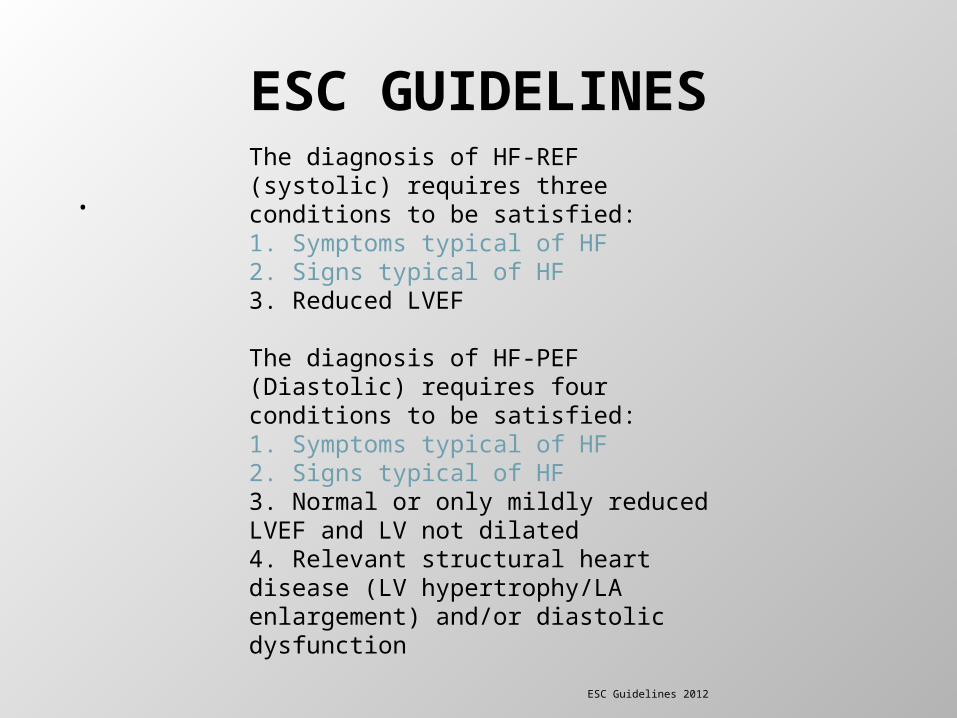
ESC GUIDELINES
.The diagnosis of HF-REF (systolic) requires three conditions to be satisfied:1. Symptoms typical of HF2. Signs typical of HF3. Reduced LVEF
The diagnosis of HF-PEF (Diastolic) requires four conditions to be satisfied:1. Symptoms typical of HF2. Signs typical of HF3. Normal or only mildly reduced LVEF and LV not dilated4. Relevant structural heart disease (LV hypertrophy/LAenlargement) and/or diastolic dysfunction
ESC Guidelines 2012
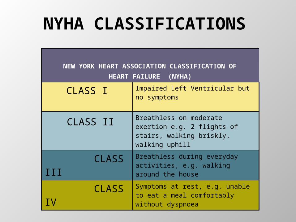
NYHA CLASSIFICATIONS
NEW YORK HEART ASSOCIATION CLASSIFICATION OF HEART FAILURE (NYHA)
CLASS I Impaired Left Ventricular but no symptoms
CLASS II Breathless on moderate exertion e.g. 2 flights of stairs, walking briskly, walking uphill
CLASS III Breathless during everyday activities, e.g. walking around the house
CLASS IV Symptoms at rest, e.g. unable to eat a meal comfortably without dyspnoea
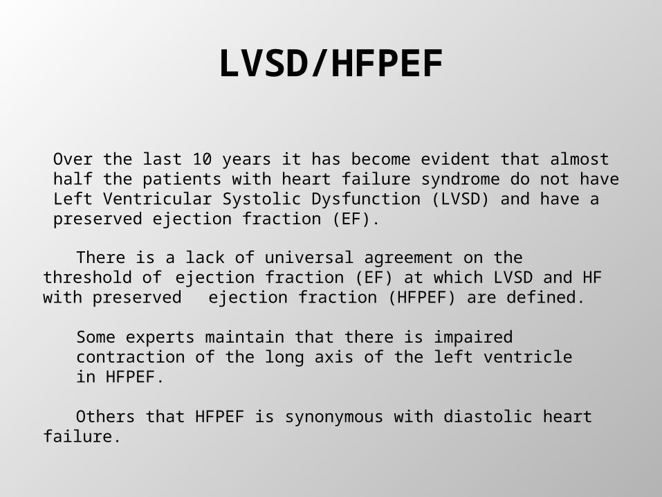
LVSD/HFPEF
Over the last 10 years it has become evident that almost half the patients with heart failure syndrome do not have Left Ventricular Systolic Dysfunction (LVSD) and have a preserved ejection fraction (EF).
There is a lack of universal agreement on the threshold of ejection fraction (EF) at which LVSD and HF with preserved ejection fraction (HFPEF) are defined.
Some experts maintain that there is impaired contraction of the long axis of the left ventricle in HFPEF.
Others that HFPEF is synonymous with diastolic heart failure.
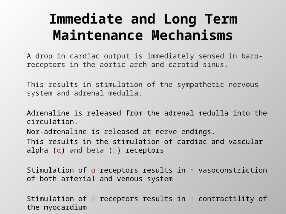
Immediate and Long Term Maintenance Mechanisms
A drop in cardiac output is immediately sensed in baro-receptors in the aortic arch and carotid sinus.
This results in stimulation of the sympathetic nervous system and adrenal medulla.
Adrenaline is released from the adrenal medulla into the circulation. Nor-adrenaline is released at nerve endings. This results in the stimulation of cardiac and vascular alpha (α) and beta (β) receptors
Stimulation of α receptors results in ↑ vasoconstriction of both arterial and venous system
Stimulation of β receptors results in ↑ contractility of the myocardium
↑ increased heart rate
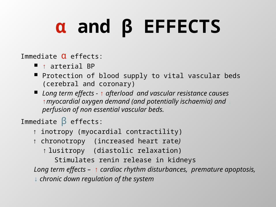
α and β EFFECTSImmediate α effects:
↑ arterial BP Protection of blood supply to vital vascular beds (cerebral and
coronary) Long term effects - ↑ afterload and vascular resistance
causes ↑myocardial oxygen demand (and potentially ischaemia) and ↓ perfusion of non essential vascular beds.
Immediate β effects:
↑ inotropy (myocardial contractility)↑ chronotropy (increased heart rate)
↑ lusitropy (diastolic relaxation) Stimulates renin release in kidneys
Long term effects – ↑ cardiac rhythm disturbances, premature apoptosis,
↓ chronic down regulation of the system
HEART FAILURE WITH REDUCED EJECTION FRACTION (HFREF)
VERSUSHEART FAILURE WITH PRESERVED EJECTION
FRACTION (HFPEF)
As heart failure has a poor prognosis, early pharmacological treatment is important and can have a great affect on future prognosis. The improved prognosis of heart failure patients with left ventricular systolic dysfunction in the last decade is likely to be related to the greater use of and earlier intervention with, pharmacological treatment.
Mortality within the first month of diagnosis remains high at 6%.
HFREFDiagnosis and Treatment

Diagnosis
Studies show diagnosis incorrect in approx 40‐50% of cases based on signs and symptoms. An echocardiogram is essential for diagnostic purposes4
Chest crepitations, oedema, tachycardia –not specific
S3, ↑JVP, displaced apex –insensitive, poor inter‐observer agreement
Many patients have symptoms only –dyspnoea, fatigue are non specific
Diagnosis
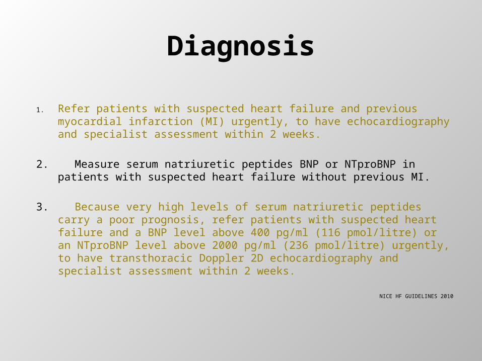
1. Refer patients with suspected heart failure and previous myocardial infarction (MI) urgently, to have echocardiography and specialist assessment within 2 weeks.
2. Measure serum natriuretic peptides BNP or NTproBNP in patients with suspected heart failure without previous MI.
3. Because very high levels of serum natriuretic peptides carry a poor prognosis, refer patients with suspected heart failure and a BNP level above 400 pg/ml (116 pmol/litre) or an NTproBNP level above 2000 pg/ml (236 pmol/litre) urgently, to have transthoracic Doppler 2D echocardiography and specialist assessment within 2 weeks.
NICE HF GUIDELINES 2010
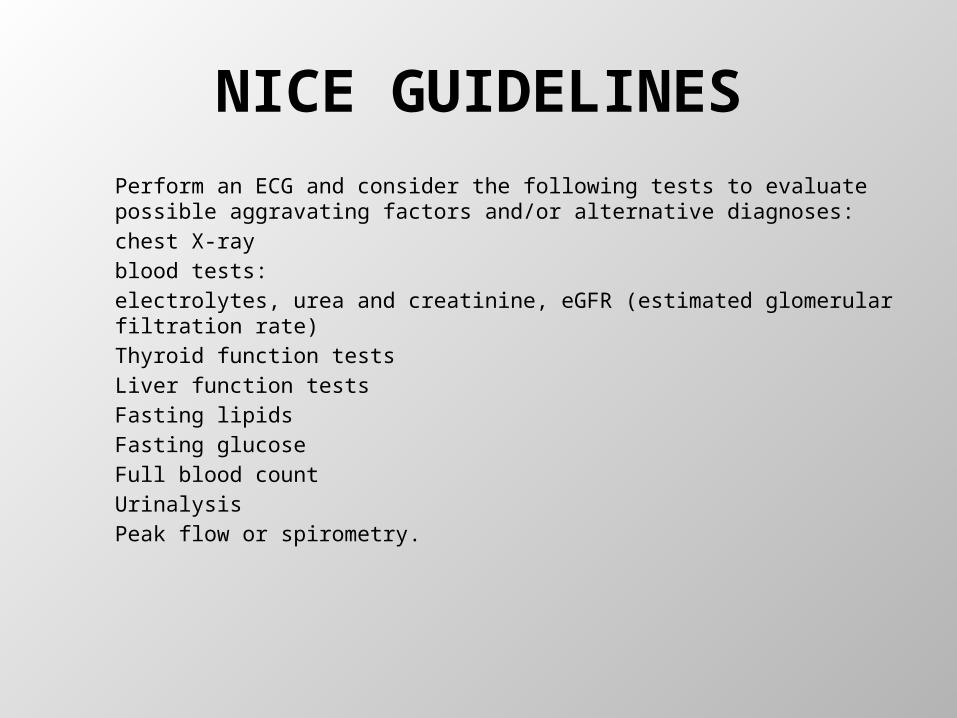
NICE GUIDELINESPerform an ECG and consider the following tests to evaluate possible aggravating factors and/or alternative diagnoses:chest X-rayblood tests:electrolytes, urea and creatinine, eGFR (estimated glomerular filtration rate)Thyroid function testsLiver function testsFasting lipidsFasting glucoseFull blood countUrinalysisPeak flow or spirometry.
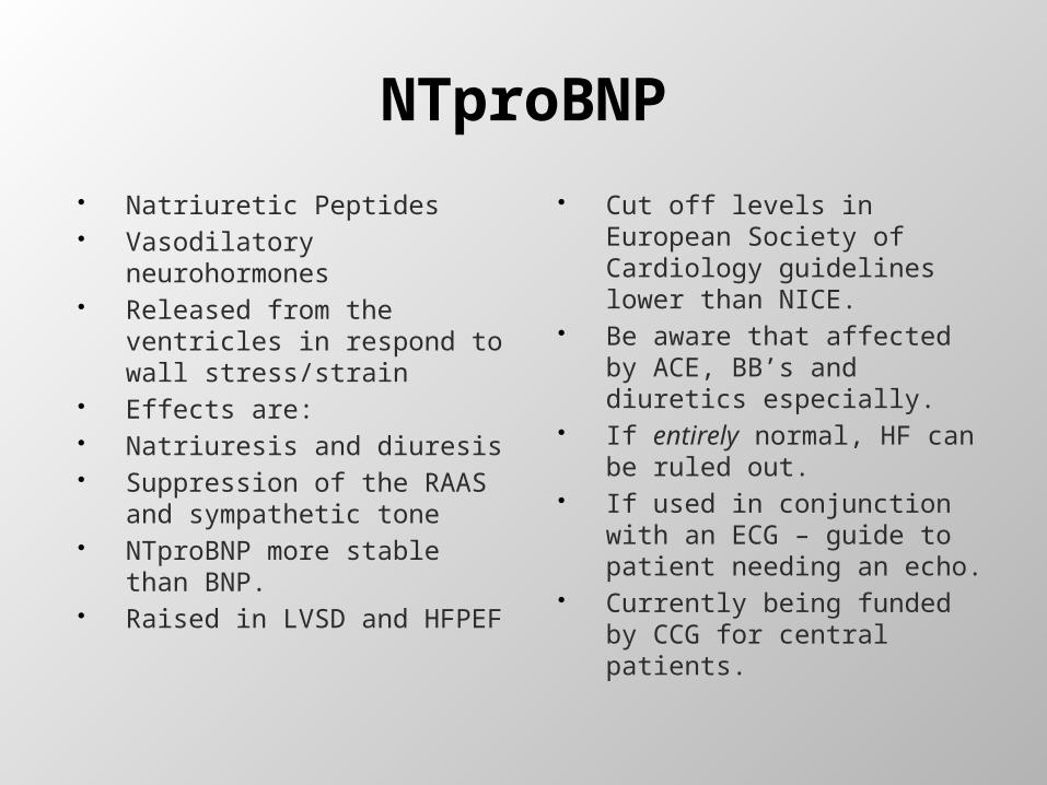
NTproBNP
Natriuretic Peptides Vasodilatory neurohormones Released from the ventricles
in respond to wall stress/strain
Effects are: Natriuresis and diuresis Suppression of the RAAS
and sympathetic tone NTproBNP more stable than
BNP. Raised in LVSD and HFPEF
Cut off levels in European Society of Cardiology guidelines lower than NICE.
Be aware that affected by ACE, BB’s and diuretics especially.
If entirely normal, HF can be ruled out.
If used in conjunction with an ECG – guide to patient needing an echo.
Currently being funded by CCG for central patients.
NTproBNP
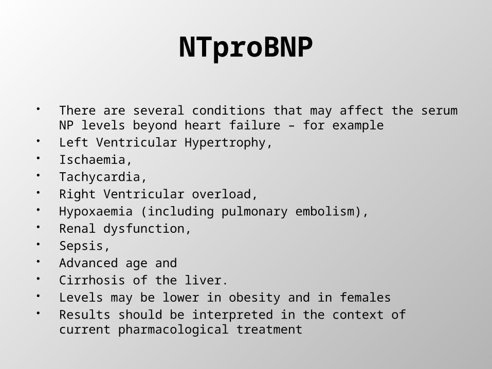
There are several conditions that may affect the serum NP levels beyond heart failure – for example
Left Ventricular Hypertrophy, Ischaemia, Tachycardia, Right Ventricular overload, Hypoxaemia (including pulmonary embolism), Renal dysfunction, Sepsis, Advanced age and Cirrhosis of the liver. Levels may be lower in obesity and in females Results should be interpreted in the context of current
pharmacological treatment

NTproBNP A high natriuretic peptide level is not only of diagnostic significance, but also of
prognostic significance.
Thresholds:
BNP>400 pg/ml (>116 pmol/l) or NT-proBNP>2000 pg/ml (>236 pmol/l): Need anechocardiogram and specialist clinical assessment no longer than 2 weeks from thetime of presentation.
BNP 100-400 pg/ml (29-116 pmol/l) or NT-proBNP 400-2000 pg/ml (47-236 pmol/l):Need an echocardiogram and clinical assessment by the Specialist within 6 weeksfrom the time of presentation. BNP <100 pg/ml (<29 pmol/l) or NT-proBNP <400 pg/ml (<47 pmol/l), in the absenceof heart failure therapy: Heart Failure is an unlikely cause for the presentation
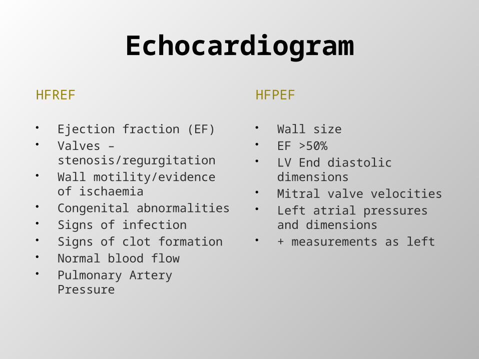
Echocardiogram
HFREF
Ejection fraction (EF) Valves –
stenosis/regurgitation Wall motility/evidence of
ischaemia Congenital abnormalities Signs of infection Signs of clot formation Normal blood flow Pulmonary Artery Pressure
HFPEF
Wall size EF >50% LV End diastolic dimensions Mitral valve velocities Left atrial pressures and
dimensions + measurements as left
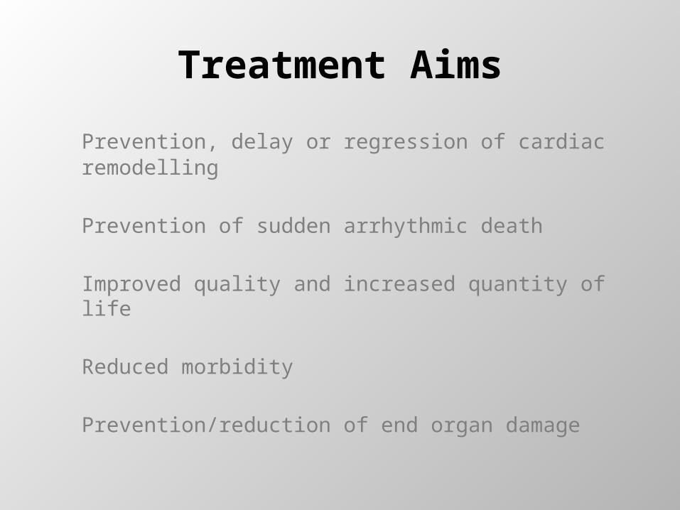
Treatment Aims
Prevention, delay or regression of cardiac remodelling
Prevention of sudden arrhythmic death
Improved quality and increased quantity of life
Reduced morbidity
Prevention/reduction of end organ damage
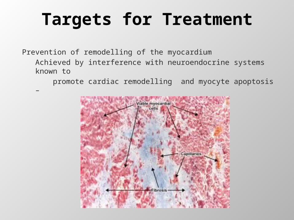
Targets for Treatment
Prevention of remodelling of the myocardiumAchieved by interference with neuroendocrine systems known to
promote cardiac remodelling and myocyte apoptosis –
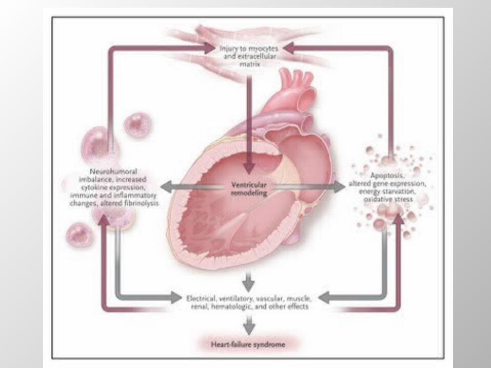
Pathophysiology

MERIT-HF Study Group. Effect of Metoprolol CR/XL in chronic heart failure: Metoprolol CR/XL randomized intervention trial in congestive heart failure (MERIT-HF). LANCET. 1999;353:2001-07.
Causes of Death
12%
24%
64%
CHF
Other
SuddenDeath
n = 103
NYHA II
26%
15%
59%
CHF
Other
SuddenDeath
n = 103
NYHA III
56%
11%
33%
CHF
Other
SuddenDeath
n = 27
NYHA IV
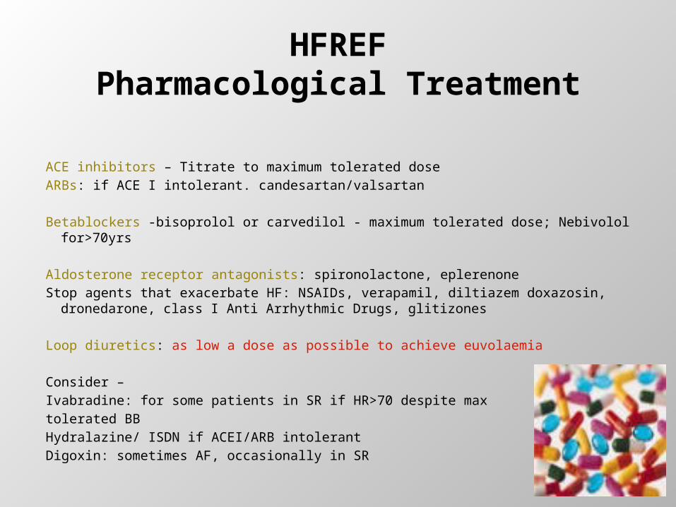
HFREFPharmacological Treatment
ACE inhibitors – Titrate to maximum tolerated doseARBs: if ACE I intolerant. candesartan/valsartan
Betablockers -bisoprolol or carvedilol - maximum tolerated dose; Nebivolol for>70yrs
Aldosterone receptor antagonists: spironolactone, eplerenoneStop agents that exacerbate HF: NSAIDs, verapamil, diltiazem doxazosin,
dronedarone, class I Anti Arrhythmic Drugs, glitizones
Loop diuretics: as low a dose as possible to achieve euvolaemia
Consider –Ivabradine: for some patients in SR if HR>70 despite max tolerated BBHydralazine/ ISDN if ACEI/ARB intolerantDigoxin: sometimes AF, occasionally in SR
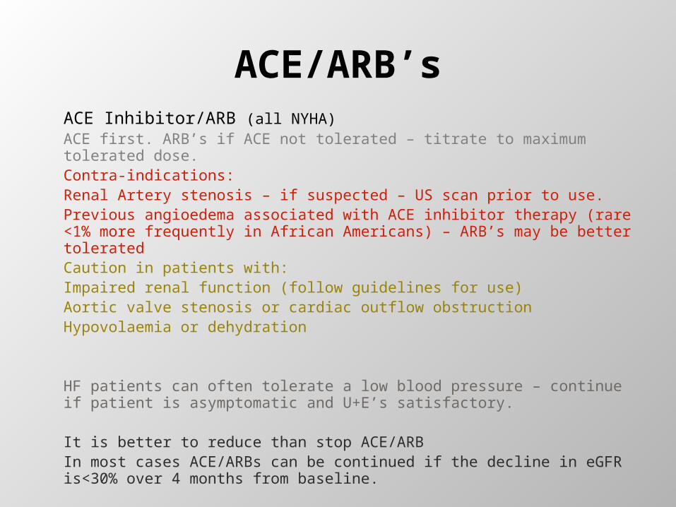
ACE/ARB’sACE Inhibitor/ARB (all NYHA)ACE first. ARB’s if ACE not tolerated – titrate to maximum tolerated dose.Contra-indications:Renal Artery stenosis – if suspected – US scan prior to use.Previous angioedema associated with ACE inhibitor therapy (rare <1% more frequently in African Americans) – ARB’s may be better tolerated Caution in patients with:Impaired renal function (follow guidelines for use)Aortic valve stenosis or cardiac outflow obstruction Hypovolaemia or dehydration
HF patients can often tolerate a low blood pressure – continue if patient is asymptomatic and U+E’s satisfactory.
It is better to reduce than stop ACE/ARBIn most cases ACE/ARBs can be continued if the decline in eGFR is<30% over 4 months from baseline.
ACE/ARB’s
An ACE licensed for heart failure should always be used prior to an ARB.
Ramipril may have a less hypotensive affect than Lisinopril
Some ACE/ARB is better than none, however, always titrate to maximum evidenced for optimal prevention of remodelling, blocking of the RAAS and reduction in hospitalisations.
Consider an ARB licensed for heart failure as an alternative to an ACE inhibitor for patients with heart failure due to left ventricular systolic dysfunction who have intolerable side effects with ACE inhibitors. ARB’s are less likely to cause angioedema, however it can occur.
Monitor serum urea, electrolytes, creatinine and eGFR for signs of renal impairment or hyperkalaemia.
ß-blockers
β-blocker (all NYHA)
Reduce absolute annual mortality by 6-8%Contra-indications – true brittle asthmaCOPD with reversibilityAny greater than 1st degree heart block
To reduce cardiac oxygen and energy demand, remodelling and premature apoptosis, titrate to maximum tolerated recommended dose.
Remember some beta-blocker is better than no beta-blocker – if necessary, take titration very slowly and monitor HR, BP and clinical response.
HR TARGET APPROX 60-70 BPM
Betablockers
There is no evidence that selective betablockers will worsen these patients‟ pulmonary function. (Salpeter 2005).
Beta-blockers are often avoided in patients with peripheral vascular disease for fear of exacerbating intermittent claudication, but this concern is unfounded (Radack 1991).
Switch stable patients who are already taking a beta-blocker for a co-morbidity (for example, angina or hypertension), and who develop heart failure due to left ventricular systolic dysfunction, to a beta-blocker licensed for heart failure. [new to NICE GUIDELINES 2010
Betablocker Clinical Tips
Clinical tips:
Transient pulmonary congestion could occur at times during up-titration of beta-blockers. If clinically indicated, diuretics can ameliorate symptoms during introduction and titration of betablockers and the use (or increase if necessary) of diuretics should be considered at this stage to aid titration.In order to promote adherence the patient should be informed that they may feel worse/more fatigued before they feel better due to the blunting of the beta-adrenergic response.Try to titrate to trail evidence to maximise betablockade which in turn helps reduce stimulation of the RAAS.Patients may not feel a real benefit for up to three months post maximal titration – they should be informed of the mortality benefits and and the importance to their heart of persistence with betablockade. Titration can be taken as slow as necessary.Bisoprolol is generally better tolerated in patients, however Carvedilol may be more useful in patients with PVD due to its alpha blocking qualities
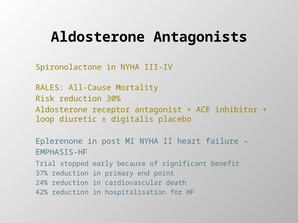
Aldosterone Antagonists
Spironolactone in NYHA III‐IV
RALES: All‐Cause MortalityRisk reduction 30%Aldosterone receptor antagonist + ACE inhibitor + loop diuretic ± digitalis placebo
Eplerenone in post MI NYHA II heart failure –EMPHASIS-HFTrial stopped early because of significant benefit37% reduction in primary end point24% reduction in cardiovascular death42% reduction in hospitalisation for HF
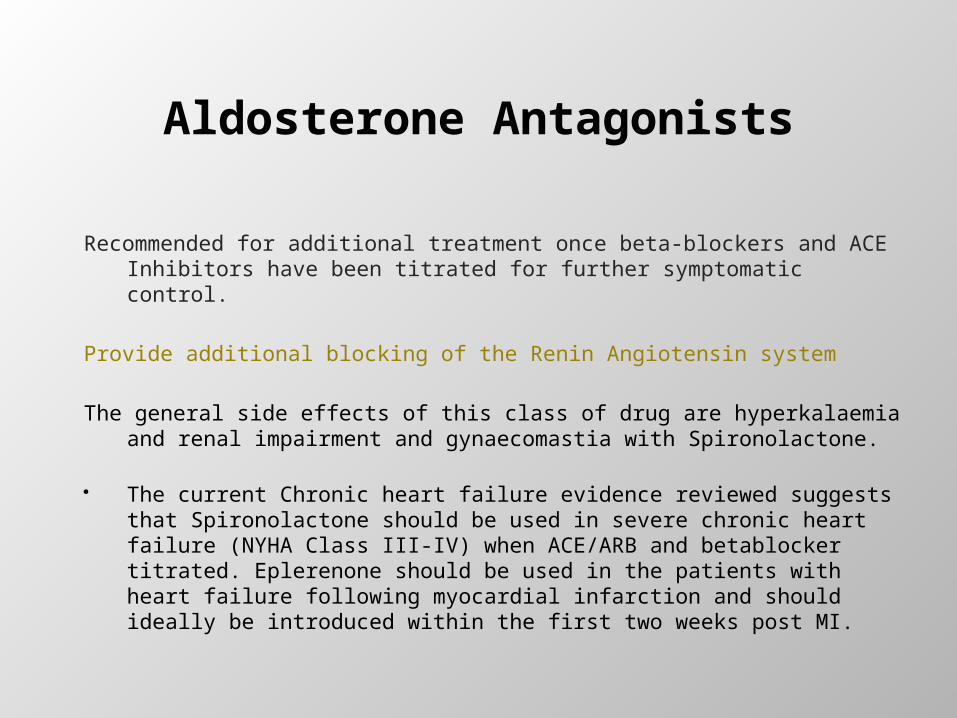
Aldosterone Antagonists
Recommended for additional treatment once beta-blockers and ACE Inhibitors have been titrated for further symptomatic control.
Provide additional blocking of the Renin Angiotensin system
The general side effects of this class of drug are hyperkalaemia and renal impairment and gynaecomastia with Spironolactone.
The current Chronic heart failure evidence reviewed suggests that Spironolactone should be used in severe chronic heart failure (NYHA Class III-IV) when ACE/ARB and betablocker titrated. Eplerenone should be used in the patients with heart failure following myocardial infarction and should ideally be introduced within the first two weeks post MI.
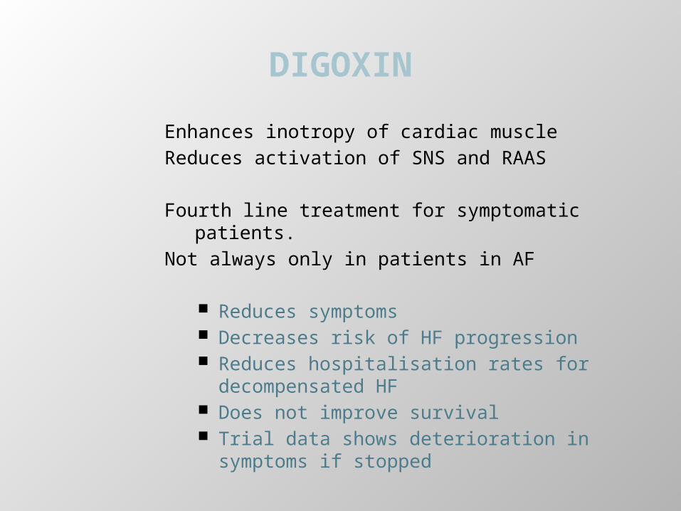
DIGOXIN
Enhances inotropy of cardiac muscleReduces activation of SNS and RAAS
Fourth line treatment for symptomatic patients.Not always only in patients in AF
Reduces symptoms Decreases risk of HF progression Reduces hospitalisation rates for
decompensated HF Does not improve survival Trial data shows deterioration in symptoms
if stopped
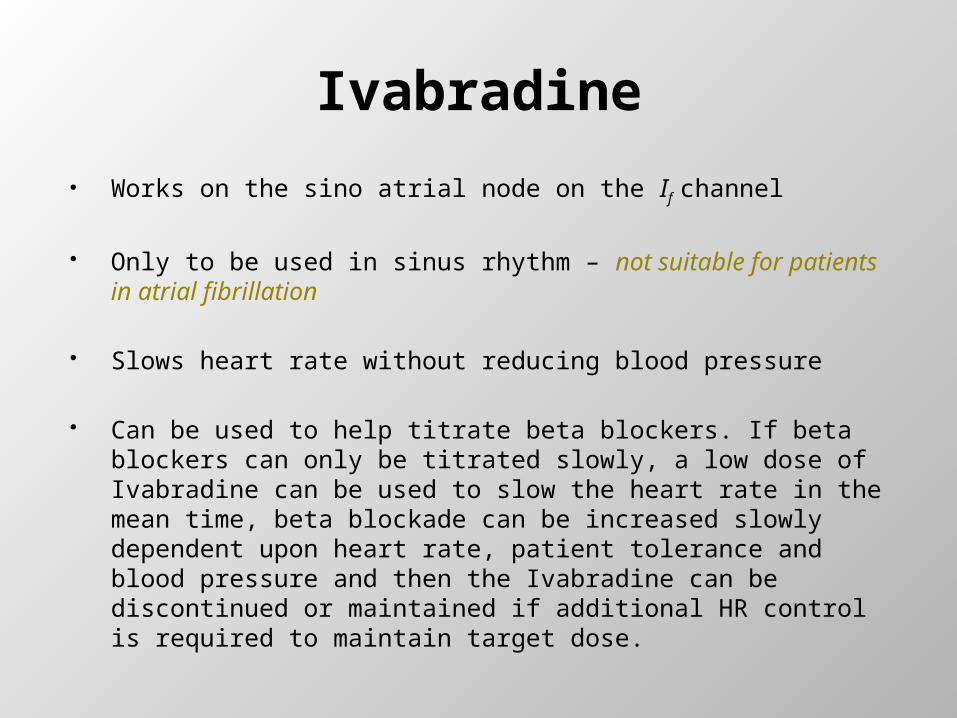
Ivabradine
Works on the sino atrial node on the If channel
Only to be used in sinus rhythm – not suitable for patients in atrial fibrillation
Slows heart rate without reducing blood pressure
Can be used to help titrate beta blockers. If beta blockers can only be titrated slowly, a low dose of Ivabradine can be used to slow the heart rate in the mean time, beta blockade can be increased slowly dependent upon heart rate, patient tolerance and blood pressure and then the Ivabradine can be discontinued or maintained if additional HR control is required to maintain target dose.
Diuretics
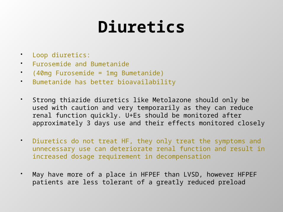
Loop diuretics: Furosemide and Bumetanide (40mg Furosemide = 1mg Bumetanide) Bumetanide has better bioavailability
Strong thiazide diuretics like Metolazone should only be used with caution and very temporarily as they can reduce renal function quickly. U+Es should be monitored after approximately 3 days use and their effects monitored closely
Diuretics do not treat HF, they only treat the symptoms and unnecessary use can deteriorate renal function and result in increased dosage requirement in decompensation
May have more of a place in HFPEF than LVSD, however HFPEF patients are less tolerant of a greatly reduced preload
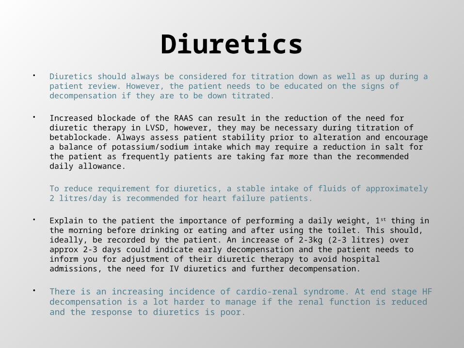
Diuretics Diuretics should always be considered for titration down as well as up during a patient
review. However, the patient needs to be educated on the signs of decompensation if they are to be down titrated.
Increased blockade of the RAAS can result in the reduction of the need for diuretic therapy in LVSD, however, they may be necessary during titration of betablockade. Always assess patient stability prior to alteration and encourage a balance of potassium/sodium intake which may require a reduction in salt for the patient as frequently patients are taking far more than the recommended daily allowance.
To reduce requirement for diuretics, a stable intake of fluids of approximately 2 litres/day is recommended for heart failure patients.
Explain to the patient the importance of performing a daily weight, 1st thing in the morning before drinking or eating and after using the toilet. This should, ideally, be recorded by the patient. An increase of 2-3kg (2-3 litres) over approx 2-3 days could indicate early decompensation and the patient needs to inform you for adjustment of their diuretic therapy to avoid hospital admissions, the need for IV diuretics and further decompensation.
There is an increasing incidence of cardio-renal syndrome. At end stage HF decompensation is a lot harder to manage if the renal function is reduced and the response to diuretics is poor.
MEDICATIONS TO STOP/REVIEW
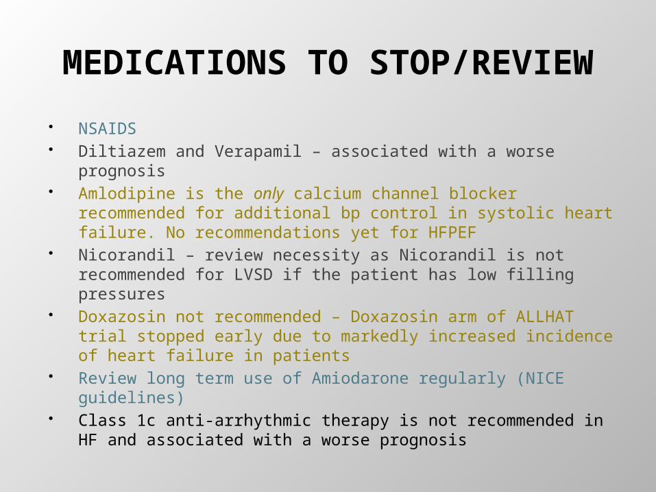
NSAIDS Diltiazem and Verapamil – associated with a worse prognosis Amlodipine is the only calcium channel blocker recommended
for additional bp control in systolic heart failure. No recommendations yet for HFPEF
Nicorandil – review necessity as Nicorandil is not recommended for LVSD if the patient has low filling pressures
Doxazosin not recommended – Doxazosin arm of ALLHAT trial stopped early due to markedly increased incidence of heart failure in patients
Review long term use of Amiodarone regularly (NICE guidelines) Class 1c anti-arrhythmic therapy is not recommended in HF and
associated with a worse prognosis
Cardiac Resynchronisation Therapy (CRT)
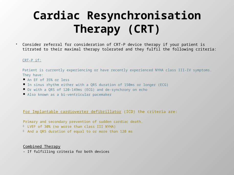
Consider referral for consideration of CRT-P device therapy if your patient is titrated to their maximal therapy tolerated and they fulfil the following criteria:
CRT-P if:
Patient is currently experiencing or have recently experienced NYHA class III-IV symptoms.They have: An EF of 35% or less In sinus rhythm either with a QRS duration of 150ms or longer (ECG) Or with a QRS of 120-149ms (ECG) and de-synchrony on echo Also known as a bi-ventricular pacemaker
For Implantable cardioverter defibrillator (ICD) the criteria are:
Primary and secondary prevention of sudden cardiac death.- LVEF of 30% (no worse than class III NYHA)- And a QRS duration of equal to or more than 120 ms
Combined Therapy- If fulfilling criteria for both devices
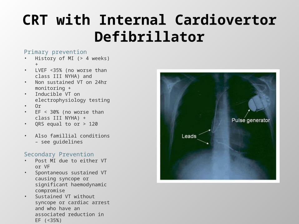
CRT with Internal Cardiovertor Defibrillator
Primary prevention• History of MI (> 4 weeks) +• LVEF <35% (no worse than
class III NYHA) and• Non sustained VT on 24hr
monitoring +• Inducible VT on
electrophysiology testing • Or• EF < 30% (no worse than
class III NYHA) +• QRS equal to or > 120
• Also famillial conditions – see guidelines
Secondary Prevention• Post MI due to either VT or VF• Spontaneous sustained VT
causing syncope or significant haemodynamic compromise
• Sustained VT without syncope or cardiac arrest and who have an associated reduction in EF (<35%)

HFPEF
LVDD TO LVSDA CONTINUUM
The prevailing paradigm of hypertensive heart disease is that it first leads to concentric ventricular hypertrophy, followed by the development of ventricular dilation and contractile impairment with systolic dysfunction and a reduced left ventricular ejection fraction
Effective treatment of hypertension leads to regression of LVH and a few studies have demonstrated decreases in LV mass
Berenji et al (2013)
HFPEF
Variability of the left ventricular ejection fraction measured by different imaging modalities, and the lack of universal agreement on the threshold of ejection fraction at which LVSD and preserved ejection fraction are defined.
Some assert that even in patients with HFPEF, there is an impairment of the contraction of the long axis of the left ventricle
Characterised by increased wall volume, elevated filling pressures and abnormal LV filling.
Tolerate tachycardia poorlyAbrupt increases in blood pressure can precipitate decompensationAs yet there are no conclusive treatment options and current advice
suggests treatment according to guidelines of underlying diseases ie diabetes, hypertension, IHD.

Pathophysiology of HFPEF
The hormone aldosterone promotes fibrosis in the heart and contributes to diastolic stiffness
Most patients with HFPEF complain of symptoms predominantly during exercise and have a reduced response to increased demand
Enhancement of contractility during exercise in HFPEF may be related to subtle resting contractile dysfunction, abnormal calcium handling, passive stiffening, ischemia, oxidative stress, or abnormal myocardial energy utilisation.
LVDD is considered by some to lie on a continuum to LVSD and the reduction/prevention of cardiac remodelling could be considered a target
However........... The term diastolic dysfunction is not synonymous with HFPEF

ACE IN HFPEF
In one trial ACE, compared with placebo, significantly reduced HF hospitalisation up to 12 months
No significant difference for All cause mortality or unplanned hospitalisations (12 months) All cause mortality 6-12months and 12-54 months HF hospitalisations 12-54 months Quality of life up to 6 months NYHA class up to 6 months
Clinical evidence showed that ACEI therapy did not improve mortality but it significantly reduced hospital admissions in patients with heart failure and preserved LVEF. Given that ACEI treatment is relatively cheap; the use of this therapy in patients with HFPEF is likely to be cost-effective, however it was decided that the evidence was inadequate to support the use of ACEI in HFPEF.
NICE GUIDELINES 2010
Betablockers in HFPEF
One third of the population recruited into the SENIORS study of Nebivolol in heart failure in older adults were patients with an EF>40%
The effect in this sub group was insignificant therefore NICE guidelines felt unable to currently recommend use of betablockers in HFPEF until further research has been performed.
Patients with HFPEF are intolerant of tachycardia, especially in AF (which has been found to be increased in elderly female patients with diastolic dysfunction). However, there is no current HR target in HFPEF treatment modalities.
If patients can be considered to be on a continuum from Diastolic remodelling to LVSD then there may be a place for betablocker therapy in long term prevention.
Diuretic therapy in HFPEF May be more of a requirement in HFPEF for diuretics.
Patients are more friable and likely to decompensate and develop pulmonary oedema easily with increases in HR and blood pressure.
Note however that they are less able to cope with a greatly reduced preload.
Some evidence for use of thiazide diuretics – however, this may be related purely to their blood pressure reducing function.
Encourage patient to monitor their weight everyday and educate them on the signs and symptoms of early decompensation and what action to take with an increase in weight of 2-3kg.
Encourage the patient to balance their potassium and sodium intake (which may require a reduction in salt intake to current UK guidelines). An imbalance in intake of sodium contributes to hypertension and can cause fluid retention.
HFPEF
Emerging evidence/current trials:
StatinsCalcium Channel blockersNovel treatments to reduce oxidative stressAldosterone Antagonists to help prevent fibrotic changes
More research is being carried out into ACE/ARB’s and BB’s
Current advice is to use underlying disease processes guidance to treat patients – ie hypertension, diabetes guidelines as there is currently no concrete evidence.

Patient Education
Patient Education is essential to the prognosis and treatment aims of both types of heart failure and may be particularly important in management of heart failure with preserved ejection fraction.
Weight monitoring Fluid intake – including alcohol Diet & Nutrition and reduction of salt intake to current
guidelines and balancing with potassium intake Smoking cessation Immunisation Rest/activity & Exercise Sexual activity and erectile dysfunction Travel/flying/driving Follow up and contact details
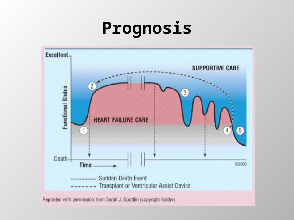
Prognosis
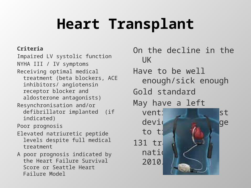
Heart Transplant
Criteria Impaired LV systolic functionNYHA III / IV symptomsReceiving optimal medical
treatment (beta blockers, ACE inhibitors/ angiotensin receptor blocker and aldosterone antagonists)
Resynchronisation and/or defibrillator implanted (if indicated)
Poor prognosisElevated natriuretic peptide
levels despite full medical treatment
A poor prognosis indicated by the Heart Failure Survival Score or Seattle Heart Failure Model
On the decline in the UKHave to be well
enough/sick enoughGold standardMay have a left
ventricular assist device as a bridge to transplant
131 transplants nationwide in 2010/11

End of Life
Difficult to predict in HF
Uncertainty creates barriers & problems in advance planning.
Patients either die very suddenly –Sudden Cardiac Death
Or they deteriorate slowly- become more fatigued, weak, sleepy, cachexic.
GSF, 2005
Gold Standards Framework
Difficult to address end of life with heart failure patients due to uncertain trajectory and changing prognosis of heart failure patients
Consideration should be made for placing the patient on the GSF so that appropriate conversations can take place
Discussions around de-activation of ICD’s should take place early so as not to cause anxiety and difficulties at the end of life.
A patient with an ICD should be made aware that de-activation of the defibrillation action of his/her device does not de activate the pacemaker facility and their heart will not just ‘stop’.
If the patient’s preferred place to die is at home, help should if possible be made available to support this within the home environment. Difficulties around decompensation and diuretic management and increasing breathlessness often end with a relative admitting the patient to hospital due to increase anxiety with home management.
End of Life
Oxygen for dyspnoeaStimulants for fatigueBenzodiazepines/counselling for anxietyExercise limbs to help relieve fatigue and dyspnoeaCPAP/02 for sleep disordered breathingTreat depression -investigation and interventionPeople often die because of multi-organ failure
which may trigger inappropriate investigations and admissions
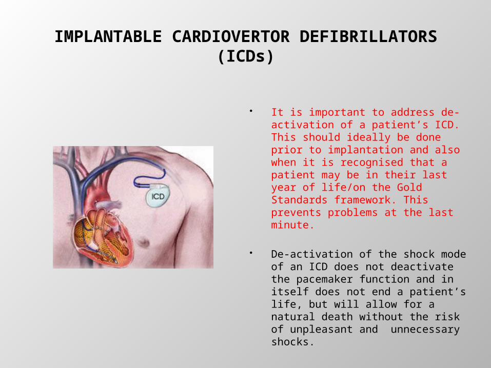
IMPLANTABLE CARDIOVERTOR DEFIBRILLATORS (ICDs)
It is important to address de-activation of a patient’s ICD. This should ideally be done prior to implantation and also when it is recognised that a patient may be in their last year of life/on the Gold Standards framework. This prevents problems at the last minute.
De-activation of the shock mode of an ICD does not deactivate the pacemaker function and in itself does not end a patient’s life, but will allow for a natural death without the risk of unpleasant and unnecessary shocks.
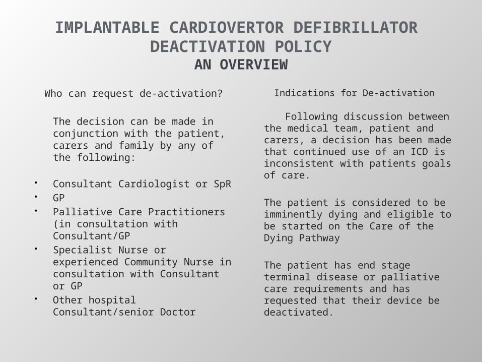
IMPLANTABLE CARDIOVERTOR DEFIBRILLATOR DEACTIVATION POLICY
AN OVERVIEW
Who can request de-activation?
The decision can be made in conjunction with the patient, carers and family by any of the following:
Consultant Cardiologist or SpR GP Palliative Care Practitioners (in
consultation with Consultant/GP Specialist Nurse or experienced
Community Nurse in consultation with Consultant or GP
Other hospital Consultant/senior Doctor
Indications for De-activation
Following discussion between the medical team, patient and carers, a decision has been made that continued use of an ICD is inconsistent with patients goals of care.
The patient is considered to be imminently dying and eligible to be started on the Care of the Dying Pathway
The patient has end stage terminal disease or palliative care requirements and has requested that their device be deactivated.
ICD DEACTIVATION POLICYPROCEDURE
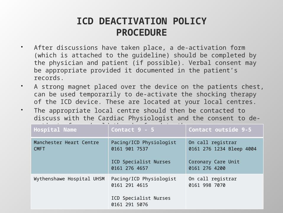
After discussions have taken place, a de-activation form (which is attached to the guideline) should be completed by the physician and patient (if possible). Verbal consent may be appropriate provided it documented in the patient’s records.
A strong magnet placed over the device on the patients chest, can be used temporarily to de-activate the shocking therapy of the ICD device. These are located at your local centres.
The appropriate local centre should then be contacted to discuss with the Cardiac Physiologist and the consent to de-activate form should then be faxed to them.
Hospital Name Contact 9 - 5 Contact outside 9-5
Manchester Heart CentreCMFT
Pacing/ICD Physiologist0161 901 7537
ICD Specialist Nurses0161 276 4657
On call registrar0161 276 1234 Bleep 4004
Coronary Care Unit0161 276 4200
Wythenshawe Hospital UHSM Pacing/ICD Physiologist0161 291 4615
ICD Specialist Nurses0161 291 5076
On call registrar0161 998 7070
Policy
The new de-activation policy can be found on the Greater Manchester and Cheshire Cardiovascular Network website.
Follow clinical working groups – heart rhythm management and look under documents:
http://www.gmccsn.nhs.uk/cardiac/clinical-working-groups/heart-rhythm-management-group/
The consent form that is attached to the document has not yet been confirmed for use.
To Remember
It is important to always record a blood pressure and manual pulse taking note of rhythm as well as rate every time the patient comes in to surgery. This needs coding on the EMIS system
HF patients should be reviewed at least every 6 months when stabilised and their medications titrated.
It is very important that every patient has education and assessment for referral to an exercise program. They need an understanding of the need for compliance with medications, signs and symptoms of decompensation, what individual medications do and the importance of lifestyle to help delay the progression of the disease due to the poor prognosis.
Consider referral to ACM for unstable patients with multiple co-morbidities – new heart failure pathway being developed to help care for patients in the community to prevent hospitalisation

CODINGIt is essential that every patient is coded correctly – ensuring that either
585f – left ventricular systolic dysfunction (on echo)OR 585g - left ventricular diastolic dysfunction codes are used correctly (on echo)
If an echo is abnormal with no evidence of heart failure the code 58531 can be used as a standalone code
Patients need the correct code for LVSD/LVDD or abnormal echo and a heart failure diagnosis on the system.
QOF CODING
HF001 - The contractor establishes and maintains a register of patients with heart failure
HF002 - The percentage of patients with a diagnosis of heart failure (diagnosed on or after 1 April 2006) which has been confirmed by an echocardiogram or by specialist assessment 3 months before or 12 months after entering on to the register
HF003. In those patients with a current diagnosis of heart failure due to left ventricular systolic dysfunction, the percentage of patients who are currently treated with an ACE-I or ARB
HF004. In those patients with a current diagnosis of heart failure due to left ventricular systolic dysfunction who are currently treated with an ACE-I or ARB, the percentage of patients who are additionally currently treated with a beta-blocker licensed for heart failure

Any Questions?