sustainable urban development projects - umeå kommun
TRANSCRIPT

Conserved S-Layer-Associated Proteins Revealed by ExoproteomicSurvey of S-Layer-Forming Lactobacilli
Brant R. Johnson,a,b Jeffrey Hymes,b Rosemary Sanozky-Dawes,b Emily DeCrescenzo Henriksen,b Rodolphe Barrangou,a,b
Todd R. Klaenhammera,b
Graduate Program in Microbiology, College of Agriculture and Life Sciences, North Carolina State University, Raleigh, North Carolina, USAa; Department of Food,Bioprocessing, and Nutrition Sciences, North Carolina State University, Raleigh, North Carolina, USAb
The Lactobacillus acidophilus homology group comprises Gram-positive species that include L. acidophilus, L. helveticus, L.crispatus, L. amylovorus, L. gallinarum, L. delbrueckii subsp. bulgaricus, L. gasseri, and L. johnsonii. While these bacteria areclosely related, they have varied ecological lifestyles as dairy and food fermenters, allochthonous probiotics, or autochthonouscommensals of the host gastrointestinal tract. Bacterial cell surface components play a critical role in the molecular dialoguebetween bacteria and interaction signaling with the intestinal mucosa. Notably, the L. acidophilus complex is distinguished intwo clades by the presence or absence of S-layers, which are semiporous crystalline arrays of self-assembling proteinaceous sub-units found as the outermost layer of the bacterial cell wall. In this study, S-layer-associated proteins (SLAPs) in the exopro-teomes of various S-layer-forming Lactobacillus species were proteomically identified, genomically compared, and transcrip-tionally analyzed. Four gene regions encoding six putative SLAPs were conserved in the S-layer-forming Lactobacillus species butnot identified in the extracts of the closely related progenitor, L. delbrueckii subsp. bulgaricus, which does not produce anS-layer. Therefore, the presence or absence of an S-layer has a clear impact on the exoproteomic composition of Lactobacillusspecies. This proteomic complexity and differences in the cell surface properties between S-layer- and non-S-layer-forming lac-tobacilli reveal the potential for SLAPs to mediate intimate probiotic interactions and signaling with the host intestinal mucosa.
Bacterial cell surface proteins play a critical role in the molecu-lar dialogue between bacteria and their interaction with the
host. For beneficial microbes, such as probiotics, these proteinsmediate health-promoting functions through gastrointestinal ad-hesion, competitive exclusion of pathogens, enhancement of in-testinal barrier function, and activation of gut mucosal immunity(1, 2). Probiotics are defined by the FAO/WHO as “live microor-ganisms that, when administered in adequate amounts, confer ahealth benefit on the host” (3). Some beneficial actions of theseorganisms are strain specific and can be harnessed to treat or re-duce the risk of multiple maladies, including acute infectious di-arrhea, irritable bowel syndrome, vaginal infections, ulcerativecolitis, lactose maldigestion, and necrotizing enterocolitis (4). Infact, the efficacy of probiotic treatment depends largely on thevarious cell surface components that mediate this specificity (5).Therefore, the characterization of effector cell surface ligands andtheir health-promoting interactions with the host is of increasingscientific and medical interest.
Some of the most prevalent and well-studied probiotics arelactobacilli, many of which are members of the Lactobacillus aci-dophilus homology group (6). The L. acidophilus group is a cladeof homologous Gram-positive Lactobacillus species that includesL. acidophilus, L. helveticus, L. crispatus, L. amylovorus, L. gallina-rum, L. delbrueckii subsp. bulgaricus, L. gasseri, and L. johnsonii(7–11). Although these bacteria are closely related phylogeneti-cally, they have varied ecological lifestyles ranging from dairy andfood fermentations to allochthonous probiotics or autochtho-nous commensals of the host gastrointestinal and urogenitaltracts. Biochemically, they are obligately homofermentative; theyalmost exclusively ferment sugar (�85%) to lactate via the Emb-den-Meyerhof-Parnas pathway. Early taxonomic descriptionswere based on the metabolic end products of fermentation, result-ing in a seemingly indistinguishable group of microbes, which
were all called L. acidophilus (10). However, DNA-DNA hybrid-ization studies revealed the heterogeneity in the group (11, 12).Since then, genome sequencing and comparative genomic analy-ses have clearly established and solidified the current descriptionof the L. acidophilus group (13, 14). Notably, these closely relatedstrains can be dichotomized based on their ability to create surface(S)-layer protein arrays as the outermost constituent of the cellwall (15).
Bacterial S-layers are semiporous proteinaceous crystalline ar-rays composed of self-assembling (glyco)protein subunits calledS-layer proteins (SLPs) (15). They can be found in both Gram-positive and Gram-negative bacteria and species of Archaea butare not ubiquitous in all microorganisms. When present, S-layersform two-dimensional lattices on the outermost layer of the cell,which are tethered through noncovalent interactions with the cellwall (15). S-layers from various species of the L. acidophilus ho-mology group have been characterized for their roles in intestinaladhesion, competitive exclusion of pathogens, and immuno-modulation of the gastrointestinal mucosa. In vitro studies using
Received 11 June 2015 Accepted 13 October 2015
Accepted manuscript posted online 16 October 2015
Citation Johnson BR, Hymes J, Sanozky-Dawes R, Henriksen ED, Barrangou R,Klaenhammer TR. 2016. Conserved S-layer-associated proteins revealed byexoproteomic survey of S-layer-forming lactobacilli. Appl Environ Microbiol82:134 –145. doi:10.1128/AEM.01968-15.
Editor: H. Nojiri
Address correspondence to Todd R. Klaenhammer, [email protected].
Supplemental material for this article may be found at http://dx.doi.org/10.1128/AEM.01968-15.
Copyright © 2015 Johnson et al. This is an open-access article distributed underthe terms of the Creative Commons Attribution 4.0 International license.
crossmark
134 aem.asm.org January 2016 Volume 82 Number 1Applied and Environmental Microbiology
on April 5, 2019 by guest
http://aem.asm
.org/D
ownloaded from

intestinal epithelial cell lines suggest that the S-layer is a majorfactor in intestinal adhesion for L. acidophilus (16, 17), L. crispatus(18–20), L. helveticus (21), and L. amylovorus (22). In fact, thisadhesion has been shown to competitively exclude enteropatho-genic bacteria by both L. crispatus (23) and L. helveticus (24, 25).Compelling studies have begun to reveal the mechanisms of gas-trointestinal immunomodulation. For example, SlpA, the pri-mary constituent of the S-layer in L. acidophilus NCFM, was foundto bind to dendritic cell (DC) orthologous C-type lectin receptors(CLR), DC-specific intercellular adhesion molecule 3 (ICAM-3)-grabbing nonintegrin (DC-SIGN) (26), and a specific intracellularadhesion molecule-3-grabbing nonintegrin homolog-related 3(SIGNR-3) (27). This SlpA-CLR interaction exerts regulatory sig-nals, which have been reported to mitigate inflammatory diseasestates and promote the maintenance of healthy intestinal barrierfunction (27). Similar experiments have aimed to elucidate theroles of the S-layer in modulating gastrointestinal immunity for L.crispatus (28), L. helveticus (29), and L. amylovorus (22).
The S-layer-forming species of the L. acidophilus homologygroup form S-layers composed of a dominant protein constituent,SlpA/Slp1 (�46 kDa), and the minor constituents SlpB/Slp2 (�47kDa) and SlpX (�51 kDa) (30). Recent evidence, however, sug-gests that the S-layer may not be as monomorphic as previouslyproposed. In L. acidophilus NCFM, proteomic analysis revealedthe presence of 37 noncovalently bound extracellular S-layer-as-sociated proteins (SLAPs), 23 of which are putative/uncharacter-ized proteins of unknown function (31). In this study, the nonco-valent exoproteomes of various S-layer- and non-S-layer-formingLactobacillus strains were proteomically identified, genomicallycompared, and transcriptionally analyzed. These data reveal boththe conservation and variability of SLAPs across lactobacilli andtheir potential to mediate intimate interactions with the intestinalmucosa.
MATERIALS AND METHODSBacterial strains and growth conditions. The bacterial strains used in thisstudy are reported in Table 1. Lactobacillus strains were propagated stati-
cally at 37°C under ambient atmospheric conditions in de Man-Rogosa-Sharpe (MRS) broth (Difco Laboratories, Inc., Detroit, MI).
DiversiLab analysis of strains. L. crispatus and L. helveticus strainswere typed using the repetitive extragenic palindromic-PCR (Rep-PCR)-based DiversiLab typing system (bioMérieux, Durham, NC). DNA fromthe Lactobacillus strains was extracted using a Mo Bio UltraClean micro-bial DNA isolation kit (Mo Bio, Carlsbad, CA) and quantified using aNanoDrop 1000 spectrophotometer (Thermo Scientific, Waltham, MA).The DNA was then normalized to 20 ng �l�1 with UltraPure distilledwater (Invitrogen, Carlsbad, CA). Rep-PCR was performed in prepara-tion for typing using the Lactobacillus DiversiLab kit (bioMérieux). DNAamplification was performed in a Bio-Rad MyCycler thermal cycler (Bio-Rad, Hercules, CA), programmed for 2 min at 94°C (initial denaturation)and 35 cycles of 30 s at 94°C (denaturation), 30 s at 55°C (annealing), and90 s at 70°C (extension), followed by a final extension cycle of 3 min at70°C using AmpliTaq DNA polymerase from Applied Biosystems (Carls-bad, CA). The reaction mixture was pipetted into the DiversiLab systemchip along with the DiversiLab DNA reagents and supplies (bioMérieux),according to the manufacturer’s protocol. The chip samples were ana-lyzed using the DiversiLab software version 3.4, and the similarity of thestrains was determined by comparing the resulting electropherogram/barcodes.
Extraction of extracellular noncovalently bound cell surface pro-teins. Noncovalently bound cell surface proteins, including S-layer pro-teins and S-layer-associated proteins, were extracted from the Lactobacil-lus strains using LiCl denaturing salt, as described previously (31). Briefly,cells were grown in 200 ml of MRS broth to stationary phase (16 h),centrifuged at 2,236 � g for 10 min (4°C), and washed twice with 25 ml ofcold phosphate-buffered saline (PBS) (Gibco) (pH 7.4). The cells wereagitated for 15 min at 4°C following the addition of 5 M LiCl (FisherScientific). Supernatants containing SLPs and SLAPs were harvested viacentrifugation at 8,994 � g for 10 min (4°C), transferred to a 6,000- to8,000-kDa Spectra/Por molecular porous membrane (Spectrum Labora-tories), and dialyzed against cold distilled water for 24 h. The precipitatewas harvested at 20,000 � g for 30 min and agitated for a second time with1 M LiCl at 4°C for 15 min to disassociate the SLAPs from the SLPs. Thesuspension was then centrifuged at 20,000 � g for 10 min, and the SLAPsupernatants were separated from the SLP pellet, transferred to the 6,000-to 8,000-kDa Spectra/Por molecular porous membrane, and dialyzedagainst cold distilled water for 24 h. Finally, the precipitate was harvested
TABLE 1 Strains used in this study
Organism (strain)a Study designation Sourceb Origin S-layer Reference
L. acidophilus (NCFM) NCK56 Human intestinal isolate � 47L. helveticus (1846) NCK230 NCDO Dairy isolate � 48L. helveticus (481-C) NCK246 NCDO Dairy isolate � 49L. helveticus NCK338 NCDO Dairy isolate � 50L. helveticus (CNRZ32) NCK936 CNRZ Industrial cheese starter culture � 51L. helveticus (ATCC 15009) NCK1088 ATCC Dairy isolate � 52L. crispatus (ATCC 33820) NCK777 ATCC Human isolate � 53, 54L. crispatus NCK953 Chicken isolate �L. crispatus (CZ6) NCK1351 Human endoscopy isolate � 55L. amylovorus (ATCC 33620) NCK776 ATCC Cattle feces � 56, 57L. gallinarum (ATCC 33199) NCK778 ATCC Chicken isolate � 58L. gallinarum NCK1560 Chicken isolate �L. delbrueckii subsp. bulgaricus NCK1561 Dairy isolate �L. gasseri (ATCC 33323) NCK334 ATCC Human isolate � 59L. johnsonii (ATCC 33200) NCK779 ATCC Human isolate � 58L. reuteri (ATCC 23272)c NCK702 ATCC Human feces � 11L. casei (ATCC 393)c NCK125 ATCC Dairy isolate � 60a Proteins from organisms indicated in bold were proteomically identified using LC-MS/MS.b NCDO, National Collection of Dairy Organisms; ATCC, American Type Culture Collection; CNRZ, Centre National de Recherches Zootechniques.c Species outside the L. acidophilus homology group.
S-Layer-Associated Proteins in Lactobacillus Species
January 2016 Volume 82 Number 1 aem.asm.org 135Applied and Environmental Microbiology
on April 5, 2019 by guest
http://aem.asm
.org/D
ownloaded from

via centrifugation at 20,000 � g for 30 min to pellet the SLAPs. Both SLPand SLAP pellets were resuspended in 10% (wt/vol) SDS (Fisher). Pro-teins were quantified via a bicinchoninic acid assay kit (Thermo Scientific)and visualized via SDS-PAGE using precast 4% to 20% Precise Tris-HEPES protein gels (Thermo Scientific). The gels were stained using Ac-quaStain (Bulldog Bio), according to the manufacturer’s instructions.SLAP extractions were performed with two biological replicates for eachstrain and visualized through SDS-PAGE to confirm that the resultantbanding patterns were reproducible.
Proteomic identification and analysis. SLAPs extracted from the var-ious Lactobacillus species were identified using liquid chromatography-tandem mass spectrometry (LC-MS/MS) from the Genome Center Pro-teomics Core at the University of California, Davis, CA, as describedpreviously (31). Proteomic screenings were performed once per strainand used as a tool for selecting candidate SLAPs within each strain. Tan-dem mass spectra were extracted and the charge state deconvoluted usingMM File Conversion version 3. All MS/MS samples were analyzed usingX! Tandem (Tornado version; The GPM [www.thegpm.org/]). UniProtsearches were performed using proteome databases for the respective pro-teins isolated from L. acidophilus NCFM, L. helveticus CNRZ32, L. crispa-tus ST1, and L. amylovorus GRL1112. X! Tandem was searched with afragment ion mass tolerance and parent ion tolerance of 20 ppm. Theiodoacetamide derivative of cysteine was specified in X! Tandem as a fixedmodification. The deamination of asparagine and glutamine, oxidation ofmethionine and tryptophan, sulfonation of methionine, tryptophan oxi-dation to formylkynurenine of tryptophan, and acetylation of the N ter-minus were specified in X! Tandem as variable modifications. Scaffold(version Scaffold_3.6.1; Proteome Software) was used to validate MS/MS-based peptide and protein identifications. Peptide identifications wereaccepted if they exceeded specific database search engine thresholds. X!Tandem identifications required scores of �1.2 with a mass accuracy of 5ppm. Protein identifications were accepted if they contained at least twoidentified peptides. Using the parameters described above, the false-dis-covery rate was calculated to be 1.1% at the protein level and 0% at thepeptide level. Proteins that contained similar peptides and that could notbe differentiated based on MS/MS analysis alone were grouped to satisfythe principles of parsimony. For this study, only proteins with uniquespectral counts of �20 were considered significant. For all analyses, totalspectral counts were utilized as a semiquantitative indicator of proteinabundance (32). Two-way clustering of total spectral counts was per-formed using JMP Genomics (version 5; SAS). Protein domains wereidentified for analysis using the Pfam protein family database (33).
Genomic in silico analyses. Genomic analysis was performed on ge-nomes curated from the genome library of the National Center for Biotech-nology Information (NCBI [http://www.ncbi.nlm.nih.gov/genome/]), in-cluding L. acidophilus NCFM (GenBank accession no. NC_006814.3), L.helveticus CNRZ32 (GenBank accession no. NC_021744.1), L. amylovorusGRL1112 (GenBank accession no. NC_014724.1), L. crispatus ST1(GenBank accession no. NC_014106.1), L. delbrueckii subsp. bulgaricusATCC 11842 (GenBank accession no. NC_008054.1), and L. casei ATCC334 (GenBank accession no. NC_008526.1). Identified genes were com-pared using the BLASTn and BLASTp features of NCBI (http://blast.ncbi.nlm.nih.gov/Blast.cgi). SignalP 4.1 was used to predict the signal pepti-dase cleavage site of each identified protein (34). Genomes wereuploaded to Geneious 8.0.5 (35) for comparative genomic and pro-moter analyses of the identified SLAP genes. The genetic context ofSLAP genes was examined using the chromosomal graphical interfacein Geneious 8.0.5. In silico promoter elements were identified in theupstream intergenic regions of SLAP genes using PromoterWise (http://www.ebi.ac.uk/Tools/psa/promoterwise/). To identify conservedpromoter elements between the various SLAP genes, genome-widesequence motifs of the putative �10 and �35 regions were scannedagainst the four S-layer-forming genomes using Geneious 8.0.5, with avariable spacer length of 16 to 23 nucleotides (nt) between the �10 and�35 regions.
RNA extraction, sequencing, and transcriptional analysis. Cellswere grown to mid-log phase (8 h) and flash-frozen for RNA extractionand sequencing. RNA was extracted using the Zymo Direct-zol RNAMiniPrep kit (Zymo Research, Irvine, CA) and analyzed for quality usingan Agilent 2100 Bioanalyzer (Agilent Technologies, Santa Clara, CA). Li-brary preparation and RNA sequencing were performed at the High-Throughput Sequencing and Genotyping Unit of the Roy J. Carver Bio-technology Center, University of Illinois at Urbana-Champaign, IL. Foreach sample, rRNA was removed with the Ribo-Zero bacterial kit (Illu-mina, San Diego, CA), followed by library preparation with the TruSeqstranded RNA sample preparation kit (Illumina). Single-read RNA se-quencing was performed using an Illumina HiSeq 2500 ultrahigh-throughput sequencing system) with a read length of 180 nt. Raw se-quencing reads were assessed for quality using FastQC version 0.11.3(http://www.bioinformatics.babraham.ac.uk/projects/fastqc/) and pro-cessed using Geneious 8.0.5 (35). Briefly, after the adaptor sequences weretrimmed, the raw reads were quality trimmed to remove sequence readswith an error probability limit of 0.001 (Phred score, 30) and filtered toremove reads �20 nt. These quality trimmed and filtered sequences werethen mapped to the reference genomes of the S-layer-forming Lactobacil-lus spp. using Bowtie 2 (36), with default settings within Geneious 8.0.5(35). The sequencing coverage depths were calculated to be 767�, 730�,727�, and 665� for L. acidophilus NCFM strain NCK56, L. amylovorusATCC 33620 strain NCK776, L. crispatus ATCC 33820 strain NCK777,and L. helveticus CNRZ32 strain NCK938, respectively. Transcriptionalanalyses were based on the normalized transcripts per million (TPM)calculation within Geneious 8.0.5 (35).
RESULTSProteomic identification of noncovalently bound extracellularproteins in S-layer- and non-S-layer-forming lactobacilli. Basedon the previous identification of S-layer-associated proteins(SLAPs) in L. acidophilus NCFM (31), we performed exopro-teome screenings on multiple S-layer- and non-S-layer-formingstrains of Lactobacillus. Thus, five S-layer- and five non-S-layer-forming Lactobacillus species were analyzed (Fig. 1). Seventeenstrains were tested in total, comprising 12 S-layer- and 5 non-S-layer-producing lactobacilli (Table 1). Notably, 15 of the strainsare members of the closely related L. acidophilus homology group.
Electrophoresis of SLAP extractions revealed a surprisingly di-verse array of protein banding patterns in the S-layer-formingspecies and a notable absence of proteins in the non-S-layer-form-ing species (Fig. 2). SLAP extractions were performed on two bi-ological replicates, and the SDS-PAGE banding patterns of theSLAPs extracted from each strain did not differ in the major band-ing patterns between replicates. Further, the LiCl extract of L.acidophilus demonstrated a banding profile similar to that of theSLAPs identified previously (28) (Fig. 2, lane 1). Proteins from theother S-layer-forming strains, including L. crispatus, L. amylovo-rus, L. gallinarum, and L. helveticus, were not only distinct from L.acidophilus but also from one another. Moreover, there was alsoheterogeneity in the protein banding between various strainswithin each species. In the five L. helveticus strains, there weredistinctive differences between the various dairy isolates NCK936,NCK338, NCK230, NCK246, and NCK1088 (Fig. 2A, lanes 2 and6 to 9). The three L. crispatus strains were also discrete from oneanother (Fig. 2A, lanes 3, 10, and 11). Rep-PCR-based DiversiLabstrain typing was performed on the five L. helveticus and three L.crispatus strains to examine genomic similarities (Fig. 2B and C).The five L. helveticus strains clustered into two groups with �93%and �98% similarity (Fig. 2B), and the L. crispatus strainswere �85% similar (Fig. 2C). Remarkably, the L. helveticus strains
Johnson et al.
136 aem.asm.org January 2016 Volume 82 Number 1Applied and Environmental Microbiology
on April 5, 2019 by guest
http://aem.asm
.org/D
ownloaded from

NCK338 and NCK230, and NCK1088 and NCK936, distinctlyvaried in terms of the isolated extracellular proteins (Fig. 2A) de-spite �98% and �95% similarity between the Rep-PCR typing pat-terns (Fig. 2B). A similar trend was observed among the L. crispatusstrains. Thus, there was no correlation between the genotype clus-tering and the exoproteome profiles revealed by SDS-PAGE.
There were very few proteins isolated from the non-S-layer-forming species of Lactobacillus, as observed in the gel lanes of theSDS-PAGE (Fig. 2, lanes 13 to 17). L. johnsonii and L. gasseri of theL. acidophilus homology group exhibited no discernible proteinsin the gel lanes (Fig. 2, lanes 15 and 17). L. delbrueckii subsp.bulgaricus, the non-S-layer-producing strain, which is the mostclosely related and progenitor to the other S-layer-forming mem-bers of the L. acidophilus homology group (Fig. 1), showed only a
small number of proteins isolated from the LiCl extract (Fig. 2,lane 13). Distantly related L. casei, devoid of any S-layer, also ex-hibited few proteins (Fig. 2, lane 14). To identify the electropho-resed proteins, lanes with visible proteins in the gel were sent forproteomic identification (Table 1, in bold).
Of the 12 S-layer-forming strains, seven were selected for pro-teomic identification, including three L. helveticus strains, three L.crispatus strains, and one L. amylovorus strain (Table 1, under-lined). Notably, L. gallinarum was not selected for analysis, asthere are no publically available genomes or proteomes publishedfor this species to date. From the five non-S-layer-forming speciestested, only L. delbrueckii subsp. bulgaricus and L. casei were se-lected from proteomic screening, as they were the only non-S-layer-forming species in which proteins were isolated from the
FIG 1 16S rRNA dendrogram of the S-layer-forming (red) and non-S-layer-forming (blue) species of the L. acidophilus homology group. The tree is rooted bythe non-S-layer-forming species L. casei and L. reuteri, which are not members of the L. acidophilus homology group.
FIG 2 Noncovalently bound exoproteomes were extracted using LiCl and electrophoresed on SDS-PAGE gels. These gels are representative of protein extrac-tions from two biological replicates of each strain. (A) The S-layer-forming strains of the L. acidophilus (L. aci) homology group presented a diverse array ofproteins in the LiCl extracts, including many anticipated S-layer-associated proteins (SLAPs). In contrast, the non-S-layer-forming species harbored very fewproteins in the cell surface extracts. Five strains of L. helveticus (L. hel) (B) and three strains of L. crispatus (L. cris) (C) were typed using the Rep-PCR-basedDiversiLab typing system. L. gal, L. gallinarum; L. amy, L. amylovorus; L. bulg, L. delbrueckii subsp. bulgaricus; L. cas, L. casei; L. john, L. johnsonii; L. reut, L. reuteri;L. gas, L. gasseri.
S-Layer-Associated Proteins in Lactobacillus Species
January 2016 Volume 82 Number 1 aem.asm.org 137Applied and Environmental Microbiology
on April 5, 2019 by guest
http://aem.asm
.org/D
ownloaded from

SLAP extraction (Table 1, underlined). Proteins were identifiedfrom the LiCl extracts of the seven S-layer- and two non-S-layer-forming Lactobacillus species using liquid chromatography-tan-dem mass spectrometry (see Table S1 in the supplemental mate-
rial). Two-way clustering was performed based on the totalspectral counts of identified proteins and visualized using a two-way clustering heat map (Fig. 3A). The proteins identified in thetwo non-S-layer-forming strains, L. casei and L. delbrueckii subsp.
FIG 3 (A) A total of 2,929 proteins were identified from the S-layer-forming strains (red) of L. crispatus, L. amylovorus, and L. helveticus and the non-S-layer-forming strains (blue) of L. delbrueckii subsp. bulgaricus and L. casei. Two-way clustering was performed on the identified proteins based on their similaritybetween strains and visualized using a red-blue heat map. The colors in the heat map represent the spectral counts of the identified proteins (semiquantitativemeasure of protein abundance), with red being the most present (400 to 1,000 total spectral counts), gray being somewhat present (12 to 400 total spectralcounts), and blue being low or no presence (0 to 12 total spectral counts). Regarding the S-layer-forming strains, there were three main clusters of proteins: SLAPsspecific to L. crispatus (B), L. amylovorus (C), and L. helveticus (D). These three clusters have been noted with the corresponding UniProt and protein annotationsof the identified proteins.
Johnson et al.
138 aem.asm.org January 2016 Volume 82 Number 1Applied and Environmental Microbiology
on April 5, 2019 by guest
http://aem.asm
.org/D
ownloaded from

bulgaricus, are unambiguously distinct from the other seven S-layer-forming strains. Furthermore, almost all of the proteins identified inthe non-S-layer-forming strains were predicted intracellular pro-teins, likely presented extracellularly as the result of cell death occur-ring at stationary phase. With regard to the S-layer-forming Lactoba-cillus species, there were three main groupings of proteinsidentified: SLAPs specific to L. crispatus (Fig. 3B), SLAPs specificto L. amylovorus (Fig. 3C), and SLAPs specific to L. helveticus (Fig.3D). Surprisingly, although each group had distinctive homolo-gies, the same types of proteins were observed in each group. Infact, these proteins, which included multiple putative uncharac-terized proteins, cell surface proteases, and group 3 bacterial Ig-like domain proteins, were the same types of proteins identified asSLAPs in L. acidophilus NCFM (see Table S2 in the supplementalmaterial). Notably, these putative SLAPs were not found in thenon-S-layer-producing strains analyzed, which were L. casei andL. delbrueckii subsp. bulgaricus.
Functional exoproteomic analysis of S-layer- and non-S-lay-er-forming lactobacilli. After proteomic identification, selectedputative SLAPs and noncovalently bound extracellular proteins
were functionally analyzed based on predicted protein domains.Four predominant protein domains were found consistently in theS-layer-forming species tested (Fig. 4A), including SLAP (PF03217),Big_3 (PF07523), SH3_8 (PF13457), and fn3 (PF00041). We proposethat the SLAP (PF03217) domain, responsible for the noncovalentattachment of SLP and other extracellular proteins in lactobacilli,be redesignated the noncovalent attachment domain (NCAD).This domain designation prevents confusion with the abbrevia-tion for S-layer-associated proteins, SLAPs. Notably, the NCADwas the most abundant protein domain identified in the extracel-lular fractions tested (Fig. 4A). Other domains associated withbacterial extracellular proteins, including group 3 bacterial Ig-likedomains (Big_3), SH3-like domains (SH3_8), and fibronectintype III domains (fn3), were found in the proteomic analysis of theS-layer-forming species but were absent from the non-S-layer-forming species (Fig. 4A). Notably, only two NCAD-containingproteins were identified within the exoproteome of L. delbrueckiisubsp. bulgaricus, while none of these domains were identified inthe exoproteome of the non-S-layer-forming L. casei.
Identified proteins were functionally categorized based on pu-
FIG 4 (A) Four protein domains found consistently in the proteins identified within the LiCl extracts: NCAD (white), BIg_3 (black), SH3_8 (dots), and fn3(diagonal lines). Dot plots were created using the semiquantitative total spectral counts from the identified proteins of each strain. Plotted are the extracellularFn3 proteins and extracellular BIg_3/SH3_8 proteins (B), putative annotated S-layer proteins and uncharacterized extracellular proteins (C), and intracellularand ribosomal proteins (D). The proteins in panel C contain the NCAD, while the proteins in panel D do not.
S-Layer-Associated Proteins in Lactobacillus Species
January 2016 Volume 82 Number 1 aem.asm.org 139Applied and Environmental Microbiology
on April 5, 2019 by guest
http://aem.asm
.org/D
ownloaded from

tative domains and placed into one of six groupings: extracellularfn3 domain proteins and extracellular BIg3/SH3_8 proteins (Fig.4B), putatively annotated SLPs and uncharacterized extracellularproteins (Fig. 4C), and intracellular proteins and ribosomalproteins (Fig. 4D). The distribution of the proteins within thesefunctional groupings was plotted for each of the strains using thesemiquantitative total spectral counts identified through the LC-MS/MS survey (Fig. 4B to D). Group 3 bacterial Ig-like domainproteins, which contain the Big_3 and SH3_8 domains, were onlyfound in the SLAP fractions of the S-layer-forming lactobacilli(Fig. 4B). Similarly, uncharacterized proteins putatively anno-tated as SLPs and fibronectin-binding proteins were found solelyin the S-layer-forming species of Lactobacillus (Fig. 4B and C).There was an increase in both the occurrence and abundance ofNCAD-containing uncharacterized extracellular proteins in theSLAP fractions from the S-layer strains compared to the non-S-layer strains (Fig. 4C). Furthermore, there was an increase in thepresence of intracellular proteins, including ribosomal proteins,in the non-S-layer strains (Fig. 4D), as measured by total spectralcounts. These data reveal a pattern of noncovalently bound pro-teins identified in S-layer species of Lactobacillus compared tonon-S-layer-forming lactobacilli.
Genomic characterization of genes corresponding to the ex-tracellular S-layer-associated proteins. The putative SLAPs iden-tified in this study, along with the previously identified SLAPs of L.
acidophilus NCFM, were curated to the genomes of L. acidophilusNCFM, L. helveticus CNRZ32, L. amylovorus GRL 1112, and L.crispatus ST1 (see Table S2 in the supplemental material). By vi-sualizing the corresponding genes on the four genomes, four con-served genetic regions containing six genes were consistently ob-served (Fig. 5). Two cell division-related genes, including anN-acetylmuramidase and autolysin, are found in region I. RegionII is composed of genes encoding fn3 domain-containing fi-bronectin-binding proteins. Region III also contains two cell di-vision-related genes, including the gene encoding cell divisionprotein A (cdpA) (33). Finally, region IV includes genes encodinggroup 3 bacterial Ig-like proteins, which contain the domainsBig_3 and SH3_8. The relative positions of the four gene regionswere conserved among the four genomes, with the exception ofregions II and III in L. helveticus, which were translocated to theminus strand leading away from the origin of replication (Fig. 5).
In addition, the genetic context of each region was examinedwithin the four strains. Notably, there was synteny observed be-tween the four chromosomal regions of each organism (Fig. 6).Although region I was the least syntenic overall, it is noteworthythat the N-acetylmuramidase and autolysin/amidase genes werepositioned directly downstream of the genes encoding the pri-mary S-layer protein, slpA and slpB. Conversely, region II exhib-ited increased conservation of genetic loci near the SLAP geneencoding a fibronectin-binding protein, including genes for a
FIG 5 All open reading frames (ORFs) from the positive (�) and negative (�) strands of L. helveticus CNRZ32, L. acidophilus NCFM, L. crispatus ST1, and L.amylovorus GRL1112 were mapped onto circular chromosomes with an annotated origin of replication (Ori). Four conserved SLAP gene regions were identifiedbased on position between strains. Blue, region I; yellow, region II; green, region III; red, region IV.
Johnson et al.
140 aem.asm.org January 2016 Volume 82 Number 1Applied and Environmental Microbiology
on April 5, 2019 by guest
http://aem.asm
.org/D
ownloaded from
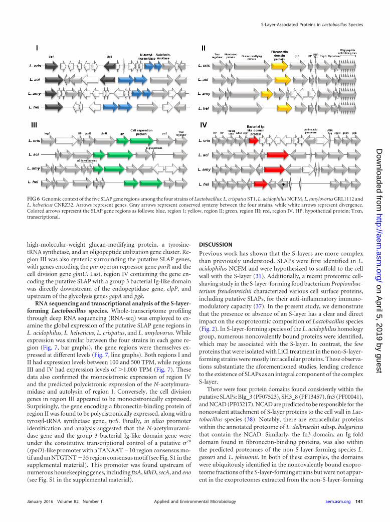
high-molecular-weight glucan-modifying protein, a tyrosine-tRNA synthetase, and an oligopeptide utilization gene cluster. Re-gion III was also syntenic surrounding the putative SLAP genes,with genes encoding the pur operon repressor gene purR and thecell division gene glmU. Last, region IV containing the gene en-coding the putative SLAP with a group 3 bacterial Ig-like domainwas directly downstream of the endopeptidase gene, clpP, andupstream of the glycolysis genes gapA and pgk.
RNA sequencing and transcriptional analysis of the S-layer-forming Lactobacillus species. Whole-transcriptome profilingthrough deep RNA sequencing (RNA-seq) was employed to ex-amine the global expression of the putative SLAP gene regions inL. acidophilus, L. helveticus, L. crispatus, and L. amylovorus. Whileexpression was similar between the four strains in each gene re-gion (Fig. 7, bar graphs), the gene regions were themselves ex-pressed at different levels (Fig. 7, line graphs). Both regions I andII had expression levels between 100 and 500 TPM, while regionsIII and IV had expression levels of �1,000 TPM (Fig. 7). Thesedata also confirmed the monocistronic expression of region IVand the predicted polycistronic expression of the N-acetylmura-midase and autolysin of region I. Conversely, the cell divisiongenes in region III appeared to be monocistronically expressed.Surprisingly, the gene encoding a fibronectin-binding protein ofregion II was found to be polycistronically expressed, along with atyrosyl-tRNA synthetase gene, tyrS. Finally, in silico promoteridentification and analysis suggested that the N-acetylmurami-dase gene and the group 3 bacterial Ig-like domain gene wereunder the constitutive transcriptional control of a putative 70
(rpoD)-like promoter with a TANAAT �10 region consensus mo-tif and an NTGTNT �35 region consensus motif (see Fig. S1 in thesupplemental material). This promoter was found upstream ofnumerous housekeeping genes, including ftsA, ldhD, secA, and eno(see Fig. S1 in the supplemental material).
DISCUSSION
Previous work has shown that the S-layers are more complexthan previously understood. SLAPs were first identified in L.acidophilus NCFM and were hypothesized to scaffold to the cellwall with the S-layer (31). Additionally, a recent proteomic cell-shaving study in the S-layer-forming food bacterium Propionibac-terium freudenreichii characterized various cell surface proteins,including putative SLAPs, for their anti-inflammatory immuno-modulatory capacity (37). In the present study, we demonstratethat the presence or absence of an S-layer has a clear and directimpact on the exoproteomic composition of Lactobacillus species(Fig. 2). In S-layer-forming species of the L. acidophilus homologygroup, numerous noncovalently bound proteins were identified,which may be associated with the S-layer. In contrast, the fewproteins that were isolated with LiCl treatment in the non-S-layer-forming strains were mostly intracellular proteins. These observa-tions substantiate the aforementioned studies, lending credenceto the existence of SLAPs as an integral component of the complexS-layer.
There were four protein domains found consistently within theputative SLAPs: BIg_3 (PF07523), SH3_8 (PF13457), fn3 (PF00041),and NCAD (PF03217). NCAD are predicted to be responsible for thenoncovalent attachment of S-layer proteins to the cell wall in Lac-tobacillus species (38). Notably, there are extracellular proteinswithin the annotated proteome of L. delbrueckii subsp. bulgaricusthat contain the NCAD. Similarly, the fn3 domain, an Ig-folddomain found in fibronectin-binding proteins, was also withinthe predicted proteomes of the non-S-layer-forming species L.gasseri and L. johnsonii. In both of these examples, the domainswere ubiquitously identified in the noncovalently bound exopro-teome fractions of the S-layer-forming strains but were not appar-ent in the exoproteomes extracted from the non-S-layer-forming
FIG 6 Genomic context of the five SLAP gene regions among the four strains of Lactobacillus: L. crispatus ST1, L. acidophilus NCFM, L. amylovorus GRL1112 andL. helveticus CNRZ32. Arrows represent genes. Gray arrows represent conserved synteny between the four strains, while white arrows represent divergence.Colored arrows represent the SLAP gene regions as follows: blue, region 1; yellow, region II; green, region III; red, region IV. HP, hypothetical protein; Trxn,transcriptional.
S-Layer-Associated Proteins in Lactobacillus Species
January 2016 Volume 82 Number 1 aem.asm.org 141Applied and Environmental Microbiology
on April 5, 2019 by guest
http://aem.asm
.org/D
ownloaded from

strains. These observations suggest that the S-layer may be animportant scaffold for extracellular proteins with NCAD.
From the numerous putative SLAPs, six were found to be con-served among the four S-layer-forming strains, L. acidophilus, L.crispatus, L. amylovorus, and L. helveticus, into four genomic re-gions. These four genomic regions include genes encoding the celldivision protein CdpA, an N-acetylmuramidase, an uncharacter-ized fibronectin-binding protein, and an uncharacterized group 3bacterial Ig-like domain protein. The cell division protein CdpAwas first functionally described in L. acidophilus NCFM (39). Spe-cifically, phenotypic analysis of a cdpA knockout strain revealed astrain with increased chain length, aberrant cell morphology, de-creased resistance to environmental stressors, and decreased adhe-sion to Caco-2 epithelial cells (39). The direct mechanisms regardingthe function of CdpA and the aforementioned phenotypes were un-clear but were thought to be a pleiotropic response to the modifiedcell wall structure. Notably, the results of the current study offerfurther insight into this mechanism. First, the protein has two ofthe NCAD, suggesting localization to the cell wall along with theS-layer. Second, CdpA is one of the most prevalent SLAPs in theS-layer-forming strains but is not found in any non-S-layer-form-ing Lactobacillus species. It is possible that CdpA is a structuralintermediary between the cell wall and the S-layer and otherSLAPs during cell division. There is evidence for this in the origi-nal study in which the cdpA-deficient strain was treated with gua-nidine HCl, and the extracted extracellular SLAPs and SLPs werereduced compared to those of the parent strain (39). These obser-
vations indicate that CdpA may be an important component ofS-layer structure and function.
The conserved SLAP gene regions were organized into fourregions, which demonstrated remarkable conservation in genomeposition within the overall chromosome architectures (Fig. 5).Strand location of genes on the bacterial chromosome is an im-portant factor for codon usage, which correlates with gene expres-sion (40–42). Moreover, genes of low-G�C-content Gram-posi-tive bacteria illustrate a strand bias for the positive and negativeleading strands diverging from the origin of replication (43, 44).The conserved SLAP genes reflect this bias, as they were all foundon the leading strands of the positive and negative strands of thechromosomes (Fig. 5).
The transcription of these genes, as measured by RNA se-quencing, was similar among the four strains, albeit their rates oftranscription were not uniform throughout all four gene regions(Fig. 7). In fact, the genes encoding the N-acetylmuramidase andgroup 3 bacterial Ig-like domain protein appeared to be under thecontrol of a putative 70(rpoD)-like promoter. The �10 regionfollowed the TANAAT consensus described by Pribnow (45),while the �35 region followed an NTGTNT consensus. Thesemotifs are similar to the 70-like promoters of housekeeping genesidentified in Lactobacillus plantarum (46). Housekeeping genes,such as ftsA, ldhD, secA, and eno, were identified as genes undersimilar transcriptional control.
Taken together, the genomic architecture and transcriptiondata suggest that the conserved SLAPs found in the S-layer-form-
FIG 7 Transcription levels of the four conserved SLAP genomic regions were measured through RNA sequencing. (I to IV) Illustrated expression shown in eachregion: blue, region I; yellow, region II; green, region III; red, region IV. The bar graphs for each panel present the normalized TPM, while the line graphs presentRNA coverage across each gene from the SLAP regions in L. acidophilus NCFM (light blue), L. helveticus CNRZ32 (dark red), L. crispatus ST1 (light green), andL. amylovorus GRL1112 (purple).
Johnson et al.
142 aem.asm.org January 2016 Volume 82 Number 1Applied and Environmental Microbiology
on April 5, 2019 by guest
http://aem.asm
.org/D
ownloaded from
ing strains of Lactobacillus are housekeeping genes expressed atconstitutive levels. Given their conservation, we conclude thatthey likely participate in various essential cell processes, such ascell wall hydrolysis, maintenance of cell shape, protein turnover,and cell adhesion. It is notable that genes encoding SLAPs withrudimentary function, such as cdpA and the N-acetylmuramidasegene, are absent in non-S-layer-forming strains. There also re-main the two uncharacterized proteins, the fibronectin-bindingprotein and the group 3 bacterial Ig-like domain proteins, whichhave yet to be functionally characterized and are functionally as-sociated with S-layer-forming strains.
Given the extracellular localization of these proteins, theSLAPs identified in this study may have unexplored, potentiallyimportant roles in probiotic-host interactions and signaling.Among the conserved SLAPs explored, both the fibronectin-bind-ing protein and the group 3 bacterial Ig-like domain protein haveIg-like folds within their respective amino acid tertiary structures,which may be involved in cell-to-cell adhesion or cell-to-host ad-hesion. Furthermore, all of these proteins, regardless of their cel-lular function, are accessible for intimate interactions with the gutepithelium and mucosal immune system (31, 37). In this study, allproteomic and genomic comparisons made for L. helveticus, L.crispatus, and L. amylovorus were made with only one respectivegenome for each species (L. helveticus CNRZ32, L. crispatus ST1,and L. amylovorus GRL1112). A more complete picture could bemade if the genomes of each strain tested were utilized as pro-teomic and genomic references.
Despite being prevalent among all bacterial types, little isknown about the evolutionary function of S-layers. Here, we pres-ent the S-layer as a scaffold for numerous noncovalently attachedsecreted proteins. These S-layer-associated proteins are conservedamong S-layer-forming species and absent in non-S-layer-form-ing species. It is unambiguously clear that the noncovalent exo-proteomes of the S-layer-forming strains are more diverse anddynamic than those of the non-S-layer-forming strains. The un-derstanding of these exoproteins opens new avenues for the func-tional characterization of the S-layer and the health-promotingmechanisms of probiotic-host signaling and cross talk.
ACKNOWLEDGMENTS
This work was supported through funding from the North Carolina Ag-riculture Foundation (Raleigh, NC) and DuPont Nutrition & Health(Madison, WI).
We thank Sarah O’Flaherty and Yong Jun Goh for insightful discus-sion and critical reading of the manuscript. We acknowledge Brett Phin-ney and Michelle Salemi at the University of California, Davis ProteomicsCore Facility for LC-MS/MS services, and Alvaro G. Hernandez at theUniversity of Illinois at Urbana-Champaign Roy J. Carver BiotechnologyCenter for RNA sequencing services.
FUNDING INFORMATIONNC Agricultural Foundation provided funding to Todd R. Klaenhammerunder grant number 6-60780. DuPont/Danisco Nutrition and Healthprovided funding to Todd R. Klaenhammer under grant number 5-51509.
REFERENCES1. Lebeer S, Vanderleyden J, De Keersmaecker SCJ. 2010. Host interactions
of probiotic bacterial surface molecules: comparison with commensalsand pathogens. Nat Rev Microbiol 8:171–184. http://dx.doi.org/10.1038/nrmicro2297.
2. Johnson BR, Klaenhammer TR. 2014. Impact of genomics on the field ofprobiotic research: historical perspectives to modern paradigms. Antonie
Van Leeuwenhoek 106:141–156. http://dx.doi.org/10.1007/s10482-014-0171-y.
3. Hill C, Guarner F, Reid G, Gibson GR, Merenstein DJ, Pot B, MorelliL, Canani RB, Flint HJ, Salminen S, Calder PC, Sanders ME. 2014.Expert consensus document. The International Scientific Association forProbiotics and Prebiotics consensus statement on the scope and appropri-ate use of the term probiotic. Nat Rev Gastroenterol Hepatol 11:506 –514.
4. Sanders ME, Guarner F, Guerrant R, Holt PR, Quigley EMM, SartorRB, Sherman PM, Mayer EA. 2013. An update on the use and investiga-tion of probiotics in health and disease. Gut 62:787–796. http://dx.doi.org/10.1136/gutjnl-2012-302504.
5. Bron PA, Tomita S, Mercenier A, Kleerebezem M. 2013. Cell surface-associated compounds of probiotic lactobacilli sustain the strain-specificity dogma. Curr Opin Microbiol 16:262–269. http://dx.doi.org/10.1016/j.mib.2013.06.001.
6. Klein G, Pack A, Bonaparte C, Reuter G. 1998. Taxonomy and physi-ology of probiotic lactic acid bacteria. Int J Food Microbiol 41:103–125.http://dx.doi.org/10.1016/S0168-1605(98)00049-X.
7. Schleifer KH, Ludwig W. 1995. Phylogeny of the genus Lactobacillus andrelated genera. Syst Appl Microbiol 18:461– 467. http://dx.doi.org/10.1016/S0723-2020(11)80404-2.
8. Hammes WP, Hertel C. 2003. The genera Lactobacillus and Carnobacte-rium. In Dworkin M (ed), The prokaryotes: an evolving electronic re-source for the microbiological community, 3rd ed. Springer-Verlag, NewYork, NY.
9. Dellaglio F, Felis GE. 2005. Taxonomy of Lactobacilli and Bifidobacteria,p 25–50. In Tannock GW (ed), Probiotics and prebiotics: scientific as-pects. Caister Academic Press, Dorset, United Kingdom.
10. Hammes WP, Vogel RF. 1995. The genus Lactobacillus, p 19 –54. InWood BJB, Holzapfel WH (ed), The genera of lactic acid bacteria. BlackieAcademic & Professional, Glasgow, Scotland.
11. Kandler O, Stetter K-O, Köhl R. 1980. Lactobacillus reuteri sp. nov., a newspecies of heterofermentative lactobacilli. Zentralbl Bakteriol Hyg Abt IOrig Reihe C 1:264 –269.
12. Johnson JL, Phelps CF, Cummins CS, London J, Gasser F. 1980.Taxonomy of the Lactobacillus acidophilus Group. Int J Syst Bacteriol 30:53– 68. http://dx.doi.org/10.1099/00207713-30-1-53.
13. Felis GE, Dellaglio F. 2007. Taxonomy of Lactobacilli and Bifidobacteria.Curr Issues Intest Microbiol 8:44 – 61.
14. Berger B, Pridmore RD, Barretto C, Delmas-Julien F, Schreiber K,Arigoni F, Brüssow H. 2007. Similarity and differences in the Lacto-bacillus acidophilus group identified by polyphasic analysis and compara-tive genomics. J Bacteriol 189:1311–1321. http://dx.doi.org/10.1128/JB.01393-06.
15. Hynönen U, Palva A. 2013. Lactobacillus surface layer proteins: structure,function and applications. Appl Microbiol Biotechnol 97:5225–5243.http://dx.doi.org/10.1007/s00253-013-4962-2.
16. Buck BL, Altermann E, Svingerud T, Klaenhammer TR. 2005. Func-tional analysis of putative adhesion factors in Lactobacillus acidophilusNCFM. Appl Environ Microbiol 71:8344 – 8351. http://dx.doi.org/10.1128/AEM.71.12.8344-8351.2005.
17. Frece J, Kos B, Svetec IK, Zgaga Z, Mrsa V, Suskovic J. 2005. Impor-tance of S-layer proteins in probiotic activity of Lactobacillus acidophilusM92. J Appl Microbiol 98:285–292. http://dx.doi.org/10.1111/j.1365-2672.2004.02473.x.
18. Sillanpää J, Martínez B, Antikainen J, Toba T, Kalkkinen N, Tankka S,Lounatmaa K, Keränen J, Höök M, Westerlund-Wikström B, PouwelsPH, Korhonen TK. 2000. Characterization of the collagen-binding S-layer protein CbsA of Lactobacillus crispatus. J Bacteriol 182:6440 – 6450.http://dx.doi.org/10.1128/JB.182.22.6440-6450.2000.
19. Antikainen J, Anton L, Sillanpää J, Korhonen TK. 2002. Domains in theS-layer protein CbsA of Lactobacillus crispatus involved in adherence tocollagens, laminin and lipoteichoic acids and in self-assembly. Mol Micro-biol 46:381–394. http://dx.doi.org/10.1046/j.1365-2958.2002.03180.x.
20. Sun Z, Kong J, Hu S, Kong W, Lu W, Liu W. 2013. Characterizationof a S-layer protein from Lactobacillus crispatus K313 and the domainsresponsible for binding to cell wall and adherence to collagen. Appl Mi-crobiol Biotechnol 97:1941–1952. http://dx.doi.org/10.1007/s00253-012-4044-x.
21. Beganovic J, Frece J, Kos B, Leboš Pavunc A, Habjanic K, Suškovic J.2011. Functionality of the S-layer protein from the probiotic strain Lacto-bacillus helveticus M92. Antonie Van Leeuwenhoek 100:43–53. http://dx.doi.org/10.1007/s10482-011-9563-4.
S-Layer-Associated Proteins in Lactobacillus Species
January 2016 Volume 82 Number 1 aem.asm.org 143Applied and Environmental Microbiology
on April 5, 2019 by guest
http://aem.asm
.org/D
ownloaded from

22. Hynönen U, Kant R, Lähteinen T, Pietilä TE, Beganovic J, Smidt H,Uroic K, Avall-Jääskeläinen S, Palva A. 2014. Functional character-ization of probiotic surface layer protein-carrying Lactobacillus amylo-vorus strains. BMC Microbiol 14:199. http://dx.doi.org/10.1186/1471-2180-14-199.
23. Chen X, Xu J, Shuai J, Chen J, Zhang Z, Fang W. 2007. The S-layerproteins of Lactobacillus crispatus strain ZJ001 is responsible for compet-itive exclusion against Escherichia coli O157:H7 and Salmonella Typhimu-rium. Int J Food Microbiol 115:307–312. http://dx.doi.org/10.1016/j.ijfoodmicro.2006.11.007.
24. Johnson-Henry KC, Hagen KE, Gordonpour M, Tompkins TA, Sher-man PM. 2007. Surface-layer protein extracts from Lactobacillus helveticusinhibit enterohaemorrhagic Escherichia coli O157:H7 adhesion to epithe-lial cells. Cell Microbiol 9:356 –367. http://dx.doi.org/10.1111/j.1462-5822.2006.00791.x.
25. Wine E, Gareau MG, Johnson-Henry K, Sherman PM. 2009. Strain-specific probiotic (Lactobacillus helveticus) inhibition of Campylobacterjejuni invasion of human intestinal epithelial cells. FEMS Microbiol Lett300:146 –152. http://dx.doi.org/10.1111/j.1574-6968.2009.01781.x.
26. Konstantinov SR, Smidt H, de Vos WM, Bruijns SCM, Singh SK,Valence F, Molle D, Lortal S, Altermann E, Klaenhammer TR, vanKooyk Y. 2008. S layer protein A of Lactobacillus acidophilus NCFM reg-ulates immature dendritic cell and T cell functions. Proc Natl Acad SciU S A 105:19474 –19479. http://dx.doi.org/10.1073/pnas.0810305105.
27. Lightfoot YL, Selle K, Yang T, Goh YJ, Sahay B, Zadeh M, Owen JL,Colliou N, Li E, Johannssen T, Lepenies B, Klaenhammer TR, Mohama-dzadeh M. 2015. SIGNR3-dependent immune regulation by Lactobacillusacidophilus surface layer protein A in colitis. EMBO J 34:881– 895. http://dx.doi.org/10.15252/embj.201490296.
28. Abramov V, Khlebnikov V, Kosarev I, Bairamova G, Vasilenko R,Suzina N, Machulin A, Sakulin V, Kulikova N, Vasilenko N, KarlyshevA, Uversky V, Chikindas ML, Melnikov V. 2014. Probiotic properties ofLactobacillus crispatus 2,029: homeostatic interaction with cervicovaginalepithelial cells and antagonistic activity to genitourinary pathogens. Pro-biotics Antimicrob Proteins 6:165–176. http://dx.doi.org/10.1007/s12602-014-9164-4.
29. Taverniti V, Stuknyte M, Minuzzo M, Arioli S, De Noni I, ScabiosiC, Cordova ZM, Junttila I, Hämäläinen S, Turpeinen H, Mora D,Karp M, Pesu M, Guglielmetti S. 2013. S-layer protein mediates thestimulatory effect of Lactobacillus helveticus MIMLh5 on innate immu-nity. Appl Environ Microbiol 79:1221–1231. http://dx.doi.org/10.1128/AEM.03056-12.
30. Goh YJ, Azcarate-Peril MA, O’Flaherty S, Durmaz E, Valence F, JardinJ, Lortal S, Klaenhammer TR. 2009. Development and application of aupp-based counterselective gene replacement system for the study of theS-layer protein SlpX of Lactobacillus acidophilus NCFM. Appl EnvironMicrobiol 75:3093–3105. http://dx.doi.org/10.1128/AEM.02502-08.
31. Johnson B, Selle K, O’Flaherty S, Goh YJ, Klaenhammer T. 2013.Identification of extracellular surface-layer associated proteins in Lactoba-cillus acidophilus NCFM. Microbiology 159:2269 –2282. http://dx.doi.org/10.1099/mic.0.070755-0.
32. Liu H, Sadygov RG, Yates JR, III. 2003. A model for random samplingand estimation of relative protein abundance in shotgun proteomics. AnalChem 76:4193– 4201.
33. Finn RD, Bateman A, Clements J, Coggill P, Eberhardt RY, Eddy SR,Heger A, Hetherington K, Holm L, Mistry J, Sonnhammer ELL, Tate J,Punta M. 2014. Pfam: the protein families database. Nucleic Acids Res42:D222–D230. http://dx.doi.org/10.1093/nar/gkt1223.
34. Petersen TN, Brunak S, von Heijne G, Nielsen H. 2011. SignalP 4.0:discriminating signal peptides from transmembrane regions. Nat Meth-ods 8:785–786. http://dx.doi.org/10.1038/nmeth.1701.
35. Kearse M, Moir R, Wilson A, Stones-Havas S, Cheung M, Sturrock S,Buxton S, Cooper A, Markowitz S, Duran C, Thierer T, Ashton B,Meintjes P, Drummond A. 2012. Geneious Basic: an integrated andextendable desktop software platform for the organization and analysis ofsequence data. Bioinformatics 28:1647–1649. http://dx.doi.org/10.1093/bioinformatics/bts199.
36. Langmead B, Salzberg SL. 2012. Fast gapped-read alignment with Bowtie2. Nat Methods 9:357–359. http://dx.doi.org/10.1038/nmeth.1923.
37. Le Maréchal C, Peton V, Plé C, Vroland C, Jardin J, Briard-Bion V,Durant G, Chuat V, Loux V, Foligné B, Deutsch S-M, Falentin H, JanG. 2015. Surface proteins of Propionibacterium freudenreichii are involved
in its anti-inflammatory properties. J Proteomics 113:447– 461. http://dx.doi.org/10.1016/j.jprot.2014.07.018.
38. Boot HJ, Kolen CP, Pouwels PH. 1995. Identification, cloning, andnucleotide sequence of a silent S-layer protein gene of Lactobacillus aci-dophilus ATCC 4356 which has extensive similarity with the S-layer pro-tein gene of this species. J Bacteriol 177:7222–7230.
39. Altermann E, Buck LB, Cano R, Klaenhammer TR. 2004. Identificationand phenotypic characterization of the cell-division protein CdpA. Gene342:189 –197. http://dx.doi.org/10.1016/j.gene.2004.08.004.
40. McInerney JO. 1998. Replicational and transcriptional selection oncodon usage in Borrelia burgdorferi. Proc Natl Acad Sci U S A 95:10698 –10703. http://dx.doi.org/10.1073/pnas.95.18.10698.
41. Lafay B, Lloyd AT, McLean MJ, Devine KM, Sharp PM, Wolfe KH.1999. Proteome composition and codon usage in spirochaetes: species-specific and DNA strand-specific mutational biases. Nucleic Acids Res27:1642–1649. http://dx.doi.org/10.1093/nar/27.7.1642.
42. Grocock RJ, Sharp PM. 2002. Synonymous codon usage in Pseudomonasaeruginosa PAO1. Gene 289:131–139. http://dx.doi.org/10.1016/S0378-1119(02)00503-6.
43. Karlin S, Theriot J, Mrázek J. 2004. Comparative analysis of gene expres-sion among low G�C Gram-positive genomes. Proc Natl Acad Sci U S A101:6182– 6187. http://dx.doi.org/10.1073/pnas.0401504101.
44. Rocha EPC. 2004. The replication-related organization of bacterial ge-nomes. Microbiology 150:1609 –1627. http://dx.doi.org/10.1099/mic.0.26974-0.
45. Pribnow D. 1975. Nucleotide sequence of an RNA polymerase bindingsite at an early T7 promoter. Proc Natl Acad Sci U S A 72:784 –788. http://dx.doi.org/10.1073/pnas.72.3.784.
46. Todt TJ, Wels M, Bongers RS, Siezen RS, van Hijum SAFT, Kleerebe-zem M. 2012. Genome-wide prediction and validation of sigma70 pro-moters in Lactobacillus plantarum WCFS1. PLoS One 7:e45097. http://dx.doi.org/10.1371/journal.pone.0045097.
47. Altermann E, Russell WM, Azcarate-Peril MA, Barrangou R, Buck BL,McAuliffe O, Souther N, Dobson A, Duong T, Callanan M, Lick S,Hamrick A, Cano R, Klaenhammer TR. 2005. Complete genome se-quence of the probiotic lactic acid bacterium Lactobacillus acidophilusNCFM. Proc Natl Acad Sci U S A 102:3906 –3912. http://dx.doi.org/10.1073/pnas.0409188102.
48. Joerger MC, Klaenhammer TR. 1990. Cloning, expression, and nucleo-tide sequence of the Lactobacillus helveticus 481 gene encoding the bacte-riocin helveticin J. J Bacteriol 172:6339 – 6347.
49. Joerger MC, Klaenhammer TR. 1986. Characterization and purificationof helveticin J and evidence for a chromosomally determined bacteriocinproduced by Lactobacillus helveticus 481. J Bacteriol 167:439 – 446.
50. Barefoot SF, Klaenhammer TR. 1983. Detection and activity of lactacinB, a bacteriocin produced by Lactobacillus acidophilus. Appl Environ Mi-crobiol 45:1808 –1815.
51. Broadbent JR, Hughes JE, Welker DL, Tompkins TA, Steele JL. 2013.Complete genome sequence for Lactobacillus helveticus CNRZ 32, an in-dustrial cheese starter and cheese flavor adjunct. Genome Announc 1(4):e00590-13. http://dx.doi.org/10.1128/genomeA.00590-13.
52. Ventura M, Callegari ML, Morelli L. 1999. Surface layer variationsaffecting phage adsorption on seven Lactobacillus helveticus strains. AnnMicrobiol Enzymol 49:45–53.
53. Cato EP, Moore WEC, Johnson JL. 1983. Synonymy of strains of “Lac-tobacillus acidophilus” group A2 (Johnson et al. 1980) with the type strainof Lactobacillus crispatus (Brygoo and Aladame 1953) Moore and Holde-man 1970. Int J Syst Bacteriol 33:426 – 428. http://dx.doi.org/10.1099/00207713-33-2-426.
54. Ojala T, Kuparinen V, Koskinen JP, Alatalo E, Holm L, Auvinen P,Edelman S, Westerlund-Wikström B, Korhonen TK, Paulin L,Kankainen M. 2010. Genome sequence of Lactobacillus crispatus ST1. JBacteriol 192:3547–3548. http://dx.doi.org/10.1128/JB.00399-10.
55. Kullen MJ, Sanozky-Dawes RB, Crowell DC, Klaenhammer TR. 2000.Use of the DNA sequence of variable regions of the 16S rRNA gene forrapid and accurate identification of bacteria in the Lactobacillus acidophi-lus complex. J Appl Microbiol 89:511–516. http://dx.doi.org/10.1046/j.1365-2672.2000.01146.x.
56. Nakamura LK. 1981. Lactobacillus amylovorus, a new starch-hydrolyzingspecies from cattle waste-corn fermentations. Int J Syst Bacteriol 31:56 –63. http://dx.doi.org/10.1099/00207713-31-1-56.
57. Kant R, Paulin L, Alatalo E, de Vos WM, Palva A. 2011. Genome
Johnson et al.
144 aem.asm.org January 2016 Volume 82 Number 1Applied and Environmental Microbiology
on April 5, 2019 by guest
http://aem.asm
.org/D
ownloaded from

sequence of Lactobacillus amylovorus GRL1112. J Bacteriol 193:789 –790.http://dx.doi.org/10.1128/JB.01365-10.
58. Fujisawa T, Benno Y, Yaeshima T, Mitsuoka T. 1992. Taxonomic studyof the Lactobacillus acidophilus group, with recognition of Lactobacillusgallinarum sp. nov. and Lactobacillus johnsonii sp. nov. and synonymy ofLactobacillus acidophilus group A3 (Johnson et al. 1980) with the typestrain of Lactobacillus amylovorus (Nakamura 1981). Int J Syst Bacteriol42:487– 491. http://dx.doi.org/10.1099/00207713-42-3-487.
59. Azcarate-Peril MA, Altermann E, Goh YJ, Tallon R, Sanozky-DawesRB, Pfeiler EA, O’Flaherty S, Buck BL, Dobson A, Duong T, Miller MJ,Barrangou R, Klaenhammer TR. 2008. Analysis of the genome sequence
of Lactobacillus gasseri ATCC 33323 reveals the molecular basis of an au-tochthonous intestinal organism. Appl Environ Microbiol 74:4610 – 4625.http://dx.doi.org/10.1128/AEM.00054-08.
60. Judicial Commission of the International Committee on Systematics ofBacteria. 2008. The type strain of Lactobacillus casei is ATCC 393, ATCC334 cannot serve as the type because it represents a different taxon, thename Lactobacillus paracasei and its subspecies names are not rejected andthe revival of the name “Lactobacillus zeae” contravenes rules 51b (1) and(2) of the International Code of Nomenclature of Bacteria. Opinion 82.Int J Syst Evol Microbiol 58:1764 –1765. http://dx.doi.org/10.1099/ijs.0.2008/005330-0.
S-Layer-Associated Proteins in Lactobacillus Species
January 2016 Volume 82 Number 1 aem.asm.org 145Applied and Environmental Microbiology
on April 5, 2019 by guest
http://aem.asm
.org/D
ownloaded from