sustained effector function of il-12/15/18–preactivated nk
TRANSCRIPT

Article
The Rockefeller University Press $30.00J. Exp. Med. 2012 Vol. 209 No. 13 2351-2365www.jem.org/cgi/doi/10.1084/jem.20120944
2351
NK cells are potent antitumor effector cells (Cerwenka and Lanier, 2001; Ljunggren and Malmberg, 2007; Terme et al., 2008; Vivier et al., 2008). Accordingly, individuals with low NK cell activity display an increased risk to develop cancer (Imai et al., 2000), and high numbers of intratumoral NK cells are often correlated with improved prognosis for cancer patients (Coca et al., 1997; Villegas et al., 2002). Human tumors frequently express low levels of MHC class I molecules that interact with inhibitory NK cell receptors. For instance, alterations in the 2m gene can lead to an almost complete and irreversible lack of MHC class I in mela-noma cells (D’Urso et al., 1991). In addition, many tumor cells express high levels of ligands for activating NK cell receptors (Raulet and Guerra, 2009), leading to efficient recogni-tion by NK cells (Vivier et al., 2008; Pegram et al., 2011). So far, NK cell–based therapy was mainly successful in patients suffering from leukemia (Moretta et al., 2011). Acute myeloid leukemia patients that received hap-loidentical bone marrow grafts from Killer
immunoglobulin receptor (KIR)–mismatched donors displayed a significantly increased 5-yr disease-free survival (Ruggeri et al., 2002). In addition, clinical benefits were observed upon infusion of KIR-mismatched NK cells after stem cell transplantation (Passweg et al., 2004; Miller et al., 2005; Geller and Miller, 2011; Geller et al., 2011). However, adoptive transfer of autologous IL-2–activated NK cells in pa-tients suffering from solid tumors such as mela-noma or renal cell carcinoma did not result in clinical benefits (Parkhurst et al., 2011). Thus, novel strategies are urgently needed to improve the antitumor activity of transferred NK cells against solid tumors.
During certain viral infections (Sun et al., 2009a) and contact hypersensitivity reactions (O’Leary et al., 2006), persistent NK cell sub-populations mounting recall responses were
CORRESPONDENCE Adelheid Cerwenka: [email protected]
Abbreviation used: RT, radiation therapy.
Sustained effector function of IL-12/15/18–preactivated NK cells against established tumors
Jing Ni,1 Matthias Miller,1 Ana Stojanovic,1 Natalio Garbi,1,2 and Adelheid Cerwenka1
1Junior Research Group Innate Immunity, German Cancer Research Center (DKFZ), 69120 Heidelberg, Germany2Institutes of Molecular Medicine and Experimental Immunology, University of Bonn, 53105 Bonn, Germany
Natural killer cell (NK cell)–based immunotherapy of cancer is hampered by the transient effector function of NK cells. Recently, mouse IL-12/15/18–preactivated NK cells were shown to persist with sustained effector function in vivo. Our study investigated the antitumor activity of such NK cells. A single injection of syngeneic IL-12/15/18–preactivated NK cells, but neither naive nor IL-15– or IL-2–pretreated NK cells, combined with irradiation substantially reduced growth of established mouse tumors. Radiation therapy (RT) was essential for the antitumor activity of transferred NK cells. IL-12/15/18–preactivated NK cells expressed high levels of IL-2R (CD25), and their rapid in vivo proliferation depended on IL-2 produced by CD4+ T cells. IL-12/15/18–preactivated NK cells accumulated in the tumor tissue and persisted at high cell numbers with potent effector function that required the presence of CD4+ T cells. RT greatly increased numbers and function of transferred NK cells. Human IL-12/15/18–preactivated NK cells also displayed sustained effector function in vitro. Our study provides a better understanding for the rational design of immunotherapies of cancer that incorporate NK cells. Moreover, our results reveal an essential role of CD4+ T cell help for sustained antitumor activity by NK cells linking adaptive and innate immunity.
© 2012 Ni et al. This article is distributed under the terms of an Attribution–Noncommercial–Share Alike–No Mirror Sites license for the first six months after the publication date (see http://www.rupress.org/terms). After six months it is available under a Creative Commons License (Attribution–Noncommercial–Share Alike 3.0 Unported license, as described at http://creativecommons.org/licenses/by-nc-sa/3.0/).
The
Journ
al o
f Exp
erim
enta
l M
edic
ine
Dow
nloaded from http://rupress.org/jem
/article-pdf/209/13/2351/1207454/jem_20120944.pdf by guest on 13 D
ecember 2021

2352 Cytokine-activated NK cells for tumor therapy | Ni et al.
IL-12/15/18–preactivated NK cells improves current proto-cols of immunotherapy of cancer. Our study reveals that a single injection of IL-12/15/18–preactivated NK cells, but neither naive nor of IL-15– or IL-2–pretreated NK cells, combined with radiation therapy (RT), substantially reduced growth of established mouse tumors. Our results raise the possibilities for the development of novel NK cell–based therapeutic strategies for clinical application.
RESULTSAdoptive transfer of IL-12/15/18–preactivated NK cells in combination with RT delays growth of established tumorsOur study aimed at establishing protocols for the in vitro generation of NK cells that effectively reduce tumor growth upon adoptive transfer. In our tumor model, we applied 106
detected, indicating previously unappreciated memory prop-erties of NK cells (Paust and von Andrian, 2011; Sun et al., 2011; Vivier et al., 2011). In addition, NK cells preactivated with IL-12, IL-15, and IL-18 in vitro for 15 h were detectable at high numbers 3 wk after transfer into RAG-1/ mice and produced high levels of IFN- upon restimulation (Cooper et al., 2009). Much lower cell numbers and IFN- production were observed when IL-15–preactivated NK cells were trans-ferred. Thus, the activation of NK cells with certain cytokines resulted in an NK cell population with enhanced effector function upon restimulation, indicating that NK cells are able to retain memory of prior activation.
Because IL-12/15/18–preactivated NK cells were shown to persist with sustained effector function after restimulation (Cooper et al., 2009), we investigated whether application of
Figure 1. Adoptive transfer of IL-12/15/18–preactivated NK cells in com-bination with RT delays RMA-S tumor growth. (a) C57BL/6 mice were s.c. inoculated with 106 RMA-S tumor cells. After 7 d, tumor-bearing mice received 106 NK cells i.v. that were preactivated in vitro with IL-15 or IL-12/15/18 for 16 h. Tumor growth and survival were monitored. The graph of tumor growth displays mean + SEM (n = 5). Survival data are outlined from two experi-ments (n = 9, 5, and 10 for None, IL-15 NK, and IL-12/15/18 NK groups, respectively) and presented as a Kaplan–Meier survival curve. (b) RMA-S tumor–bearing mice were irradi-ated with 5 Gy of total body RT on day 7 after tumor cell inoculation. 106 IL-15– or IL-12/15/18–preactivated NK cells were trans-ferred i.v. 3 h after irradiation. The graph of tumor growth displays mean + SEM (n = 5–8). Survival data are outlined from five experi-ments (n = 24, 38, 11, and 23 for None, RT, RT+IL-15 NK, and RT+IL-12/15/18 NK groups, respectively) and presented as a Kaplan–Meier survival curve. *, P < 0.05; and **, P < 0.01 compared with the group with RT treatment. (c) C57BL/6 mice (n = 10–12) were i.v. injected with 106 B16–RAE-1 tumor cells and received RT and NK cell infusion (106) at day 7. Lungs were dissected at day 14 after tumor inocula-tion, and metastases were enumerated by counting the nodules. Numbers of lung me-tastases from individual mice are indicated, and their geometric mean values are shown. Nodules >500 are depicted as 500. Data are pooled from two independent experiments.
Dow
nloaded from http://rupress.org/jem
/article-pdf/209/13/2351/1207454/jem_20120944.pdf by guest on 13 D
ecember 2021

JEM Vol. 209, No. 13
Article
2353
NK cell infusion with total body RT of 5 Gy, which repre-sents a sublethal dose of radiation. RMA-S tumor–bearing mice received RT and a single dose of 106 IL-12/15/18–preactivated NK cells on day 7 after tumor inoculation. RT by itself transiently delayed RMA-S tumor growth. Strikingly, adoptive transfer of IL-12/15/18–preactivated NK cells in mice that received RT significantly reduced tumor growth (Fig. 1 b, left) and significantly prolonged survival of recip-ient mice (Fig. 1 b, right). 22% of treated mice completely rejected tumor and remained tumor free. In contrast, adop-tive transfer of control IL-15–pretreated NK cells did not affect the RT-mediated delay of tumor growth (Fig. 1 b). Similarly, transfer of neither naive NK cells nor NK cells
MHC class I–deficient RMA-S cells s.c. (Kärre et al., 1986), leading to progressive tumor growth. IL-12/15/18–preactivated NK cells were previously shown to persist for 3 wk after adoptive transfer with high effector function upon restimula-tion (Cooper et al., 2009). To address their therapeutic antitu-mor activity, 106 syngeneic NK cells preactivated in vitro with IL-12/15/18 or IL-15 alone for 16 h were adoptively trans-ferred at day 7 into RMA-S tumor–bearing mice when the tumor diameter reached 5 mm. No therapeutic effect of the adoptively transferred cells was observed (Fig. 1 a). Because RT was shown to improve the antitumor activity of adop-tively transferred T cells (Ganss et al., 2002; Gattinoni et al., 2005; Quezada et al., 2010; Xie et al., 2010), we combined
Figure 2. Adoptive transfer of IL-12/15/18–preactivated NK cells leads to high numbers of transferred NK cells in spleen and tumor. (a) C57BL/6 mice were inoculated with RMA-S tumor cells (day 7). Tumor-bearing mice received RT on day 0 or remained untreated as indicated. 4 d later, the numbers of CD3+CD4+, CD3+CD8+, CD3NK1.1+, and CD4+FoxP3+ cells in spleen were determined. The graph indicates mean + SD (n = 3–6). Data are representative of two independent experiments. (b) C57BL/6 mice (CD45.2+) were inoculated with RMA-S tumor cells (day 7). Tumor-bearing mice received RT or remained nonirradiated and 106 IL-15– or IL-12/15/18–preactivated NK cells (CD45.1+) on day 0 as indicated. 4 d later, the numbers of host and transferred NK cells were analyzed. (c and d) Host (CD45.1) and transferred (CD45.1+) NK cells in spleen (c) and tumor (d) are depicted. Cells were gated on CD3NK1.1+ NK cells, and one represen-tative dot plot from each group is shown (top). Numbers indicate percentages of host and transferred NK cells. Numbers of NK cells in spleen and the percentages of NK cells among total cells in the tumor are depicted in the bottom panels. Graphs indicate mean + SD (n = 3). Data are representative of two independent experiments.
Dow
nloaded from http://rupress.org/jem
/article-pdf/209/13/2351/1207454/jem_20120944.pdf by guest on 13 D
ecember 2021
2354 Cytokine-activated NK cells for tumor therapy | Ni et al.
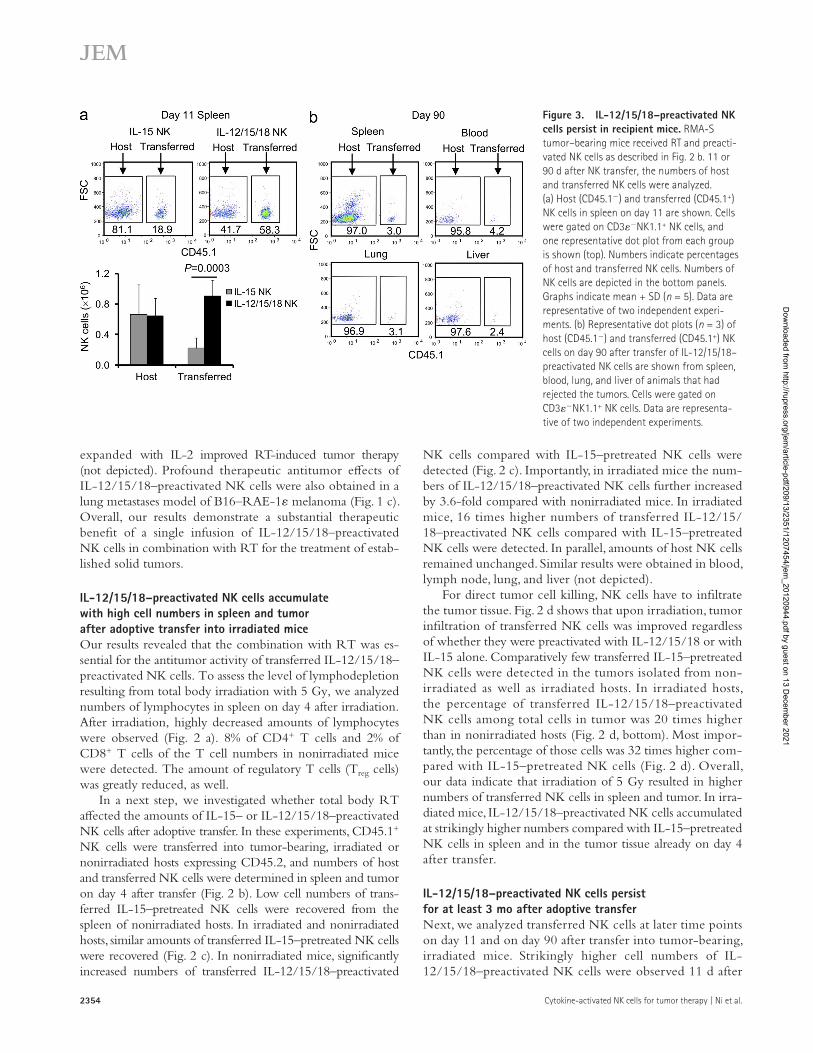
NK cells compared with IL-15–pretreated NK cells were detected (Fig. 2 c). Importantly, in irradiated mice the num-bers of IL-12/15/18–preactivated NK cells further increased by 3.6-fold compared with nonirradiated mice. In irradiated mice, 16 times higher numbers of transferred IL-12/15/ 18–preactivated NK cells compared with IL-15–pretreated NK cells were detected. In parallel, amounts of host NK cells remained unchanged. Similar results were obtained in blood, lymph node, lung, and liver (not depicted).
For direct tumor cell killing, NK cells have to infiltrate the tumor tissue. Fig. 2 d shows that upon irradiation, tumor infiltration of transferred NK cells was improved regardless of whether they were preactivated with IL-12/15/18 or with IL-15 alone. Comparatively few transferred IL-15–pretreated NK cells were detected in the tumors isolated from non-irradiated as well as irradiated hosts. In irradiated hosts, the percentage of transferred IL-12/15/18–preactivated NK cells among total cells in tumor was 20 times higher than in nonirradiated hosts (Fig. 2 d, bottom). Most impor-tantly, the percentage of those cells was 32 times higher com-pared with IL-15–pretreated NK cells (Fig. 2 d). Overall, our data indicate that irradiation of 5 Gy resulted in higher numbers of transferred NK cells in spleen and tumor. In irra-diated mice, IL-12/15/18–preactivated NK cells accumulated at strikingly higher numbers compared with IL-15–pretreated NK cells in spleen and in the tumor tissue already on day 4 after transfer.
IL-12/15/18–preactivated NK cells persist for at least 3 mo after adoptive transferNext, we analyzed transferred NK cells at later time points on day 11 and on day 90 after transfer into tumor-bearing, irradiated mice. Strikingly higher cell numbers of IL-12/15/18–preactivated NK cells were observed 11 d after
expanded with IL-2 improved RT-induced tumor therapy (not depicted). Profound therapeutic antitumor effects of IL-12/15/18–preactivated NK cells were also obtained in a lung metastases model of B16–RAE-1 melanoma (Fig. 1 c). Overall, our results demonstrate a substantial therapeutic benefit of a single infusion of IL-12/15/18–preactivated NK cells in combination with RT for the treatment of estab-lished solid tumors.
IL-12/15/18–preactivated NK cells accumulate with high cell numbers in spleen and tumor after adoptive transfer into irradiated miceOur results revealed that the combination with RT was es-sential for the antitumor activity of transferred IL-12/15/18–preactivated NK cells. To assess the level of lymphodepletion resulting from total body irradiation with 5 Gy, we analyzed numbers of lymphocytes in spleen on day 4 after irradiation. After irradiation, highly decreased amounts of lymphocytes were observed (Fig. 2 a). 8% of CD4+ T cells and 2% of CD8+ T cells of the T cell numbers in nonirradiated mice were detected. The amount of regulatory T cells (Treg cells) was greatly reduced, as well.
In a next step, we investigated whether total body RT affected the amounts of IL-15– or IL-12/15/18–preactivated NK cells after adoptive transfer. In these experiments, CD45.1+ NK cells were transferred into tumor-bearing, irradiated or nonirradiated hosts expressing CD45.2, and numbers of host and transferred NK cells were determined in spleen and tumor on day 4 after transfer (Fig. 2 b). Low cell numbers of trans-ferred IL-15–pretreated NK cells were recovered from the spleen of nonirradiated hosts. In irradiated and nonirradiated hosts, similar amounts of transferred IL-15–pretreated NK cells were recovered (Fig. 2 c). In nonirradiated mice, significantly increased numbers of transferred IL-12/15/18–preactivated
Figure 3. IL-12/15/18–preactivated NK cells persist in recipient mice. RMA-S tumor–bearing mice received RT and preacti-vated NK cells as described in Fig. 2 b. 11 or 90 d after NK transfer, the numbers of host and transferred NK cells were analyzed. (a) Host (CD45.1) and transferred (CD45.1+) NK cells in spleen on day 11 are shown. Cells were gated on CD3NK1.1+ NK cells, and one representative dot plot from each group is shown (top). Numbers indicate percentages of host and transferred NK cells. Numbers of NK cells are depicted in the bottom panels. Graphs indicate mean + SD (n = 5). Data are representative of two independent experi-ments. (b) Representative dot plots (n = 3) of host (CD45.1) and transferred (CD45.1+) NK cells on day 90 after transfer of IL-12/15/18–preactivated NK cells are shown from spleen, blood, lung, and liver of animals that had rejected the tumors. Cells were gated on CD3NK1.1+ NK cells. Data are representa-tive of two independent experiments.
Dow
nloaded from http://rupress.org/jem
/article-pdf/209/13/2351/1207454/jem_20120944.pdf by guest on 13 D
ecember 2021
JEM Vol. 209, No. 13
Article
2355
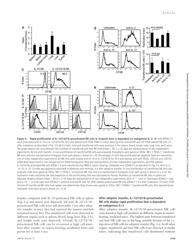
After adoptive transfer, IL-12/15/18–preactivated NK cells display rapid proliferation that is dependent on endogenous IL-2After adoptive transfer, IL-12/15/18–preactivated NK cells were found at high cell numbers in different organs in tumor-bearing, irradiated mice. The highest ratio between transferred and host NK cells was in the lung, possibly because of the i.v. injection of NK cells for adoptive transfer (Fig. 4 a). In all other organs, transferred and host NK cells were detected at similar ratios, indicating that transferred cells distributed without
transfer compared with IL-15–pretreated NK cells in spleen (Fig. 3 a) and tumor (not depicted). Of note, IL-12/15/18–preactivated NK cells were still detectable 3 mo after adop-tive transfer in mice that had rejected the tumors and had remained tumor free. The transferred cells were detected in different organs such as spleen, blood, lung, liver (Fig. 3 b), and lymph node (not depicted). Overall, IL-12/15/18–preactivated NK cells can be recovered at high cell num-bers after transfer in tumor-bearing, irradiated hosts and persist for at least 3 mo.
Figure 4. Rapid proliferation of IL-12/15/18–preactivated NK cells in recipient mice is dependent on endogenous IL-2. NK cells (CD45.1+) were pretreated with IL-15 or IL-12/15/18 for 16 h and labeled with CFSE. RMA-S tumor–bearing mice received RT and 106 CFSE-labeled NK cells 3 h after irradiation as described in Fig. 2 b. (a) 4 d later, host and transferred cells were analyzed in the spleen, blood, lymph node, lung, liver, and tumor. The graph depicts the ratio between the numbers of transferred and host NK cells (mean + SD; n = 5). Data are representative of two independent experiments. (b) 4 d after transfer, in vivo proliferation of transferred NK cells was analyzed. Histograms were gated on CD3NK1.1+CD45.1+ transferred NK cells, and one representative histogram from each group is shown (n = 5). Percentages of cells that proliferated are depicted. Data are representa-tive of three independent experiments. (c) NK cells were treated with IL-15 or IL-12/15/18 for 16 h and stained with anti-CD25, -CD122, and -CD132 mAbs (bold black lines) or the isotype control (filled histograms). Data are representative of three independent experiments. (d) CFSE-labeled IL-12/15/18–pretreated NK cells (CD45.1+) were transferred into RMA-S tumor–bearing, irradiated mice (CD45.2+) as described in Fig. 2 b. Anti–IL-2, –IL-15, or –IL-12 mAb was applied as described in Materials and methods. 4 d after adoptive transfer, in vivo proliferation of transferred NK cells was analyzed. Cells were gated on CD3NK1.1+CD45.1+ transferred NK cells, and one representative histogram from each group is shown (n = 3–5). The replication index indicating the fold expansion of the proliferating cells was calculated by FlowJo. Numbers of transferred NK cells in spleen are depicted. Graphs indicate mean + SD (n = 3–5). Data are representative of two independent experiments. (e) IL-2/ mice or littermates (CD45.2+; top) and IL-15/ or wild-type mice (CD45.2+; bottom) received RT and 106 CFSE-labeled preactivated NK cells (CD45.1+) 3 h after irradiation. 4 d later, CFSE dilution of transferred NK cells from spleen was determined. Data shown were gated on CD3NK1.1+CD45.1+ transferred NK cells. One representative histogram from each group is shown (n = 3–5).
Dow
nloaded from http://rupress.org/jem
/article-pdf/209/13/2351/1207454/jem_20120944.pdf by guest on 13 D
ecember 2021

2356 Cytokine-activated NK cells for tumor therapy | Ni et al.
In contrast, very low percentages of IL-15–pretreated NK cells had proliferated at this early time point in spleen (Fig. 4 b) and other organs (not depicted).
To determine factors involved in the rapid prolifera-tion, expression of cell surface molecules implicated in NK cell proliferation was analyzed on preactivated NK cells before adoptive transfer. Our results reveal elevated expression of CD25 (IL-2R -chain) and CD132 (IL-2R -chain) and slightly lower expression of CD122 (IL-2R -chain) on IL-12/15/18–preactivated NK cells compared with IL-15–pretreated NK cells (Fig. 4 c). Upon culture of MACS-sorted NK cells in IL-12 or IL-18, low levels of CD25 were induced. The presence of both IL-12 and IL-18 was re-quired to induce high expression of CD25 on purified NK cells during the activation (not depicted). CD127 (IL-7R -chain) and CD28 were not detectable (not depicted).
Because IL-2R chains were highly expressed on IL-12/15/18–preactivated NK cells, we determined the require-ment of IL-2 (binding to the IL-2R -, -, and -chains) and IL-15 (binding to the IL-2R - and -chains) for their rapid in vivo proliferation. IL-12/15/18–preactivated NK cells showed significantly less proliferation in spleen after IL-2 neutralization (Fig. 4 d) or upon transfer into IL-2/ mice (Fig. 4 e, top). Neutralization of IL-15 did not signifi-cantly affect their rapid proliferation (Fig. 4 d). Accordingly, proliferation was not reduced in IL-15/ hosts (Fig. 4 e, bottom). IL-12 has been implicated in the proliferation of Ly49H+ NK cells during MCMV infection (Sun et al., 2009b). In our experimental model, neutralization of IL-12 in the recipient mice did not delay proliferation of transferred NK cells. Importantly, upon in vivo neutralization of IL-2, but not of IL-15 or IL-12, significantly lower numbers of transferred NK cells were detected (Fig. 4 d). Together, our data reveal high expression of CD25 on IL-12/15/18–preactivated NK cells before transfer and an indispensible role of IL-2 for their rapid proliferation in vivo.
The rapid proliferation of IL-12/15/18–preactivated NK cells after adoptive transfer depends on the presence of CD4+ T cells that produce IL-2In a next step, we investigated the source of IL-2 in our exper-imental model. Fig. 5 a shows that on day 4 after irradiation and NK cell transfer, IL-2 production in spleen was mainly detected in CD4+ T cells. Other cell populations (CD4) pro-duced negligible levels of IL-2. Because T cells are the main source of IL-2, depletion of CD4+ or CD8+ T cells was per-formed using the respective antibodies. Depletion of CD4+ T cells resulted in a pronounced reduction in proliferation of transferred IL-12/15/18–preactivated NK cells (Fig. 5 b) that was comparable with IL-2 neutralization (Fig. 4 d). Signifi-cantly lower numbers of transferred NK cells were detected in spleen (Fig. 5 b). Depletion of CD8+ T cells did not signifi-cantly affect the rapid proliferation (Fig. 5 b) but significantly reduced the numbers of transferred NK cells, although to a much lesser extent compared with CD4+ T cell depletion. Of note, CFSE dilution of transferred IL-12/15/18–preactivated
tropism for certain organs. To investigate whether high numbers of transferred NK cells were associated with proliferation, we transferred CSFE-labeled IL-15– or IL-12/15/18–pretreated NK cells into tumor-bearing, irradiated mice. 4 d after transfer, >95% of IL-12/15/18–preactivated NK cells had proliferated with more than eight daughter generations in spleen (Fig. 4 b), blood, lymph node, lung, liver, and tumor (not depicted).
Figure 5. The rapid proliferation of IL-12/15/18–preactivated NK cells is dependent on CD4+ T–producing IL-2. CFSE-labeled IL-12/15/18–pretreated NK (CD45.1+) cells were transferred into RMA-S tumor–bearing, irradiated mice (CD45.2+) as described in Fig. 2 b. Anti-CD4 or anti-CD8 mAb was applied as described in Materials and methods. 4 d later, spleen cells were analyzed. (a) Spleen cells were restimulated by PMA/ionomycin, and IL-2 expression was determined by intracellular staining. One representative dot plot gated on total splenocytes is shown. Numbers indicate percentages among total splenocytes. Percentages of IL-2–producing CD4+ or CD4 cells (after subtraction of the isotype con-trol) in spleen are shown in the graph (mean + SD; n = 3). n.d., not detectable. Data are representative of three independent experiments. (b) CFSE-dilution in spleen was determined. Data shown were gated on CD3NK1.1+CD45.1+ transferred NK cells, and one representative histo-gram from each group is shown (n = 3). The replication index of trans-ferred NK cells was calculated by FlowJo. Numbers of transferred NK cells in spleen are shown. Graphs indicate mean + SD (n = 3). Data are repre-sentative of three independent experiments.
Dow
nloaded from http://rupress.org/jem
/article-pdf/209/13/2351/1207454/jem_20120944.pdf by guest on 13 D
ecember 2021

JEM Vol. 209, No. 13
Article
2357
increased levels of IFN-, granzyme B, and perforin were detected among spleen cells (Fig. 7, b and c). Percentages of IL-15– or IL-12/15/18–preactivated NK cells were greatly increased in irradiated hosts. Most importantly, in irradiated hosts, significantly higher levels of IFN-, granzyme B, and perforin were detected in transferred IL-12/15/18 NK cells compared with nonirradiated hosts (Fig. 7, b and c). In fact, six times more IFN-–expressing transferred IL-12/15/18–preactivated NK cells were observed in irradiated compared with nonirradiated hosts (Fig. 7 c).
To investigate whether IFN- production and direct per-forin-dependent NK cell–mediated killing were required for the antitumor activity, NK cells from wild-type, IFN-/, and perforin/ mice were preactivated with IL-12/15/18 and transferred into tumor-bearing, irradiated mice. Wild-type NK cells prolonged survival of mice compared with RT treat-ment, whereas IFN-/ and perforin/ NK cells did not significantly improve tumor therapy compared with RT alone (Fig. 7 d). Of note, the numbers of transferred IFN-/ and perforin/ NK cells in spleen on day 11 after adoptive transfer were similar to wild-type NK cells (not depicted). Collectively, these results demonstrate that IL-12/15/18–preactivated NK cells display greatly enhanced effector function after transfer in tumor-bearing, irradiated hosts and that expression of both IFN- and perforin is required for their antitumor activity.
NK cells on day 4 after transfer remained unchanged in diph-theria toxin–treated DC-deficient CD11c.DOG mice (not de-picted; Hochweller et al., 2008). Overall, our results indicate that IL-2 and host CD4+ T cells promote the rapid prolifera-tion and expansion of IL-12/15/18–preactivated NK cells in tumor-bearing, irradiated hosts.
IL-12/15/18–preactivated NK cells display a mature phenotype and potent effector function after transfer into irradiated recipientsBefore and after adoptive transfer, IL-12/15/18–preactivated NK cells displayed a mature phenotype characterized by CD11bhighCD27low, KLRG1high, and CD43high expression (Fig. 6, a and b) that was similar to memory NK cells during MCMV infection (Sun et al., 2009a). In contrast, IL-15–pretreated NK cells expressed lower levels of KLRG1 and CD43 before and after transfer. Moreover, before transfer, IL-12/15/18–preactivated NK cells produced substantially higher levels of IFN-, granzyme B, and perforin com-pared with IL-15–pretreated NK cells (Fig. 7 a).
In a next step, we assessed the effector function of IL-15– or IL-12/15/18–preactivated NK cells after transfer in tumor-bearing, irradiated or nonirradiated mice. In nonirradiated hosts, higher percentages of IL-12/15/18–preactivated NK cells compared with IL-15–pretreated NK cells that expressed
Figure 6. IL-12/15/18–preactivated NK cells display a mature phenotype before and after transfer. NK cells (CD45.1+) were treated with IL-15 or IL-12/15/18 for 16 h and transferred into tumor-bearing, irradiated mice (CD45.2+; n = 5) as described in Fig. 2 b. (a) Dot plots show the expression of CD11b and CD27 on splenic naive NK cells (gated on CD3NK1.1+) and IL-15– or IL-12/15/18–preacti-vated NK cells before (gated on CD3NK1.1+) and after transfer (gated on CD3NK1.1+CD45.1+ trans-ferred NK cells) on day 11. Numbers indicate percent-ages. (b) Histograms show expression of KLRG1 and CD43 on NK cells. Numbers indicate geometric mean fluorescence. All data are representative of two inde-pendent experiments.
Dow
nloaded from http://rupress.org/jem
/article-pdf/209/13/2351/1207454/jem_20120944.pdf by guest on 13 D
ecember 2021

2358 Cytokine-activated NK cells for tumor therapy | Ni et al.
Figure 7. IL-12/15/18–preactivated NK cells show high effector function before and after transfer into irradiated recipients. (a) NK cells were pretreated with IL-15 or IL-12/15/18 for 16 h. Expression of IFN-, granzyme B, and perforin (bold black lines) or the isotype control (filled his-tograms) was determined by flow cytometry. Data are representative of three independent experiments. (b and c) IL-15– or IL-12/15/18–pretreated NK cells (CD45.1+) were transferred into untreated (No RT) or irradiated (RT) tumor-bearing mice (CD45.2+) as described in Fig. 2 b. 4 d later, spleno-cytes were restimulated by RMA-S cells and stained with anti–IFN-, anti–granzyme B, and anti-perforin mAbs (bold black lines) or the isotype control
Dow
nloaded from http://rupress.org/jem
/article-pdf/209/13/2351/1207454/jem_20120944.pdf by guest on 13 D
ecember 2021

JEM Vol. 209, No. 13
Article
2359
CD4+ T cells are essential for the antitumor activity of IL-12/15/18–preactivated NK cells after adoptive transferTo investigate whether CD4+ or CD8+ T cells were required for the sustained effector function of IL-12/15/18–preactivated NK cells in tumor-bearing, irradiated hosts, depletion of CD4+ or CD8+ T cells was performed. Of note, neither CD4 nor CD8 expression was detectable on RMA-S lymphoma cells (not depicted). Fig. 8 (a and b) shows that depletion of CD4+ T cells in tumor-bearing, irradiated mice severely impaired IFN- and granzyme B production by trans-ferred IL-12/15/18–preactivated NK cells. In addition, the effector function of host NK cells was also impaired in CD4+ T cell–depleted mice. Depletion of CD8+ T cells did not affect the effector function of transferred NK cells (Fig. 8, a and b). Importantly, depletion of CD4+ T cells abrogated the delay in tumor growth mediated by transfer of IL-12/15/18–preactivated NK cells (Fig. 8 c). Immuno-histochemical analyses revealed that both CD4+ T cells and transferred IL-12/15/18–preactivated NK cells (CD45.1+) were detected in similar areas within the tumor tissue (Fig. 8 d). Low numbers of CD8+ T cells were detected in the tumors. Our results demonstrate an indispensible role of CD4+ T cells for efficient antitumor effects mediated by IL-12/15/18–preactivated NK cells.
Human NK cells preactivated with IL-12/15/18 proliferate rapidly in vitro, are recovered at high cell numbers, and maintain their capacity of producing high levels of IFN-Next, we investigated the effect of preactivation of human NK cells by IL-12/15/18. Human NK cells were preacti-vated with IL-12/15/18 or IL-15 as a control for 16 h, labeled with CFSE, and cultured in low-dose (100 U/ml) IL-2 (Fig. 9 a). In line with results obtained with mouse NK cells, IL-12/15/18–preactivated human NK cells also displayed higher expression of CD25 compared with IL-15–pretreated NK cells (Fig. 9 b). Importantly, IL-12/15/18–preactivated human NK cells showed pronounced rapid proliferation already after 2 d of culture in IL-2 (Fig. 9 c). Proliferation of IL-15–pretreated human NK cells was greatly delayed (Fig. 9 c). In parallel, significantly higher numbers of IL-12/15/18–preactivated human NK cells were re-covered on days 6 and 8 after IL-2 culture (Fig. 9 d). Most strikingly, those NK cells maintained the ability to produce high levels of IFN- upon restimulation by IL-12/15 or by the tumor cell line K562 after 4 and 8 d of in vitro culture (Fig. 9 e). Overall, our results indicate that similar to mouse
NK cells, IL-12/15/18–preactivated human NK cells dis-play high cell recovery after in vitro culture and produce high levels of IFN- upon restimulation.
DISCUSSIONImmune cell–based therapy is a promising, innovative strat-egy of personalized cancer treatment. In our study we aimed at improving current protocols of NK cell adoptive therapy of cancer. Our results demonstrate profound therapeutic antitumor effects of a single injection of 106 IL-12/15/18–preactivated NK cells in lymphoma or melanoma-bearing, irradiated mice. The combination with irradiation was es-sential for the antitumor activity of transferred IL-12/15/18–preactivated NK cells. Furthermore, our results reveal an important role for CD4+ T cells in the rapid in vivo prolif-eration and effector function of IL-12/15/18–preactivated NK cells in irradiated, tumor-bearing mice.
A recent clinical trial revealed that adoptive transfer of autologous IL-2–activated NK cells into patients suffering from solid tumors did not lead to substantial clinical responses (Parkhurst et al., 2011). Accordingly, adoptive transfer of neither 106 naive nor IL-2–expanded or IL-15–pretreated NK cells on day 7 after tumor inoculation showed thera-peutic effects in our models. In this context, it was reported that adoptive transfer of 3 × 10⁶ IL-2–expanded NK cells into RAG2/c/ mice on days 0 and 1 after inoculation of a sarcoma cell line resulted in a significant delay in tumor growth (Pegram et al., 2010). In a CT26 lung metastasis model, IL-15–expanded NK cells injected at high numbers (4 × 10⁶) together with tumor cells prolonged survival (Salagianni et al., 2011). It is possible that differences in experimental protocols such as the tumor model, the time points of cell transfer (prophylactic vs. therapeutic model), and the doses of transferred cells accounted for the different outcomes in these studies. In several clinical trials, IL-2 was applied in vivo to maintain high numbers of transferred cells (Rosenberg et al., 1985). However, IL-2 infusions are often associated with severe side effects (Rosenberg et al., 1985) and result in the expansion of Treg cells (Bachanova et al., 2010). Importantly, in our experiments, adoptive NK cell transfers were performed without additional cytokine appli-cation in vivo.
Our results indicate that the combination with total body RT of 5 Gy, a sublethal dose of radiation, was essen-tial for the therapeutic antitumor effects of IL-12/15/18–preactivated NK cells. No beneficial effects were observed
(filled histograms; b). Dot plots (left) depict whole splenocytes. Histograms (right) are gated on CD45.1+NK1.1+ cells as indicated in the left panel. Numbers indicate percentages of IFN-–producing cells among the transferred NK cells or mean fluorescence intensity (MFI) of granzyme B and per-forin expression. One representative dot plot or histogram from each group is shown. (c) Numbers of IFN-–producing transferred NK cells and mean fluorescence intensity of granzyme B and perforin detected in transferred NK cells are depicted. Graphs indicate mean + SD (n = 3). Data shown are representative of two independent experiments. (d) NK cells from wild-type, IFN-/, or perforin/ mice were stimulated with IL-12/15/18 for 16 h and transferred into tumor-bearing, irradiated mice 7 d after tumor inoculation. Survival data are outlined from two experiments (n = 15, 9, 10, and 10 for RT, RT+NK wild-type, RT+NK IFN-/, and RT+NK perforin/ groups, respectively) and presented as a Kaplan–Meier survival curve. ** , P < 0.01 compared with the other groups.
Dow
nloaded from http://rupress.org/jem
/article-pdf/209/13/2351/1207454/jem_20120944.pdf by guest on 13 D
ecember 2021

2360 Cytokine-activated NK cells for tumor therapy | Ni et al.
by irradiation (Garbi et al., 2004; Quezada et al., 2008). Im-portantly, RT enhanced levels of IFN- and perforin by transferred IL-12/15/18–preactivated NK cells that were essential for their therapeutic effects. Although expression of ligands for the activating NK cell receptors was shown to be induced on tumor cells by irradiation in vitro (Gasser et al., 2005; Cerwenka, 2009), we did not observe up-regulation of the NKG2D ligands Rae-1, H60, and MULT-1 or the DNAM-1 ligand CD155 on tumor cells on day 2 after irradiation in our experimental model (unpublished data). In recent clinical trials, T cell adoptive immunotherapy was combined with nonmyeloablative lymphodepleting regi-men such as radiotherapy with a sublethal dose of irradiation or chemotherapy with cyclophosphamide and fludarabine
in the absence of irradiation. In this context, improved anti-tumor activity of T cell infusions was reported in combina-tion with RT (Ganss et al., 2002; Gattinoni et al., 2005, 2006; Quezada et al., 2010; Xie et al., 2010). Our results show that the total body irradiation with 5 Gy induced pro-found lymphopenia, including a reduction in Treg cells. Be-cause Treg cells express high levels of CD25 and consume IL-2 (Pandiyan et al., 2007), reduction of Treg cell numbers might be beneficial for the IL-2–driven rapid proliferation of IL-12/15/18–preactivated NK cells. In irradiated recipients, we also observed higher amounts of transferred cells in the tumor tissue. In this context, previous studies demonstrated that adhesion molecules on the tumor vasculature promoting immune cell infiltration in the tumor tissue were modulated
Figure 8. Host CD4+ T cells are indispensible for potent effector function and antitumor activity of IL-12/15/18–preactivated NK cells. (a and b) IL-12/15/18–pretreated NK cells (CD45.1+) were transferred into tumor-bearing, irradiated mice (CD45.2+) as described in Fig. 2 b. Anti-CD4 or anti-CD8 mAb was applied as described in Materials and methods. 11 d later, splenocytes were restimulated by RMA-S cells and stained for IFN- (a) or granzyme B (b). Dot plots and histograms shown are gated on CD3NK1.1+ cells. Numbers indicate percentages among the host or transferred NK cells. One representative staining from each group is shown. Percentages of IFN- and levels of granzyme B produced by host (CD3NK1.1+CD45.1) and transferred (CD3NK1.1+CD45.1+) NK cells are depicted in the bottom panels. Graphs indicate mean + SD (n = 3). Data shown are representative of two independent experiments. (c) NK cells were preactivated with IL-12/15/18 for 16 h and transferred into tumor-bearing, irradiated mice as described in Fig. 1 b. Anti-CD4 mAb was applied as described in Materials and methods. Tumor growth was monitored. Graphs display mean + SEM (n = 4–8). *, P < 0.05 compared with the group with RT treatment. Data shown are representative of two independent experiments. (d) Representative staining (n = 3) of frozen tumor sections obtained from mice 4 d after RT and transfer of IL-12/15/18–preactivated NK cells is depicted. Transferred NK cells and CD4+ and CD8+ T cells were stained with anti-CD45.1, anti-CD8, and anti-CD4 mAbs. Data shown are representative of two independent experiments. Bars, 25 µm.
Dow
nloaded from http://rupress.org/jem
/article-pdf/209/13/2351/1207454/jem_20120944.pdf by guest on 13 D
ecember 2021

JEM Vol. 209, No. 13
Article
2361
Importantly, our experiments reveal that IL-12/15/18–preactivated NK cells expressed high levels of CD25 before transfer and that neutralization of IL-2 and depletion of CD4+ T cells that produced IL-2 significantly reduced the rapid proliferation and cell numbers of IL-12/15/18–preactivated NK cells in vivo. In a recent study, Lee et al. (2012) showed that addition of IL-12 by itself in cultures of whole spleno-cytes induced expression of CD25 on a substantial percent-age (36%) of NK cells. In our study, MACS-sorted NK cells were cultured in the presence of IL-12, IL-15, or IL-18. Low levels of CD25 (11%) were induced on purified NK cells by IL-12 by itself. Both IL-12 and IL-18 were required to induce expression of CD25 on a substantial percentage (37%) of NK cells (unpublished data). It is likely that the differences in the experimental set-ups contributed to the different results observed in these studies.
Furthermore, depletion of CD4+ T cells impaired the sus-tained effector function and abrogated the delay in tumor
(Dudley et al., 2002, 2008; Muranski et al., 2006). Our data indicate that a combination with sublethal radiotherapy might improve clinical benefits of adoptively transferred NK cells. Our future studies will address whether increased antitumor activity of transferred IL-12/15/18–preactivated NK cells will also be observed in combination with chemotherapy.
Already 4 d after adoptive transfer into tumor-bearing, irradiated mice, almost all IL-12/15/18–preactivated NK cells proliferated with many daughter cell generations, whereas very low percentages of IL-15–pretreated NK cells had started to proliferate. At day 7 after transfer, IL-15–pretreated NK cells had started to proliferate to a much lower extent compared with IL-12/15/18–preactivated NK cells (unpublished data). In our study, host-derived IL-15 was not required for the rapid proliferation of IL-12/15/18–preactivated NK cells, which is in line with previous studies showing that IL-15 was dispensable for the lymphopenia (Jamieson et al., 2004)- and MCMV-driven (Sun et al., 2009b) expansion of NK cells.
Figure 9. IL-12/15/18–preactivated human NK cells show increased proliferation, cell recovery, and effector function compared with IL-15–pretreated cells. (a) Human NK cells were pretreated with IL-15 or IL-12/15/18 for 16 h, labeled with CFSE, and cultured in low-dose IL-2 (100 U/ml). In vitro proliferation and cell numbers were analyzed at the indicated time points. (b) CD25 expression on NK cells pretreated for 16 h with IL-15 or IL-12/15/18 is depicted. Isotype control is shown in the filled histograms; CD25 is shown by the bold black lines. (c) CFSE dilution was assessed on days 2, 4, and 6 of culture with IL-2. (d) Numbers of IL-2–cultured, IL-15– or IL-12/15/18–pretreated NK cells on days 0, 4, 6, and 8 of culture are depicted. Graph indicates mean + SD (cell counts from duplicate cultures). (e) IFN- production upon restimulation by IL-12/15 or K562 at an E/T ratio of 1:1 after culture for 4 or 8 d is shown. Supernatants were harvested after 24 h, and IFN- levels were determined by ELISA. Graph indicate mean + SD (duplicates from ELISA). (a–e) Data are representative of four independent donors.
Dow
nloaded from http://rupress.org/jem
/article-pdf/209/13/2351/1207454/jem_20120944.pdf by guest on 13 D
ecember 2021

2362 Cytokine-activated NK cells for tumor therapy | Ni et al.
indicating that additional T cell–independent unknown fac-tors affect proliferation.
In our study, depletion of CD8+ T cells in tumor-bearing, irradiated mice significantly reduced the numbers of transferred IL-12/15/18 NK cells, although to a much lesser extent com-pared with CD4+ T cell depletion. The function of IL-12/15/18–preactivated NK cells in vivo remained unaffected by the depletion of CD8+ T cells, suggesting a subordinate role of CD8+ T cells on NK cell activation in our model. In addi-tion, much lower amounts of CD8+ T cells compared with CD4+ T cells were detected in the tumors. It is possible that in our study the impact of CD4+ T cells on NK cell activation was greater compared with CD8+ T cells because experiments were performed in the RMA-S lymphoma model in which tumor cells are deficient in MHC class I.
In many human (Coca et al., 1997; Villegas et al., 2002) and mouse (Wendel et al., 2008) tumors, high numbers of intratumoral NK cells are correlated with improved prognosis. Accordingly, in our study, after transfer into tumor-bearing, irradiated hosts, strikingly higher numbers of IL-12/15/18–preactivated NK cells compared with IL-15–pretreated NK cells were detected in the tumor tissue, correlating with a delay in tumor growth. Furthermore, IL-12/15/18–preactivated NK cells produced high levels of IFN- and granzyme B upon restimulation by RMA-S tumor cells 11 d after adop-tive transfer. Thus, the short preactivation of NK cells with IL-12/15/18 in vitro completely altered their behavior as well as the behavior of their daughter cells in vivo. NK cells preactivated with IL-12/15/18 persisted for at least 90 d in adoptive hosts that had rejected tumor. Whether these NK cells can mount protective memory responses against tumor will be addressed in future studies. In line with a previous study (Pegram et al., 2010), IFN- and perforin expression by IL-12/15/18–preactivated NK cells were required for their antitumor activity. The importance of perforin expression for direct tumor cell killing by NK cells is well established (van den Broek et al., 1995). Multiple antitumor mechanisms were reported for IFN-, including the inhibition of angiogen-esis (Qin et al., 2003), the repolarization of tumor-infiltrating macrophages (Corthay et al., 2005; Galani et al., 2010), or the subsequent activation of adaptive immune cells (Martín-Fontecha et al., 2004). The exact role of IFN- produced by NK cells in our tumor model needs further investigation.
Similar to mouse NK cells, human IL-12/15/18–preactivated NK cells displayed higher levels of CD25 compared with IL-15–pretreated NK cells. Upon culture in IL-2, IL-12/15/18–preactivated NK cells displayed increased cell numbers and sustained effector function upon restimulation in vitro. In a recent study by Romee et al. (2012), higher cell numbers and increased IFN- production of IL-12/15/18–preactivated NK cells upon culture in IL-15 were observed. These data suggest that IL-12/15/18–preactivated NK cells maintain an enhanced effector function not only in cultures containing IL-2, but also IL-15. Overall, our results demonstrate pro-found therapeutic antitumor effects of a single injection of 106 IL-12/15/18–preactivated NK cells upon adoptive transfer.
growth mediated by IL-12/15/18–preactivated NK cells. Both NK cells and CD4+ T cells were detected in similar areas throughout the tumor tissue, suggesting that their interaction might occur within the tumors. 4 d after RT, 8% of CD4+ T cells compared with nonirradiated recipients were detected in spleen. This amount of CD4+ T cells was sufficient to sup-port the rapid proliferation of transferred NK cells. Depletion of CD8+ T cells resulted in a reduction in cell numbers of transferred IL-12/15/18–preactivated NK cells in spleen on day 4 after transfer, but the rapid proliferation and effector function of transferred cells remained unchanged. Because negligible levels of IL-2 were detected in CD8+ T cells, we assume that their effect on NK cell numbers is independent of IL-2. Whether the presence of CD8+ T cells affects the survival or distribution of transferred NK cells is currently unknown. Using diphtheria toxin–treated DC-deficient CD11c.DOG mice, we observed that the presence of DCs was not required for the rapid proliferation of IL-12/15/18–preactivated NK cells, suggesting that CD4+ T cell help for NK cells occurred independently of priming by DCs.
A critical role of CD4+ T cell help for effective pri-mary and memory CD8+ T cell responses has been well addressed (Wiesel and Oxenius, 2012). Evidence exists that also certain NK cell–mediated immune responses benefit by help from CD4+ T cells. An in vitro study revealed that human CD56bright NK cells in the lymph node can be acti-vated by T cell–derived IL-2 (Fehniger et al., 2003). In addition, the cross talk between human T cells and NK cells was shown to be required for NK cell–mediated IFN- responses against influenza (He et al., 2004)- and Plasmodium falciparum–infected erythrocytes (Horowitz et al., 2010). In a mouse model of Leishmania major infection in vivo, Bihl et al. (2010) demonstrated that primed antigen-specific CD4+ T cells were required for NK cell activation. Recent studies also addressed the importance of the T/NK cross talk in cancer models. These studies revealed that the CD4+ T cell–mediated control of tumor growth required the presence of NK cells (Perez-Diez et al., 2007) and that CD4+ T cells were required for the IFN- production by innate immune cells carrying markers of NK cells (Li et al., 2007). Our study exploits the NK cell/CD4+ T cell cross talk for the therapeutic usage of adoptively transferred NK cells. Our results define the requirement of preactivation of NK cells by IL-12/15/18 to induce high expression of CD25 and highlight the importance of IL-2 and CD4+ T cells for the expansion and antitumor activity of adoptively transferred IL-12/15/18–preactivated NK cells. A previous study dem-onstrated that the OX40–OX40L interaction was involved in the CD4+ T cell–NK cell interaction in vitro (Zingoni et al., 2004). The molecules involved in the NK/CD4+ T cell cross talk during antitumor immune responses need to be further characterized. Upon neutralization of IL-2 or depletion of CD4+ T cells, the inhibition of the rapid proliferation was not complete. In this context, Cooper et al. (2009) demon-strated proliferation of IL-12/15/18–preactivated NK cells on day 7 after transfer into nonirradiated RAG-1/ mice,
Dow
nloaded from http://rupress.org/jem
/article-pdf/209/13/2351/1207454/jem_20120944.pdf by guest on 13 D
ecember 2021
JEM Vol. 209, No. 13
Article
2363
Ex vivo stimulation of mouse NK cells. Cells were isolated from spleen of treated mice and co-cultured with RMA-S cells (106 spleen cells/5 × 105 RMA-S cells) for 22 h. GolgiStop (BD) was added 4 h before the end of co-culture. Cells were stained for surface markers, fixed, and permeabilized (eBioscience), followed by intracellular staining of IFN- and granzyme B. For intracellular staining of IL-2, splenocytes were restimulated with 50 ng/ml PMA (Sigma-Aldrich) and 500 ng/ml ionomycin (Sigma-Aldrich) for 4 h in the presence of GolgiStop.
In vivo proliferation assay. In vitro activated NK cells were labeled with 1.5 µM CFSE (Sigma-Aldrich) at room temperature for 15 min. After three washes with PBS, cells were transferred into tumor-bearing, irradiated mice. 4 d later, single-cell suspensions from spleen and other organs were prepared, stained, and analyzed by flow cytometry. The replication index indicating the fold expansion of the proliferating cells was calculated by FlowJo. 500 µg anti–IL-2 (S4B6 and JES6-1A12, 1:1; Bio X Cell), 200 µg anti-CD4 (GK1.5; Bio X Cell), and 200 µg anti-CD8 (2.43; Bio X Cell) mAbs were i.p. injected 2 d before adoptive transfer of CFSE-labeled NK cells and every second day. 750 µg anti–IL-12 (C17.8; Bio X Cell) mAb was i.p. injected 1 d before NK cell infusion; 25 µg anti–IL-15/IL-15R (GRW15PLZ; eBioscience) mAb was i.p. injected 1 d before NK cell infusion and subsequently every second day.
Immunohistochemistry. Freshly isolated tumors were embedded in O.C.T. (optimal cutting temperature) compound, frozen in liquid nitrogen, and stored at 80°C until use. Cryosections of 6 µm in thickness were air dried for 10 min at room temperature and fixed for 10 min at 4°C with acetone. The following primary antibodies (at 1:200 dilution) were used: rat anti-CD4 (H129.19), rat anti-CD8 (53-6.7), and mouse anti-CD45.1-Biotin (A20). The anti–rat Ig HRP Detection kit (BD) was used for detection according to the manufacturer’s protocol. Sections were counterstained with Hematoxylin (Mayer’s hemalam solution; Applichem). Images were digitally captured on a BX51 microscope (Olympus) and imaged using cell^D software (Olympus).
Human NK cells. PBMCs from healthy donors were isolated by Ficoll separation (LSM 1077 lymphocyte separation medium; PAA). NK cells were purified by negative selection (Human NK cell isolation kit; Miltenyi Bio-tec) with a purity of CD3CD56+ NK cells >95%. NK cells were preacti-vated in SCGM medium (CellGenix) containing 20% human serum (PAA), 1% penicillin, and 1% streptomycin (Invitrogen) with 10 ng/ml IL-12 (PeproTech), 20 ng/ml IL-15, and 100 ng/ml IL-18 (MBL) for 16 h. To assess in vitro proliferation, preactivated NK cells were labeled with 2 µM CFSE (Sigma-Aldrich) and cultured in 100 U/ml recombinant human IL-2 (National Institutes of Health; day 0). On days 2, 4, 6, and 8, cells were counted using a hemocytometer, and CFSE dilution was analyzed by flow cytometry on a FACSCalibur (BD). Dead cells were excluded by gating on 7-AAD cells. For IFN- production, NK cells were harvested on days 4 and 8 and restim-ulated with either 10 ng/ml IL-12 and 50 ng/ml IL-15 or K562 cells in the presence of 100 U/ml IL-2 at an E/T ratio of 1:1. Supernatants were har-vested after 24 h, and IFN- was measured by ELISA (BioLegend).
Statistics. The statistical significance of results from experimental groups in comparison with control groups was determined by the Student’s t test. Survival data were analyzed with the log-rank test. All tests were two tailed, and P < 0.05 was considered to be statistically significant.
We thank A. Arnold for excellent technical support; the Animal Laboratory Services of the German Cancer Research Center (DKFZ) for the animal care and irradiation; Prof. B. Arnold for providing the IL-15/ mice; Prof. B. Kyewski for providing IFN-/ mice; Dr. Amiya K. Patra for providing the IL-2/ mice; and Prof. G. Hämmerling, A. Rölle, and E. Schlecker for critical reading of the manuscript and helpful discussions. We thank Prof. Jürgen Debus (University Hospital, Heidelberg, Germany) for advice with radiation experiments.
This work is supported by the Deutsche Krebshilfe (#109174; to A. Cerwenka) and by the Cooperation Program in Cancer Research of the DKFZ and Israel’s Ministry of Science and Technology (to A. Cerwenka).
The authors do not have any competing financial interests.
Because our results reveal the importance of CD4+ T cell help for efficient antitumor activity of IL-12/15/18–preactivated NK cells, clinical protocols that combine NK cell–based im-munotherapy with treatments leading to simultaneous CD4+ T cell activation should be considered.
MATERIALS AND METHODSMice. C57BL/6 (CD45.2+) 8-wk-old mice were purchased from Charles River. Perforin/ mice of C57BL/6 background were purchased from the Jackson Laboratory. IL-15/ mice of C57BL/6 background were pur-chased from Taconic or provided by B. Arnold (German Cancer Research Center [DKFZ], Heidelberg, Germany). IFN-/ mice of C57BL/6 back-ground were provided by B. Kyewski (DKFZ). IL-2/ mice of C57BL/6 background and the littermates (IL-2+/) were provided by A.K. Patra (University of Würzburg, Würzburg, Germany). C57BL/6 (CD45.1+), RAG-2/ (CD45.1+ or CD45.2+), and CD11c.DOG mice (Hochweller et al., 2008) of C57BL/6 background were bred at the DKFZ animal facil-ity. Mice were housed under specific pathogen–free conditions and in accordance with all standards of animal care. All animal experiments were approved by the Regierungspräsidium Karlsruhe.
Antibodies and flow cytometry. Anti–mouse CD3 (145-2C11), NK1.1 (PK136), CD4 (H129.19), CD45.1 (A20), CD11b (M1/70), CD25 (PC61), CD27 (LG.3A10), CD43 (1B11), CD122 (TM-1), CD132 (TUGm2), KLRG1 (2F1), IL-2 (JES6-5H4), IFN- (XMG1.2), Granzyme B (16G6), and anti–human CD25 (BC96) were obtained from BD, BioLegend, eBiosci-ence, Invitrogen, and SouthernBiotech. Flow cytometric analyses were performed with a FACSCanto II (BD), and data were analyzed using FlowJo software (Tree Star). For staining of surface molecules, dead cells were excluded by gating on 7-AAD–negative cells.
NK cell isolation from mouse spleen and in vitro activation. NK cells were isolated by negative selection from spleens of wild-type or RAG2/ mice with the NK cell isolation kit (Miltenyi Biotec) and treated with 10 ng/ml IL-12 (PeproTech), 10 ng/ml IL-15 (PeproTech), and 50 ng/ml IL-18 (MBL) or 10 ng/ml IL-15 for 16 h. To obtain IL-2–expanded NK cells, NK cells were cultured with 1,700 U/ml recombinant human IL-2 (Chiron) for 7 d. The cells were stained and analyzed by flow cytome-try or washed four times with PBS before adoptive transfer. The purity of the NK cells (CD3NK1.1+) before adoptive transfer was >90%.
Tumor cells and mouse tumor models. The MHC class I–deficient lym-phoma cell line RMA-S was cultured in RPMI-1640 (Sigma-Aldrich) supple-mented with 10% FCS, 1% l-glutamine, 1% penicillin, and 1% streptomycin (Invitrogen). Mice were s.c. injected with 106 RMA-S lymphoma cells that were washed three times in PBS. On day 7 after tumor cell inoculation, tumor-bearing mice were treated with 5 Gy of total body RT (0.49 Gy/min). 106 IL-15– or IL-12/15/18–treated syngeneic NK cells were i.v. injected 3 h after irradiation. Tumor diameters were measured by a caliper. Mice were eu-thanized when the tumors reached the mean diameter of 1.5 cm. The tumor volume was calculated as large diameter × small diameter × depth.
The melanoma cell line B16 ectopically expressing RAE-1 was cultured in DMEM (Sigma-Aldrich), 10% FCS, 1% l-glutamine, 1% penicillin, and 1% streptomycin (Invitrogen). Mice were i.v. injected with 106 B16–RAE-1 cells and received total body irradiation and adoptive transfer of NK cells at day 7 after tumor cell inoculation as described for the RMA-S tumor model. On day 14, lungs were dissected and fixed in Bouin’s buffer (Sigma-Aldrich), and numbers of nodules were counted under a dissecting microscope (S8AP0; Leica).
Preparation of single-cell suspension from tumors. Tumors were re-moved, cut into small pieces, and digested with 0.5 mg/ml hyaluronidase (Sigma-Aldrich) and 0.5 mg/ml DNase I (Sigma-Aldrich) at 37°C for 30 min. Percentages of NK cells were calculated among all viable cells in the tumors (gated on 7-AAD population, including both tumor cells and tumor-infiltrating leukocytes).
Dow
nloaded from http://rupress.org/jem
/article-pdf/209/13/2351/1207454/jem_20120944.pdf by guest on 13 D
ecember 2021

2364 Cytokine-activated NK cells for tumor therapy | Ni et al.
Gattinoni, L., S.E. Finkelstein, C.A. Klebanoff, P.A. Antony, D.C. Palmer, P.J. Spiess, L.N. Hwang, Z. Yu, C. Wrzesinski, D.M. Heimann, et al. 2005. Removal of homeostatic cytokine sinks by lymphodepletion enhances the efficacy of adoptively transferred tumor-specific CD8+ T cells. J. Exp. Med. 202:907–912. http://dx.doi.org/10.1084/jem .20050732
Gattinoni, L., D.J. Powell Jr., S.A. Rosenberg, and N.P. Restifo. 2006. Adoptive immunotherapy for cancer: building on success. Nat. Rev. Immunol. 6:383–393. http://dx.doi.org/10.1038/nri1842
Geller, M.A., and J.S. Miller. 2011. Use of allogeneic NK cells for cancer im-munotherapy. Immunotherapy. 3:1445–1459. http://dx.doi.org/10.2217/ imt.11.131
Geller, M.A., S. Cooley, P.L. Judson, R. Ghebre, L.F. Carson, P.A. Argenta, A.L. Jonson, A. Panoskaltsis-Mortari, J. Curtsinger, D. McKenna, et al. 2011. A phase II study of allogeneic natural killer cell therapy to treat patients with recurrent ovarian and breast cancer. Cytotherapy. 13:98–107. http://dx.doi.org/10.3109/14653249.2010.515582
He, X.S., M. Draghi, K. Mahmood, T.H. Holmes, G.W. Kemble, C.L. Dekker, A.M. Arvin, P. Parham, and H.B. Greenberg. 2004. T cell-dependent production of IFN-gamma by NK cells in response to influ-enza A virus. J. Clin. Invest. 114:1812–1819.
Hochweller, K., J. Striegler, G.J. Hämmerling, and N. Garbi. 2008. A novel CD11c.DTR transgenic mouse for depletion of dendritic cells reveals their requirement for homeostatic proliferation of natural killer cells. Eur. J. Immunol. 38:2776–2783. http://dx.doi.org/10.1002/eji.200838659
Horowitz, A., K.C. Newman, J.H. Evans, D.S. Korbel, D.M. Davis, and E.M. Riley. 2010. Cross-talk between T cells and NK cells generates rapid effector responses to Plasmodium falciparum-infected erythrocytes. J. Immunol. 184:6043–6052. http://dx.doi.org/10.4049/jimmunol.1000106
Imai, K., S. Matsuyama, S. Miyake, K. Suga, and K. Nakachi. 2000. Natural cytotoxic activity of peripheral-blood lymphocytes and cancer in-cidence: an 11-year follow-up study of a general population. Lancet. 356:1795–1799. http://dx.doi.org/10.1016/S0140-6736(00)03231-1
Jamieson, A.M., P. Isnard, J.R. Dorfman, M.C. Coles, and D.H. Raulet. 2004. Turnover and proliferation of NK cells in steady state and lym-phopenic conditions. J. Immunol. 172:864–870.
Kärre, K., H.G. Ljunggren, G. Piontek, and R. Kiessling. 1986. Selective re-jection of H-2-deficient lymphoma variants suggests alternative immune defence strategy. Nature. 319:675–678. http://dx.doi.org/10.1038/ 319675a0
Lee, S.H., M.F. Fragoso, and C.A. Biron. 2012. Cutting Edge: A novel mechanism bridging innate and adaptive immunity: IL-12 induction of CD25 to form high-affinity IL-2 receptors on NK cells. J. Immunol. 189:2712–2716. http://dx.doi.org/10.4049/jimmunol.1201528
Li, Z., F. Pradera, T. Kammertoens, B. Li, S. Liu, and Z. Qin. 2007. Cross-talk between T cells and innate immune cells is crucial for IFN-gamma-dependent tumor rejection. J. Immunol. 179:1568–1576.
Ljunggren, H.G., and K.J. Malmberg. 2007. Prospects for the use of NK cells in immunotherapy of human cancer. Nat. Rev. Immunol. 7:329–339. http://dx.doi.org/10.1038/nri2073
Martín-Fontecha, A., L.L. Thomsen, S. Brett, C. Gerard, M. Lipp, A. Lanzavecchia, and F. Sallusto. 2004. Induced recruitment of NK cells to lymph nodes provides IFN-gamma for T(H)1 priming. Nat. Immunol. 5:1260–1265. http://dx.doi.org/10.1038/ni1138
Miller, J.S., Y. Soignier, A. Panoskaltsis-Mortari, S.A. McNearney, G.H. Yun, S.K. Fautsch, D. McKenna, C. Le, T.E. Defor, L.J. Burns, et al. 2005. Successful adoptive transfer and in vivo expansion of human haploidentical NK cells in patients with cancer. Blood. 105:3051–3057. http://dx.doi .org/10.1182/blood-2004-07-2974
Moretta, L., F. Locatelli, D. Pende, E. Marcenaro, M.C. Mingari, and A. Moretta. 2011. Killer Ig-like receptor-mediated control of natural killer cell allo-reactivity in haploidentical hematopoietic stem cell transplantation. Blood. 117:764–771. http://dx.doi.org/10.1182/blood-2010-08-264085
Muranski, P., A. Boni, C. Wrzesinski, D.E. Citrin, S.A. Rosenberg, R. Childs, and N.P. Restifo. 2006. Increased intensity lymphodepletion and adoptive immunotherapy—how far can we go? Nat. Clin. Pract. Oncol. 3:668–681. http://dx.doi.org/10.1038/ncponc0666
Author contributions: A. Cerwenka and J. Ni designed the study; J. Ni and M. Miller performed experiments; J. Ni and M. Miller collected and analyzed data; A. Stojanovic and N. Garbi provided mice; and A. Cerwenka and J. Ni wrote the manuscript.
Submitted: 2 May 2012Accepted: 5 November 2012
REFERENCESBachanova, V., L.J. Burns, D.H. McKenna, J. Curtsinger, A. Panoskaltsis-
Mortari, B.R. Lindgren, S. Cooley, D. Weisdorf, and J.S. Miller. 2010. Allogeneic natural killer cells for refractory lymphoma. Cancer Immunol. Immunother. 59:1739–1744. http://dx.doi.org/10.1007/s00262-010- 0896-z
Bihl, F., J. Pecheur, B. Bréart, G. Poupon, J. Cazareth, V. Julia, N. Glaichenhaus, and V.M. Braud. 2010. Primed antigen-specific CD4+ T cells are required for NK cell activation in vivo upon Leishmania major infection. J. Immunol. 185:2174–2181. http://dx.doi.org/10.4049/jimmunol.1001486
Cerwenka, A. 2009. New twist on the regulation of NKG2D ligand ex-pression. J. Exp. Med. 206:265–268. http://dx.doi.org/10.1084/jem .20090225
Cerwenka, A., and L.L. Lanier. 2001. Natural killer cells, viruses and cancer. Nat. Rev. Immunol. 1:41–49. http://dx.doi.org/10.1038/35095564
Coca, S., J. Perez-Piqueras, D. Martinez, A. Colmenarejo, M.A. Saez, C. Vallejo, J.A. Martos, and M. Moreno. 1997. The prognostic significance of intratumoral natural killer cells in patients with colorectal carcinoma. Cancer. 79:2320–2328. http://dx.doi.org/10.1002/(SICI)1097-0142(19970615)79:12<2320::AID-CNCR5>3.0.CO;2-P
Cooper, M.A., J.M. Elliott, P.A. Keyel, L. Yang, J.A. Carrero, and W.M. Yokoyama. 2009. Cytokine-induced memory-like natural killer cells. Proc. Natl. Acad. Sci. USA. 106:1915–1919. http://dx.doi.org/10.1073/pnas .0813192106
Corthay, A., D.K. Skovseth, K.U. Lundin, E. Røsjø, H. Omholt, P.O. Hofgaard, G. Haraldsen, and B. Bogen. 2005. Primary antitumor im-mune response mediated by CD4+ T cells. Immunity. 22:371–383. http://dx.doi.org/10.1016/j.immuni.2005.02.003
D’Urso, C.M., Z.G. Wang, Y. Cao, R. Tatake, R.A. Zeff, and S. Ferrone. 1991. Lack of HLA class I antigen expression by cultured melanoma cells FO-1 due to a defect in B2m gene expression. J. Clin. Invest. 87: 284–292. http://dx.doi.org/10.1172/JCI114984
Dudley, M.E., J.R. Wunderlich, P.F. Robbins, J.C. Yang, P. Hwu, D.J. Schwartzentruber, S.L. Topalian, R. Sherry, N.P. Restifo, A.M. Hubicki, et al. 2002. Cancer regression and autoimmunity in patients after clonal repopulation with antitumor lymphocytes. Science. 298:850–854. http://dx.doi.org/10.1126/science.1076514
Dudley, M.E., J.C. Yang, R. Sherry, M.S. Hughes, R. Royal, U. Kammula, P.F. Robbins, J. Huang, D.E. Citrin, S.F. Leitman, et al. 2008. Adoptive cell therapy for patients with metastatic melanoma: evaluation of inten-sive myeloablative chemoradiation preparative regimens. J. Clin. Oncol. 26:5233–5239. http://dx.doi.org/10.1200/JCO.2008.16.5449
Fehniger, T.A., M.A. Cooper, G.J. Nuovo, M. Cella, F. Facchetti, M. Colonna, and M.A. Caligiuri. 2003. CD56bright natural killer cells are present in human lymph nodes and are activated by T cell-derived IL-2: a potential new link between adaptive and innate immunity. Blood. 101:3052–3057. http://dx.doi.org/10.1182/blood-2002-09-2876
Galani, I.E., M. Wendel, A. Stojanovic, M. Jesiak, M.M. Müller, C. Schellack, E. Suri-Payer, and A. Cerwenka. 2010. Regulatory T cells control mac-rophage accumulation and activation in lymphoma. Int. J. Cancer. 127: 1131–1140. http://dx.doi.org/10.1002/ijc.25132
Ganss, R., E. Ryschich, E. Klar, B. Arnold, and G.J. Hämmerling. 2002. Combination of T-cell therapy and trigger of inflammation induces remod-eling of the vasculature and tumor eradication. Cancer Res. 62:1462–1470.
Garbi, N., B. Arnold, S. Gordon, G.J. Hämmerling, and R. Ganss. 2004. CpG motifs as proinflammatory factors render autochthonous tumors per-missive for infiltration and destruction. J. Immunol. 172:5861–5869.
Gasser, S., S. Orsulic, E.J. Brown, and D.H. Raulet. 2005. The DNA damage pathway regulates innate immune system ligands of the NKG2D receptor. Nature. 436:1186–1190. http://dx.doi.org/10.1038/nature03884
Dow
nloaded from http://rupress.org/jem
/article-pdf/209/13/2351/1207454/jem_20120944.pdf by guest on 13 D
ecember 2021

JEM Vol. 209, No. 13
Article
2365
with metastatic cancer. N. Engl. J. Med. 313:1485–1492. http://dx.doi .org/10.1056/NEJM198512053132327
Ruggeri, L., M. Capanni, E. Urbani, K. Perruccio, W.D. Shlomchik, A. Tosti, S. Posati, D. Rogaia, F. Frassoni, F. Aversa, et al. 2002. Effectiveness of donor natural killer cell alloreactivity in mismatched hematopoietic transplants. Science. 295:2097–2100. http://dx.doi.org/ 10.1126/science.1068440
Salagianni, M., E. Lekka, A. Moustaki, E.G. Iliopoulou, C.N. Baxevanis, M. Papamichail, and S.A. Perez. 2011. NK cell adoptive transfer com-bined with Ontak-mediated regulatory T cell elimination induces effec-tive adaptive antitumor immune responses. J. Immunol. 186:3327–3335. http://dx.doi.org/10.4049/jimmunol.1000652
Sun, J.C., J.N. Beilke, and L.L. Lanier. 2009a. Adaptive immune features of natural killer cells. Nature. 457:557–561. http://dx.doi.org/10.1038/ nature07665
Sun, J.C., A. Ma, and L.L. Lanier. 2009b. Cutting edge: IL-15-indepen-dent NK cell response to mouse cytomegalovirus infection. J. Immunol. 183:2911–2914. http://dx.doi.org/10.4049/jimmunol.0901872
Sun, J.C., S. Lopez-Verges, C.C. Kim, J.L. DeRisi, and L.L. Lanier. 2011. NK cells and immune “memory”. J. Immunol. 186:1891–1897. http://dx.doi.org/10.4049/jimmunol.1003035
Terme, M., E. Ullrich, N.F. Delahaye, N. Chaput, and L. Zitvogel. 2008. Natural killer cell-directed therapies: moving from unexpected re-sults to successful strategies. Nat. Immunol. 9:486–494. http://dx.doi .org/10.1038/ni1580
van den Broek, M.F., D. Kägi, R.M. Zinkernagel, and H. Hengartner. 1995. Perforin dependence of natural killer cell-mediated tumor control in vivo. Eur. J. Immunol. 25:3514–3516. http://dx.doi.org/10.1002/ eji.1830251246
Villegas, F.R., S. Coca, V.G. Villarrubia, R. Jiménez, M.J. Chillón, J. Jareño, M. Zuil, and L. Callol. 2002. Prognostic significance of tumor infiltrating natural killer cells subset CD57 in patients with squamous cell lung cancer. Lung Cancer. 35:23–28. http://dx.doi.org/10.1016/ S0169-5002(01)00292-6
Vivier, E., E. Tomasello, M. Baratin, T. Walzer, and S. Ugolini. 2008. Functions of natural killer cells. Nat. Immunol. 9:503–510. http://dx.doi .org/10.1038/ni1582
Vivier, E., D.H. Raulet, A. Moretta, M.A. Caligiuri, L. Zitvogel, L.L. Lanier, W.M. Yokoyama, and S. Ugolini. 2011. Innate or adaptive immunity? The example of natural killer cells. Science. 331:44–49. http://dx.doi.org/10.1126/science.1198687
Wendel, M., I.E. Galani, E. Suri-Payer, and A. Cerwenka. 2008. Natural killer cell accumulation in tumors is dependent on IFN-gamma and CXCR3 ligands. Cancer Res. 68:8437–8445. http://dx.doi.org/10.1158/ 0008-5472.CAN-08-1440
Wiesel, M., and A. Oxenius. 2012. From crucial to negligible: functional CD8+ T-cell responses and their dependence on CD4+ T-cell help. Eur. J. Immunol. 42:1080–1088. http://dx.doi.org/10.1002/eji.201142205
Xie, Y., A. Akpinarli, C. Maris, E.L. Hipkiss, M. Lane, E.K. Kwon, P. Muranski, N.P. Restifo, and P.A. Antony. 2010. Naive tumor-specific CD4+ T cells differentiated in vivo eradicate established melanoma. J. Exp. Med. 207:651–667. http://dx.doi.org/10.1084/jem.20091921
Zingoni, A., T. Sornasse, B.G. Cocks, Y. Tanaka, A. Santoni, and L.L. Lanier. 2004. Cross-talk between activated human NK cells and CD4+ T cells via OX40-OX40 ligand interactions. J. Immunol. 173:3716–3724.
O’Leary, J.G., M. Goodarzi, D.L. Drayton, and U.H. von Andrian. 2006. T cell- and B cell-independent adaptive immunity mediated by natural killer cells. Nat. Immunol. 7:507–516. http://dx.doi.org/10.1038/ ni1332
Pandiyan, P., L. Zheng, S. Ishihara, J. Reed, and M.J. Lenardo. 2007. CD4+CD25+Foxp3+ regulatory T cells induce cytokine deprivation-mediated apoptosis of effector CD4+ T cells. Nat. Immunol. 8:1353–1362. http://dx.doi.org/10.1038/ni1536
Parkhurst, M.R., J.P. Riley, M.E. Dudley, and S.A. Rosenberg. 2011. Adoptive transfer of autologous natural killer cells leads to high levels of circulating natural killer cells but does not mediate tumor regression. Clin. Cancer Res. 17:6287–6297. http://dx.doi.org/10.1158/1078-0432 .CCR-11-1347
Passweg, J.R., A. Tichelli, S. Meyer-Monard, D. Heim, M. Stern, T. Kühne, G. Favre, and A. Gratwohl. 2004. Purified donor NK-lymphocyte infusion to consolidate engraftment after haploidentical stem cell transplantation. Leukemia. 18:1835–1838. http://dx.doi.org/10.1038/sj.leu.2403524
Paust, S., and U.H. von Andrian. 2011. Natural killer cell memory. Nat. Immunol. 12:500–508. http://dx.doi.org/10.1038/ni.2032
Pegram, H.J., N.M. Haynes, M.J. Smyth, M.H. Kershaw, and P.K. Darcy. 2010. Characterizing the anti-tumor function of adoptively transferred NK cells in vivo. Cancer Immunol. Immunother. 59:1235–1246. http://dx.doi.org/10.1007/s00262-010-0848-7
Pegram, H.J., D.M. Andrews, M.J. Smyth, P.K. Darcy, and M.H. Kershaw. 2011. Activating and inhibitory receptors of natural killer cells. Immunol. Cell Biol. 89:216–224. http://dx.doi.org/10.1038/icb.2010.78
Perez-Diez, A., N.T. Joncker, K. Choi, W.F. Chan, C.C. Anderson, O. Lantz, and P. Matzinger. 2007. CD4 cells can be more efficient at tumor rejection than CD8 cells. Blood. 109:5346–5354. http://dx.doi.org/10 .1182/blood-2006-10-051318
Qin, Z., J. Schwartzkopff, F. Pradera, T. Kammertoens, B. Seliger, H. Pircher, and T. Blankenstein. 2003. A critical requirement of interferon gamma-mediated angiostasis for tumor rejection by CD8+ T cells. Cancer Res. 63:4095–4100.
Quezada, S.A., K.S. Peggs, T.R. Simpson, Y. Shen, D.R. Littman, and J.P. Allison. 2008. Limited tumor infiltration by activated T effector cells restricts the therapeutic activity of regulatory T cell depletion against established melanoma. J. Exp. Med. 205:2125–2138. http://dx.doi .org/10.1084/jem.20080099
Quezada, S.A., T.R. Simpson, K.S. Peggs, T. Merghoub, J. Vider, X. Fan, R. Blasberg, H. Yagita, P. Muranski, P.A. Antony, et al. 2010. Tumor-reactive CD4+ T cells develop cytotoxic activity and eradicate large established melanoma after transfer into lymphopenic hosts. J. Exp. Med. 207:637–650. http://dx.doi.org/10.1084/jem.20091918
Raulet, D.H., and N. Guerra. 2009. Oncogenic stress sensed by the im-mune system: role of natural killer cell receptors. Nat. Rev. Immunol. 9:568–580. http://dx.doi.org/10.1038/nri2604
Romee, R., S.E. Schneider, J.W. Leong, J.M. Chase, C.R. Keppel, R.P. Sullivan, M.A. Cooper, and T.A. Fehniger. 2012. Cytokine activa-tion induces human memory-like NK cells. Blood. http://dx.doi.org/ 10.1182/blood-2012-04-419283
Rosenberg, S.A., M.T. Lotze, L.M. Muul, S. Leitman, A.E. Chang, S.E. Ettinghausen, Y.L. Matory, J.M. Skibber, E. Shiloni, J.T. Vetto, et al. 1985. Observations on the systemic administration of autologous lym-phokine-activated killer cells and recombinant interleukin-2 to patients
Dow
nloaded from http://rupress.org/jem
/article-pdf/209/13/2351/1207454/jem_20120944.pdf by guest on 13 D
ecember 2021